Review

Prevalence of Human and Animal African Trypanosomiasis in Nigeria: A Scoping Review

by
Chinwe Chukwudi
1,*,
Elizabeth Odebunmi
2 and
Chukwuemeka Ibeachu
2
1
Department of Veterinary Pathology and Microbiology, University of Nigeria Nsukka, Nsukka 410001, Nigeria
2
Department of Public Health, School of Health and Society, University of Wolverhampton, Wolverhampton WV1 1LY, UK
*
Author to whom correspondence should be addressed.
Parasitologia 2025, 5(4), 53; https://doi.org/10.3390/parasitologia5040053 (registering DOI)
Submission received: 23 June 2025 / Revised: 8 September 2025 / Accepted: 10 October 2025 / Published: 11 October 2025

Simple Summary

Human African Trypanosomiasis (HAT) is a zoonotic NTD targeted for elimination by 2030. There is a scarcity of HAT epidemiological studies/data from Nigeria. There is a disconnect between WHO records and peer-reviewed data on HAT in Nigeria. But the disease remains prevalent, with a complex epidemiology and zoonotic potential. Inadequate surveillance and diagnostic challenges limit HAT reporting from Nigeria

Abstract

African trypanosomiasis is a protozoan disease that affects both humans and animals. Human African Trypanosomiasis (HAT) is a Neglected Tropical Disease targeted for elimination in 2030. Although WHO has not reported HAT from Nigeria in the last decade, there are published studies reporting seroprevalence, parasite detection/isolation, and animal reservoirs potentially involved in HAT transmission in Nigeria. Interestingly, the burden of Animal African Trypanosomiasis (AAT) continues to increase. In this study, we synthesized published reports on the prevalence of HAT and AAT in Nigeria from 1993–2021, the trypanosome species involved, the spread of animal reservoirs, and the variability in diagnostic methodologies employed. A scoping review was performed following the methodological framework outlined in PRISMA-ScR checklist. Sixteen eligible studies published between 1993 and 2021 were reviewed: 13 for AAT and 3 for HAT. Varying prevalence rates were recorded depending on the diagnostic methods employed. The average prevalence reported from these studies was 3.3% (HAT), and 27.3% (AAT). Diagnostic methods employed include microscopy, PCR and Card Agglutination Test for Trypanosomiasis (CATT). Cattle, pigs, and dogs were identified as carriers of human-infective trypanosomes. This study highlights the scarcity of HAT epidemiological studies/data from Nigeria, the high prevalence, complex epidemiology, limited attention and surveillance of African Trypanosomiasis in Nigeria. Remarkably, WHO records do not reflect the published data showing evidence of HAT prevalence/cases in Nigeria. Unfortunately, diagnostics challenges and unrealistic disease reporting protocols seem to limit HAT reporting from Nigeria. Therefore, adequately coordinated epidemiological surveys and targeted intervention policies are imperative to ascertain the true epidemiological status of HAT in Nigeria and prevent disease re-emergence towards achieving WHO’s elimination targets. The presence of animal carriers of human-infective trypanosomes underscores the importance of a one-health approach to combat African trypanosomiasis effectively.

1. Introduction

African Trypanosomiasis (AT) is one of the Neglected Tropical Diseases (NTDs) with an estimated 60 million people in sub-Saharan Africa at risk of infection [1,2], alongside various animals such as dogs, cattle, pigs, sheep and goats [3,4]. This vector-borne disease, caused by protozoan parasites of the genus Trypanosoma, is transmitted by tsetse flies (Glossina sp.) [5].
Human African Trypanosomiasis (HAT), also known as sleeping sickness is caused by two sub-species of Trypanosoma brucei. Trypanosoma brucei gambiense (Tbg), found in 24 countries in West and Central Africa, accounts for about 97% of all reported cases, and typically causes a chronic form of the disease characterized by neurological symptoms. The first stage of the disease (hemolymphatic stage) presents with mild symptoms such as raised, red sore (chancre) at the site of the tsetse bite, headache, intermittent/chronic fever, malaise, muscle aches, and swollen lymph nodes. The second stage (neurological stage) involves the central nervous system, manifesting as sleep disturbances, hallucination, confusion, tremors, seizures, and if untreated, coma and death [5,6]. Trypanosoma brucei rhodesiense (Tbr), found in 13 countries in East and Southern Africa, is responsible for 3% of reported cases. It causes an acute disease considered zoonotic in both humans and animals [7,8].
Animal African Trypanosomiasis (AAT), also known as Nagana, is caused by various trypanosome species, including T. brucei, T. congolense, T. vivax, T. equiperdum, T. evansi, T. simiae, T. suis, and T. theileri [9,10]. Cattle are the most affected, though goats, dogs, sheep, pigs, and wild animals are also susceptible [11]. Notably, T. equiperdum causes a venereal disease (Dourine) in horses and donkeys, while T. evansi, causes a form of trypanosomiasis known as Surra in horses, camels, buffaloes, mules, and deer [12]. Clinical signs of AAT include intermittent fever, anemia, edema, abortion, reduced fertility, emaciation, neurologic disorders, and death [11].
The transmission of both HAT and AAT depends largely on the tsetse fly, exclusively found in sub-Saharan Africa [13]. However, maternal and sexual transmission have been suggested as alternative transmission routes for HAT, although their epidemiological significance have not been fully explored [14]. The coexistence of humans, animal reservoirs/hosts, vectors, and parasites within the conducive environment of the tropical trypanosomiasis belt of Africa makes the disease a significant public health challenge, with detrimental effects on health, the economy, poverty levels, and agricultural productivity [15,16].
Reported cases of HAT have substantially declined globally to fewer than 1000 cases between 2019 and 2020 [7]. This decline is attributed to HAT control strategies and inclusion in the Neglected Tropical Diseases, with a roadmap aiming to eliminate gHAT as a public health concern by 2020 and zero transmission to humans by 2030 [17]. However, disease monitoring and epidemiological surveillances are grossly inadequate in many endemic countries [18]. For Nigeria, WHO records indicate that HAT was last reported from Nigeria in 2012 (except for one case that was diagnosed in a Nigerian in the UK in 2016) [19]. This has resulted in the neglect of the country in HAT surveillance and control programs, since it is believed that the country is on course to eliminate the disease. However, Cameroon which shares borders with the Nigerian HAT foci continues to report cases (Figure 1). Interestingly, there has been reports of finding the human-infective trypanosomes in animals in Nigeria, especially in companion animals (dogs). This raises questions about the existence and true prevalence of HAT in Nigeria, especially with regard to the source of infection and transmission route for the animal carriers. Neglecting any HAT foci or infection site, no matter how small, poses serious risks to the achievement of the NTD elimination targets. These locations could serve as sources of continuous re-infection and may become potential hotspots for new epidemic strains of the parasite, leading to disease re-emergence. Therefore, adequate epidemiological information about the co-circulation of human and animal trypanosomes is critical to inform control or eradication policies and strategies.
Hence, this review examined published evidence on the prevalence of African trypanosomiasis in both humans and animals in Nigeria. The study is aimed at synchronizing published HAT and AAT epidemiological data from Nigeria in order to understand the disease incidence/occurrence, estimate the prevalence, identify the host spread, parasite species involved, diagnostic techniques being used, and explore the possibilities of cross infection between humans and animals.

2. Methods

The study protocol was developed in 2023, and thoroughly revised by the research team before commencement of the study. This scoping review protocol was not prospectively registered in PROSPERO or any other systematic review registry, but was strictly followed throughout the conduct of the review in line with the University of Wolverhampton guidelines and ethics. The conduct and reporting also followed the Preferred Reporting Items for Systematic Reviews and Meta-Analyses (PRISMA-ScR) four-stage framework, encompassing identification, screening, eligibility assessment, and inclusion to ensure a transparent, accurate, and high-quality reporting of the review’s findings [20].

2.1. Search Strategy

The searches were performed using MEDLINE, CINAHL, and EMBASE in July 2023. The search terms included ‘Neglected Tropical Disease,’ ‘African trypanosomiasis,’ ‘human African trypanosomiasis,’ ‘animal African trypanosomiasis,’ ‘sleeping sickness,’ ‘nagana,’ ‘trypanosoma,’ ‘trypanosomosis,’ ‘trypanosomes.’ These terms were combined using Boolean operators ‘OR’, as well as ‘AND’ to retrieve relevant articles. A manual search was also performed using google search engine to ensure that studies published in non-indexed journals are captured. See Table S1 for final search strategy and result for MEDLINE database.

2.2. Eligibility Criteria

The eligibility criteria for this review were informed by participants (P), phenomena of interest (I), and context (Co) or PICo, where P is humans/animals with AT, I is African trypanosomiasis infection and Co is Nigeria. Primary research studies including cross-sectional, cohort, surveys, and prevalence studies that reported animal and human African trypanosomiasis infection were included. Specific inclusion criteria included studies from Nigeria, published in English language (as Nigeria is an English-speaking country), in the last 30 years (1993–2023), with a well detailed sample, prevalence of trypanosomiasis in sampled population, method of diagnosis and Trypanosoma species identified.

2.3. Study Selection

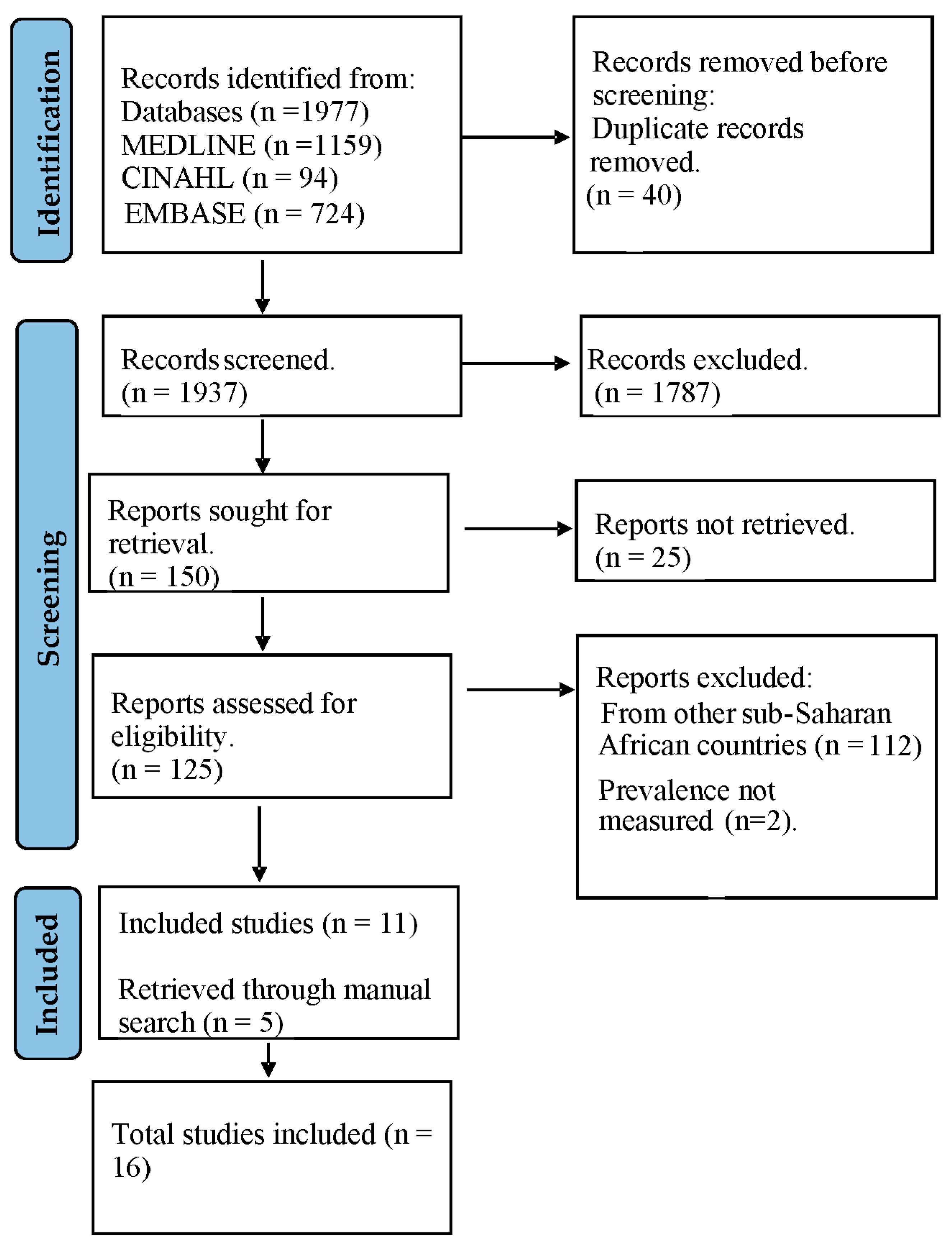
Study selection was performed independently by the three authors. Discrepancies between the authors were resolved through discussion until a consensus was reached. The studies included in this systematic review underwent a two-stage selection process. Following the search, all identified citations were collated and uploaded into EndNote version 20 (Clarivate Analytics, Philadelphia, PA, USA) and duplicates removed. Initially, all potentially relevant studies were screened based on their titles and abstracts to identify studies that potentially met the eligibility criteria. Subsequently, full-text articles of these studies were retrieved and further assessed against the pre-defined inclusion and exclusion criteria for this review. Reasons for exclusion of papers at the full text review stage that did not meet the inclusion criteria are recorded and reported in Figure 2. A manual search was also performed to ensure that all relevant studies missed by the electronic search were captured. The results of the search and the study inclusion process are reported and presented in a Preferred Reporting Items for Systematic Reviews and Meta-analyses (PRISMA) flow diagram [21].
For this review, the selected studies underwent critical appraisal using the Joanna Briggs Institute’s (JBI) critical appraisal checklist for cross-sectional studies [22]. This checklist consists of eight criteria that assess various aspects of methodological quality, including the selection criteria of subjects, study subjects and setting, sample size, measurement of the condition, measurement of outcomes, suitability of statistical analysis, and the method used to identify the disease condition. Each response to the screening questions was assigned a score. A score of ‘1’ is allocated for ‘Yes,’ while ‘No’ and ‘Unclear’ responses are scored as ‘0’ (Table S2). The interpretation of the results obtained using the JBI Critical Appraisal Checklist for Cross-Sectional Studies is categorized as follows:
Low Risk of Bias: his designation is assigned to studies that align with most or all the methodological criteria and exhibit a low risk of bias in their design, conduct, analysis, and reporting, resulting in a score between 6 and 8.
Moderate Risk of Bias: Studies that partially meet the methodological criteria but possess certain limitations or concerns that may impact the reliability of their findings fall into this category, with scores ranging from 4 to 5.
High Risk of Bias: This classification applies to studies with significant methodological limitations, deviations from best practices, or substantial concerns regarding bias. Studies scoring below the specified benchmarks (<4) for moderate-level appraisals of each type of research are included in this category.
Finally, studies that met the eligibility criteria were included for further data extraction.

2.4. Data Extraction

After identifying the pertinent articles for inclusion, the next crucial step involves extracting and synthesizing the relevant information from these selected articles. To facilitate this process and ensure consistency in data classification, a tailored data extraction form was developed, aligning with the specific research question that the review aims to address.
The following key data points were extracted and documented within a pre-prepared Microsoft Excel spreadsheet: author and year of publication, study subject (human, animal), type of study design, study size, Trypanosoma species identified, diagnostic method used, number of positive cases, prevalence, or percentages.
This structured approach to data extraction ensured that critical details from each article were captured accurately and comprehensively. By utilizing a standardized format and organized spreadsheet, the data was efficiently managed and synthesized, facilitating the subsequent stages of analysis, and reporting within the systematic review process.

2.5. Data Synthesis

A narrative summary was performed to synthesize findings from this review, and categorized into human and animal studies. All studies were further structured by the diagnostic techniques employed.

3. Results

A total of 1977 articles were retrieved from the search of three databases. 59% (1159) from MEDLINE, 36% (724) from EMBASE and 5% (94) from CINAHL. After removal of 40 duplicates, 1937 articles were screened for titles and abstracts. A total of 1787 non-relevant articles were excluded in accordance with the predetermined criteria. Following full text retrieval, another 25 studies were excluded as their full texts were not accessible. Upon review of the full texts of the remaining 125 articles, a total of 11 were deemed eligible for inclusion in the study. An additional 5 articles were identified from a search of reference lists. Finally, 16 articles were included. The study selection process is represented in Figure 2.

3.1. Description of Eligible Studies

All included studies were published between 1993 and 2021, with the number of study subjects ranging from 19 to 7143 and an overall sample size of 18,091. Of the 16 included studies, three investigated human African trypanosomiasis [23,24,25], while thirteen studies reported animal African trypanosomiasis. Five of these reported infection in cattle—bovine trypanosomiasis [26,27,28,29,30]. Two studies investigated trypanosomiasis infection in pigs—porcine trypanosomiasis [31,32]. Three studies investigated infection in ruminants—sheep, goat, cattle [33,34,35]—while one study investigated in dogs—canine trypanosomiasis [36]. The remaining two studies investigated infection in multiple animal hosts including monkeys [37,38]. Table 1 provides a summary of the included studies.

3.2. Prevalence of HAT

A total of 1974 individuals underwent testing for the disease across three studies. Among them, 65 individuals were seropositive for the disease in two of the studies using the CATT kit [23,24]. Trypanosomes were detected from the blood of 23 seropositive individuals by various methods such as microscopic examination of blood films or buffy coat, and in vivo inoculation into mice/rats. The detection of the parasites in the blood did not reflect how strongly positive the CATT test was, as parasites were detected in patients that were weakly, moderately and strongly positive in the CATT test. One study also examined the CSF, and detected parasites in the CSF of 4 patients. The third study, conducted in the northern part of the country, did not detect any positive case within the sampled population [25]. The seroprevalence of HAT reported by these studies for various study sites across the country ranged from 0–9.6%, with a calculated average of 3.3% (65/1974). The actual prevalence of HAT as confirmed by parasite detection was as high as 4.8% in one study site (Urhouka), with a calculated average of 1.2% (23/1974) across all studies. Trypanosoma brucei gambiense type 1 was reported as the causative parasite specie, as detected by the gambiense-specific TgsGP-PCR.

3.3. Prevalence of AAT

Thirteen included studies reported AAT with an overall sample size of 16,177. The highest sampled animal was cattle at 12,193, while the least is monkey at 1 sample. Others are 577 sheep, 418 goat, 2639 pigs, 31 dogs, and 318 small ruminants (unspecified). Out of the total sampled animals, 4404 were found positive for trypanosomiasis infection from all the 13 studies. The prevalence of AAT as reported in these studies ranged between 0.8–46.8% for various animal species in various study sites, with a calculated mean prevalence of 27.3% (4404/16,177).
These studies detected the animal trypanosomes: T. vivax, T. congolense, T. brucei, T. simiae, T. evansi, T. theileri in the studied samples, with T. vivax, T. congolense, and T. brucei reported as the most prevalent trypanosome species in animals. In addition to these trypanosomes, three of these studies detected the human infective parasite Trypanosoma brucei gambiense in animals [32,36,37].

3.4. Diagnostic Techniques

The included studies for this review utilized various diagnostic methods, including microscopy, Card Agglutination Test for Trypanosomiasis (CATT), and Polymerase Chain Reaction (PCR) in the detection of trypanosomes. 11 out of the 16 included studies utilized microscopy for diagnosis, while 6 used PCR and 4 used CATT. 5 studies used a combination of diagnostic techniques (Microscopy + PCR = 2, Microscopy + CATT = 1, PCR + CATT = 2. For the studies that utilized a single diagnostic approach, 8 studies utilized microscopy alone, while 2 used PCR alone and 1 used CATT alone. Overall, microscopy was the most utilized diagnostic technique in these studies for the diagnosis of African Trypanosomiasis.

4. Discussion

The findings from this review provide insights into the prevalence, host range, species distribution, and diagnostics techniques of African trypanosomiasis in humans and animals in Nigeria. According to this study’s findings, the calculated mean prevalence of AAT in Nigeria is 27.3% (4404 out of 16,117), while that of HAT is 1.2% by parasite identification and 3.3% seroprevalence, indicating that African trypanosomiasis is still endemic in both humans and animals in Nigeria. The presence of various trypanosome species and use of different diagnostic methods contributes to the complexity of this disease, its epidemiological studies in Nigeria and its public health significance. The study identified several trypanosomes, including T. vivax, T. congolense, T. brucei brucei, T. simiae, T. evansi, T. theileri, and T. brucei gambiense as the causative pathogens of African trypanosomiasis in animals and humans in Nigeria. Additionally, the most used diagnostic technique in Nigeria for detecting these trypanosomes is microscopic examination, particularly in animals. Bearing in mind that gHAT is characterized by extremely low parasitaemia, and hence, is very difficult to detect through microscopic examination of blood smears, the identification of T. b gambiense by microscopy in the few studies available indicates that the prevalence of HAT in the country might be underestimated.
While these mean prevalence rates provide a general overview, they mask substantial regional variation and heterogeneity in study design. AAT prevalence varied widely across locations, reflecting differences in local ecology, host species, vector density, and diagnostic methods. The three HAT studies reviewed were conducted in distinct regions; Kaduna, Gboko, and Abraka, and differed in sample size and diagnostic approach. Kaduna reported zero cases using CATT only in 300 participants; Gboko reported 1.8% (CATT) and 0.6% (PCR) among 1200 participants; and Abraka reported 9.3% (microscopy) and 3.4% (CATT) among 474 participants. This heterogeneity complicates direct comparisons and precludes definitive conclusions about national trends, but suggests that prevalence is not uniform and certain locations may have higher infection burdens.
These variations underscore the importance of regionalized surveillance strategies tailored to local risk factors, host populations, and vector distribution. They also highlight the influence of methodological differences on reported prevalence: less sensitive diagnostic methods such as microscopy or CATT may underestimate true infection rates, whereas molecular techniques like PCR can detect otherwise missed cases. Synthesizing across studies, the data indicate that AAT remains a widespread animal health concern, while HAT is focal but potentially under-reported due to low parasitaemia, limited surveillance, and diagnostic constraints. These findings emphasize the need for integrated, geographically informed surveillance and standardized diagnostic protocols to more accurately assess the burden of African trypanosomiasis and guide effective control measures.

4.1. The Prevalence of Human African Trypanosomiasis in Nigeria

Despite the World Health Organization reporting, only eight new HAT cases in Nigeria between 2010 and 2016 [39], the results of this analysis present a different perspective. During this period, two of the studies included in this analysis collectively identified 65 HAT seropositive individuals out of a total of 1974 assessed, with the parasites isolated from 7 individuals in one study and detected in the blood and CSF of 16 and 4 individuals, respectively, in another study [23,24]. This means that in the same period when WHO data recorded 8 cases, there were 23 confirmed cases reported in research studies. This notable difference suggests that the true prevalence of HAT in Nigeria may be under-reported due to limited surveillance, diagnostic resources, and policy gaps. This further buttresses the fact that the only reported case from 2013 was diagnosed in the UK, indicating that many clinical cases may be missed in Nigeria due to lack of diagnostic capacity/resources. This could be attributed to the challenges of clinical HAT diagnosis especially in the resource-limited settings of the disease foci in the country, and non-inclusion of HAT into routine medical checks in Nigeria’s healthcare facilities [24]. More importantly, there is very little HAT surveillance going on in the country, and when done, are restricted to the previously known HAT foci. The focal nature of the disease has made the sparse surveillance efforts to be limited to the traditionally known foci, while totally neglecting the vast majority of the country which are prone to disease expansion due to climate change and human/animal migration. This concentration on known foci also introduces bias, as areas outside recognized hotspots are rarely surveyed, and silent carriers in apparently healthy individuals may remain undetected. Considering that some of the studies reported silent infections in human carriers who were apparently healthy, and that microscopic detection of the parasites in HAT is quite challenging due to extremely low parasitaemia and difficulty in obtaining CSF, it is apparent that the true burden of HAT in Nigeria is largely unknown. Prevalence studies are grossly inadequate in Nigeria (as only 3 eligible studies were found within the period reviewed), despite being an endemic country. This underscores the limited attention that HAT receives in some endemic regions, especially Nigeria [40].
Human African trypanosomiasis has been included in the Neglected Tropical Diseases Road map, aiming to eliminate HAT as a public health concern by 2020 and interrupt the transmission of Trypanosoma brucei gambiense (Tbg) to humans by 2030 [17]. The criteria for elimination are set at fewer than 2000 reported cases annually and no more than 1 case per 10,000 residents in areas at moderate or high risk [18]. Several endemic African countries, such as Benin, Côte d’Ivoire, Equatorial Guinea, Togo, and Uganda, have successfully eliminated gambiense Human African Trypanosomiasis (gHAT) as a public health problem [7]. However, Nigeria still faces challenges in achieving this goal. The prevalence rates of HAT from these studies in Nigeria are much higher when compared to 0.06% in Côte d’Ivoire and 0.88% in Uganda [41,42]. With infection rates (seroprevalence) as high as 9.6%, parasites detected in blood and CSF in 4.8% and 1.8%, respectively, and parasites isolated from 1.5% of the population in specific study sites in the country [23,24], these reviewed studies suggest that Nigeria may have a higher incidence/prevalence rate than is being reported by WHO. The disparity in the WHO reported data and published research data indicates that WHO reports may not necessarily reflect the actual disease status in Nigeria, and therefore could be misleading. These findings suggest that the disease may still pose a significant health concern in the country, but surveillance and reporting are grossly inadequate. In view of the possibility of genetic admixture with sympatric animal parasites that may lead to new parasites clones and disease/epidemic re-emergence, this situation could present a huge drawback to the planned gHAT elimination targets by 2030. It is also important to note that the three HAT studies included in this review were conducted in different locations, Kaduna, Gboko, and Abraka (Delta), and employed varying diagnostic methods and sample sizes. Recognizing these differences provides context for interpreting reported prevalence and highlights the heterogeneity of HAT epidemiology across the country.
In addition to methodological and regional heterogeneity, structural and policy barriers significantly influence HAT surveillance and control in Nigeria. Diagnostic capacity varies widely, with limited availability of trained personnel, molecular diagnostic tools, and functional laboratories in many regions. In most areas, surveillance relies primarily on microscopy or serological screening, which can underestimate true infection rates. Existing control efforts are largely restricted to previously identified HAT foci, leaving vast areas unmonitored and potentially allowing silent infections to persist. Where interventions have been implemented, success has been constrained by inadequate disease monitoring occasioned by lack of funding for disease surveillance, poor integration of human and animal health services, and limited public awareness. These barriers highlight the need for a coordinated, multi-sectoral approach that strengthens diagnostic infrastructure, expands surveillance coverage, and aligns policy with WHO elimination targets.

4.2. Prevalence of AAT in Nigeria

This study has indicated a calculated mean prevalence rate of 27.3% for animal African trypanosomiasis, which is higher than the 16.1% recorded in a previous review [43]. This indicates increasing AAT burden. A notable finding from this study is the presence of the human-infective T. brucei gambiense in pigs, dogs, and cattle in Nigeria [32,36,37]. Similar findings have been documented in various sub-Saharan African countries [44,45,46]. This raises additional concerns regarding the potential role of animals in the epidemiology of HAT, and the possibility of HAT re-emergence with new epidemic clones of the parasite. Although the precise role of these animal hosts/carriers in gHAT transmission is not entirely clear [47], their presence poses a potential threat to the goal of eliminating HAT transmission to humans by 2030 [48]. While the zoonotic potential of T. brucei gambiense has been a subject of significant discussion over the years, the confirmation of its presence in animal hosts highlights the possibility of transmission between humans and animals that should not be neglected. The existence of these animal reservoirs in Nigeria, could contribute to the persistence and potential spread of Human African Trypanosomiasis (HAT) beyond the known foci. One of the prerequisites for a disease to be eradicable is the absence of animal reservoirs [49]. Therefore, in addition to undetected human infections and inadequate diagnostic tools/capacity, the presence of potential animal reservoir hosts is a major challenge to the complete elimination of HAT in Nigeria and across sub-Saharan Africa [50,51]. Without addressing the knowledge gaps regarding T. b. gambiense reservoirs, complete gHAT elimination may be unattainable by 2030 [48]. The complex genetic and immunologic interactions in humans and animals co-hosting various parasite species/subspecies and consequent changes in disease transmission dynamics in these sites could potentially present conducive breeding grounds for new epidemic strains of the parasites. Therefore, further research to gain a comprehensive understanding of the specific role played by these animal reservoirs in the transmission of HAT is crucial. This underscores the importance of integrated human–animal surveillance under a One Health framework. Animal reservoirs may undermine progress towards elimination if not addressed through coordinated veterinary and public health strategies.

4.3. The Diagnosis of Trypanosomiasis

The diagnosis of trypanosomiasis relies on various methods like parasitological, serological, and molecular tests, each with different levels of accuracy, ease of use, and cost [11]. For instance, less sensitive and less expensive diagnostic procedures such as microscopy are more likely to give false negative results, leading to an underestimation of the true prevalence of the disease. Conversely, more sensitive and more expensive diagnostic tests like PCR can detect more positive cases that might be missed by less sensitive methods, resulting in higher reported prevalence rates [52,53], but are not easily accessible/affordable. Consequently, the choice of diagnostic methods and their accuracy can significantly affect the detection of infected individuals or animals and the reported prevalence rates [54]. Microscopic examination was found to be the most used method for identifying trypanosomiasis infections in both animals and humans in Nigeria. However, this method has faced criticism for its low sensitivity, especially in cases with low parasitaemia characteristic of human infections with TbG, often resulting in the underestimation of HAT prevalence rates [55]. On the other hand, Polymerase Chain Reaction has been acknowledged as the preferred diagnostic method for epidemiological investigations concerning Trypanosoma sp. due to its high sensitivity and capacity for processing many samples [56]. This aligns with the results of this review, where the use of both microscopy and PCR on the same group of samples revealed that microscopic examination detected a lower prevalence rate compared to PCR.
As observed in one of the studies, the prevalence of AAT in cattle using microscopy was 15.1% while PCR detected a higher prevalence of 63.7% [30]. This substantial gap in prevalence rates can be attributed to the varying sensitivities of the two methods. Similarly, when examining T. b. gambiense infections in humans using the Card Agglutination Test for Trypanosomiasis (CATT) and PCR, different prevalence rates were detected [24]. In one study, CATT showed an overall prevalence rate of 1.8%, whereas PCR indicated a lower rate of 0.6% among the same sampled population of 1200 individuals. This discrepancy in prevalence rates between the two diagnostic techniques underscores the higher specificity of PCR and the potential for false-positive results from CATT due to cross-reactivity with antibodies against other endemic protozoan diseases such as malaria [57]. On the other hand, there is a likelihood of encountering false negative results with CATT, primarily attributable to discrepancies in antigen types utilized in the CATT assay kit especially in Nigeria and Cameroon [58,59].
The variation in prevalence rates has also been linked to the capacity of diagnostic tools to identify small amounts of trypanosomes in infected samples. Unlike the PCR method, which has the capability to detect very small quantities of infecting parasites [45], the microscopy technique is limited in its ability to detect trypanosomes in the characteristically low parasitaemia in gHAT, and to distinguish between different species and subspecies [36]. In these studies, the polymerase chain reaction approach was successful in detecting the human-infective parasite T. b. gambiense in animals [36,37].
In summary, the choice of diagnostic methods significantly impacts the reported prevalence rates of trypanosomiasis. This emphasizes the importance of carefully considering the methods used when interpreting and comparing prevalence data across different studies and locations. However, these choices are often influenced by factors such as the availability of adequately skilled personnel, cost, ease of use, and usability of the diagnostic tools [60]. Nigeria, being a developing nation, faces challenges related to limited resources, funding, and healthcare personnel. This may explain why microscopy is frequently used for trypanosomiasis detection in Nigeria compared to other methods as it requires minimal equipment and is a cost-effective way to detect the disease [11]. These factors highlight how the choice of diagnostic methods strongly shapes prevalence estimates. Over time, improvements in diagnostics have also influenced reported rates of infection in Nigeria. Earlier studies that relied predominantly on microscopy tended to report lower prevalence, whereas more recent investigations employing serological and molecular tools such as CATT and PCR have often detected higher rates. This methodological shift explains some of the variation observed across study periods. These observations demonstrate that diagnostic capacity is central to understanding the true burden of African Trypanosomiasis. The continued reliance on low-sensitivity, low-cost methods such as microscopy, though practical in resource-limited settings, means that many infections are likely being missed. Without sustained investment in more sensitive diagnostics and standardized surveillance approaches, Nigeria risks continued underestimation of disease prevalence, which may in turn undermine national and global targets for gHAT elimination.
However, a limitation of this review pertains to the methodology used in the primary studies. Many of these studies did not provide all the necessary data, including confidence intervals, which is essential for conducting a meta-analysis. Consequently, a meta-analysis could not be performed for this review, a statistical analysis which would have combined prevalence data from the included studies to generate a more precise overall estimate. Nevertheless, a narrative summary of the findings was employed to provide an understanding of the prevalence of African trypanosomiasis in both animals and humans in Nigeria.

5. Conclusions

This study highlights the high and widespread prevalence of African trypanosomiasis infection in both humans and animals in Nigeria, thereby presenting evidence of the persistent threat that the disease poses to the health of humans and animals. The identification of pigs, dogs, and cattle as carriers for the human-infective parasite Trypanosoma brucei gambiense underscores the potential for transmission between humans and animals and cross-species hybridization, thus highlighting the zoonotic potential of gambiense Human African Trypanosomiasis (gHAT). This presents a significant challenge to the 2030 objective of interrupting gHAT transmission to human populations. In addition to the zoonotic risk of T. b. gambiense, the widespread burden of animal African trypanosomiasis (AAT) remains a critical concern, causing major losses in livestock productivity and threatening food security. To enhance the efficacy of gHAT control measures, additional research is crucial to elucidate the precise roles played by animal reservoirs or carriers. There is also a need to establish more effective diagnostic techniques for the detection of the trypanosomes in low resource endemic areas. Specifically, longitudinal studies are needed to determine infection dynamics in animal reservoirs, while field validation of molecular and serological tools is required to overcome the limited sensitivity of CATT and microscopy. Strengthening surveillance systems is also essential to reduce underdiagnosis and address the gap between WHO-reported cases and research-confirmed infections. Moving forward, integrated One Health–based interventions should be prioritized. Collaboration between veterinarians, public health professionals, and policymakers to strengthen diagnostic capacity, enhance surveillance, and develop sustainable strategies for the control of both HAT and AAT should be strengthened.

Supplementary Materials

The following supporting information can be downloaded at: https://www.mdpi.com/article/10.3390/parasitologia5040053/s1, Table S1: Final Search Strategy and Results for MEDLINE Database Using EBSCO Platform; Table S2: Quality assessment of included studies using JBI checklist for Cross-Sectional studies.

Author Contributions

Conception/design: C.C., C.I.; Data acquisition/analysis: E.O. and C.I.; data interpretation: C.C. and C.I.; Initial manuscript draft: E.O.; Substantive manuscript revision: C.C.; Manuscript editing and approval: E.O., C.I. and C.C. All authors have read and agreed to the published version of the manuscript.

Funding

The authors received no specific funding for this work.

Institutional Review Board Statement

Not applicable.

Data Availability Statement

The dataset supporting the conclusions of this article is included within the article.

Conflicts of Interest

The authors declare no conflicts of interest.

References

  1. The Disease|Programme Against African Trypanosomosis (PAAT)|Food and Agriculture Organization of the United Nations. Available online: https://www.fao.org/paat/the-programme/the-disease/en/ (accessed on 15 August 2025).
  2. Papagni, R.; Novara, R.; Minardi, M.L.; Frallonardo, L.; Panico, G.G.; Pallara, E.; Cotugno, S.; Bartoli, T.A.; Guido, G.; Vita, E.; et al. Human African Trypanosomiasis (Sleeping Sickness): Current Knowledge and Future Challenges. Front. Trop. Dis. 2023, 4, 1087003. [Google Scholar] [CrossRef]
  3. Balasegaram, M.; Balasegaram, S.; Malvy, D.; Millet, P. Neglected Diseases in the News: A Content Analysis of Recent International Media Coverage Focussing on Leishmaniasis and Trypanosomiasis. PLoS Negl. Trop. Dis. 2008, 2, e234. [Google Scholar] [CrossRef]
  4. Simarro, P.P.; Diarra, A.; Postigo, J.A.R.; Franco, J.R.; Jannin, J.G. The Human African Trypanosomiasis Control and Surveillance Programme of the World Health Organization 2000–2009: The Way Forward. PLoS Negl. Trop. Dis. 2011, 5, e1007. [Google Scholar] [CrossRef]
  5. About Sleeping Sickness|Sleeping Sickness (African Trypanosomiasis)|CDC. Available online: https://www.cdc.gov/sleeping-sickness/about/index.html (accessed on 18 August 2025).
  6. Ortiz-Martínez, Y.; Kouamé, M.G.; Bongomin, F.; Lakoh, S.; Henao-Martínez, A.F. Human African Trypanosomiasis (Sleeping Sickness)—Epidemiology, Clinical Manifestations, Diagnosis, Treatment, and Prevention. Curr. Trop. Med. Rep. 2023, 10, 222. [Google Scholar] [CrossRef] [PubMed]
  7. Trypanosomiasis, Human African (Sleeping Sickness). Available online: https://www.who.int/news-room/fact-sheets/detail/trypanosomiasis-human-african-(sleeping-sickness) (accessed on 18 August 2025).
  8. Franco, J.R.; Simarro, P.P.; Diarra, A.; Jannin, J.G. Epidemiology of Human African Trypanosomiasis. Clin. Epidemiol. 2014, 6, 257–275. [Google Scholar] [CrossRef] [PubMed]
  9. Stevens, J.R.; Brisse, S. Systematics of Trypanosomes of Medical and Veterinary Importance. In The Trypanosomiases; Cabi Publishing: Wallingford, UK, 2004; pp. 1–23. [Google Scholar] [CrossRef]
  10. Steverding, D. The History of African Trypanosomiasis. Parasit. Vectors 2008, 1, 3. [Google Scholar] [CrossRef]
  11. Desquesnes, M.; Gonzatti, M.; Sazmand, A.; Thévenon, S.; Bossard, G.; Boulangé, A.; Gimonneau, G.; Truc, P.; Herder, S.; Ravel, S.; et al. A Review on the Diagnosis of Animal Trypanosomoses. Parasit. Vectors 2022, 15, 64. [Google Scholar] [CrossRef]
  12. Ponte-Sucre, A. An Overview of Trypanosoma Brucei Infections: An Intense Host-Parasite Interaction. Front. Microbiol. 2016, 7, 232783. [Google Scholar] [CrossRef]
  13. Brun, R.; Blum, J.; Chappuis, F.; Burri, C. Human African Trypanosomiasis. Lancet 2010, 375, 148–159. [Google Scholar] [CrossRef]
  14. Lindner, A.K.; Priotto, G. The Unknown Risk of Vertical Transmission in Sleeping Sickness—A Literature Review. PLoS Negl. Trop. Dis. 2010, 4, e783. [Google Scholar] [CrossRef]
  15. Fèvre, E.M.; Wissmann, B.V.; Welburn, S.C.; Lutumba, P. The Burden of Human African Trypanosomiasis. PLoS Negl. Trop. Dis. 2008, 2, e333. [Google Scholar] [CrossRef]
  16. Ilemobade, A.A. Tsetse and Trypanosomosis in Africa: The Challenges, the Opportunities. Onderstepoort J. Vet. Res. 2009, 76, 35–40. [Google Scholar] [CrossRef]
  17. Accelerating Work to Overcome the Global Impact of Neglected Tropical Diseases—A Roadmap for Implementation. Available online: https://www.who.int/publications/i/item/WHO-HTM-NTD-2012.1 (accessed on 15 August 2025).
  18. Franco, J.R.; Cecchi, G.; Priotto, G.; Paone, M.; Diarra, A.; Grout, L.; Simarro, P.P.; Zhao, W.; Argaw, D. Monitoring the Elimination of Human African Trypanosomiasis at Continental and Country Level: Update to 2018. PLoS Negl. Trop. Dis. 2020, 14, e0008261. [Google Scholar] [CrossRef]
  19. Luintel, A.; Lowe, P.; Cooper, A.; MacLeod, A.; Büscher, P.; Brooks, T.; Brown, M. Case of Nigeria-Acquired Human African Trypanosomiasis in the United Kingdom, 2016. Emerg. Infect. Dis. 2016, 23, 1225–1227. [Google Scholar] [CrossRef] [PubMed]
  20. Moher, D.; Shamseer, L.; Clarke, M.; Ghersi, D.; Liberati, A.; Petticrew, M.; Shekelle, P.; Stewart, L.A.; Estarli, M.; Barrera, E.S.A.; et al. Preferred Reporting Items for Systematic Review and Meta-Analysis Protocols (PRISMA-P) 2015 Statement. Rev. Esp. Nutr. Humana Diet. 2016, 20, 148–160. [Google Scholar] [CrossRef] [PubMed]
  21. Page, M.J.; McKenzie, J.E.; Bossuyt, P.M.; Boutron, I.; Hoffmann, T.C.; Mulrow, C.D.; Shamseer, L.; Tetzlaff, J.M.; Akl, E.A.; Brennan, S.E.; et al. The PRISMA 2020 Statement: An Updated Guideline for Reporting Systematic Reviews. BMJ 2021, 372, n71. [Google Scholar] [CrossRef] [PubMed]
  22. Munn, Z.; MClinSc, S.M.; Lisy, K.; Riitano, D.; Tufanaru, C. Methodological Guidance for Systematic Reviews of Observational Epidemiological Studies Reporting Prevalence and Cumulative Incidence Data. Int. J. Evid. Based Healthc. 2015, 13, 147–153. [Google Scholar] [CrossRef]
  23. Nmorsi, O.P.G.; Isaac, C.; Igbinosa, I.B.; Umukoro, D.O.; Aitaikuru, D.P. Human African Trypanosomiasis in Endemic Focus of Abraka, Nigeria. Asian Pac. J. Trop. Med. 2010, 3, 448–450. [Google Scholar] [CrossRef]
  24. Karshima, S.N.; Lawal, I.A.; Okubanjo, O.O. Silent Human Trypanosoma Brucei Gambiense Infections around the Old Gboko Sleeping Sickness Focus in Nigeria. J. Parasitol. Res. 2016, 2016, 2656121. [Google Scholar] [CrossRef]
  25. Uba, B.V.; Aliyu, A.; Abubakar, A.; Uba, S.A.; Gidado, S.; Edukugho, A.; Anagbogu, I.; Kalejaiye, J.; Nguku, P. Knowledge and Prevalence of Human African Trypanosomiasis among Residents of Kachia Grazing Reserve, Kachia Local Government Area, Kaduna State, Nigeria, 2012. Pan Afr. Med. J. 2016, 23, 89. [Google Scholar] [CrossRef]
  26. Daniel, A.D.; Dadah, A.J.; Kalejaiye, J.O.; Dalhatu, A.D. Prevalence of Bovine Trypanosomiasis in Gongola State of Northern Nigeria. Rev. Elev. Med. Vet. Pays. Trop. 1993, 46, 571–574. Available online: https://pubmed.ncbi.nlm.nih.gov/7915428/ (accessed on 15 August 2025). [CrossRef]
  27. Enwezor, F.N.C.; Umoh, J.U.; Esievo, K.A.N.; Halid, I.; Zaria, L.T.; Anere, J.I. Survey of Bovine Trypanosomosis in the Kachia Grazing Reserve, Kaduna State, Nigeria. Vet. Parasitol. 2009, 159, 121–125. [Google Scholar] [CrossRef] [PubMed]
  28. Habeeb, I.F.; Chechet, G.D.; Kwaga, J.K.P. Molecular Identification and Prevalence of Trypanosomes in Cattle Distributed within the Jebba Axis of the River Niger, Kwara State, Nigeria. Parasit. Vectors 2021, 14, 560. [Google Scholar] [CrossRef] [PubMed]
  29. Majekodunmi, A.O.; Fajinmi, A.; Dongkum, C.; Picozzi, K.; Thrusfield, M.V.; Welburn, S.C. A Longitudinal Survey of African Animal Trypanosomiasis in Domestic Cattle on the Jos Plateau, Nigeria: Prevalence, Distribution and Risk Factors. Parasit. Vectors 2013, 6, 239. [Google Scholar] [CrossRef] [PubMed]
  30. Takeet, M.I.; Fagbemi, B.O.; De Donato, M.; Yakubu, A.; Rodulfo, H.E.; Peters, S.O.; Wheto, M.; Imumorin, I.G. Molecular Survey of Pathogenic Trypanosomes in Naturally Infected Nigerian Cattle. Res. Vet. Sci. 2013, 94, 555–561. [Google Scholar] [CrossRef]
  31. Omeke, B.C. Pig trypanosomosis: Prevalence and significance in the endemic Middle Belt zone of southern Nigeria. Rev. Elev. Med. Vet. Pays. Trop. 1994, 47, 381–386. Available online: https://pubmed.ncbi.nlm.nih.gov/7770662/ (accessed on 15 August 2025). [CrossRef] [PubMed]
  32. Onah, D.N.; Ebenebe, O.O. Isolation of a Human Serum-Resistant Trypanosoma Brucei from a Naturally Infected Pig in the Nsukka Area of Enugu State. Niger. Vet. J. 2004, 24, 37–43. [Google Scholar] [CrossRef]
  33. Daniel, A.D.; Joshua, R.A.; Kalejaiye, J.O.; Dada, A.J. Prevalence of Trypanosomiasis in Sheep and Goats in a Region of Northern Nigeria. Rev. D’élevage Médecine Vétérinaire Pays Trop. 1994, 47, 295–297. [Google Scholar] [CrossRef] [PubMed]
  34. Kalu, A.U.; Lawani, F.A. Observations on the Epidemiology of Ruminant Trypanosomosis in Kano State, Nigeria. Rev. D’élevage Médecine Vétérinaire Pays Trop. 1996, 49, 213–217. [Google Scholar] [CrossRef] [PubMed]
  35. Wayo, B.; Samdi, S.M.; Fajinmi, A.O.; Bizi, R.; Dauda, H.; Muhammad, A.A.; Kalejaiye, J.O. Prevalence of Trypanosomiasis in Sheep in the Kachia Grazing Reserve, Kachia, Kaduna State, Nigeria. Afr. J. Clin. Exp. Microbiol. 2017, 18, 120. [Google Scholar] [CrossRef]
  36. Umeakuana, P.U.; Gibson, W.; Ezeokonkwo, R.C.; Anene, B.M. Identification of Trypanosoma Brucei Gambiense in Naturally Infected Dogs in Nigeria. Parasit. Vectors 2019, 12, 420. [Google Scholar] [CrossRef] [PubMed]
  37. Karshima, S.N.; Lawal, I.A.; Bata, S.I.; Barde, I.J.; Adamu, P.V.; Salihu, A.; Dross, P.N.; Obalisa, A. Animal Reservoirs of Trypanosoma Brucei Gambiense around the Old Gboko Sleeping Sickness Focus in Nigeria. J. Parasitol. Vector Biol. 2016, 8, 47–54. [Google Scholar] [CrossRef]
  38. Enwezor, F.N.C.; Emmanuel, R.; Olanrewaju, T.O.; Yarnap, J.E.; Bizi, R.L.; David, K.; Ezebuiro, O.G.C.; Yusuf, R.J.; Kugama, M.A.; Abubakar, S.; et al. Investigation of livestock for presence of Trypanosoma brucei gambiense in Tafa local government area of Niger state, Nigeria. Sci. World J. 2019, 14, 39–44. [Google Scholar]
  39. Human African Trypanosomiasis (T.b. Gambiense), Cases, Reported Number. Available online: https://www.who.int/data/gho/data/indicators/indicator-details/GHO/hat-tb-gambiense (accessed on 18 August 2025).
  40. Mwiinde, A.M.; Simuunza, M.; Namangala, B.; Chama-Chiliba, C.M.; Machila, N.; Anderson, N.E.; Atkinson, P.M.; Welburn, S.C. Healthcare Management of Human African Trypanosomiasis Cases in the Eastern, Muchinga and Lusaka Provinces of Zambia. Trop. Med. Infect. Dis. 2022, 7, 270. [Google Scholar] [CrossRef]
  41. Selby, R.; Wamboga, C.; Erphas, O.; Mugenyi, A.; Jamonneau, V.; Waiswa, C.; Torr, S.J.; Lehane, M. Gambian Human African Trypanosomiasis in North West Uganda. Are We on Course for the 2020 Target? PLoS Negl. Trop. Dis. 2019, 13, e0007550. [Google Scholar] [CrossRef]
  42. Koné, M.; Kaba, D.; Kaboré, J.; Thomas, L.F.; Falzon, L.C.; Koffi, M.; Kouamé, C.M.; Ahouty, B.; Compaoré, C.F.A.; N’gouan, E.K.; et al. Passive Surveillance of Human African Trypanosomiasis in Côte d’ivoire: Understanding Prevalence, Clinical Symptoms and Signs, and Diagnostic Test Characteristics. PLoS Negl. Trop. Dis. 2021, 15, e0009656. [Google Scholar] [CrossRef]
  43. Odeniran, P.O.; Ademola, I.O. A Meta-Analysis of the Prevalence of African Animal Trypanosomiasis in Nigeria from 1960 to 2017. Parasit. Vectors 2018, 11, 280. [Google Scholar] [CrossRef]
  44. Boundenga, L.; Mombo, I.M.; Augustin, M.O.; Barthélémy, N.; Nzassi, P.M.; Moukodoum, N.D.; Rougeron, V.; Prugnolle, F. Molecular Identification of Trypanosome Diversity in Domestic Animals Reveals the Presence of Trypanosoma Brucei Gambiense in Historical Foci of Human African Trypanosomiasis in Gabon. Pathogens 2022, 11, 992. [Google Scholar] [CrossRef]
  45. Simo, G.; Asonganyi, T.; Nkinin, S.W.; Njiokou, F.; Herder, S. High Prevalence of Trypanosoma Brucei Gambiense Group 1 in Pigs from the Fontem Sleeping Sickness Focus in Cameroon. Vet. Parasitol. 2006, 139, 57–66. [Google Scholar] [CrossRef]
  46. Njiokou, F.; Nimpaye, H.; Simo, G.; Njitchouang, G.R.; Asonganyi, T.; Cuny, G.; Herder, S. Domestic Animals as Potential Reservoir Hosts of Trypanosoma Brucei Gambiense in Sleeping Sickness Foci in Cameroon. Parasite 2010, 17, 61–66. [Google Scholar] [CrossRef]
  47. Franco, J.R.; Simarro, P.P.; Diarra, A.; Ruiz-Postigo, J.A.; Jannin, J.G. The Journey towards Elimination of Gambiense Human African Trypanosomiasis: Not Far, nor Easy. Parasitology 2014, 141, 748–760. [Google Scholar] [CrossRef]
  48. Mehlitz, D.; Molyneux, D.H. The Elimination of Trypanosoma Brucei Gambiense? Challenges of Reservoir Hosts and Transmission Cycles: Expect the Unexpected. Parasite Epidemiol. Control 2019, 6, e00113. [Google Scholar] [CrossRef]
  49. Molyneux, D.H.; Hopkins, D.R.; Zagaria, N. Disease Eradication, Elimination and Control: The Need for Accurate and Consistent Usage. Trends Parasitol. 2004, 20, 347–351. [Google Scholar] [CrossRef] [PubMed]
  50. Büscher, P.; Bart, J.M.; Boelaert, M.; Bucheton, B.; Cecchi, G.; Chitnis, N.; Courtin, D.; Figueiredo, L.M.; Franco, J.R.; Grébaut, P.; et al. Do Cryptic Reservoirs Threaten Gambiense-Sleeping Sickness Elimination? Trends Parasitol. 2018, 34, 197–207. [Google Scholar] [CrossRef] [PubMed]
  51. Geerts, M.; Chen, Z.; Bebronne, N.; Savill, N.J.; Schnaufer, A.; Büscher, P.; Van Reet, N.; Van den Broeck, F. Deep Kinetoplast Genome Analyses Result in a Novel Molecular Assay for Detecting Trypanosoma Brucei Gambiense-Specific Minicircles. NAR Genom. Bioinform. 2022, 4, lqac081. [Google Scholar] [CrossRef] [PubMed]
  52. Hall, M.K.; Kea, B.; Wang, R. Recognising Bias in Studies of Diagnostic Tests Part 1: Patient Selection. Emerg. Med. J. 2019, 36, 431–434. [Google Scholar] [CrossRef]
  53. Shreffler, J.; Huecker, M.R. Diagnostic Testing Accuracy: Sensitivity, Specificity, Predictive Values and Likelihood Ratios; StatPearls: Treasure Island, FL, USA, 2023. [Google Scholar]
  54. Okello, I.; Mafie, E.; Eastwood, G.; Nzalawahe, J.; Mboera, L.E.G. African Animal Trypanosomiasis: A Systematic Review on Prevalence, Risk Factors and Drug Resistance in Sub-Saharan Africa. J. Med. Entomol. 2022, 59, 1099–1143. [Google Scholar] [CrossRef]
  55. Picozzi, K.; Tilley, A.; Fèvre, E.M.; Coleman, P.G.; Welburn, S.C. The Diagnosis of Trypanosome Infections: Applications of Novel Technology for Reducing Disease Risk. Adv. J. Microbiol. Res. 2002, 1, 39–45. [Google Scholar]
  56. Contreras Garcia, M.; Walshe, E.; Steketee, P.C.; Paxton, E.; Lopez-Vidal, J.; Pearce, M.C.; Matthews, K.R.; Ezzahra-Akki, F.; Evans, A.; Fairlie-Clark, K.; et al. Comparative Sensitivity and Specificity of the 7SL SRNA Diagnostic Test for Animal Trypanosomiasis. Front. Vet. Sci. 2022, 9, 868912. [Google Scholar] [CrossRef]
  57. Inojosa, W.O.; Augusto, I.; Bisoffi, Z.; Josenado, T.; Abel, P.M.; Stich, A.; Whitty, C.J.M. Diagnosing Human African Trypanosomiasis in Angola Using a Card Agglutination Test: Observational Study of Active and Passive Case Finding Strategies. Br. Med. J. 2006, 332, 1479–1481. [Google Scholar] [CrossRef]
  58. Dukes, P.; Gibson, W.; Gashumba, J.; Hudson, K.; Bromidge, T.; Kaukus, A.; Asonganyi, T.; Magnus, E. Absence of the LiTat 1.3 (CATT Antigen) Gene in Trypanosoma Brucei Gambiense Stocks from Cameroon. Acta Trop. 1992, 51, 123–134. [Google Scholar] [CrossRef]
  59. Van Meirvenne, N.; Magnus, E.; Büscher, P. Evaluation of Variant Specific Trypanolysis Tests for Serodiagnosis of Human Infections with Trypanosoma Brucei Gambiense. Acta Trop. 1995, 60, 189–199. [Google Scholar] [CrossRef]
  60. Chappuis, F.; Loutan, L.; Simarro, P.; Lejon, V.; Büscher, P. Options for Field Diagnosis of Human African Trypanosomiasis. Clin. Microbiol. Rev. 2005, 18, 133–146. [Google Scholar] [CrossRef]
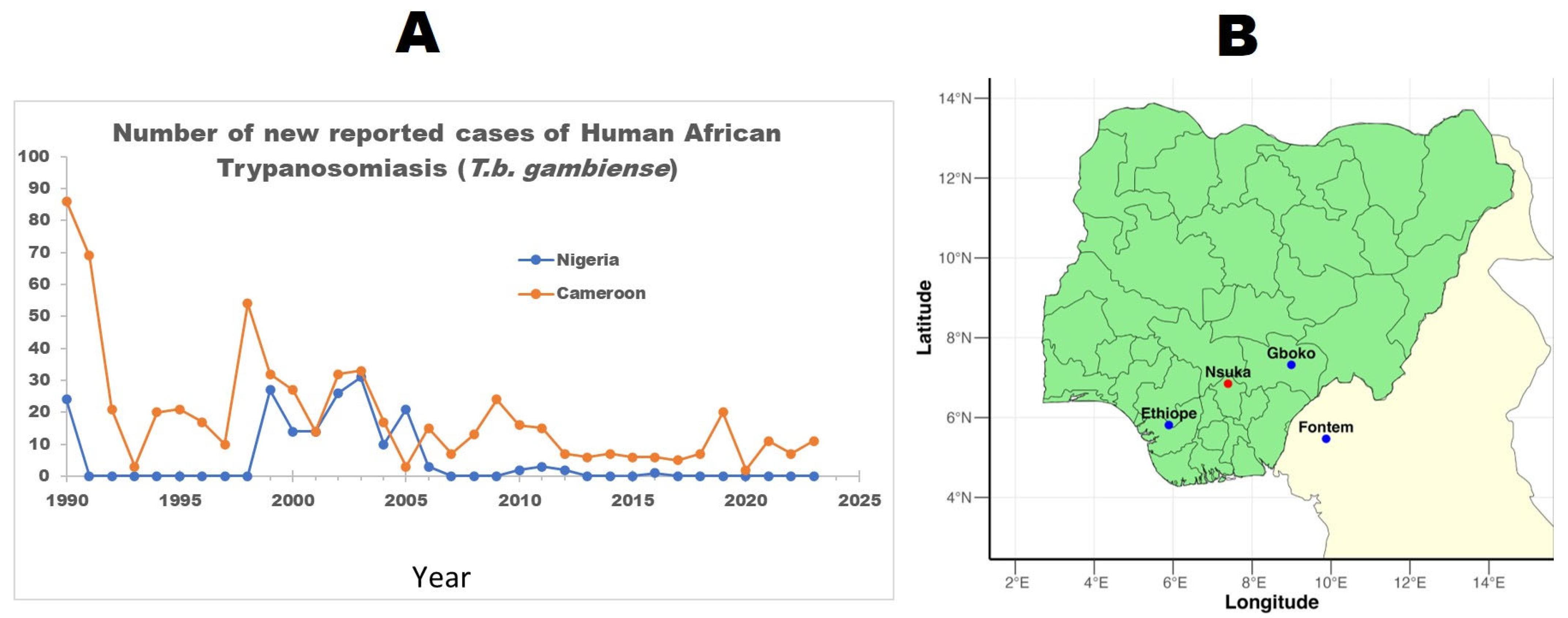
Figure 1. WHO HAT prevalence records for Nigeria and neighboring Cameroon showing that Nigeria has not reported any case in several years, while Cameroon continues to report cases (A), despite the close proximity of both countries’ known HAT foci and trans-border activities. (B)—Blue dots represent major HAT foci. Graph data was assessed from https://www.who.int/data/gho/data/themes/topics/human-african-trypanosomiasis (accessed on 15 March 2024).
Parasitologia 05 00053 g001
Figure 2. Flow chart of study selection process and article inclusion/exclusion for the review.
Parasitologia 05 00053 g002
Table 1. Summary of the characteristics of the reviewed articles.
Author/YearTitleAimCity/AreaStudy DesignStudy PopulationSample SizeOutcome MeasuredResultPrevalence RateDiagnostic MethodSpecies Identified
Daniel et al., 1993 [26]Prevalence of bovine trypanosomiasis in Gongola State of Northern NigeriaTo assess the prevalence of bovine trypanosomosis in Karim Lamido and Numan local government areas of Gongola State.Adamawa: Formerly Gongola (Karim Lamido and Numan LGAs)Cross-sectional SurveyCattle1065Bovine trypanosomiasisA total of 42 (3.9%) Cattle were found to be infected with trypanosomes. Out of which 27 (64.3%) were due to T. vivax, 13 (31%) to T. congolense and 2 (4.8%) to T. brucei.3.90%MicroscopyT. vivax
T. congolense
T. brucei
Daniel et al., 1994 [33]Prevalence of trypanosomiasis in sheep and goats in a region of northern NigeriaTo investigate the sensitivity of four techniques currently in use for the parasitological diagnosis of trypanosomosis.Bauchi (Alkaleri and Gombe LGAs)Cross-sectional SurveySheep
Goat
615 (258 sheep and 357 goats)Animal African trypanosomiasisA total of 19 (7.4%) sheep and 18 (5.0%) goats were positive giving a total infection rate of 37 (6.0%), 22 being positive with T vivax, 9 with T. congolense and 6 with T. brucei6%MicroscopyT. vivax
T. congolense
T. brucei
Enwezor et al., 2009 [27]Survey of bovine trypanosomosis in the Kachia Grazing Reserve, Kaduna State, NigeriaTo assess the prevalence of trypanosomes in cattle at the Kachia Grazing Reserve (KGR)KadunaCross-sectional SurveyCattle1293Bovine trypanosomiasisA total of 109 cattle were infected with trypanosomes giving an overall prevalence of 8.4%. Out of these, 105 (96.3%) was due to T. vivax, 2 (1.9%) to T. congolense, 1 (0.9%) to T. b. brucei and a mixed infection of T. congolense and T. vivax 1 (0.9%).8.40%MicroscopyT. vivax
T. congolense
T. brucei brucei
Enwezor et al., 2019 [38]Investigation of livestock for presence of Trypanosoma brucei gambiense In Tafa Local
Government Area of Niger State, Nigeria
Aimed at screening livestock for possible presence of T. b. gambiense within Tafa Local Government Area (LGA) of Niger State, Nigeria.NigerCross-sectional SurveyCattle
Sheep
Goats
Local dogs
Monkey
460 animals (177 cattle, 209 sheep, 61 goats, 12 local dogs and 1 monkey)Animal African trypanosomiasisA total of 10 animals were positive for trypanosomes with an overall prevalence of 2.17%. The morphological identification indicated the presence of T. brucei, T. vivax and a mixed infection of T. brucei and T. congolense. Details by animal species showed 3 cattle (1.69%), 6 sheep (3.02%) and 1 goat (1.64%) infected with trypanosomes. Neither the dogs nor the monkeys were positive for trypanosomes.2.17%MicroscopyT. brucei
T. congolense
T. vivax
Habeeb et al., 2021 [28]Molecular identification and prevalence of trypanosomes in cattle distributed within the Jebba axis of the River Niger, Kwara state, NigeriaTo investigate the prevalence of trypanosome infection in cattle, and molecularly identified the species of trypanosomes in infected cattle and the spatial distribution of trypanosome-infected herds along the Jebba axis of the River Niger.KwaraCross-sectional SurveyCattle398Bovine trypanosomiasisA total of 3 Cattle were positive by microscopy, representing 0.8% prevalence, while 12 samples representing 3.0% tested positive by nested PCR. With T. congolense more prevalent (50.0%).Microscopy: 0.8%
PCR: 3.0%
PCR
Microscopy
T. theileri 
T. evansi 
T. simiae
T. congolense 
T. brucei
T. vivax
Kalu et al., 1996 [34]Observations on the epidemiology of ruminant trypanosomosis in Kano State, NigeriaTo evaluate the prevalence of trypanosomosis among ruminants in Kano State and to elucidate aspects of the disease transmission in the areaKanoCross-sectional SurveyRuminants (Cattle, sheep, goat)1424 (1106 cattle and 318 small ruminants)Animal African trypanosomiasis59 cattle were infected with trypanosomes giving a prevalence of 5.3% with a confidence interval of ±1.3. Three out of 318 small ruminants sampled were infected (prevalence 0.9% ± 1.0).Cattle: 5.3% (95% CI: 4.0–6.6%)
Small ruminants (95% CI: −0.1–1.9%)
MicroscopyT. vivax
T. congolense
T. brucei
Karshima et al., 2016 [24]Silent Human Trypanosoma brucei gambiense Infections around the Old Gboko Sleeping Sickness Focus in NigeriaTo conduct an active screening of T. b. gambiense in humans in the old Gboko sleeping sickness focus in Nigeria and characterized isolates using TgsGP-polymerase chain reaction.GbokoCross-sectional SurveyHumans1200Human African trypanosomiasisOut of sampled people, a total of 28 were found positive. The CATT revealed an overall infection rate of 1.8% of the 1200 samples studied. PCR revealed an overall infection rate of 0.6% of the 1200 samples analyzed. Trypanosomes (TbG) were isolated from 7 of the samplesCATT: 1.8%
Parasite isolation/PCR: 0.6%
CATT
PCR
T. b. gambiense
Karshima et al., 2016 [37]Animal reservoirs of Trypanosoma brucei gambiense around the old Gboko sleeping sickness focus in NigeriaTo ascertain the possible role of animal reservoirs in the epidemiology of the parasite in the old Gboko sleeping sickness focus in Nigeria and characterized isolates using TgsGP polymerase chain reaction.GbokoCross-sectional SurveyCattle
Pigs
Animal African trypanosomiasisA total of 118 animals were positive (46 cattle, 72 pigs). The overall infection rates for the CATT and TgsGP-PCR were 8.9 and 0.9%, respectively. Trypanosomes of animal origin identified by ITS 1 PCR were T. brucei (4.2%), T. congolense forest (3.2%), T. congolense savannah (2.0%), T. vivax (2.2%) and mixed infections (1.5%) in cattle as well as T. brucei (4.8%), T. congolense forest (1.8%), T. congolense savannah (1.0%) and mixed infections (1.2%) in pigs. T. brucei gambiense and other animal trypanosomes were identified among animals in the focus, indicating the existence of animal reservoirs of human infective T. b. gambiense.CATT: 8.9%
PCR: 0.9%
CATT
PCR
T. brucei (T. b. gambiense)
T. congolense forest
T. congolense savannah
T. vivax
Majekodunmi et al., 2013 [29]A longitudinal survey of African animal trypanosomiasis in domestic cattle on the Jos Plateau, Nigeria: Prevalence, distribution, and risk factorsTo determine seasonal variations in the prevalence of AAT across the Jos PlateauJosCross-sectional SurveyCattle7143Bovine trypanosomiasis3, 342 cattle were found positive. The prevalence of bovine trypanosomiasis was found to be high at 46.8% (39.0–54.5%) and significant, seasonal variation was observed between the dry and the end of the wet season. T. b. brucei was observed at a prevalence of 3.2% (1–5.5%); T. congolense at 27.7% (21.8–33.6%) and T. vivax at 26.7% (18.2–35.3%).46.8% (39.0–54.5%)PCRT. congolense
T. vivax
T. b. brucei
Nmorsi et al., 2010 [23]Human African trypanosomiasis in endemic focus of Abraka, NigeriaTo investigate the prevalence of human African trypanosomiasis (HAT), caused by Trypanosoma brucei gambiense in an endemic focus of Nigeria, as it relates to age, sex and occupational differences.Abraka, DeltaCross-sectional SurveyHumans474Human African trypanosomiasisOf the 474 screened, 44(9.3%) were seropositive with seroprevalence of 22(9.6%) in Urhouka, 14(9.5%) in Umeghe and 8(7.9%) for Ugonu. CATT: 9.30%
Microscopy (blood): 3.4%
Microscopy (CSF): 0.8%
Microscopy
CATT
T. b. gambiense
Omeke, 1994 [31]Pig trypanosomosis: prevalence and significance in the endemic Middle Belt zone of southern NigeriaTo determine the prevalence of trypanosome species pathogenic to pigs and the significance of pig trypanosomosis in the Middle Belt zone of Southern Nigeria.Anambra and Benue statesCross-sectional SurveyPigs1954Porcine trypanosomiasis524 (26.8%) were positive for trypanosome infections, 348 (66.5%) of which had a mixed T. brucei and T. congolense burden, while 125 (23.9%) and 43 (8.2%) others had single T. brucei and T. congolense infections, respectively. 26.80%MicroscopyT. congolense
T. brucei
Onah and Ebenebe 2004 [32]Isolation of a human serum-resistant Trypanosoma brucei from a naturally infected pig in the Nsukka area of Enugu StateTo re-evaluate the role of domestic pig as a reservoir for T. b. gambienseEnuguCross-sectional SurveyPigs85Porcine trypanosomiasis19 positive cases were identified from the 85 sampled pigs. 15 (78.96%) were identified as single infection caused by T. brucei. While the remaining 4 (21.05%) were due to mixed infections of T. brucei and T. congolense.22.35%MicroscopyT. brucei
T. congolense T. brucei
gambiense
Takeet et al., 2013 [30]Molecular survey of pathogenic trypanosomes in naturally infected Nigerian cattleTo determine the prevalence and characteristics of trypanosome species and strains in Nigerian cattle using PCR for the first time.Ogun and Kaduna statesCross-sectional SurveyCattle411Bovine trypanosomiasisParasite detection by microscopy observation showed 62 samples infected by one or more species of Trypanosomes, for a prevalence of 15.1% (95% CI, 12–18%).
PCR detection showed 262 samples infected by one or more species of Trypanosoma, for an overall prevalence of 63.7% (95% CI, 59.4–68.8%) and T congolense was the most prevalent 48.7% (95% CI, 4.2–54.3), followed by T. vivax 26.0% (95% CI, 21.8–31.1%) and T. brucei 4.4% (95% CI, 3.3–7.1%).
Prevalence of mixed infections was 13.9% (95% CI, 10.6–17.4%) being co-infection by T. congolense and T. vivax.
Microscopy: 15.1% (95% CI, 12–18%).
PCR: 63.7% (95% CI, 59.4–68.8%)
Microscopy
PCR
T. brucei
T. vivax
T. congolense
Uba et al., 2016 [25]Knowledge and prevalence of human african trypanosomiasis among residents of kachia grazing reserve, Kachia local government area, Kaduna state, Nigeria, 2012To determine the knowledge, practices and prevalence of HAT among residents of the grazing reserve.KadunaCross-sectional SurveyHumans300Human African TrypanosomiasisOf the 300 respondents that were examined and screened for HAT, none had palpable cervical lymph nodes enlargement, and none tested positive on CATT; hence HAT prevalence was zero.0.00%CATT
Umeakuana et al., 2019 [36]Identification of Trypanosoma brucei gambiense in naturally infected dogs in NigeriaTo determine which trypanosome species, cause canine trypanosomosis in the Nsukka area of Nigeria and whether any dogs harbor the human-infective trypanosome, Tbg1.NsukkaCross-sectional SurveyDogs19Canine African Trypanosomiasis (CAT)All 19 dogs sampled had canine trypanosomosis caused by trypanosomes of the T. brucei group. Two of the dogs were shown to be infected with the human pathogen T. b. gambiense Group 1 (Tbg1).Tbg: 10.52%PCRT. b. gambiense
T. brucei
T. congolense
Wayo, 2017 [35]Prevalence of trypanosomiasis in sheep in the Kachia grazing reserve, Kachia, Kaduna State, NigeriaTo update information on the prevalence of trypanosomiasis in small ruminants in the area, to allow for proper planning of control activities and serve as valuable scientific data.KadunaCross-sectional SurveySheep110Animal African trypanosomiasisA total of 45 (40.9%) animals were found positive. The trypanosomes observed were T. congolense (40.0%), T. Brucei (28.8%), T. vivax (17.7%) and mixed infections (13.3%)40.9MicroscopyT. brucei
T. vivax
T. congolense
Disclaimer/Publisher’s Note: The statements, opinions and data contained in all publications are solely those of the individual author(s) and contributor(s) and not of MDPI and/or the editor(s). MDPI and/or the editor(s) disclaim responsibility for any injury to people or property resulting from any ideas, methods, instructions or products referred to in the content.