Article

Empirical Anthelmintic Therapy for Patients with Eosinophilia in Nepal: A Prospective Cohort Study

1
Critical Care Division, Rambam Medical Center, Haifa 3109601, Israel
2
Dhulikhel Hospital, Kathmandu University Hospital, Dhulikhel 45200, Nepal
3
Travel Medicine & Tropical Diseases, Rambam Medical Center, Haifa 3109601, Israel
4
Sackler Faculty of Medicine, Tel Aviv University, Tel Aviv 6997801, Israel
5
Department of Infectious Diseases and Epidemiology, Sourasky Medical Center, Tel Aviv 6423906, Israel
6
Gastroenterology Unit, Rambam Medical Center, Haifa 3109601, Israel
7
The Center for Geographic Medicine and Tropical Diseases, Chaim Sheba Medical Center, Tel Hashomer 5262000, Israel
*
Author to whom correspondence should be addressed.
These authors contributed equally to this work.
Parasitologia 2023, 3(2), 160-171; https://doi.org/10.3390/parasitologia3020017
Submission received: 20 January 2023 / Revised: 2 April 2023 / Accepted: 11 April 2023 / Published: 22 April 2023
(This article belongs to the Special Issue Global Change and Parasites)

Abstract

:
Eosinophilia is common in low-resource countries and usually implies helminthiasis. Since helminthiasis is a common cause of eosinophilia and its diagnosis is cumbersome, we hypothesized that broad-spectrum anthelmintic therapy may decrease the eosinophil count and eventually cure helminthiasis, whether microbiologic diagnosis is established or not. We recruited patients with eosinophilia aged 5 years and older who presented to Dhulikhel hospital, Nepal. Patients were treated with albendazole and ivermectin. A stool sample for microscopy was obtained. Of a total of 113 patients, 106 had a follow-up visit and 56 were classified as responders to treatment (defined as a decrease in eosinophil count to below 500 cells/µL, or an absolute decrease of more than 1000 cells/µL). For all patients, we found an absolute decrease in the eosinophil count and for the responding group (more than 50% of the whole cohort), the eosinophil count decreased substantially. All stool samples were negative. The reason for a lack of response in the remaining patients is unclear. In order to ascertain whether eosinophilia should be an indication for anthelmintic treatment, a randomized controlled study of empirical treatment after a thorough microbiologic workup is needed.

1. Introduction

Eosinophilia is linked to a broad spectrum of clinical conditions, such as helminthic and parasitic infections, allergic disorders, malignant and primary hematologic diseases, and auto-immune disorders. Infections are by far the leading cause of eosinophilia worldwide, especially in low-resource countries [1]. According to the World Health Organization (WHO), almost one billion people in low- and medium-resource countries in sub-Saharan Africa, Asia, and Latin America acquire helminth infections as a result of low standards of hygiene and the lack of access to clean food and water. Impaired mental and physical development, higher rates of anemia, weight loss, malnutrition, and neurological disorders are linked to such infections, which may also exacerbate other conditions, such as malaria and human immunodeficiency virus infection [2].
In many countries, medical care is only partially accessible to impoverished populations, which by and large lack any form of reliable medical insurance. The microbiologic diagnosis of helminthic infections in these settings has proven to be difficult, time consuming and, above all, too expensive. Multiple microscopic examinations of stool specimens have a low sensitivity and specificity, and are extremely laborious. Serologic tests, stool antigen detection tests, and nucleic-acid amplification tests are usually not widely available due to their cost, and do not exist for many helminths [3].
In Nepal, helminthic infections are common and the prevalence of eosinophilia is considerable [4]. A previous study conducted at Dhulikhel Hospital on a pediatric population between the years 2009 and 2011 revealed the incidence of persistent eosinophilia to be 2.4% [5].
Helminths, which are one of the causes of eosinophilia in Nepal, are most commonly soil-transmitted helminths (STH), such as Ascaris lumbricoides, Strongyloides stercoralis, Trichuris trichuria Toxocara spp., hookworms, and tapeworms such as Hymenolepis nana and Taenia spp. [6,7,8,9,10,11,12]. One study demonstrated a high detection rate of anti-Toxocara spp. antibodies [13]. Filariasis occurs more commonly in the tropical region of Nepal, but also in the Kathmandu Valley region [14,15].
Several studies have shown that empirical anthelmintic therapy leads to a decrease in the eosinophil count, whether a comprehensive microbiological diagnosis was obtained [16,17] or not [18,19,20,21]. These studies were all conducted in developed countries, and included participants who were either immigrants or returning travelers from the tropics.
Mass drug administration of anthelmintic drugs is common policy in endemic regions, but it is usually performed without any laboratory follow-up.
In the current study, we aimed to follow the decrease in eosinophil counts in patients with eosinophilia who receive broad-spectrum anthelmintic therapy in a low-resource setting and to assess the factors that are associated with response to treatment.

2. Results

During the study period, we enrolled 113 patients with eosinophilia; 106 (93.8%) returned for a follow-up visit, of whom 68 were males and 38 were females. The study group included both adults (44 patients) and children (62 patients), with a median age of 47.4 ± 16 and 8.5 ± 3.6 years, respectively.
The vast majority of patients who took part in the study resided in the Kavre-palanchowk district in the Kathmandu Valley region. The majority, 75/106 (70%), came from rural areas, whereas 31/106 came from towns or cities such as Dhulikhel, Banepa, and Kathmandu. Most of the patients used public transportation to reach the hospital (travel duration ranged from ten minutes to one day). Among adult patients, 36/44 (81%) were farmers, whereas 8 had non-farming occupations (e.g., private business, building, or housekeeping). The major reasons for hospital admissions included cardiorespiratory and infectious diseases, as shown in Table 1.
A stool test for ova and parasites was conducted for 97 patients at the hospital lab. Seven samples detected a presence of parasites: two were Giardia spp. and five Entamoeba histolytica. No helminths were detected. Nine patients could not pass stool during their hospital stay.
After administration of the anthelmintic treatment with ivermectin and albendazole, a follow-up visit was ordered after 2–8 weeks.
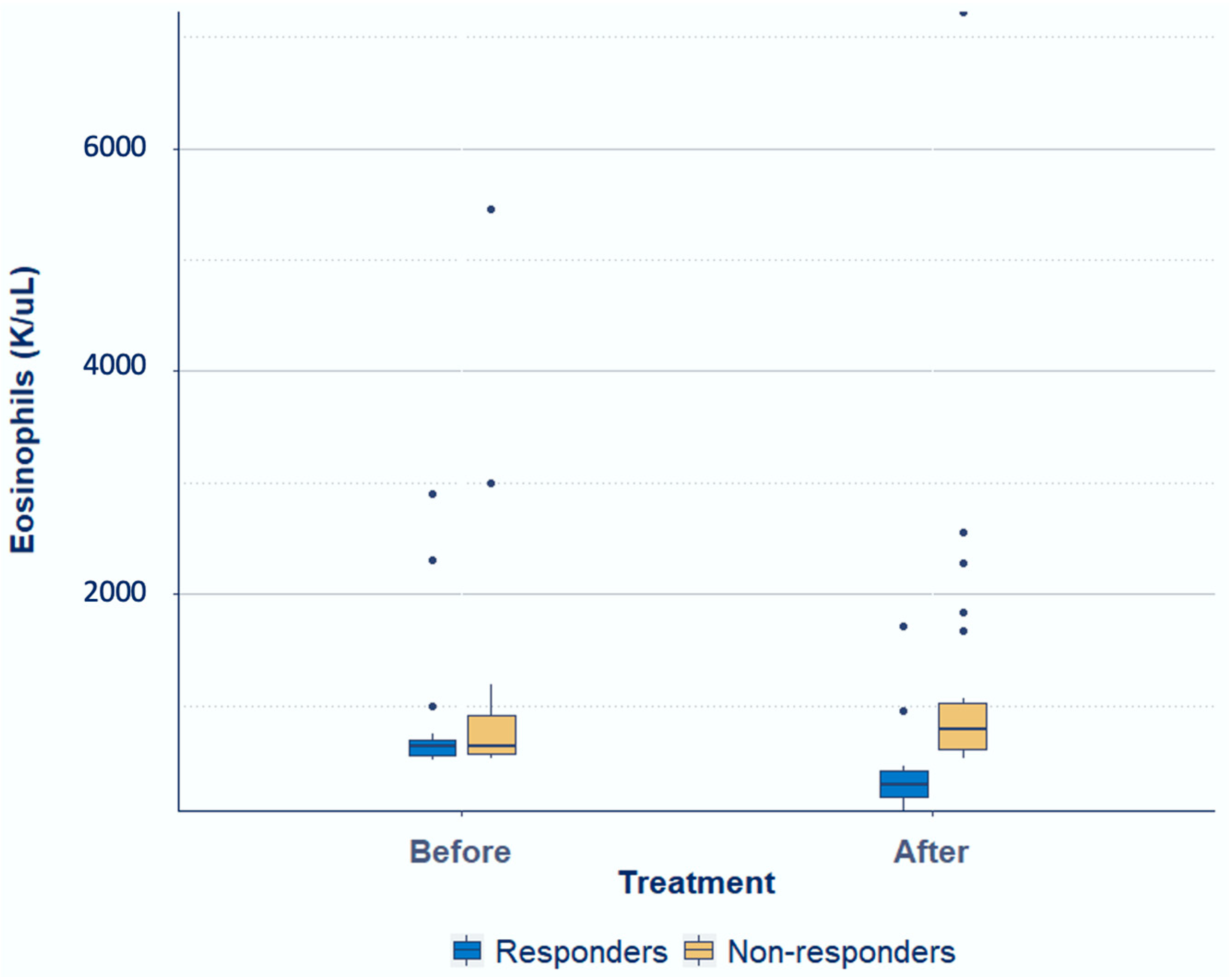
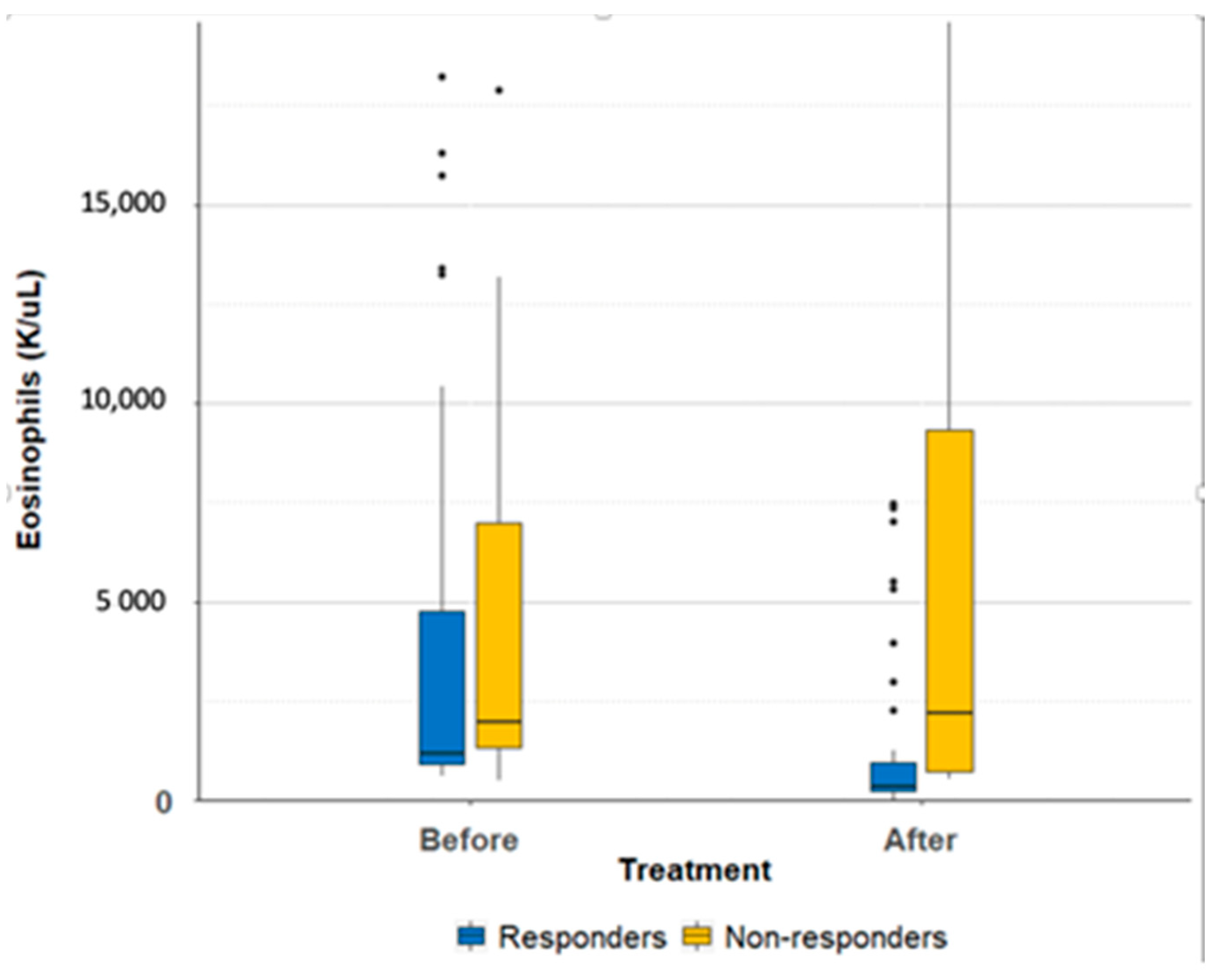
For the whole cohort (N = 106), we found a decrease in the eosinophil count before and after treatment: mean eosinophil count 2850 ± 4153 cells/μL, and 2220 ± 3689 cells/μL, respectively (mean of difference 629, p-value 0.03) (Table 2). Dividing the group into responders and non-responders, 56 (52.8%) were classified as responders and 50 as non-responders. Within the responders group (N = 56), the mean eosinophil count on first evaluation was 2823 ± 4517 cells/μL and 1027 ± 1900 cells/μL after treatment (mean of difference 1795, p-value < 0.001). Within the non-responders group, the mean eosinophil counts were 2879 ± 3749 cells/μL and 3555 ± 4655 cells/μL, before and after treatment, respectively (mean of difference 675, p-value 0.025), Table 2, Figure 1. When analysis was carried out for the adult and pediatric participants separately, results were similar, although the increases in eosinophil counts among non-responders were statistically insignificant, as shown in Table 2 and Figure 2 and Figure 3.
The distribution of the sociodemographic characteristics between responders and non-responders is summarized in Table 3. It is notable that living conditions, which could be potentially associated with a higher burden of parasitic infections and treatment failure, were similar in the two study groups. Among responders, 76.8% of the patients came from rural areas, and 3.6% worked in farming. Among the non-responders, the figures were 64% and 12% (p-values of 0.2 and 0.14, respectively). A total of 23% of the responders reported seeing worms in their stool, compared with 12% of the non-responders (p-value 0.2).
Comparisons of medical history, parameters of hygiene, and physical examination findings between responders and non-responders did not reveal statistically significant differences. Although there was a decrease in rashes, chest pains, gastrointestinal symptoms, weight loss, fever, myalgia, and headache following treatment among responders and non-responders (p < 0.05 for all comparisons), as shown in Table 3, a direct cause and effect relation could not be established, as patients were admitted for other, possibly unrelated, conditions.

3. Discussion

In this study, we treated 106 patients with incidental eosinophilia in Nepal with ivermectin and albendazole. Fifty-six patients met the predefined response criteria of a decrease in the eosinophil count to below 500 cells/µL, or an absolute drop of more than 1000 cells/µL.
It should be noted that only half of the patients in this cohort responded with a substantial decrease in eosinophil counts, and many non-responders’ eosinophil counts even rose (Figure 1), This unexpected finding could be attributed to several factors: (1) Theoretically, a helminthic reinfection is likely, although we are unable to prove it due to the unavailability of diagnostics in these low resource settings. (2) Initial infections with parasites that respond poorly or slowly to treatment (e.g., toxocariasis). (3) Poor compliance with albendazole treatment. (4) A longer period needed to observe a decrease in eosinophil counts after treatment. (5) Less likely, but non-infectious causes of eosinophilia. (6) Immunostimulation following the disintegration of worms and the release of their antigenic components. This phenomenon has been previously described for schistosomiasis and lymphatic filariasis [22]; a follow-up interval of a minimum of two weeks could skip immunostimulation. However, this is still a possibility.
Previous studies, conducted only in high-income countries, have shown that treating immigrants and travelers from the tropics empirically for eosinophilia was beneficial in terms of eosinophil counts [16,17,18]. The response rate in these studies was much higher than in the current study. For example, in a post-travel clinic in Israel, empirical treatment with albendazole was given to travelers returning from the tropics presenting with eosinophilia, resulting in a significant decrease in the eosinophil count in 27 out of 30 patients (90%). Of note, in these patients, schistosomiasis was ruled out, but a definite etiology for eosinophilia could not be obtained [16]. Similar results were shown in another study conducted in Spain, where a resolution of eosinophilia was observed in 31 out of 33 immigrants (93.9%) after giving an empirical treatment with albendazole, ivermectin, and praziquantel [17].
To our knowledge, our study is the first to be conducted in a developing country, suggesting that repeated infection or a higher burden of initial parasitic infection are key factors in low-resource settings. However, a missed non-infectious etiology for eosinophilia is also possible.
Non-responders had higher eosinophil counts at the first encounter. This could imply that the higher the eosinophil count, the less likely a response to treatment will be, either because of infection with a difficult to treat organism (e.g., Toxocara spp.), a heavier burden of infection, or a higher risk of reinfection. These findings call for a prospective study of the causes of eosinophilia in patients with high eosinophil counts.
As expected, gastrointestinal symptoms were not present in all patients with eosinophilia, as many infections are asymptomatic. Similarly, relevant findings in physical exams, such as abdominal pain, were absent in most patients. The alleviation of each gastrointestinal symptom in patients following treatment cannot be directly attributed to the therapy as there was no control group, and patients were admitted for various unrelated diseases that have been treated. The decrease in all gastrointestinal symptoms in both the responders and non-responders was not significantly different between both groups (p-value 0.25). Only by conducting a placebo-controlled trial were we able to conclude that empirical therapy did indeed alleviate symptoms. As symptoms and physical signs are unreliable for the diagnosis of helminthic infections, and since microbiological diagnosis is cumbersome and expensive, the rational approach is to treat patients with eosinophilia empirically.
Demographic differences between responders and non-responders were not significant. However, conditions that favor the transmission of helminthic infection were very common in most cases, and it is likely that in high-endemicity settings most of the population, regardless of socioeconomic status, is at a high risk of infection. Access to clean water and food, and educational efforts aiming at raising awareness as to how these infections spread, are warranted.
Ivermectin is effective in the treatment of lymphatic filariasis [23], strongyloidiasis [24], and STHs such as T. trichuria and A. lumbricoides [25]. It has, however, been found to be ineffective against hookworms, which are common in Nepal. Albendazole is used worldwide for STHs such as hookworms and A. lumbricoides, but its effectiveness against T. trichuria is limited [26]. Periodic and long-term mass drug treatment with ivermectin was found to decrease the prevalence and intensity of lymphatic filariasis [27], but did not have a long-term effect on the prevalence of many STHs, such as A. lumbricoides and hookworms [28]. On the other hand, when given as mass treatment, albendazole is effective against ascariasis with a single dose, and against hookworms with two doses [29]. Praziquantel, the drug of choice against tapeworms such as H. nana and Taenia spp. was not added to our regimen. However, some studies have showed that albendazole is a reasonable alternative to praziquantel in treating infections with H. nana and Taenia spp. [30,31]. A combined treatment regimen that includes both albendazole and ivermectin, although effective against most STHs and relatively safe, may not cure some infections that cause eosinophilia (e.g., toxocariasis) [32,33].
Advanced molecular diagnostic tests and the Kato-Katz technique were not used, as they are not available at the hospital laboratory. Instead, a direct smear was performed after using a concentration solution. Our failure in detecting helminths in stool can be explained by several factors. Firstly, the test is insensitive and usually needs to be repeated three times to optimize its sensitivity in order to diagnose some helminths such as s. stercoralis [34]; this was not achievable in our study due to the fact that most of our participants were outpatients visiting the hospital for a short period of time. Secondly, stool tests could miss the detection of larva given an intermittent excretion into the gastrointestinal tract [34]. Lastly, some endemic helminths in Nepal, such as lymphatic filariasis, cannot be detected in stool as they do not involve the gastrointestinal system. Not using Kato-Katz flotation or advanced techniques to diagnose helminths indeed explains our failure to detect helminth species. The lack of more sophisticated tests (i.e., polymerase chain reaction-based molecular techniques and serologic assays) is likely to decrease the yield of a laboratory workup, and prompts the need to look for simpler methods in order to diagnose helminthic infections.
The time until the repeated eosinophil test following treatment was 2–8 weeks. The difference between short and long follow-up intervals, defined as 2–4 weeks for short and 4–8 weeks for long, had no significant impact on the change in eosinophil count between responders and non-responders (p-value 0.3).
Studies conducted over the past few decades have assessed the immediate and long-term effects of anthelmintic therapy administered individually for infected patients, or as part of a mass treatment program of a susceptible population [35,36,37,38]. None of these studies used eosinophil count as a possible screening tool for patients who could have derive a greater benefit from empirical treatment.
Our study has several limitations: (1) It is a single-center study. (2) Selection bias is unavoidable, as only patients that could afford the transportation to the hospital were recruited. (3) We could not ascertain if patients’ symptoms provided by the questionnaire were related to helminthiasis or other non-infectious medical conditions. (4) Apart from stool microscopy, other laboratory tests were not performed in order to check for the presence of helminths. (5) The effect of the anthelmintic therapy would theoretically have taken more time to affect the eosinophil count, so that early follow-ups might have missed additional patients who responded to treatment at a later time. (6) There were nine patients who did not provide stool samples for analysis. However, this is unlikely to change our conclusions, as in all other 97 samples no helminths were detected. (7) This is a non-comparative observational study assessing a continuous variable (eosinophil count) after a pre-specified intervention. Since such study has never been performed among patients residing in low-income countries, the predicted effect was unknown. Therefore, a formal calculation of a sample size was not possible. (8) The study is limited by the absence of a control group. In fact, some changes could represent the natural history of eosinophilia in a population with continuous exposure to parasitic infections, rather than the effect of any medication.

4. Materials and Methods

4.1. Setting

Dhulikhel hospital is a non-governmental, non-profit, independent medical center situated 30 km northeast of Kathmandu. It accommodates a mostly rural population from the surrounding villages and has 475 beds.

4.2. Study Design and Participants

A prospective interventional trial, non-blinded and non-randomized, was performed between October 2014 and July 2017. During the study period, Nepal’s devastating earthquake in April 2015 affected patients’ recruitment for several months (April to December of that year); apart from that, patients’ recruitment was completed through all the months of the years. The institutional review committee of Kathmandu University School of Medical Science/Dhulikhel hospital approved the study (approval number 106/14). Patients aged 5 years and older were examined in the internal medicine and pediatric ward at Dhulikhel hospital in the outpatient clinics or as inpatients. Those who were found to have an absolute eosinophil count greater than 500 cells/microliter, regardless of symptoms, were included in the study. Exclusion criteria included: overt non-infectious causes of eosinophilia (such as allergic reactions, inflammatory conditions, paraneoplastic phenomena), treatment with an anthelmintic drug within the previous three months, pregnancy or breast feeding, known hypersensitivity to either albendazole or ivermectin, inability to swallow oral medications, significant liver disease, and treatment with vitamin K antagonists. Detailed explanations were provided in Nepali. All patients who agreed to participate in the study signed a consent form in Nepali. For the pediatric group, the parents had to sign the consent form.

4.3. Intervention

At first encounter, demographic and socioeconomic characteristics, as well as parameters associated with personal hygiene, were recorded on a structured questionnaire, supplement Table S1, and a structured physical examination was conducted. A complete blood count test was obtained and processed at the hospital’s laboratory with an automatic coulter counter. Stool samples were examined with a direct smear after a concentration solution was used. All patients were treated orally with a 400 mg dose of albendazole, taken twice daily for 3 days, and a 200 mcg/kg dose of ivermectin, administered as a single dose. Patients took the remainder of the albendazole tablets on the second and third day of their own accord, without supervision. In case patients experienced any adverse events, they were told to return to the hospital.
Two to eight weeks after taking the drugs, patients were invited to attend a follow-up visit, which included assessment of any adverse effects and changes in symptoms and repeat blood tests for an absolute eosinophil count.

4.4. Outcome Measures

The primary outcome measure was response to treatment in terms of the change in absolute eosinophil count obtained before and after treatment with ivermectin and albendazole. Any patient with a decrease in eosinophil count to below 500 cells/µL, or an absolute decrease of more than 1000 cells/µL in eosinophil count was considered to have met the criteria for the primary outcome. The secondary outcome measures were improvement in symptoms for patients who were found to be symptomatic before treatment.

4.5. Statistics

We compared dichotomous variables with the use of chi-square or Fisher’s exact text, where appropriate. Continuous variables were compared with the use of either the Student’s t-test, or the Mann–Whitney test for variables with a non-normal distribution. A significance level of 0.05 was utilized as the threshold for determining statistical significance.

5. Conclusions

In this interventional study, we demonstrated that just over 50% of Nepali patients with incidental eosinophilia had a significant decrease in eosinophilia following treatment with ivermectin and albendazole. The reasons for lack of response in terms of eosinophil counts among other patients are unclear. In order to assess whether such an empirical and cheap treatment is indeed effective, and to determine which patients could benefit from it, a randomized–placebo-controlled trial, which should include a thorough microbiologic workup, is warranted.

Supplementary Materials

The following supporting information can be downloaded at: https://www.mdpi.com/article/10.3390/parasitologia3020017/s1, Table S1: A Questionnaire for patients with eosinophilia at first encounter and after therapy.

Author Contributions

Conceptualization, E.S. and A.N.; methodology, K.B., P.P. and M.A.; validation, E.S., A.N. and K.B.; formal analysis, Y.G.; investigation, K.B., P.P., S.S., S.K.M. and M.A.; resources, E.S. and A.N.; data curation, N.Z. and Y.P.; writing—original draft preparation, K.B. and P.P.; writing—review and editing, K.B., E.S. and A.N.; visualization, N.Z. and Y.P.; supervision, E.S.; project administration, K.B. and M.A.; funding acquisition, E.S. All authors have read and agreed to the published version of the manuscript.

Funding

The funder of this study was the Israeli Embassy in Kathmandu, Nepal. The Israeli embassy played no role in study design, data collection, data analysis, data interpretation, or writing of the report.

Institutional Review Board Statement

The institutional review committee of Kathmandu University School of Medical Science/Dhulikhel hospital approved the study (approval number 106/14).

Informed Consent Statement

Detailed explanations were provided in Nepali. All patients who agreed to participate in the study signed a consent form in Nepali. For the pediatric group, the parents had to sign the consent form.

Data Availability Statement

The data presented in this study are available on request from the corresponding author. The data are not publicly available as they contain information that could compromise the privacy of research participants.

Acknowledgments

We thank Dhulikhel Hospital staff for their support.

Conflicts of Interest

The authors declare no conflict of interest.

References

  1. Savini, H.; Simon, F. Blood eosinophilia in the tropics. Méd. Santé Trop. 2013, 23, 132–144. [Google Scholar] [CrossRef]
  2. World Health Organization & UNICEF/UNDP/World Bank/WHO Special Programme for Research and Training in Tropical Diseases. Global Report for Research on Infectious Diseases of Poverty 2012. World Health Organization. 2012. Available online: https://apps.who.int/iris/handle/10665/44850 (accessed on 18 April 2023).
  3. Miswan, N.; Singham, G.V.; Othman, N. Advantages and Limitations of Microscopy and Molecular Detections for Diagnosis of Soil-transmitted Helminths: An Overview. Helminthologia 2022, 59, 321–340. [Google Scholar] [CrossRef] [PubMed]
  4. Devleesschauwer, B.; Ale, A.; Torgerson, P.; Praet, N.; de Noordhout, C.M.; Pandey, B.D.; Pun, S.B.; Lake, R.; Vercruysse, J.; Joshi, D.D.; et al. The Burden of Parasitic Zoonoses in Nepal: A Systematic Review. PLoS Negl. Trop. Dis. 2014, 8, e2634. [Google Scholar] [CrossRef] [PubMed]
  5. Shrestha, S.; Dongol Singh, S.; Shrestha, N.C.; Shrestha, R.P.B. Clinical and laboratory profile of children with eosinophilia at Dhulikhel hospital. Kathmandu Univ. Med. J. 2012, 10, 58–62. [Google Scholar] [CrossRef] [PubMed]
  6. Agrawal, P.K.; Rai, S.K.; Khanal, L.K.; Ghimire, G.; Banjara, M.R.; Singh, A. Intestinal Parasitic Infections among Patients Attending Nepal Medical College Teaching Hospital, Kathmandu, Nepal. Nepal Med. Coll. J. 2012, 14, 80–83. [Google Scholar]
  7. Gyawali, N.; Amatya, R.; Nepal, H.P. Intestinal parasitosis in school going children of Dharan municipality, Nepal. Trop. Gastroenterol. 2009, 30, 145–147. [Google Scholar]
  8. Khanal, L.K.; Choudhury, D.R.; Rai, S.K.; Sapkota, J.; Barakoti, A.; Amatya, R.; Hada, S. Prevalence of Intestinal Worm Infestations among School Children in Kathmandu, Nepal. Nepal Med. Coll. J. 2011, 13, 272–274. [Google Scholar]
  9. Sharma, B.K.; Rai, S.K.; Rai, D.R.; Choudhury, D.R. Prevalence of Intestinal Parasitic Infestation in School Children in the Northeastern Part of Kathmandu Valley, Nepal. Southeast Asian J. Trop. Med. Public Health 2004, 35, 501–505. [Google Scholar]
  10. Shrestha, A.; Rai, S.K.; Basnyat, S.R.; Rai, C.K.; Shakya, B. Soil Transmitted Helminthiasis in Kathmandu, Nepal. Nepal Med. Coll. J. 2007, 9, 166–169. [Google Scholar]
  11. Shrestha, A.; Narayan, K.; Sharma, R. Prevalence of Intestinal Parasitosis Among School Children in Baglung District of Western Nepal. Kathmandu Univ. Med. J. 2012, 10, 62–65. [Google Scholar] [CrossRef]
  12. Dhital, S.; Pant, N.D.; Neupane, S.; Khatiwada, S.; Gaire, B.; Sherchand, J.B.; Shrestha, P. Prevalence of enteropathogens in children under 15 years of age with special reference to parasites in Kathmandu, Nepal; a cross sectional study. SpringerPlus 2016, 5, 1813. [Google Scholar] [CrossRef]
  13. Rai, S.K.; Uga, S.; Ono, K.; Nakanishi, M.; Shrestha, H.G.; Matsumura, T. Seroepidemiological study of Toxocara infection in Nepal. Southeast Asian J. Trop. Med. Public Health 1996, 27, 286–290. [Google Scholar]
  14. Sherchand, J.B.; Obsomer, V.; Thakur, G.D. Hommel, M. Mapping of lymphatic filariasis in Nepal. Filaria J. 2003, 2, 7. [Google Scholar]
  15. Watanabe, K.; Itoh, M.; Matsuyama, H.; Hamano, S.; Kobayashi, S.; Shirakawa, T.; Suzuki, A.; Sharma, S.; Acharya, G.P.; Itoh, K.; et al. Bancroftian filariasis in Nepal: A survey for circulating antigenemia of Wuchereria bancrofti and urinary IgG4 antibody in two rural areas of Nepal. Acta Trop. 2003, 88, 11–15. [Google Scholar] [CrossRef]
  16. Meltzer, E.; Percik, R.; Shatzkes, J.; Sidi, Y.; Schwartz, E. Eosinophilia among returning travelers: A practical approach. Am. J. Trop. Med. Hyg. 2008, 78, 702–709. [Google Scholar] [CrossRef]
  17. Salas-Coronas, J.; Cabezas-Fernández, M.T.; Vázquez-Villegas, J.; Soriano-Pérez, M.J.; Lozano-Serrano, A.B.; Pérez-Camacho, I.; Cabeza-Barrera, M.I.; Cobo, F. Evaluation of eosinophilia in immigrants in Southern Spain using tailored screening and treatment protocols: A prospective study. Travel Med. Infect. Dis. 2015, 13, 315–321. [Google Scholar] [CrossRef]
  18. Harries, A.D.; Myers, B.; Bhattacharrya, D. Eosinophilia in caucasians returning from the tropics. Trans. R. Soc. Trop. Med. Hyg. 1986, 80, 327–328. [Google Scholar] [CrossRef]
  19. Checkley, A.M.; Chiodini, P.L.; Dockrell, D.H.; Bates, I.; Thwaites, G.E.; Booth, H.L.; Brown, M.; Wright, S.G.; Grant, A.D.; Mabey, D.C.; et al. Eosinophilia in returning travellers and migrants from the tropics: UK recommendations for investigation and initial management. J. Infect. 2010, 60, 1–20. [Google Scholar] [CrossRef]
  20. Cañas García-Otero, E.; Praena-Segovia, J.; Ruiz-Pérez de Pipaón, M.; Bosh-Guerra, X.; Sánchez-Agüera, M.; Álvarez-Martínez, D.; Cisneros-Herreros, J.M. Clinical approach to imported eosinophilia. Enferm. Infecc. Microbiol. Clin. 2016, 34, 661–684. [Google Scholar] [CrossRef]
  21. Vaisben, E.; Brand Md, R.; Kadakh, A.; Nassar, F. The Role of Empirical Albendazole Treatment in Idiopathic Hypereosinophilia—A Case Series. Can. J. Infect. Dis. Med. Microbiol. 2015, 26, 323–324. [Google Scholar] [CrossRef]
  22. Ottesen, E.A.; Weller, P.F. Eosinophilia following treatment of patients with schistosomiasis mansoni and Bancroft's filariasis. J. Infect. Dis. 1979, 139, 343–347. [Google Scholar] [CrossRef] [PubMed]
  23. Cao, W.; Ploeg, C.P.B.; Plaisier, A.P.; Sluijs, I.J.S.; Habbema, J.D.F. Ivermectin for the chemotherapy of bancroftian filariasis: A meta-analysis of the effect of single treatment. Trop. Med. Int. Health 2007, 2, 393–403. [Google Scholar] [CrossRef]
  24. Zaha, O.; Hirata, T.; Kinjo, F.; Saito, A.; Fukuhara, H. Efficacy of ivermectin for chronic strongyloidiasis: Two single doses given 2 weeks apart. J. Infect. Chemother. 2002, 8, 94–98. [Google Scholar] [CrossRef] [PubMed]
  25. Naquira, C.; Jimenez, G.; Guerra, J.G.; Bernal, R.; Nalin, D.R.; Neu, D.; Aziz, M. Ivermectin for human strongyloidiasis and other intestinal helminths. Am. J. Trop. Med. Hyg. 1989, 40, 304–309. [Google Scholar] [CrossRef] [PubMed]
  26. Keiser, J.; Utzinger, J. Efficacy of current drugs against soil-transmitted helminth infections: Systematic review and meta-analysis. JAMA 2008, 299, 1937–1948. [Google Scholar] [CrossRef]
  27. Crump, A.; Ōmura, S. Review Ivermectin, “Wonder drug” from Japan: The human use perspective. Proc. Jpn. Acad. Ser. B Phys. Biol. Sci. 2011, 87, 13–28. [Google Scholar] [CrossRef]
  28. Moncayo, A.L.; Vaca, M.; Amorim, L.; Rodriguez, A.; Erazo, S.; Oviedo, G.; Quinzo, I.; Padilla, M.; Chico, M.; Lovato, R.; et al. Impact of long-term treatment with ivermectin on the prevalence and intensity of soil-transmitted helminth infections. PLoS Negl. Trop. Dis. 2008, 2, e293. [Google Scholar] [CrossRef]
  29. Adegnika, A.A.; Zinsou, J.F.; Issifou, S.; Ateba-Ngoa, U.; Kassa, R.F.; Feugap, E.N.; Honkpehedji, Y.J.; Agobe, J.-C.D.; Kenguele, H.M.; Massinga-Loembe, M.; et al. Randomized, Controlled, Assessor-Blind Clinical Trial to Assess the Efficacy of Single- versus Repeated-Dose Albendazole to Treat Ascaris lumbricoides, Trichuris trichiura, and Hookworm Infection. Antimicrob. Agents Chemother. 2014, 58, 2535–2540. [Google Scholar] [CrossRef]
  30. Prasad, K.N. My experience on taeniasis and neurocysticercosis. Trop. Parasitol. 2021, 11, 71–77. [Google Scholar] [CrossRef]
  31. Khan, W.; Khan, J.; Rahman, A.; Ullah, H.; Salim, M.; Iqbal, M.; Khan, I.; Salman, M.; Munir, B. Albendazole in the treatment of Hymenolepiasis in school children. Pak. J. Pharm. Sci. 2018, 31 (Suppl. 1), 305–309. [Google Scholar]
  32. McCarty, T.R.; Turkeltaub, J.A.; Hotez, P.J. Global progress towards eliminating gastrointestinal helminth infections. Curr. Opin. Gastroenterol. 2014, 30, 18–24. [Google Scholar] [CrossRef]
  33. Mohammed, K.A.; Haji, H.J.; Gabrielli, A.-F.; Mubila, L.; Biswas, G.; Chitsulo, L.; Bradley, M.H.; Engels, D.; Savioli, L.; Molyneux, D.H. Triple Co-Administration of Ivermectin, Albendazole and Praziquantel in Zanzibar: A Safety Study. PLoS Negl. Trop. Dis. 2008, 2, e171. [Google Scholar] [CrossRef]
  34. Vandenberg, O.; Van Laethem, Y.; Souayah, H.; Kutane, W.T.; van Gool, T.; Dediste, A. Improvement of routine diagnosis of intestinal parasites with multiple sampling and SAF-fixative in the triple-faeces-test. Acta Gastro-Enterol. Belg. 2006, 69, 361–366. [Google Scholar]
  35. Bronzan, R.N.; Dorkenoo, A.M.; Agbo, Y.M.; Halatoko, W.; Layibo, Y.; Adjeloh, P.; Teko, M.; Sossou, E.; Yakpa, K.; Tchalim, M.; et al. Impact of community-based integrated mass drug administration on schistosomiasis and soil-transmitted helminth prevalence in Togo. PLoS Negl. Trop. Dis. 2018, 12, e0006551. [Google Scholar] [CrossRef]
  36. Aye, N.N.; Lin, Z.; Lon, K.N.; Linn, N.Y.Y.; Nwe, T.W.; Mon, K.M.; Ramaiah, K.; Betts, H.; Kelly-Hope, L.A. Mapping and modelling the impact of mass drug adminstration on filariasis prevalence in Myanmar. Infect. Dis. Poverty 2018, 7, 56. [Google Scholar] [CrossRef]
  37. Shamsuzzaman, A.K.M.; Haq, R.; Karim, M.J.; Azad, M.B.; Mahmood, A.S.M.S.; Khair, A.; Rahman, M.M.; Hafiz, I.; Ramaiah, K.D.; Mackenzie, C.D.; et al. The significant scale up and success of Transmission Assessment Surveys “TAS” for endgame surveillance of lymphatic filariasis in Bangladesh: One step closer to the elimination goal of 2020. PLoS Negl. Trop. Dis. 2017, 11, e0005340. [Google Scholar] [CrossRef]
  38. Ahuja, A.; Baird, S.; Hicks, J.H.; Kremer, M.; Miguel, E. Economics of Mass Deworming Programs. In Child and Adolescent Health and Development, 3rd ed.; Bundy, D.A.P., Silva, N.D., Horton, S., Jamison, D.T., Patton, G.C., Eds.; The International Bank for Reconstruction and Development/The World Bank: Washington, DC, USA, 2017; Volume 29. [Google Scholar] [PubMed]
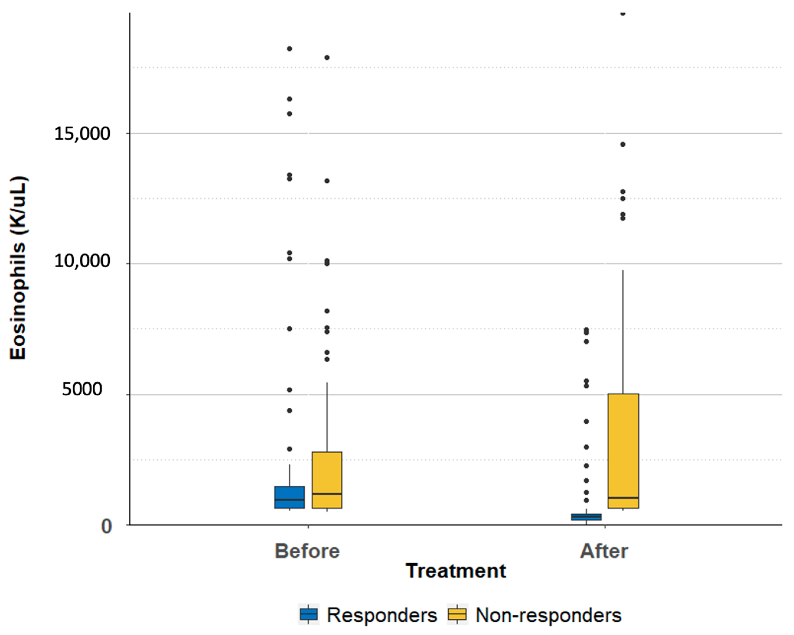
Figure 1. The change in eosinophil count among responders and non-responders in all patients after treatment with ivermectin and albendazole.
Parasitologia 03 00017 g001
Figure 2. The change in eosinophil count among responders and non-responders in adult group after treatment with ivermectin and albendazole.
Parasitologia 03 00017 g002
Figure 3. The change in eosinophil count among responders and non-responders in pediatric group after treatment with ivermectin and albendazole.
Parasitologia 03 00017 g003
Table 1. Sociodemographic characteristics of patients with eosinophilia in Dhulikhel, Nepal (N = 106).
Adults (N = 44)Children (N = 62)
Age (years) 49.4 ± 3.58.4 ± 4
Gender (females %) 21 (48%)17 (27%)
Place of residency
Village 30 (68%)45 (73%)
Town/city 14 (32%)17 (27%)
Occupation
Farmer 36 (82%)Not Relevant
Non-farmer 8 (18%)Not Relevant
Cause for admission (N = 98) N = 36N = 62
Infectious disease 28 (78%)31 (50%)
Respiratory disease 17 (47%)24 (39%)
Cardiac disease 07 (11%)
Gastrointestinal disease 2 (6%)9 (15%)
Kidney disease 06 (8%)
Neurological disease 3 (8%)3 (5%)
Others 14 (39%)13 (20%)
Table 2. Eosinophil counts before and after treatment among all participants, responders, non-responders, adults, and children.
MeanSDMedianIQRMean of DifferencepValue95% CI
All participants: pretreatment (N = 106)2850415399413996290.03(−621, 197)
All participants: post-treatment (N = 106)222036895891719
Responders: pretreatment (N = 56)282345179548151795<0.001(956, 2634)
Responders: post-treatment (N = 56)10271900296233
Non-responders: pretreatment (N = 50)2879374911612158−6750.025(−1263, −88)
Non-responders: post-treatment (N = 50)3555465510234388
Pediatric responders: pretreatment (N = 35)40255369118538732602<0.001(1315, 3889)
Pediatric responders post-treatment (N = 35)14232309339719
Pediatric non-responders: pretreatment (N = 27)4457445619805664−10720.051(−2149, 5)
Pediatric non-responders: post-treatment (N = 27)5529550722058621
Adult responders: pretreatment (N = 21)820610630143451<0.001(136, 766)
Adult responders: post-treatment (N = 21)369361288236
Adult non-responders: pretreatment (N = 23)10281093636346−2100.09(−458, 38)
Adult non-responders: post-treatment (N = 23)12381422784411
Table 3. Epidemiological and clinical data: responders versus non-responders.
Responders
(N = 56)
Non-Responders
(N = 50)
p-Value
Gender
Males (N = 68)60.7%68%0.54
Rural area (N = 75)76.8%64%0.2
Occupation
Children (N = 62)62%54%0.43
Farmers (N = 36)3.6%12%0.14
Self-reported hygiene (N = 106)
Wash hands after using the toilet 94%96%1
Use soap for washing hands76.8%64%0.2
Use latrine83.9%72%0.16
Walk barefoot 98.2%94%0.34
Wash vegetables94.6%96%1
Eat raw meat or vegetables 25%32%0.52
Saw worms in stool 23.2%12%0.2
Used anthelmintic agents in the past 62%56%0.55
Had allergic disorders 18.2%10%0.27
Cause of admission (N = 98)
Infection (N = 38)43.6%30%0.2
Respiratory disease (N = 41)44.6%32%0.22
Cardiac disease (N = 7)5.4%8%0.7
Gastrointestinal symptoms (N = 11)8.9%12%0.75
Urinary tract condition (N = 6)10.7%0%0.03
Neurologic condition (N = 6)012%0.01
Clinical presentation (N = 106)
Skin changes before treatment41.1%34%0.55
Skin changes after treatment8.9%6%0.72
Hemoptysis before treatment7.1%00.12
Hemoptysis after treatment1.8%01
Chest pain before treatment39.3%40%1
Chest pain after treatment14.3%12%0.78
Abdominal pain before treatment48.2%58%0.34
Abdominal pain post-treatment7.1%14%0.34
Diarrhea before treatment20%8.9%0.16
Diarrhea after treatment0%2%0.47
Nausea or vomiting before treatment25%40%0.14
Nausea or vomiting after treatment1.8%10%0.1
Weight loss before treatment55.4%44%0.33
Weight loss after treatment12.5%14%1
Constipation before treatment12.5%16%0.78
Constipation after treatment7.1%4%0.68
Fever before treatment58.6%48%0.33
Fever after treatment17.6%12%0.43
Chills before treatment32.1%36%0.69
Chills after treatment1.8%2%1
Myalgia before treatment44.6%36%0.43
Myalgia after treatment12.5%10%0.77
Headache before treatment50%48%0.85
Headache after treatment14.3%18%0.79
Throat irritation before treatment30.4%18%0.18
Throat irritation after treatment12.5%8%0.53
Limb swelling before treatment5.4%16%0.11
Limb swelling after treatment1.8%8%0.19
Eye redness before treatment8.9%26%0.04
Eye redness after treatment3.6%6%0.66
Disclaimer/Publisher’s Note: The statements, opinions and data contained in all publications are solely those of the individual author(s) and contributor(s) and not of MDPI and/or the editor(s). MDPI and/or the editor(s) disclaim responsibility for any injury to people or property resulting from any ideas, methods, instructions or products referred to in the content.