1. Introduction
The application of DNA analysis in the identification of deceased individuals holds indisputable forensic significance, particularly in cases involving advanced decomposition and fragmented remains, with its role well-established and widely recognized [
1]. In routine forensic practice, biological samples such as blood, hair roots, or saliva are commonly collected for recently deceased individuals, whereas in cases with prolonged postmortem intervals or advanced decomposition, soft tissues, teeth, or bone may be utilized [
2]. However, hard tissues often require extensive processing or repeated analysis, while soft tissues are often degraded or entirely absent, rendering them unsuitable for DNA analysis [
3]. Consequently, in Croatia, forensic identification of decomposed remains typically relies on more durable tissues such as bone or teeth, which require more time-consuming and technically challenging procedures for DNA analysis compared to blood or saliva. Bone and tooth samples, while generally considered the gold standard in postmortem DNA analysis, require time-intensive decontamination, decalcification, and extraction procedures, often necessitating specialized equipment and repeat sampling to achieve viable DNA profiles [
3,
4,
5,
6]. These limitations can delay identification, particularly in cases involving mass disasters, war victims, or exhumations of long-buried remains [
7,
8]. Given these constraints, there is a growing need to identify alternative biological matrices that can yield high-quality DNA in a more efficient manner [
4,
9,
10]. Nails, as keratinized structures, exhibit high resistance to postmortem degradation and environmental factors, and have increasingly been recognized as a potential source of genomic DNA. To date, only a limited number of studies have explored the extraction of DNA from nail samples derived from decomposed human remains for forensic identification purposes [
4,
5,
6,
7,
8,
11,
12,
13]. Compared to other biological substrates, such as teeth or bones, nails remain a relatively under-investigated matrix in forensic practice. Therefore, it is essential to investigate more possibilities of isolating, amplifying, and detecting targeted DNA fragments for genotyping and forensic identification from those biological samples [
9,
10].
The main goal of this case report was to explore the efficacy of DNA extraction and genotyping from a nail sample collected from a decomposed body, and to improve identification protocols in challenging forensic contexts, especially regarding flexibility in taking samples. Nails are composed primarily of keratin, a highly durable protein that provides structural protection to DNA against enzymatic degradation and microbial activity [
2,
10,
14]. Their compact and keratinized structure, combined with their anatomical location, makes them relatively resistant to intrinsic factors such as tissue decomposition, and extrinsic influences including moisture, temperature fluctuations, and prolonged burial—conditions that often compromise the integrity of soft tissues and other biological samples [
15,
16,
17]. Also, nails are much more accessible than bone or tooth samples during autopsy or exhumation. These properties make nails a valuable supplement when other samples are unavailable or unsuitable due to severe decomposition [
10,
11]. However, the literature lacks consensus on standard protocols for their processing, and their utility in severely decomposed cases remains underexplored. Additionally, DNA extracted from nails has been shown to remain stable even in highly decomposed bodies and in various environmental conditions [
11]. The application of PCR amplification kits specifically designed for degraded samples has further enhanced the ability to obtain reliable DNA profiles from such biological material [
12]. Both intrinsic and extrinsic factors influence DNA preservation in skeletal remains, and these principles apply similarly to nails due to their protective keratin matrix [
18]. Some studies suggest nails may be a good alternative to bone or tooth in certain forensic scenarios due to easier collection and less invasive processing [
13,
19].
Recommendations from mass fatality management highlight the importance of diversifying sample types to improve DNA recovery success, especially in forensic challenging contexts. In this regard, nails may serve as a practical sample choice for successful identification of altered human remains [
20,
21]. This case report seeks to contribute to those findings by presenting a successful application of nail-derived DNA in a forensic identification case and by briefly reviewing relevant literature.
Case Presentation
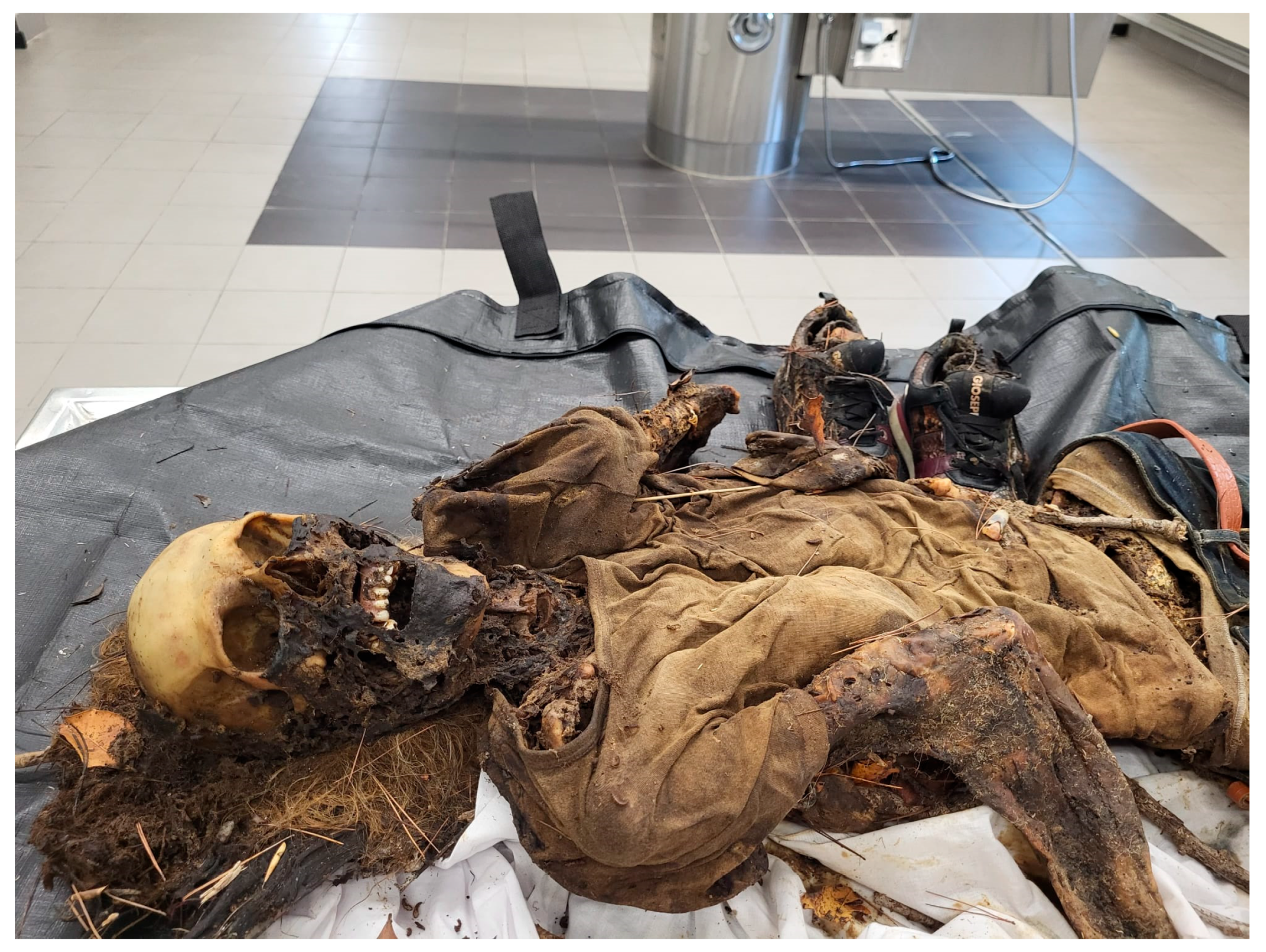
On 28 August 2024, human remains in an advanced stage of decomposition were discovered in the Marjan forest park in Split, Croatia. The body, located in a remote woodland area, was exposed to prolonged summer heat, resulting in partial mummification (
Figure 1). At the scene, personal documents and currency were recovered, and no visible signs of trauma were observed. Additionally, mobile phone signal data confirmed that the last recorded location of the missing person’s device corresponded with the site of discovery. Based on these preliminary findings and the timeline of disappearance, authorities suspected that the remains belonged to a female individual reported missing in July 2024. The body was subsequently transferred to the Department for Forensic Medicine at University Hospital Split for further examination, autopsy, and forensic identification. A thorough external and internal examination was conducted. However, due to the extent of postmortem changes, including desiccation and soft tissue degradation, the exact cause of death could not be determined. Notably, both the autopsy findings and the crime scene assessment indicated no evidence of inflicted injuries or violent death. Toxicological testing of available tissues returned negative results for common substances of abuse and medications.
Given the advanced decomposition and the poor condition of soft tissues, conventional biological samples (such as blood or muscle) were unavailable for DNA extraction. Therefore, teeth and fingernail samples from the right hand were collected during the autopsy as alternative DNA sources. These samples were selected based on their anatomical resilience and likelihood of preserving nucleated cells despite environmental exposure. STR DNA profiling was successfully performed on the selected samples and compared to reference profiles obtained from buccal swabs of the putative biological parents. Genotype concordance with both reference profiles supported a strong likelihood of biological parentage, thereby allowing identification of the deceased.
2. Materials and Methods
2.1. Sample Preparation
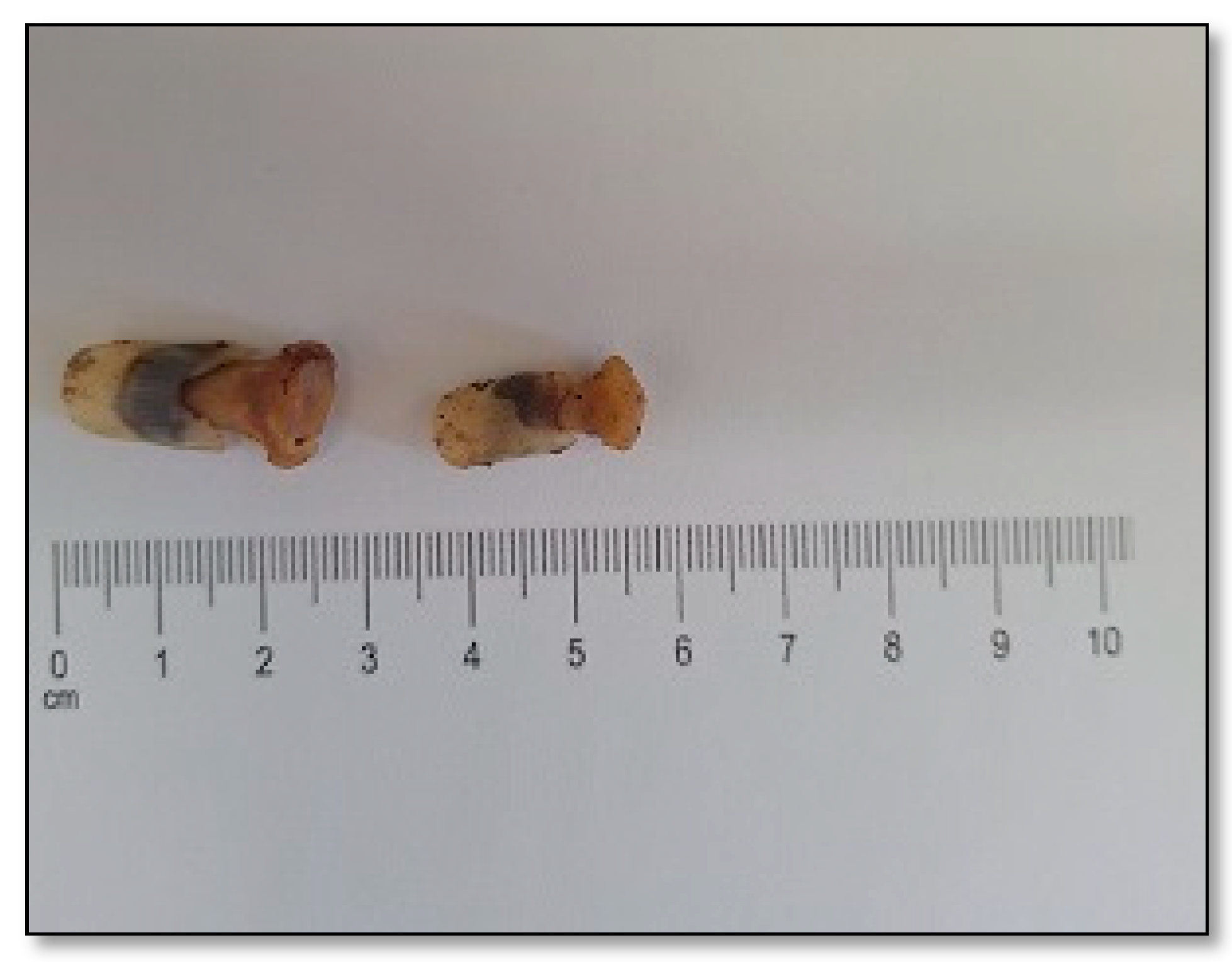
The sampling of fingernails was performed during autopsy and involved the removal of the entire nail from two fingers using autopsy pliers. The samples were taken non-selectively from the fingers of the right hand and stored in a sterile, labeled polypropylene container with a screw cap (
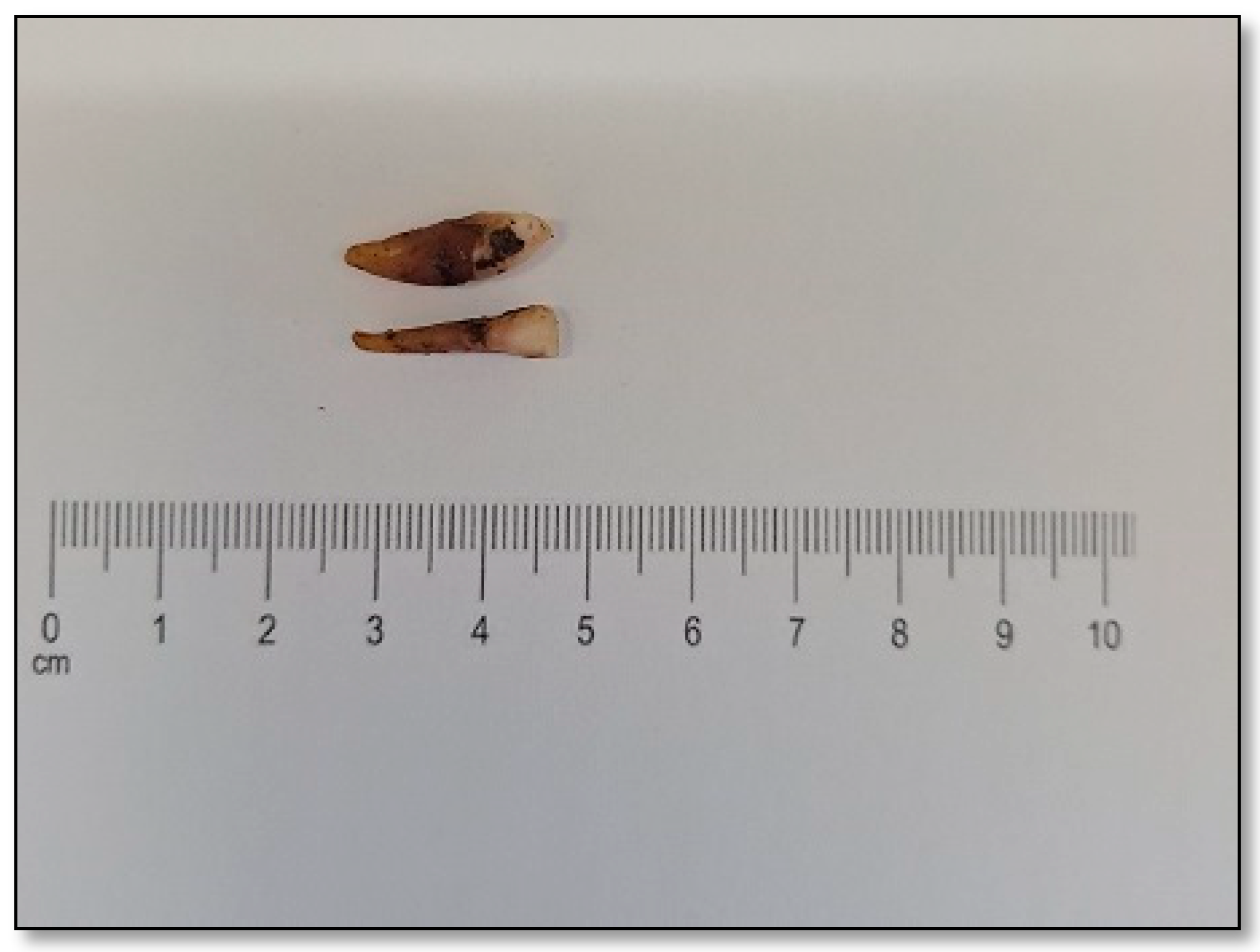
Figure 2). Two single-rooted teeth—specifically, a mandibular lateral incisor and a mandibular canine were collected during autopsy due to their easy accessibility during autopsy and anatomical characteristics that facilitate faster and more efficient laboratory processing (
Figure 3). Due to their relatively small size and single-rooted structure, these teeth are easier to decontaminate, section, and pulverize into fine powder—an essential step for maximizing DNA yield from dental tissues. All biological material was stored at 4–8 °C within 2 h and was delivered to the Laboratory for Forensic Genetics at the Department for Forensic Medicine, where a unique laboratory identification number was assigned to it. Photographs of the samples were taken with a Samsung Galaxy A54 (Samsung Electronics Co., Ltd., Suwon, Republic of Korea) cellular phone and stored as JPEG files.
Strict contamination prevention measures were implemented throughout the DNA extraction and analysis process. All equipment and work surfaces were sterilized with DNA-degrading agents (a 1% solution of sodium hypochlorite, sprayed on the surface and left for 5 min before drying and wiping with 70% ethanol) and ultraviolet irradiation. Decontamination was conducted before each procedure and for every workspace. Personnel adhered to appropriate personal protective equipment (PPE) protocols, including the use of gloves, masks, and lab coats. In addition, all molecular analyses were conducted using physically separated pre-PCR and post-PCR laboratory areas in order to prevent cross-contamination. One of two fingernail samples was selected and subjected to a multistep decontamination protocol, which included mechanical cleaning using a sterile scalpel to remove visible biological residues, soil, and impurities. The nail was subsequently subjected to sequential washes with bidistilled water and 80% ethanol (5 min in sterile water followed by 30 s in 80% ethanol), repeated three times. The sample was then air-dried under sterile conditions prior to further processing. All decontamination steps were carried out in line with the general principles of previously published protocols for keratinized tissues [
4,
10,
11].
2.2. DNA Extraction and Quantification
Once dried, a 5 mm × 2 mm fragment (approx. 0.005 g) was extracted from the proximal (root) part of the nail using a sterile scalpel and tweezers, as this region is most likely to contain viable nucleated cells and is better protected from external environmental factors, and the fragment was then placed in a sterile 1.5 mL microcentrifuge tube. Of the two fingernail samples collected during autopsy, only one was selected for DNA extraction. It was processed individually, beginning with rinsing in distilled water, followed by drying. Its surface was then mechanically abraded using a handheld grinder to reduce the risk of bacterial contamination, followed by a second rinse in a hypochlorite solution and drying. The tooth was subsequently powdered using a freezer mill, and 100 mg of the resulting powder was used for DNA extraction. DNA extraction was performed using the PrepFiler Forensic DNA Extraction Kit for the fingernail sample and PrepFiler BTA Forensic DNA Extraction Kit for the tooth samples (Applied Biosystems, Thermo Fisher Scientific, Waltham, MA, USA), following the manufacturer’s standard protocols for keratinized and calcified tissues, respectively. For nail samples, lysis incubation with proteinase K (20 mg/mL), 1.0 M DTT (dithiothreitol), and lysis buffer was performed at 56 °C for up to 2 h. For tooth samples, the incubation was carried out overnight at 56 °C to ensure complete digestion of the calcified matrix. DNA was then bound to magnetic particles, followed by a series of washing steps to remove potential inhibitors, and finally eluted in 50 μL of elution buffer. The laboratory workflow for the tooth sample required a two-day processing period due to additional steps such as mechanical grinding and overnight lysis, whereas the nail sample required less pre-processing time and was prepared for DNA extraction within standard laboratory working hours.
The concentration of extracted DNA was measured using the Qubit dsDNA HS Assay Kit (Invitrogen, Thermo Fischer Scientific, Waltham, MA, USA) and diluted to 0.3 ng/µL with nuclease-free water per manufacturès instructions. A negative control (blank sample) was included during Qubit quantification to monitor background fluorescence and confirm the absence of contamination. Previous studies have demonstrated that the Qubit fluorometer is suitable for forensic applications, including those involving degraded or postmortem samples [
22,
23,
24]. In routine forensic practice, real-time qPCR–based quantification is also widely used in many laboratories and represents an established alternative approach for assessing the amount of amplifiable human DNA.
2.3. STR Amplification and Capillary Electrophoresis
DNA amplification was performed using the ProFlex PCR System (Applied Biosystems; Thermo Fischer Scientific) with the GlobalFiler PCR Amplification Kit (Applied Biosystems; Thermo Fischer Scientific), following the manufacturer’s recommended thermal cycling conditions and reaction setup. Negative (no-template) and positive controls (control DNA 007) were included during amplification to monitor for potential contamination. GlobalFiler PCR Amplification kit allows the amplification of 24 nuclear DNA short tandem repeats (STR) markers, including the gender determination marker amelogenin. Each PCR reaction was prepared in a final volume of 25 μL, including 7.5 μL GlobalFiler Master Mix (Applied Biosystems, Thermo Fisher Scientific, Waltham, MA, USA), 2.5 μLPrimer Set and 15 μL DNA template Amplification was conducted using the ProFlex™ PCR System under the cycling conditions wherw initial denaturation was at 95 °C for 1 min followed with 29 cycles of denaturation (at 94 °C for 10 s), annealing (at 59 °C for 1 min), extension (at 72 °C for 30 s) and final extension at 60 °C for 8 min. Final hold was at 4 °C.
Capillary electrophoresis and fragment analysis were performed using the SeqStudio Genetic Analyzer (Applied Biosystems; Thermo Fischer Scientific), and data collection and analysis were carried out with GeneMapper ID-X v1.6 software.
Buccal swabs were collected at the police department in Split as standard reference samples from the presumed biological mother and father, in accordance with national forensic procedures, and were subsequently submitted to the Laboratory for Forensic Genetics. The tentative identity of the deceased was initially based on circumstantial evidence found near the body, including personal belongings. Reference DNA profiles obtained from the buccal swabs were compared with the STR profile generated from the fingernail sample to support the identification of the deceased.
2.4. Kinship Analysis
Kinship evaluation was performed under a trio hypothesis, comparing the STR profile of the unidentified deceased individual with the profiles of the presumed biological mother and father. Paternity testing was conducted through likelihood ratio-based evaluation of genotype concordance.
For each autosomal STR locus, a Paternity Index (PI) was calculated (PI = Likelihood ratio/frequency of obligate allele), representing the ratio between the probability of observing the genetic results if the tested individual is the biological father and the probability of observing those same results if a randomly selected unrelated man from the general population were the father. These individual indices were then multiplied to obtain the Combined Paternity Index (CPI), which represents the cumulative likelihood ratio expressing the evidential strength of the DNA findings under the two competing hypotheses of paternity and non-paternity. Finally, the Probability of Paternity (PP) was calculated from the CPI using Bayesian formulae. All calculations were performed using Microsoft Excel, applying formulas established in the scientific literature [
22], as detailed in
Table 1. Allele frequency data used for statistical evaluation were obtained from published population data for Croatia, ensuring that the calculations were population-specific and reflective of the genetic background of the tested individuals [
25]. Although specialized forensic software such as Familias exists, our laboratory has successfully applied this Excel-based method in routine paternity testing for over two decades [
26,
27,
28]. Importantly, results obtained using this approach are regularly accepted in court as valid and forensically credible, and have been used in numerous real-case scenarios involving postmortem identification and kinship testing. The formulas implemented in the spreadsheet follow internationally accepted forensic genetics guidelines for likelihood ratio and paternity index calculations. In accordance with long-standing Croatian forensic practice, all statistical calculations were performed using the standard trio likelihood approach (mother–child–father). When both putative parents are genotyped, this trio framework is mathematically equivalent to full likelihood ratio reasoning through the relationship LR = MI × PI, where MI denotes the maternity index and PI the paternity index. This approach is scientifically valid, transparent, and legally accepted in operational postmortem identification casework in our jurisdiction. The key locus-specific parameters and resulting indices are presented in
Table 1.
Ethical approval for the scientific use and publication of this anonymized case report was obtained from the Ethics Committee of the University Hospital Centre Split on 24 October 2024 (Class: 520-03/24-01/203; Reg. No.: 2181-147/01-06/I.J.Z.-24-02).
3. Results
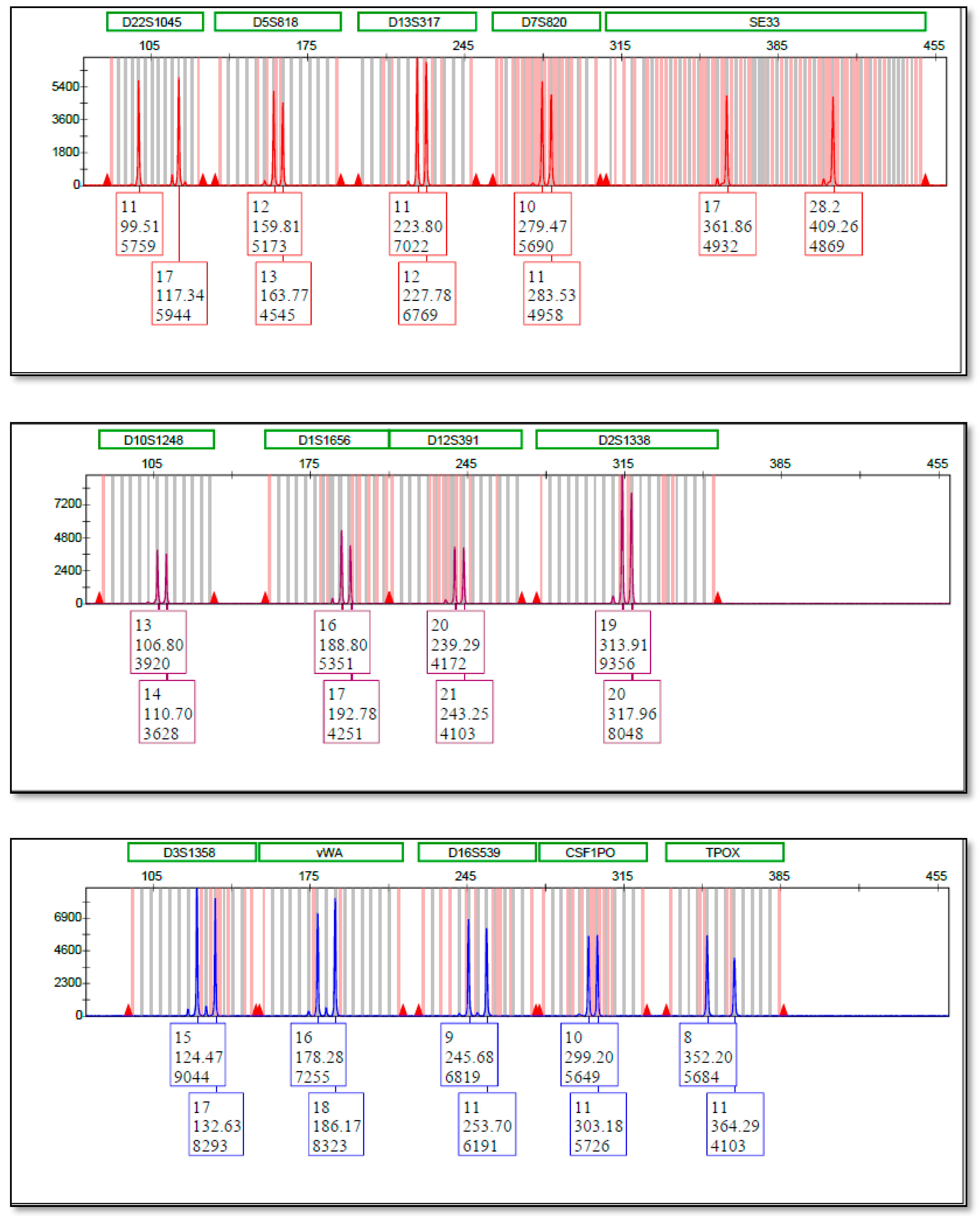
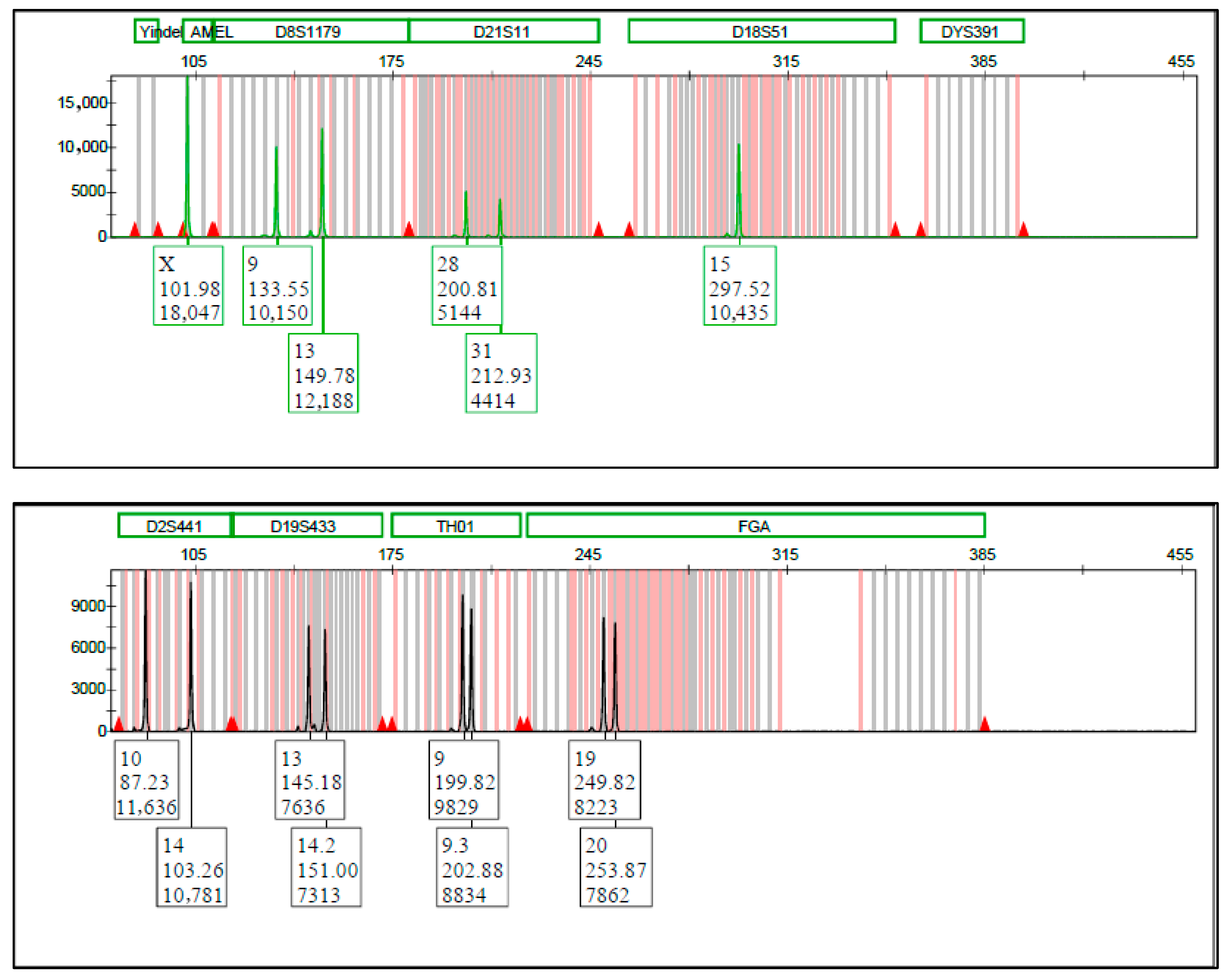
Large amounts of DNA were recovered from fingernails. The concentration obtained from a small fingernail sample was 3.87 ng/µL, while the tooth sample yielded 2.18 ng/µL. However, all 24 STR loci were successfully amplified from the fingernail sample (
Figure A1), indicating that DNA from the nail was well preserved, whereas no STR profile was obtained from the tooth sample. Notably, the nail-derived DNA extract exceeded the minimum threshold required for optimal template input in STR profiling reactions conducted in this study. The failure of the tooth sample to yield an STR profile, despite an apparently sufficient DNA concentration, may be attributed to advanced DNA fragmentation or the presence of PCR inhibitors, which are not always detectable during fluorometric quantification methods such as Quibit. While Qubit fluorometry is widely used in forensic laboratories due to its sensitivity and ease of use, it cannot distinguish between human and non-human DNA, nor can it assess DNA degradation. This highlights a known limitation in forensic DNA analysis of degraded samples: total DNA concentration does not always correlate with amplification success. In contrast, the successful generation of a full and high-quality STR profile from the nail sample emphasizes its robustness as a DNA source, even in the context of advanced decomposition.
Additionally, complete STR profiles were successfully obtained from the buccal swab samples of the presumed parents. The most important finding was the high quality of the electropherogram obtained from the nail sample. All peaks were sharp, well-resolved, and properly sized with no evidence of artefacts or unexplained additional peaks, meeting all criteria for a reliable STR profile. All peaks were above the analytical threshold, which was set at 100 RFU (Relative Fluorescent Unit). No evidence of allele dropout or other artifacts was observed; peak height patterns were consistent with a high-quality profile, and all expected loci were successfully amplified (
Figure A1). No mixed DNA profile was observed. The obtained genotypes of the presumed child, mother, and father were compared across all 24 loci, and the obligatory alleles inherited from the biological father were identified, as shown in
Table 2. The results demonstrate a complete genetic concordance across all analyzed loci. This high level of genetic compatibility strongly supports the hypothesis of a biological relationship between the individuals in question, as reflected in the resulting likelihood ratios.
The paternity index (PI) was calculated using corresponding formulas, previously described in the literature [
22], as shown in
Table S1.
Also, the combined paternity index (CPI) was calculated by multiplying the PI values calculated for each STR locus. In accordance with Bayes’ theorem, CPI can be combined with a prior probability to obtain the posterior probability of identity. In forensic kinship and identity assessment, prior odds reflect the weight of non-genetic evidence available prior to DNA analysis.
In routine casework in our Laboratory—particularly in cases involving decomposed, unidentified remains, without direct reference samples—prior probabilities are set conservatively whenever the amount of non-genetic information supplied by the authorities is limited. It is important to note that the contextual information described earlier in the manuscript (e.g., items found at the scene) was obtained retrospectively from police documentation for the purposes of scientific reporting. These details were not provided to the laboratory at the time of analysis, and therefore did not contribute to the prior probability assessment. At the moment of receiving the samples, the only information available to the laboratory was that the remains belonged to an unidentified missing person in an advanced stage of decomposition. Consequently, a conservative prior probability was applied in accordance with our long-standing operational practice. In this case, both the customary neutral prior probability of 0.5 and the conservative prior of 0.01 were used for CPI calculations, and both results are presented in
Table S1. For the official forensic report submitted to the competent authorities, a conservative prior probability of 0.01 (1%) was used. The selection of a conservative prior does not affect the scientific conclusion of the case, as both prior probabilities yield an overwhelmingly high posterior probability fully supporting the identification. Reports using this framework have been routinely accepted in judicial and medico-legal proceedings, demonstrating the robustness and legal reliability of this approach.
Based on allele frequencies in the Croatian population for 20 STR loci, and using formulas for calculating the paternity index (PI) for each locus as well as the combined paternity index (CPI), a probability of paternity of 99.99999812% was obtained, supporting the conclusion that the tested man was the biological father. The SE33 locus, although included in the GlobalFiler™ kit, was excluded from the statistical evaluation due to the lack of published allele frequency data for the Croatian population. The applied formulas are grounded in Mendelian inheritance, while the allele frequencies used in the likelihood ratio calculations are derived from population data estimated under Hardy–Weinberg equilibrium assumptions.
4. Discussion
This case highlights the effectiveness of nail samples in forensic identification, particularly when standard biological materials are degraded or compromised due to advanced decomposition. Despite growing forensic interest, research on DNA extraction from nails collected from decomposed remains is scarce. Only a limited number of studies have focused on DNA extraction from nail samples obtained from decomposed human remains for identification purposes [
4,
5,
7,
10,
11,
12], while most published research has concentrated on forensic applications involving fingernail scrapings collected from living individuals or victims in assault cases.
Rapid postmortem changes, including microbial and enzymatic degradation, severely compromise standard biological samples such as blood, muscle tissue, and internal organs, thereby limiting their utility in forensic investigations [
2,
18]. In such cases, bones and teeth are generally considered the gold standard for DNA isolation, as their mineral matrix protects DNA from degradation. The efficiency of DNA recovery from skeletal remains can vary depending on bone density, porosity, and preservation conditions [
3,
13,
19,
20,
21]. Also, processing such samples requires intensive labor, extended processing time, and specialized equipment, which prolongs laboratory analysis time and increases costs [
3,
4,
13].
In this context, we present a case that contributes to the limited but growing body of literature addressing the viability of nails for postmortem DNA identification from decomposed remains. Nails offer several practical advantages compared to bones and teeth. Sampling is minimally invasive, rapid, and can be performed immediately upon body discovery, preserving remains for further examination [
10,
29,
30,
31]. Their keratinized structure provides a physical barrier against microbial invasion, insect activity, and environmental degradation [
2,
4,
10,
31]. Numerous studies have demonstrated successful STR profiling from nail samples, even when recovered weeks or months postmortem, or in extreme environments such as marine settings or soil [
6,
8,
11,
32]. Given their resilience and accessibility, nails are especially useful in disaster scenarios or missing persons cases, where conventional samples may be absent or degraded [
8,
9,
11,
15]. Della Rocca et al. reported successful STR profiles from nails in bodies recovered from seawater, while other authors highlighted their utility in long-term postmortem intervals [
11,
29,
32]. Nail samples may also complement skeletal remains in complex identifications, offering an additional DNA source when bone or dental samples are insufficient.
DNA is usually obtained from fingernail clippings due to their resistance to environmental degradation; however, the literature also reports other approaches, such as swabbing and scraping, which are primarily used in contexts involving exogenous DNA recovery (e.g., assault cases) [
29,
30,
32]. While partial nail clipping is often sufficient and less invasive, in this case, removal of the entire nail was warranted due to advanced decomposition and compromised nail beds, which limited the feasibility of a more conservative sampling approach.
In this case, DNA extracted from a fingernail sample collected during the autopsy of decomposed remains yielded a complete STR profile. The PrepFiler™ Forensic DNA Extraction Kit used for DNA isolation, and GlobalFiler™ PCR Amplification used for STR amplification, are well-established systems in modern forensic DNA laboratories, known for their high sensitivity and reproducibility, even in samples with low template DNA [
1,
12].
Although no postmortem profile could be generated from the extracted tooth, the complete STR profile obtained from the nail was directly compared with reference profiles of the presumed biological parents. Because both biological parents were genotyped and population allele frequencies were available for each locus, the trio-based MI × PI approach fully satisfies the likelihood ratio framework for reverse-parentage identification and provides a statistically robust basis for the positive identification in this case. The resulting kinship analysis yielded a probability of paternity (PP) of 99.99999812%, providing strong genetic support for the identification of the deceased. While the DNA evidence was highly informative, it is important to emphasize that, in forensic casework, the formal attribution of identity typically relies on the integration of genetic results with corroborative medico-legal findings, in accordance with jurisdictional standards. This outcome underscores the forensic utility of nail samples in postmortem identification and kinship testing [
16,
32]. Importantly, the success of STR profiling—rather than the absolute DNA concentration—serves as the most definitive indicator of DNA suitability for forensic identification purposes.
Although numerous studies have demonstrated that nails can yield reliable DNA using various extraction protocols, there is no standardized protocol for DNA isolation and amplification that is universally established across all forensic laboratories. Inter-laboratory variability persists due to differences in reagents, instruments, and handling practices. Furthermore, there is a scarcity of comprehensive comparative studies that directly evaluate these methods.
Several extraction methods from nail samples have been studied in previous studies, including organic extraction, commercial column-based kits, and magnetic bead-based protocols. Magnetic bead methods, such as the PrepFiler™ kit used in this case, are advantageous due to their automation potential and low inhibitor retention, while other methods, such as organic extraction, yield higher amounts of DNA but pose greater contamination risk and require longer processing time [
6,
17,
19]. Further studies are needed to develop standardized extraction protocols optimized for nails, taking into account their keratinized nature and structural properties that distinguish them from other biological materials such as soft tissues, bones, and teeth [
17,
30]. As suggested by several studies, cross-validation between laboratories is needed to ensure reproducibility, especially in the context of disaster victim identification [
8,
13,
15].
Being external appendages, nails are particularly prone to both environmental and handling-related contamination [
14,
29]. The DNA yield may vary depending on factors such as the nail’s condition, size, chemical exposure, and postmortem interval [
6,
29]. To minimize the risk of exogenous DNA interference, appropriate contamination control measures must be implemented, both to avoid contamination during laboratory procedures and to effectively remove any surface contaminants prior to extraction. Although protocols differ across studies, they typically share common decontamination steps, including mechanical cleaning and multiple chemical treatments involving water, ethanol, and, in some cases, bleach or detergent solutions [
10,
11,
12]. Nonetheless, it is important to note that low levels of exogenous DNA, if present, generally do not interfere with STR profiling, as they typically remain below the analytical detection threshold—commonly less than 5% relative to the dominant endogenous DNA originating from the nail tissue itself. This is particularly true in the absence of heavy contamination, such as visible blood, mixed biological material, or direct contact with other decomposed bodies [
33].
In forensic systems where nail samples are not yet routinely utilized for postmortem DNA identification—such as in Croatia—there is a pressing need for method harmonization and inter-laboratory validation. Standardization would not only improve reproducibility but also reinforce the legal robustness and admissibility of such evidence in judicial proceedings.
The admissibility of DNA evidence derived from nail samples depends on the use of scientifically validated protocols. An increasing number of case reports and studies support the forensic and legal relevance of nails, particularly when interpreted in conjunction with kinship analysis and contextualized by complementary disciplines such as forensic pathology and anthropology [
1,
16,
17]. This case adds to the growing body of evidence that supports the routine consideration of nails as viable forensic samples in postmortem identification. Successful integration of DNA analysis from nails into the standard forensic workflow in Croatia also requires institutional support, including appropriate training of personnel in sample collection, handling, and processing protocols. Continued research is essential not only to optimize extraction techniques and implement effective contamination control measures, but also to build a scientifically robust framework for the broader forensic acceptance of nail-derived DNA evidence.
This case highlights the viability of nails as a reliable DNA source for high-quality STR profiling, even under conditions of advanced decomposition. Their anatomical resilience, ease of sampling, and relative resistance to degradation make them a valuable alternative when conventional materials are unavailable. Notably, this is the first documented forensic case in Croatia in which nail material served as the primary—and ultimately successful—biological sample for postmortem DNA identification within a judicial context, thereby underscoring both its practical utility and evidentiary relevance.
To support broader implementation in routine forensic workflows, especially within the Croatian medico-legal context, future research should include larger-scale studies evaluating DNA recovery from nails across various decomposition stages and environmental conditions. Such data would provide the empirical foundation needed to formally recognize nail material as a standard biological sample in postmortem identification protocols.