1. Introduction
In forensic anthropology, the construction of a biological profile—comprising estimates of age, sex, population affinity, and stature—serves as a valuable auxiliary tool when primary methods of human identification, such as DNA analysis, fingerprinting, or dental comparison, are unavailable or inconclusive. This approach helps narrow the pool of potential matches and supports the overall identification process, especially in cases involving skeletonized remains [
1]. This profile includes estimates of age, sex, population affinity, and stature—key attributes that guide the identification of unknown individuals and assist in narrowing down potential matches during investigations [
2]. Stature estimation is a crucial component among these parameters, providing a quantifiable trait that can be compared with available records or databases of missing persons [
3,
4,
5].
It is important to distinguish between biological profiling and positive identification. Biological profiling involves estimating characteristics, such as age at death, sex, population affinity, and stature, typically from skeletal remains, and is used to narrow down the pool of possible identities. In contrast, positive identification requires the comparison of individualizing traits, such as DNA, dental records, or fingerprints. The stature estimation equations used in this study were derived from dry skeletal remains and are therefore applicable to forensic cases involving human remains, not to the estimation of stature in living individuals.
Stature estimation is traditionally performed using anatomical or mathematical methods. Other approaches have been developed, focusing on DNA and utilizing a polygenic score [
6]. This approach enables the estimation of stature using biological samples. Other techniques include anatomical methods, such as the Fully method, which are considered the gold standard due to their comprehensive nature [
2]. These methods provide accurate and detailed stature estimations, measuring almost all skeletal elements. However, their practical application is often hindered by the time-consuming procedures involved and the frequent unavailability of intact skeletal remains, especially in forensic cases involving decomposition, fragmentation, or trauma [
7].
As in most cases, skeletal remains are incomplete [
8]; mathematical methods offer a more accessible alternative. These techniques use regression models constructed from measurements of specific bones, particularly long bones such as the femur, tibia, and humerus. Lower limb bones are generally considered the most reliable predictors of stature due to their direct involvement in weight bearing and locomotion, resulting in a stronger biological correlation with stature. This is supported by several studies reporting higher correlation coefficients and lower estimation errors when using bones such as the femur and tibia for stature estimation [
9,
10].
The reconstruction of living stature from skeletal elements relies on the correlation between stature and long bone length and assumes that the body proportions of every individual are equal. Yet variations in body proportions occur and relate to sex and population affinity. Rattanachet stated that White Americans have shorter limbs relative to stature than African-Americans [
11]. Therefore, estimating population affinity before estimating stature is mandatory, as these methods are population-specific and rely on established correlations between bone dimensions and overall stature. For instance, in Portugal, validated models for stature estimation using the metatarsals, humerus, and femur are well-documented and widely utilized [
4,
12,
13,
14]. Despite their utility, the reliance on long bones can pose challenges when these specific skeletal elements are missing, poorly preserved, or otherwise unavailable for measurement. In such cases, other alternative methods involving other skeletal structures are helpful.
The correlation between stature and the length of the sternum has also been investigated. Tumram et al. investigated an Indian population. They found that the sternum had a moderate positive correlation and relatively low reliability in estimating stature, with limited forensic value [
15]. Other authors obtained similar results [
16].
Pelvic measurements have also been performed. Imai et al. [
17] stated that pelvic height measured on CT images was a reliable predictor of living stature in a Japanese population. Comparable results were obtained by other authors [
18], claiming that such measurements could be useful in mass disaster situations.
Other bone structures that have been studied include the scapula [
19], the clavicle [
20], the vertebrae [
21], and the sacrum and coccyx [
22]. The authors concluded that all structures can be used as stature predictors, particularly when long bones are not available, suggesting limited accuracy in stature estimation.
The relationship between tooth length and stature has also been explored. In deciduous teeth, no correlation between body stature and crown height was found [
23]. In permanent teeth, a positive correlation between stature was found, but without the accuracy required in forensic situations [
24,
25]. A 2024 systematic review on the subject stated the following: “Dental measurements are not reliable for stature estimation in the forensic field” [
26].
In recent years, there has been growing interest in exploring cranial and mandibular measurements for stature estimation [
5,
25,
27,
28]. The skull and mandible are often better preserved than long bones, particularly in archeological and forensic contexts where environmental factors or taphonomic processes have affected the remains [
29]. Studies conducted in various populations have demonstrated that maximum cranial length, facial height, and mandibular dimensions can serve as proxies for stature estimation [
5,
25,
27,
28]. However, these methods are also population-specific and require extensive validation to ensure accuracy and reliability.
In Portugal, while regression models using long bones are well-established [
30], limited research exists on applying cranial and mandibular measurements for stature estimation. Developing robust, population-specific models that incorporate these alternative measurements could, therefore, be useful to estimate stature, particularly in cases where traditional skeletal elements are unavailable.
This study seeks to address this gap by investigating the relationship between cranial and mandibular measurements and stature in a sample of Portuguese individuals.
2. Materials and Methods
2.1. Study Design and Sample Description
The sample consisted of 84 skeletons and included only adult individuals (age at death over 18 years), 43 (51.2%) of which were female and 41 of which were males (48.8%); 70 belonged to the “Luís Lopes” collection from the Natural History Museum of Lisbon (MHUNAC), and 14 belonged to the “CEIC–“Coleção de Esqueletos Identificados da CESPU” collection [
31]. Both of these are Portuguese-identified skeleton collections.
2.2. The Collections
The XXI Century CEIC collection comprises 98 adult skeletons of Portuguese individuals who died between 1946 and 2007. The individuals were recovered from two cemeteries in Porto—Cemitério Prado do Repouso and Cemitério de Agramonte—and are believed to represent a low-to-middle socioeconomic background. Although stature records were not available, demographic information, such as age at death, sex, and dates of birth and death, was provided by cemetery authorities and kept confidential. This collection is considered contemporary and relevant for forensic research, as it reflects a modern population affected by historical and environmental factors such as post-war living conditions, urbanization, and possible nutritional or health-related stressors [
31].
The Luís Lopes collection from the Natural History Museum in Lisbon similarly comprises individuals from urban cemeteries and is also representative of the lower-to-middle socioeconomic population. Although historical context and detailed health information are limited, these collections provide valuable anthropological data for studying variation in stature and other biological parameters in 20th-century Portuguese populations [
32].
2.3. Inclusion and Exclusion Criteria
The inclusion criteria required that each individual had an intact left or right humerus, mandible, and complete skull, with closed cranial sutures and preserved external acoustic meatus. Only adult individuals of Portuguese nationality were included.
The exclusion criteria encompassed visible skeletal pathologies or alterations that could compromise measurement accuracy. These included, but were not limited to, post-traumatic bone remodeling (e.g., evidence of fracture healing), severe alveolar resorption associated with tooth loss, pronounced osteoarthritic changes, congenital bone deformities, or extensive periosteal reactions. Bones were examined macroscopically by trained observers prior to data collection, and any specimen displaying such alterations was excluded. No radiological or clinical records were available to support diagnosis, so exclusion was based on direct visual inspection.
2.4. Methodological Calibration
Before data collection, a calibration session was conducted to ensure consistency in measurement techniques. Observers practiced measurements on a subset of bones to standardize procedures and minimize interobserver and intraobserver variability.
All measurements were performed following standard anthropometric protocols as defined by Buikstra and Ubelaker (1994) and White and Folkens (2005) [
33,
34]. The cranial and mandibular landmarks used were selected for their reproducibility and relevance in stature estimation studies.
Measurements were performed using an osteometry board, a caliper, and a spreading caliper. The left humerus (or, in its absence, the right) was placed on an osteometry board, with the posterior face upwards, and the distance between the humeral head’s most proximal point to the condyle’s most distal one (
Figure 1) was measured.
The left humerus was systematically preferred for analysis to ensure consistency across the sample, as recommended in osteological research, where one side is consistently chosen to minimize bilateral variation [
34]. The right humerus was only used in cases where the left side was absent or not sufficiently preserved for measurement.
Stature was determined using the model developed by Mendonça for the Portuguese population (
Table 1) [
2]. This model was developed using dry bones, and not on living individuals, and the obtained value was considered the individual’s humeral stature (St) and used as a gold standard for comparison.
It should be noted that this stature is itself an estimate based on humeral length and not a documented or anatomically reconstructed value, which may introduce an additional layer of estimation error into the subsequent analysis.
The measurements performed on the skull were the following:
- (a)
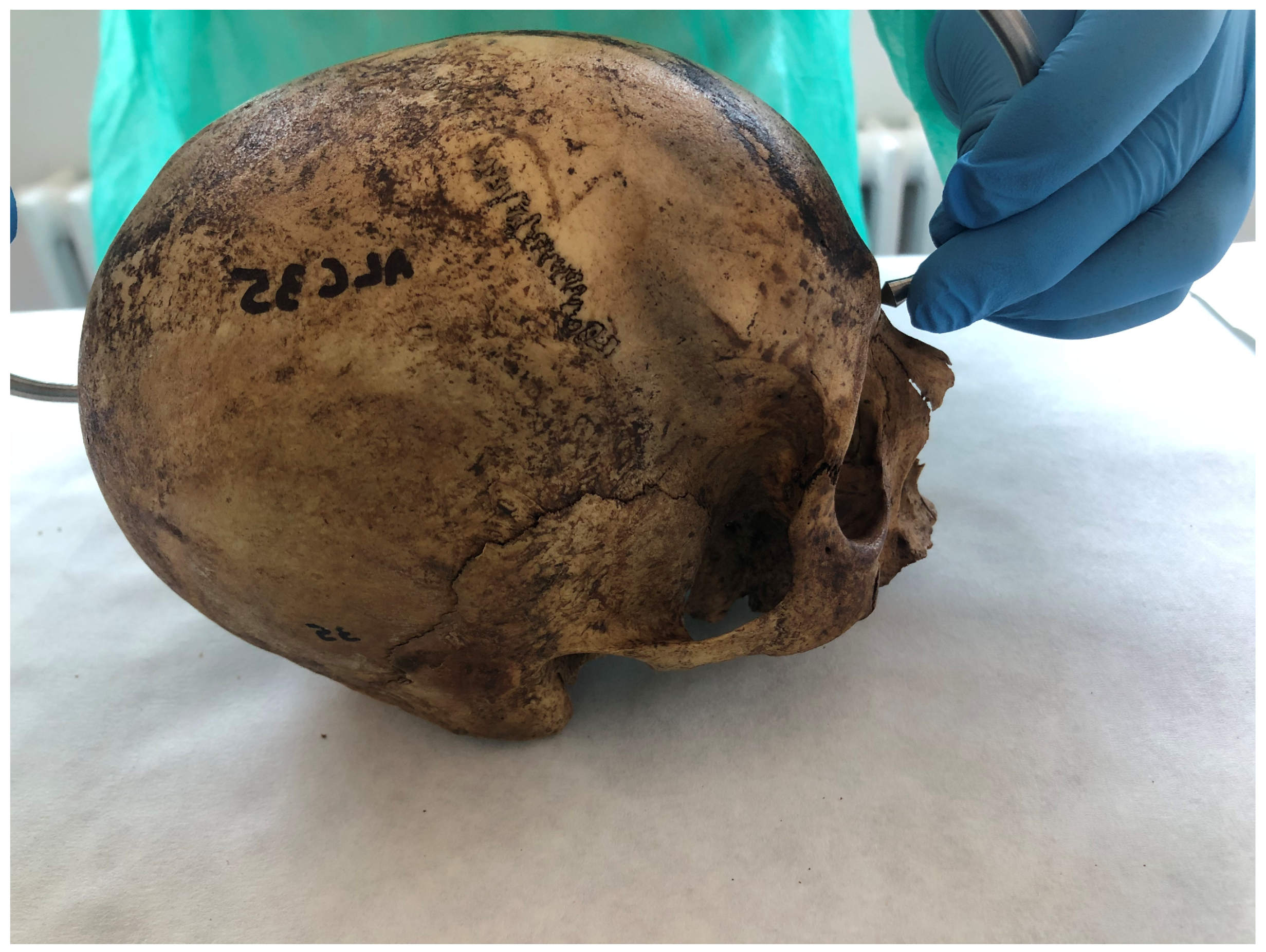
Maximum skull length (MSL) was measured as the straight-line distance between the glabella (g)—the most prominent anthropometric landmark between the eyebrows in the mid-sagittal plane—and the opisthocranion (op)—the most posterior anthropometric landmark on the skull in the mid-sagittal plane—using a spreading caliper (
Figure 2).
- (b)
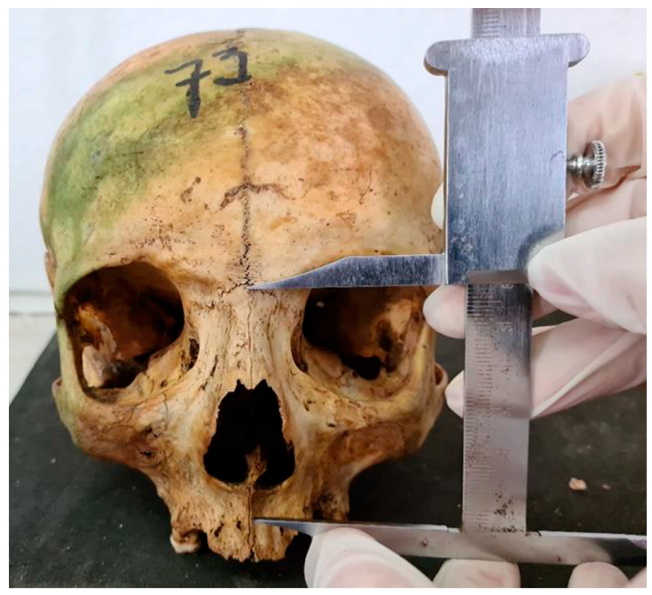
Upper facial height (UFH) was measured between the nasion and point A, using a manual caliper, from the most anterior anthropometric landmark of the frontonasal suture and the deepest point of the maxillary concavity (
Figure 3).
- (c)
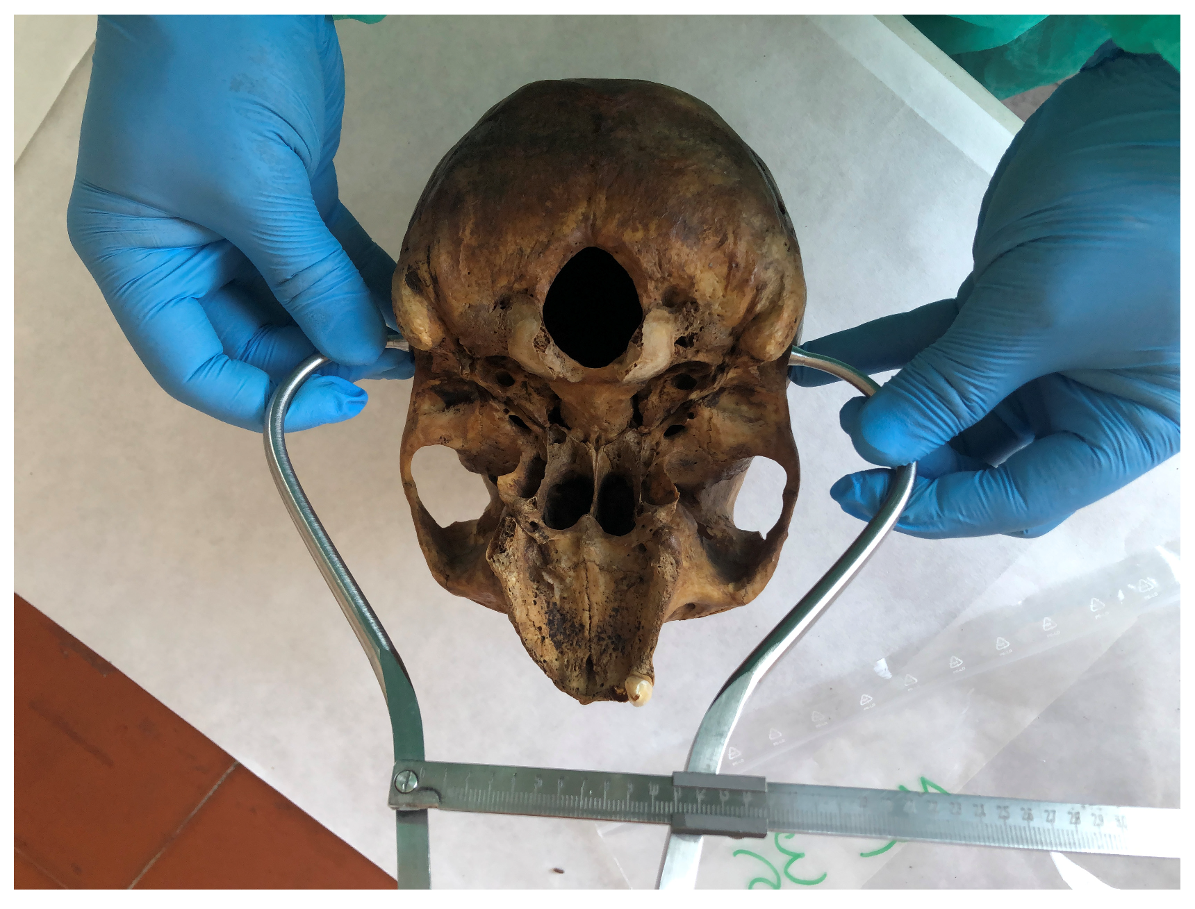
Porion-porion distance (binaural distance) (PoE-PoD) was measured with a spreading caliper, between the left and right porion, with the porion point corresponding to the most superior end of the external acoustic meatus (
Figure 4).
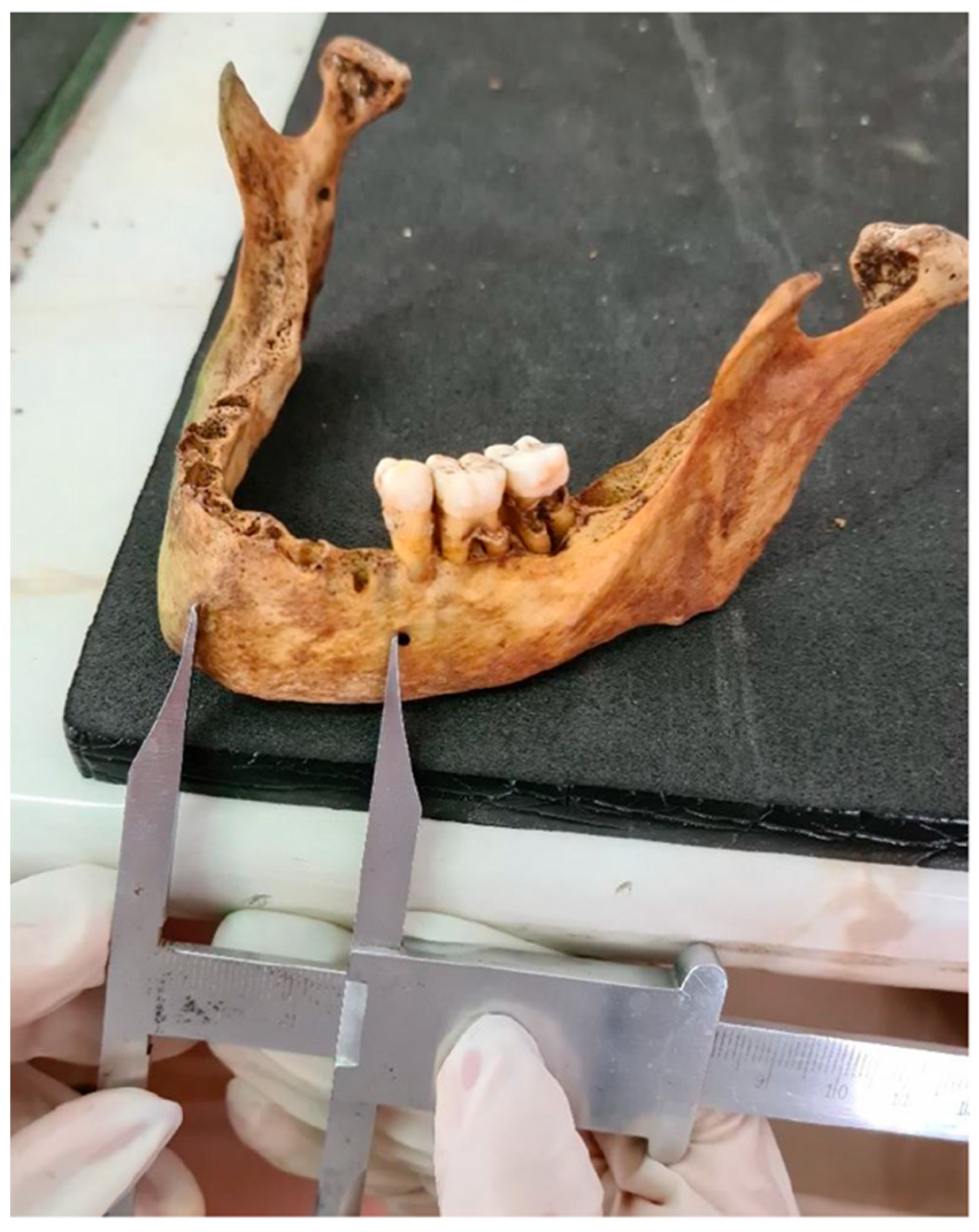
In the mandible, using a caliper, the distance between the mental symphysis central anthropometric landmark to the most anterior portion of the mental foramen (SMFM) on the median sagittal plane was measured (
Figure 5).
2.5. Reliability of Measurements
A random subset of 20 skeletons was measured twice by the same observer (MJC), one week apart, and by a second observer (IMC). Intraclass and interclass correlation coefficients (ICCs) were calculated to assess repeatability and reproducibility. In accordance with the criteria applied by Fleiss [
35], ICC values were interpreted as follows: below 0.4 = poor reliability; 0.4–0.75 = moderate-to-good reliability; and above 0.75 = excellent reliability. The coefficients for each measurement were the following:
Maximum skull length (MSL): Displayed the highest reliability, with an intraclass ICC of 0.988 and an interclass ICC of 0.980, indicating nearly perfect agreement.
Upper facial height (UFH): Also demonstrated strong reliability, with an intraclass ICC of 0.912 and an interclass ICC of 0.901.
Porion-porion distance (binaural distance) (PoE-PoD): Showed high reliability, with an intraclass ICC of 0.911 and an interclass ICC of 0.901.
Distance between the mental symphysis central anthropometric landmark to the most anterior portion of the mental foramen (SMFM) on the median sagittal plane: Had the lowest reliability among the four parameters, with an intraclass ICC of 0.901 and an interclass ICC of 0.853, remaining within acceptable levels of agreement.
Overall, the data indicate that the methods for measuring these parameters are reliable for intra- and interobserver agreement. All the following measurements were made by the same person (MJC).
2.6. Statistical Analysis
Statistical analysis was performed using SPSS (Statistical Package for the Social Sciences) version 26 and Excel software (2013). The following statistical approaches were employed:
- (a)
Continuous variables were characterized using descriptive analysis (mean, standard deviation, minimum, and maximum values), whereas categorical variables were described using frequencies and percentages.
- (b)
The correlations between the variables were analyzed using Pearson’s correlation coefficient, and linear regression models were developed to estimate stature. Linear regression analyses were performed using a stepwise method with a backward elimination approach, in which all predictor variables were initially included in the model and then sequentially removed based on their statistical significance (p > 0.05) to identify the most parsimonious model. This approach allows for the selection of the most relevant variables while minimizing overfitting.
Assumptions for the use of Pearson’s correlation and linear regression were considered. Although formal normality tests were not conducted, the sample sizes for each variable exceeded 30; therefore, based on the Central Limit Theorem, approximate normality was assumed [
36]. Scatterplots were examined to confirm the presence of linear relationships between variables, and multicollinearity diagnostics (tolerance and VIF) were within acceptable thresholds, supporting the validity of the parametric analyses performed.
The established significance level was set at a minimum of 5%.
3. Results
A descriptive analysis of the measurements is presented in
Table 2.
All variables had a statistically significant correlation with stature, but none presented a strong correlation value (over 0.7) (
Table 3).
Subsequently, a backward stepwise linear regression was performed using five predictors: sex, maximum skull length (MSL), upper facial height (UFH), porion-porion distance (PoE-PoD), and the distance between the mental symphysis and the mental foramen (SMFM). The initial model yielded an R2 of 0.535 and an adjusted R2 of 0.505, indicating that approximately 53.5% of the variance in stature was explained by the predictors combined (F (5. 78) = 17.94. p < 0.001).
Among the predictors, sex (
p < 0.001) and SMFM (
p = 0.007) were statistically significant contributors to the model. MSL (
p = 0.266), UFH (
p = 0.967), and PoE-PoD (
p = 0.199) were not significant at the 0.05 level and were subsequently removed in the stepwise process (
Table 4).
In the second step of the backward regression, upper facial height (UFH) was already removed. The model was recalculated using the remaining predictors: sex, maximum skull length (MSL), porion-porion distance (PoE-PoD), and the mandibular measurement SMFM. The model remained statistically significant (R2 = 0.535; adjusted R2 = 0.511; F (4, 79) = 22.71; p < 0.001).
Of the remaining variables, only sex (
p < 0.001) and SMFM (
p = 0.007) remained statistically significant. MSL (
p = 0.262) and PoE-PoD (
p = 0.188) did not meet the inclusion criteria (
p < 0.05) and were considered for exclusion in the following steps (
Table 5).
In the third step (
Table 6), following the removal of PoE-PoD, the model retained sex, SMFM, and MSL as predictors. The overall model remained highly significant (R
2 = 0.524; adjusted R
2 = 0.507; F (3, 80) = 29.41;
p < 0.001). Among the three predictors, sex (
p < 0.001) and SMFM (
p = 0.0037) remained statistically significant, whereas MSL did not reach significance (
p = 0.163) and was therefore excluded in the next step.
In the final step of the backward regression, only sex and the mandibular measurement (SMFM) remained in the model. Both predictors were statistically significant (
p < 0.001 and
p = 0.002, respectively) (
Table 7). The model accounted for approximately 51.3% of the variance in stature (R
2 = 0.513; adjusted R
2 = 0.501; F (2, 81) = 42.60;
p < 0.001), with a standard error of 4.39 cm.
The final model‘s stature was equal to 138.32 + 7.51 × SEX + 0.59 × SMFM (with the value of sex being 0 for males and 1 for females); SMFM is the distance between the mental symphysis central point to the most anterior portion of the left mental foramen on the median sagittal plane.
4. Discussion
The present study aimed to evaluate the potential of cranial and mandibular measurements for estimating human stature in the absence of long bones. Through backward stepwise regression analysis, only two predictors—sex and the mandibular measurement between the mental symphysis and the mental foramen (SMFM)—were retained in the final model. This simplified model accounted for approximately 51.3% of the variance in stature, with a standard error of 4.39 cm. These results suggest that mandibular landmarks, particularly SMFM, may serve as useful alternatives for stature estimation in forensic contexts where postcranial elements are missing or compromised.
Both practical and scientific considerations guided the selection of the anatomical landmarks. From a forensic perspective, structures with a higher likelihood of preservation in challenging contexts, such as advanced decomposition or fragmentation, were preferred. In the case of the mandible, the SMFM measurement was chosen as a bony analog to the dental landmarks used in Carrea’s method [
37], which relies on the mesiodistal dimensions of the anterior teeth (central incisor, lateral incisor, and canine) to estimate stature. As these teeth are single-rooted and thus more likely to be absent under extreme conditions, we intended to replicate the method’s anatomical region using more stable osteological reference points, unaffected by dental loss, prosthetic work, or diastemas.
For the cranial measurements, the selection was influenced by previous studies demonstrating potential correlations between specific cranial dimensions and stature, notably those by Chiba and Terazawa (1998) [
38] and Kyllonen et al. (2017) [
28]. While the chosen measurements do not replicate theirs exactly, they target comparable regions of the skull and were selected for their reproducibility and forensic applicability, especially in cases where long bones are missing or severely damaged.
In this research, the measurements performed on the skull did not relate to stature and were, therefore, excluded from the final model. That does not necessarily mean that stature cannot be predicted using the skull, but rather that perhaps other measurements should be considered. For instance, Angel, in 1982 [
39], stated that skull base height is a sensitive indicator of childhood growth stress, showing significant increases with improved nutrition and health conditions. The author explained that better nutrition and health conditions significantly increase skull base height. Therefore, this could serve as a sensitive indicator of childhood growth stress, thereby measuring growth efficiency. Thus, this measure could potentially be applied in stature estimation, as it correlates with changes in stature. Chiba and Terzazawa [
38] also developed models for stature estimation using the skull, specifically the distance between the glabella and external protuberance, and the length around the skull through two points: the glabella and the external protuberance. The authors argued that although the standard errors provided by the model are more extensive than those obtained using other models using different body parts (up to 7.95 cm), the models can be helpful in cases where identification is required, using only the skull. Similar results were described by Giurazza et al. [
5], who reported that the length of the cranial base and distance from the posterior extremity of the cranial base to the inferior point of the nasal bone diameter significantly correlated with stature and provided regression equations for males and females. However, the measurements were obtained using CT scans, rather than being directly measured as in the present study, which may explain the differences. It should also be considered that one methodological limitation of this study is that the stature values used were themselves estimated using regression equations rather than known (documented) or anatomically reconstructed statures. This may have introduced compounded errors in the correlation analysis between cranial/mandibular measurements and stature.
More in line with the present results were the results described by Bimos and Adebesin [
27], who also reported a correlation between stature and some skull measurements in South Africa, namely the basion–opistocranium height and basion–nasion length, yet the authors claimed the skull measurements had a low-to-moderate correlation with stature, and they should only be used in the absence of intact long bones and other skeletal elements. Similarly, Kyllonen et al. [
28] said that cranial measurements can be used for stature estimation, but the resulting 95% confidence intervals produced stature ranges too broad to be helpful in forensic investigations.
Regarding mandibular measurements, the distance between the mental symphysis central point and the most anterior portion of the mental foramen (SMFM) was significantly correlated with stature. As stated, this measurement was selected based on the Carrea method [
37]. The original method considered diastemas and crowding but did not consider missing teeth, orthodontic treatments, and prosthetic treatments. Furthermore, there are reports that it may not work as robustly in cases with diastemas [
40] and crowding [
41,
42]. Thus, the idea was to replicate these points more stably, without the impact of dental factors by using the mandible’s structure.
SMFM was significantly correlated with stature, exhibiting a moderate correlation coefficient. The obtained correlation value is similar to the one reported by Hamza et al. (R = 0.439) [
43] and higher than those found in other studies that also used horizontal mandibular measurements. For example, Aragão et al. [
3], who used the length of the whole mandibular arch (i.e., the distance between the mandibular angles), referred to much lower statistically significant correlation values of the length of the mandibular arch and stature (R = 0.177 and R = 0.271 for men and women, respectively), suggesting that the mandible anterior section may be more related to stature than the posterior zone. As for other studies, the literature is scarce, yet a study involving a sample of 30 females depicted no correlation values between the bigonical distance and stature [
44]; nevertheless, the details of the research are low, making it hard to draw any conclusion.
The accuracy of the proposed regression model is moderate, accounting for approximately 51% of the variance in stature with a standard error of 4.39 cm. This level of precision, while lower than that achieved by models using long bones or full anatomical reconstructions (e.g., the Fully method [
11]), is still within an acceptable range for forensic applications, particularly in cases where postcranial elements are unavailable. Compared to imaging-based methods such as CT-derived cranial measurements, which have demonstrated higher correlation coefficients and narrower confidence intervals in some studies [
5], direct osteometric approaches like the present one are more accessible in resource-limited settings and do not require specialized equipment. However, imaging allows for non-invasive application and can measure structures that may be inaccessible in dry bone contexts. Therefore, while this model cannot replace traditional methods when those are available, it offers a valuable alternative in degraded or fragmented remains and should be viewed as a complementary tool in the forensic identification process.
The present findings suggest potential for further exploration. Therefore, increasing the sample size can provide a more robust assessment of this correlation and help determine whether additional measurements may also contribute meaningfully to stature estimation. Furthermore, confirming these results in a larger and more diverse Portuguese population could enhance the validity of the method. Future research should also consider evaluating the applicability of the method to different populations, which would help determine whether population-specific adaptations are necessary to improve accuracy and generalizability. Additionally, it would be highly valuable to assess the applicability of the current osteological profiling methods using imaging-based approaches, such as CT or radiographic scans, to determine if comparable results can be achieved without direct skeletal manipulation. This would enhance the model’s relevance in clinical and forensic settings where non-invasive techniques are preferred or required.
5. Conclusions
This study contributes to the existing body of forensic anthropological research by demonstrating that mandibular measurements—specifically the distance between the mental symphysis and the mental foramen (SMFM)—can be used, along with sex, to estimate human stature when long bones are unavailable.
While most of the cranial measurements analyzed did not demonstrate strong or statistically significant correlations with stature, one mandibular measurement—the distance between the mental symphysis and the mental foramen (SMFM)—showed a statistically significant moderate correlation. A regression model based on SMFM and sex was developed, accounting for approximately 51% of the variance in stature, suggesting potential forensic utility in cases where long bones are unavailable.
These findings highlight the value of mandibular measurements, particularly SMFM, as a supplementary tool for stature estimation in forensic contexts.


