Article

Modern vs. Pre-Hispanic Skeletal Variation: A Non-Metric Study of the Calcaneus in the Canary Islands

by
Samuel James Cockerill
1,*,
Emilio González-Reimers
2 and
Matilde Arnay-De-La-Rosa
1
1
Laboratory of Archaeology and Prehistory, Faculty of Humanities, University of La Laguna, 38200 Santa Cruz de Tenerife, Spain
2
Department of Internal Medicine, Dermatology and Psychiatry, University of La Laguna, 38200 Santa Cruz de Tenerife, Spain
*
Author to whom correspondence should be addressed.
Forensic Sci. 2025, 5(2), 25; https://doi.org/10.3390/forensicsci5020025
Submission received: 28 March 2025 / Revised: 7 May 2025 / Accepted: 29 May 2025 / Published: 4 June 2025
(This article belongs to the Special Issue Forensic Anthropology and Human Biological Variation)

Abstract

:
The calcaneus is very useful in archaeological contexts where human remains may be commingled. When DNA is impossible and the context is commingled, non-metric traits of the calcaneus may be used to identify phenotypical differences between human remains. Background/Objectives: We compared the prevalence of several calcaneal non-metric traits of pre-Hispanic and modern inhabitants of the Canary islands in order to (1) test the skeletal variation between pre-Hispanic and modern (17–18th century) Canarian samples and (2) gather information on biological and lifestyle differences between the samples using the calcaneal bone. Methods: We used a total of 364 calcanei (164 modern Canarian [72 left and 92 right] and 200 pre-Hispanic Canarian [92 left and 108 right] calcanei) and available non-metric traits to show differences between modern and pre-Hispanic Canarian population samples. Results: Our results highlight that some particular activity traits were common among the pre-Hispanic sample, while other traits, such as articular facet type Ib and the medial root of the inferior extensor retinaculum, show similar frequencies between pre-Hispanic and modern samples, which may indicate a genetic proponent influencing these similarities. Conclusions: Our results suggest that at least two traits, facet type Ib and the medial root of the inferior extensor retinaculum, may be influenced by genetics due to the persistence in modern samples despite the change of lifestyle between pre-Hispanic and modern Canarians.

1. Introduction

The Canary Islands consist of an archipelago of seven main islands and smaller islets in the Atlantic Ocean near the North-Western African coast. The pre-Hispanic inhabitants of the islands arrived around the 3rd century CE [1,2], and in 1497, the Spanish conquered the islands, giving way to an influx of Spanish colonialists to the islands. Genetic studies performed on ancient pre-Hispanic populations, on post-conquest populations of the 17th and 18th century, and on modern population groups revealed that a great proportion of pre-Hispanic genes were still present in the 17–18th century population of the Canary Islands (as well as in the contemporary one; [3]).
On the other hand, the Spanish conquest led to very marked changes in economic activity and social structure, resulting in profound changes in the lifestyle of the conquered population.
Skeletal human variants, often called non-metric traits, are morphological features of human bone that are usually recorded as dichotomic present/absent, or following an ordinal recording method using grades of intensity. Some skeletal variations have been found to be inheritable and genetically determined [4,5], but others are acquired during life [6]. Nevertheless, environmental influence can vary depending on the trait [7]. Therefore, many postcranial traits become affected by acquired factors and can be used to examine lifestyle habits and levels of activity within a population [8,9,10]. When recorded in combination, postcranial traits can be highly useful to identify relationships in cemeteries and burial grounds with documented contexts, regardless of their etiology [11,12,13].
The calcaneus is very useful for osteological research in archaeological contexts due to its high bone density, which enables higher levels of preservation [14], and also because its involvement in the standing position and walking allow the development of several traits that may provide information related to lifestyle and weight-bearing activities. The usually good preservation of the calcaneus is especially important in commingled, disturbed, and/or looted burial grounds and cemeteries where cranial non-metric trait analysis is often impossible due to damage. Additionally, well-preserved postcranial bones like the calcaneus can be used to discover relationships between skeletal individuals when the archaeological context is highly disturbed and DNA analysis is impossible due to different conditions causing contamination [12]. These commingled conditions are common in the Canary Islands, where many pre-Hispanic cave burials are communal burial deposits, in which skeletal remains are often found disarticulated and intertwined with other individuals of previous deposits [15,16].
Moreover, during the three centuries after the conquest of the Islands (completed towards the end of the 15th century), and in the 18th century, Catholics had to be interred in the floor of churches, leading to space constraints, especially during fatal epidemics or famine episodes, in which several individuals were buried together or in nearby locations.
Several traits are described in the calcaneus—some of them possibly related to activity, others more dependent on genetics, although some uncertainty still exists (Table 1). Those included in this study are the retrotrochlear eminence, Extra Facet Extension of Posterior Facet (EFEOPF), pseudo-facet, medial root of inferior extensor retinaculum (MRIER), talo-calcaneal facets, and 
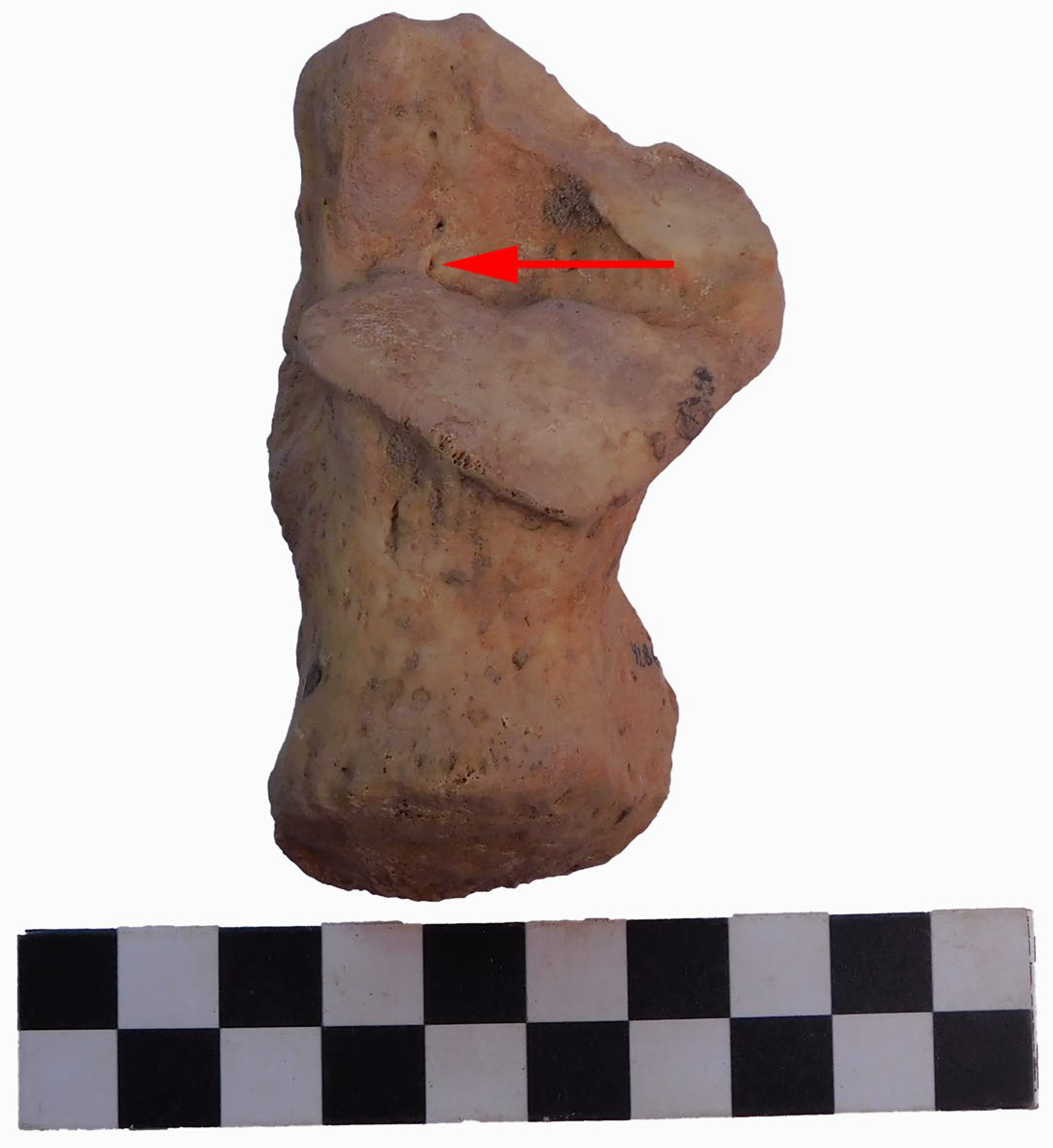
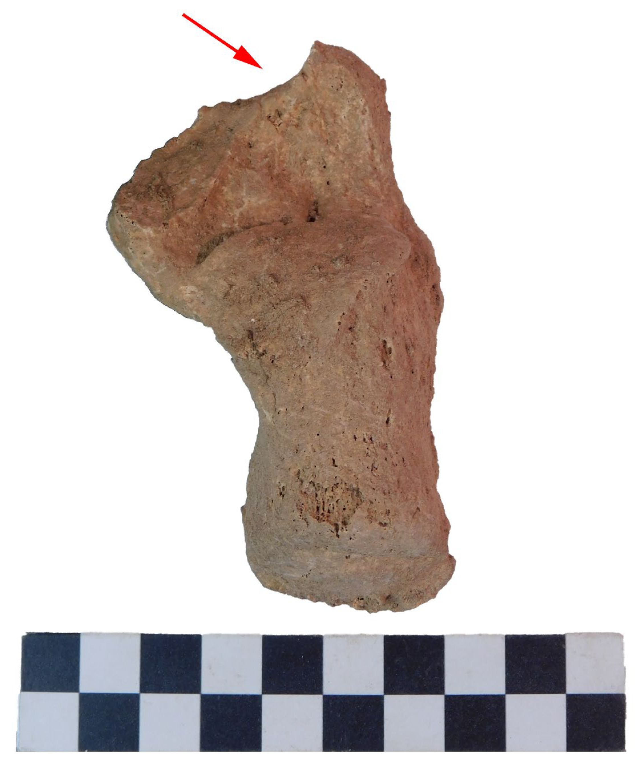
calcaneus secundarius. The retrotrochlear eminence is a frequent insertion site of accessory muscles and a common insertion site for the peroneus quartus muscle. This muscle insertion is located near to, and should not be confused with, the peroneal tubercle [
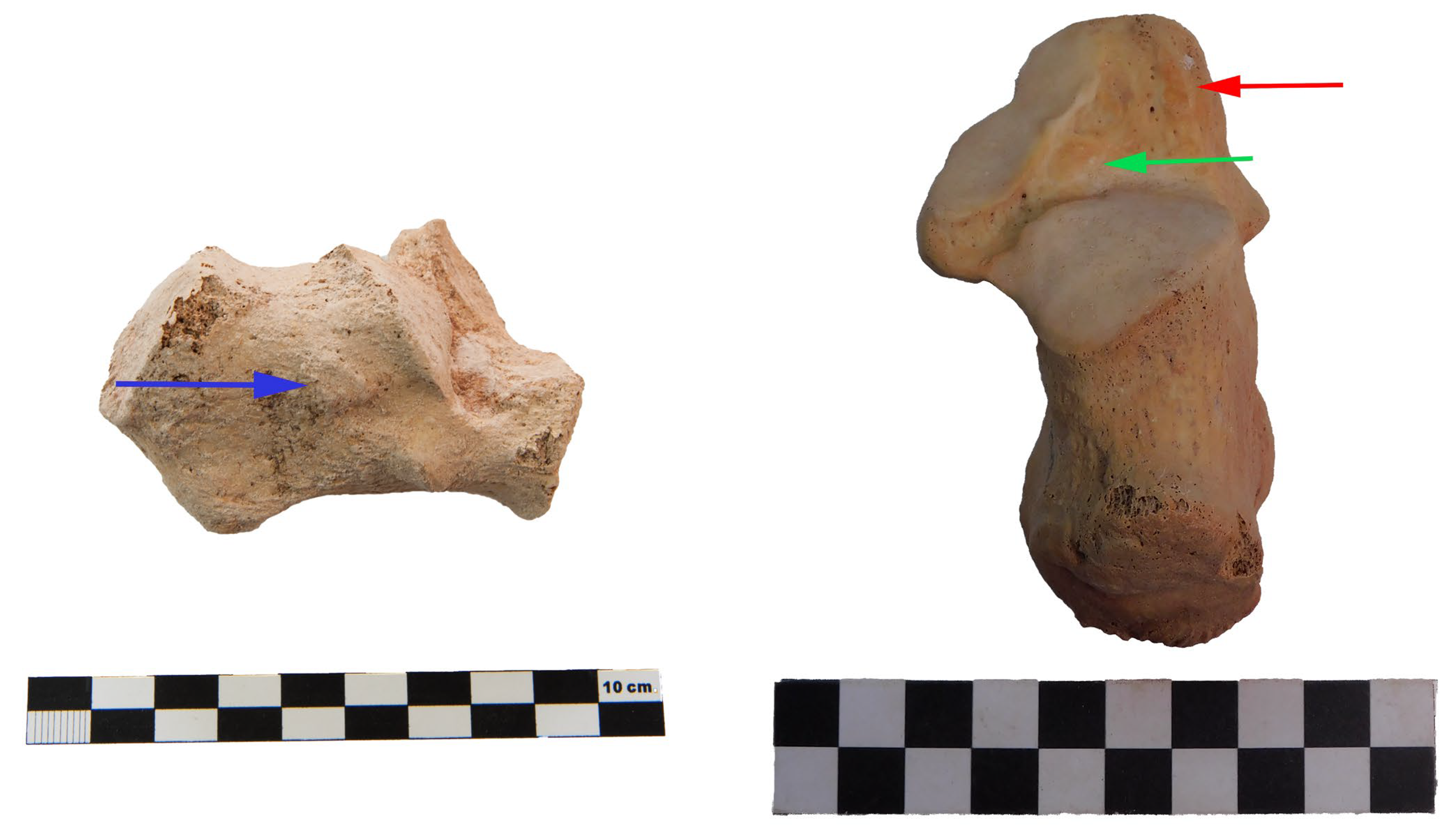
17]. The EFEOPF trait consists of a facet believed to be caused by friction from the accessory anterolateral talar facet on the talus bone due to the angle; normally, the talocalcaneal ligaments and retinaculum would prohibit friction with the talus bone during certain squatting postures, but in these cases the retinaculum and ligaments are genetically absent [18]. The pseudo-facet is located anteriorly on the calcaneus and may appear due to a low foot arch or flat foot syndrome [19]. The MRIER is an insertion location for the inferior extensor retinaculum. A fibrous band attaches to the anterior–lateral part of the calcaneus, wrapping around the ankle region, and the presence of this trait may be caused by genetic–environmental factors [20,21]. The talo-calcaneal facets are the joints between the calcaneus and the talus bone that support the weight and movement of the ankle. These facets are considered to be genetic in origin as they can start to develop early in development, but ankle activity may also influence changes in facet morphology [22,23,24,25]. The calcaneus secundarius is an ossicle and connection site for the ossicle located on the anterior region of the calcaneus. This ossicle can be joined by cartilage with a porous texture on a semi-circular notch on the calcaneus. Type 1 calcaneus secundarius consists of a smooth semi-circular notch on the bone, while type 2 is a porous notch [26]. For more information on these traits, we recommend consulting the atlas of non-metric traits of the calcaneus [26].
Each trait has its significance in its inclusion in this study. All of these traits appear to have lifestyle and genetic factors influencing their presence [26]. Regardless of whether genetics influence the appearance of these traits, lifestyle is an important factor that can differ between populations and individuals. Thus, due to their shared etiology, each trait included in this study may aid in differentiating the modern Canarian population from pre-Hispanic Canarians due to lifestyle differences.
In addition, considering that a genetic homology exists among the population living on the islands during the 18th century and the pre-Hispanic era, in the face of marked differences in economic activity, this study potentially aids in clarifying the genetic or acquired nature of some of these traits. Therefore, taking advantage of the opportunity to analyze large population groups of pre-conquest and 17th-18th century post-conquest inhabitants of the Islands, in this study we compare the prevalence of several calcaneal non-metric traits of pre-Hispanic and modern inhabitants of the islands in order to (1) test the skeletal variation between pre-Hispanic and modern (17–18th century) Canarian samples and (2) gather information on biological and lifestyle differences between the samples using the calcaneal bone.

2. Materials and Methods

2.1. Samples

Our sample consists of a total of four sub-samples: La Concepción Church, Convent of San Francisco de Las Palmas, Maspalomas, and La Guancha–El Agujero. La Concepción Church and the Convent of San Francisco de Las Palmas are modern samples, while Maspalomas and La Guancha–El Agujero are pre-Hispanic samples. In total, 364 calcanei (164 modern Canarian [72 left and 92 right] and 200 pre-Hispanic Canarian [92 left and 108 right] calcanei) were included in this study (see Table 2). A total of 270 calcanei (103 modern and 167 pre-Hispanic) were used to calculate sex chi-square tests. In some of these archaeological sites, the preservation of the skeletal remains within the burials was relatively good. This was the case of the pre-Hispanic burials (Maspalomas, La Guancha and El Agujero), in contrast to the modern samples from La Concepción and San Francisco Convent, in which skeletal remains were frequently commingled, in most cases due to the frequent re-utilization of the same burial, as documented in La Concepción during the archaeological excavation (directed by one of us (MAR)). In addition, some calcanei were partially eroded, impeding adequate measurement and/or accurate evaluation of a given non-metric trait. Therefore, some calcanei were excluded due to taphonomic damage, which may result in differing frequencies in the results’ tables.

2.1.1. Age at Death

We only selected calcanei belonging to individuals with complete fusion of the epiphysis and the medial and lateral processes of the body of the calcaneus. Thus, no subadults were included in this study. Age at death was always over 19–20 years [27].

2.1.2. Sex Assessment

In the 89 cases in which complete skeletons were available, sex was estimated following standard procedures [28,29], using cranial and pelvic morphological traits, such as observation of the pubis, the sciatic notch, the presence or absence of the preauricular sulcus, the observation of cranial features such as the nuchal crest, the mastoid process, the surpraorbital ridge, the glabellar region, and the mandible. In the 56 cases in which skeletal remains were incomplete, estimation of sex was performed using discriminant functions of the calcaneus following previous studies [30,31]. Sex estimation for pre-Hispanic samples was confirmed by genetic sexing among at least 108 skeletons (with well-preserved calcanei), following the same procedure described in previous research [32]. Taphonomic alterations precluded accurate measurements in the remaining cases. All samples are located in Museo Canario, Las Palmas de Gran Canaria, with the exception of the La Concepción Church collection, which is located in the Laboratory of Prehistory, Department of Geography and History at the Faculty of Humanities of the University of La Laguna.
All non-metric trait observations were performed by an experienced specialist. The non-metric traits selected (see Table 1) for this research have been exhaustively studied and are defined in an atlas of calcaneus non-metric traits published recently by our team, in which all current information and the location of each trait and more is presented in detail [26,33].
Table 1. Influences on the appearance of the traits selected for this study.
TraitAcquired During LifeGenetic Influence
Retrotrochlear eminenceInsertion site for accessory peroneal muscles [34,35]The accessory muscles must be present for the trait to appear on bone [34,35]
EFEOPFCaused by squatting posture [18]Necessary muscle attachments must be genetically absent [18]
Pseudo-facetPossibly connected to a low foot arch or the cervical ligament attachment site [19,36]N/A
MRIERIt is the site of a ligament insertion, possibly caused by squatting positions [18]N/A
Talo-calcaneal facetsMay be affected by pronation and supination ankle movements [26]May be affected by genetics due to observed population differences [37,38,39]
Calcaneus secundariusPossibly caused by microtraumatism during early childhood [40], among other possible causes [26]N/A
Table 2. Samples used in this study.
Samples
ModernPre-HispanicTotal
La Concepción ChurchConvent of San Francisco (LP)MaspalomasLa Guancha–El Agujero
Calcaneus1451915149364
164200364

2.2. Traits Included

The traits included in this study were the retrotrochlear eminence, Extra Facet Extension of Posterior Facet (EFEOPF), pseudo-facet, medial root of inferior extensor retinaculum (MRIER), talo-calcaneal facets (type Ia, Ib, IIa, IIb, IIc, III, IV, and V), and calcaneus secundarius (type 1 and 2). The traits were classified as present or absent and as categorical data. If the trait was minimally observed, the trait was recorded as present, and if the trait was not observed at all, it was classified as absent. This process is described and defined in previous research (see [26]).

2.3. Statistical Analysis

Regarding the statistical methods used in this study, chi-square analysis with Yates correction was applied. Additionally, the phi value was calculated following previous studies [41]. Previous research has highlighted the statistical issues with p-values, and for this reason we decided to include phi values in this study to indicate the strength or effect size of each significant p-value, offering an alternative measurement that has been defended by many statisticians [41,42]. The phi-value is an effect size that can be interpreted as <0.1 = very weak association, 0.1–0.2 = weak association, 0.2–0.3 = medium association or >0.3 = strong association, following statisticians [43].
Chi-square is a hypothesis test using contingency tables and is utilized to test if there are statistical disproportions between samples. The chi-square test allows us to test if the null hypothesis is true or false depending on the proportions of the samples. If the p-value is below 0.05, the null hypothesis is false [44]. Several logistic regression analyses were also performed (see Results) in order to assess which variables were independently related to the presence or absence of a given trait. All statistical procedures in this study were performed with IBM SPSS Statistics for Windows, Version 20.0. Armonk, NY, USA: IBM Corp.

2.4. Archaeological Background

La Concepción Church is located in Santa Cruz in Tenerife. In use between the 16th and 18th century, it is one of the oldest churches that was used as a cemetery: individuals were buried underneath the floor of the church, a practice that continued until 1829 [45]. The individuals buried here were from varied occupations within the port city, including both the city’s elite and paupers, who were given a burial out of charity. Due to the church building being located close to a ravine, floods are frequent. However, many recent burials (for individuals in the last decades of the 18th and very early 19th centuries) provided acceptably well-preserved skeletal remains, including many calcanei, recovered from an emergency excavation campaign in 1995 [16].
The Convent of San Francisco of Las Palmas de Gran Canaria is the second archaeological site from which the second modern Canarian osteological collection originates. This convent was used during the 16–18th centuries. During archaeological excavations in 1992, human remains were found in the outskirts of the inner convent church, which was an area used as an ossuary [46]. The human remains may have consisted of friars or paupers. Regarding the pre-Hispanic archaeological sites from which the osteological collections are derived, the first site is Maspalomas, located in the south of Gran Canaria, and is one of the best contextualized pre-Hispanic burial sites of the Canary Islands, with a total dimension of 2000 m2 and more than 100 burials. The burials are mainly singular, with some cases of multiple burials in the same grave, and some graves are cist burials. Carbon dating of human remains showed that the site was used as a cemetery between the 12th and 15th century CE [47]. The second pre-Hispanic archaeological site is La Guancha–El Agujero, which is located in northern Gran Canaria, and consists of seven burial mounds with cist burials. These have been interpreted as social elite burial grounds due to the complexity in construction, and cranial and postcranial non-metric studies have shown phenotypical relationships between members of the same mounds and between different mounds [48]. C-14 dating yields and antiquity ranging 875 ± 60 BP [49] (see Table 3).

3. Results

The results of our study are shown in the following Table 4, Table 5, Table 6, Table 7, Table 8 and Table 9:

3.1. General Proportions of the Calcaneal Traits

As shown in Table 4, many traits show statistical significance and disproportions between pre-Hispanic and modern samples, such as retrotrochlear eminence, EFEOPF (see Figure 1), pseudo-facet, MRIER (see Figure 2), facet type Ib, IIa, and IIb, and CalSec2. The pseudo-facet is the trait with the highest frequency in the pre-Hispanic sample. Regarding the talo-calcaneal facets, type IIc was absent in both samples, while the presence of type III, IV, and V was minimal and rare among both samples. Notably, the pre-Hispanic sample presented many more cases of type Ib, with a high phi value. On the other hand, the modern sample presents a more distributed frequency of the talo-calcaneal types, showing higher frequency in type IIa and IIb. The main distribution differences can be observed in pseudo-facet and EFEOPF, shown by high phi values. Although, these results are regardless of sex and side. Below, we will explore the traits including these variables.

3.2. Non-Metric Trait Distribution According to Side

In order to assure that the same individual was not included twice, we report the prevalence of the traits observed in right bones and left bones separately. Comparisons between pre-Hispanic and post-conquest individuals yield similar results to those shown in Table 4. The exception is the retrotrochlear eminence, with which the differences between the pre-Hispanic and modern samples are slightly more marked in the right bones than in the left ones, although the differences in chi-square and p-values are not very high (0.04), and the phi value of the association observed with the right bones yields only a low–medium effect size. Furthermore, EFEOPF, pseudo-facet, MRIER, type Ib, IIa, and IIb, and CalSec2 appear as statistically significantly similar to the previous results above. Notable results are provided by the pseudo-facet, which shows the highest effect size. Traits such as retrotrochlear eminence, MRIER, and facet type IIb show unilateral significance, while EFEOPF, pseudo-facet, facet type Ib, IIa, and CalSec2 show bilateral significant differences between pre-Hispanic and modern samples.

3.3. Non-Metric Trait Distribution According to Sex of Right and Left Calcanei Separately

In Table 6, we show the results of the right and left calcanei chi-square tests separately between pre-Hispanic and modern Canarians pooled together. No differences were found, with only one exception of talo-calcaneal facet type IIb, which was observed in the left calcanei of males. The trait was completely absent among females of the samples.

3.4. X2 Tests Between Males and Females of Pre-Hispanic and Post-Conquest (Modern) Samples

Below in Table 7, some traits show statistical significance, with high phi values between pre-Hispanic males and modern males, such as EFEOPF on the left calcanei, pseudo-facet on the right calcanei, MRIER on the left calcanei, and talo-calcaneal facet type Ib on the left calcanei and type IIa on right and left calcanei.
As observed in Table 8, pre-Hispanic females and modern females show various trait proportion differences, such as the retrotrochlear eminence on the right calcanei, the EFEOPF on both the right and left calcanei, the pseudo-facet on the right and left calcanei, talocalcaneal facet type Ib on the right and left calcanei, type IIb on the right calcanei, and calcaneus secundarius type 2 on the right calcanei (Figure 3).

3.5. Logistic Regression Analysis

To further support the results shown above, we performed a multivariate logistic regression on all non-metric traits included in this study. The results are shown in Table 8. We tested the only variables available for the included samples, which were site (modern or pre-Hispanic), sex (male or female), and side (right or left). As can be seen in Table 9, retrotrochlear eminence, EFEOPF, pseudo-facet, MRIER, facet type Ib, IIa, and IIc, and CalSec2 were only dependent on the variable site (pre-Hispanic or modern). Facet IIc had insufficient cases to perform the analysis, and the rest had no dependent variables.

4. Discussion

There are statistically significant differences between pre-Hispanic and modern Canarians regarding the skeletal variations in the calcanei of these samples. Variations and non-metric trait differences of the calcaneus and the ankle, in general, can likely be attributed to many factors, including a mixture of genetic and environmental influences, as seen in previous studies [42,43]. Furthermore, different traits of the calcaneus have differing etiologies, and previous research has shown that some traits of the calcaneus are considered more influenced by activity than others [23,44,45]. However, considering the genetic homology existing among the pre-Hispanic and modern population groups, the results observed in this study generally support that the traits with significantly different proportions among the two population groups are related to differences in economic activity. In any case, each trait must be studied separately to understand the differences in proportion between the samples.
In this study, we have described the prevalence of several calcaneal traits and the typology of calcaneal facets, comparing them among two periods defined by marked differences in socioeconomic status. Although the genetic composition of the population included in each of the two groups (modern/pre-Hispanic) is not the same, the estimated indigenous genetic contribution to the 17–18th century population reaches 40%, based on Y-chromosome and mtDNA haplogroup frequencies [3]. Despite this, some non-metric traits show striking differences among the pre-Hispanic and modern populations of the Canary Archipelago. For instance, the prevalence of EFEOPF was by far higher among the pre-Hispanic inhabitants (i.e., regarding the left bone, EFEOPF was observed in more than 31% of the pre-Hispanic inhabitants, but it was absent in the modern population), in sharp contrast with the similar prevalence of some other commonly observed traits, such as the type Ia facet, which was present in about 14% of the pre-Hispanic population and 19% of the modern one. Therefore, this indicates that some traits strongly depend on different activities performed by both populations, whereas others must be dependent on a common genetic inheritance. Interestingly, no differences were observed among males and females regarding these two traits, both in the pre-Hispanic and modern samples.
The presence of EFEOPF has been attributed to different types of squatting positions, and/or high activity levels with angular ankle movements [50]. Normally, the talo-calcaneal ligaments and the retinaculum would stop the talus from advancing into the sinus tarsi, but in a forced partial dorsiflexion, the talus can advance slightly more than usual [51]. The facet shape has also been described in relation to anatomical variations in ligaments [18]. The very high prevalence of this trait among the pre-Hispanic population is fully in accordance with the marked differences in lifestyle before and after the Spanish conquest, moving from agropastoralist activities to an urban way of life. It is also of interest to consider that EFEOPF was similarly common among pre-Hispanic men (i.e., 31%) and women (32% of the left bones), so both sexes performed similar activity, at least regarding the information derived from this trait.
Another striking result is the presence of a pseudo-facet, a trait that affects most of the pre-Hispanic population (88.9% among males and 86.1% among females), but only 32% of the modern one (36.1% of females and 26% of males). This trait has previously been associated with an area of friction with the talus bone in cases of flat foot syndrome [19], although other authors suggest that it is a cervical ligament attachment site, where the extensor digitorum brevis muscle attaches to the calcaneus [19], adopting the appearance of a facet, as other muscle attachments are shaped as depressions [21]. In any case, the prevalence of this trait among the modern population of the Canary Islands is also very high (>30%), compared with that derived from other studies, such as that of Madhavi et al. [19], who described the trait in only 4% (9 out of 225 cases) of the Indian population. Therefore, our results strongly suggest that the trait must be influenced not only by a strong muscle attachment (derived from activity), but also by genetics, since it is by far more common among both pre-Hispanic and modern Canarians than among other populations, although the higher prevalence among pre-Hispanics suggests that lifestyle may also play a role. Perhaps, the lack of supportive footwear may account for the high prevalence of this trait. As with the EFEOPF trait, the prevalence of this trait was similar among pre-Hispanic males and females.
On the other hand, an explanation for the high prevalence of calcaneus secundarius among the pre-Hispanic population is quite challenging. The calcaneus secundarius is a small accessory ossicle that can be identified as a porous notch, and is either deep, small, and semicircular, or large and shallow, usually located on the anterior facet of the calcaneus. Until now, there are no studies that have proven a hereditary transmission of the trait [52]. It seems more likely to be a trait related to physical activity performed during childhood, similar to the pathogenesis of os trigonum in talar bones. It was found in nearly 12% of the pre-Hispanic sample (both males and females), a figure by far higher than that reported for other population groups, and only comparable with the figures observed for the French population [53] and that (11.8%) of a Neolithic–Chalcolithic population in Portugal [54]. Some authors argue a genetic origin, based on observations on individuals where many other ossicle-type traits are present [53]. In the present study, the finding of only 1 case among the modern population in contrast to the 19 cases (in which sex was estimated) observed in the pre-Hispanic one strongly supports that the trait is not a genetic one, but acquired via physical activity during childhood [40].
The retrotrochlear eminence is regarded as the insertion site for the peroneus quartus muscle [55]. It was very frequently observed among the population studied. Overall, the prevalence of this trait is high (84%) in some studies [56], in which its presence was evaluated dichotomically (present/absent), as undertaken in this work. However, other authors have determined its size, and considered its presence only if it reaches a given size (5 mm following [57]), thus making comparisons difficult: [57] reports a prevalence of 17% of total cases. Logically, one can expect that, as with other muscle insertions, the stronger the muscle, the greater the enthesis. Although it seems that the trait is slightly more frequent among the pre-Hispanics, the differences are not very marked and only restricted to right female bones—although perhaps indicating again more evidence of the higher level of exercise performed by the pre-Hispanics—and, overall, we can consider that the prevalence is in the range of reported studies, especially considering that this trait may become sometimes difficult to identify due to taphonomic alterations.
The medial root of the inferior extensor retinaculum (MRIER) trait relates to the insertion area on the calcaneus where the fibrous tissue wraps around the ankle and inserts into the tibial malleolus on the other side of the foot [20,21]. Due to this trait being an insertion for a muscle, it is likely an activity marker that can indicate dorsiflexion and plantarflexion movements during life [26]. This may explain why we found a by far larger proportion of this trait among the pre-Hispanic population (≈50%) than in the post-conquest one (≈35%). In any case, both figures are also larger than the 13.1% reported by Morimoto (1960) [18], therefore suggesting that a genetic component also exists regarding this trait.
A similar argument may be used to explain the prevalence of the type Ib talocalcaneal facet. It is the most frequent variant, especially among the pre-Hispanic population, reaching a figure (nearly 74% among left male bones and 69% among female ones) higher than any other reported [26,58]. The prevalence observed among the post-conquest population is also quite high (≈40%)—higher, for instance, than that reported for the Spanish population (25%) in [38]. It is important to consider that among individuals with type I facets, there are only two facets that support the total body weight in the ankle, reducing weight support and allowing the foot to slide, thus allowing osteoarthritis to form much more easily [59,60]. As the results of this study also suggest (high prevalence even in the modern population), the talo-calcaneal types have often been associated with genetic population differences in previous studies [19,61,62], although acquired factors were also surely present, explaining the even higher proportion among pre-Hispanics. Thus, the differences observed in this study are likely due to other factors rather than genetics. The talo-calcaneal facet association with osteoarthritis shown in previous research [19,36] may be due to ankle movement and posture influencing the shape of the facets and, as a consequence, causing osteoarthritis.
It is also important to highlight the lack of statistically significant differences in prevalence among males and females regarding facet type, both in the pre-Hispanic and modern samples. Future research should focus on the factors other than genetics that influence the presence of these calcaneal traits, as well as other postcranial skeletal variations. Research focused on genetic aspects of the traits would be beneficial for kinship studies and heritability studies alike. Therefore, multidisciplinary approaches where geneticists and archaeologists collaborate to discover particular genes that influence the appearance of postcranial traits would be highly valuable for heritability studies.
A limitation of this study is the possible difference existing between the two modern Canarians samples included in this study, which may have influenced the results of this study. On one hand, the La Concepción Church collection provides skeletal individuals from multiple lifestyles from a port city environment of the 18th century, while the Convent of San Francisco of Las Palmas, Gran Canaria, is a collection that perhaps contained a larger proportion of friars. However, the convent was also a “sacred” place, and a desired place for Catholics to be buried, and, indeed, many of the individuals buried at that place were citizens living in an urban, port environment, similar to that of Santa Cruz. In any case, subtle lifestyle differences may have existed among the two sites. Another limitation is that although genetic homology was considerable between pre-Hispanic and modern individuals, the modern population is mixed, formed by people of pre-Hispanic ancestry but also Spaniards, other Europeans, some sub-Saharan Africans, and even some South American natives [16], so we cannot fully assure that the appearance of a given trait only depends on differences in activity. However, as commented, the higher proportions of some traits, such as MRIER, both among pre-Hispanic and modern Canarians, do support the relative genetic homogeneity of pre-Hispanic and modern Canarians compared with other population groups.
Also, in this study, the most “modern” individuals analyzed were at least 200 years old. In these two centuries, some further changes in lifestyle may have occurred that may have modified the prevalence or some features of the analyzed traits, limiting the ability to apply the results obtained to forensic research on contemporary population of the Canary Islands. In any case, some traits such as the retrotrochlear eminence, EFEOPF, pseudo-facet, MRIER, talocalcaneal facets, and calsec2 are more common among pre-Hispanics. Regardless of whether these traits are highly influenced by genetics or lifestyle, they may aid in the identification of “modern” vs. pre-Hispanic skeletal remains due to the highly statistical differences shown in this study. As shown, the majority of calcaneal non-metric traits included in this study are statistically different in frequency between pre-Hispanic and modern Canarians, and thus could be used for identification. This may be useful to help separate modern forensic cases from pre-Hispanic archaeological cases.
Furthermore, the use of non-metric traits, specifically trait combinations, in individual identification has shown promising results [12]. Therefore, following our results, it may be possible that combinations of postcranial traits, such as calcaneal variation, may aid in personal identification. Previous research has already demonstrated potential trait combinations to identify relationships in archaeological contexts [13].

5. Conclusions

In this study, we analyzed the prevalence of several traits in two groups of a genetically related population, separated by 4–5 centuries, during which profound socioeconomic changes ensued. We showed that some traits are possibly linked to increased ankle activity, such as the presence of the EFEOPF, retrotrochlear eminence, pseudo-facet, and calcaneus secundarius, which were more commonly observed among the pre-Hispanic population, underscoring the importance of acquired factors in their development (instead of genetic ones). By contrast, some others, such as type Ib articular facet and MRIER, although more common among pre-Hispanics, also show an unusually high prevalence among the post-conquest, modern population, showing a by far higher prevalence than among other population groups, including Spaniards. This suggests that at least these two traits are heavily dependent on genetics, constituting, in this sense, a confirmation of the paleogenomic analyses that show a 40% persistence of pre-Hispanic DNA among modern inhabitants of the Islands. Another important conclusion is that no sex differences were observed, at least regarding some of the traits possibly related to activity included in this study (for instance, EFEOPF, pseudo-facet, and calcaneus secundarius), a finding that may suggest the performance of similar activities by both males and females. Our results may aid forensic anthropologists during biological profiling, and the use of these traits may be useful in forensic fieldwork where particular calcaneal traits are studied, providing additional information on the lifestyle and background of skeletal remains found in forensic contexts.

Author Contributions

All authors contributed equally to the development of this article. All authors have read and agreed to the published version of the manuscript.

Funding

This research received no external funding.

Institutional Review Board Statement

Not applicable.

Informed Consent Statement

Not applicable.

Data Availability Statement

Dataset available on request from the authors.

Conflicts of Interest

The authors declare no conflict of interest.

Abbreviations

The following abbreviations are used in this manuscript:
EFEOPFExtra Facet Extension of Posterior Facet
MRIERMedial Root of Inferior Extensor Retinaculum
CalSecCalcaneus Secundarius

References

  1. Santana, J.; del Pino, M.; Morales, J.; Fregel, R.; Hagenblad, J.; Morquecho, A.; Brito-Mayor, A.; Henríquez, P.; Jiménez, J.; Serrano, J.G.; et al. The Chronology of the Human Colonization of the Canary Islands. Proc. Natl. Acad. Sci. USA 2024, 121, e2302924121. [Google Scholar] [CrossRef]
  2. Serrano, J.G.; Ordóñez, A.C.; Santana, J.; Sánchez-Cañadillas, E.; Arnay, M.; Rodríguez-Rodríguez, A.; Morales, J.; Velasco-Vázquez, J.; Alberto-Barroso, V.; Delgado-Darias, T.; et al. The Genomic History of the Indigenous People of the Canary Islands. Nat. Commun. 2023, 14, 4641. [Google Scholar] [CrossRef]
  3. Fregel, R.; Gomes, V.; Gusmão, L.; González, A.M.; Cabrera, V.M.; Amorim, A.; Larruga, J.M. Demographic History of Canary Islands Male Gene-Pool: Replacement of Native Lineages by European. BMC Evol. Biol. 2009, 9, 181. [Google Scholar] [CrossRef] [PubMed]
  4. Ansorge, H. Assessing Non-Metric Skeleton Characters as a Morphological Tool. Zoology 2001, 104, 268–277. [Google Scholar] [CrossRef]
  5. Sangari, S.K.; Dossous, P.-M.; Heineman, T.; Mtui, E.P. Dimensions and Anatomical Variants of the Foramen Transversarium of Typical Cervical Vertebrae. Anat. Res. Int. 2015, 2015, 391823. [Google Scholar] [CrossRef]
  6. Hauser, G.; De Stefano, G.F. Epigenetic Variants of the Skull; E. Schweizerbartsche Verlagsbuchhandlung: Stuttgart, Germany, 1989. [Google Scholar]
  7. Falconer, D.S. The Inheritance of Liability to Certain Diseases, Estimated from the Incidence among Relatives. Ann. Hum. Genet. 1965, 29, 51–76. [Google Scholar] [CrossRef]
  8. Baykara, I.; Yilmaz, H.; Gültekin, T.; Güleç, E. Squatting Facet: A Case Study Dilkaya and Van-Kalesi Populations in Eastern Turkey. Coll. Antropol. 2010, 34, 1257–1262. [Google Scholar]
  9. Brukner Havelková, P.; Dulíková, V.; Bejdová, Š.; Vacková, J.; Velemínský, P.; Bárta, M. Ancient Egyptian Scribes and Specific Skeletal Occupational Risk Markers (Abusir, Old Kingdom). Sci. Rep. 2024, 14, 13317. [Google Scholar] [CrossRef]
  10. Bühler, B.; Kirchengast, S. A Life on Horseback? Prevalence and Correlation of Metric and Non-Metric Traits of the “horse-Riding Syndrome“ in an Avar Population (7th-8th Century AD) in Eastern Austria. Anthr. Rev. 2022, 85, 67–82. [Google Scholar] [CrossRef]
  11. Cockerill, S.J.; Alberto-Barroso, V.; Arnay-De-La-Rosa, M.; González-Reimers, E. Intracemetery Relationships: Non-Metric Trait Combination Analysis of Maspalomas of Gran Canaria (Canary Islands, Spain). Anthr. Anz. 2024, 81, 433–447. [Google Scholar] [CrossRef]
  12. Cockerill, S.J.; Arnay de la Rosa, M.; González Reimers, E. Guild Burials or Family Graves? Intrapopulation Relationship Analysis of 18th Century La Concepción Church Cemetery in Santa Cruz de Tenerife, Canary Islands. J. Archaeol. Sci. Rep. 2021, 37, 102915. [Google Scholar] [CrossRef]
  13. Palamenghi, A.; Borlando, A.; De Angelis, D.; Sforza, C.; Cattaneo, C.; Gibelli, D. Exploring the Potential of Cranial Non-Metric Traits as a Tool for Personal Identification: The Never-Ending Dilemma. Int. J. Leg. Med. 2021, 135, 2509–2518. [Google Scholar] [CrossRef] [PubMed]
  14. Hoover, K.C.; Berbesque, J.C. Early Holocene Morphological Variation in Hunter-Gatherer Hands and Feet. PeerJ 2018, 6, e5564. [Google Scholar] [CrossRef] [PubMed]
  15. Alberto Barroso, V. Rozando La Eternidad: La Muerte Entre Los Antiguos Canarios, 1st ed.; Ediciones del Cabildo de Gran Canaria: Las Palmas de Gran Canaria, Spain, 2020. [Google Scholar]
  16. Arnay de la Rosa, M.; Pérez Álvarez, A.R. Estudio de Un Espacio Sepulcral Del Siglo XVIII En La Iglesia de La Concepción de Santa Cruz de Tenerife. Rev. Tabona 2002, 11, 131–167. [Google Scholar]
  17. Yammine, K. The Accessory Peroneal (Fibular) Muscles: Peroneus Quartus and Peroneus Digiti Quinti. A Systematic Review and Meta-Analysis. Surg. Radiol. Anat. 2015, 37, 617–627. [Google Scholar] [CrossRef]
  18. Morimoto, I. The Influence of Squatting Posture on the Calcaneus in the Japanese. J. Anthropol. Soc. Nippon. 1960, 68, 16–22. [Google Scholar] [CrossRef]
  19. Madhavi, C.; Madhuri, V.; George, V.M.; Antonisamy, B. South Indian Calcaneal Talar Facet Configurations and Osteoarthritic Changes. Clin. Anat. 2008, 21, 581–586. [Google Scholar] [CrossRef]
  20. Kelikian, A.S.; Sarrafian, S. Sarrafian’s Anatomy of the Foot and Ankle: Descriptive, Topographic, Functional; Lippincott Williams & Wilkins: Philadelphia, PA, USA, 2011; ISBN 978-0-7817-9750-4. [Google Scholar]
  21. Drake, R.L.; Wayne Vogl, A.; Mitch, A.W.M. Gray’s Anatomy for Students: With Student Consult Online Access, 3rd ed.; Elsevier: Philadelphia, PA, USA, 2014; ISBN 978-0-7020-5131-9. [Google Scholar]
  22. Norris, C.M. Lower Limb Motion during Walking, Running and Jumping. In Managing Sports Injuries; Elsevier: Amsterdam, The Netherlands, 2011; pp. 133–142. ISBN 978-0-7020-3473-2. [Google Scholar]
  23. Placzek, J.D.; Boyce, D.A. Orthopaedic Physical Therapy Secrets, 2nd ed.; Hanley & Belfus: St. Louis, MO, USA, 2006; ISBN 978-1-56053-708-3. [Google Scholar]
  24. Mahan, S.T.; Prete, V.I.; Spencer, S.A.; Kasser, J.R.; Bixby, S.D. Subtalar Coalitions: Does the Morphology of the Subtalar Joint Involvement Influence Outcomes After Coalition Excision? J. Foot Ankle Surg. 2017, 56, 797–801. [Google Scholar] [CrossRef]
  25. Bunning, P.S.C. Some Observations on the West African Calcaneus and the Associated Talo-Calcaneal Interosseous Ligamentous Apparatus. Am. J. Phys. Anthropol. 1964, 22, 467–472. [Google Scholar] [CrossRef]
  26. Cockerill, S.J.; Arnay-de-la-Rosa, M.; González-Reimers, E. An Atlas of Anatomical Variants of the Human Calcaneus. J. Morphol. 2024, 285, e21706. [Google Scholar] [CrossRef]
  27. Coqueugniot, H.; Weaver, T.D. Brief Communication: Infracranial Maturation in the Skeletal Collection from Coimbra, Portugal: New Aging Standards for Epiphyseal Union. Am. J. Phys. Anthropol. 2007, 134, 424–437. [Google Scholar] [CrossRef]
  28. Black, S.M. Forensic Anthropology: 2000 to 2010; Black, S.M., Ferguson, E., Eds.; CRC Press: Boca Raton, FL, USA, 2011; ISBN 978-1-4398-4588-2. [Google Scholar]
  29. Langley, N.R. Forensic Anthropology: A Comprehensive Introduction, 2nd ed.; Langley, N.R., Tersigni-Tarrant, M.A., Eds.; CRC Press: Boca Raton, FL, USA; Taylor & Franci Group: New York, NY, USA, 2017; ISBN 978-1-4987-3612-1. [Google Scholar]
  30. Alemán Aguilera, I.; Botella López, M.C.; Du Souich Henrici, P. Aplicación de Las Funciones Discriminantes En La Determinación Del Sexo. Estud. Antropol. Biológica 1999, IX, 221–230. [Google Scholar]
  31. Alemán Aguilera, I.; Botella López, M.C.; Ruiz Rodríguez, L. Determinación Del Sexo En El Esqueleto Postcraneal. Estudio de Una Población Mediterránea Actual. Arch. Esp. Morfol. 1997, 2, 69–79. [Google Scholar]
  32. Cockerill, S.J.; Calderón Ordóñez, A.; Guamán Chulunchana, R.G.; González Serrano, J.; Fregel, R.; Díaz-Pérez, C.; González-Reimers, E.; Arnay-de-la-Rosa, M. The Talus of the Pre-Hispanic Population from Punta Azul (El Hierro, Canary Islands): Variability and Sexual Dimorphism of Non-Metric Traits. Int. J. Osteoarchaeol. 2022, 33, 1198–1212. [Google Scholar] [CrossRef]
  33. Cockerill, S.J.; Arnay-de-la-Rosa, M.; González-Reimers, E. An Atlas of Anatomical Variants of the Human Talus. J. Morphol. 2025, 286, e70023. [Google Scholar] [CrossRef]
  34. Cheung, Y.Y.; Rosenberg, Z.S.; Ramsinghani, R.; Beltran, J.; Jahss, M.H. Peroneus Quartus Muscle: MR Imaging Features. Radiology 1997, 202, 745–750. [Google Scholar] [CrossRef]
  35. Sobel, M.; Levy, M.E.; Bohne, W.H.O. Congenital Variations of the Peroneus Quartus Muscle: An Anatomic Study. Foot Ankle 1990, 11, 81–89. [Google Scholar] [CrossRef]
  36. Drayer-Verhagen, F. Arthritis of the Subtalar Joint Associated with Sustentaculum Tali Facet Configuration. J. Anat. 1993, 183, 631–634. [Google Scholar]
  37. Laxmi, V.; Mehra, R.; Sharma, R. A Morphological and Morphometric Study of Human Calcanei and Their Articular Facets. Indian. J. Med. Res. Pharm. Sci. 2018, 5, 34–42. [Google Scholar] [CrossRef]
  38. Forriol Campos, F.; Gomez Pellico, L. Talar Articular Facets (Facies Articulares Talares) in Human Calcanei. Cells Tissues Organs 1989, 134, 124–127. [Google Scholar] [CrossRef]
  39. Kori, D.; Prasad, G.; Rani, A.; Dewan, R.; Singh, R.; Singh, P. Study of Variations in Talar Articular Facets of Human Calcanei and Their Association with Calcaneal Spurs in North Indian Population. Int. J. Anat. Res. 2016, 4, 2710–2716. [Google Scholar] [CrossRef]
  40. Mann, R.W. Calcaneus Secundarius: Description and Frequency in Six Skeletal Samples. Am. J. Phys. Anthropol. 1990, 81, 17–25. [Google Scholar] [CrossRef]
  41. Karpen, S.C. P Value Problems. Am. J. Pharm. Educ. 2017, 81, 6570. [Google Scholar] [CrossRef]
  42. Cohen, J. Statistical Power Analysis for the Behavioral Sciences; L. Erlbaum Associates: Hillsdale, NJ, USA, 1988; ISBN 978-0-8058-0283-2. [Google Scholar]
  43. Campbell, I. Chi-Squared and Fisher–Irwin Tests of Two-by-Two Tables with Small Sample Recommendations. Stat. Med. 2007, 26, 3661–3675. [Google Scholar] [CrossRef]
  44. Sanz de Magallanes, J.M. In Memoriam: Enterramientos En La Parroquia Matriz de La Concepción; Imprenta Editora Católica: Santa Cruz de Tenerife, Spain, 2002. [Google Scholar]
  45. Cuenca Sanabria, J.; Anaya Hernández, L.A.; Betancor Rodríguez, A.; Cuenca Sanabria, A.; Lobo Cabrera, M.; Toledo Ponce, D.; Torres Palenzuela, J.A. La investigación histórico-arqueológica del desaparecido Convento de San Francisco de Las Palmas de Gran canaria. Investig. Arqueol. 1995, 4, 9–198. [Google Scholar]
  46. Alberto Barroso, V.; Delgado Darias, T.; Moreno Benítez, M.; Velasco Vázquez, J. La Dimensión Temporal y El Fenómeno Sepulcral Entre Los Antiguos Canarios. Zephyrus Rev. Prehist. Y Arqueol. 2019, 84, 139–160. [Google Scholar] [CrossRef]
  47. Cabrera López, R. El Estudio de Los Caracteres Discretos En Los Espacios Funerarios Prehispánicos de Gáldar; Universidad de Las Palmas: Las Palmas de Gran Canaria, Spain, 2012. [Google Scholar]
  48. Velasco-Vázquez, J. Economía y dieta de las poblaciones prehistóricas de Gran Canaria: Una aproximación bioantropológica. Complutum 1998, 9, 137–160. [Google Scholar]
  49. Trinkaus, E. Squatting among the Neandertals: A Problem in the Behavioral Interpretation of Skeletal Morphology. J. Archaeol. Sci. 1975, 2, 327–351. [Google Scholar] [CrossRef]
  50. Smith, J.W. The Ligamentous Structures in the Canalis and Sinus Tarsi. J. Anat. 1958, 92, 616–620. [Google Scholar]
  51. Case, D.T.; Jones, L.B.; Offenbecker, A.M. Skeletal Kinship Analysis Using Developmental Anomalies of the Foot. Int. J. Osteoarchaeol. 2017, 27, 192–205. [Google Scholar] [CrossRef]
  52. Verna, E.; Villotte, S. Several Infra-Cranial Non-Metric Variations in a Single Neolithic Individual: A Common Genetic Underlying Predisposition? Int. J. Osteoarchaeol. 2016, 26, 1113–1118. [Google Scholar] [CrossRef]
  53. Silva, A.M. Foot Anomalies in the Late Neolithic/Chalcolithic Population Exhumed from the Rock Cut Cave of São Paulo 2 (Almada, Portugal). Int. J. Osteoarchaeol. 2011, 21, 420–427. [Google Scholar] [CrossRef]
  54. Bilgili, M.G.; Kaynak, G.; Botanlioğlu, H.; Basaran, S.H.; Ercin, E.; Baca, E.; Uzun, I. Peroneus Quartus: Prevalance and Clinical Importance. Arch. Orthop. Trauma. Surg. 2014, 134, 481–487. [Google Scholar] [CrossRef] [PubMed]
  55. Ersoz, E.; Tokgoz, N.; Kaptan, A.Y.; Ozturk, A.M.; Ucar, M. Anatomical Variations Related to Pathological Conditions of the Peroneal Tendon: Evaluation of Ankle MRI with a 3D SPACE Sequence in Symptomatic Patients. Skelet. Radiol. 2019, 48, 1221–1231. [Google Scholar] [CrossRef]
  56. Saupe, N.; Mengiardi, B.; Pfirrmann, C.W.A.; Vienne, P.; Seifert, B.; Zanetti, M. Anatomic Variants Associated with Peroneal Tendon Disorders: MR Imaging Findings in Volunteers with Asymptomatic Ankles. Radiology 2007, 242, 509–517. [Google Scholar] [CrossRef]
  57. Boyan, N.; Ozsahin, E.; Kizilkanat, E.; Soames, R.; Oguz, O. Morphometric Measurement and Types of Articular Facets on the Talus and Calcaneus in an Anatolian Population. Int. J. Morphol. 2016, 34, 1378–1385. [Google Scholar] [CrossRef]
  58. Anbumani, S.; Sridharan, R.; Selvi, A.T. An Anatomical Study of Morphology and Morphometric Analysis of Calcaneum and Its Talar Articular Surfaces. Int. J. Anat. Res. 2017, 5, 4223–4229. [Google Scholar] [CrossRef]
  59. Bruckner, J. Variations in the Human Subtalar Joint. J. Orthop. Sports Phys. Ther. 1987, 8, 489–494. [Google Scholar] [CrossRef] [PubMed]
  60. Anjaneyulu, K.; Philips, C.; Tamang, B.K.; Kumar, A. Patterns of Talar Articulating Facets in Adult Human Calcanei from North-East India and Their Clinical Correlation. Asian J. Med. Sci. 2014, 5, 89–93. [Google Scholar] [CrossRef]
  61. Kori, S. Time since Death from Rigor Mortis: Forensic Prospective. J. Forensic Sci. Crim. Investig. 2018, 9, 555771. [Google Scholar] [CrossRef]
  62. Palamenghi, A.; Gibelli, D.; Mazzarelli, D.; De Angelis, D.; Sforza, C.; Cattaneo, C. Rumor Has It: A Narrative Review on the Use of Skeletal Non-Metric Traits and Variants for Personal Identification. Leg. Med. 2023, 65, 102316. [Google Scholar] [CrossRef] [PubMed]
Figure 1. Superior view of EFEOPF (red arrow) from La Guancha–El Agujero (pre-Hispanic sample).
Forensicsci 05 00025 g001
Figure 2. Lateral image of calcaneus, showing retrotrochlear eminence (blue arrow), superior image of pseudo-facet (red arrow), and MRIER (green arrow) on right calcaneus from La Guancha–El Agujero (pre-Hispanic sample).
Forensicsci 05 00025 g002
Figure 3. Superior image of CalSec type 2 (red arrow), showing the curve of the ossicle insertion on right calcaneus from La Guancha–El Agujero (pre-Hispanic sample). The ossicle is not present in the image.
Forensicsci 05 00025 g003
Table 3. Archaeological samples with time period and number of calcanei included in this study.
SiteTime PeriodCalcanei
La Concepción Church16–18th century145
Convent of San Francisco (LP)16–18th century19
Maspalomas12–15th century151
La Guancha–El Agujero11–12th century49
Table 4. Chi-square tests on calcaneal non-metric traits, showing differences between pre-Hispanic and modern samples. EFEOPF = Extra Facet Extension of Posterior Facet. MRIER = medial root of inferior extensor retinaculum. CalSec1 = Calcaneus secundarius type 1. CalSec2 = Calcaneus secundarius type 2.
Non-Metric TraitPre-Hispanic (%)Modern (%)x2 Valuep-ValuePhi Value
Retrotrochlear eminenceAbsent36 (22.2)26 (36.4)6.980.000.17
Present126 (77.8)40 (60.6)
EFEOPFAbsent127 (67.9)160 (98.2)53.970.000.39
Present60 (32.1)3 (1.8)
Pseudo-facetAbsent20 (11.0)101 (69.7)118.460.000.60
Present161 (89.0)44 (30.3)
MRIERAbsent90 (47.6)108 (68.8)15.700.000.21
Present99 (52.4)49 (31.2)
Type IaAbsent165 (84.2)122 (78.2)2.060.15-
Present31 (15.8)34 (21.8)
Type IbAbsent56 (28.6)93 (59.6)34.290.000.31
Present140 (71.4)63 (40.4)
Type IIaAbsent181 (92.8)118 (75.6)20.260.000.24
Present14 (7.2)38 (24.4)
Type IIbAbsent190 (96.9)141 (90.4)6.650.010.13
Present6 (3.1)15 (9.6)
Type IIcAbsent195 (100.0)156 (100.0)---
Present0 (0.0)0 (0.0)
Type IIIAbsent195 (99.5)153 (96.8)3.690.09-
Present1 (0.5)5 (3.2)
Type IVAbsent192 (98.0)156 (100.0)3.220.13-
Present4 (2.0)0 (0.0)
Type VAbsent196 (100.0)155 (99.4)1.260.44-
Present0 (0.0)1 (0.6)
CalSec1Absent189 (98.4)148 (98.0)0.081.00-
Present3 (1.6)3 (2.0)
CalSec2Absent171 (89.1)154 (99.4)15.300.000.21
Present21 (10.9)1 (0.6)
Table 5. Chi-square tests on calcaneal non-metric traits of pre-Hispanic and modern samples. EFEOPF = Extra Facet Extension of Posterior Facet. MRIER = medial root of inferior extensor retinaculum. CalSec1 = Calcaneus secundarius type 1. CalSec2 = Calcaneus secundarius type 2.
Non-Metric TraitSideAbsent/PresentPre-Hispanic (%)Modern (%)x2 Valuep-ValuePhi Value
Retrotrocholear eminenceRightAbsent18 (20.2)13 (38.2)4.230.040.18
Present71 (79.8)21 (61.8)
LeftAbsent18 (24.7)13 (40.6)2.730.09-
Present55 (75.3)19 (59.4)
EFEOPFRightAbsent71 (69.6)88 (96.7)24.320.000.35
Present31 (30.4)3 (3.3)
LeftAbsent56 (65.9)72 (100.0)30.130.000.44
Present29 (34.1)0 (0.0)
Pseudo-facetRightAbsent11 (11.1)62 (73.8)74.500.000.64
Present88 (88.9)22 (26.2)
LeftAbsent9 (11.0)39 (63.9)43.990.000.56
Present73 (89.0)22 (36.1)
MRIERRightAbsent56 (55.4)58 (65.9)2.150.14-
Present45 (44.6)30 (34.1)
LeftAbsent34 (38.6)50 (72.5)17.790.000.34
Present54 (61.4)19 (27.5)
Type IaRightAbsent89 (85.6)71 (81.6)0.550.45-
Present15 (14.4)16 (18.4)
LeftAbsent76 (82.6)51 (73.9)1.790.18-
Present16 (17.4)18 (26.1)
Type IbRightAbsent28 (26.9)51 (58.6)19.620.000.32
Present76 (73.1)36 (41.4)
LeftAbsent28 (30.4)42 (60.9)14.860.000.30
Present64 (69.6)27 (39.1)
Type IIaRightAbsent97 (93.3)66 (75.9)11.470.000.25
Present7 (6.7)21 (24.1)
LeftAbsent84 (92.3)52 (75.4)8.840.000.24
Present7 (7.7)17 (24.6)
Type IIbRightAbsent102 (98.1)76 (87.4)8.580.000.21
Present2 (1.9)11 (12.6)
LeftAbsent88 (95.7)65 (94.2)0.180.73-
Present4 (4.3)4 (5.8)
Type IIcRightAbsent103 (100.0)87 (100.0)---
Present0 (0.0)0 (0.0)
LeftAbsent92 (100.0)69 (100.0)---
Present0 (0.0)0 (0.0)
Type IIIRightAbsent103 (99.0)87 (97.8)0.520.59-
Present1 (1.0)2 (2.2)
LeftAbsent92 (100.0)66 (95.7)4.080.07-
Present0 (0.0)3 (4.3)
Type IVRightAbsent101 (97.1)87 (100.0)2.550.25-
Present3 (2.9)0 (0.0)
LeftAbsent91 (98.9)69 (100.0)0.761.0-
Present1 (1.1)0 (0.0)
Type VRightAbsent104 (100.0)86 (98.9)1.200.46-
Present0 (0.0)1 (1.1)
LeftAbsent92 (100.0)69 (100.0)---
Present0 (0.0)0 (0.0)
CalSec 1RightAbsent100 (99.0)80 (96.4)1.480.32-
Present1 (1.0)3 (3.6)
LeftAbsent89 (97.8)68 (100.0)1.510.50-
Present2 (2.2)0 (0.0)
CalSec 2RightAbsent89 (88.1)86 (100.0)10.920.000.24
Present12 (11.9)0 (0.0)
LeftAbsent82 (90.1)68 (98.6)4.780.040.17
Present9 (9.9)1 (1.4)
Table 6. Sex chi-square tests on calcaneal non-metric traits, including right and left calcanei of pre-Hispanic and modern samples separately. EFEOPF = Extra Facet Extension of Posterior Facet. MRIER = medial root of inferior extensor retinaculum. CalSec1 = Calcaneus secundarius type 1. CalSec2 = Calcaneus secundarius type 2.
Non-Metric TraitSideAbsent/PresentMale (%)Female (%)x2 Valuep-ValuePhi Value
Retrotrocholear eminenceRightAbsent12 (24.5)13 (26.0)0.030.86-
Present37 (75.5)37 (74.0)
LeftAbsent19 (35.2)7 (20.0)2.370.12-
Present35 (64.8)28 (80.0)
EFEOPFRightAbsent47 (75.8)68 (87.2)3.050.08-
Present15 (24.2)10 (12.8)
LeftAbsent53 (80.3)44 (81.5)0.031.0-
Present13 (19.7)10 (18.5)
Pseudo-facetRightAbsent16 (26.7)32 (42.1)3.500.06-
Present44 (73.3)44 (57.9)
LeftAbsent18 (28.6)16 (32.0)0.160.69-
Present45 (71.4)34 (68.0)
MRIERRightAbsent30 (50.8)51 (64.6)2.620.10-
Present29 (49.2)28 (35.4)
LeftAbsent30 (46.2)29 (52.7)0.520.47-
Present35 (53.8)26 (47.3)
Type IaRightAbsent49 (80.3)68 (85.0)0.540.46-
Present12 (19.7)12 (15.0)
LeftAbsent59 (86.8)42 (76.4)2.240.14-
Present9 (13.2)13 (23.6)
Type IbRightAbsent23 (37.7)32 (40.0)0.080.78-
Present38 (62.3)48 (60.0)
LeftAbsent26 (38.2)24 (43.6)3.680.54-
Present42 (61.8)31 (56.4)
Type IIaRightAbsent54 (88.5)69 (86.2)0.160.68-
Present7 (11.5)11 (13.8)
LeftAbsent58 (85.3)44 (81.5)0.320.57-
Present10 (14.7)10 (18.5)
Type IIbRightAbsent59 (96.7)74 (92.5)1.150.46-
Present2 (3.3)6 (7.5)
LeftAbsent61 (89.7)55 (100.0)6.000.010.22
Present7 (10.3)0 (0.0)
Type IIcRightAbsent60 (100.0)80 (100.0)---
Present0 (0.0)0 (0.0)
LeftAbsent68 (100.0)55 (100.0)---
Present0 (0.0)0 (0.0)
Type IIIRightAbsent62 (100.0)78 (97.5)1.570.50-
Present0 (0.0)2 (2.5)
LeftAbsent68 (100.0)54 (98.2)1.250.44-
Present0 (0.0)1 (1.8)
Type IVRightAbsent59 (95.7)80 (100.0)2.660.18-
Present2 (3.3)0 (0.0)
LeftAbsent68 (100.0)55 (100.0)---
Present0 (0.0)0 (0.0)
Type VRightAbsent61(100.0)79 (98.8)0.771.0-
Present0 (0.0)1 (1.2)
LeftAbsent68 (100.0)55 (100.0)---
Present0 (0.0)0 (0.0)
CalSec 1RightAbsent59 (98.3)76 (98.7)0.031.0-
Present1 (1.7)1 (1.3)
LeftAbsent65 (98.5)55 (100.0)0.841.0-
Present1 (1.5)0 (0.0)
CalSec 2RightAbsent55 (90.2)73 (93.6)0.550.53-
Present6 (9.8)5 (6.4)
LeftAbsent61 (91.0)52 (94.5)0.540.51-
Present6 (9.0)3 (5.5)
Table 7. Trait chi-square tests on separate right and left calcanei between pre-Hispanic males and modern males. EFEOPF = Extra Facet Extension of Posterior Facet. MRIER = medial root of inferior extensor retinaculum. CalSec1 = Calcaneus secundarius type 1. CalSec2 = Calcaneus secundarius type 2.
Non-Metric TraitSideAbsent/PresentPre-Hispanic Male (%)Modern Male (%)x2 Valuep-ValuePhi Value
Retrotrocholear eminenceRightAbsent9 (22.5)4 (33.3)1.270.42-
Present31 (77.5)6 (60.0)
LeftAbsent12 (30.8)9 (52.9)2.480.11-
Present27 (69.2)8 (47.1)
EFEOPFRightAbsent34 (72.3)14 (87.5)1.510.31-
Present13 (27.7)2 (12.5)
LeftAbsent29 (69.0)26 (100.0)9.950.000.38
Present13 (31.0)0 (0.0)
Pseudo-facetRightAbsent5 (10.9)12 (80.0)26.890.000.66
Present41 (89.1)3 (20.0)
LeftAbsent4 (9.5)16 (72.7)25.150.000.62
Present38 (90.5)7 (30.4)
MRIERRightAbsent25 (53.2)5 (41.7)0.500.47-
Present22 (46.8)7 (58.3)
LeftAbsent14 (32.6)16 (72.7)9.440.000.38
Present29 (67.4)6 (27.3)
Type IaRightAbsent38 (79.2)11 (84.6)0.191.00-
Present10 (20.8)2 (15.4)
LeftAbsent40 (87.0)19 (86.4)0.001.00-
Present6 (13.0)3 (13.6)
Type IbRightAbsent16 (33.3)7 (53.8)1.830.20-
Present32 (66.7)6 (46.2)
LeftAbsent12 (26.1)14 (63.6)8.880.000.36
Present34 (73.9)8 (36.4)
Type IIaRightAbsent46 (95.8)8 (61.5)11.840.000.44
Present2 (4.2)5 (38.5)
LeftAbsent44 (95.7)14 (63.6)12.160.000.42
Present2 (4.3)8 (13.6)
Type IIbRightAbsent46 (95.8)13 (100.0)0.561.00-
Present2 (4.2)0 (0.0)
LeftAbsent42 (91.3)19 (86.4)0.390.67-
Present4 (8.7)3 (13.6)
Type IIcRightAbsent47 (100.0)13 (100.0)---
Present0 (0.0)0 (0.0)
LeftAbsent46 (100.0)22---
Present0 (0.0)0 (0.0)
Type IIIRightAbsent48 (100.0)14 (100.0)---
Present0 (0.0)0 (0.0)
LeftAbsent46 (100.0)22 (100.0)---
Present0 (0.0)0 (0.0)
Type IVRightAbsent46 (95.8)13 (100.0)0.561.00-
Present2 (4.2)0 (0.0)
LeftAbsent46 (100.0)22 (100.0)---
Present0 (0.0)0 (0.0)
Type VRightAbsent48 (100.0)13 (100.0)---
Present0 (0.0)0 (0.0)
LeftAbsent46 (100.0)22 (100.0)---
Present0 (0.0)0 (0.0)
CalSec 1RightAbsent48 (100.0)11 (91.7)4.060.20-
Present0 (0.0)1 (8.3)
LeftAbsent44 (97.8)21 (100.0)0.471.00-
Present1 (2.2)0 (0.0)
CalSec 2RightAbsent42 (87.5)13 (100.0)1.800.32-
Present6 (12.5)0 (0.0)
LeftAbsent40 (88.9)21 (95.5)0.780.65-
Present5 (11.1)1 (4.5)
Table 8. Trait chi-square tests on separate right and left calcanei between pre-Hispanic females and modern females. EFEOPF = Extra Facet Extension of Posterior Facet. MRIER = medial root of inferior extensor retinaculum. CalSec1 = Calcaneus secundarius type 1. CalSec2 = Calcaneus secundarius type 2.
Non-Metric TraitSideAbsent/PresentPre-Hispanic FemaleModern Femalex2 Valuep-ValuePhi Value
Retrotrocholear eminenceRightAbsent5 (15.2)8 (47.1)5.930.020.34
Present28 (84.8)9 (52.9)
LeftAbsent4 (16.7)3 (27.3)0.530.65-
Present20 (83.3)8 (72.7)
EFEOPFRightAbsent28 (75.7)40 (97.6)8.330.000.32
Present9 (24.3)1 (2.4)
LeftAbsent21 (67.7)23 (100.0)9.100.000.41
Present10 (32.3)0 (0.0)
Pseudo-facetRightAbsent5 (13.9)27 (67.5)22.340.000.54
Present31 (86.1)13 (32.5)
LeftAbsent4 (13.8)12 (57.1)10.510.000.45
Present25 (86.2)9 (42.9)
MRIERRightAbsent23 (60.5)28 (68.3)0.520.47-
Present15 (39.5)13 (31.7)
LeftAbsent14 (43.8)15 (65.2)2.470.11-
Present18 (56.2)8 (34.8)
Type IaRightAbsent34 (87.2)34 (82.9)0.280.59-
Present5 (12.8)7 (17.1)
LeftAbsent25 (78.1)17 (73.9)0.130.71-
Present7 (21.9)6 (26.1)
Type IbRightAbsent10 (25.6)22 (53.7)6.530.010.28
Present29 (74.4)19 (46.3)
LeftAbsent10 (31.2)14 (60.9)4.770.020.29
Present22 (68.8)9 (39.1)
Type IIaRightAbsent35 (89.7)34 (82.9)0.780.37-
Present4 (10.3)7 (17.1)
LeftAbsent28 (90.3)16 (69.6)3.770.07-
Present3 (9.7)7 30.4)
Type IIbRightAbsent39 (100.0)35 (85.4)6.170.020.27
Present0 (0.0)6 (14.6)
LeftAbsent32 (100.0)23 (100.0)---
Present0 (0.0)0 (0.0)
Type IIcRightAbsent39 (100.0)41 (100.0)---
Present0 (0.0)0 (0.0)
LeftAbsent32 (100.0)23 (100.0)---
Present0 (0.0)0 (0.0)
Type IIIRightAbsent38 (97.4)40 (97.6)0.001.00-
Present1 (2.6)1 (2.4)
LeftAbsent32 (100.0)22 (95.7)1.410.41-
Present0 (0.0)1 (4.3)
Type IVRightAbsent39 (100.0)41 (100.0)---
Present0 (0.0)0 (0.0)
LeftAbsent32 (100.0)23 (100.0)---
Present0 (0.0)0 (0.0)
Type VRightAbsent39 (100.0)40 (97.6)0.961.00-
Present0 (0.0)1 (2.4)
LeftAbsent32 (100.0)23 (100.0)---
Present0 (0.0)0 (0.0)
CalSec 1RightAbsent37 (100.0)39 (100.0)0.931.00-
Present0 (0.0)1 (2.5)
LeftAbsent32 (100.0)23 (100.0)---
Present0 (0.0)0 (0.0)
CalSec 2RightAbsent32 (86.5)41 (100.0)5.920.020.27
Present5 (13.5)0 (0.0)
LeftAbsent29 (90.6)23 (100.0)2.280.25-
Present3 (9.4)0 (0.0)
Table 9. Logistic regression of calcaneal non-metric traits. N/A was stated when 0 cases were observed in a variable. EFEOPF = Extra Facet Extension of Posterior Facet. MRIER = medial root of inferior extensor retinaculum. CalSec1 = Calcaneus secundarius type 1. CalSec2 = Calcaneus secundarius type 2.
TraitDependent VariableBS.EWalddfSig.EXP (B) (Odds Ratio)
Retrotrochlear EminenceSite0.8730.3506.21110.012.394
EFEOPFSite2.5950.61217.97810.0013.393
Pseudo-facetSite2.7080.33166.76710.0015.000
MRIERSite0.7330.2657.65610.002.080
Facet type Ia-------
Facet type IbSite1.1960.26620.23510.003.308
Facet type IIaSite-1.6520.38518.38710.000.192
Facet type IIb-------
Facet type IIcN/AN/AN/AN/AN/AN/AN/A
Facet type III-------
Facet type IV-------
Facet type V-------
CalSec1-------
CalSec2Site2.5671.0346.15710.01313.021
Disclaimer/Publisher’s Note: The statements, opinions and data contained in all publications are solely those of the individual author(s) and contributor(s) and not of MDPI and/or the editor(s). MDPI and/or the editor(s) disclaim responsibility for any injury to people or property resulting from any ideas, methods, instructions or products referred to in the content.

