3. Results
Taxonomy
Order Orthoptera Olivier, 1789
Suborder Ensifera Chopard, 1921
Superfamily Grylloidea Laicharting, 1781
Family Phalangopsidae Blanchard, 1845
Subfamily Paragryllinae Desutter-Grandcolas, 1987
Tribe Paragryllini Desutter-Grandcolas, 1987
Subtribe Paragryllina Desutter-Grandcolas, 1987
Grunagryllus gen. nov. Mendes-Carvalho & Ferreira
Type species. Grunagryllus aquaeclarae sp. nov.
Diagnosis. Combination of the following characteristics: head longer than wide; vertex with a pair of dark brown stripes extending from the occiput toward the fastigium and diverging at both ends; mirror of the right forewing crossed by two transverse veins; foretibia bearing tympana on both faces; hind tibia with three inner subapical spurs; hind basitarsus with two rows of spines; supra-anal plate with a tripartite modification in the shape of an inverted lowercase omega (ω); phallic complex laterally symmetrical; pseudepiphallic parameres dorsally curved, with the apex dorsoventrally bifurcated, the inner surface concave, and the dorsal projection more developed than the ventral; ectophallic fold and endophallic sclerites weakly developed.
Comparative diagnosis. Grunagryllus gen. nov. can be distinguished from all other genera of Paragryllina by the unique combination of characters presented above. It differs from
Benoistella,
Izerskya,
Silvastella, and
Rumea in having three inner subapical spurs on the hind tibia (
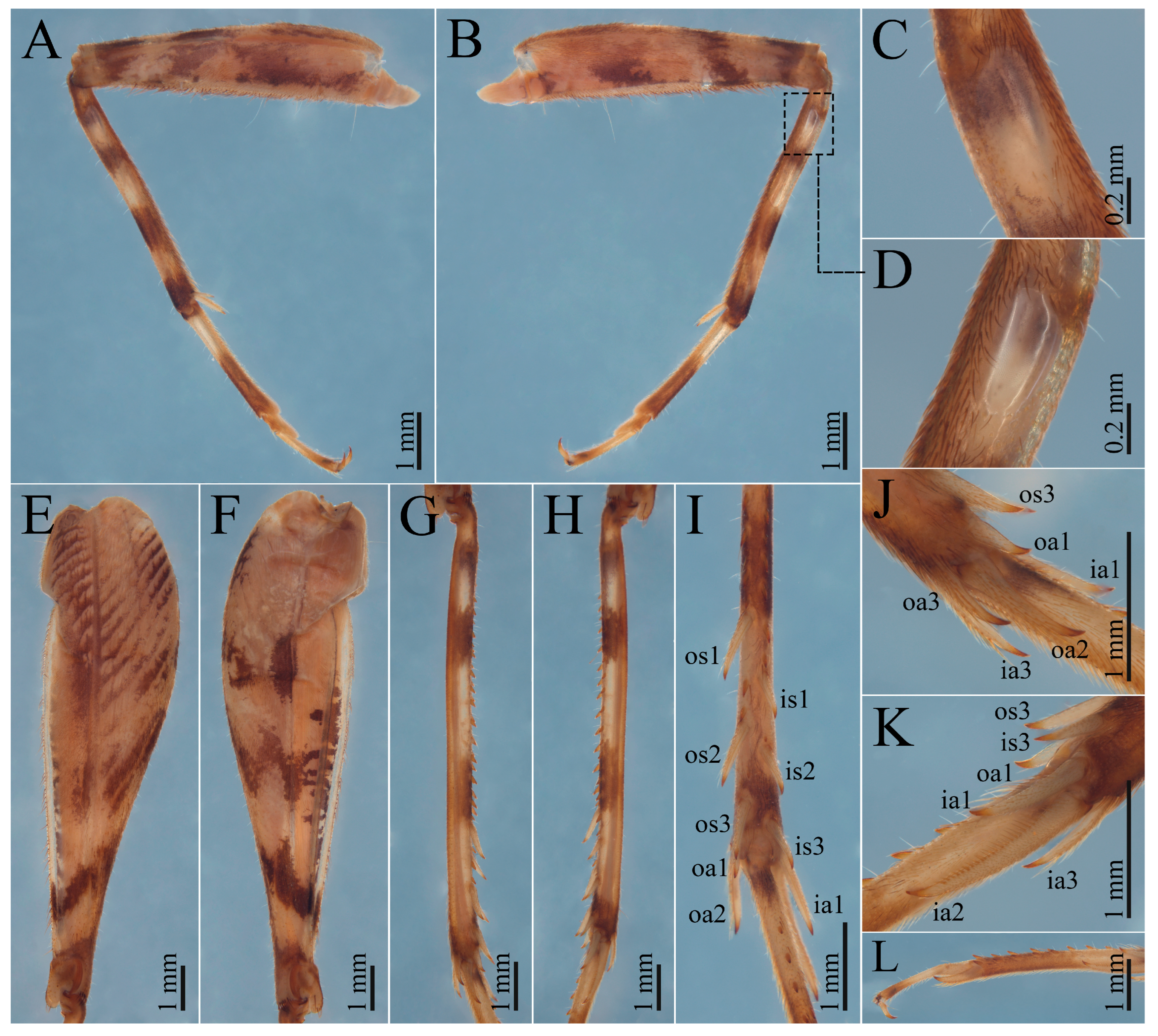
Figure 4G–I and
Figure 9G–I). It further differs from all other Paragryllina, except
Rumea, in possessing two rows of spines on the hind basitarsus (
Figure 4L and
Figure 9L).
Adult males of
Grunagryllus gen. nov. differ from
Aclogryllus,
Alfarogryllus,
Bolivacla,
Dambachia,
Paragryllus, and
Rumea by having forewings that do not extend beyond the abdomen and a mirror crossed by two transverse veins (
Figure 2B,
Figure 3A and
Figure 8B). Additionally, it differs from
Paragryllus in lacking a swollen dorsal inner apical spur on the hind tibia (
Figure 4J,K and
Figure 9J,K), and from
Dambachia in lacking a modified distal subapical spur on the hind tibia (
Figure 4G–K and
Figure 9G–K).
The supra-anal plate of
Grunagryllus gen. nov. is modified, as in
Paragryllus and
Benoistella lyra Gorochov, 2009, but differs from both in being tripartite rather than bipartite (
Figure 2H,I and
Figure 8E).
Regarding the phallic complex,
Grunagryllus gen. nov. differs from
Dambachia in having a laterally symmetrical phallic complex (
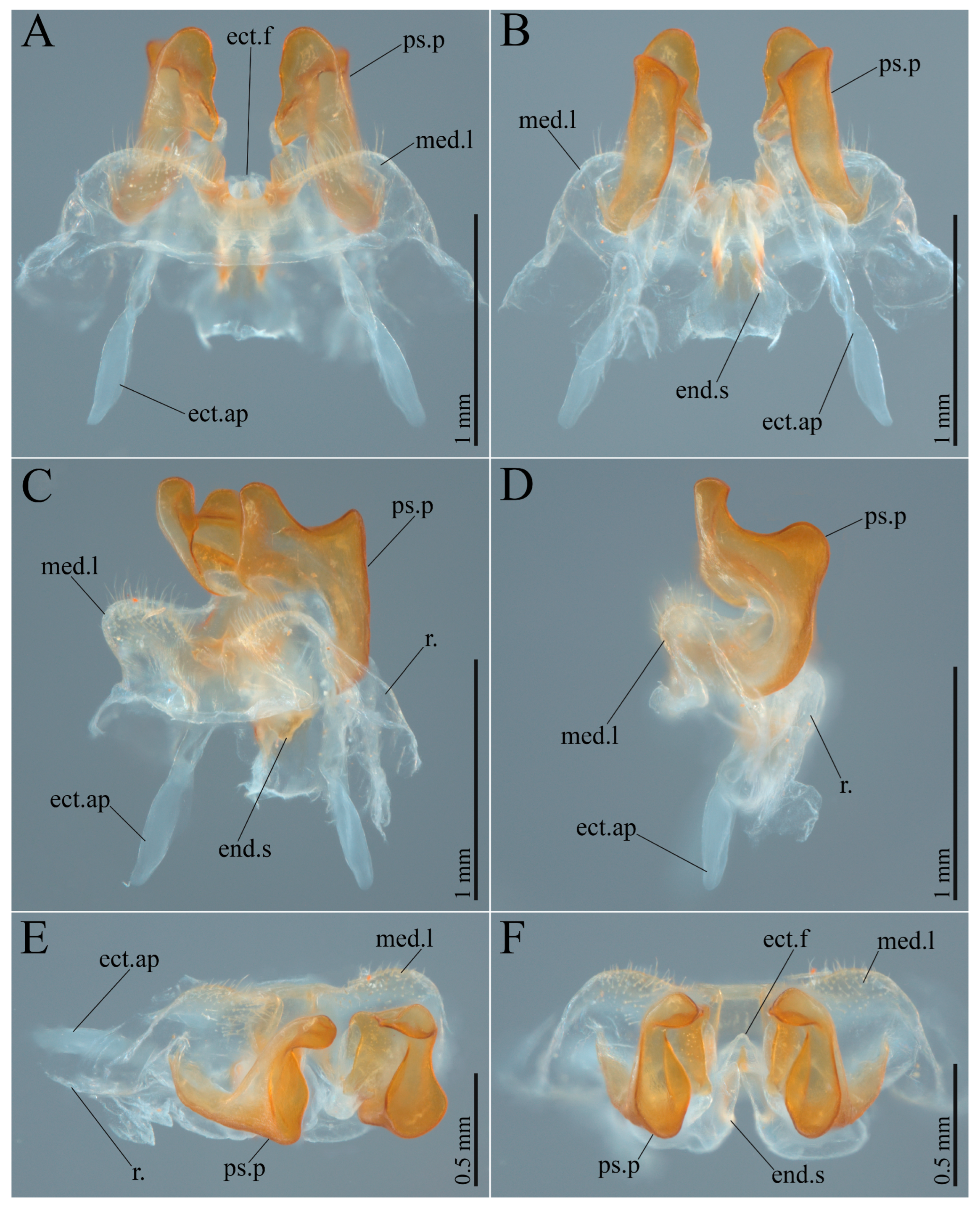
Figure 5 and
Figure 10). The pseudepiphallic parameres differ from those of all other Paragryllina in overall shape: they are considerably shorter than in
Aclogryllus and
Rumea, dorsally curved, differing from
Benoistella and
Izerskya, and do not cross distally, differing from
Paragryllus. Additionally, the median lophi lack ventral or lateral projections, further distinguishing the new genus from
Alfarogryllus and
Paragryllus, respectively. Finally, the ectophallic fold and endophallic sclerites are considerably less developed than in
Aclogryllus and
Bolivacla.
Etymology. The generic name Grunagryllus is derived from gruna, a traditional local term used to refer to caves in the Serra do Ramalho region, southwestern Bahia, where the type species was collected, combined with gryllus (Latin for “cricket”), an element commonly associated with cricket taxa, particularly genera assigned to the subtribe Paragryllina. The name thus alludes both to the subterranean habitat in which the type species was collected and to its taxonomic affinities. The gender of the genus is masculine.
Description. Body coloration ranging from beige to light brown, with dark brow or grey spots and stripes; body size varying from approximately 16 to 21 mm (
Table 1).
Head: pubescent; longer than wide in frontal view; compound eyes developed; lateral ocelli closer to each other than to the median ocellus, which is subapical; fastigium bearing long, thick setae and approximately as wide as the antennal scape; maxillary palp short; apical maxillary palpomeres longer than third and fourth palpomeres, broadened at the tip, with the apex rounded.
Thorax: pronotum pubescent, anterior and posterior margins bearing long, thick setae; pronotum anteriorly and posteriorly irregularly dark brown along the anterior and posterior portions, dorsal area separated from the lateral lobes by a sinuous dark brown stripe extending anteroposteriorly; lateral lobes directed anteriorly; metanotum apparently lacking glandular structures; metasternite with posterior margin broadly arched.
Male right forewing: slightly shorter than or as long as the abdomen; darkly colored, with light-colored crossveins; mirror wider than long, crossed by two arched transverse veins; harp with one anteriorly bifurcated crossvein on the left, followed by four or five thick, simple transverse veins decreasing in length from left to right; stridulatory file present, composed of hundreds of closely spaced teeth; apical area present, moderately elongated; lateral field with two thick, parallel, and closely spaced longitudinal veins, and multiple (16–18) dorsoventral crossveins.
Hindwings: translucent; longer than the forewings.
Legs: femora and tibiae with dark brown rings; foretibiae bearing a well-developed oval tympanum on both faces and a pair of ventral apical spurs, the inner longer than the outer; middle tibiae also with two ventral apical spurs; hind tibiae dorsally serrulated on the proximal and median portions, intersubapical spur margins bearing one or no spines; hind tibiae with three inner (is1–is3) and three outer (os1–os3) subapical spurs concentrated on the distal portion of the tibia, inner spurs shorter than the outer; hind tibiae with three outer (oa1–oa3) and three inner (ia1–ia3) apical spurs; dorsal outer apical spur (oa1) shorter than the ventral (oa3), which, in turn, is approximately half the length of the median outer apical spur (oa2); dorsal (ia1) and ventral (ia3) inner apical spurs subequal and approximately half the length of the median inner apical spur (ia2); subapical and apical spurs lacking any modifications; hind basitarsi with two rows of spines, the inner row composed of 3–5 spines and shorter than the outer row, which bears 5–8 spines; hind basitarsi with a pair of ventral apical spurs, the inner longer than the outer; tarsal claws smooth, symmetrical, and more strongly sclerotized at the apex.
Male abdomen: cerci elongated, pubescent, covered with short, elongate and globose setae, the latter concentrated on the proximal portion of the structure; subgenital plate subtriangular, with the distal margin medially emarginate; supra-anal plate trapezoidal, bearing a tripartite modification in the shape of an inverted lowercase omega (ω).
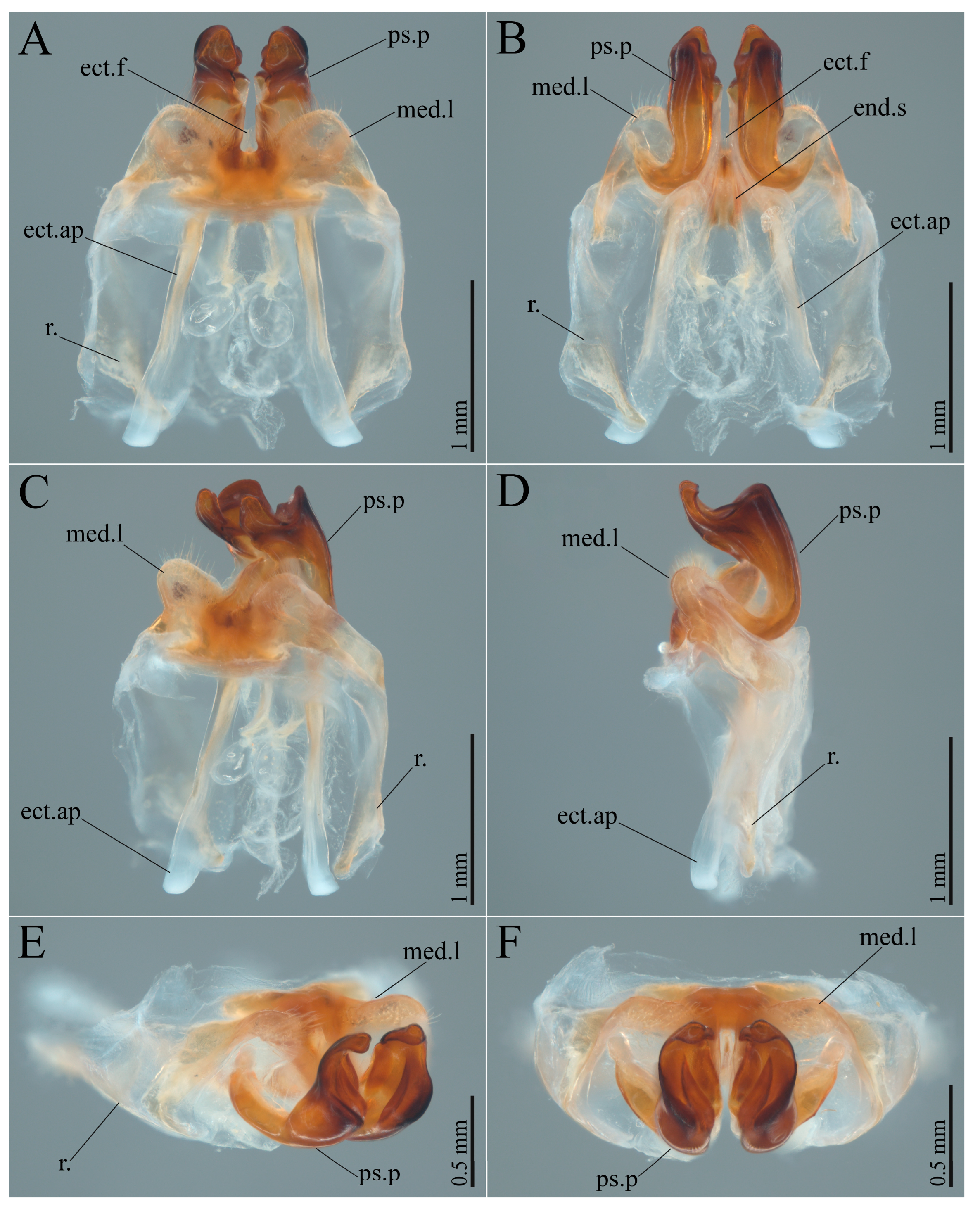
Phallic complex: pseudepiphallic median lophi (med.l) posteriorly rounded, densely covered with long setae, and with a median emargination approximately square or rectangular in shape, with moderately sclerotized margins; med.l lacking lateral projections; pseudepiphallic parameres (ps.p) well developed, with the apex dorsoventrally bifurcated, the inner surface concave, and the dorsal projection more developed than the ventral one, curving posteriorly and inward as a broad hook; ectophallic fold (ect.f) reduced; ectophallic apodemes (ect.ap) well developed; endophallic sclerite (end.s) shaped like an inverted V, with a pair of less sclerotized apodemes extending outwardly.
Figure 2.
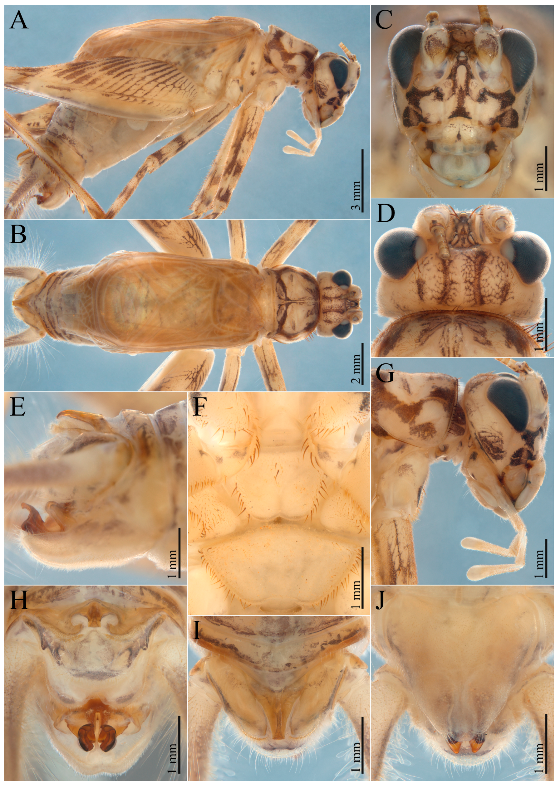
Adult male (ISLA50485, holotype) of Grunagryllus aquaeclarae gen. et sp. nov. (A) Body, lateral view; (B) body, dorsal view; (C) head, frontal view; (D) head, dorsal view; (E) abdominal tip, lateral view; (F) meso- and metasternite, ventral view; (G) head and pronotum, lateral view; (H) abdominal tip, axial view; (I) supra-anal plate, dorsal view; (J) subgenital plate, ventral view.
Figure 2.
Adult male (ISLA50485, holotype) of Grunagryllus aquaeclarae gen. et sp. nov. (A) Body, lateral view; (B) body, dorsal view; (C) head, frontal view; (D) head, dorsal view; (E) abdominal tip, lateral view; (F) meso- and metasternite, ventral view; (G) head and pronotum, lateral view; (H) abdominal tip, axial view; (I) supra-anal plate, dorsal view; (J) subgenital plate, ventral view.
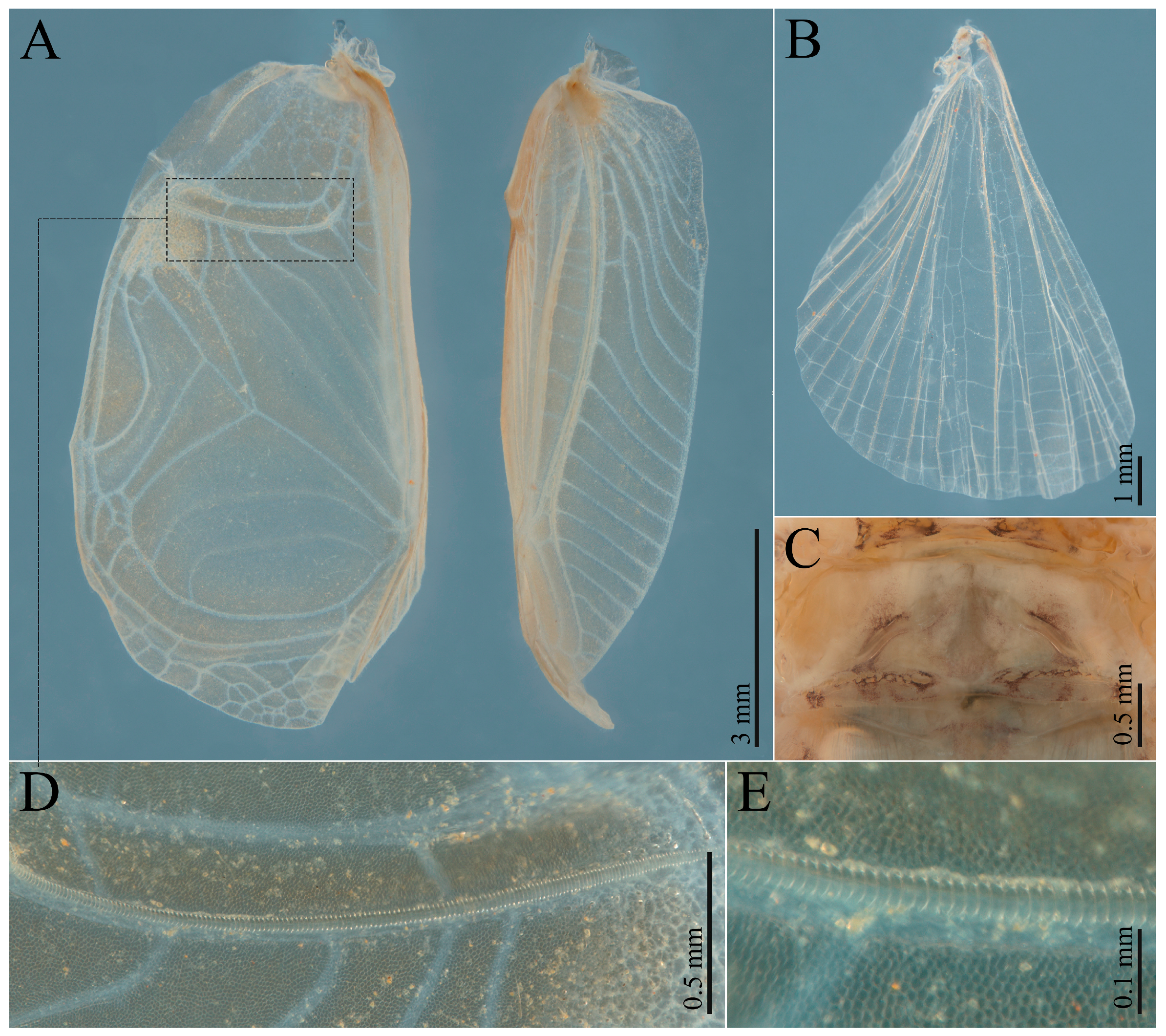
Figure 3.
Wings and metanotum of adult male (ISLA50489, paratype) of Grunagryllus aquaeclarae gen. et sp. nov. (A) Right forewing, dorsal (left) and lateral (right) views; (B) right hind wing; (C) metanotum, dorsal view; (D) stridulatory file, ventral view; (E) stridulatory file teeth, ventral view.
Figure 3.
Wings and metanotum of adult male (ISLA50489, paratype) of Grunagryllus aquaeclarae gen. et sp. nov. (A) Right forewing, dorsal (left) and lateral (right) views; (B) right hind wing; (C) metanotum, dorsal view; (D) stridulatory file, ventral view; (E) stridulatory file teeth, ventral view.
Female right forewing: also darkly colored, with light-colored anastomotic crossveins; lacking specialized areas, except for the lateral field.
Female abdomen: subgenital plate longer than wide, with a deep distal emargination; supra-anal plate trapezoidal and proximally elevated, bearing what appears to be an underdeveloped version of the modification observed in adult males; ovipositor slightly curved dorsally, longer than the hind femur.
Copulatory papilla: subtriangular, with a strongly sclerotized median dorsal carina; anterior ventral margin slightly arcuate.
Grunagryllus aquaeclarae sp. nov. Mendes-Carvalho & Ferreira
Comparative diagnosis. Grunagryllus aquaeclarae gen. et sp. nov. differs from
G. sanctijosephi gen. et sp. nov. by the absence of a broad dark brown stripe in the transitional area between the vertex and gena, extending from the compound eye toward the occiput (present in the latter species) (
Figure 2D,G); less conspicuous dark brown markings on the frons (
Figure 2C); a clypeus bearing a single narrow median dark brown spot rather than multiple irregular dark brown spots (
Figure 2C); a more developed tripartite modification of the supra-anal plate, covering nearly its entire surface (covering approximately one third in
G. sanctijosephi sp. nov.) (
Figure 2E,H,I); a narrower apical portion of the pseudepiphallic parameres (
Figure 5D); apical portion of the pseudepiphallic parameres differently shaped, with the inner concavity bearing a leaf-shaped carina (
Figure 5F); and ectophallic apodemes longer and more developed (
Figure 5B,C).
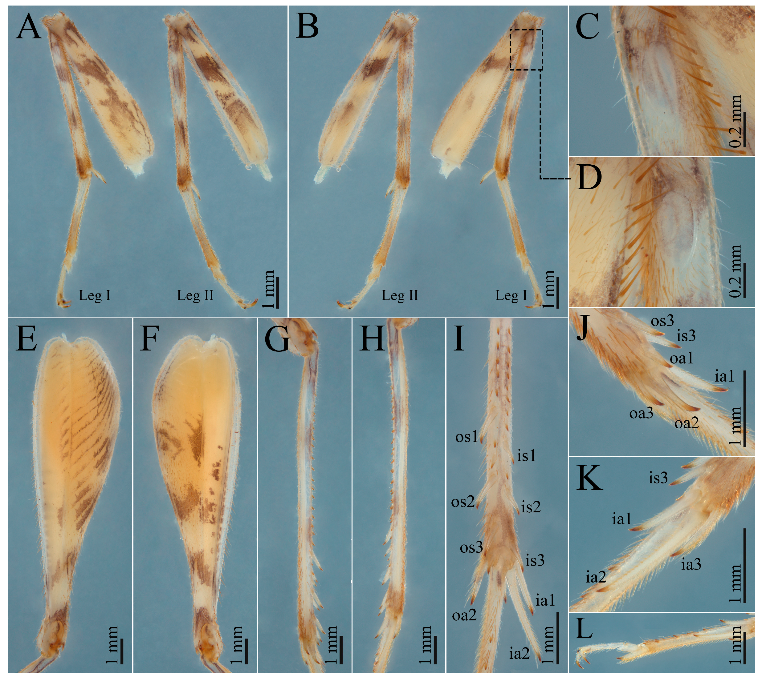
Figure 4.
Legs of adult male (ISLA50489, paratype) of Grunagryllus aquaeclarae gen. et sp. nov. (A) Left fore and middle legs, posterior view; (B) left fore and middle legs, anterior view; (C) tympanum on the foretibia, posterior (outer) view; (D) tympanum on the foretibia, anterior (inner) view; (E) left hind femur, anterior (outer) view; (F) left hind femur, posterior (inner) view; (G) left hind tibia, anterior (outer) view; (H) left hind tibia, posterior (inner) view; (I) left hind tibia, dorsal view; (J) left hind tibia, anterior (outer) apical spurs; (K) left hind tibia, posterior (inner) apical spurs; (L) left hind tarsus. Abbreviations: ia, inner apical spur; is, inner subapical spur; oa, outer apical spur; os, outer subapical spur.
Figure 4.
Legs of adult male (ISLA50489, paratype) of Grunagryllus aquaeclarae gen. et sp. nov. (A) Left fore and middle legs, posterior view; (B) left fore and middle legs, anterior view; (C) tympanum on the foretibia, posterior (outer) view; (D) tympanum on the foretibia, anterior (inner) view; (E) left hind femur, anterior (outer) view; (F) left hind femur, posterior (inner) view; (G) left hind tibia, anterior (outer) view; (H) left hind tibia, posterior (inner) view; (I) left hind tibia, dorsal view; (J) left hind tibia, anterior (outer) apical spurs; (K) left hind tibia, posterior (inner) apical spurs; (L) left hind tarsus. Abbreviations: ia, inner apical spur; is, inner subapical spur; oa, outer apical spur; os, outer subapical spur.
Etymology. The specific epithet aquaeclarae derives from the Latin aquae (“of water”) and clarae (“clear, bright”), meaning “of clear waters.” The name refers to the caves of the Água Clara Cave System, where the type series was collected. The epithet is to be treated as a noun in apposition.
Material examined. Holotype (ISLA50485), male ♂, Lapa dos Peixes I cave (13°48′43.66″ S; 43°57′30.26″ W), São Felix do Coribe, Bahia, Brazil, R.F.V. Cerqueira leg., 14 October 2017; condition: antennae and cerci broken, phallic complex dissected and stored alongside the specimen. Paratype (ISLA50489), male ♂, same data as the holotype; condition: fore and hind wings, fore, middle and hind left legs, fore right leg, and left cercus detached, phallic complex dissected, all structures stored alongside the specimen. Paratype (ISLA155510), female ♀, Gruna da Água Escura I cave (13°49′3.70″ S; 43°57′1.30″ W), Carinhanha, Bahia, Brazil, R.L. Ferreira leg., 17 September 2021; condition: right hind leg missing, cerci and antennae broken, right foreleg detached, copulatory papilla dissected, both stored alongside the specimen.
Figure 5.
Phallic complex of adult male (ISLA50485, holotype) of Grunagryllus aquaeclarae gen. et sp. nov. (A) Dorsal view; (B) ventral view; (C) dorsolateral view; (D) lateral view; (E) lateroaxial view; (F) axial view. Abbreviations: med.l, pseudepiphallic median lophi; ps.p, pseudepiphallic paramere; r., rami; ect.f, ectophallic fold; ect.ap, ectophallic apodeme; end.s, endophallic sclerite.
Figure 5.
Phallic complex of adult male (ISLA50485, holotype) of Grunagryllus aquaeclarae gen. et sp. nov. (A) Dorsal view; (B) ventral view; (C) dorsolateral view; (D) lateral view; (E) lateroaxial view; (F) axial view. Abbreviations: med.l, pseudepiphallic median lophi; ps.p, pseudepiphallic paramere; r., rami; ect.f, ectophallic fold; ect.ap, ectophallic apodeme; end.s, endophallic sclerite.
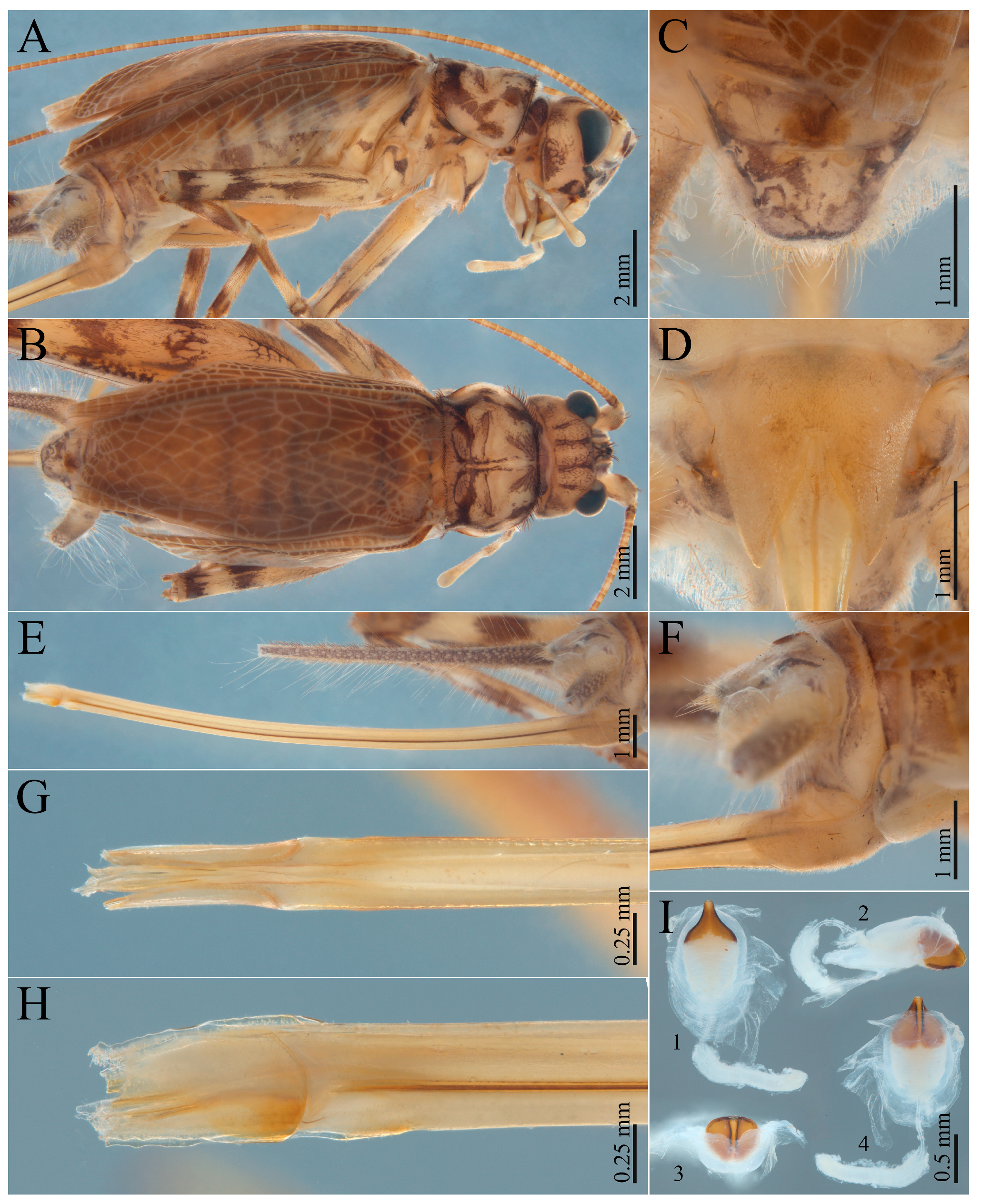
Figure 6.
Adult female (ISLA155510, paratype) of Grunagryllus aquaeclarae gen. et sp. nov. (A) Body, lateral view; (B) body, dorsal view; (C) supra-anal plate, dorsal view; (D) subgenital plate, ventral view; (E) ovipositor, lateral view; (F) abdominal tip, lateral view; (G) ovipositor apex, dorsal view; (H) ovipositor apex, lateral view; (I) copulatory papilla, (1) ventral, (2) lateral, (3) axial, and (4) dorsal views.
Figure 6.
Adult female (ISLA155510, paratype) of Grunagryllus aquaeclarae gen. et sp. nov. (A) Body, lateral view; (B) body, dorsal view; (C) supra-anal plate, dorsal view; (D) subgenital plate, ventral view; (E) ovipositor, lateral view; (F) abdominal tip, lateral view; (G) ovipositor apex, dorsal view; (H) ovipositor apex, lateral view; (I) copulatory papilla, (1) ventral, (2) lateral, (3) axial, and (4) dorsal views.
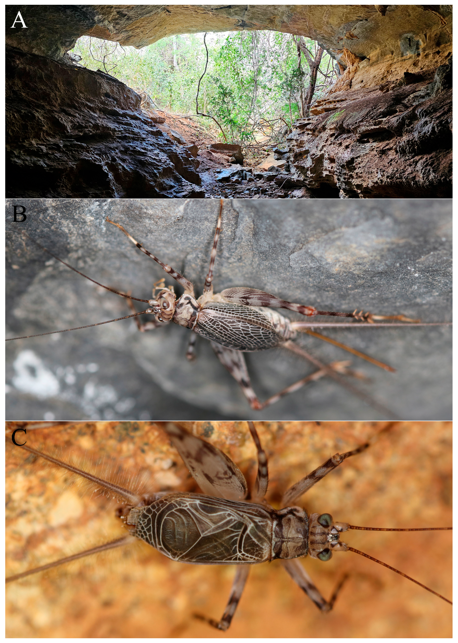
Figure 7.
Habitat and live specimens of Grunagryllus aquaeclarae gen. et sp. nov. (A) Cave entrance, Água Clara Cave System, Serra do Ramalho region; (B) adult female of G. aquaeclarae gen. et sp. nov.; (C) adult male of G. aquaeclarae gen. et sp. nov.
Figure 7.
Habitat and live specimens of Grunagryllus aquaeclarae gen. et sp. nov. (A) Cave entrance, Água Clara Cave System, Serra do Ramalho region; (B) adult female of G. aquaeclarae gen. et sp. nov.; (C) adult male of G. aquaeclarae gen. et sp. nov.
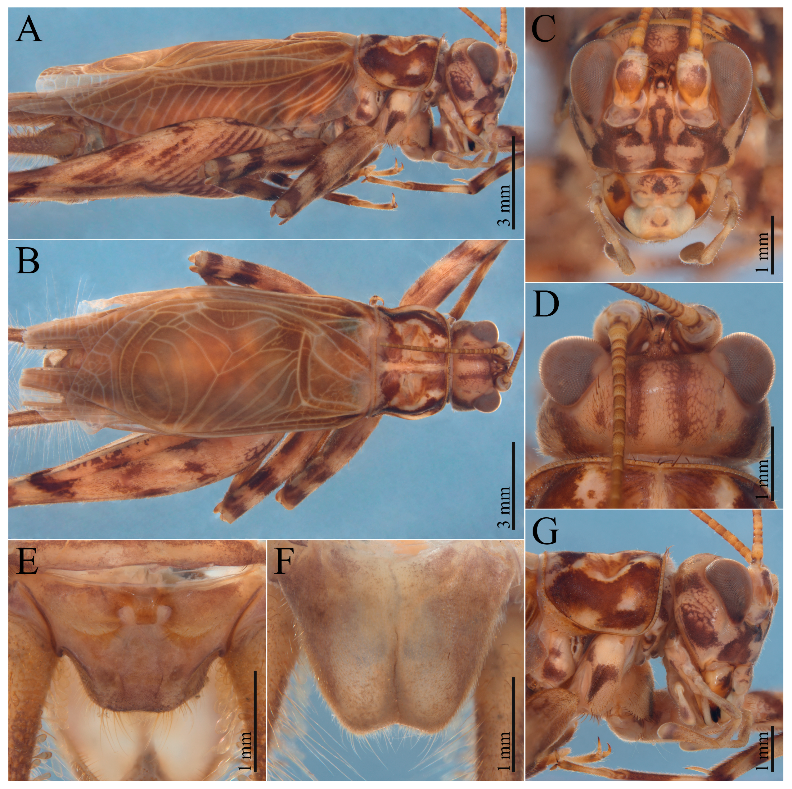
Figure 8.
Adult male (ISLA155526, holotype) of Grunagryllus sanctijosephi gen. et sp. nov. (A) Body, lateral view; (B) body, dorsal view; (C) head, frontal view; (D) head, dorsal view; (E) supra-anal plate, dorsal view; (F) subgenital plate, ventral view; (G) head and pronotum, lateral view.
Figure 8.
Adult male (ISLA155526, holotype) of Grunagryllus sanctijosephi gen. et sp. nov. (A) Body, lateral view; (B) body, dorsal view; (C) head, frontal view; (D) head, dorsal view; (E) supra-anal plate, dorsal view; (F) subgenital plate, ventral view; (G) head and pronotum, lateral view.
Description. Body coloration beige, with dark grey spots and stripes.
Head (
Figure 2A–D,G): pubescent; longer than wide in frontal view; compound eyes developed; antennal scape with a dark brown spot anteriorly; lateral ocelli closer to each other than to the median ocellus, which is subapical; fastigium bearing long, thick setae and approximately as wide as the antennal scape; vertex with a pair of dark brown stripes extending from the occiput toward the fastigium and diverging at both ends, area between the compound eye and the occiput with a dark brown spot; transitional area between vertex and gena without a broad dark brown stripe extending from the compound eye toward the occiput; gena with a posteromedian dark brown spot; frons with a pair of broad, irregular lateral dark brown stripes extending from the compound eye to the clypeus, a median dark brown stripe with a central interruption roughly in the shape of an inverted triangle, lateral stripes connected to the median stripe through a pair of small dark brown spots; clypeus with a single median spot; labrum whitish, without a dark brown spot; mandibles with a median dark brow spot, continuous with the color pattern of the frons; maxillary palp short; apical maxillary palpomeres longer than third and fourth palpomeres, broadened at the tip, with the apex rounded.
Figure 9.
Legs of adult male (ISLA155526, holotype) of Grunagryllus sanctijosephi gen. et sp. nov. (A) Left foreleg, posterior view; (B) left foreleg, anterior view; (C) tympanum on the foretibia, posterior (outer) view; (D) tympanum on the foretibia, anterior (inner) view; (E) left hind femur, anterior (outer) view; (F) left hind femur, posterior (inner) view; (G) left hind tibia, anterior (outer) view; (H) left hind tibia, posterior (inner) view; (I) left hind tibia, dorsal view; (J) left hind tibia, anterior (outer) apical spurs; (K) left hind tibia, posterior (inner) apical spurs; (L) left hind tarsus. Abbreviations: ia, inner apical spur; is, inner subapical spur; oa, outer apical spur; os, outer subapical spur.
Figure 9.
Legs of adult male (ISLA155526, holotype) of Grunagryllus sanctijosephi gen. et sp. nov. (A) Left foreleg, posterior view; (B) left foreleg, anterior view; (C) tympanum on the foretibia, posterior (outer) view; (D) tympanum on the foretibia, anterior (inner) view; (E) left hind femur, anterior (outer) view; (F) left hind femur, posterior (inner) view; (G) left hind tibia, anterior (outer) view; (H) left hind tibia, posterior (inner) view; (I) left hind tibia, dorsal view; (J) left hind tibia, anterior (outer) apical spurs; (K) left hind tibia, posterior (inner) apical spurs; (L) left hind tarsus. Abbreviations: ia, inner apical spur; is, inner subapical spur; oa, outer apical spur; os, outer subapical spur.
Thorax (
Figure 2A,B,F,G, and
Figure 3C): pronotum pubescent, anterior and posterior margins bearing long, thick setae; pronotum anteriorly and posteriorly irregularly dark brown along the anterior and posterior portions, dorsal area separated from the lateral lobes by a narrow sinuous dark brown stripe extending anteroposteriorly; lateral lobes directed anteriorly; metanotum apparently lacking glandular structures; metasternite with posterior margin broadly arched.
Male right forewing (
Figure 2A,B, and
Figure 3A,D,E): slightly shorter than or as long as the abdomen; darkly colored, with light-colored crossveins; mirror wider than long, crossed by two arched transverse veins; harp bearing one anteriorly bifurcated crossvein on the left, followed by four (paratype ISLA155510) or five (holotype, ISLA50485) thick, simple transverse veins decreasing in length from left to right; stridulatory file present, composed of 179 closely spaced teeth (n = 1); apical area present, moderately elongate; lateral field with two thick, parallel, and closely spaced longitudinal veins, and multiple (16–18) dorsoventral crossveins.
Male left forewing: similar to the right forewing but mirrored, less sclerotized, and with less conspicuous venation; stridulatory file absent.
Hindwings (
Figure 3B): translucent; longer than the forewings.
Figure 10.
Phallic complex of adult male (ISLA155526, holotype) of Grunagryllus sanctijosephi gen. et sp. nov. (A) Dorsal view; (B) ventral view; (C) dorsolateral view; (D) lateral view; (E) lateroaxial view; (F) axial view. Abbreviations: med.l, pseudepiphallic median lophi; ps.p, pseudepiphallic paramere; r., rami; ect.f, ectophallic fold; ect.ap, ectophallic apodeme; end.s, endophallic sclerite.
Figure 10.
Phallic complex of adult male (ISLA155526, holotype) of Grunagryllus sanctijosephi gen. et sp. nov. (A) Dorsal view; (B) ventral view; (C) dorsolateral view; (D) lateral view; (E) lateroaxial view; (F) axial view. Abbreviations: med.l, pseudepiphallic median lophi; ps.p, pseudepiphallic paramere; r., rami; ect.f, ectophallic fold; ect.ap, ectophallic apodeme; end.s, endophallic sclerite.
Legs (
Figure 4A–L): femora and tibiae with dark brown rings; foretibiae bearing a well-developed oval tympanum on both faces and a pair of ventral apical spurs, the inner longer than the outer; middle tibiae also with two ventral apical spurs; hind tibiae dorsally serrulated on the proximal and median portions, intersubapical spur margins bearing one or no spines; hind tibiae with three inner (is1–is3) and three outer (os1–os3) subapical spurs concentrated on the distal portion of the tibia, inner spurs shorter than the outer; hind tibiae with three outer (oa1–oa3) and three inner (ia1–ia3) apical spurs; dorsal outer apical spur (oa1) shorter than the ventral (oa3), which, in turn, is approximately half the length of the median outer apical spur (oa2); dorsal (ia1) and ventral (ia3) inner apical spurs subequal and approximately half the length of the median inner apical spur (ia2); subapical and apical spurs lacking any modifications; hind basitarsi with two rows of spines, the inner row composed of 3–5 spines and shorter than the outer row, which bears 5–8 spines; hind basitarsi with a pair of ventral apical spurs, the inner longer than the outer; tarsal claws smooth, symmetrical, and more strongly sclerotized at the apex.
Male abdomen (
Figure 2A,B,E,H–J): cerci elongated, pubescent, covered with short, elongate and globose setae, the latter concentrated on the proximal portion of the structure; subgenital plate subtriangular, with the distal margin medially emarginate; supra-anal plate trapezoidal, bearing a tripartite modification in the shape of an inverted lowercase omega (ω) covering almost entirely the dorsal surface of the supra-anal plate.
Phallic complex (
Figure 5A–F): pseudepiphallic median lophi (med.l) posteriorly rounded, densely covered with long setae, and with a median emargination approximately rectangular in shape, with moderately sclerotized margins; med.l lacking lateral projections; pseudepiphallic parameres (ps.p) well developed, with the apex dorsoventrally bifurcated, the inner surface concave with a conspicuous leaf-shaped carina, and the dorsal projection more developed than the ventral one, curving posteriorly and inward as a broad hook with a tapered apex; ectophallic fold (ect.f) reduced; ectophallic apodemes (ect.ap) well developed, longer than the rami, which curve inward, over the ect.ap; endophallic sclerite (end.s) shaped like an inverted V, with a pair of less sclerotized apodemes extending outwardly.
Female right forewing (
Figure 6A,B): also darkly colored, with light-colored anastomotic crossveins; lacking specialized areas, except for the lateral field.
Female abdomen (
Figure 6A–H): subgenital plate longer than wide, with a deep distal emargination; supra-anal plate trapezoidal and proximally elevated, bearing what appears to be an underdeveloped version of the modification observed in adult males; ovipositor slightly curved dorsally, longer than the hind femur, with a yellowish coloration that may indicate the specimen had recently molted; apex broken.
Copulatory papilla (
Figure 6I): subtriangular, with a strongly sclerotized median dorsal carina; anterior ventral margin slightly arcuate.
Ecological remarks. Specimens of
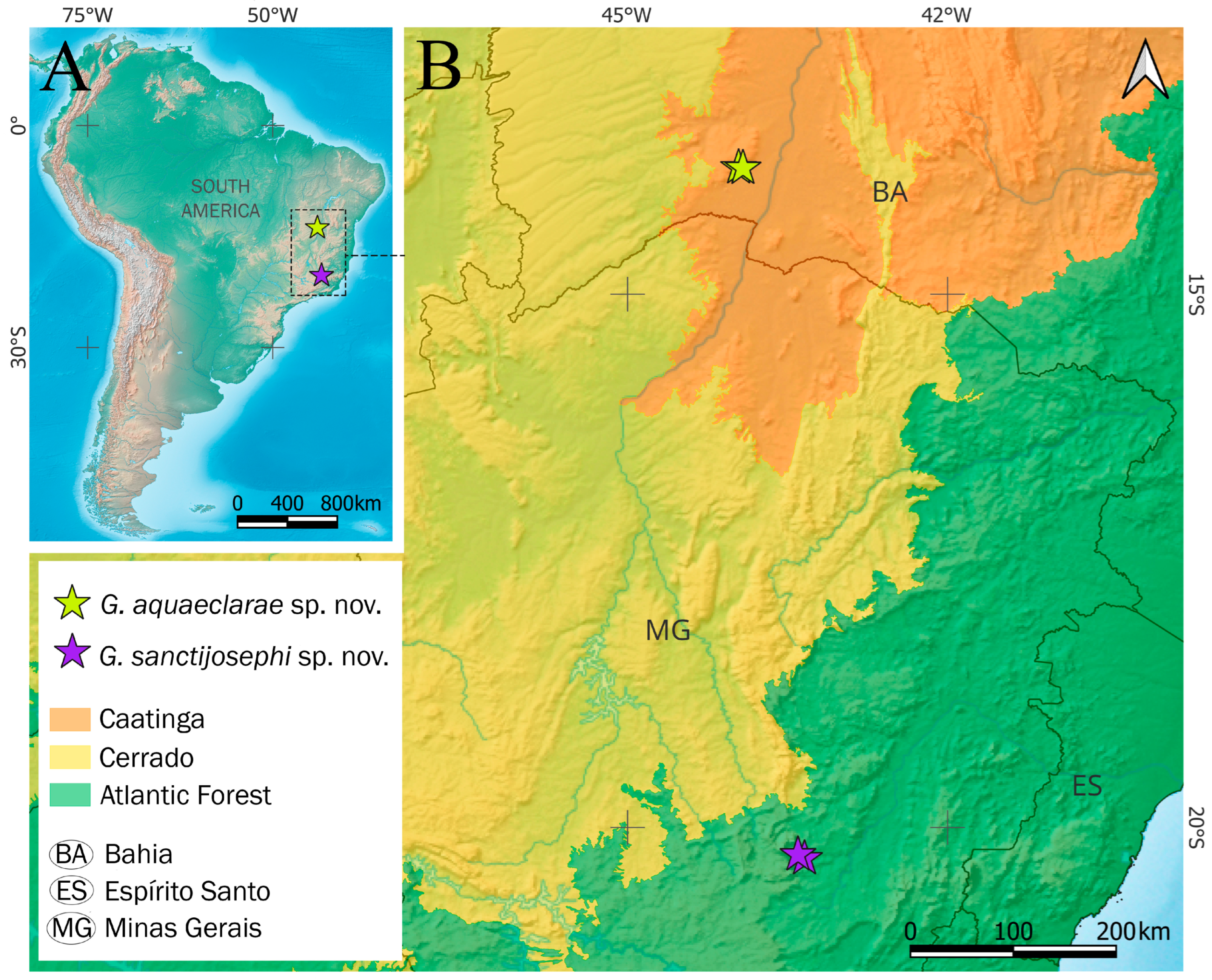
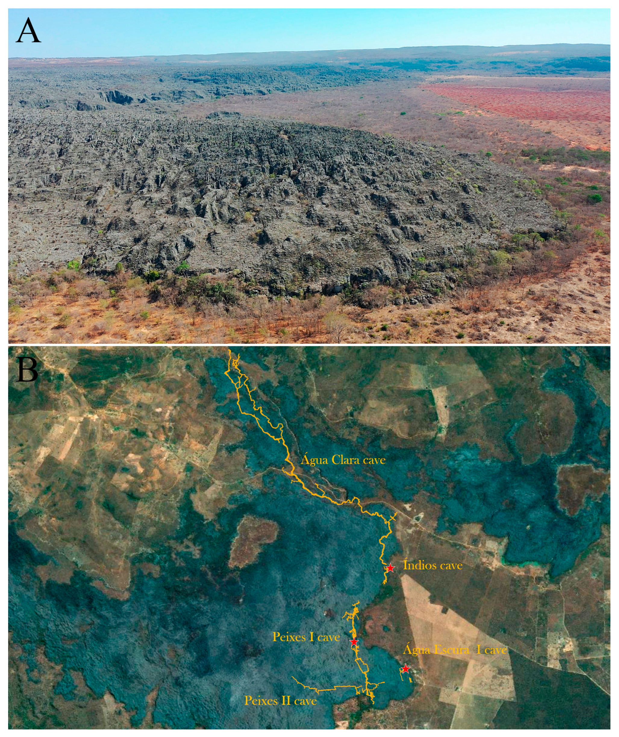
Grunagryllus aquaeclarae gen. et sp. nov. were recorded within caves of the Água Clara Cave System (ACCS), a hydrologically integrated karst network located along the eastern margin of the Serra do Ramalho massif (
Figure 11A). The ACCS comprises a series of large, interconnected conduits, including the Água Clara (13,880 m), Índios (510 m), Lapa dos Peixes I (9320 m), and Lapa dos Peixes II (2100 m) caves (
Figure 11B). A detailed account of the geological structure and hydrological dynamics of the ACCS is provided by Ferreira et al. [
30].
Individuals of
G. aquaeclarae gen. et sp. nov. were observed exclusively in entrance-associated sectors, particularly in Lapa dos Peixes I (
Figure 7A), Gruta dos Índios and Gruna da Água Escura I caves (
Figure 11B). Specimens were typically found resting on cave walls within disphotic zones (
Figure 7B,C), where light levels are reduced but not entirely absent. Individuals remained motionless in the presence of approaching observers, allowing close inspection, but responded to minor disturbances with abrupt jumping movements toward sheltered microhabitats, indicating a combination of low baseline activity and rapid escape responses. Notably, no specimens were recorded in aphotic zones, suggesting that the species does not occupy the deeper, permanently dark sectors of the caves. Instead, its occurrence appears to be restricted to ecotonal environments near cave entrances.
Although several caves in the surrounding region were surveyed, G. aquaeclarae gen. et sp. nov. was detected only within the ACCS. This pattern may indicate a geographically restricted distribution, potentially encompassing both subterranean and adjacent epigean habitats, or alternatively, a species that utilizes cave entrances opportunistically as refuges. However, the relatively high number of individuals observed in the three occupied caves (despite the limited number of collected specimens) suggests that these entrance zones are not used sporadically, but rather constitute regularly exploited microhabitats. It is important to emphasize that adjacent surface environments were not included in the sampling design, preventing a comprehensive assessment of the species’ full distributional range. Further studies are needed to better understand the ecological relationship between the new species and subterranean environments.
Grunagryllus sanctijosephi sp. nov. Mendes-Carvalho & Ferreira
Comparative diagnosis. Grunagryllus sanctijosephi gen. et sp. nov. differs from
G. aquaeclarae gen. et sp. nov. by the presence of a broad dark brown stripe in the transitional area between the vertex and gena, extending from the compound eye toward the occiput (absent in the latter species) (
Figure 8D,G); more conspicuous dark brown markings on the frons (
Figure 8C); a clypeus bearing multiple irregular dark brown spots rather than a single narrow median dark brown spot (
Figure 8C); a less developed tripartite modification of the supra-anal plate, covering approximately one third of its surface (covering nearly its entire surface in
G. aquaeclarae sp. nov.) (
Figure 8E); a broader apical portion of the pseudepiphallic parameres (
Figure 10D); apical portion of the pseudepiphallic parameres differently shaped, with the inner concavity lacking a leaf-shaped carina (
Figure 10F); and ectophallic apodemes shorter and less developed (
Figure 10B,C).
Etymology. The specific epithet sanctijosephi derives from the Latin sancti (“of Saint”) and josephi (“of Joseph”), meaning “of Saint Joseph.” The name refers to International Workers’ Day, traditionally associated with Saint Joseph the Worker, on which the holotype was fortuitously discovered among material deposited in the Collection of Subterranean Invertebrates of Lavras (ISLA). The epithet is to be treated as a noun in apposition.
Material examined. Holotype (ISLA155526), male ♂, GER-0051 cave (20°17′32.6″ S; 43°22′23.6″ W), Mariana, Minas Gerais, Brazil, R.A. Castro-Souza leg., 10 October 2018; condition: antennae and cerci broken, right forewing detached, left hind femur slightly damaged, abdominal apex damaged prior to the present study, phallic complex previously dissected and stored alongside the specimen. Other material examined: (ISLA155527), juvenile male ♂, CMR-0207 cave (20°15′21.2″ S; 43°24′06.9″ W), Mariana, Minas Gerais, Brazil, R.A. Castro-Souza leg., 3 July 2019.
Description. Body coloration light brown, with dark brown spots and stripes; body size slightly smaller than G. aquaeclarae gen. et sp. nov.
Head (
Figure 8A–D,G): pubescent; longer than wide in frontal view; compound eyes developed; antennal scape with a dark brown spot anteriorly; lateral ocelli closer to each other than to the median ocellus, which is subapical; fastigium bearing long, thick setae and approximately as wide as the antennal scape; vertex with a pair of dark brown stripes extending from the occiput toward the fastigium and diverging at both ends, area between the compound eye and the occiput with a dark brown spot; transitional area between vertex and gena with a broad dark brown stripe extending from the compound eye toward the occiput; gena with a posteromedian dark brown spot; frons with a pair of broad, irregular lateral dark brown stripes extending from the compound eye to the clypeus, a median dark brown stripe with a central interruption roughly in the shape of an inverted triangle, lateral stripes connected to the median stripe through a pair of conspicuous dark brown spots; clypeus with multiple brown spots; labrum light yellow, with a small dark brown spot; mandibles with a median dark brow spot, continuous with the color pattern of the frons; maxillary palp short; apical maxillary palpomeres longer than third and fourth palpomeres, broadened at the tip, with the apex rounded.
Thorax (
Figure 8A,B,G): pronotum pubescent, anterior and posterior margins bearing long, thick setae; pronotum anteriorly and posteriorly irregularly dark brown along the anterior and posterior portions, dorsal area separated from the lateral lobes by a broad sinuous dark brown stripe extending anteroposteriorly; lateral lobes directed anteriorly; metanotum apparently lacking glandular structures; metasternite with posterior margin broadly arched.
Male right forewing (
Figure 8A,B): as long as the abdomen; darkly colored, with light-colored crossveins; mirror wider than long, crossed by two arched transverse veins; harp bearing one anteriorly bifurcated crossvein on the left, followed by four thick, simple transverse veins decreasing in length from left to right; stridulatory file present, composed of 208 closely spaced teeth (n = 1); apical area present, moderately elongate; lateral field with two thick, parallel, and closely spaced longitudinal veins, and multiple (16–18) dorsoventral crossveins.
Male left forewing: similar to the right forewing but mirrored and less sclerotized; stridulatory file absent; mirror crossed by two arched transverse veins, the posterior one incomplete, not reaching the left margin and instead connected to the posterior margin by two short longitudinal veins.
Hindwings: translucent; longer than the forewings.
Legs (
Figure 9A–L): femora and tibiae with dark brown rings; foretibiae bearing a well-developed oval tympanum on both faces and a pair of ventral apical spurs, the inner longer than the outer; middle tibiae also with two ventral apical spurs; hind tibiae dorsally serrulated on the proximal and median portions, intersubapical spur margins bearing one or no spines; hind tibiae with three inner (is1–is3) and three outer (os1–os3) subapical spurs concentrated on the distal portion of the tibia, inner spurs shorter than the outer; hind tibiae with three outer (oa1–oa3) and three inner (ia1–ia3) apical spurs; dorsal outer apical spur (oa1) shorter than the ventral (oa3), which, in turn, is approximately half the length of the median outer apical spur (oa2); dorsal (ia1) and ventral (ia3) inner apical spurs subequal and approximately half the length of the median inner apical spur (ia2); subapical and apical spurs lacking any modifications; hind basitarsi with two rows of spines, the inner row composed of 5 spines and shorter than the outer row, which bears 8 spines; hind basitarsi with a pair of ventral apical spurs, the inner longer than the outer; tarsal claws smooth, symmetrical, and more strongly sclerotized at the apex.
Male abdomen (
Figure 8E,F): cerci elongated, pubescent, covered with short, elongate and globose setae, the latter concentrated on the proximal portion of the structure; subgenital plate subtriangular, with the distal margin medially emarginate; supra-anal plate trapezoidal, bearing a tripartite modification in the shape of an inverted lowercase omega (ω) covering one third of the dorsal surface of supra-anal plate.
Phallic complex (
Figure 10A–F): pseudepiphallic median lophi (med.l) posteriorly rounded, densely covered with long setae, and with a median emargination approximately square in shape, with moderately sclerotized margins; med.l lacking lateral projections; pseudepiphallic parameres (ps.p) well developed, with the apex dorsoventrally bifurcated, the inner surface concave, and the dorsal projection more developed than the ventral one, anteroposteriorly broadened, curving posteriorly and inward as a broad hook; ectophallic fold (ect.f) reduced; ectophallic apodemes (ect.ap) well developed; endophallic sclerite (end.s) shaped like an inverted V, with a pair of less sclerotized apodemes extending outwardly.
Female: Unknown.
Ecological remarks. Specimens of
Grunagryllus sanctijosephi gen. et sp. nov. were recorded in two quartzite caves situated on a mountaintop and surrounded by shrub and arboreal campo rupestre vegetation [
31]. As the specimens were collected during environmental licensing surveys conducted by biologists associated with environmental consulting companies, detailed information on the ecology and behavior of the species is unfortunately unavailable. As with
G. aquaeclarae gen. et sp. nov., further studies will be necessary to clarify the ecological relationship between the new species and subterranean environments.