
Article

Nymphal Development, Morphology and Life History of the Madeiran Endemic Planthoppers Cyphopterum retusum and C. fauveli (Hemiptera: Flatidae): Taxonomic and Conservation Implications

1
Faculdade de Ciências da Vida, Universidade da Madeira, 9000-390 Funchal, Madeira, Portugal
2
CIBIO, Centro de Investigação em Biodiversidade e Recursos Genéticos, Universidade do Porto, Campus de Vairão, Rua Padre Armando Quintas, nº 7, Vairão, 4485-661 Porto, Portugal
*
Author to whom correspondence should be addressed.
Taxonomy 2026, 6(3), 44; https://doi.org/10.3390/taxonomy6030044
Submission received: 20 May 2026 / Revised: 25 June 2026 / Accepted: 1 July 2026 / Published: 15 July 2026

Abstract

The Macaronesian archipelagos harbor a highly endemic fauna of the flatid genus Cyphopterum Melichar, 1905, characterized by pronounced insular diversification. Despite approximately 80% of its species being unique to the region, the morphology and biology of their immature stages remain virtually unknown. This lack of ontogenetic data has long hindered more thorough studies on these planthoppers. To address this gap, the present study provides the first comprehensive description of all five nymphal instars of Cyphopterum retusum (Walker, 1851) and C. fauveli Noualhier, 1897 from the Madeira archipelago. Nymphs were reared under controlled laboratory conditions to analyze developmental duration, survival rates, and morphological features. Our results demonstrate that nymphal morphology offers robust diagnostic characters. Specifically, body size, species-specific coloration, and integumentary textures distinguish the two species, whereas body size combined with the number of metatibial spines and tarsomeres and the number and arrangement of sensory pits, allow for reliable instar determination. These findings fill a significant gap in the knowledge of the family Flatidae and provide essential tools for the conservation and monitoring of these endemic lineages in vulnerable Macaronesian ecosystems.

1. Introduction

Oceanic islands are widely regarded as natural laboratories of evolution, offering exceptional opportunities to study fundamental processes such as colonization, geographic isolation, and adaptive radiations [1,2,3]. Insects illustrate these dynamics remarkably well, as many lineages undergo extensive insular radiations often accompanied by high levels of endemism and ecological specialization [4,5]. Within the Macaronesian archipelagos—Azores, Madeira, Salvagens, Canary Islands, and Cape Verde—such patterns are especially pronounced, reflecting both complex geological histories and long-term isolation [6,7,8,9].
The flatid planthopper genus Cyphopterum Melichar, 1905, provides a compelling model for studying this diversification. Distributed throughout the Southwestern Palaearctic, this genus comprises 45 valid species, 80% of which are endemic to Macaronesian archipelagos [10,11,12,13,14,15,16,17]. While species richness peaks in the Canary Islands (26 species), the Madeira archipelago serves as a critical area for understanding the genus’ evolutionary reach, being home to two endemic species: Cyphopterum retusum (Walker, 1851) and Cyphopterum fauveli Noualhier, 1897.
Despite their evolutionary significance, the taxonomy of Cyphopterum remains particularly challenging. Species identification within the genus often relies on subtle morphologically differences, frequently requiring the examination of male genitalia, such as the aedeagal appendages and the tenth abdominal segment [11,18]. While some species are more easily distinguishable, others—particularly the Madeiran endemics, present significant diagnostic difficulties. In these cases, male genitalia may exhibit significant intraspecific variation or overlapping forms rendering diagnosis dependent on body size and broad habitat preferences, traits that have never been rigorously assessed [18]. Historically, C. retusum is described as a larger, polyphagous species widely distributed on the main island of Madeira often associated with shrubs like Rubus spp. [16,18]. In contrast, C. fauveli appears restricted to coastal halophytic vegetation in eastern Madeira, the Desertas, and Porto Santo islands [16,18].
This lack of taxonomic clarity combined with a deficit of ecological data, directly hinders conservation efforts. Currently, no species of Cyphopterum has been evaluated under IUCN criteria, largely due to an insufficient understanding regarding population dynamics, biology, host-plant associations, and precise geographic ranges. The absence of data on immature stages furthers complicates the protection of these endemic lineages, as many habitats in Madeira are under increasing pressure from tourism and urban development. Therefore, the study of immature stages is not merely a descriptive endeavor but a requirement for effective conservation. In Fulgoromorpha, nymphal characters can provide valuable diagnostic information that complements adult morphology, potentially offering more stable traits for certain groups [19,20]. Furthermore, accurate recognition of nymphal instars is essential for interpreting life cycles and phenology, ensuring that management strategies protect the resources necessary for the species’ entire ontogeny.
To date, no descriptions of nymphal instars exist for any species of Cyphopterum. This reflects a broader deficiency within the Flatidae (ca 1500 species) [10], where descriptions of immatures are scarce and mainly restricted to species of economic importance [21,22,23,24]. The present study aims to fill this critical gap by providing the first detailed descriptions of the nymphal instars of C. retusum and C. fauveli, alongside data on their development under laboratory conditions. These data provide a necessary foundation for future taxonomic and ecological research and contribute to a broader understanding of the diversification and conservation of insular Flatidae.

2. Materials and Methods

2.1. Study Area

The Madeira archipelago is of volcanic origin, located in the North Atlantic Ocean (32°22′–33°10′ N; 16°16′–17°16′ W). It comprises the islands of Madeira (741 km2; max. elevation 1862 m) and Porto Santo (42 km2; max. elevation 517 m), separated by approximately 43 km, and the Desertas Islands (14 km2) [25].
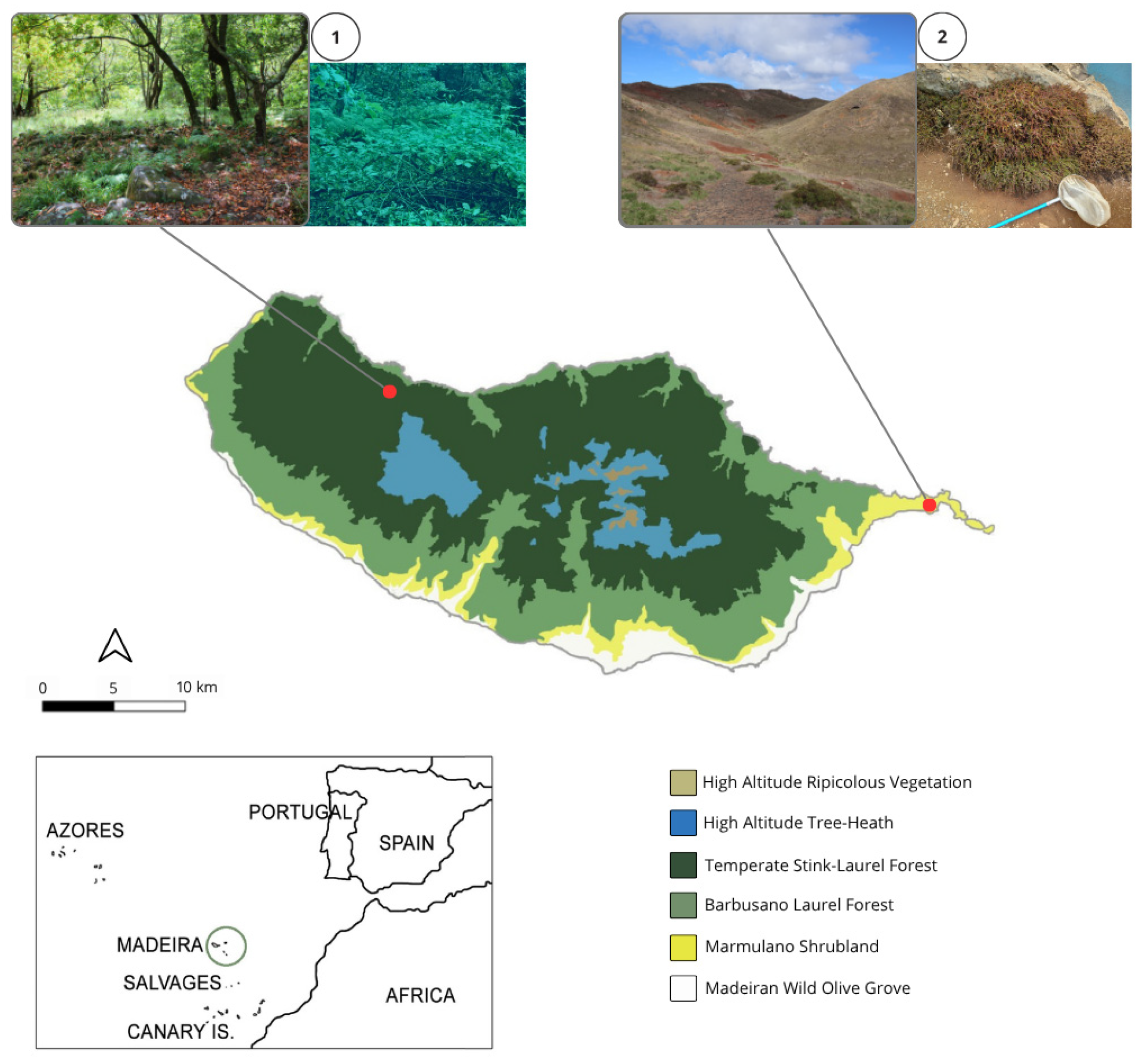
Madeira Island is characterized by a central mountain ridge, oriented east–west, creating a marked climatic contrast between the northern and southern slopes. The northern sector, exposed to prevailing trade winds, experiences higher precipitation, while the southern sector is comparatively drier. Approximately 20% of Madeira is covered by the Laurissilva (laurel forest), a UNESCO World Heritage dominated by native evergreen trees of Lauraceae such as Ocotea foetens (Aiton) Baill. (1870) and Laurus novocanariensis Rivas Mart., Lousã, Fern. Prieto, E.Días, J.C.Costa & C.Aguiar (2002) (Figure 1). In contrast, the eastern peninsula (Ponta de São Lourenço), and the Desertas islands constitute a semi-arid zone with xerophilous vegetation, notably halophytic shrubs of Suaeda vera Forssk. ex J.F.Gmel. (1776) (Amaranthaceae).

2.2. Sampling and Laboratory Rearing

Nymphs were collected in mid-April from two ecologically distinct localities in the Madeira island (Figure 1): Chão da Ribeira (Laurissilva), where C. retusum was found on Rubus cf. bollei Focke, 1887 (Rosaceae); and Ponta de São Lourenço (semi-arid peninsula), where C. fauveli was sampled from Suaeda vera using a sweep-net. A total of 155 nymphs were collected in Chão da Ribeira, and 66 in Ponta de São Lourenço.
Host plant material and nymphs were collected in the field and transported to the laboratory inside cool boxes; plant stems were kept in a container with water during transit. In the laboratory, plants were maintained in the shade with their stems in water. For the experimental setup, small young twigs (approx. 10 cm)—were selected because nymphs are typically observed on them—were removed from the host plant branches, placed in water-filled microtubes (Eppendorf), and wrapped in cotton wool to ensure plant hydration and prevent nymphs from falling into the water.
Each twig-microtube unit was then placed inside an individual transparent plastic container cup (10.5 cm height × 7 cm diameter) covered with fine mesh, into which the nymphs and the twigs were introduced. The insects were kept under natural photoperiod, and twigs were replaced with fresh ones 2–3 times per week, or whenever they were no longer in good condition. Nymphs were monitored daily around 16:00 h to record colour variation and the presence of exuviae; environmental temperature and humidity were also recorded (Table S1). Freshly cast exuviae and specimens that died during the study were preserved in 70% ethanol with 2% glycerol for later identification. Plant identification followed Press and Short [26].

2.3. Morphological Description and Morphometrics

A total of 88 C. retusum and 38 C. fauveli specimens was examined for morphological description and morphometric analysis (Table 1).
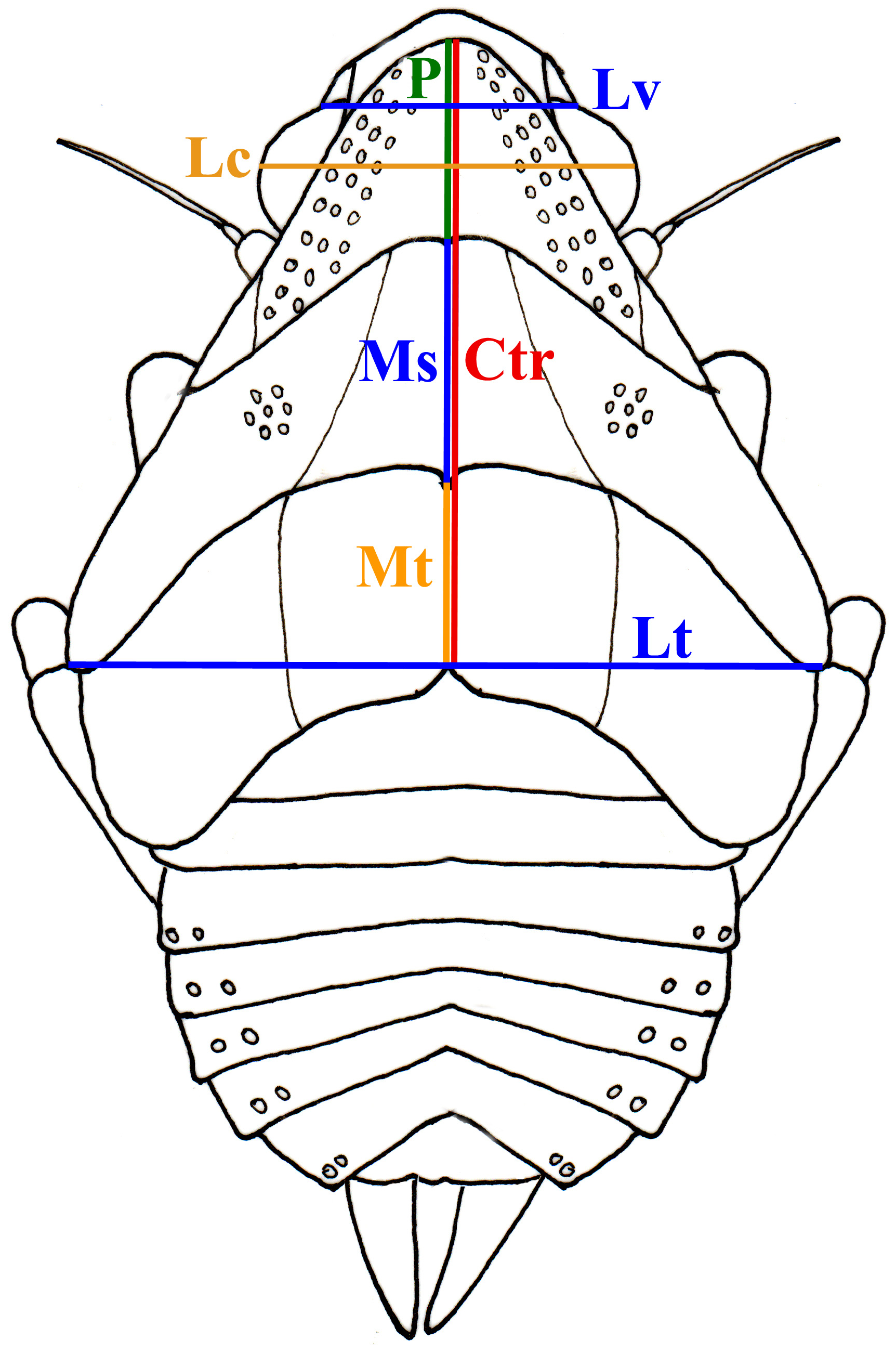
Morphometric measurements were based on live specimens. For this purpose, live laboratory-reared specimens were photographed upon their arrival at the laboratory and following each molt, using an Olympus DP11 digital camera coupled to an Olympus SZ60 stereomicroscope (Olympus Corporation, Tokyo, Japan). Measurements were taken using the Olympus Camedia Master (Olympus Corporation, Tokyo, Japan) and Olympus DP-Soft (version 3.0) image analysis software systems (Olympus Corporation, Münster, Germany). Seven morphometric parameters were measured for each specimen (Figure 2): Lt: Maximum body width; Ctr: Total thorax length (from the anterior margin of the pronotum to the posterior margin of the metanotum); Lc: Head width; Lv: Maximum width of vertex; P: Midline length of the pronotum; Ms: Midline length of the mesonotum; Mt: Midline length of the metanotum.
Description, Illustrations and Image Processing
The description of the first nymphal instar was based on field-captured nymphs. The remaining instars were described based on both laboratory-reared and field-captured specimens. All insects used for formal descriptions were preserved in 70% ethanol with 2% glycerin. The description of each instar was based on a variable number of individuals, and coloration was cross-checked using photographs of live specimens. Photographs of nymphs were stacked using Zerene Stacker (version 1.04), and plates were prepared with Adobe Photoshop CS6.
Regarding the nymphal illustrations, detailed drawings were prepared from photographs of live nymphs taken during morphometric analysis. Final illustrations were drawn with India ink on tracing paper. Only the sensory pits clearly visible from a strictly dorsal perspective were included; those located on the extreme lateral margins or pleural regions may not be represented.

2.4. Data Analysis

Statistical analyses were performed to compare body measurements among instars of the same and of different species. Data normality was assessed using Kolmogorov–Smirnov test, and homogeneity of variances followed Fowler and Cohen [27]. When the assumption of homogeneity was violated, Levene’s correction was applied. Student’s t-tests were employed for pairwise group comparisons; however, the first and second instar of C. fauveli were excluded from these analyses due to an insufficient number of specimens resulting from total mortality during these early stages.
Pearson’s correlation coefficient demonstrated strong positive relationships between all variables (Ctr, P, Ms, Mt, Lc, Lv, and Lt). Since the variable Ctr corresponds to the sum of measurements P, Ms, and Mt, the latter three were excluded from subsequent analyses to avoid redundancy. Among all measured variables, Ctr and Lc showed the lowest correlation with each other. Therefore, Ctr was selected as the primary metric for the identification key due to its stability (indicated by the smallest CV) and ease of measurement.

3. Results

3.1. General Nymphal Development and Morphology

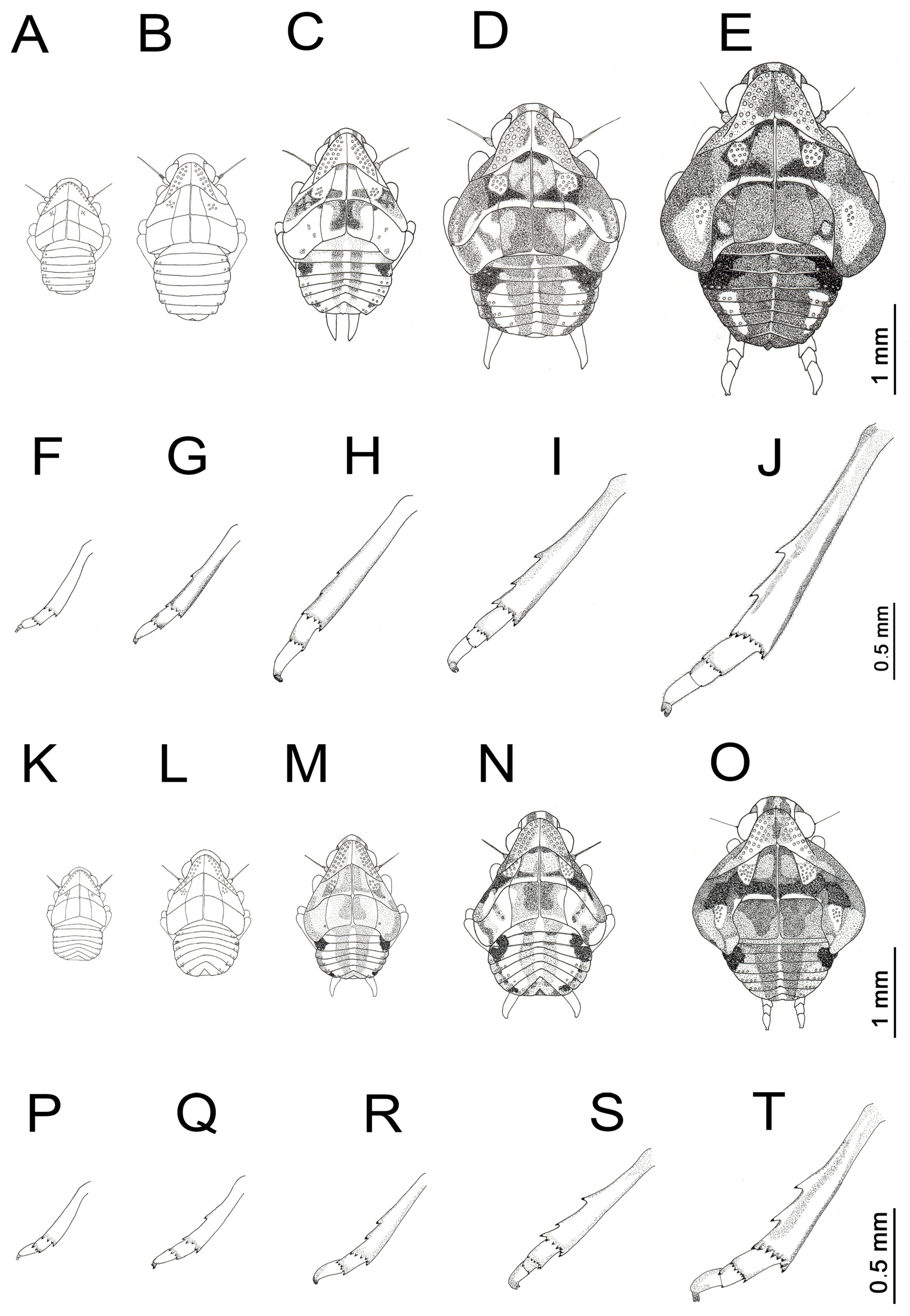
The nymphal development comprises five instars, characterized by a consistent increase in total body dimensions and wing pad (thoracic paranotal expansions) enlargement. A key diagnostic feature is the ontogenetic development of the hind legs and tarsi. Specifically, the number of tarsomeres increases from the two in the 1st, 2nd, and 3rd instars, to three in the 4th to 5th instars. Additionally, lateral metatibial spines develop progressively: they are absent in the 1st instar, a single spine emerges in the 2nd instar, and two spines are present from the 3rd instar onwards; furthermore, metatibial apical spines increase in number throughout the subsequent instars (Figure 3).
Ontogenetic shifts in pigmentation are significant as the nymphs mature. The progression of pigmentation from light to dark phenotypes is not random; instead, it follows a fixed pattern categorized into three main phases:
  • Instars 1–2 (Pale Phase): Base coloration is cream or pale ivory, with brown markings nearly absent. By the 2nd instar, pigmentation, when present, is limited to vestigial punctations on the metanotum and small abdominal eyespots.
  • Instars 3–4 (Transition Phase): Contrast increases and species-specific dark markings become clearer. Cyphopterum fauveli displays a highly contrastive “checkerboard” pattern on the thorax, defined by the intersection of dark blocks and the clean, pale median carina. In contrast, C. retusum exhibits a distinct, sharp geometric marking on the mesonotum, often resembling ‘X’ shape of dark fuscous pigment against the pale background. In the third instar the four longitudinal abdominal stripes become defined and by the 4th instar, paracentral abdominal markings may begin to coalesce, especially in C. retusum.
  • Instar 5 (Melanic Phase): Dark phenotypes exhibit significant coalescence of dark pigmentation, with expanding mesonotal markings. Abdominal bands often fuse across multiple segments, giving the nymph a predominantly fuscous appearance. This melanism is particularly pronounced in C. retusum, while in C. fauveli the pattern remains slightly more fragmented.
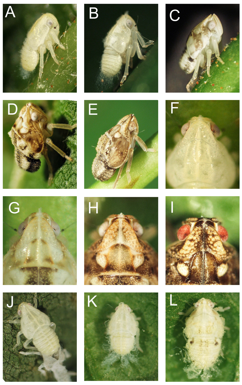
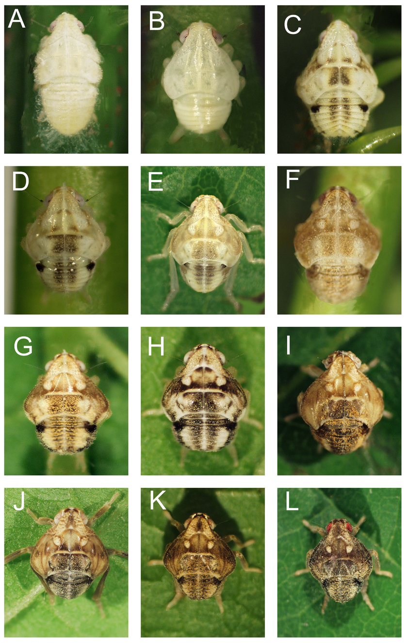
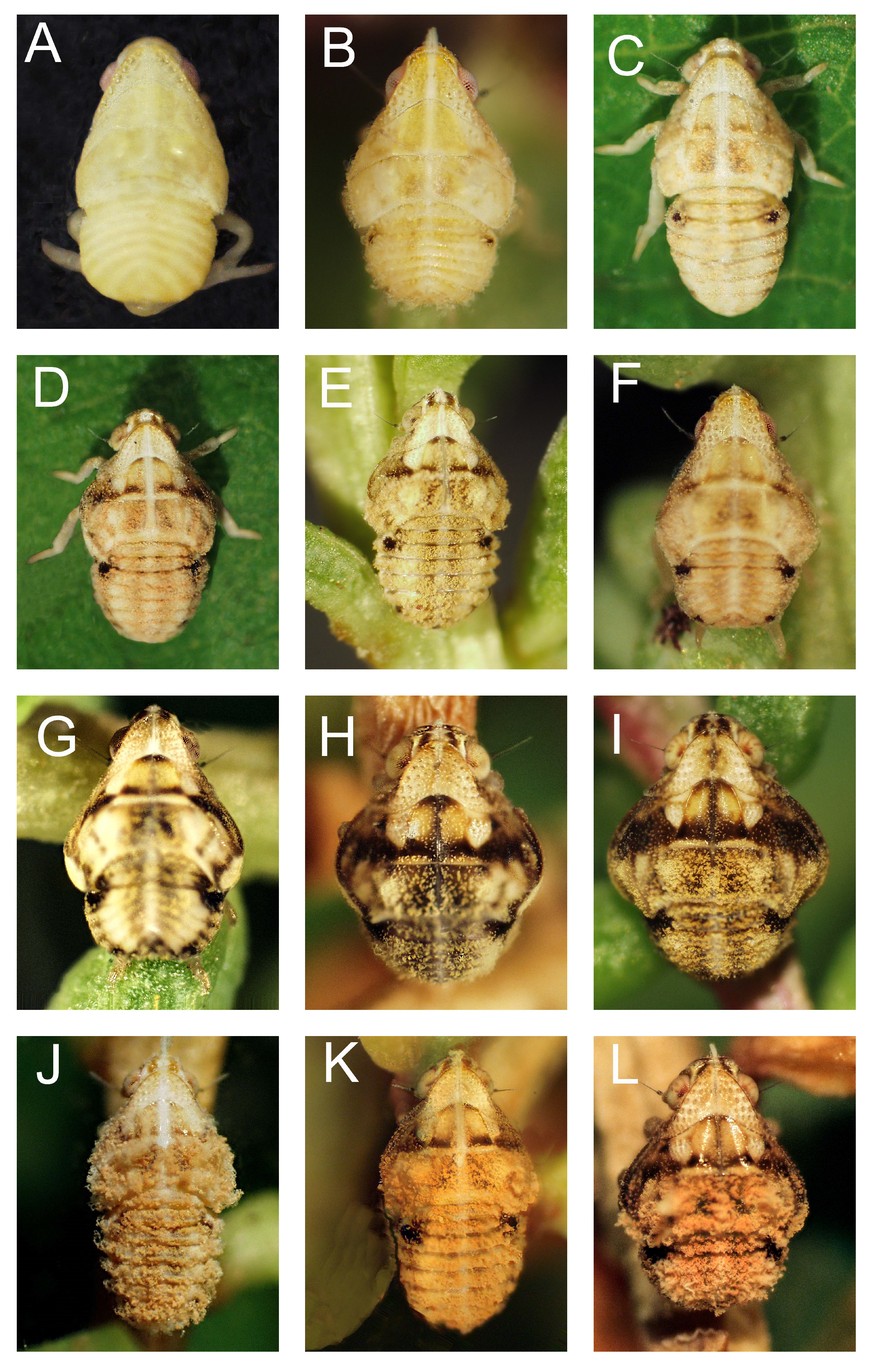
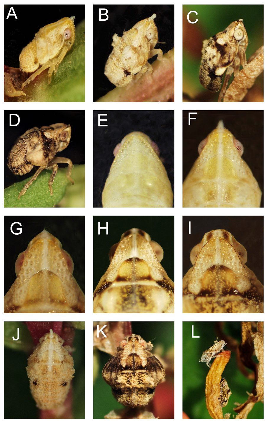
The coloration of the tibiae consistently follows these primary body patterns throughout all developmental stages. Detailed morphological features and pigmentation patterns for C. retusum are illustrated in Figure 4 and Figure 5, while the corresponding characteristics for C. fauveli are presented in Figure 6 and Figure 7.

3.2. Systematic Descriptions

3.2.1. Cyphopterum retusum (Walker, 1851)

(Figure 3A–J, Figure 4A–L and Figure 5A–L)
Issus retusus Walker, 1851: 371 [28].
Cyphopterum curvipenne Walker, 1858: 45 [29] (mislabelled locality: “Port Philip”, Australia); synonymized by Distant, 1910: 316 [30]
Cyphopterum obtusatum Melichar, 1923: 93 [31] (mislabelled locality: “Cape Verde”); synonymized by Leise, 1992: 8 [18]
Cyphopterum retusum: Melichar, 1905: 477 [32]; China, 1938: 54 [33]; Lindberg, 1961: 56 [16]; Leise, 1992: 260 [18].
Cyphopterum retusum (misidentifications):
—Lallemand, 1928: 10 [34], Seabra, 1930a: 260 [35], 1930b: 359 [36], 1939a: 175 [37], 1941: 12 [38] (all records from mainland Portugal, misidentified and subsequently corrected to C. adscendens (Herrich-Schäeffer, 1835) by Leise [18]).
First Instar (Figure 3A,F, Figure 4A and Figure 5A)
Body measurements (mm). CTR = 0.68; LC = 0.45; LV = 0.31; LT = 0.76
Body. Broadly oval and subcylindrical, widest across the metathorax (Figure 3A). In lateral view, thorax slightly humped, tapering posteriorly (Figure 5A). Coloration uniformly pale yellowish white to creamy, covered in fine white powdery wax (Figure 4A).
Head. Vertex short, wider than long, and rounded anteriorly, with carinae on the anterolateral margins. Frons more or less oval, broadest just beneath eyes, anterior margin convex and ventral margin concave; three carinae present (two marginal, one medial), with a longitudinal row of sensory pits between lateral and outer carina. Rostrum three-segmented, extending to metacoxae. Eyes globular and reddish-brown. Antennae three-segmented; first and second antennomere cylindrical; third segment small, globose with a black, bearing a bristle-like flagellum (Figure 5A).
Thorax. Semi-oval, divided by a mid-dorsal line into three pairs of plates; wing pads absent (Figure 3A). Pronotum subtriangular, with an anterolateral carina curving posterolaterally, and a row of ca. 10 pits parallel to the outer margin; posterior border sinuate; medial carina extending the entire length of the thorax. Mesonotum with a midline length like that of pronotum; posterior margin slightly sinuate, with two longitudinal carinae configuring two sub-rectangular plates. Outer half of each mesonotal plate bearing a cluster of about 5 pits arranged in a circle located midway between the midline and the lateral margin (Figure 3A); the lateral-most pits are not visible from strictly dorsal view. Metanotum sub-rectangular, also divided by two plates, each subdivided by a longitudinal carinae. Legs whitish. All tarsi with the second tarsomere subconical and slightly curved, terminating in a claw. Metatibiae bearing an apical row of four black-tipped spines ventrally (Figure 3F). Metatarsi two-segmented; metatarsomere I cylindrical, bearing an apical row of four apical, black-tipped spines ventrally.
Abdomen. Subcylindrical, nine-segmented, pale yellowish without markings, widest across segments 2–4. Segments 8–9 telescoped, giving abdomen a truncate, flattened appearance caudally (Figure 4A). Tergites with marginal pits; delicate long waxy filaments often present at the caudal end.
Second Instar (Figure 3B,G, Figure 4B, Figure 5B,F,J,K)
Body measurements (mm). CTR = 0.87; LC = 0.55; LV = 0.36; LT = 1.00.
Body: Shape like 1st instar but more robust. Thoracic hump more pronounced (Figure 5B). Coloration predominantly whitish, though subtle darker yellowish shading may appear on thoracic plates (Figure 3B and Figure 4B).
Head. Vertex yellowish or whitish (Figure 5F).
Thorax. Pronotum with two rows of ca. 13 sensory pits, and a group of small pits located ventrally to the eyes (not seen in dorsal view) (Figure 3B and Figure 4B). Mesonotum slightly longer than pronotum at its widest point, with ca. five pits on the median part of each plate. Metanotum wider, with midline length equal to that of mesonotum; similar to the previous instar, except for the presence of a few pits on the outer lateral margin. Legs pale. Metatibia now bears a medial, black-tipped spine in addition to an apical row of five black-tipped spines (Figure 3G). Metatarsomere I with five black-tipped spines.
Abdomen. increased number of sensory pits on lateral margins. A faint eyespot may appear on the basal lateral margin. Waxy filaments often longer than in previous stage (Figure 5K). Waxy filaments absent in recently molted specimens (Figure 5J).
Otherwise, similar to 1st instar.
Third instar (Figure 3C,H, Figure 4C,D and Figure 5C,G,L)
Body measurements (mm). CTR =1.14; LC = 0.69; LV = 0.47; LT = 1.38
Body: Significant colour transition. Coloration remains pale to yellow with a distinct whitish line from the head to the abdomen and dorsal surface with a geometric pattern of dark fuscous maculation on the thorax (Figure 3C and Figure 4C).
Head. Vertex more defined, often with two faint brownish mottling posteriorly (Figure 5G). Frons with brownish carinae.
Thorax. Wing pads emerge as small lateral expansions; lateral margin of mesonotum covering approximately one-third of that of the metanotum (Figure 3C). Pronotum often with brownish along lateral carinae and below eyes, with three rows of ca. 23 pits, and more sensory pits ventrally to the eyes (not visible in dorsal view) than in the second instar (Figure 3C and Figure 5G). Mesonotum with four central dark brown spots, two upper and two lower, flanking the midline between the medial carina; brownish wing pads; each plate with a cluster of ca. 7 pits arranged in a whitish circle located at the level of eyes, and few along the outer margins of the wing pads (Figure 4C,D). Metanotum with two large, sub-rectangular “X” dark maculae, between the medial carina more saturated toward the posterior edge, corresponding to the pattern on the mesonotum. Legs light beige with dark spotting at the joints (Figure 5C). Metatibia with two lateral black-tipped spines and an apical row of six spines. Metatarsomere I with a row of six apical spines (Figure 3H).
Abdomen. Four longitudinal dark strips (two paracentral and two lateral) create a striped effect, with the paracentral corresponding to the pattern on the metanotum. A distinct, circular black eyespot is present on each side of the third visible abdominal segment, continuing as two thin lateral strips across the subsequent segments (Figure 4C). Terminal segments predominantly pale (Figure 5L). Lateral margins with a higher number of sensory pits than in 2nd instar (Figure 3C).
Colour variation. The size of the spots throughout the body and the intensity of the coloration are highly variable. There are pale forms (ivory or creamy-white with faint brownish markings; Figure 5L), intermediate forms (the standard phenotype, with contrast between the pale ground and well-defined dark brown macula), and dark (melanic) forms with saturated and expanded dark brown pigment (Figure 4C) often overing a larger surface area, sometimes merging into continuous dark bands across the mesonotum.
Fourth Instar (Figure 3D,I, Figure 4E–H and Figure 5D,H)
Body measurements (mm). CTR = 1.56; LC = 0.88; LV = 0.61; LT = 1.91
Body. Shape wider than 3rd instar. Strongly mottled, with basic pattern similar to that of the 3rd instar but with darker and more extensive brown maculae. Some specimens with a distinct whitish midline extending from the head to the abdomen, and longitudinal abdominal stripes more defined (Figure 3D). Colouration remains highly variable in both the number of spots and intensity (Figure 4E–H).
Head. Vertex with two dark spots; central and lateral carinae whitish (Figure 5H).
Thorax. Wing pads significantly elongated, covering half of the metanotum margin, with brownish veins visible (Figure 5D). Pronotum with marginal carina brownish and central carina carinae whitish; anterior half with brown markings or entirely whitish; each plate with three rows of ca. 19 pits, and with more sensory pits ventrally to the eyes (not visible in dorsal view) than in the third instar (Figure 3D). Mesonotum plates dark, with posterior margin whitish at the middle and two distinct circular whitish maculae at the level of eyes, bearing ca. 11 sensorial pits; wing pads dark brownish (Figure 4F,H). Metanotum with two large central dark maculae between medial carina, corresponding to those on the mesonotum, flanked by distinct whitish circular spots, often adjacent to some mottle brown markings; margins whitish (Figure 4H). Legs yellowish-brown, darker on femora and tibiae. Tarsi now three-segmented. Metatibia with two apical spines and an apical row of 7 spines (Figure 3I). Metatarsomere I with seven black-tipped apical spines; metatarsomere II with two apical spines.
Abdomen. With contrasting longitudinal dark stripes and circular eyespots on the margins of third visible segment, highly visible regardless of the nymph’s overall pigmentation; in darker specimens, these stripes continue as two large lateral dark bands. Pale segments form two distinct oval whitish strips next to the paracentral dark strips, which are larger in some specimens (Figure 4H). Number of lateral sensory pits increased compared to the third instar (Figure 3C).
Otherwise, similar to 3rd instar.
Fifth Instar (Figure 3E,J, Figure 4I–L and Figure 5E,I)
Body measurements (mm). CTR = 2.06; LC = 1.12; LV = 0.81; LT = 2.73
Body. Body sub-romboidal in dorsal view; widest across the wing pads (Figure 3E). Dorsal color pattern distinctly reticulate and mosaiform, characterized by a complex network of irregular dark brown striae and dots overlaid upon a lighter ocreous background, particularly on the pronotum and anterior abdomen (Figure 4I–L).
Head. Vertex short, with symmetrical dark brownish infusions that accentuate its height; lateral carina whitish. Frons darker.
Thorax. Wing pads fully developed; lateral margin of the mesonotum completely overlapping and extending beyond the lateral margin of the metanotum, reaching the third visible abdominal segment (Figure 4I–J); distinct veins towards the apices. Pronotum with a dark brown spot along the central carina; each plate with ca. 24 pits arranged in four rows; number of sensory pits to ventral to eyes greater than in the fourth instar. Mesonotum distinctly humped (Figure 5E); dark brown pigmentation confined to the depressions between carinae and along wing pads; two distinct circular whitish maculae present at the level of eyes, bearing ca. 11 pits, and ca. 7 pits located along the wind pad margin. Metanotum dark, with two-dot pattern on the lateral portion. Legs. Brown. Metatibiae and metatarsomere I each with 8 apical spines (Figure 3J)., metatarsomere II with two apical spines.
Abdomen. Longitudinal bands often reaching their highest contrast (dark brown to fuscous), with two distinct oval whitish maculae adjacent to the paracentral dark strips. Terminal segments generally paler (Figure 4I–L), possessing a higher number of marginal sensory pits than the fourth instar.
Colour variation. Ranges from Ochreous Form (warm-brownish-red tone with less distinct; Figure 4I–J) to a Dark Form (dorsal surface nearly black; Figure 4K–L) but may exhibit variation in the number of spots and in the intensity of pigmentation.
Otherwise, like 4th instar.
Integumental Wax in C. retusum
In Cyphopterum retusum, the secretory strategy is characterized by a high degree of localization. In early instars (1st and 2nd), wax is primarily emerging as discrete, elongated white filaments at the anal extremity (Figure 5A,B,K) and more or less rare dorsally, leaving the ivory-colored integument exposed. From the third instar onwards (Figure 4E–L), waxy secretions remain restricted. In the final instars (Figure 4I–L), despite the variation between heavily “peppered” and highly melanic forms, the cuticle maintains a relatively dry and polished appearance. Unlike its congener, the sensory pits and the sharp carinae of the head and thorax in C. retusum remain clearly defined and are not obscured by flocculent secretions, even in the most mature stages (Figure 4K–L and Figure 5I).
Remarks. Cyphopterum retusum has a history of extensive misidentification and geographical errors in the literature. Historical records from mainland Portugal (e.g., Leiria) by Lallemand [34] and Seabra [35,36,37,38] were shown to be based on specimens of C. adscendens (Herrich-Schäeffer, 1835) (see [18]).
Further confusion arises from the type locality of C. curvipenne Walker, 1858, cited as “Port Philip”. Although Distant [30] synonymized it with C. retusum without examining the type material, the locality refers to Victoria, Australia—a region where the genus Cyphopterum does not occur. It is highly probable that the specimen was mislabelled by its donor, Captain Parry, during his travels from Madeira to Australia.
Similarly, C. obtusatum Melichar, 1923, described from a single female allegedly from Cape Verde [33], was synonymized with C. retusum by Leise [18]. Morphological examination revealed no differences between the two, and biogeographical evidence supports a mislabelling: all other Cape Verdean Cyphopterum according to Lindberg belong to the subgenus Phocypterum Lindberg, 1962, making the presence of a Cyphopterum s. str. in that archipelago unlikely. This conclusion is further reinforced by the fact that intensive sampling in Cape Verde by Lindberg failed to rediscover the species.

3.2.2. Cyphopterum fauveli (Noualhier, 1897)

(Figure 3K–T, Figure 6A–L and Figure 7A–L)
Hysteropterum fauveli Noualhier, 1897: 79 [39]
Cyphopterum fauveli: Melichar, 1905: 476 [32]; China, 1938: 53 [33]; Lindberg, 1961: 58 [16]; Leise, 1992: 259 [18]
Cyphopterum fauveli (misidentifications):
—Horváth, 1909: 299 [40] (records from La Palma, Canary Islands, misidentified and subsequently corrected to C. grossum Lindberg, 1954 [13] by Leise, 1992 [18])
—Lallemand, 1929: 8 [41]; Seabra, 1930a: 259 [35], 1930b: 359 [36], 1939a: 175 [37], 1941: 12 [38] (all records from mainland Portugal, misidentified and subsequently corrected to C. adscendens (Herrich-Schäeffer, 1835) by Leise [18]).
First Instar (Figure 3K,P, Figure 6A and Figure 7E)
Body measurements (mm). CTR = 0.45; LC = 0.37; LV = 0.34; LT = 0.56
Body. Broadly oval, elongate, and subcylindrical, widest across the metathorax (Figure 3K). In lateral view, thorax slightly humped. Colouration uniformly pale stramineous (Figure 6A).
Head. Vertex short, rounded anteriorly, and distinctively wider than long (Figure 7E). Frons roughly oval with three carinae; sensory pits arranged in longitudinal rows between the lateral and outer carinae. Eyes globular, reddish. Antennae three-segmented; first antennomere; second and third segments brownish; first and second antennomere cylindrical; third segment small and globose bearing a black bristle-like flagellum.
Thorax. Wing pads absent; segments divided by a pale mid-dorsal line into three pair of plates. Coloration pale stamineous, with weak, diffuse yellowish pigmentation forming ill-defined median shadows. Pronotum subtriangular, with an anterolateral carina curving posterolaterally and a row of ca. 9 sensory pits parallel to the outer margin (Figure 7E). Mesonotum with a midline length similar to that of the pronotum; posterior margin sinuate with two longitudinal carinae; at the level of eyes, two circular clusters of ca. 5 sensory pits located midway between midline and the lateral margin (Figure 3K). Metanotum slightly curved at the posterior margin, subdivided by two longitudinal carinae. Legs same colour as body. Metatibiae bearing an apical row of four black-tipped spines ventrally (Figure 3P). Tarsi two-segmented; metatarsomere I cylindrical, bearing four apical, black-tipped spines.
Abdomen. Nine-segmented, stamineous, occasionally with a faint, darker yellowish hue along the midline. Posterior end of the abdomen telescoped, providing a truncate appearance (Figure 6A). Marginal sensorial pits present; often, long delicate waxy filaments at the caudal end.
Second Instar (Figure 3L,Q, Figure 6B and Figure 7A,F)
Body measurements (mm). CTR = 0.63; LC = 0.48; LV = 0.31; LT = 0.8
Body. Similar to the 1st instar but more robust (Figure 3L). In lateral view, thoracic hump more pronounced (Figure 7A). Coloration remains predominantly pale stamineous to yellow, although subtle amber or light brown shading may appear on the thoracic plates; a distinct longitudinal whitish thin strip extends from the head to the abdomen along the medial carina (Figure 6B and Figure 7A).
Head. Vertex yellowish or stamineous (Figure 7F). Carinae on the vertex and frons concolorous with the vertex and slightly more distinct than in the previous stage.
Thorax. Wing pads emerged as small lateral expansions (Figure 3M). Pronotum yellowish medially, with two rows of ca. 14 sensory pits parallel to the anterior margin; sensory pits ventral to the eyes present (Figure 7F). Mesonotum yellowish between longitudinal carina, with faint brownish markings at the posterior margin; each plate with two clusters of ca. 5 pits each, located at the level of ayes and adjacent to yellowish markings.
Metanotum wider, featuring a central yellowish spot without sensory pits. Legs pale stamineous (Figure 7A). Metatibiae bearing one lateral spine in addition to an apical row of five black-tipped spines (Figure 3Q). Metatarsomere I with five black-tipped spines ventrally.
Abdomen. Pale stamineous, often bearing a faint eyespot-like macula on the lateral margins of the first visible segments (Figure 7A) and two additional faint maculae at posterior end of abdomen. Increased number of lateral sensory pits compared to the first instar. Waxy filaments often present at the caudal end.
Otherwise, similar to 1st instar.
Third instar (Figure 3M,R, Figure 6C–F,J,K and Figure 7B,G,J)
Body measurements (mm). CTR = 0.96; LC = 0.6; LV = 0.28; LT = 1.15
Body. More robust than the 2nd instar, with a broadly oval shape (Figure 3M). General coloration predominantly pale whitish to stramineous with a pale whitish mid-dorsal strip clearly visible (Figure 7J); first appearance of diffuse brownish markings on the thoracic and abdominal plates (Figure 6C–F) forming a “checkerboard” pattern on the thorax (Figure 6C,D). Overall contrast moderate.
Head. Vertex more defined; carinae becoming more prominent and whitish with fine dark brown marking on vertex, the frons and near the base of the antennae, giving the head a slightly “dusty” appearance. (Figure 7G). Eyes maintaining a reddish hue.
Thorax. Wing pads emerge as discrete, rounded lateral lobs on the mesonotum; they are short and do not cover the lateral margins of the metanotum (Figure 3M). Pronotum stamineous with three irregular rows of ca. 21 sensory pits paralleling lateral margins. Mesonotum with four dark brown marking between longitudinal carinae and two amber spots in the middle; a cluster of ca. seven pits present in a whitish round spot at the level of the eyes. Metanotum with two large, sub-rectangular dark maculae between longitudinal carinae. Legs light beige, translucent with dark spotting at the joints (Figure 7B). Metatibiae with two lateral spines and an apical row of six spines (Figure 3R).
Abdomen. Four longitudinal rows (two paracentral, two lateral) of light brown bands in pairs, separated by distinct whitish stripes. Lateral eyespots present. Waxy exudation on posterior end often shows a light dusting of white wax, and the overall body surface may appear slightly pulverulent (powdery) (Figure 6J,K).
Otherwise, similar to 2nd instar.
Fourth Instar (Figure 3N,S, Figure 6G,H and Figure 7C,H)
Body measurements (mm). CTR = 1.16; LC = 0.73; LV = 0.34; LT = 1.44
Body. Significant increase in body volume. The coloration evolves into a more defined “mosaic” pattern, with dark brown spots contrasting sharply against the stramineous background. The stamineous ground color is increasingly obscured by a light tan “over-wash,” giving the nymph a significantly darker, earthier tone than the 3rd instar. The mesonotum features expanded dark median areas that contrast sharply with paler lateral regions.
Head. Vertex marked by brown stippling and distinct punctures on vertex (Figure 7H). Frontal carinae well-defined with darker pigment emphasizing the sensory pits.
Thorax. Wing pads significantly elongated, extending posteriorly and laterally to cover the metanotum but not reaching the abdominal segments (Figure 3N and Figure 7C). Pronotum predominantly whitish, with ca. 20 pits arranged in three irregular rows. Mesonotum inter-carinal spaces filled with two amber spots and four deep chocolate or fuscous markings; those at the base irregular and “blotchy,” with pigment often bleeding onto the wing pads in a marbled or speckled pattern, forming a continuous a characteristic brown strip along posterior margin. A cluster of ca. 10 sensory pits within a whish circular spot. Metanotal plates with central dark spots more visible and one sensory pit in the distal third. Legs darker than in previous instars. Metatibia with two lateral spines and an apical row of seven spines (Figure 3S). Metatarsomere I with seven apical spines.
Abdomen. Longitudinal stripes more defined but often interrupted, featuring the same “earthy” stippling found on the rest of the body. Lateral black maculae (eyespots) highly contrasting, giving a stronger striped or checkered appearance (Figure 6F). Pulverulent wax, ranging from orange to light cream, often covers the thorax and abdomen (Figure 6H,K).
Colour variation. Nymphs display notable chromatic variation, ranging from nearly lighter individuals (Figure 6G) to intermediate and heavily pigmented dark forms (Figure 6H). Regardless of overall pigment intensity, the large circular “eyespots” on the abdomen remain consistently dark and distinct.
Otherwise, similar to 3rd instar.
Fifth Instar (Figure 3O,T, Figure 6I,L and Figure 7D,I,K,L)
Body measurements (mm). CTR = 1.51; LC = 0.89; LV = 0.43; LT = 1.97
Body. Maximum nymphal size. The pigmentation reaches its peak complexity, characterized by dark brown to black markings. Displays a complete and highly contrasted pattern (Figure 6I).
Head. Vertex almost entirely fuscous or dark brown with dense stippling, exhibiting a distinct “blunt” profile (Figure 7I). Frons dark with well-defined carina.
Thorax. Wing pads fully developed, and pointed, clearly reaching the 3rd or 4th abdominal segment. The mesonotal pads completely overlap the metanotal ones in lateral view (Figure 3O). Pronotum plates stamineous, occasionally with a central dark carinae and ca. 23 pits arranged in four lateral rows; median area whitish (Figure 6I) Mesonotum large and distinctly humped in lateral view (Figure 7D). Dark pattern deep chocolate-to-blackish brown, featuring two distinctive, vivid amber spots in the middle. In a shift from the 4th instar, the dark blotches are largely coalescent, and the previously seen “checkerboard” pattern is replaced by a more solid dark field on the mesonotal disk. Each mesonotal plate with ca. 16 pits within a roundish white spot located at the midline, and ca. 9 pits along the internal margin of the wing pads. Metanotum features with a distinct big dark macula in the middle. Legs. Metatibiae with two lateral spines and metatarsomere I with a row of 8 apical spines (Figure 3T).
Abdomen. Black maculae on the flanks reach their maximum size, forming dark continuous longitudinal bands. A dense, light cream to ochre-coloured waxy secretion frequently covers the thorax and abdomen (Figure 6L and Figure 7K), partially obscuring the underlying pattern. This pulverulent waxy secretion remains on the exuviae after moulting (Figure 7L).
Otherwise, like 4th instar.
Integumental Wax in C. fauveli
In contrast to its congener, Cyphopterum fauveli exhibits a “powdered” or “shaggy” wax strategy, characterized by extensive production distributed across the entire body surface (Figure 6 and Figure 7). From the early transition stages, nymphs develop a distinct granular appearance that softens the sharp anatomical lines of the body (Figure 6D,E). This waxy exudation is not limited to the anal extremity; it forms a diffuse, dust-like layer over the head, thorax, and abdomen, often giving the insects a “frosted” or matte finish.
As the nymphs reach the final stages (5th instar), this production intensifies significantly. In many individuals (e.g., Figure 6J and Figure 7J), the wax accumulates into thick, flocculent masses that can obscure the underlying dark brown marbled pigmentation. This broad exudation results in a granular texture that provides high structural contrast, especially on the head; the pale median carina and the sensory pits often appear as recessed or highlighted structures amidst a field of waxy granules (Figure 7H,I). In some cases (Figure 6L), the wax may even trap environmental particles or appear as “woolly” clumps. This extensive coating stands in stark contrast to the polished and dry appearance of its congener, C. retusum.
Remarks. Cyphopterum fauveli has been historically subject to several misidentifications that led to erroneous distribution records. Leise [18] clarified that Horváth’s [40] records of this species from La Palma (Canary Islands) correspond to C. grossum Lindberg, 1954. Similarly, the records from mainland Portugal attributed to C. fauveli by Lallemand [41] and Seabra [35,36,37,38] refer to C. adscendens (Herrich-Schäeffer, 1835). These corrections, following Leise’s [18] revision, confirm that C. fauveli is not present in the Canary Islands or mainland Europe, being strictly endemic to the Madeira archipelago.

3.3. Ontogeny and Distribution of Sensory Pits

The sensory pits in both Cyphopterum retusum and C. fauveli serve as precise morphological instar markers, following a strictly synchronized ontogenetic progression throughout their postembryonic development. These structures are distributed across the frontal plate of the head, the thoracic and abdominal tergites, and both the fore and hind wing buds in both species. The spatial arrangement of the pits is non-random and non-homogeneous, characterized by a significantly higher concentration of organs on the anterior part of the body. While the lateral edges of the frontal plate and the pronotum are covered by dense arrays of pits, other regions, such as the wing pads and abdominal tergites, bear relatively few of these structures.
The total number of sensory pits increases progressively with each moult. This numerical growth is most evident on the pronotum, which increases from approximately 20 pits in the first instar to around 46 in the fifth, while across the mesonotum, the count progresses from 10 pits in the first instar to nearly 22 in the fifth. Throughout these stages, the pits exhibit almost perfect bilateral symmetry regarding their size, location, and orientation. Comparative data suggest that while the fundamental arrangement and numerical progression are conserved between C. retusum and C. fauveli, their visual prominence creates a distinct interspecific profile. In C. fauveli, the pits provide a significantly higher structural contrast, appearing as recessed, high-contrast craters against the dark, waxy cuticle (Figure 7H,I). Conversely, in C. retusum, the pits remain clearly defined but “clean,” as they are not obscured or highlighted by flocculent waxy exudations, remaining fully exposed against the polished nature of the integument (Figure 5I).

3.4. Nymphal Duration and Mortality Under Laboratory Conditions

Developmental duration and mortality rates varied notably between the two species. For Cyphopterum retusum, the mean duration of development increased progressively from the second to the fifth instar (9.4 ± 1.99, 10.76 ± 3.37, 12.15 ± 3.82 and 16.8 ± 5.4 days, respectively). In contrast, C. fauveli exhibited a more prolonged development in the final instar stages, requiring 23.7 ± 7.59 for the fourth and 20.2 ± 4.49 days for the fifth instar (Table 2). High coefficients of variation (CV) in both species indicate significant intraspecific plasticity in developmental timing.
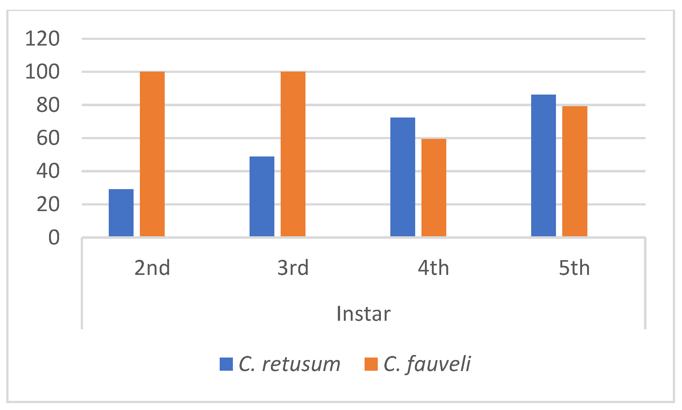
Laboratory rearing was successful in producing adults for morphological confirmation but mortality was high (Figure S1). Both species showed a high cumulative mortality. While Table 2 provides the specific demographic breakdown and environmental parameters for each stage, the survival trends are visually summarized in Figure 8 and Table S4. C. fauveli showed a final mortality rate of 60%, whereas C. retusum 84%, highlighting the critical nature of the initial nymphal transitions. (N1–N3). In C. retusum, mortality increased progressively through ontogeny, reaching 86.1% by the final instar. C. fauveli demonstrated extreme sensitivity in the initial stages, though individuals that reached the 4th instar showed a stabilized, albeit slow, progression to the adult stage.

3.5. Comparative Analysis of C. retusum and C. fauveli Nymphs

The nymphs of Cyphopterum retusum and C. fauveli can be reliably distinguished throughout their ontogeny by combining morphometric data with stage-specific chromatic and textural characters. While both species increase in size between successive instars (Table S2), C. fauveli remains consistently smaller than C. retusum across all stages for all morphometric variables from the third to fifth instar (Table S3).
Despite intraspecific variation dorsal pigmentation patterns and size serve as the most reliable diagnostic characters. In the early instars (1st and 2nd; pale phase), both species are virtually indistinguishable by color alone, presenting a uniform cream or ivory base. Separation at this stage relies almost exclusively on body size and metatibial spination (Table 1 and Table S5).
During the intermediate instars (3rd and 4th; transition phase), species-specific patterns become prominent on the thorax and abdomen. C. fauveli displays sharp, geometric markings and a highly contrastive thoracic pattern on the mesonotum, characterized by a clean, pale median carina (Figure 6C,D,H). In contrast, C. retusum exhibits a more fragmented, marbled appearance, with grainy, speckled, and muddier fuscous spots (Figure 4C,D). By the final instar (5th; melanic phase), differentiation in pigmentation peaks: C. retusum often reaches a nearly black (fuscous) appearance due to the total coalescence of the dorsal abdominal bands (Figure 4J–L). Conversely, C. fauveli maintains a grainier, yellowish-brown to amber tone, often highlighted by distinct amber markings on the mesonotum (Figure 6I,L).
Furthermore, diagnostic landmarks on the abdomen and head clearly separate the two species:
Abdominal eyespots: In C. fauveli, these spots form focal, circular black ocelli (Figure 6D–F), whereas in C. retusum, they are less circular, often broadening and merging into transverse “saddles” or bands (Figure 4C,D).
Cephalic carinae: In C. fauveli, the head morphology is clearly defined and dominated by a clean, pale median carina. In C. retusum, the cephalic carinae are often obscured by dense, grainy stippling in the final instars.
Thoracic pattern: The mesonotum markings in C. retusum tend to form an irregular, coalescent dark blotch, while C. fauveli exhibits a more structured geometric pattern.
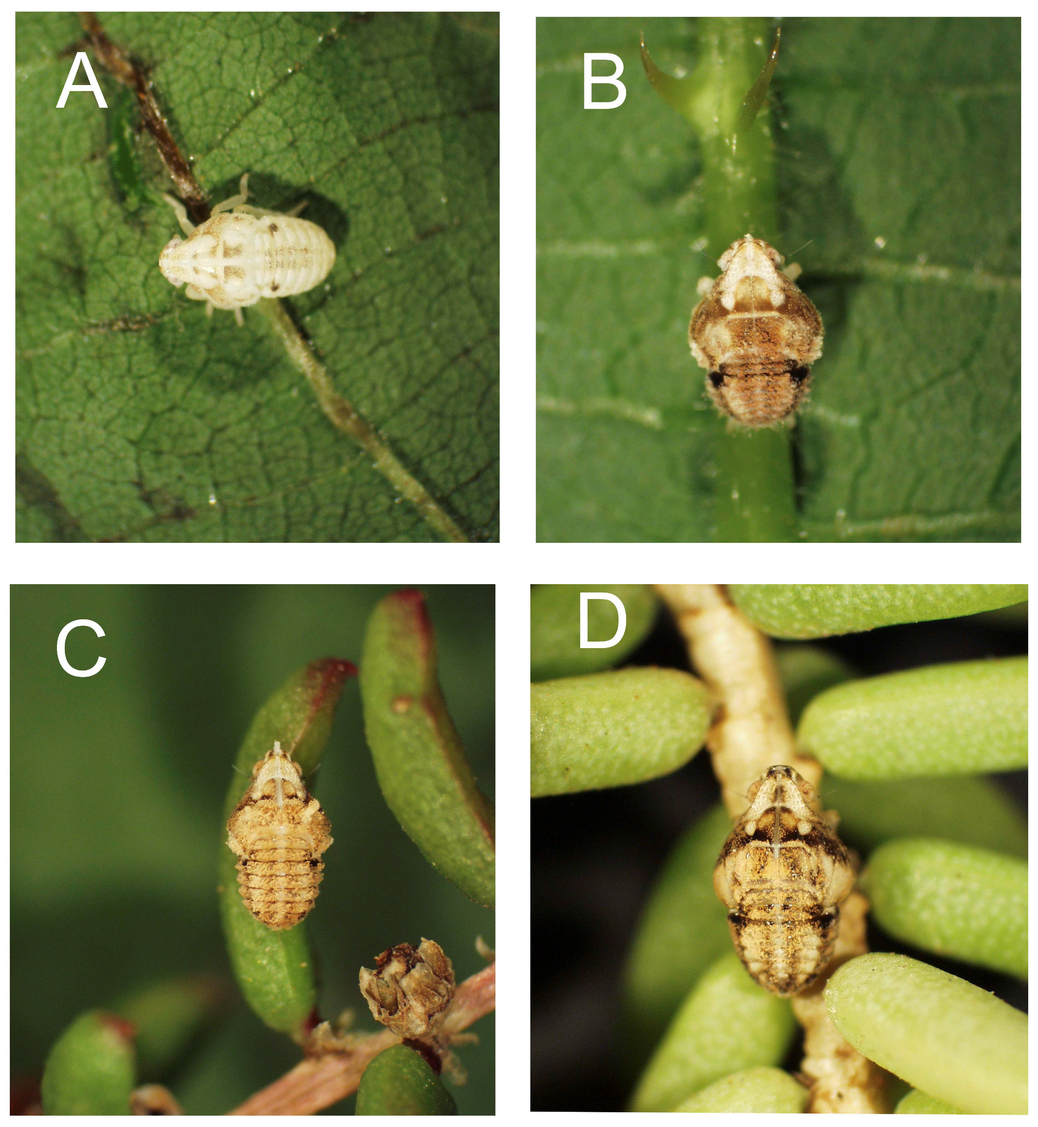
The most striking morphological divergence lies in the wax production and integument texture. C. retusum presents a “dry,” and polished appearance (Figure 4 and Figure 5); wax exudation is primarily restricted to the anal extremity, forming discrete, elongated white filaments. This localized secretion ensures that the underlying pigmentation and the sharp definition of the body’s carinae and sensory pits remain highly visible, facilitating a camouflage strategy where the nymph aligns with leaf veins (Figure 9B) or breaks its outline against plant stems (Figure 9A).
Conversely, C. fauveli presents a diffuse and extensive wax cover (Figure 6 and Figure 7). From the intermediate stages onward, these nymphs develop a granular or “frosted” appearance as flocculent wax masses accumulate across the entire body (Figure 9C). This broad exudation often obscures the underlaying marbled pigmentation, creating a structural contrast on the head where sensory pits and the pale median carina emerge from a field of waxy granules (Figure 7H,I). This textural camouflage strategy allows the nymphs to blend seamlessly into the irregular stems of their host plant (Figure 9D).
Finally, while the fundamental arrangement of sensory pits is conserved —increasing from approximately 20 pits on the head in the first instar to around 60 in the fifth—their visual prominence differ. In C. fauveli, the pits provide a higher contrast against the dark, waxy background, whereas in C. retusum, they remain clearly defined due to the polished, non-waxy nature of the cuticle. These contrasting states—the polished, dry finish of C. retusum versus the granular, broad exudation of C. fauveli—represent the most consistent characters for species separation in the field and laboratory.

3.6. Key to the Nymphal Instars of Cyphopterum retusum and C. fauveli

Nymphs of both species share a similar body plan but can be distinguished by a combination of size, coloration, and spination pattern. Thorax length (Ctr) is used here as the primary metric for stage separation, complemented by the number of spines on the metatibia and number of metatarsomeres.
Key to instars (both species)
  • 1. Wing pads entirely absent or much reduced (Figure 3A,B,K,L); body uniformly pale, white, or light yellowish with no distinct patterns (maculation) or, if present, reduced to two faint spots on abdomen (Figure 4A,B and Figure 6A,B)................................................................2
  • –Wing pads distinct (Figure 3C–E,M–O); body colour beige, brown, or dark, often with distinct dark/brown colour patterns (maculation) on thorax and abdomen (Figure 4C–L and Figure 6C–L)....................................................................................................................................... 5
  • 2. Body uniformly pale/whitish, entirely lacking dorsal maculae on thorax and abdomen (Figure 3A–K, Figure 4A, Figure 5A and Figure 6A); metatibia lacking lateral spines and bearing 4 apical spines (Figure 3F,P)..............................................................................................[1st instar].…. 3
  • –Body pale to light yellowish, dorsal surface predominantly immaculate (Figure 3B,L and Figure 4B) or, if maculation is present, restricted to two faint spots (Figure 6B); metatibia with 1 lateral spine and 5 apical spines (Figure 3G,Q)...............................................[2nd instar]…. 4
  • 3. Thoracic length > 0.6 mm; body colour cream whitish, translucent (Figure 4A and Figure 5A).................................................................................................................C. retusum 1st instar
  • –Thoracic length < 0.6 mm; body colour predominantly yellowish (Figure 6A and Figure 7E)...............................…………………………….……………………....…...C. fauveli 1st Instar
  • 4. Thoracic length > 0.7 mm; vertex broadly rounded in dorsal view (Figure 5F); dorsum completely immaculate and uniformly pale cream, entirely lacking dark dots, shading, or punctures on the dorsal surface (Figure 4B and Figure 5B,J–L)...................... C. retusum 2nd instar
  • –Thoracic length < 0.7 mm; vertex slightly produced anteriorly (Figure 7F); dorsum pale yellowish-cream but with a white longitudinal strip and faint, darker markings (Figure 6B and Figure 7A);.................................................................................................... C. fauveli 2nd instar
  • 5. Wing pads present only as short, subtle lateral sketches (Figure 3C,M); body colour whitish or beige with distinct dorsal dark markings restricted or fragmented into isolated patches; thoracic color pattern distinctly variegated, with the lighter ground color widely distributed and continuous across the vertex, pronotum, and wing pads (Figure 4C,D and Figure 6C–F); metatibia with 6 apical spines and 2 tarsomeres (Figure 3H,R)....[3rd instar]…6
  • –Wing pads large, prominent, distinct (Figure 3D–E,N–O); body colour brown to dark brown with extensive dark macula; thoracic region showing a strong coalescence or sharp definition of dark pigment, either forming a prominent, continuous dark median longitudinal band across the vertex and pronotum, or rendering the thoracic dorsum almost entirely and uniformly dark brown/blackish (Figure 4E–L and Figure 6G–L); metatibia with 7 or 8 apical spines and 3 tarsomeres (Figure 3I,J,S)…............................................................................................................................................ 7
  • 6. Thoracic length > 1.0 mm; vertex pale and rounded (Figure 5G); dorsal colouration more fragmented and marbled, characterized by diffuse, grainy and speckled fuscous markings rather than sharply delimited transverse bands (Figure 4C,D and Figure 5C,L)...................................................................................................................... C. retusum 3rd instar
  • –Thoracic length ≤ 1.0 mm; vertex acute-tending (Figure 7G); dorsal colouration predominantly ochre-brown, with darker brown transverse bands and a high-contrast thoracic pattern on the mesonotum with a distinct pale whitish median dorsal stripe (Figure 6C,F)................................................................................................ C. fauveli 3rd instar
  • 7. Wing pads relatively short (Figure 3D–N); dorsal dark markings more restricted or fragmented into isolated patches; thoracic color pattern variegated, with the lighter ground color widely distributed (Figure 4E,H and Figure 6G,H); metatibia with 7 apical spines (Figure 3I,S)........................................................................................................... [4th instar]…. 8
  • –Wing pads elongated, long, clearly extending posteriorly (Figure 3E,O); dorsal dark markings highly expanded, continuous, or structurally dominant; thoracic region showing a strong coalescence of dark pigment (Figure 4I–L and Figure 6I–L); metatibia with 8 apical spines (Figure 3J)……………………………………………………….. [5th instar]….. 9
  • 8. Thoracic length > 1.3 mm; dorsal maculation extensive, forming irregular, coalescent dark blotches; dorsal thorax with two large, distinct, dark brown sub-quadrate patches arranged symmetrically next to the median line; overall coloration predominantly brownish with well-defined, continuous longitudinal dark bands on the abdomen (Figure 3D, Figure 4E–H and Figure 5D–H)................................................................... C. retusum 4th instar
  • –Thoracic length < 1.3 mm; maculation on median dorsum showing a structured geometric pattern; dorsal thorax centrally pale with scattered dark punctures, completely lacking large, solid, sub-quadrate dark patches adjacent to the median line; overall coloration predominantly ochraceous or pale cream with isolated dark spots (Figure 6G,H and Figure 7C,H)................................................................................................... C. fauveli 4th instar
  • 9. Thoracic length > 1.7 mm; dorsal pigmentation strongly reticulate, yielding a nearly black (fuscous) appearance due to the total coalescence of the dorsal abdominal bands; thorax and wing pads predominantly solid dark chocolate brown, with pale areas restricted to two small, isolated circular spots on the mesonotum; abdomen heavily darkened (Figure 4I–L and Figure 5E–I); paranotal expansions broadly rounded in dorsal outline (Figure 3E).................................................................................................. C. retusum 5th instar
  • –Thoracic length < 1.7 mm; dark pigmentation concentrated medially; thorax and wing pads with a highly contrasting, variegated pattern of dark brown and broad golden-yellow areas; overall appearance grainier, yellowish-brown, often highlighted by distinct amber markings on the mesonotum (Figure 6I–L and Figure 7D,I,K); paranotal expansions less rounded (Figure 3O)................................................................................... C.fauveli 5th instar

4. Discussion

4.1. Ontogeny and Developmental Insights

The genus Cyphopterum is a vital component of Macaronesian fauna, yet the life history of this and many other Flatidae remains largely obscure. Our study provides the first biological insights into the ontogeny of these insular endemics, confirming a likely univoltine life cycle with the five-instar development typical of the family [23,24,42,43,44]. Through daily laboratory monitoring of individual nymphs and the collection of successive exuviae, we were able to reliably assign instar stages and document the complete moulting sequence from first-instar field-collected specimens to adulthood. Although the initial emergence from the egg was not directly observed, the combination of morphological markers—minimal body dimensions and metatibial spination patterns consistent with neonate Flatidae—and the recording of four distinct moulting events per individual provides compelling evidence that the earliest stage described indeed corresponds to the first instar.
A notable finding was the significantly slower development of C. fauveli compared to C. retusum. This potentially reflects a reduced metabolic rate—an adaptation to the thermally stressed and resource-limited coastal environments it inhabits. However, the high nymphal mortality and developmental plasticity observed under laboratory conditions suggest that Cyphopterum species are highly specialized to their respective micro-habitats and may not readily adapt to controlled environmental conditions.
The peak in mortality during the early instars (N1–N3) likely reflects a critical ‘establishment window’ where nymphs are most vulnerable to fluctuations in host plant quality and humidity. For the coastal specialist C. fauveli, the lower survival in early stages under stable laboratory conditions may indicate a dependency on specific environmental cues, such as salt spray, high solar radiation, or particular microclimatic conditions which are absent in controlled environments. These life-history traits, combined with their limited dispersal as flightless nymphs, emphasize the vulnerability of these endemic planthoppers to habitat fragmentation. While high mortality underscore that these species are likely highly sensitive to nutritional and humidity fluctuations outside their natural niche, it also highlights the importance of preserving intact micro-habitats that support the complete ontogenetic sequence.
Future research should prioritize the investigation of egg structure and oviposition microhabitats, as these represent critical knowledge gaps for a comprehensive understanding of the reproductive biology of Macaronesian Flatidae. Determining whether oviposition occurs endophytically (inserted into plant tissue), epiphytically (on the plant surface), or epigeically (within the soil or leaf litter) would provide invaluable insights for identifying critical breeding habitats and informing targeted management strategies.
Although our morphological comparisons were primarily made with West Palaearctic and Nearctic Flatidae—given their closer biogeographical proximity to the Macaronesian fauna—we recognize that a broader comparison with Oriental lineages, would be important for a comprehensive understanding of larval morphological evolution within the family.

4.2. Diagnostic Value of Nymphal Morphology and Sensory Pits

While adult Cyphopterum from Madeira are notoriously difficult to distinguish due to conservative genitalia and large overlapping intraspecific variation in body size [18], nymphal stages offer new diagnostic tools. Stable ontogenetic markers—such as the transition from two to three tarsal segments at the 4th instar and the progressive increase in metatibial spines—allow for reliable stage identification [19,45].
The sensory pits deserve special mention as precise taxonomic anchors. Our data align with observations in other Fulgoromorpha [46,47], showing that the number, size, and bilateral symmetry of these pits increase predictably with each moult. In Cyphopterum, these structures are under strict ontogenetic control and are less plastic than total body length, making them ideal for unequivocal instar identification. Furthermore, the interaction between these pits and waxy exudations—which provide protection against abiotic stress [48]—highlights the functional complexity of the nymphal integument. While our study establishes the first baseline for the number and arrangement of sensory pits in Cyphopterum, future taxonomic revisions of the genus should prioritize the systematic counting of these structures across larger populations to determine their stability as diagnostic characters for closely related species. However, the restricted visibility of these structures reduces their practicality for field-based identification.
Regarding the terminal wax glands, they were not investigated in the present work. Our observations were strictly limited to the external wax filaments visible on the specimens. The wax glands may possess diagnostic potential but are typically concealed by the wax secretions; their visualization require dedicated specimen preparation—including cleaning, dissection, and ideally scanning electron microscopy—which was beyond the scope of this study. Our primary aim was to develop a practical field identification key for living nymphs, based on easily observable external characters that allow rapid recognition without destructive sampling. This approach was deliberately chosen to support conservation monitoring programs targeting these endemic species to ensure usability by non-specialists involved in field surveys. Although not investigated here, nymphal Fulgoroidea possess also a unique interlocking gear mechanism on the metatrochanters that synchronises hind leg movements during jumping [49]. This remarkable evolutionary trait appears to be a conserved character across the superfamily.

4.3. Specific Diagnostic Coloration and Integumentary Texture

The chromatic and textural divergence observed from the third instar onwards provides a reliable non-morphometric basis for species separation. C. retusum is characterized by high-contrast markings [50], including distinct patterns and lateral abdominal maculae that remain visible throughout late development. These characters allow for immediate identification even when specimens are found on non-typical host plants.
In contrast, C. fauveli exhibits a consistent “frosted” or granular appearance due to widespread waxy exudations that partially obscure the underlying marbled pigmentation. This textural difference is a stable taxonomic character that separates it from the “polished” and dry integument of C. retusum. While the ecological drivers of these traits suggest adaptation to distinct micro-habitats—foliage for C. retusum and woody/saline stems for C. fauveli—their primary value in the current context is a robust morphological tool for field and laboratory diagnosis. The distinctness of these chromatic and textural features, observable from the third instar onwards, reinforces the utility of nymphal characters for rapid species identification in the field.

4.4. Conservation Implications for Insular Endemics

The ability to identify nymphs is a significant advancement for the monitoring of Madeiran biodiversity. This is particularly urgent for C. fauveli, which is restricted to vulnerable coastal habitats threatened by tourism and rising sea levels. Since our results demonstrate that early instars (N1–N3) are more highly sensitive environmental conditions, even minor degradations of Suaeda communities could trigger immediate population crashes.
During this study, C. retusum and C. fauveli were recorded associated with 25 plant species, of which 6 (24%) are endemic to the Madeiran archipelago (unpublished data). This relatively high proportion of endemic host plants underscores the ecological specialization of these planthoppers and highlights the potential vulnerability of their populations to habitat degradation or loss of key plant species. Although the detailed assessment of host specificity is not the scope of the present study, it suggests that the preservation of their endemic host plants as a priority.
Furthermore, the limited dispersal capacity of Cyphopterum suggests that populations may be easily isolated by habitat fragmentation. Conservation strategies must therefore shift from protecting adults to preserving the specific host-plant mosaics that support the entire ontogenetic sequence. This includes identifying these “vulnerable juveniles” in the field allows for a proactive rather than reactive approach to island conservation management, moving beyond simple species presence toward ensuring the long-term viability of their developmental niches.
Much more important information is necessary of the understanding and safeguarding critical oviposition sites, which remain unknown, and maintenance of the structural and floristic integrity of the vegetation communities upon which these planthoppers depend. The high proportion of endemic host plants recorded in this study reinforces the notion that the conservation of Cyphopterum species is inextricably linked to the preservation of Madeira’s unique flora.

Supplementary Materials

The following supporting information can be downloaded at: https://www.mdpi.com/article/10.3390/taxonomy6030044/s1, Table S1: Summary of environmental conditions (temperature and relative humidity) recorded during the nymphal rearing period under laboratory conditions (May to August); Table S2. Results of the comparison of nymphal instars of Cyphopterum retusum and C. fauveli using Student’s t-test (t) based on body measurements. Values highlighted in red were corrected using Levene’s test. df = degrees of freedom; p = significance level. * p < 0.001; Table S3: Statistical comparison of nymphal body measurements of Cyphopterum retusum and C. fauveli using Student’s t-test (t). Red values indicate tests adjusted with Levene’s correction for unequal variances. df = degrees of freedom; p = significance level. * p < 0.001; Table S4: Nymphal mortality per instar (2nd to 5th) of Cyphopterum retusum and C. fauveli under laboratory rearing conditions. Abbreviations: n = number of individuals entering the instar; N = number surviving to the next instar; Dead = number dying during the instar; Percentage = (Dead/n) × 100. 1st-instar mortality was not recorded because early-instar nymphs could not be reliably identified until the moult to the 2nd instar; Table S5: Summary of diagnostic characters for the identification of nymphal instars of Cyphopterum retusum and C. fauveli. Colour pattern is the most readily observable character under field conditions. Metatibial spine and tarsomere counts should be confirmed with hand lens or under stereomicroscopy. Thoracic length is provided as a complementary metric for species separation within each instar. For detailed morphology, refer to Figure 3 and Figure 7; Figure S1: Adult habitus photographs of Cyphopterum fauveli (Noualhier, 1897) (A,B) and C. retusum (Walker, 1851) (C,D). Dorsal views (A,C) and lateral views (B,D). Specimens reared from nymphs collected in the field. Note that external morphological differences between the two species are subtle, reliable species identification requires examination of male genitalia.

Author Contributions

Conceptualization, D.A.-P.; methodology, S.M. and D.A.-P.; software, S.M. and D.A.-P.; validation, D.A.-P.; formal analysis, S.M. and D.A.-P., investigation, S.M.; resources, D.A.-P.; data curation, D.A.-P.; writing—original draft preparation, S.M. and D.A.-P.; writing—review and editing, D.A.-P.; visualization, S.M. and D.A.-P.; supervision, D.A.-P. All authors have read and agreed to the published version of the manuscript.

Funding

This research received no external funding.

Data Availability Statement

The raw data supporting the conclusions of this article will be made available by the authors on request.

Acknowledgments

The authors would like to thank Luena Soraya for her skillful preparation of Figure 1 and for contributing the photographs used in its composition.

Conflicts of Interest

The authors declare no conflicts of interest.

References

  1. Gillespie, R.G.; Bennett, G.M.; De Meester, L.; Feder, J.L.; Fleischer, R.C.; Harmon, L.J.; Losos, J.B.; Revell, L.J.; Ricklefs, R.E.; Tsutsui, N.D.; et al. Comparing adaptive radiations across space, time, and taxa. J. Hered. 2020, 111, 1–20. [Google Scholar] [CrossRef] [Scilit] [PubMed]
  2. Losos, J.B.; Ricklefs, R.E. Adaptation and diversification on islands. Nature 2009, 457, 830–836. [Google Scholar] [CrossRef] [Scilit] [PubMed]
  3. Whittaker, R.J.; Fernández-Palacios, J.M.; Matthews, T.J.; Borregaard, M.K.; Triantis, K.A. Island biogeography: Taking the long view of nature’s laboratories. Science 2017, 357, eaam8326. [Google Scholar] [CrossRef] [Scilit] [PubMed]
  4. Gillespie, R.G. Island time and the interplay between ecology and evolution in species diversification. Evol. Appl. 2016, 9, 53–73. [Google Scholar] [CrossRef] [Scilit] [PubMed]
  5. Gillespie, R.G.; Roderick, G.K. Arthropods on islands: Colonization, speciation, and conservation. Annu. Rev. Entomol. 2002, 47, 595–632. [Google Scholar] [CrossRef] [Scilit] [PubMed]
  6. Bastin, S.; Reyes-Betancort, J.A.; Siverio de la Rosa, F.; Percy, D.M. Origins of the central Macaronesian psyllid lineages (Hemiptera; Psylloidea) with characterization of a new island radiation on endemic Convolvulus floridus (Convolvulaceae) in the Canary Islands. PLoS ONE 2024, 19, e0297062. [Google Scholar] [CrossRef] [PubMed]
  7. Florencio, M.; Patiño, J.; Nogué, S.; Traveset, A.; Borges, P.A.; Schaefer, H.; Roura-Pascual, N.; Arnedo, M.A.; Ávila, S.P.; Santos, A.M. Macaronesia as a fruitful arena for ecology, evolution, and conservation biology. Front. Ecol. Evol. 2021, 9, 718169. [Google Scholar] [CrossRef] [Scilit]
  8. Jordal, B.H.; Hewitt, G.M. The origin and radiation of Macaronesian beetles breeding in Euphorbia: The relative importance of multiple data partitions and population sampling. Syst. Biol. 2004, 53, 711–734. [Google Scholar] [CrossRef] [Scilit] [PubMed]
  9. Juan, C.; Emerson, B.C.; Oromí, P.; Hewitt, G.M. Colonization and diversification: Towards a phylogeographic synthesis for the Canary Islands. Trends Ecol. Evol. 2000, 15, 104–109. [Google Scholar] [CrossRef] [Scilit] [PubMed]
  10. Dmitriev, D.A.; Angelova, R.; Anufriev, G.A.; Bartlett, C.R.; Blanco-Rodríguez, E.; Borodin, O.I.; Cao, Y.-H.; Cara, C.; Deitz, L.L.; Dietrich, C.H.; et al. World Auchenorrhyncha Database. TaxonPages. Available online: https://hoppers.speciesfile.org/ (accessed on 9 May 2026).
  11. Leise, T.; Remane, R. Fünf neue Arten der Gattung Cyphopterum Mel., 1905 (Homoptera Auchenorrhyncha Flatidae) von den Kanarischen Inseln. Marbg. Entomol. Publ. 1994, 2, 47–76. [Google Scholar]
  12. Lindberg, H. Hemiptera Insularum Canariensium (Systematik, Ökologie und Verbreitung der Kanarischen Heteropteren und Cicadinen). Comment. Biol. 1953, 14, 1–304. [Google Scholar]
  13. Lindberg, H. Zur Kenntnis der Hemipteren-fauna der Azorischen Inseln. Comment. Biol. 1954, 13, 1–9. [Google Scholar]
  14. Lindberg, H. Hemiptera Insularum Caboverdensium. Systematik, Ökologie und Verbreitung der Heteropteren und Cicadinen der Kapverdischen Inseln. Comment. Biol. 1958, 19, 1–246. [Google Scholar]
  15. Lindberg, H. A new species of the genus Cyphopterum Amyot (Hom., Flatidae) from the Selvage Islands. Not. Entomol. 1959, 39, 18–21. [Google Scholar]
  16. Lindberg, H. Hemiptera Insularum Madeirensium. Comment. Biol. 1961, 24, 1–110. [Google Scholar]
  17. Lindberg, H. Die Cyphopterum-Arten (Hom. Flatidae) der Purpurarien. Zool. Beitr. 1965, 11, 129–135. [Google Scholar]
  18. Leise, T. Zur Phylogenie der Sudwest-Palaarktischen Zikaden-Gattung Cyphopterum Melichar, 1905 (Homoptera: Auchenorrhyncha: Flatidae). Ph.D. Thesis, University of Marburg, Lahn, Germany, 1992; pp. 1–271. [Google Scholar]
  19. Emeljanov, A.F. Larval characters and their ontogenetic development in Fulgoroidea (Homoptera, Cicadina). Zoosyst. Ross. 2001, 9, 101–121. [Google Scholar]
  20. Emeljanov, A.F. Evolutionary transformations of abdominal wax-plates in the larvae of the Fulgoroidea (Homoptera, Cicadina). Entomol. Rev. 2009, 89, 1035–1054. [Google Scholar] [CrossRef] [Scilit]
  21. Swezey, O.H. Life-history notes on two Fulgoridae. Ohio Nat. 1903, 3, 354–357. [Google Scholar]
  22. Wheeler, A.G., Jr.; Hoebeke, E.R. Host plants and nymphal descriptions of Acanalonia pumila and Cyarda sp. near acutissima (Homoptera, Fulgoroidea: Acanaloniidae and Flatidae). Fla. Entomol. 1982, 65, 340–349. [Google Scholar] [CrossRef] [Scilit]
  23. Wilson, S.W.; McPherson, J.E. Life histories of Anormenis septentrionalis, Metcalfa pruinosa, and Ormenoides venusta with descriptions of immature stages. Ann. Entomol. Soc. Am. 1981, 74, 299–311. [Google Scholar] [CrossRef] [Scilit]
  24. Wilson, S.W.; Tsai, J.H. Ormenaria rufifascia (Homoptera: Fulgoroidea: Flatidae): Descriptions of nymphal instars and notes on field biology. J. N. Y. Entomol. Soc. 1984, 92, 307–315. [Google Scholar] [CrossRef] [Scilit]
  25. Aguín-Pombo, D.; Pinheiro de Carvalho, M.A.A. Madeira Archipelago. In Encyclopedia of Islands; Gillespie, R., Clague, D., Eds.; University of California Press: Oakland, CA, USA, 2009; pp. 582–585. [Google Scholar]
  26. Press, J.R.; Short, M.J. (Eds.) Flora of Madeira; Natural History Museum: London, UK, 1994; pp. 1–574. [Google Scholar]
  27. Fowler, J.; Cohen, L.; Jarvis, P. Practical Statistics for Field Biology, 2nd ed.; John Wiley & Sons: Chichester, UK, 2013. [Google Scholar]
  28. Walker, F. List of the Specimens of Homopterous Insects in the Collection of the British Museum. Part II.; Edward Newman: London, UK, 1851; Volume 2, pp. 261–636. [Google Scholar]
  29. Walker, F. Homoptera. In Insecta Saundersiana: Or characters of undescribed insects in the collection of William Wilson Saunders. Esq.; John Van Voorst: London, UK, 1858; pp. 1–117. [Google Scholar]
  30. Distant, W.L. Rhynchotal notes. Ann. Mag. Nat. Hist. 1910, 5, 297–322. [Google Scholar] [CrossRef] [Scilit]
  31. Melichar, L. Homoptera, fam. Acanaloniidae, Flatidae et Ricaniidae. In Genera Insectorum, Fascicule 182; Wytsman, P., Ed.; Louis Desmet-Verteneuil: Bruxeles, Belgium, 1923; pp. 1–185. [Google Scholar]
  32. Melichar, L. Genera tria Fulgoridarum mundi antiqui. Ann. Hist.-Nat. Mus. Natl. Hung. 1905, 3, 473–477. [Google Scholar]
  33. China, W.E. Die Arthropodenfauna von Madeira nach den Ergebnissen der Reise von Prof. Dr. O. Lundblad Juli–August 1935. III. Terrestrial Hemiptera (Hemiptera and Homoptera Auchenorrhyncha). Ark. Zool. 1938, 30, 1–68. [Google Scholar]
  34. Lallemand, V. Liste d’Homoptères du Portugal recueillis par Mr. de Seabra. Mem. Estud. Mus. Zool. Univ. Coimbra Sér. I 1928, 28, 1–10. [Google Scholar]
  35. de Seabra, A.F. Subsídios para o conhecimento da fauna das matas nacionais. Conclusões de estudos realizados durante os meses de Julho e Agôsto de 1925 na Mata de Leiria. Arq. Secc. Biol. Parasitol. 1930, 1, 215–260. [Google Scholar]
  36. de Seabra, A.F. Observações sobre algumas espécies da Fam. Cicadoideae e da Fam. Fulguroidea existentes nas colecções do Museu de Coimbra. Arq. Secc. Biol. Parasitol. 1930, 1, 307–359. [Google Scholar]
  37. de Seabra, A.F. Contribuição para a história da entomologia em Portugal. Catálogo das coleções entomológicas do Laboratório de Biologia Florestal em 1937. Dir. Geral Serv. Florest. Agric. 1939, 6, 155–301. [Google Scholar]
  38. de Seabra, A.F. Contribuições para o inventário da fauna lusitânica. Insecta Homoptera (Cicadoideae e Fulgoroideae). Mem. Estud. Mus. Zool. Univ. Coimbra 1941, 121, 1–14. [Google Scholar]
  39. Noualhier, J.M. Hémiptères recueillis par M. A. Fauvel à Madère, en Mai et Juin 1896. Rev. Fr. Entomol. 1897, 16, 76–80. [Google Scholar]
  40. Horváth, G. Hémiptères recueillis par M. Th. Becker aux îles Canaries. Ann. Hist.-Nat. Mus. Natl. Hung. 1909, 7, 289–301. [Google Scholar]
  41. Lallemand, V. 2.e Liste d’Homoptères du Portugal recueillis par M. de Seabra. Mem. Estud. Mus. Zool. Univ. Coimbra Sér. I 1929, 39, 1–8. [Google Scholar]
  42. Wilson, S.W.; Lucchi, A. Distribution and ecology of Metcalfa pruinosa and associated planthoppers in North America (Homoptera: Fulgoroidea). Atti Accad. Naz. Ital. Entomol. Rend. 2001, 49, 121–130. [Google Scholar]
  43. Oliveira, C.M.; Frizzas, M.R. Bio-ecology of Poekilloptera phalaenoides (Hemiptera: Flatidae) under the influence of climatic factors in the Brazilian Cerrado. Ann. Entomol. Soc. Am. 2015, 108, 263–271. [Google Scholar] [CrossRef] [Scilit]
  44. Chen, C.; Yang, C.-T. The metatarsi of the Fulgoroidea (Homoptera: Auchenorrhyncha). Formos. Entomol. 1995, 15, 257–269. [Google Scholar]
  45. Bräunig, P.; Krumpholz, K.; Baumgartner, W. Sensory pits—Enigmatic sense organs of the nymphs of the planthopper Issus coleoptratus (Auchenorrhyncha, Fulgoromorpha). Arthropod Struct. Dev. 2012, 41, 443–458. [Google Scholar] [CrossRef] [Scilit] [PubMed]
  46. Ortega-Gómez, A.; Selfa, J.; Sendra, A.; Hoch, H. Postembryonic development of the troglobitic planthopper species Valenciolenda fadaforesta Hoch & Sendra, 2021 (Hemiptera, Fulgoromorpha, Kinnaridae), with a key to nymphal instars. Subterr. Biol. 2022, 44, 51–68. [Google Scholar] [CrossRef] [Scilit]
  47. McDonald, C.L.; Alcalde, G.T.; Jones, T.C., III; Laude, R.A.P.; Yap, S.A.; Bhamla, S. Wax “tails” enable planthopper nymphs to self-right midair and land on their feet. Integr. Comp. Biol. 2024, 64, 619–631. [Google Scholar] [CrossRef] [Scilit] [PubMed]
  48. Lucchi, A.; Santini, L. Morpho-biological notes on the immature stages of Metcalfa pruinosa (Say) (Homoptera, Flatidae). Frustula Entomol. 1993, 16, 175–185. [Google Scholar]
  49. Burrows, M.; Sutton, G.P. Interacting gears synchronize propulsive leg movements in a jumping insect. Science 2013, 341, 1254–1256. [Google Scholar] [CrossRef] [Scilit] [PubMed]
  50. Stevens, M.; Cuthill, I.C.; Windsor, A.M.; Walker, H.J. Disruptive contrast in animal camouflage. Proc. R. Soc. B 2006, 273, 2433–2438. [Google Scholar] [CrossRef] [Scilit] [PubMed]
Figure 1. Study area and sampling localities in Madeira Island. The main map displays the natural vegetation zones of the island; red dots indicate the sampling sites. Map (bottom left) shows the geographic position of Madeira archipelago within the Macaronesian region. (1) Chão da Ribeira: habitat of Cyphopterum retusum within the Temperate Stink-Laurel Forest (Laurissilva), showing the host plant Rubus cf. bollei; (2) Ponta de São Lourenço: semi-arid habitat of Cyphopterum fauveli characterized by Marmulano Shrubland, showing the host plant Suaeda vera and the sweep-net sampling method.
Taxonomy 06 00044 g001
Figure 2. Morphometric parameters and measurement scheme for Cyphopterum nymphs. The coloured lines indicate the specific dimensions recorded for each specimen: Ctr—total thorax length (combined midline length of pronotum (P), mesonotum (Ms), and metanotum (Mt); Lt—maximum body width (measured across the metanotum/wing pads); Lv—maximum width of the vertex; Lc—head width. The location of sensory pits is highlighted with small circles (○) on the dorsal view; only those pits clearly visible from a strictly dorsal perspective are represented. Scale bar = 1 mm.
Taxonomy 06 00044 g002
Figure 3. Comparative nymphal morphology of Cyphopterum retusum (Walker, 1851) (AJ) and Cyphopterum fauveli Noualhier, 1897 (KT). Instars: 1st instar (A,F,K,P); 2nd instar (B,G,L,Q); 3rd instar (C,H,M,R); 4th instar (D,I,N,S); 5th instar (E,J,O,T). C. retusum: (AE) dorsal view of 1st to 5th nymphal instars, respectively, showing the progressive development of wing pads and intensification of fuscous maculation; (FJ) detail of the metatibia and tarsomeres of 1st to 5th instars, highlighting the increasing number of metatibial apical and lateral spines. C. fauveli: (KO) dorsal view of 1st to 5th nymphal instars, respectively; (PT) detail of the metatibia and tarsomeres of 1st to 5th instars. The location of sensory pits is highlighted with small circles (○); only sensory pits clearly visible from a strictly dorsal perspective are represented. Note the differences in body proportions and maculation patterns between the two species at corresponding stages. Scale bars: (AE,KO) = 1 mm; (FJ,PT) = 0.5 mm.
Taxonomy 06 00044 g003
Figure 4. Nymphal development of Cyphopterum retusum (Walker, 1851), dorsal view. Instars: 1st instar (A); 2nd instar (B); 3rd instar (C,D); 4th instar (E,H); 5th instar (IL). (A) 1st instar, characterized by a uniform pale coloration and absence of wing pads; (B) 2nd instar, showing increased body volume while maintaining pale pigmentation; (C,D) 3rd instar, illustrating the first appearance of lobate wing pads and the onset of fuscous maculation; (EH) 4th instar, showing well-developed wing pads and a significant transition toward darker, mottled patterns; (IL) 5th instar, reaching maximum nymphal size with elongated wing pads covering the anterior abdominal segments and prominent dark marbled pigmentation.
Taxonomy 06 00044 g004
Figure 5. Morphological details of five life stages of Cyphopterum retusum (Walker, 1851). Instars: 1st instar (A); 2nd instar (B,F,J,K); 3rd instar (C,G,L); 4th instar (D,H); 5th instar (E,I). (AE) Lateral view of the five nymphal instars: (A) 1st instar; (B) 2nd instar; (C) 3rd instar; (D) 4th instar; (E) 5th instar, highlighting the progressive enlargement of the thoracic hump and the development of wing pads; (FI) Frontal/dorsal close-up of the head: (F) 2rd instar; (G) 3rd instar; (H) 4th instar; (I) 5th instar, showing the arrangement of sensory pits, prominent carinae, and the intensification of eye pigmentation; (J) Second nymph after ecdysis, with the waxy secretions adhering to the discarded exuviae; (K,L) Detail of the caudal end of 2nd and 3rd instars, illustrating the production of long, delicate white waxy filaments.
Taxonomy 06 00044 g005
Figure 6. Nymphal development of Cyphopterum fauveli Noualhier, 1897, dorsal view. Instars: 1st instar (A); 2nd instar (B); 3rd instar (CF); 4th instar (G,H); 5th instar (IL). (A) 1st instar, showing a predominantly uniform pale-yellow coloration and oval body shape; (B) 2nd instar, with slightly increased sclerotization; (CF) 3rd instar, exhibiting the first appearance of small wing buds and the onset of lateral dark spots on the abdomen; (G,H) 4th instar, showing more pronounced dark brown (fuscous) maculation and further development of the mesonotal wing pads; (I,L) 5th instar, reaching maximum nymphal size with fully developed wing pads and intense marbled pigmentation. Note the presence of white waxy secretions covering parts of the body in fourth (J,K), and fifth instars (L).
Taxonomy 06 00044 g006
Figure 7. Morphological details and life stages of Cyphopterum fauveli Noualhier, 1897. Instars: 1st instar (E); 2nd instar (A,F); 3rd instar (B,G,J); 4th instar (C,H); 5th instar (D,I,K,L). (AD) Lateral view of nymphs. (C,D) 4th and 5th instar, illustrating the progressive development of wing pads and the transition from pale to dark mottled coloration. (EI) Frontal/dorsal close-up of the head across different instars (1st to 5th), showing the distinct arrangement of sensory pits and the prominence of the carinae. (J,K) Dorsal view of 3rd and 5th instar nymphs highlighting the extensive accumulation of white waxy filaments on the abdominal tergites and margins (L) 5th nymph on the host plant Suaeda vera recently emerged after ecdysis, shown adjacent to its discarded exuviae; note that the newly emerged instar lacks the waxy filaments which remain attached to the exuviae.
Taxonomy 06 00044 g007
Figure 8. Nymphal mortality per instar (2nd to 5th) of Cyphopterum retusum and C. fauveli under laboratory rearing conditions. Bars represent the percentage of individuals that died during each instar. Sample sizes (number of individuals entering each instar) are as follows: C. retusum—2nd instar, n = 31; 3rd instar, n = 41; 4th instar, n = 47; 5th instar, n = 36; C. fauveli—2nd instar, n = 4; 3rd instar, n = 6; 4th instar, n = 32; 5th instar, n = 24. Raw mortality counts and additional details are provided in Table S4. Mortality in the 1st instar was not recorded because individuals could not be reliably identified until they molted to the 2nd instar, due to morphological similarity among early-instar nymphs.
Taxonomy 06 00044 g008
Figure 9. Ecological morphology and cryptic strategies of Cyphopterum nymphs on their food plants. Instars: 3rd instar (A,C); 4th instar (B); 5th instar (D). (A,B) C. retusum nymph showing a high-contrast pattern (A) and aligned with the central vein of a Rubus sp. leaf (B), and (C,D) C. fauveli nymph exhibiting extensive waxy exudation (“frosted effect”) and granular pigmentation similar to the host (C) and integrated into the woody texture of a Suaeda vera stem (D).
Taxonomy 06 00044 g009
Table 1. Measures from nymphs of Cyphopterum retusum and C. fauveli. Ctr = thorax length; Lc = head width; Lv = vertex width; Lt = total width; N = number of measured individuals; SD = standard deviation; CV = coefficient of variation. Values in mm.
C. retusumC. fauveli
InstarStatisticsCtrLcLvLtCtrLcLvLt
n61
1stMean0.68 ± 0.040.45 ± 0.020.31 ± 0.010.76 ± 0.04
Min–Max0.63–0.730.43–0.480.3–0.330.69–0.80.450.370.240.56
C.V. (%)5.783.213.95
n191
2ndMean0.87 ± 0.050.55 ± 0.030.36 ± 0.031.05 ± 0.05
Min–Max0.77–0.980.49–0.630.31–0.440.9–1.110.630.480.310.8
C.V. (%)5.595.688.235.09
n207
3rdMean1.14 ± 0.060.69 ± 0.030.47 ± 0.031.38 ± 0.090.96 ± 0.040.6 ± 0.040.28 ± 0.021.15 ± 0.08
Min.1.06–1.270.64–0.740.42–0.541.26–1.510.89–10.54–0.660.24–0.321.05–1.26
C.V. (%)5.084.056.786.293.735.928.246.54
n2314
4thMean1.56 ± 0.070.88 ± 0.060.61 ± 0.051.91 ± 0.151.16 ± 0.050.73 ± 0.040.34 ± 0.011.44 ± 0.08
Min–Max1.39–1.680.65–0.930.46–0.691.41–2.081.07–1.260.68–0.790.31–0.361.34–1.61
C.V. (%)4.596.658.447.584.3454.195.24
n2015
5thMean2.06 ± 0.141.12 ± 0.090.81 ± 0.092.73 ± 0.191.51 ± 0.080.89 ± 0.050.43 ± 0.031.97 ± 0.09
Min–Max1.84–2.280.83–1.240.63–0.952.4–3.061.39–1.660.82–0.970.4–0.491.85–2.15
C.V. (%)6.638.4211.287.115.395.426.544.55
Table 2. Developmental parameters of Cyphopterum retusum and C. fauveli under laboratory conditions. n = initial number of nymphs; N = number of individuals completing the instar; A = range (min-max) of days for transition between instars; CV = coefficient of variation. Environmental data (Temp/RH) represents means recorded for each specific period. Asterisks (*) denote variables calculated only for surviving individuals.
SpeciesInstarnNMean Nº of DaysDevelopment *Laboratory Conditions *
ACV (%)Minimum Temp. (°C)Maximum Temp. (°C)Relative Humidity (%)
C. retusum2nd31229.4 ± 1.996–1321.1718.35 ± 0.442.67 ± 0.7466.83 ± 0.76
3rd412110.76 ± 3.376–1731.3219.29 ± 0.6924.58 ± 1.0466.69 ± 0.97
4th471312.15 ± 3.827–2131.4420.39 ± 0.7825.07 ± 1.0768.8 ± 2.02
5th36516.8 ± 5.413–2332.1421.18 ± 0.1925.2 ± 0.3568.2 ± 2.07
C. fauveli2nd40
3rd60
4th321323.7 ± 7.5915–3832.0420.32 ± 0.2424.2 ± 0.2064.7 ± 0.17
Disclaimer/Publisher’s Note: The statements, opinions and data contained in all publications are solely those of the individual author(s) and contributor(s) and not of MDPI and/or the editor(s). MDPI and/or the editor(s) disclaim responsibility for any injury to people or property resulting from any ideas, methods, instructions or products referred to in the content.