
Systematic Review

Dentists’ Knowledge of Radiation Protection, Justification, and Optimization Practices: A Systematic Review


1
Faculty of Dental Medicine and Health, Josip Juraj Strossmayer University of Osijek, 31000 Osijek, Croatia
2
Medical Faculty Osijek, Josip Juraj Strossmayer University of Osijek, 31000 Osijek, Croatia
*
Author to whom correspondence should be addressed.
Submission received: 31 May 2026 / Revised: 5 July 2026 / Accepted: 20 July 2026 / Published: 22 July 2026

Highlights

What are the main findings?
  • There is a consistent and significant gap between guidelines on radiation protection and their clinical application in dental medicine, especially regarding justification and optimization of dental X-ray imaging.
  • Dentists demonstrate a heterogeneous, often insufficient level of knowledge regarding ALARA/ALADA principles, technical device parameters, and the correct use of protective measures (collimation, FOV, exposure parameters, protective equipment), with marked variability between countries and levels of practice.
What are the implications of the main findings?
  • Implementation of regulatory and professional guidelines (including DRLs) should be strengthened, quality control and equipment maintenance should be improved, and actions for reducing potentially unjustified use of dental X-ray imaging in daily practice should be implemented.
  • Radiation protection education should be increased and possibly included in the basic dental curriculum, along with systematic, standardized, and continuous education for dentists in this field, emphasizing the clinical application of justification and optimization principles, including appropriate imaging indications and rational use of CBCT.

Abstract

Background/Objectives: Radiation protection in dental medicine is based on the principles of justification and optimization, yet available evidence indicates inconsistent application of these principles in everyday practice. The aim of this systematic review was to assess dentists’ understanding of radiation protection and their adherence to guidelines for justification and optimization of dental X-ray procedures. Methods: A systematic search of the PubMed and Scopus databases was conducted from January 2010 to April 2026, following PRISMA guidelines. The systematic review included surveys and cross-sectional studies that examined dentists’ knowledge of radiation protection, as well as justification and optimization practices. The quality of the included studies was assessed using the Newcastle–Ottawa scale adapted for cross-sectional studies. Results: The systematic review included 22 original scientific articles that assessed knowledge of radiation protection, justification, and optimization practices among 5277 dentists from 12 countries. The findings indicate a high theoretical understanding of the basic rules of radiation protection, but a lower level of understanding of the technical and optimization factors of dental X-ray devices. A relevant inconsistency was observed in the justification process, with frequent referral for X-ray imaging without clinical indication and inconsistent adherence to guidelines. The optimization of X-ray imaging varied, especially in failing to adjust exposure parameters and in the restricted use of rectangular collimators. Inadequate quality control of X-ray devices and insufficient continuous professional training in radiation protection and imaging optimization are examples of organizational and infrastructural factors that lead to higher radiation exposure. Conclusions: Although many guidelines exist, there remains a noteworthy disparity between theoretical understanding and clinical practice in dental radiation protection. To reduce patients’ and dentists’ exposure to ionizing radiation, it is important to include radiation protection education in the basic dental curriculum, establish and improve continuous training, standardize clinical procedures, and enable broader application of systems for optimization and quality control.

1. Introduction

Dental radiology is an integral component of modern dental medicine, and the use of dental imaging continues to increase globally. Studies in different countries have confirmed increases in dental X-ray equipment in private dental practices, as well as the adoption of newer imaging modalities, among which cone-beam computed tomography (CBCT) has seen a substantial increase in recent years. It is estimated that the number of annual dental X-ray images increased by about 41% from 1988 to 2008, accounting for about 15% of all radiological procedures in medicine [1,2]. National data from various countries confirm this trend. Thus, in the USA, an increase in intraoral and panoramic dental X-ray imaging has been observed [3], while in Switzerland, dental X-ray imaging accounts for almost half of all radiological examinations [4]. However, this trend is not present in all countries. In Finland, the Radiation and Nuclear Safety Authority reported an increase in the number of dental X-ray units. However, the number of dental X-ray images decreased from 2018 to 2021. For intraoral X-ray imaging, a decrease of 0.2% was recorded; for panoramic X-ray imaging, a decrease of 9.2%; and for CBCT imaging, a decrease of 18% in the mentioned period [5].
Dental X-ray imaging often increases cumulative exposure to ionizing radiation, which can have stochastic or deterministic effects on patient health [6]. For this reason, dentists must adhere to the ALARA (As Low As Reasonably Achievable) or, even better, the ALADA (As Low As Diagnostically Acceptable) principles to reduce exposure to ionizing radiation while still obtaining diagnostic benefit. Additionally, some states have developed their own national guidelines for patient referral and the safe use of dental X-rays [7,8,9,10,11,12,13]. However, despite the existence of those guidelines and dentists’ familiarity with them, many do not apply them in clinical practice [14]. Specifically, the justification for dental X-ray indications is often insufficient, exposure parameters for dental X-ray devices are not adjusted, and the devices’ effective doses are unknown [15,16]. Although the literature indicates that regulatory frameworks and guidelines for dental radiology are continually updated to enhance protection against ionizing radiation and reduce effective doses, their inconsistent application in clinical practice persists [17], especially due to a lack of understanding of the principles of dental X-ray devices and their optimization.
Today, dentists often use one of the three dental X-ray modalities to diagnose, plan, and monitor treatment procedures [18,19]. Intraoral X-ray is the most widely used, enabling morphological assessment of individual teeth and periapical tissues with relatively low effective radiation doses. On the other hand, the panoramic X-ray provides complete insight into the dentoalveolar system and the jaw’s anatomical structures, but at a slightly higher effective dose. CBCT is the most recent modality in dental radiology, providing 3D imaging of dental and maxillofacial structures and offering high resolution. However, it also has a higher effective radiation dose than the two most commonly used 2D modalities. For the above reasons, a strict personalized clinical indication is required to justify CBCT imaging [20,21].
The starting point of dental radiology is the justification for referring patients for dental X-rays, which must be based on a clear clinical indication, and the benefit of exposure to ionizing radiation during the diagnostic procedure must significantly outweigh the risk to the patient [22,23]. Referral guidelines are a primary tool for rational clinical decision-making, but their availability and implementation in daily practice vary significantly across countries [17]. In European and Central Asian countries, such guidelines are available in 55–70% of countries, which directly contributes to inconsistent clinical practice [24]. As a result, a significant proportion of dental X-ray imaging remains clinically unjustified. In Germany, dental X-ray imaging was justified in only 70.5% of cases, while almost one-third of all dental X-rays were performed without a valid indication [16].
Data from clinical studies further illustrate the high variability in dental X-ray referral practices for new patients. In Sweden, 6% of dentists performed complete-mouth X-ray imaging at the first examination of each new patient, while 64% routinely indicated bitewing imaging regardless of individual clinical assessment [25]. Additionally, dental X-rays are often performed according to established routine protocols, without systematic analysis of the risk-benefit ratio for each patient. This practice is especially pronounced in countries with limited regulatory frameworks [23], as seen in clinical practice in developing countries [24]. Particular emphasis should be placed on the use of CBCT imaging, which has a significantly higher effective dose than 2D modalities and should be reserved for patients who do not provide sufficient diagnostic data with 2D modalities [26,27]. In CBCT imaging, justification is more stringent than in intraoral or panoramic radiography because the higher radiation dose and 3D data acquisition demand a clear expectation of additional diagnostic benefits that cannot be obtained with lower-dose 2D modalities [28].
Furthermore, an important part of dental radiology is optimizing radiation doses in accordance with the ALARA and ALADA principles. This implies that X-ray imaging is of sufficient quality for treatment procedures while keeping the patient’s exposure to ionizing radiation as low as possible. In intraoral X-ray imaging, an effective way to reduce dose is to use a rectangular collimator. Namely, a rectangular collimator reduces the area of irradiated tissue, which can reduce the dose by up to 60% compared to a circular collimator [29]. Despite the overall increase in dental X-ray imaging systems, the transition to digital systems has reduced cumulative ionizing radiation dose, and the use of rectangular collimation has contributed significantly to this reduction.
Furthermore, adjusting exposure parameters, such as the X-ray tube voltage (kV), current (mA), and exposure time (mAs), based on an individual patient’s age and body composition can further reduce the patients’ effective dose. In panoramic X-ray, the dose can be further reduced by proper patient positioning and selecting an appropriate imaging program based on the referral diagnosis [30]. CBCT optimization is somewhat more complex because it requires greater technical knowledge and familiarity with the equipment’s operation. Namely, for optimal radiation dose in CBCT imaging, it is necessary to adjust the field of view (FOV), voxel, and exposure parameters [26,27], which is why there is a large variability in effective doses between CBCT devices, but significant variations in doses depending on the people using them [30]. Optimization in CBCT is tailored to the clinical task, whereas in conventional dental radiography it primarily focuses on minimizing dose while maintaining adequate image quality [28]. Therefore, it is difficult to create standardized CBCT imaging protocols because, in addition to the many technical parameters that need to be adjusted for each patient, there is a wide range in dentists’ knowledge of CBCT use.
Diagnostic reference levels (DRLs) are a well-established method for dose optimization and monitoring of ionizing radiation exposure in dental radiology [31,32], and enable comparisons of radiation doses across devices and institutions, thereby contributing to optimization. However, despite the existence of DRLs, their application in clinical practice remains inconsistent [33,34,35,36], and significant variability in typical values used for dental radiology has been reported, depending on the device and imaging protocol [31,32]. This inconsistency in implementation has been associated with higher cumulative patient doses [37]. Various studies have shown that knowledge of DRLs and their values across dental radiology modalities is limited. A study of dental students in the United Arab Emirates found that 53.8% were familiar with the quality control of dental X-ray equipment [37]. However, 73.8% were familiar with techniques for optimizing dental X-ray imaging [37]. Moreover, testing 119 intraoral units in Serbia showed that 60% of users are not adequately trained to handle such devices [32]. This indicates shortcomings in the education of future dentists and in the application of acquired knowledge in clinical practice.
Although guidelines exist to optimize radiological procedures, data from various studies indicate that their implementation in practice is inadequate [38,39]. A major problem is that many dentists independently operate diagnostic dental X-ray equipment without formal education in radiation safety or protection against ionizing radiation [40]. A study in Jordan found that about one-third (34.7%) of dentists take their own patients’ X-ray imaging [41]. This can lead to increased exposure to ionizing radiation for both the patient and the dentist performing such imaging, because most dentists are not familiar with the technical side of dental X-ray devices and do not adjust the exposure parameters for each patient or correctly position patients during imaging, and often do not apply the recommended radiation protection protocols [37,41,42,43]. Furthermore, in many dental offices, there are no separate rooms for radiological equipment, and dental X-ray devices are located in the same working area [41,44], leading to increased unjustified occupational exposure to ionizing radiation, especially when dentists image a large number of patients and the workload is high [45,46].
A special part of the optimization is the regular maintenance and quality assurance of the dental X-ray device. This mainly refers to regular technical services and device calibration, as well as regular confirmation of the consistency of exposure parameters [47]. Namely, implementing quality assurance can detect technical deviations, prevent uncontrolled increases in radiation dose, and maintain the quality of the X-ray image [21,48]. However, many do not comply with the assigned regulations, and equipment maintenance and calibration are reduced to being done only when necessary, most often when the quality of the X-ray image declines. Thus, in Brazil, even 90% of intraoral X-ray devices do not meet national radiation protection regulations [42], while 53% of Turkish dentists do not conduct regular checks of their X-ray devices [49]. Due to such a systematic lack of quality assurance, significant technical deviations in the device itself can occur, increasing the effective radiation dose and decreasing the quality of the X-ray image.
An important part of radiation protection in dental radiology is the level of professional knowledge of all healthcare professionals involved in the dental X-ray imaging process. However, dentists bear the greatest responsibility because they conduct the initial assessment and justify the indication for dental X-ray imaging. However, despite education and available guidelines, there is a significant gap between formal knowledge and its application in everyday work. Namely, although dentists know the basic principles of radiation protection and that a clinical indication is required to justify dental X-ray imaging, practice varies significantly and often deviates from all recommendations [44,50,51,52]. The largest deviations were recorded for the indication for CBCT imaging, knowledge and application of DRLs, and adaptation of exposure parameters to the patient’s personalized needs [37], which can lead to unnecessary radiation and/or increased patient exposure to ionizing radiation.
The above shows that knowledge, radiation dose optimization, and justification for referring patients for dental X-rays are interconnected and functionally integrated parts of the overall radiation protection system in dental medicine. Inadequacy of any of these components inevitably compromises the overall effectiveness of the system. Therefore, this systematic review aims to collect and critically integrate current scientific evidence on dentists’ knowledge of three aspects of ionizing radiation protection in dental radiology: justification of dental X-rays, optimization of ionizing radiation dose, and ionizing radiation protection.

2. Materials and Methods

The databases Scopus and PubMed were searched for studies on dentists’ knowledge of radiation protection, justification, and optimization from January 2010 to April 2026. The search strategy is described in Supplementary Table S1. The database search was conducted in accordance with PRISMA guidelines (Supplementary Files S1 and S2), and the study protocol is registered in PROSPERO (CRD420261389498).
All retrieved articles were screened for studies that met specific criteria: cross-sectional or survey studies focused on dentists’ knowledge of radiation protection, justification, and optimization practices. Exclusion criteria were duplicate records, review articles, guidelines, case reports, editorials, surveys involving dental patients, studies on the knowledge of dental or radiography students regarding radiation protection, and studies involving other dental professionals, such as dental hygienists, dental assistants, and radiographers. No language restrictions were applied.
Two authors (I.Š. and D.F.) independently screened the studies and collected data on the author, year of publication, study design, number of dentists included, knowledge of the ALARA principle, ICRP (International Commission on Radiological Protection), NCRP (National Council on Radiation Protection and Measurements), referral guidelines, or national referral guidelines and the safe use, DRL, radiation protection measures used (such as lead aprons, thyroid collars, distance rule), and knowledge of equipment operation and maintenance. Any disagreements during screening or regarding the interpretation of the eligibility criteria were resolved by consensus, with a third author consulted when necessary to address inconsistencies. The quality of the studies was assessed using the Newcastle–Ottawa Scale (NOS) adapted for cross-sectional studies, which ranges from one to ten stars and includes three components: selection, comparability, and ascertainment of outcomes [53,54]. Studies with more stars are considered higher quality. Studies scoring 9–10 stars were classified as very good quality, those scoring 7–8 stars as good quality, those scoring 5–6 stars as satisfactory, and those scoring 0–4 stars as unsatisfactory (poor) quality.

3. Results

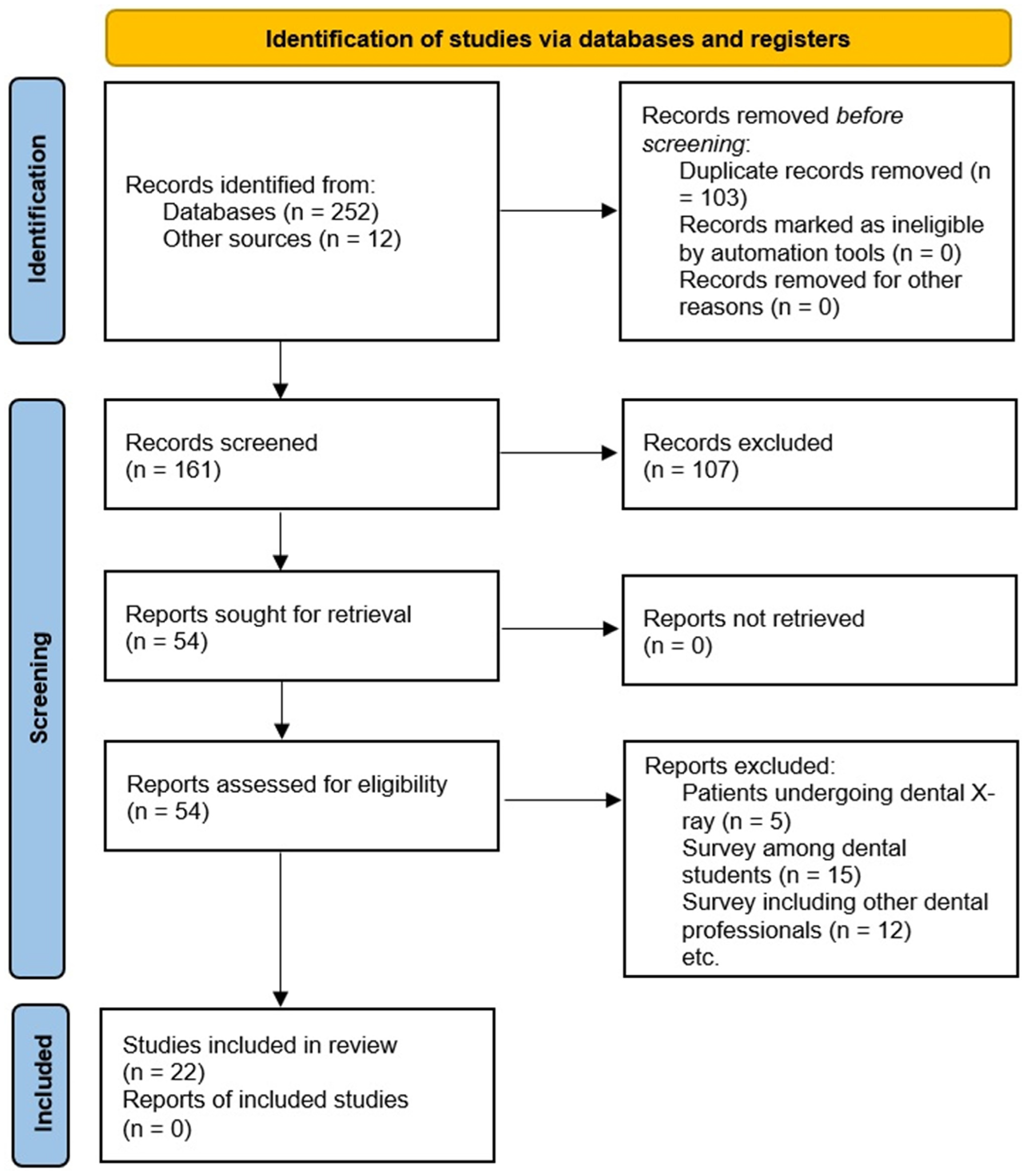
A total of 161 articles were retrieved after duplicates were removed. One hundred and seven articles were excluded after screening titles and abstracts. An additional 32 articles were excluded based on the inclusion and exclusion criteria. Ultimately, 22 articles were included in the qualitative analysis, assessing the knowledge of 5277 dentists. The study selection process is shown in Figure 1. The quality scores of the selected studies ranged from six to nine (Supplementary Table S2). Of the 22 included studies, nine were rated very good (9 points out of a maximum 10), 11 were rated good (four with 8 points and seven with 7 points), and two were rated satisfactory (6 points) on the Newcastle–Ottawa quality assessment scale adapted for cross-sectional studies.
The characteristics of the included studies are shown in Table 1. Most studies were conducted in India (n = 9), followed by Turkey and Korea (n = 2 each), and one study each in Australia, Iran, Iraq, Jordan, Malaysia, Morocco, Singapore, the United Kingdom, and the USA. All studies used a cross-sectional survey design among dentists, and some included dental specialists such as oral surgeons, periodontists, orthodontists, pediatric dentists, and endodontists, in addition to general dentists.
The level of knowledge about indications for radiological examinations and referral guidelines varies across populations and geographic regions. However, frequent use of dental X-ray imaging during initial examinations has been reported in Korea [64], indicating insufficient application of the principle of justification. Only one study in Korea examined adherence to referral guidelines (FDA-ADA, Food and Drug Administration and American Dental Association) for referring patients for dental X-rays, and only 1.9% of dentists followed the recommended referral criteria [57]. In contrast, one-third of dentists (34.1%) used predetermined routine examinations [57]. Dental X-ray imaging of new patients before clinical evaluation ranged from 1.2% in India [75] to 99% in Korea [64] (Table 2). Although pregnancy is not a contraindication for dental X-rays, the rate of acceptable dental imaging among pregnant women varied significantly, ranging from 17% among dentists in India [60] to 95.2% in emergency cases in Australia [68] (Table 2). Most studies do not mention that pregnant women can undergo dental X-rays in emergencies. However, dentists believe that pregnant women are more sensitive to radiation [59] and that, if exposed to dental X-rays, they must wear a lead apron [63]. In Australia, dentists believe the best approach for a pregnant woman who requires a dental X-ray is an emergency intraoral scan [68]. Many dentists in Turkey (62%) still ask women if they are pregnant before referring them for a dental X-ray [69], while 62.4% of dentists in Iraq believe that dental X-rays are contraindicated in pregnant women [70].
There is significant variability in practice across countries and a notable gap between knowledge and the implementation of protective measures. Dentists’ awareness of the ALARA principle ranged from 35% in Morocco [72] to 84.3% in India [59] (Table 3). Protective equipment, such as lead aprons and thyroid collars, is often used inconsistently and sometimes in ways that contradict current guidelines. Infrastructure, such as separate rooms for X-ray equipment, remains limited in many practices. Thirty percent of dentists in Iran reported that panoramic exposure delivers a higher radiation dose to patients than full-mouth periapical radiography [55], a finding similar to that of 45.6% of dentists in Iraq [70]. In contrast, in 2016, 69.1% of dentists in India reported that panoramic radiography results in less patient radiation exposure than full-mouth radiography [60]. In 2016 in India, 80.3% of dentists had a separate section for radiographic equipment, 14.7% were aware of the annual whole-body radiation limit for radiation workers, and 21% knew about DRLs for patients [59]. In Korea, 61% of dentists knew the definition of DRL [64], while other studies did not measure knowledge of DRL. Dentists’ knowledge of radiographic equipment, its maintenance, and the adjustment of imaging parameters for patients based on age and body weight is shown in Table 3. Sixty to seventy percent of dentists in Malaysia had a satisfactory level of knowledge about the basic principles of radiation protection. However, fewer than 50% correctly applied all protective measures in daily practice [76]. Similar results were found in Turkey, where more than 70% of dentists recognized the importance of protection, but fewer than 40% consistently used all recommended protocols [69].
In contrast, research in Morocco showed that only 35–45% of dentists had adequate knowledge of radiation doses for different diagnostic methods [72]. Rectangular collimation can reduce the radiation dose, but it is used in fewer than 50% of cases, ranging from 3.5% in India [65] to 42% in the UK [56]. However, a rectangular collimator is required for all intraoral devices in the EU [77]. A circular collimator is more common, with usage ranging from 38% [66] to 96.5% in India [65] (Table 3).
Although the use of protective equipment for either the dentist or the patient, including thyroid collars, is not mandatory, its usage varies significantly among dentists in different countries. In 2016, 90.3% of dentists in India did not provide any radiation safety measures for their patients [59]. The percentage of dentists educated on radiation protection in the past five years ranged from 38.1% in Australia [68] and 46% in Singapore [73] to 83% in Korea [64] and 89% in the USA [61]. In India, only 2% of dentists used dosimeters to measure occupational radiation exposure [59], compared to 83.7% in Iraq [70]. The use of lead aprons or thyroid collars varies significantly, from 2% in India [60] to 100% in Korea [64] (Table 4). Among dentists in India who perform imaging themselves, the percentage of X-ray rooms with lead-covered walls ranges from 4.7% [65] to 37.9% [58]. Awareness of the distance rule of 2 m and an angle of 90–135° to the central X-ray beam ranges from 33.3% [62] to 55.8% [65] among dentists. For intraoral imaging, dentists in Singapore most often use film holders (85.6%) [73], in Iraq, patients usually hold the film themselves (79.3%) [70], and in Korea, dentists most often hold the film (60%) [57]. Dentists’ knowledge of radiation protection (such as the distance rule, film holders, and collimators) and the frequency of use of protective equipment, such as lead aprons and thyroid collars, are shown in Table 4.

4. Discussion

This study conducted a systematic review to integrate current scientific evidence on dentists’ knowledge of the justification for dental X-rays, optimization of ionizing radiation dose, and radiation protection in dental radiology. Considerable variability was observed in the justification of dental X-ray procedures and in the optimization of imaging techniques. Various factors, such as national regulations and additional education in radiation protection or the handling of certain dental X-ray modalities, influence all of this.
A contradiction was observed in the justification for dental X-rays. Although most dentists are familiar with referral guidelines [7,8,9,10,11,12,13], few follow them in practice. According to these guidelines, dentists should not refer new patients for a dental X-ray without first conducting a clinical examination and providing medical justification. In certain studies in India, almost all dentists take a medical history and perform a clinical examination before referring patients for dental X-rays: 98.5% [66] and 98.8% [75], respectively. However, in Korea, slightly more than one-third of dentists (34.1%) routinely refer patients for dental X-ray imaging, and very few (1.9%) adhere to the guidelines [57] (Table 2). A similar observation was made in Australia, where one-third of dentists request a panoramic X-ray or existing images without a clear clinical indication [68]. Moreover, a study in Saudi Arabia found that 68% of dentists were familiar with guidelines for dental X-ray referrals; however, about 28% did not follow them [78]. Pregnancy is not a contraindication for dental X-rays, and a pregnant woman should be referred for imaging when the estimated benefit outweighs the potential harm to the fetus that could result from delaying imaging until after delivery. This mainly applies to emergencies such as dental trauma and toothache [7]. However, analysis of the articles included in the systematic review shows that only a small proportion of dentists are willing to refer pregnant women for dental X-rays. Most dentists believe it is not safe to perform dental X-rays on pregnant women, with rates ranging from 62.4% in Iraq [70] to 86.4% in Turkey [69]. All this indicates that justification is only formally satisfied. At the same time, there is no clear clinical indication, and almost one-third of dental radiographic imaging is unjustified [16], further confirming the inconsistency in the application of the guidelines [17]. A major issue is the referral to CBCT, which should be reserved for special cases or when the resolution of the 2D modality is insufficient for appropriate treatment [79,80]. However, some studies demonstrate good practice in CBCT. In Singapore, CBCT was performed only after a clinical examination in 88.6% of cases [73].
Variability in the optimization of dental X-ray procedures is even more noticeable, especially in technical and organizational factors. A large proportion of dentists, as many as 74%, do not adjust the exposure time for each patient or according to the anatomical region being imaged [60]. Similarly, almost two-thirds of dentists do not adjust exposure parameters based on tooth position [62]. In contrast, 42% of dentists reported adjusting exposure parameters based on patient body weight [63] (Table 3).
Today’s digital dental X-ray technologies have reduced the effective dose [58], but their limited use in practice remains a concern. According to the European Commission’s criteria for the acceptability of medical radiological equipment used in diagnostic radiology, all intraoral devices in the EU must have a rectangular collimator [77]. In countries outside the EU, such as Serbia, India, and Korea, rectangular collimation is not mandatory. Although rectangular collimation has been shown to greatly reduce radiation doses, its use is limited, with only 9.1% of dentists in Turkey [69], 14.6% in Korea [57], and 20.6% in India [58] using it in practice (Table 3). The use of film holders is also inconsistent, and a large proportion of dentists still use inappropriate methods, such as holding the receptor by hand, namely 60% in Korea [57] and 44.6% in India [75] (Table 4). Such practices can greatly increase exposure to ionizing radiation and hinder the optimization process.
The optimization is additionally influenced by the fact that a large number of dentists perform dental X-ray imaging by themselves, as many as 86% in India [65] and 83.3% in Singapore [73], and often without the basic prerequisites for radiation protection. Namely, there was a considerable lack of dedicated spaces for radiological devices (86%) and protective partitions (84.9%) [65], indicating serious safety gaps in the application of basic radiation protection standards. Inadequate dental education on self-performed dental X-ray imaging certainly contributes to this [41]. Besides inadequate radiation protection, dentists rarely adjust exposure parameters based on the patient’s body weight, age, and tooth position, and often image patients without a clear clinical indication or perform multiple follow-up radiographs without a clear clinical indication. All this contributes to increased cumulative radiation exposure for both the patient and the dentists who perform dental X-rays [81,82].
Additionally, increased frequency of dental X-ray imaging and high clinical workloads may lead to greater cumulative occupational exposure to ionizing radiation, especially when optimization and shielding practices are insufficient [83,84]. A noteworthy concern is the lack of standardized quality control for dental X-ray devices, resulting in substantial variation. Thus, in Iran, even 61% of dentists do not conduct regular examinations of their X-ray devices [55]. In contrast, in systems with stricter quality controls and national regulatory guidelines, this is not the case. In Australia, 92.1% of dentists regularly inspect radiographic equipment every 12 to 24 months [68] (Table 3).
Knowledge of DRLs and their use in practice is limited, inconsistent, and insufficient, highlighting an inadequate understanding of effective doses and the technical indicators of X-ray device operation. Specifically, knowledge of DRL’s definition is limited among dentists, with only 61% of dentists in Korea knowing its definition [64]. A study in Jordan found that only 30.3% of dentists and dental students were aware of DRLs, and among those who were, 73.1% knew the exact DRLs for intraoral X-rays [41]. A substantial number of dentists do not know the basic technical parameters of the device, including the X-ray tube voltage and exposure current [58,67,72]. Insufficient use and knowledge of DRLs, as well as technical parameters of X-ray devices, are directly reflected in clinical practice [85,86], confirming that optimizing ionizing radiation in dental radiology should begin with appropriate education starting in basic dental school and continuing through professional practice.
There is a marked discrepancy between self-reported knowledge and actual understanding of the principles of protection against ionizing radiation among dentists [59,87]. Although dentists reported being well acquainted with the ALARA principles, ranging from 71.8% in Malaysia [62] to 81.1% in India [75] (Table 3), the application of international guidelines in clinical practice is extremely low [76]. Thus, members of the European Academy of Dentomaxillofacial Radiology reported that more than 50% use standard CBCT protocols without adjusting parameters to the individual patient, while only 30% adjust FOV and exposure parameters based on the patient [88]. Similarly, rectangular collimators, which reduce radiation dose, are used in fewer than 50% of cases in Canada [29]. The application of guidelines in practice depends particularly on clinical experience; dentists routinely refer patients for dental X-rays, while students have somewhat greater theoretical knowledge, but do not have the opportunity to apply it [44]. Also, quantitative assessments of dentists’ knowledge indicate that, on average, it is suboptimal, as confirmed by a study conducted in Turkey, which found that dentists’ average knowledge was 8.3 on a scale of 1 to 17 [69].
It is important to point out the discrepancy between knowledge and practice. Despite the importance of preventive radiation protection procedures, their practical application deviates from the guidelines. Most dentists know that the thyroid gland is one of the most sensitive organs to ionizing radiation, but fewer use thyroid collars, while lead aprons are used somewhat more often [55,62,69] (Table 4). According to recommendations, lead aprons may be used, but they are not mandatory. The observed variability in the use of thyroid collars and lead aprons likely reflects changes in radiation protection guidelines over time, as well as differences in national regulatory frameworks during the 2012–2025 study period. In addition, many dentists are not familiar with the concept of the distance rule and the 90–135° angle to the central X-ray beam, and even fewer apply it in practice [60,62] (Table 4). The distance rule is especially important for occupational radiation exposure, as more dental practices now have their own dental X-ray equipment and experience a high workload [89,90].
Education is a major factor, and dentists who participate in systematic training demonstrate greater consistency in applying radiation protection guidelines in practice [68]. Only 38.1% of Australian dentists participate in radiation protection and X-ray equipment training [68], suggesting that attendance at such training programs is neither comprehensive nor consistent. Despite the relatively high CBCT usage rate in the USA of 68% [61], training in CBCT operation is a particular barrier. More than half (59.8%) of dentists acknowledge the need for further training [61], and 7.5% do not consistently implement optimization measures, such as FOV adjustment, for each patient [61].
This study has several limitations. Most of the included studies relied on self-reported knowledge and familiarity with radiation protection guidelines, which introduce biases such as recall bias, selection bias, and socially desirable responses and may lead to overestimation of knowledge and compliance with guidelines compared with clinical practice. Therefore, the results should be interpreted with caution, especially in the context of actual adherence to radiation protection principles. Furthermore, the cross-sectional design of included studies limits the ability to draw causal conclusions, and it is challenging to directly compare outcomes due to methodological diversity and the lack of validated questionnaires.

5. Conclusions

The enclosed data demonstrate that the fundamental challenges of radiation protection in dental medicine are systemic and persist despite inconsistent and often suboptimal justification practices, knowledge and education gaps, and varying levels of optimization. Improvement requires a multi-level approach that includes standardizing clinical protocols, mandating the implementation of DRLs, strengthening quality control systems, and providing continuous, structured education focused on the practical application of radiation protection principles.

Supplementary Materials

The following supporting information can be downloaded at: https://www.mdpi.com/article/10.3390/oral6040092/s1, Supplementary File S1: PRISMA 2020 Checklist [91]; Supplementary File S2: PRISMA 2020 for Abstracts Checklist; Table S1: Search strategy: PubMed and Scopus from 2010 to April 2026; Table S2: Quality assessment of included studies by Newcastle–Ottawa scale adapted for cross-sectional studies.

Author Contributions

Conceptualization, D.F.; methodology, I.Š. and D.F.; formal analysis, I.Š. and D.F.; investigation, I.Š. and D.F.; resources, D.F.; data curation, I.Š., D.F. and A.M.; writing—original draft preparation, I.Š.; writing—review and editing, I.Š., D.F. and A.M.; visualization, I.Š.; project administration, D.F.; funding acquisition, D.F. All authors have read and agreed to the published version of the manuscript.

Funding

This research was funded by the European Union—NextGenerationEU and dedicated to institutional financing of scientific activity at the Josip Juraj Strossmayer University of Osijek, Faculty of Dental Medicine and Health, Osijek, Croatia, ‘Improvement of radiological safety during dental radiology procedures with special emphasis on radiosensitive groups of patients’—RAD-DENT (grant no. 581-UNIOS-45). However, the views and opinions expressed are solely those of the authors and do not necessarily reflect those of the European Union or the European Commission. Neither the European Union nor the European Commission can be held responsible for them.

Institutional Review Board Statement

Not applicable.

Informed Consent Statement

Not applicable.

Data Availability Statement

The original contributions presented in this study are included in the article. Further inquiries can be directed to the corresponding author.

Acknowledgments

During the preparation of this manuscript, the authors used the Grammarly AI tool for Chrome version 14.1311.0 to check spelling and grammar. The authors reviewed and edited the output and take full responsibility for the content of this publication.

Conflicts of Interest

The authors declare no conflicts of interest. The funders had no role in the design of the study, the collection, analysis, or interpretation of data, the writing of the manuscript, or the decision to publish the results.

Abbreviations

The following abbreviations are used in this manuscript:
CBCTCone-Beam Computed Tomography
ALARAAs Low As Reasonably Achievable
ALADAAs Low As Diagnostically Acceptable
FOVField of view
kVKilovolt
kVpKilovolt Peak
mAMilliampere
mAsMilliampere-seconds
DRLsDiagnostic Reference Levels
ICRPInternational Commission on Radiological Protection
NCRPNational Council on Radiation Protection and Measurements

References

  1. National Council on Radiation Protection and Measurements. Ionizing Radiation Exposure of the Population of the United States: 2006. NCRP Report No. 160; Rockville: Washington, DC, USA, 2009; Volume 29. [Google Scholar]
  2. United Nations Scientific Committee on the Effects of Atomic Radiation (UNSCEAR). Report of the United Nations Scientific Committee on the Effects of Atomic Radiation 2010: Summary of Low-Dose Radiation Effects on Health; United Nations: New York, NY, USA, 2011. [Google Scholar]
  3. Spelic, D.C.; Kakar, S. Nationwide Survey of Dental Offices: Findings and Trends in Radiological Practice. Health Phys. 2025, 129, 539–546. [Google Scholar] [CrossRef] [PubMed]
  4. Viry, A.; Bize, J.; Trueb, P.R.; Ott, B.; Racine, D.; Verdun, F.R.; Lecoultre, R. Annual Exposure of the Swiss Population from Medical Imaging in 2018. Radiat. Prot. Dosim. 2021, 195, 289–295. [Google Scholar] [CrossRef] [PubMed]
  5. Suuronen, M.; Lehtonen, L.; Autti, T. Radiation safety of dentomaxillofacial radiology in Finland: Reported abnormal irradiation-related incidents during 2012–2022. Radiography 2024, 30, 1265–1271. [Google Scholar] [CrossRef] [PubMed]
  6. Talapko, J.; Talapko, D.; Katalinić, D.; Kotris, I.; Erić, I.; Belić, D.; Vasilj Mihaljević, M.; Vasilj, A.; Erić, S.; Flam, J.; et al. Health Effects of Ionizing Radiation on the Human Body. Medicina 2024, 60, 653. [Google Scholar] [CrossRef] [PubMed]
  7. Croatian Dental Society of the Croatian Medical Association. Referral Guidelines for Appropriate Use of X-Ray Imaging in Dentistry. Available online: https://rtg-dent.eu/en/referral-guidelines/ (accessed on 7 May 2026).
  8. Horner, K.; Eaton, K. (Eds.) Selection Criteria for Dental Radiography, 3rd ed.; Faculty of General Dental Practice: London, UK, 2018. [Google Scholar]
  9. American Dental Association. The Selection of Patients for Dental Radiographic Examinations; FDA: Silver Spring, MD, USA, 2012. [Google Scholar]
  10. Koong, B.; Lam, M.; Huang, T. Practical Guide to Dental Radiology; Australian Dental Association: St Leonards, Australia, 2019. [Google Scholar]
  11. European Commission: Directorate-General for Energy. Cone Beam CT for Dental and Maxillofacial Radiology: Evidence-Based Guidelines; Publications Office of the European Union: Luxembourg, 2012. [Google Scholar]
  12. Noffke, C.E.; Farman, A.G.; Nel, S.; Nzima, N. Guidelines for the safe use of dental and maxillofacial CBCT: A review with recommendations for South Africa. SADJ 2011, 66, 262–264. [Google Scholar] [PubMed]
  13. Harris, D.; Horner, K.; Gröndahl, K.; Jacobs, R.; Helmrot, E.; Benic, G.I.; Bornstein, M.M.; Dawood, A.; Quirynen, M.E.A.O. guidelines for the use of diagnostic imaging in implant dentistry 2011. A consensus workshop organized by the European Association for Osseointegration at the Medical University of Warsaw. Clin. Oral Implants Res. 2012, 23, 1243–1253. [Google Scholar] [CrossRef] [PubMed]
  14. Drage, N.A.; Davies, K.J.M. Adherence to NICE guidelines on recall intervals and the FGDP(UK) Selection Criteria for Dental Radiography. Prim. Dent. J. 2013, 2, 50–56. [Google Scholar] [CrossRef] [PubMed]
  15. Rout, J.; Brown, J. Ionizing radiation regulations and the dental practitioner: 1. The nature of ionizing radiation and its use in dentistry. Dent. Update 2012, 39, 191–203. [Google Scholar] [CrossRef] [PubMed]
  16. Hellstern, F.; Geibel, M.-A. Quality assurance in digital dental radiography--justification and dose reduction in dental and maxillofacial radiology. Int. J. Comput. Dent. 2012, 15, 35–44. [Google Scholar] [PubMed]
  17. Rusch, F.M.; Bornstein, M.M.; Dagassan-Berndt, D. The use of protection measures in dento-maxillofacial radiology in Europe: A survey among experts. Swiss Dent. J. 2023, 133, 12–20. [Google Scholar] [CrossRef] [PubMed]
  18. Shah, N.; Bansal, N.; Logani, A. Recent advances in imaging technologies in dentistry. World J. Radiol. 2014, 6, 794–807. [Google Scholar] [CrossRef] [PubMed]
  19. MacDonald, D.; Telyakova, V. An Overview of Cone-Beam Computed Tomography and Dental Panoramic Radiography in Dentistry in the Community. Tomography 2024, 10, 1222–1237. [Google Scholar] [CrossRef] [PubMed]
  20. Pauwels, R.; Cockmartin, L.; Ivanauskaité, D.; Urboniene, A.; Gavala, S.; Donta, C.; Tsiklakis, K.; Jacobs, R.; Bosmans, H.; Bogaerts, R.; et al. Estimating cancer risk from dental cone-beam CT exposures based on skin dosimetry. Phys. Med. Biol. 2014, 59, 3877–3891. [Google Scholar] [CrossRef] [PubMed]
  21. European Commission. European Guidelines on Radiation Protection in Dental Radiology; Radiation Protection 136; Office for Official Publications of the European Communities: Luxembourg, 2004. [Google Scholar]
  22. Cho, H.J.; Lee, S.S.; Kang, J.H.; Kim, J.E.; Huh, K.H.; Yi, W.J.; Heo, M.S.; Yeom, H.G.; Lee, C.; Choi, H.M.; et al. Development of 10 principles of radiation protection in oral and maxillofacial radiology. Imaging Sci. Dent. 2025, 55, 280–289. [Google Scholar] [CrossRef] [PubMed]
  23. Abdelkarim, A.A. Appropriate use of ionizing radiation in orthodontic practice and research. Am. J. Orthod. Dentofac. Orthop. 2015, 147, 166–168. [Google Scholar] [CrossRef] [PubMed]
  24. Faj, D.; Žontar, D.; Remedios, D.; Chabukovska-Radulovska, J.; Kralik, I.; Sera, T.; Ziliukas, J.; Gershan, V.; Vassileva, J. Implementation of generic and individual justification: Findings from a survey under the IAEA technical cooperation programme in Europe and Central Asia. Eur. J. Radiol. 2025, 192, 112381. [Google Scholar] [CrossRef] [PubMed]
  25. Svenson, B.; Ståhlnacke, K.; Karlsson, R.; Fält, A. Dentists’ use of digital radiographic techniques: Part I—Intraoral X-ray: A questionnaire study of Swedish dentists. Acta Odontol. Scand. 2018, 76, 111–118. [Google Scholar] [CrossRef] [PubMed]
  26. American Dental Association Council on Scientific Affairs. The use of cone-beam computed tomography in dentistry: An advisory statement from the American Dental Association Council on Scientific Affairs. J. Am. Dent. Assoc. 2012, 143, 899–902. [Google Scholar] [CrossRef] [PubMed]
  27. Oenning, A.C.; Jacobs, R.; Pauwels, R.; Stratis, A.; Hedesiu, M.; Salmon, B. Cone-beam CT in paediatric dentistry: DIMITRA project position statement. Pediatr. Radiol. 2018, 48, 308–316. [Google Scholar] [CrossRef] [PubMed]
  28. Drage, N. Cone Beam Computed Tomography (CBCT) in General Dental Practice. Prim. Dent. J. 2018, 7, 26–30. [Google Scholar] [CrossRef] [PubMed]
  29. Senior, A.; Tolentino Almeida, F.; Geha, H.; Pachêco-Pereira, C. Intraoral Imaging in Dental Private Practice—A Rectangular Collimator Study. J. Can. Dent. Assoc. 2020, 86, k16. [Google Scholar] [PubMed]
  30. Trindade, H.; Toivo Mark, E.C.; Camilleri, M.L.; Tsaggari, E.; Gilligan, P.; Pauwels, R. Diagnostic reference levels for dental cone-beam computed tomography: Current state and way forward. Phys. Medica 2025, 137, 105072. [Google Scholar] [CrossRef] [PubMed]
  31. Jasieniak, J.; Kuchcińska, A.; Podgórska, J.; Cieszanowski, A. Summary of radiation dose management and optimization: Comparison of radiation protection measures between Poland and other countries. Pol. J. Radiol. 2023, 88, 12–21. [Google Scholar] [CrossRef] [PubMed]
  32. Mirkov, Z.M.; Milošević, N.T.; Faj, D. Proposed values for diagnostic reference levels in intraoral dental radiography in Serbia. Nucl. Technol. Radiat. Prot. 2024, 39, 66–73. [Google Scholar] [CrossRef]
  33. Damilakis, J.; Brkljacic, B.; Frija, G.; De Bondt, T.; Paulo, G.; Tsapaki, V.; Vano, E. Current practices and perceptions on diagnostic reference levels: A EuroSafe Imaging Survey Analysis. Insights Imaging 2025, 16, 156. [Google Scholar] [CrossRef] [PubMed]
  34. Praskalo, J.; Beganović, A.; Milanović, J.; Stanković, K. Intraoral dental X-ray radiography in Bosnia and Herzegovina: Study for revising diagnostic reference level value. Radiat. Prot. Dosim. 2020, 190, 90–99. [Google Scholar] [CrossRef] [PubMed]
  35. Matjasic, A. Diagnostic reference levels in dental radiology: A systematic review. Med. Imaging Radiother. J. 2021, 38, 22–29. [Google Scholar] [CrossRef]
  36. Beganović, A.; Ciraj-Bjelac, O.; Dyakov, I.; Gershan, V.; Kralik, I.; Milatović, A.; Šalat, D.; Stepanyan, K.; Vladimirov, A.; Vassileva, J. IAEA survey of dental cone beam computed tomography practice and related patient exposure in nine Central and Eastern European countries. Dentomaxillofac. Radiol. 2019, 49, 20190157. [Google Scholar] [CrossRef] [PubMed]
  37. David, L.R.; Luke, A.M.; Anithadevi, T.S.; England, A.; McEntee, M.; Ravichandran, S.; Uthman, A.T.; Alam, M.K.; Al-Bayatti, S.; Sukumar, S.; et al. Knowledge and Practice of Diagnostic Reference Levels, Radiation Protection, and Safety among Dental Professionals and Dental Undergraduate Students in the United Arab Emirates: A Multicentric Study. J. Datta Meghe Inst. Med. Sci. Univ. 2024, 19, 508–515. [Google Scholar] [CrossRef] [PubMed]
  38. Torresan, T.T.; Rodrigues, I.C.; Poletto, M.C.; Bringmann, D.R.; Flores, I.L.; Gamba, T.d.O. Radioprotection in Dentistry: Knowledge and practices. Res. Soc. Dev. 2021, 10, e583101422429. [Google Scholar] [CrossRef]
  39. Jacobs, R.; Vanderstappen, M.; Bogaerts, R.; Gijbels, F. Attitude of the Belgian dentist population towards radiation protection. Dentomaxillofac. Radiol. 2004, 33, 334–339. [Google Scholar] [CrossRef] [PubMed]
  40. Absi, E.G.; Drage, N.A.; Thomas, H.S.; Newcombe, R.G.; Nash, E.S. Continuing dental education in radiation protection: Monitoring the outcomes. Dentomaxillofac. Radiol. 2009, 38, 127–133. [Google Scholar] [CrossRef] [PubMed]
  41. Al-Mousa, D.S.; Alakhras, M.; AlSa’di, A.G.; Chau, M.; Hayre, C.; Mahasneh, A.M. Attitudes and practices of radiation protection among Jordanian dental radiography practitioners. Radiography 2024, 30, 1556–1562. [Google Scholar] [CrossRef] [PubMed]
  42. Miguel, C.; Barros, F.S.; da Rocha, A.S.P.S.; Tilly Junior, J.G.; De Almeida, C.D. Evaluation of radiation protection conditions in intraoral radiology. Rev. Bras. Eng. Biomed. 2016, 32, 105–110. [Google Scholar] [CrossRef]
  43. Aps, J.K.M. Flemish general dental practitioners’ knowledge of dental radiology. Dentomaxillofac. Radiol. 2010, 39, 113–118. [Google Scholar] [CrossRef] [PubMed]
  44. Gameraddin, M.; Alessa, A.A.; Aloufi, H.S.; Alzaidi, S.D.; Gareeballah, A.; Alsaedy, H.I.; Alsultan, K.; Supair, M.K.; Alharthi, A.A.; Mukhtar, E.M.; et al. Dental radiography and safety awareness: Insights from radiographers, dentists, and students in a cross-sectional study. PLoS ONE 2025, 20, e0314884. [Google Scholar] [CrossRef] [PubMed]
  45. Makdissi, J.; Pawar, R.R.; Johnson, B.; Chong, B.S. The effects of device position on the operator’s radiation dose when using a handheld portable X-ray device. Dentomaxillofac. Radiol. 2016, 45, 20150245. [Google Scholar] [CrossRef] [PubMed]
  46. Kim, Y.J.; Cha, E.S.; Lee, W.J. Occupational radiation procedures and doses in South Korean dentists. Community Dent. Oral Epidemiol. 2016, 44, 476–484. [Google Scholar] [CrossRef] [PubMed]
  47. Metsälä, E.; Henner, A.; Ekholm, M. Quality assurance in digital dental imaging: A systematic review. Acta Odontol. Scand. 2014, 72, 362–371. [Google Scholar] [CrossRef] [PubMed]
  48. Javed, M.Q.; Kolarkodi, S.H.; Riaz, A.; Nawabi, S. Quality assurance audit of digital intraoral periapical radiographs at the undergraduate dental clinics. J. Coll. Physicians Surg. Pak. 2020, 30, 1339–1342. [Google Scholar] [CrossRef] [PubMed]
  49. Dölekoǧlu, S.; Fişekçioǧlu, E.; Ilgüy, M.; Ilgüy, D. The usage of digital radiography and cone beam computed tomography among Turkish dentists. Dentomaxillofac. Radiol. 2011, 40, 379–384. [Google Scholar] [CrossRef] [PubMed]
  50. Aljamal, M.; Batakat, T.; Hamd, Z.; Abuzaid, M.; Gareeballah, A. Evaluating radiation safety knowledge and practices in dental professionals: Findings from a cross-sectional survey. BMC Med. Educ. 2025, 25, 973. [Google Scholar] [CrossRef] [PubMed]
  51. Nurrachman, A.S.; Ramadhani, N.F.; Wilujeng, S.A.; Mazlan, L.H.; Saputra, D.; Ghazali, A.B. Knowledge, practices and regulatory challenges associated with portable handheld dental X-ray devices utilization among Indonesian dentists: A cross-sectional study. J. Dentomaxillofac. Sci. 2025, 10, 123–128. [Google Scholar]
  52. Esien-Umo, E.O.; Ikamaise, V.C.; Ibe, B.S.; Erim, A.E.; Ishiekwen, C.A.; Omeke, K.C.; Chiaghanam, N.O. Radiation protection in dental radiology: Compliance, continuing education and equipment audit in imaging clinics. J. Assoc. Med. Sci. 2026, 59, 84–90. [Google Scholar] [CrossRef]
  53. Lo, C.K.L.; Mertz, D.; Loeb, M. Newcastle-Ottawa Scale: Comparing reviewers’ to authors’ assessments. BMC Med. Res. Methodol. 2014, 14, 1–5. [Google Scholar] [CrossRef]
  54. Gualdi-Russo, E.; Zaccagni, L. The Newcastle–Ottawa Scale for Assessing the Quality of Studies in Systematic Reviews. Publications 2026, 14, 4. [Google Scholar] [CrossRef]
  55. Shahab, S.; Kavosi, A.; Nazarinia, H.; Mehralizadeh, S.; Mohammadpour, M.; Emami, M. Compliance of Iranian dentists with safety standards of oral radiology. Dentomaxillofac. Radiol. 2012, 41, 159–164. [Google Scholar] [CrossRef] [PubMed]
  56. Orafi, I.; Rushton, V.E. A questionnaire study to derive information on the working environment, clinical training, use of ancillary staff and optimization of patient radiation dose within UK dental practice. Int. Endod. J. 2012, 45, 763–772. [Google Scholar] [CrossRef] [PubMed]
  57. Do Lee, B.; Ludlow, J.B. Attitude of the Korean dentists towards radiation safety and selection criteria. Imaging Sci. Dent. 2013, 43, 179–184. [Google Scholar] [CrossRef] [PubMed]
  58. Sheikh, S.; Pallagatti, S.; Singla, I.; Gupta, R.; Aggarwal, A.; Singh, R.; Gupta, D. Survey of dental radiographical practice in states of Punjab and Haryana in India. J. Investig. Clin. Dent. 2014, 5, 72–77. [Google Scholar] [CrossRef] [PubMed]
  59. Aravind, B.S.; Joy, E.T.; Kiran, M.S.; Sherubin, J.E.; Sajesh, S.; Manchil, P.R.D. Attitude and awareness of general dental practitioners toward radiation hazards and safety. J. Pharm. Bioallied Sci. 2016, 8, S53–S58. [Google Scholar] [CrossRef] [PubMed]
  60. Chaudhry, M.; Jayaprakash, K.; Shivalingesh, K.K.; Agarwal, V.; Gupta, B.; Anand, R.; Sharma, A.; Kushwaha, S. Oral radiology safety standards adopted by the general dentists practicing in national capital region (NCR). J. Clin. Diagn. Res. 2016, 10, ZC42–ZC45. [Google Scholar] [CrossRef] [PubMed]
  61. Buchanan, A.; Thachil, K.; Haggard, C.; Kalathingal, S. Predoctoral and Postdoctoral Education on Cone-Beam Computed Tomography. J. Evid. Based Dent. Pract. 2017, 17, 310–316. [Google Scholar] [CrossRef] [PubMed]
  62. Binnal, A.; Rajesh, G.; Ahmed, J.; Denny, C. Factors determining radiation protection practices among Indian general dental practitioners: An explorative study. World J. Dent. 2017, 8, 14–20. [Google Scholar] [CrossRef]
  63. Kasat, V.O.; Ladda, R.; Joshi, S.; Giri, P.A.; Pandya, M.; Shaikh, S. Knowledge and practice regarding safety standards of oral radiology among dental practitioners in western Maharashtra, India. Oral Radiol. 2017, 33, 1–7. [Google Scholar] [CrossRef]
  64. An, S.Y.; Lee, K.M.; Lee, J.S. Korean dentists’ perceptions and attitudes regarding radiation safety and protection. Dentomaxillofac. Radiol. 2018, 47, 20170228. [Google Scholar] [CrossRef] [PubMed]
  65. Kumar, T.P.; Azmi, R.; Premkumar, A.; Sujatha, S.; Devi, B.K.Y.; Rakesh, N.; Shwetha, V. Radiation safety protocol in dentistry: A neglected practice. J. Orofac. Sci. 2018, 10, 24–30. [Google Scholar] [CrossRef] [PubMed]
  66. Javali, R.; Dantu, R. Attitude and awareness about radiation protection among dental surgeons in North Karnataka: A questionnaire study. J. Indian Acad. Oral Med. Radiol. 2018, 30, 116–120. [Google Scholar] [CrossRef] [PubMed]
  67. Yasa, Y.; Sadik, E. Survey of dental radiological practice among private dentists in Ordu, Turkey. Gulhane Med. J. 2018, 60, 9–13. [Google Scholar] [CrossRef]
  68. Ihle, I.R.; Neibling, E.; Albrecht, K.; Treston, H.; Sholapurkar, A. Investigation of radiation-protection knowledge, attitudes, and practices of North Queensland dentists. J. Investig. Clin. Dent. 2019, 10, e12374. [Google Scholar] [CrossRef] [PubMed]
  69. Yurt, A.; Ayrancıoğlu, C.; Kılınç, G.; Ergönül, E. Knowledge, attitude, and behavior of Turkish dentists about radiation protection and radiation safety. Dentomaxillofac. Radiol. 2022, 51, 20210120. [Google Scholar] [CrossRef] [PubMed]
  70. Alnuaimy, A.; Alhamdani, F. Knowledge, Attitudes, and Perception toward Radiation Hazards and Protection among Dentists in Iraq. Health Educ. Health Promot. 2022, 10, 10. [Google Scholar]
  71. Anushya, P.; Jayaraman, M. Knowledge, attitude, and practice of lead aprons among dental practitioners and specialists. J. Adv. Pharm. Technol. Res. 2022, 13, 12–15. [Google Scholar] [CrossRef] [PubMed]
  72. Elmorabit, N.; Obtel, M.; Azougagh, M.; Ennibi, O. Radiation protection knowledge and practices among Moroccan dentists: A cross-sectional study. Radiat. Med. Prot. 2024, 5, 131–138. [Google Scholar] [CrossRef]
  73. Ng, A.Y.Q.; Lai, C.W.M.; Ho, C.X.H.; Lim, L.Z. Dental imaging in Singapore: A survey of 2D radiographic techniques and CBCT practices. Dentomaxillofac. Radiol. 2025, 54, 649–658. [Google Scholar] [CrossRef] [PubMed]
  74. Alshbool, B.D.; Alzoubi, M.M.; Ajwa, N.M.; Al-Mugheed, K.; Alahmedi, S.H.; Farghaly Abdelaliem, S.M.; Mohammed Alnajjar, K.; Al-Awayssa, R.M. Assessment of knowledge, availability, and practice of interventional radiation safety among dental professionals: A cross-sectional study. Medicine 2025, 104, e44877. [Google Scholar] [CrossRef] [PubMed]
  75. Giri, D.; Ghosh, R.; Chattopadhyay, S.; Boral, D.; Chaudhuri, K. Evaluation of Knowledge, Awareness, and Practices of General Dental Practitioners in West Bengal Regarding Radiation Hazards and Protection: A Cross-sectional Questionnaire-based Survey. J. Indian Acad. Oral Med. Radiol. 2025, 37, 66–70. [Google Scholar] [CrossRef] [PubMed]
  76. Meundi, M.A.; Nayak, V.S.; Francis, T.; Soe, H.H.K.; Yu, T.Q.; Yi, L.K.; Ying, K.G.Y.; Shennie, P.; Bhowal, S. Knowledge, Awareness, Attitude, and Practices on Radiation Protection and Radiation Safety Among Dental Surgeons in Melaka, Malaysia. Malays. J. Med. Heal. Sci. 2025, 21, 33–40. [Google Scholar]
  77. European Commission: Directorate-General for Energy. Criteria for Acceptability of Medical Radiological Equipment Used in Diagnostic Radiology, Nuclear Medicine and Radiotherapy; Publications Office of the European Union: Luxembourg, 2012. [Google Scholar]
  78. Alashban, Y.; Alohaly, A.; Shubayr, N.; Albeshan, S. Justification of Imaging in Dental Clinics: Does Radiation Knowledge Play a Role? Radiat. Prot. Dosim. 2023, 199, 453–461. [Google Scholar] [CrossRef] [PubMed]
  79. de Grauwe, A.; Ayaz, I.; Shujaat, S.; Dimitrov, S.; Gbadegbegnon, L.; Vande Vannet, B.; Jacobs, R. CBCT in orthodontics: A systematic review on justification of CBCT in a paediatric population prior to orthodontic treatment. Eur. J. Orthod. 2019, 41, 381–389. [Google Scholar] [CrossRef] [PubMed]
  80. Kühnisch, J.; Anttonen, V.; Duggal, M.S.; Spyridonos, M.L.; Rajasekharan, S.; Sobczak, M.; Stratigaki, E.; Van Acker, J.W.G.; Aps, J.K.M.; Horner, K.; et al. Best clinical practice guidance for prescribing dental radiographs in children and adolescents: An EAPD policy document. Eur. Arch. Paediatr. Dent. 2019, 21, 375–386. [Google Scholar] [CrossRef] [PubMed]
  81. Van Acker, J.W.G.; Pauwels, N.S.; Cauwels, R.G.E.C.; Rajasekharan, S. Outcomes of different radioprotective precautions in children undergoing dental radiography: A systematic review. Eur. Arch. Paediatr. Dent. 2020, 21, 463–508. [Google Scholar] [CrossRef] [PubMed]
  82. Benavides, E.; Bhula, A.; Gohel, A.; Lurie, A.G.; Mallya, S.M.; Ramesh, A.; Tyndall, D.A. Patient shielding during dentomaxillofacial radiography: Recommendations from the American Academy of Oral and Maxillofacial Radiology. J. Am. Dent. Assoc. 2023, 154, 826–835.e2. [Google Scholar] [CrossRef] [PubMed]
  83. Tamam, N.; Almuqrin, A.H.; Mansour, S.; Elnour, A.; Musa, M.; Omer, H.; Sulieman, A.; Bradley, D.A. Occupational and patients effective radiation doses in dental imaging. Appl. Radiat. Isot. 2021, 177, 109899. [Google Scholar] [CrossRef] [PubMed]
  84. Kamar, A.A.M.; Mavroudis, I.; Ciobica, A.; Gheban, D.; Morosan, C.; Gurzu, I.L.; Novac, O. Beyond dose: An occupational health perspective on architecture as an overlooked component of dental radiology exposure in industrial settings. Front. Public Health 2026, 14, 1803266. [Google Scholar] [CrossRef]
  85. Siiskonen, T.; Gallagher, A.; Ciraj-Bjelac, O.; Novak, L.; Sans-Mercé, M.; Farah, J.; Dabin, J.; Malchair, F.; Knezevic, Z.; Kortesniemi, M. European perspective on dental cone beam computed tomography systems with a focus on optimisation utilising diagnostic reference levels. J. Radiol. Prot. 2021, 41, 442–451. [Google Scholar] [CrossRef] [PubMed]
  86. Vañó, E.; Miller, D.; Martin, C.; Rehani, M.; Kang, K.; Rosenstein, M.; Ortiz-Lopez, P.; Mattsson, S.; Padovani, R.; Rogers, A. ICRP Publication 135: Diagnostic Reference Levels in Medical Imaging. Ann. ICRP 2017, 46, 1–144. [Google Scholar] [CrossRef] [PubMed]
  87. Furmaniak, K.Z.; Kołodziejska, M.A.; Szopiński, K.T. Radiation awareness among dentists, radiographers and students. Dentomaxillofac. Radiol. 2016, 45, 20160097. [Google Scholar] [CrossRef] [PubMed]
  88. Fakhtei, S.; Hoogeveen, R.; Berkhout, E. Dose optimization in CBCT in dentistry: A survey among EADMFR members. Dentomaxillofac. Radiol. 2026, 55, 43–51. [Google Scholar] [CrossRef] [PubMed]
  89. Snel, R.; Van De Maele, E.; Politis, C.; Jacobs, R. Digital dental radiology in Belgium: A nationwide survey. Dentomaxillofac. Radiol. 2018, 47, 20180045. [Google Scholar] [CrossRef] [PubMed]
  90. Mallya, S.; Lam, E. White and Pharoah’s Oral Radiology: Principles and Interpretation, 9th ed.; Elsevier: St. Louis, MO, USA, 2024. [Google Scholar]
  91. Page, M.J.; McKenzie, J.E.; Bossuyt, P.M.; Boutron, I.; Hoffmann, T.C.; Mulrow, C.D.; Shamseer, L.; Tetzlaff, J.M.; Akl, E.A.; Brennan, S.E.; et al. The PRISMA 2020 statement: An updated guideline for reporting systematic reviews. BMJ 2021, 372, n71. [Google Scholar] [CrossRef] [PubMed]
Figure 1. Flowchart of the review on justification, optimization, and knowledge in radiation protection among dentists.
Oral 06 00092 g001
Table 1. General characteristics of studies included in qualitative analysis.
No.AuthorYearCountryStudy TypeParticipantsNo. of DentistsAge of DentistsGender
M (%)F (%)
1.Shahab et al. [55]2012Irancross-sectional surveydentists700n/a5644
2.Orafi and Rushton [56]2012United Kingdomcross-sectional surveydentists735n/a6832
3.Lee and Ludlow [57]2013Koreacross-sectional studydentists267n/an/a
4.Sheikh et al. [58]2014Indiacross-sectional surveydentists34329.98 ± 4.06n/a
5.Aravind et al. [59]2016Indiacross-sectional studydentists300n/a17.782.3
6.Chaudhry et al. [60]2016Indiacross-sectional studydentists353n/a5941
7.Buchanan et al. [61]2017USAcross-sectional studydentists (general, oral surgeons, and periodontists120n/an/a
8.Binnal et al. [62]2017Indiacross-sectional studydentists8778.2% under 40 years51.748.3
9.Kasat et al. [63]2017Indiacross-sectional studydentists12725–66 years54.445.6
10.An et al. [64]2018Koreacross-sectional surveydentists207n/an/a
11.Kumar et al. [65]2018Indiacross-sectional studydentists8631% above 35 years59.340.7
12.Javali and Dantu [66]2018Indiacross-sectional studyoral surgeons200n/an/a
13.Yasa and Sadik [67]2018Turkeycross-sectional studydentists4743.28 ± 10.23n/a
14.Ihle et al. [68]2019Australiasurveydentists6325.4% between 30–39 years69.830.2
15.Yurt et al. [69]2022Turkeycross-sectional surveydentists6643.2 ±9.539.460.6
16.Alnuaimy and Alhamdani [70]2022Iraqcross-sectional studydentists (general and specialists)322n/a55.644.4
17.Anushya and Jayaraman [71]2022Indiacross-sectional surveydentists100n/an/a
18.Elmorabit et al. [72]2024Maroccocross-sectional studydentists32534.4% under 29, 36.3% between 30–39 years35.964.1
19.Ng et al. [73]2025Singaporecross-sectional studydentists305n/a47.552.5
20.Alshbool et al. [74]2025Jordancross-sectional studydentists (orthodontists, oral surgeons, pediatric dentists, endodontists)15834.7 ± 8.2349.450.6
21.Giri et al. [75]2025Indiaprospective cross-sectional studydentists249n/an/a
22.Meundi et al. [76]2025Malaysiacross-sectional surveyoral surgeons11744.4% between 31–40 years36.863.2
M—male; F—female; n/a—not available.
Table 2. Dentists’ practices regarding radiographic imaging of new patients and pregnant women.
No.AuthorImaging of New PatientsAcceptable Imaging for
Pregnant Women
1.Shahab et al. [55]44%61%
2.Aravind et al. [59]n/a22%
3.Chaudhry et al. [60]n/a17%
4.Binnal et al. [62]5.7%n/a
5.Kasat et al. [63]n/a62.2%
6.An et al. [64]99% of adults,
93% of pediatric
n/a
7.Javali and Dantu [66]1.5%n/a
8.Yasa and Sadik [67]n/a5%
9.Ihle et al. [68]33.3%95.2% if urgent
10.Yurt et al. [69]n/a13.6%
11.Alnuaimy and Alhamdani [70]n/a37,6%
12.Elmorabit et al. [72]n/an/a
13.Ng et al. [73]11.4%n/a
14.Giri et al. [75]1.2%n/a
15.Meundi et al. [76]n/an/a
n/a—not available.
Table 3. Dentists’ awareness of the operation and maintenance of imaging equipment.
No.AuthorAwareness of ALARA or ALADA (%)Regular Equipment Maintenance (%)Adjustment of Exposure Parameters (%)Collimator Use and Type (%)Knowledge of Equipment (Tube Voltage (kVp) and Current (mA)) (%)Dentists Perform X-Rays (%)
1.Shahab et al. [55]n/a49496 rectangularn/an/a
2.Orafi and Rushton [56]n/an/an/a42 rectangularn/an/a
3.Lee and Ludlow [57]n/an/an/a14.6 rectangularn/an/a
4.Sheikh et al. [58]n/a25.9n/a20.6 rectangular17.753.9
5.Aravind et al. [59]84.3n/an/an/an/an/a
6.Chaudhry et al. [60]n/a192696 circularn/an/a
7.Buchanan et al. [61]n/an/a7.5n/an/an/a
8.Binnal et al. [62]n/a6960.952.9 circularn/a70.2
9.Kasat et al. [63]n/a67.242 based on weight, 52 tooth position, 62.2 agen/an/a81.1
10.An et al. [64]n/an/an/a19 rectangularn/an/a
11.Kumar et al. [65]n/a7n/a3.5 rectangular, 96.5 circular58.186
12.Javali and Dantu [66]n/a60n/a43 rectangular, 38 circularn/an/a
13.Yasa and Sadik [67]n/a77.5n/a10.5 rectangular, 82.5 circular25n/a
14.Ihle et al. [68]80.392.1n/a95.1 cylindrical68.5n/a
15.Yurt et al. [69]53n/a18.59.1 rectangularn/a6.1
16.Alnuaimy and Alhamdani [70]46.1n/an/an/an/an/a
17.Anushya and Jayaraman [71]n/an/an/an/an/an/a
18.Elmorabit et al. [72]35n/an/a11.3 rectangular, 54.7 circular11n/a
19.Ng et al. [73]n/an/a43.85.6 rectangular, 89.2 circularn/a83.3
20.Alshbool et al. [74]n/an/an/a25 rectangularn/an/a
21.Giri et al. [75]81.1n/an/a90 knew about itn/an/a
22.Meundi et al. [76]71.8n/an/a60.7 usefuln/an/a
ALARA—As Low As Reasonably Achievable, ALADA—As Low As Diagnostically Acceptable; n/a—not available.
Table 4. Dentists’ knowledge of radiation protection and the frequency of use of protective equipment.
No.AuthorUse of Lead Aprons (%)Use of Thyroid Collars (%)Dentists’ Protection (%)Film Holders (%)Compliance with the Distance Rule (%)The Thyroid Gland Is a Radiosensitive Organ (%)
1.Shahab et al. [55]343470.5 protective barrier123666
2.Orafi and Rushton [56]n/an/an/a79.5n/an/a
3.Lee and Ludlow [57]21.721.7n/a60 dentistsn/an/a
4.Sheikh et al. [58]19.24n/an/an/an/a
5.Aravind et al. [59]16n/a33.371 patients28.3n/a
6.Chaudhry et al. [60]22n/an/a28.864.8
7.Buchanan et al. [61]75n/an/an/an/an/a
8.Binnal et al. [62]11.55.7n/a49.433.3n/a
9.Kasat et al. [63]23.6n/an/a39.414.7n/a
10.An et al. [64]100100n/an/an/an/a
11.Kumar et al. [65]20.9 n/a67.455.8n/a
12.Javali and Dantu [66]413470.517.5 dentists74n/a
13.Yasa and Sadik [67]45n/a67.5 protective barrier20 dentists67.5n/a
14.Ihle et al. [68]50.8n/an/an/a58.775.8
15.Yurt et al. [69]37.8n/a23.8 lead aprons37.2 patientsn/a9.1
16.Alnuaimy and Alhamdani [70]12.827.3n/a79.3 patients44.568.3
17.Anushya and Jayaraman [71]6382n/an/a85n/a
18.Elmorabit et al. [72]n/an/an/a62.1 patients40.767
19. Ng et al. [73]n/an/an/a85.6n/an/a
20.Alshbool et al. [74]86.124.1n/an/an/an/a
21.Giri et al. [75]36.9n/a41.844.6 dentists63.4n/a
22.Meundi et al. [76]85.549.6n/a50.4 patients41n/a
n/a—not available. Distance rule: 2 m between the radiation source and the patient, and an angle of 90–135° to the central X-ray beam.
Disclaimer/Publisher’s Note: The statements, opinions and data contained in all publications are solely those of the individual author(s) and contributor(s) and not of MDPI and/or the editor(s). MDPI and/or the editor(s) disclaim responsibility for any injury to people or property resulting from any ideas, methods, instructions or products referred to in the content.