1. Introduction
Thanks to the advances and widespread use of digital image acquisition and CAD/CAM (computer-aided design and computer-aided manufacturing) technology, 3D printing has become a valid and highly promising alternative for the fabrication of indirect restorations and study prototypes in dentistry [
1,
2,
3]. In 1984, Chuck Hull printed the first 3D object, which he termed “stereolithography,” and founded 3D Systems, launching the first commercial printer (SLA-1). In the dental field, the adoption of these technologies began to consolidate in the 1990s, transforming diagnosis and treatment planning through the production of anatomical models and customized surgical guides [
4]. Three-dimensional printing offers several advantages, such as the ability to fabricate complex geometries with high fidelity [
5]. Likewise, the use of photopolymerizable resins enables high resolution and dimensional accuracy of anatomical details, together with a substantial reduction in production costs [
2,
6]. However, this technology presents some limitations, including the restricted variety of certified biocompatible materials, the need for critical post-curing procedures to ensure adequate mechanical properties, and lower fracture resistance compared with traditional milled materials [
6].
Among 3D printing technologies used in dentistry, stereolithography (SLA) and digital light processing (DLP) are the most common [
5,
7]. More recently, liquid crystal display (LCD) technology has gained popularity as a more economical alternative that offers good performance compared with DLP [
8,
9].
The main advantage of DLP and LCD technologies over SLA is printing speed. In SLA systems, the laser must scan every point of each layer, whereas DLP and LCD systems project a complete digital image of the layer and cure it instantaneously [
5]. The color stability of 3D-printed restorations is intrinsically linked to the polymerization technology used. While SLA provides greater homogeneity, DLP and LCD technologies are more sensitive to variations in the post-curing protocol, in which degradation of residual photoinitiators and the higher water uptake of these resins may induce perceptible color changes greater than those observed with conventional milled materials [
10,
11].
All three technologies for model fabrication show satisfactory esthetic color outcomes for clinical application [
12]. However, most existing literature has evaluated color stability using flat geometric specimens or alternative technologies like SLA and DLP [
13,
14]. There is a lack of evidence regarding how additional post-curing (APC) protocols specifically influence the three-dimensional color coordinates of full-contour anatomical single-unit crowns printed with LCD technology [
15,
16,
17,
18]. Investigating this is crucial because anatomical configurations may affect light scattering and polymer conversion. Therefore, the present study seeks to expand current knowledge by testing realistic clinical scenarios.
Color is a relevant factor to consider in 3D-printed dental restorations, especially in the anterior region. Dental color has been shown to transcend the purely esthetic domain and to influence the perception of psychosocial traits such as attractiveness, intelligence, and success [
19,
20]. Consequently, disharmonious tooth color has been associated with feelings of embarrassment, anxiety, and low self-esteem [
19,
21]. To quantify color, the CIELab color space (L*, a*, b* coordinates) is the most widely used system for recording color objectively, where L* represents lightness (L* = 0, black; L* = 100, white), a* represents the green–red axis (negative a*, tendency toward green; positive a*, tendency toward red), and b* represents the blue–yellow axis (negative b*, tendency toward blue; positive b*, tendency toward yellow) [
22,
23]. For objective color measurement, the use of instrumental methods such as spectrophotometers is recommended, as they are faster and more precise than subjective methods based on visual shade guides [
24,
25].
The post-curing process of resins is necessary to photopolymerize monomers that do not react during printing, since these are potentially cytotoxic and irritating for soft tissues [
17,
26]. In addition, post-curing promotes the achievement of optimal mechanical properties, because the light emitted by printers does not achieve complete conversion of chemical bonds. Standard post-curing recommended by the manufacturer (SPC) further helps to stabilize the resin, which translates into improved color stability [
27].
Moreover, flexural strength, flexural modulus, and surface hardness have been shown to improve significantly with an additional 20-min post-curing cycle [
28]. Hardness has also been reported to increase in studies using additional post-curing times of 30 min [
29]. In this context, some authors suggest that additional post-curing procedures induce color changes in 3D-printed resins and that longer exposure times tend to generate greater alterations [
29,
30,
31]. The rationale for this procedure is that extensive additional post-curing minimizes the presence of residual free monomers, thereby reducing the permeability and solubility of the material in the oral environment. This need for post-curing may be due to manufacturers seeking a balance between productivity and esthetics, since prolonged exposure may induce undesirable chromatic variations [
30]. In this way, the absorption of extrinsic pigments and long-term chemical degradation are prevented, ensuring that the restoration maintains its mechanical properties and a predictable esthetic outcome throughout its clinical service time [
32,
33].
The color stability of 3D-printed dental restorations represents a critical aspect both for clinical practice and for patient satisfaction. This is particularly relevant in provisional restorations in the anterior esthetic zone, where color mismatches may affect not only esthetic perception but also the patient’s psychosocial dimensions [
34,
35]. In this context, it is necessary to rigorously compare how standard and extended post-curing protocols influence the color coordinates (L*, a*, and b*) and the overall color difference (ΔE) of contemporary dental resins. Evaluating these variations will provide useful clinical evidence for decision-making, reinforcing the validity of DLP and LCD technologies for provisional restorations with predictable color outcomes. Given the scarcity of studies in the literature addressing color changes in 3D-printed provisional restorations as a function of post-curing type and duration [
10,
33,
36], the present study seeks to expand current knowledge.
The objectives of this study are: (1) to evaluate the changes in color coordinates and color differences in 3D-printed provisional restorations fabricated with three different resins, after applying the manufacturer-recommended post-curing protocol and after subjecting them to an additional post-curing cycle, and (2) to analyze the clinical acceptability of the resins tested. The null hypotheses are as follows: (1) there are no color differences among the 3D-printed photopolymer provisional restorations evaluated in this study after an additional post-curing cycle; (2) the color changes between standard and additional post-curing are clinically acceptable according to the clinical acceptability threshold.
2. Materials and Methods
2.1. Sample Design
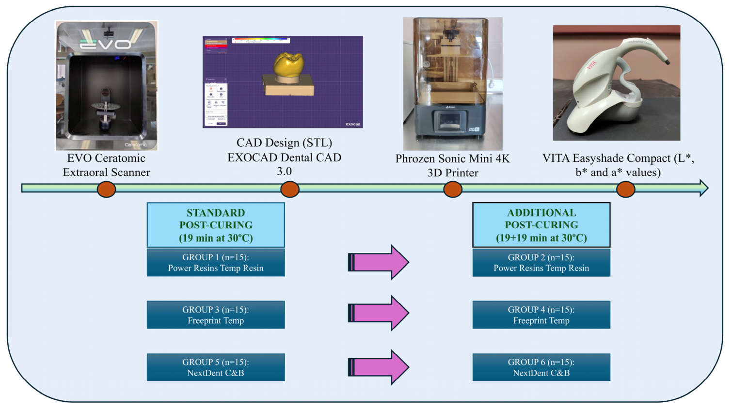
A cross-sectional comparative in vitro study was conducted to evaluate the color of provisional restorations fabricated with three different types of resins using a Phrozen Sonic Mini 4K (Phrozen Technology, Hsinchu, Taiwan) 3D printer with LCD technology, as a function of post-curing time, following all recommendations of the CRIS (Checklist for Reporting In vitro Studies) guidelines. For the fabrication of the prosthetic restorations, a metallic die of a single-unit crown of a mandibular first molar (tooth 46) was used (
Figure 1). Prior to scanning the metallic die, airborne-particle abrasion was performed with 110 µm aluminum oxide to facilitate accurate scanner reading, and the die was subsequently scanned with a laboratory scanner (EVO CERATOMIC, Ref: 420210; Protechno, Vilamalla, Spain).
A single restoration design was created using EXOCAD Dental CAD 3.0 software (Exocad GmbH, Darmstadt, Germany). A total of 90 crowns of a mandibular first molar (tooth 46) were fabricated and distributed into six groups (n = 15) according to the resin brand—Power Resins Temp (Technology Inc., Denver, CO, USA, ref: P1000TA2, 3BFAB), Freeprint Temp (DETAX GmbH, Ettlingen, Germany, ref: 04064), and NextDent C&B (3D Systems, Rock Hill, SC, USA, ref: P892-1513)—and the type of post-curing protocol (SPC vs. APC). The post-curing phase was executed using a multispectral UV-LED curing unit (Phrozen Post Curing UV Lamp; Phrozen Technology, 2024; FCUV2ST03A-4711117880138) operating simultaneously at wavelengths of 365 nm, 385 nm, and 405 nm, with a total nominal LED power of 60 W. The unit incorporates a 360-degree rotating turntable to optimize the spatial homogeneity of the irradiation spectrum across the specimens. To ensure reproducibility and standardize the radiometric conditions according to dental material standards, the device’s calibrated operational irradiance was maintained at 12.5 mW/cm2 at the specimen plane, under an automated internal thermal baseline programmed at 30 °C.
To define the absolute energy delivered to the specimens, the manufacturer’s calibrated irradiance (12.5 mW/cm
2) at the sample plane was converted into cumulative radiant exposure (UV Dose, E) via the equation:
where
t is time in seconds. Consequently, the standard post-curing group (SPC) was subjected to a total radiant exposure of 14.25 J/cm
2 (19 min–1140 s), while the additional post-curing group (APC) received 28.50 J/cm
2 (38 min–2280 s). While the chamber actively regulates the 30 °C baseline, localized exothermic micro-fluctuations during extended cycles are anticipated due to continuous LED emission. The technical specifications of the post-curing unit and parameters for specimen fabrication are presented in
Table 1. To avoid an arbitrary choice of extended curing time, the 38-min protocol was deliberately selected to evaluate the impact of a repeated manufacturer-recommended standard post-curing cycle (two consecutive 19-min cycles).
The groups were distributed as follows: Group 1, 15 crowns fabricated with Power Resins Temp and subjected to standard post-curing; Group 2, 15 crowns fabricated with Power Resins Temp and subjected to standard plus additional post-curing; Group 3, 15 crowns fabricated with Freeprint Temp and subjected to standard post-curing; Group 4, 15 crowns fabricated with Freeprint Temp and subjected to standard plus additional post-curing; Group 5, 15 crowns fabricated with NextDent C&B and subjected to standard post-curing; and Group 6, 15 crowns fabricated with NextDent C&B and subjected to standard plus additional post-curing.
The STL files obtained after design with EXOCAD Dental CAD 3.0 were sent to the Phrozen Sonic Mini 4K printer (Phrozen Technology, 2024; ref: 8000000005465), which uses LCD printing technology, to perform three-dimensional printing in 50 µm layers with a total thickness of 2 mm. Shade A2 from the VITA Classic shade guide (VITA Zahnfabrik, Bad Säckingen, Germany) was used for all three resin brands. The crowns were printed at an angulation of 180°, following the manufacturers’ instructions (
Figure 2). All resins complied with ISO 13485 and ISO 10477 standards.
After printing, the crowns were placed in a washing unit (Phrozen Technology, Hsinchu, Taiwan) with 91% isopropyl alcohol for 10 min, according to the manufacturer’s instructions. Standard post-curing was then performed, followed by trimming (fine diamond disc) and polishing of the restorations (polishing rubbers at low speed).
2.2. Color Measurement
For color measurement, the samples were evaluated using a VITA Easyshade Compact spectrophotometer (VITA Zahnfabrik; serial no. H20394). After calibration, the spectrophotometer tip was placed in contact with the middle third of the vestibular surface of each restoration, and the L*, a*, and b* coordinates were recorded in three separate measurements inside a neutral 50 × 50 × 50 cm chamber with gray walls. All measurements were performed by the same operator on the same day inside a gray chamber (L* = 50). Measurements were taken under stable ambient lighting conditions in an indoor environment with natural daylight, and the arithmetic mean of the three measurements was used for the statistical calculations.
This procedure was carried out for all six study groups (
Figure 2). The color difference (ΔE) was calculated using the following formula: ΔE (CIEL*a*b*) = √(ΔL*)
2 + (Δa*)
2 + (Δb*)
2. A color difference of ΔE < 3.3 units was used as the threshold for clinical acceptability [
20,
22]. A tooth color mismatch remaining within the range of a single intermediate shade (
half-shade), which corresponds to the clinical acceptability threshold of ΔE < 3.3, is physically perceptible under instrumental or expert evaluation; however, it is considered clinically acceptable as it does not compromise the aesthetic success of the restoration and falls within the standard limits of visual tolerance in dental practice [
20,
22].
2.3. Statistical Analysis
Sample size was determined using G*Power software (v. 3.1.9.7). For a one-way analysis of variance (ANOVA), assuming a medium-to-large effect size (f = 0.40), a significance level of α = 0.05, and a statistical power of 95% (1 − β = 0.95) for three independent groups, the calculation indicated a minimum of 84 samples. The sample was increased to a total of 90 crowns (n = 30 per resin type) to enhance the robustness of the study and compensate for potential variability.
Statistical analysis was performed using IBM SPSS Statistics 29.0.2.0 software. The Shapiro–Wilk test was used to assess the normality of data distribution. When normal distribution assumptions were met, the paired Student t-test was applied; otherwise, the Wilcoxon signed-rank test was used. The results indicated a non-normal distribution for the b* variable of the Power Resins resin and for the L* variable of the NextDent C&B resin; consequently, the non-parametric Wilcoxon test was applied to these variables. For the remaining variables, a normal distribution was confirmed (p ≥ 0.05). Additionally, differences in ΔE among groups were compared using one-way ANOVA. A p value < 0.05 was considered statistically significant.
4. Discussion
This study evaluated the impact of an additional post-curing protocol (APC) on the optical properties of 3D-printed resins, comparing the results with the manufacturer’s standard post-curing cycle (SPC). After data analysis, the null hypotheses were not rejected, since the color changes observed in the b* coordinate did not show significant differences in the intergroup analysis and did not compromise clinical acceptability. Color stability and mechanical resistance remain the main long-term restorative challenges for these resins [
6,
37].
Beyond its direct clinical implications, the present investigation contributes to the understanding of how increased radiant exposure influences the optical stability of contemporary LCD-fabricated provisional restorative materials. Optical behavior in photopolymerizable resins is intrinsically associated with polymer network formation, residual photoinitiator consumption, and light scattering phenomena occurring during post-curing. Consequently, evaluating color coordinates after controlled increases in post-curing dose provides indirect information regarding material stabilization processes. Moreover, unlike previous investigations based predominantly on standardized flat specimens, the present study employed full-anatomical provisional crowns, which represent a more clinically realistic geometry and may influence light propagation and optical response.
The findings of our study align with the work of several authors who suggest that, although additional post-curing aims to maximize polymerization, it may induce slight but measurable optical changes [
38,
39]. The generalized reduction in lightness (L*) in all three materials after the APC protocol indicates that longer post-curing times tend to densify the polymers, a phenomenon described by Song et al. as critical for long-term stability [
35].
In terms of post-curing time, Celikel et al. [
31] examined color and translucency differences in 3D-printed provisional crowns. They concluded that extending post-curing duration did not produce significant color variations compared with the manufacturer-recommended time, in agreement with the present results. In their study, the acceptable ΔE threshold was also not exceeded in the additional post-curing groups (ΔE limit: 2.25). Similarly, Siqueira et al. [
33] evaluated the effect of different post-curing times on the color stability of 3D-printed resins and reported no statistically significant color changes in any of the four groups analyzed (no post-curing, 16 min, 32 min, and 60 min).
Sahrir et al. [
40] investigated the influence of different post-curing durations and light intensities on the color stability of resins. They reported that higher light intensity combined with shorter post-curing times improved color stability and identified optimal post-curing conditions of 5 min at 860 mW/cm
2 and 10–15 min at 210 mW/cm
2. However, significant color differences were found in single-unit crowns post-cured for 5 min at 420 mW/cm
2.
The color stability observed in this study can be explained through the stabilization kinetics of the polymer network. When the printed provisional resins are exposed to a constant multispectral irradiance of 12.5 mW/cm2, a progressive cross-linking reaction takes place. During the first 19 min of exposure (SPC group), the light intensity efficiently activates the remaining photoinitiators, driving a rapid initial curing phase that establishes the baseline structure and color coordinates of the material. When the exposure is doubled to 38 min (APC group) under the exact 12.5 mW/cm2 output, the material reaches an asymptotic stabilization plateau, a phenomenon widely described in dental polymer literature. As the resin cross-links and becomes rigid, the mobility of the remaining molecules decreases drastically, slowing down the reaction. Our colorimetric results directly support this physical behavior: the minor, clinically acceptable immediate color differences (ΔE) between the 19-min and 38-min groups prove that the material had already reached its optical stability plateau during the standard cycle. Therefore, extending the post-curing time under a controlled 12.5 mW/cm2 intensity does not cause over-curing or thermal degradation, but rather acts as a safe clinical protocol that ensures the complete maturation and structural homogeneity of the provisional restoration.
These discrepancies in the literature highlight that direct comparisons remain limited due to methodological differences among studies, such as the specific light intensity outputs used, the atmospheric temperature controls and the structural configuration of the samples (anatomical crowns versus standardized flat specimen disks). Due to the limited literature on this topic, further research in this direction is recommended.
Regarding the post-curing of 3D-printed provisional restorations, several authors have investigated whether different resins exhibit differences in color stability. Jong-Eun Kim et al. [
38] reported a significant color change after 1 h of UV light post-curing, which persisted in measurements taken up to 6 months. In another study, Kim et al. [
17] highlighted that color changes increase with longer light exposure, which may be primarily related to photoinitiators.
The findings of the present study demonstrate that the post-curing protocol exerts a decisive influence on the color coordinates of 3D-printed resins, with marked material-dependent variability. When comparing SPC and APC, a trend toward stabilization of the b* coordinate was observed, particularly in Freeprint Temp and NextDent C&B, where yellowing values decreased after longer light exposure. This behavior is consistent with the findings of Sahrir et al. [
40] and Soto-Montero et al. [
32], who reported that higher conversion of residual monomers alters light transmission through the polymer. In contrast, the behavior of Power Resins Temp—showing an increase in b* in the APC group—supports Shin et al.’s [
41] thesis regarding the formulation-specific behavior of each manufacturer’s material. These findings suggest that optimizing lightness (L*) and minimizing color change require customized post-curing protocols, since generic processing could compromise the esthetics of provisional restorations in clinical practice.
The results of this study indicate that optimization of printing and post-curing parameters allows the fabrication of restorations with minimal color change (ΔE), maintaining values within accepted thresholds for clinical success. This agrees with the work of Sahrir et al. [
40], whose objective was to determine how different light intensities, exposure times, and energy levels during post-curing affect the final color of 3D-printed crowns, in order to identify protocols that minimize chromatic alteration. In this regard, the present data are also consistent with the findings of Celikel et al. [
31], who investigated the effect of different post-curing cycles—specifically, variations in duration and repetition of light exposure (0, 750, 1500, 2250, and 3000 cycles). Temizci and Kölüş [
42] provided a multifactorial perspective by evaluating the combined influence of printing angle (layer orientation) and post-curing time on color and translucency, emphasizing that visual esthetics depends on the interaction between printing geometry and light. In all these studies, the authors, as in the present work, analyzed color coordinates in detail.
A comparative analysis of photopolymerizable provisional dental resins (such as Power Resins Temp, Freeprint Temp, and NextDent C&B) shows that formulations containing 2-hydroxyethyl methacrylate (HEMA) exhibit greater hydrophilicity. This implies increased susceptibility to water absorption and reduced color stability after curing, as water may swell the polymer matrix and promote staining.
Chromatic changes in photopolymerizable resins are closely related to the composition and concentration of the photoinitiators used in their formulation. Color stability in esthetic restorative materials is a critical factor to ensure optimal integration with adjacent dental tissues and, consequently, clinical success [
41].
Celikel et al. [
31] reported that photoinitiator selection influences chromatic variability in restorations. Among the most used photoinitiators in post-curing systems are bis(2,4,6-trimethylbenzoyl) phenylphosphine oxide (BAPO), camphorquinone (CQ), and diphenyl(2,4,6-trimethylbenzoyl) phosphine oxide (TPO). In the materials evaluated in this study, the photoinitiators are monoacylphosphine oxide and bis(2,4,6-trimethylbenzoyl) phenylphosphine oxide, which absorb in the 380–420 nm range.
When analyzing the clinical acceptability of color differences, light exposure time emerges as the critical shared variable; Soto-Montero et al. [
32] showed that different post-curing times induce color alterations, highlighting exposure time as the key factor. Precision during the polymerization phase allows the material to reach the stability needed to withstand aging, as described by Shin et al. [
11], who emphasized that greater specificity in post-curing configuration (intensity, cycles, and time) is essential to ensure that a restoration is not only accurate initially but remains within clinical acceptability thresholds over time.
In the present study, ΔE values were below 3.3, the clinically acceptable threshold (Group 1: 1.7; Group 2: 1.8; Group 3: 1.7). Soto-Montero et al. [
32] analyzed color differences in resins subjected to 10- and 15-min post-curing times compared with non-post-cured resins and reported significant color differences in ΔE at both 10 and 15 min of post-curing.
The study by Soto-Montero et al. [
32] provides a useful point of comparison for the present results, as it evaluated the effect of six post-curing intervals on the optical and mechanical properties of printed resins. Their findings showed that increasing light exposure time led to reduced lightness (L*) and a progressive increase in b*, attributing these changes to photoinitiator kinetics and higher degrees of polymer conversion. Furthermore, they demonstrated that most of the chromatic alteration (ΔE) occurs in the early stages of post-curing (10 min), after which color stabilizes. This evidence supports the observation that APC is a safe strategy strictly in terms of chromatic clinical acceptability; however, its consequences on mechanical performance, structural wear and long-term behavior require further validation before full clinical feasibility can be definitively claimed.
While the present findings demonstrate immediate chromatic stability following additional post-curing, caution should be exercised when extrapolating these results to long-term clinical performance. Water sorption, hydrolytic degradation, thermal stresses, and residual photoinitiator oxidation may progressively alter the optical properties of resin-based restorations during clinical service. Consequently, accelerated aging protocols, including thermocycling, artificial saliva storage, and prolonged water immersion, should be incorporated into future investigations to determine whether the immediate optical stability observed in the present study is maintained over time [
11,
13,
32,
38].
Several limitations of the current study design must be acknowledged. First, although the post-curing chamber actively regulates a 30 °C thermal baseline, the continuous multispectral LED emission inevitably induces minor internal exothermic variations. While these thermal fluctuations were not monitored online, they are known to potentially modulate the structural relaxation and subsequent optical coordinates of the resin matrix. Second, the degree of double bond conversion (DC%) was not directly quantified via Fourier-Transform Infrared Spectroscopy (FTIR).
Although FTIR spectroscopy remains the gold standard for quantifying monomer conversion in photopolymerizable dental materials, several investigations evaluating the optical behavior of 3D-printed restorations have relied primarily on spectrophotometric analysis without simultaneous FTIR characterization. Therefore, while a direct conversion–color correlation could not be established in the present investigation, the methodology remains consistent with previously validated approaches for color stability assessment. Future studies should combine FTIR, UV-visible spectroscopy, and colorimetric measurements to elucidate the mechanistic relationship between conversion kinetics and chromatic stability [
10,
31,
38].
The irradiation protocol was intentionally restricted to two clinically relevant exposure conditions corresponding to the manufacturer-recommended cycle and its repetition. Consequently, the study was not designed to establish complete kinetic conversion or optical response curves. Although intermediate radiant exposure levels would provide additional mechanistic information regarding plateau formation, the present design reflects realistic laboratory workflows and therefore preserves its direct clinical applicability [
17,
32,
33].
Additionally, temperature-dependent effects may influence polymerization kinetics and optical behavior in photocurable materials. Although the curing unit employed maintains an internally regulated operating temperature of 30 °C, localized exothermic fluctuations were not directly monitored. Future investigations integrating infrared thermography or thermocouple-based measurements would help clarify the contribution of thermal phenomena to color development and polymer network maturation [
26,
30].
Although a quantitative radiometric mapping of the chamber was not performed, the rotating platform was specifically incorporated to reduce spatial variations in irradiance distribution and improve exposure homogeneity across the restoration surface. All specimens were positioned and processed according to an identical standardized protocol.
Additionally, this study evaluated only immediate optical properties without considering long-term oral aging conditions or concurrent mechanical testing like flexural strength. Lastly, a conventional provisional control group, such as milled PMMA or bis-acryl resins, was not included for direct comparison, meaning the results should be generalized to clinical practice with caution.
The immediate chromatic stability demonstrated in the present investigation should be interpreted as a baseline optical characterization rather than definitive evidence of long-term clinical color durability.




 ,
,