Review

Contemporary Approaches to Obstructive Sleep Apnea: A Review of Orthodontic and Non-Orthodontic Interventions in Children and Adults

by
Janvier Habumugisha
Department of Orthodontics, Graduate School of Medicine, Dentistry and Pharmaceutical Sciences, Okayama University, Okayama 700-8530, Japan
Oral 2025, 5(3), 55; https://doi.org/10.3390/oral5030055 (registering DOI)
Submission received: 14 April 2025 / Revised: 8 May 2025 / Accepted: 22 July 2025 / Published: 1 August 2025

Abstract

Background: Obstructive sleep apnea (OSA) is a prevalent disorder in both pediatric and adult populations, characterized by substantial morbidity encompassing cardiovascular, neurocognitive, and metabolic impairments. Management strategies vary by age group and underlying etiology, with orthodontic and non-orthodontic interventions playing key roles. This narrative review synthesizes the current evidence on orthodontic and non-orthodontic therapies for OSA in pediatric and adult populations, emphasizing individualized, multidisciplinary care approaches and highlighting future research directions. Methods: A narrative review was conducted using PubMed, Scopus, and Google Scholar to identify studies on diagnosis and management of OSA in children and adults from 2000 to 2025. Results: In pediatric patients, treatments such as rapid maxillary expansion (RME), mandibular advancement devices (MADs), and adenotonsillectomy have shown promising outcomes in improving airway dimensions and reducing apnea–hypopnea index (AHI). For adults, comprehensive management includes positive airway pressure (PAP) therapy, oral appliances, maxillomandibular advancement (MMA) surgery, and emerging modalities such as hypoglossal nerve stimulation. Special attention is given to long-term treatment outcomes, adherence challenges, and multidisciplinary approaches. Conclusions: The findings highlight the need for individualized therapy based on anatomical, functional, and compliance-related factors. As the understanding of OSA pathophysiology evolves, orthodontic and adjunctive therapies continue to expand their role in achieving durable and patient-centered outcomes in sleep apnea management.

1. Introduction

Obstructive sleep apnea (OSA) is a sleep-related breathing disorder characterized by repeated episodes of upper airway obstruction during sleep, leading to intermittent hypoxia and disrupted sleep architecture [1]. OSA is a major public health issue, as it is linked to chronic physical and mental health conditions, higher healthcare utilization, increased risk of motor vehicle accidents, decreased productivity, and workplace absenteeism [2]. It affects both children and adults, although etiology, clinical presentation, and treatment strategies differ markedly between these groups.
While adenotonsillar hypertrophy is a common cause of OSA in pediatric cases [3,4], adult OSA is more commonly linked to obesity, craniofacial morphology, and neuromuscular tone. If left untreated, OSA can result in serious health consequences, including cardiovascular disease, neurocognitive impairment, and diminished quality of life [5,6].
Over the years, various treatment modalities, ranging from non-invasive therapies like continuous positive airway pressure (CPAP) to surgical and dental interventions, have been developed to manage OSA. Orthodontic treatments such as mandibular advancement appliances (MAAs), rapid maxillary expansion (RME), and myofunctional therapy (MFT) are increasingly recognized for their role in modifying airway anatomy and function [7,8,9]. Simultaneously, non-orthodontic options including positive airway pressure (PAP) therapy, adenotonsillectomy, and emerging therapies like hypoglossal nerve stimulation have shown varied success across age groups [10,11].
Although many studies have assessed individual interventions [12,13], there is a lack of recent comprehensive reviews that compare both orthodontic and non-orthodontic approaches across pediatric and adult populations within a single framework. Most existing literature focuses on either a specific age group or a single modality, limiting clinicians’ ability to take an integrated, age-adapted approach to care.
This narrative review addresses this gap by synthesizing current evidence on both orthodontic and non-orthodontic treatments for OSA. By highlighting similarities, differences, and clinical considerations across pediatric and adult populations, this review aims to inform personalized and multidisciplinary treatment planning in contemporary OSA management.

2. Materials and Methods

Literature Search Strategy

A narrative review of the literature was conducted to identify relevant publications addressing the diagnosis and management of OSA in both pediatric and adult populations. Searches were conducted using databases such as PubMed, Scopus, and Google Scholar, covering publications from the past 25 years (2000–2025). Keywords included combinations of “obstructive sleep apnea”, “orthodontic treatment”, “mandibular advancement”, “rapid maxillary expansion”, “adenotonsillectomy”, “CPAP”, “oral appliance therapy”, and “hypoglossal nerve stimulation”. Studies were selected based on their clinical relevance and contribution to current understanding of both orthodontic and non-orthodontic interventions in OSA management.

3. Literature Review

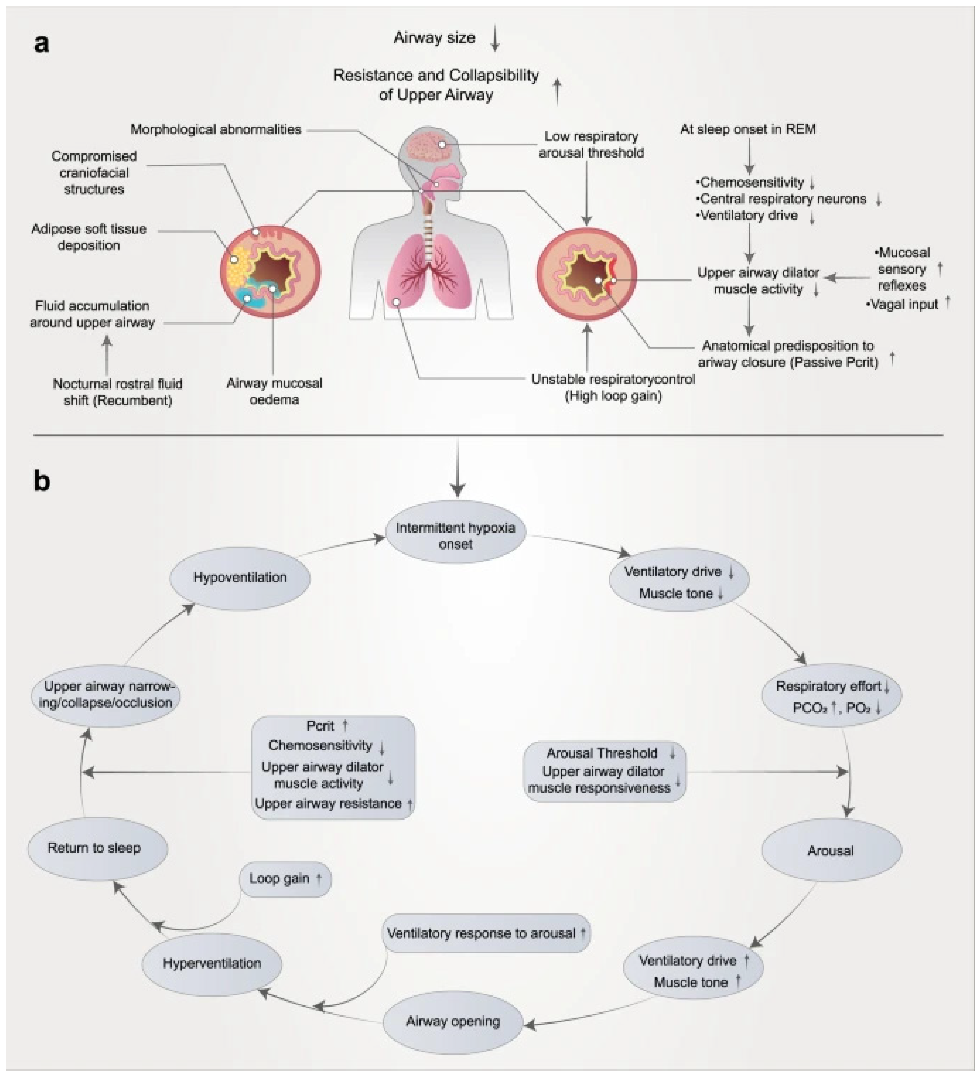
3.1. Pathophysiology of OSA

The pathophysiology of OSA is complex and multifactorial, involving a dynamic interplay between anatomical and non-anatomical factors. Anatomical contributors include upper airway narrowing due to obesity-related fat deposition, craniofacial abnormalities, soft tissue hypertrophy, and other structural variations. In addition, obesity and reduced neuromuscular tone exacerbate airway obstruction during sleep. Non-anatomical factors involve impaired neuromuscular control of the pharyngeal dilator muscles, elevated ventilatory loop gain, reduced arousal thresholds, and increased passive collapsibility of the pharyngeal airway. These elements interact to destabilize the upper airway during sleep, resulting in recurrent obstructions, intermittent hypoxia, and fragmented sleep architecture [14,15,16,17]. A summary of the OSAS pathophysiology and the interplay between contributing factors is illustrated in Figure 1.

3.2. Diagnostic Considerations

Orthodontists play a pivotal role in the early identification of patients at risk for OSA, particularly through the recognition of craniofacial and dental anomalies that may contribute to airway obstruction [18,19]. Key anatomical indicators include retrognathia (a posteriorly positioned mandible), a narrow maxillary arch, and an elongated soft palate [20,21,22,23], all of which can contribute to obstruction of the upper airway and increase susceptibility to OSA. In pediatric populations, early screening is essential, as untreated OSA can lead to behavioral problems, poor academic performance, and growth disturbances. The Pediatric Sleep Questionnaire (PSQ) is commonly utilized in these cases, providing a validated tool to assess sleep-disordered breathing symptoms in children [24].
In adult patients, screening is equally important, particularly before undergoing surgical procedures or in those presenting with suggestive symptoms such as loud snoring, daytime fatigue, or witnessed apneas. Screening instruments like the Berlin questionnaire (BQ), STOP-BANG questionnaire (SBQ), and STOP questionnaire (which stands for snoring, tiredness, observed apneas, and high blood pressure) are commonly used to detect OSA. Additionally, the Epworth Sleepiness Scale (ESS), initially created to evaluate daytime sleepiness, has been proposed as a tool for identifying OSA [25].

3.3. Orthodontic Treatment Outcomes and Evidence in Pediatric OSA

Orthodontic interventions, either as standalone therapies or in conjunction with medical and surgical treatments, have demonstrated promising results in the management of pediatric OSA. These interventions target structural contributors to upper airway obstruction, such as craniofacial abnormalities and dental arch deficiencies, with the goal of improving airway patency and reducing OSA severity [26].
Left untreated, pediatric OSA can lead to significant long-term consequences, including cognitive impairment, behavioral disturbances, poor academic performance, and cardiovascular complications [27].
While adenotonsillectomy remains the first-line therapy for children with hypertrophic tonsils and adenoids, a substantial proportion of patients, particularly those with a high preoperative apnea–hypopnea index (AHI), obesity, neuromuscular conditions, or unfavorable craniofacial anatomy, experience persistent OSA postoperatively. In such cases, orthodontic treatment modalities play a pivotal role in alleviating symptoms and promoting long-term airway stability. Consequently, optimal management of pediatric OSA often requires a multidisciplinary approach that integrates orthodontic care with pediatric sleep medicine, otolaryngology, and myofunctional therapy [8,28,29,30].

3.4. Treatment of Pediatric OSA

3.4.1. Adenotonsillectomy Effect

Adenotonsillectomy is the most common first-line surgical intervention for pediatric OSA and is indicated in cases where adenotonsillar hypertrophy is a primary contributing factor. Numerous studies [31,32,33,34] have demonstrated substantial improvements in AHI, oxygen desaturation index, and quality of life following the procedure. A pivotal randomized controlled trial by Garetz et al. [31] evaluated the impact of adenotonsillectomy compared to watchful waiting in children aged 5 to 9.9 years with OSAS. The study demonstrated that children who underwent surgery experienced significantly greater improvements in both general and OSAS-specific quality of life (QoL), as well as sleep-related symptoms, compared to the control group. These improvements were observed across multiple validated scales, including the Pediatric Quality of Life Inventory and the Pediatric Sleep Questionnaire. Notably, the findings were independent of obesity status or baseline OSAS severity, although some racial differences were noted. The authors concluded that adenotonsillectomy offers superior benefits in improving QoL and reducing symptoms in pediatric OSAS patients.
In contrast, Mitchell [32] included children with moderate to severe OSA (baseline AHI ≥ 5) and found that while adenotonsillectomy significantly reduced AHI overall, residual OSA persisted especially in those with severe preoperative disease. The persistence of postoperative symptoms such as snoring highlighted the need for ongoing monitoring. This suggests that adenotonsillectomy may not be curative in all cases, particularly in severe phenotypes. Similarly, Bhattacharjee et al. [33] identified that only about a quarter of children achieved complete resolution (AHI < 1), and older age and higher BMI were predictors of residual disease. This study emphasizes the variability in treatment response and challenges the notion of adenotonsillectomy as a standalone definitive treatment.
Collectively, these studies affirm the benefit of adenotonsillectomy as an initial intervention but reveal considerable variability in treatment response. Importantly, they highlight that surgery alone often fails to fully resolve OSA, especially in children with complex or multifactorial phenotypes. This underscores the necessity for individualized treatment approaches and the integration of adjunctive therapies, such as orthodontic or myofunctional interventions, for patients at higher risk of residual disease. Moreover, the predominance of short-term follow-up in the literature emphasizes the urgent need for long-term, prospective studies to evaluate the sustainability of surgical outcomes and to determine the most effective multimodal treatment strategies.

3.4.2. Mandibular Advancement Appliances in Pediatric Obstructive Sleep Apnea

Mandibular advancement appliances (MAAs) can enhance the lateral dimension of the velopharyngeal airway by repositioning the mandible anteriorly and decreasing airway collapsibility. Additionally, their use may promote upper airway stability by stimulating dilator muscles such as the genioglossus [35]. Consequently, several studies have evaluated oral jaw-positioning appliances for treating OSA in children presenting with various forms of malocclusion and reported their effectiveness in reducing OSA severity [36,37,38,39,40].
Previous clinical study suggests that the use of customized MAAs in children with mild to moderate OSA is associated with improvements in AHI, oxygen saturation, and overall sleep quality [41]. In a preliminary study, Zhang et al. [38] evaluated the effects of a twin-block appliance in 46 children with OSA and mandibular retrognathia, reporting a significant reduction in AHI (from 14.08 ± 4.25 to 3.39 ± 1.86; p < 0.01) and an improvement in minimum oxygen saturation. The authors concluded that the twin block appliance may be effective in enhancing both facial aesthetics and OSA symptoms in carefully selected pediatric patients presenting with OSA and mandibular retrognathia. Similar results were observed in a separate study that demonstrated significant increases in upper airway volume and cross-sectional dimensions at the oropharyngeal and hypopharyngeal levels following mandibular advancement, though nasopharyngeal changes were minimal [42]. Duan et al. [39] further supported these findings using a modified twin-block appliance (MTBA), showing both respiratory improvement and favorable skeletal changes, including anterior–superior hyoid repositioning.
In addition, the modified monobloc (MM) has also shown promise. Cozza et al. [43] reported that children with Class II skeletal patterns treated with the MM appliance experienced significant reductions in AHI (p = 0.0003), improved sleep quality, and decreased daytime sleepiness. Collectively, these studies support the therapeutic potential of mandibular advancement in pediatric OSA, particularly in patients with retrognathic features.
However, several limitations in the current evidence base must be acknowledged. Most available studies have small sample size, lack randomization, and are limited by short-term follow-up and absence of appropriate control groups. As highlighted in previous systematic reviews, while the observed improvements in AHI are encouraging, the evidence remains insufficient to confirm the long-term efficacy of MAAs in treating pediatric OSA [35]. Inconsistencies in study design, appliance type, and outcome measurement further complicate comparisons. Larger, well-controlled trials with standardized protocols and long-term data are needed to validate these findings and clarify their generalizability.

3.4.3. Rapid Maxillary Expansion

Rapid maxillary expansion is a well-established orthodontic procedure used to correct maxillary transverse deficiencies by widening the upper jaw. In children with OSA and narrow maxilla, RME can increase nasal cavity volume, reduce nasal resistance, and improve airflow. Several studies have reported significant reductions in AHI and improvement in nocturnal oxygenation following RME [8]. In a prospective clinical trial, Villa et al. [44] evaluated the effectiveness of RME in children with OSAS. They reported significant reductions in the apnea–hypopnea index, hypopnea index, and arousal index (all p < 0.01). Parental reports also indicated substantial improvement in OSA symptoms. The authors concluded that RME is a promising intervention for managing pediatric OSAS and emphasized the need for orthodontic screening in children with signs of sleep-disordered breathing. A complementary long-term study by Pirelli et al. further reinforced the efficacy of RME in pediatric OSA patients. This prospective study included 31 children with OSA characterized by isolated maxillary constriction and no adenotonsillar hypertrophy. After completing orthodontic treatment at a mean age of 8.7 years, 23 participants were followed for an average of 12 years. Polysomnographic evaluations performed in late adolescence or early adulthood confirmed sustained normalization of respiratory parameters. CT imaging demonstrated long-term stability of maxillary expansion. At the final evaluation, all 23 individuals with continuous follow-up showed no clinical signs of OSA recurrence. Additionally, all participants demonstrated normal academic performance. These findings underscore the potential of RME as a durable and effective intervention for pediatric OSA in cases associated with isolated maxillary constriction [45].
While these studies suggest RME may improve airway dimensions and reduce OSA severity in children with maxillary constriction, the overall quality of evidence remains limited. Most studies have small sample sizes, lack control groups, and are constrained by short-term follow-up. Moreover, randomized controlled trials comparing RME to watchful waiting or alternative treatments are scarce, making it difficult to distinguish true treatment effects from natural resolution of symptoms. Ethical and methodological challenges—such as blinding difficulties and narrow patient selection—further complicate high-quality research in this area. Therefore, although RME appears promising, its role in pediatric OSA management should be approached with caution pending confirmation from robust clinical trials [46].

3.4.4. Myofunctional Therapy and Orofacial Exercises

Myofunctional therapy involves exercises that target the orofacial muscles, particularly the tongue, lips, and soft palate, aiming to improve airway patency and breathing patterns during sleep [47]. In children, improper tongue posture, mouth breathing, and low muscle tone can contribute to airway collapse. Emerging evidence suggests that when used alongside orthodontic interventions like RME or MADs, myofunctional therapy may enhance long-term outcomes by retraining neuromuscular function and preventing relapse. It is especially useful for children with persistent OSA symptoms post-adenotonsillectomy.
Guilleminault et al. [48] investigated the long-term effects of myofunctional therapy in children with sleep-disordered breathing (SDB) who had previously undergone adenotonsillectomy and orthodontic treatment. In this retrospective study, 24 children with initially normalized polysomnography (PSG) were evaluated. Of these, only 11 completed a 24-month course of myofunctional therapy, while the remaining 13 did not. At follow-up (22–50 months later), children who did not receive myofunctional therapy experienced recurrence of SDB symptoms, with an average AHI of 5.3. In contrast, children who underwent myofunctional therapy maintained normal sleep and respiratory parameters. These results are complemented by findings from Suzuki et al. [29], who assessed the efficacy of oral myofunctional therapy (OMFT) using a lip trainer device over a 2-month period in young adults with elevated AHI and excessive daytime sleepiness. Their study demonstrated significant improvements in labial closure force (LCF), nocturnal oxygen saturation (SpO2), and a notable reduction in AHI following OMFT. Specifically, AHI decreased from 15.1 to 9.2 events/hour, while SpO2 improved from 90.0% to 96.8%.
Myofunctional therapy, including targeted orofacial exercises, has shown promise in enhancing upper airway stability and improving key outcomes such as AHI, oxygen saturation, and snoring severity, particularly in children with residual symptoms post-adenotonsillectomy or orthodontic treatment. While current findings support its role as a valuable adjunctive therapy in pediatric OSA, the overall quality of evidence remains limited due to small sample sizes and a lack of randomized controlled trials. Further well-designed studies with standardized protocols and long-term follow-up are needed to clarify its clinical efficacy [49,50].

3.5. Management of Adult OSA

In contrast to pediatric patients, where adenotonsillar hypertrophy is often the primary etiology, adult OSA is typically multifactorial and more complex in nature. As such, adult OSA management requires a multifaceted approach that may include behavioral, medical, orthodontic, and surgical interventions [51]. The following subsections explore key treatment strategies for adult OSA, with a particular focus on craniofacial and airway-modifying therapies. While positive airway pressure (PAP) therapy remains the cornerstone of initial management, structural and orthodontic interventions play an increasingly critical role in patients who are intolerant to PAP or require anatomical correction. Emerging modalities such as oral appliance therapy and hypoglossal nerve stimulation further expand the therapeutic landscape.

3.5.1. Maxillomandibular Advancement (MMA) Surgery

Maxillomandibular advancement (MMA) is one of the most effective surgical interventions for moderate to severe adult OSA, particularly in patients who are intolerant to positive airway pressure therapy. The procedure involves advancing both the maxilla and mandible, thereby increasing upper airway volume and tensioning the pharyngeal soft tissues [52]. Studies consistently report significant reductions in AHI, improved oxygen saturation, and enhanced quality of life postoperatively.
Boyd et al. [53] conducted a prospective cohort study to evaluate the long-term effectiveness and safety of MMA in adults with moderate to severe OSA. Thirty patients were followed for an average of 6.6 years post-surgery. Results showed a significant reduction in mean AHI from 49 to 10.9 events/hour, with 83.4% achieving an AHI ≤ 15 and nearly half reaching an AHI < 5. Additionally, patients experienced improvements in diastolic blood pressure, daytime sleepiness, and quality of life. Few long-term adverse effects were reported. The study concludes that MMA is a safe and effective long-term treatment option, especially for patients who are unable to tolerate continuous positive airway pressure (CPAP) therapy. This agrees with the findings of Jaspers et al. [54], who reported that MMA surgery showed promising long-term effects in reducing AHI and ESS (Epworth Sleepiness Scale) scores. However, they emphasized the need for larger long-term cohort studies to further validate these clinical outcomes. A recent study compared the effectiveness of MMA and mandibular advancement devices (MADs) in patients with moderate to severe OSA. MMA was found to be significantly more effective in achieving therapeutic success—defined as an AHI < 15 events/hour and at least a 50% reduction from baseline—demonstrating an odds ratio of 3.22 compared to MADs [55].
These results reinforce the superior efficacy of MMA in more severe cases of OSA and highlight its role as a definitive treatment option when conservative therapies such as MADs prove insufficient. While MADs offer a non-invasive alternative with documented benefits in mild to moderate cases, MMA provides greater long-term stability in reducing both AHI and daytime symptoms, particularly in anatomically predisposed patients.
Previous systematic reviews have consistently identified important methodological limitations in the MMA literature. These include considerable heterogeneity in how apneas and hypopneas are defined, inconsistent criteria for surgical success, and the absence of standardized reporting for key clinical outcomes. Additionally, the lack of randomized controlled trials and comparative studies, particularly between MMA and other surgical options such as upper airway stimulation (UAS)—limits the ability to draw firm conclusions about relative efficacy. Many studies also lack complete pre- and postoperative data and exclude subjective outcomes or safety metrics. These limitations highlight the urgent need for future multicenter studies with standardized protocols and comprehensive outcome reporting to better define the role of MMA and identify which patient populations derive the greatest long-term benefit [52,56].

3.5.2. Oral Appliance Therapy (Mandibular Advancement Devices)

Oral appliances, particularly mandibular advancement devices (MADs), are a non-invasive alternative for patients with mild to moderate OSA. These devices reposition the mandible anteriorly, enlarging the upper airway and reducing collapsibility. Clinical trials have demonstrated improvements in snoring, daytime sleepiness, and AHI, though typically to a lesser extent than CPAP. Custom-fitted devices by qualified dental professionals are essential for efficacy and comfort.
Pahkala et al. [57] investigated the effects of moderate mandibular advancement using an MAD on upper airway volume and OSA symptoms in 58 adults diagnosed with obstructive sleep apnea (mean AHI 19.2). After 6 months of treatment, 23 patients achieved a complete response (AHI < 5), 13 had partial improvement, and 9 showed no significant change. Significant improvements were noted in AHI, oxygen saturation, snoring, daytime sleepiness, and quality of life. The study concluded that MAD therapy can yield excellent results, although the response is multifactorial and influenced by craniofacial characteristics.
Vecchierini et al. [58] reported five-year follow-up results by evaluating the long-term effectiveness of MADs in 172 adult patients with OSA. Patients were treated with a custom-made, CAD/CAM-manufactured biblock MAD. The overall treatment success rate—defined as a ≥50% reduction in AHI—was 52% at five years, with success rates of 25%, 52%, and 63% in mild, moderate, and severe OSA groups, respectively. Although effectiveness declined compared to earlier follow-ups (79% at 3–6 months and 68% at 2 years), patient-reported symptoms and sleepiness remained stable. Adherence was high, with over 90% using the device ≥6 h/night, and 96.5% expressing willingness to continue therapy. The study concluded that MADs remain a viable and well-tolerated long-term treatment for OSA, especially in patients with moderate to severe disease who are CPAP-intolerant.
In a separate randomized crossover trial, Phillips et al. [59] compared the short-term health effects of CPAP and mandibular advancement device (MAD) therapy in 108 patients with moderate to severe OSA. Over one month of use with each device, CPAP was more effective in reducing AHI, while compliance was significantly better with MAD therapy. Despite differences in efficacy, mean arterial pressure used as the primary cardiovascular outcome was similar between the two treatments, and neither resulted in significant changes in blood pressure. Notably, both therapies yielded comparable improvements in sleepiness, driving performance, and disease-specific quality of life. These findings highlight the trade-off between therapeutic effectiveness and patient adherence, suggesting that MADs may serve as a practical and effective alternative for appropriately selected individuals.
Although there is promising evidence that MADs improve symptoms and daytime functioning in obstructive sleep apnea, their effects on comorbidities and risks such as road traffic accidents remain difficult to synthesize due to the small number of studies and inconsistent methodologies. Larger and longer randomized controlled trials are needed to better evaluate MAD treatment impacts on cardiac, metabolic, neurocognitive outcomes, and medication use. Treatment approaches should be reassessed when patients experience recurrent symptoms, significant weight changes, or develop comorbidities related to obstructive sleep apnea [60].

3.5.3. Positive Airway Pressure (PAP) Therapy

Positive airway pressure therapy, particularly CPAP, remains the first-line treatment for most adults with OSA [59]. By delivering a constant stream of air that prevents airway collapse during sleep, PAP therapy effectively reduces AHI and improves daytime functioning. Despite its efficacy, adherence remains a major challenge, with many patients reporting discomfort or intolerance. Addressing device fit, pressure settings, and nasal resistance can help improve compliance.
Beyond symptom control, CPAP therapy has also demonstrated significant benefits in comorbid conditions associated with OSA. For instance, Mansfield et al. [61] conducted a randomized controlled trial to evaluate the impact of CPAP in patients with both congestive heart failure (CHF) and OSA. Over a 3-month period, 55 patients were randomized to receive either CPAP treatment or no intervention. The study found that CPAP significantly improved left ventricular ejection fraction, reduced overnight urinary norepinephrine excretion (indicating decreased sympathetic nervous system activity), and enhanced quality of life. No significant changes in systemic blood pressure were observed. These findings suggest that CPAP therapy in CHF patients with OSA can improve cardiac function and autonomic regulation, reinforcing the importance of screening and treating sleep apnea in this population.
However, the clinical benefits of CPAP seen in controlled trials may not fully translate to everyday practice due to variable adherence. Stuck et al. [62] conducted a retrospective analysis to assess the real-world effectiveness of CPAP therapy for OSA, focusing on how patient adherence influences AHI outcomes. Data from 82 stable CPAP users were analyzed, showing that although CPAP significantly reduced mean AHI to 2.4, average nightly usage was only 4.7 h, resulting in substantial time spent sleeping without CPAP. When adherence was factored in, the average total AHI across treated and untreated sleep time rose to 11.91, demonstrating that incomplete adherence diminishes overall therapeutic benefit. The authors concluded that adherence must be considered when comparing CPAP to alternative therapies particularly those like surgery, which are not dependent on nightly compliance.
Although CPAP is clinically effective, long-term adherence remains suboptimal, with many patients using the device only 3–5 h per night [63]. This limits real-world effectiveness and may confound observed benefits in clinical trials. Evidence for strategies to improve adherences such as telemonitoring, education, and mask customization—is limited and mostly short-term. Variability in patient factors (e.g., nasal obstruction, facial anatomy) and lack of tailored interventions further complicate outcomes. High-quality, long-term RCTs are needed to optimize adherence strategies and guide individualized care.

3.5.4. Hypoglossal Nerve Stimulation (HGNS)

Hypoglossal nerve stimulation is an emerging neuromodulatory therapy for moderate to severe OSA in patients who are unable to tolerate PAP therapy. The implanted device stimulates the hypoglossal nerve during inspiration, causing tongue protrusion and airway stabilization. Studies have shown substantial reductions in AHI and improvements in sleep-related quality of life. Careful patient selection, including absence of complete concentric collapse at the soft palate, is critical to treatment success.
Kezirian et al. [64] evaluated the 12-month outcomes of hypoglossal nerve stimulation (HGNS) in 31 patients with moderate to severe OSA who were unable to tolerate PAP therapy. Using the HGNS® system (Apnex Medical. HGNS; Apnex Medical, St. Paul, MN, USA), patients experienced significant improvements in AHI, decreasing from 45.4 to 25.3 events/h, and in quality of life, with FOSQ scores increasing from 14.2 to 17.0 (both p < 0.001). Device usage averaged 5.4 h per night, with 86% nightly adherence. While three serious device-related adverse events were reported (one infection, two lead dislodgements), no new adverse events occurred after 6 months. The findings support HGNS as a safe, feasible, and effective alternative for patient intolerance to conventional PAP therapy. These results are in agreement with findings from Eastwood et al. [65], who similarly reported high adherence to HGNS, with usage at 89% of nights for an average of 5.8 h. In that 6-month trial, significant improvements were observed across multiple measures, including a reduction in AHI from 43.1 to 19.5, and improvements in ESS scores (12.1 to 8.1), Functional Outcomes of Sleep Questionnaire (FOSQ) scores (14.4 to 16.7), Sleep Apnea Quality of Life Index (SAQLI) scores (3.2 to 4.9), and Beck Depression Inventory (BDI) scores (15.8 to 9.7) (p < 0.05 for all). HGNS was shown to be safe, effective, and well tolerated, offering substantial reductions in OSA severity and related symptoms.
Together, these studies support HGNS as a safe, effective, and well-tolerated long-term treatment option for patients with moderate to severe OSA who are unable to adhere to positive airway pressure therapy. However, current evidence is limited by a lack of randomized controlled trials, short follow-up durations, and potential reporting bias. Inconsistent reporting of response and complication rates further complicates interpretation. While adherence appears higher than with CPAP, more independent, long-term studies are needed to confirm efficacy, refine patient selection, and assess safety over time [66].

4. Discussion

4.1. Summary of Evidence and Future Directions

While adenotonsillectomy (AT) is widely regarded as the most common first-line surgical intervention for pediatric OSA, it is not without limitations. Despite significant improvements in objective measures, such as the AHI and oxygen desaturation index, and in subjective outcomes like quality of life (QoL), residual OSA may persist, particularly in children with more severe preoperative disease or those who are obese. Studies have shown that a substantial proportion of children do not experience complete resolution of OSA, particularly those with higher body mass index (BMI) or those presenting with severe disease preoperatively [32,33]. This underscores the importance of ongoing clinical follow-up and potentially adjunctive therapies for patients at higher risk of residual symptoms. Thus, while AT provides substantial benefits, its efficacy in achieving complete resolution of OSA is variable, and individualized treatment strategies, including monitoring and additional interventions, may be necessary for certain patient subgroups to optimize long-term outcomes
This review highlights the evolving role of orthodontic interventions in the diagnosis and management of OSA across pediatric and adult populations. Orthodontic interventions such as mandibular advancement devices (MADs) [67], RME [8], and myofunctional therapy (MFT) [68] have demonstrated clinically meaningful improvements in airway dimensions, respiratory indices, and quality of life. Notably, studies indicate that MFT plays a crucial role in preventing the recurrence of sleep-disordered breathing (SDB) [48,69]. Research has shown that children who underwent myofunctional re-education after adenotonsillectomy and orthodontic treatment had healthier long-term outcomes, whereas those who did not receive MFT experienced a recurrence of symptoms [48]. However, the current evidence is limited by methodological shortcomings; most studies have small sample sizes, lack randomization, and are restricted by short-term follow-up and the absence of appropriate control groups, warranting further high-quality, long-term randomized controlled trials to validate these findings.
In adults, OSA management is inherently more complex due to multifactorial etiologies involving structural, neuromuscular, and lifestyle-related components. While positive airway pressure (PAP) therapy remains the gold standard, adherence issues necessitate alternative strategies [62]. MMA surgery stands out as the most effective structural intervention for moderate to severe cases, offering long-term improvements in AHI and sleep quality [53]. Other treatments, including oral appliance therapy and HGNS, offer individualized options based on patient anatomy and treatment response [25,64].
Despite the growing body of evidence supporting orthodontic and craniofacial interventions, several gaps remain. High-quality, long-term studies are needed to evaluate the durability of orthodontic outcomes and their synergistic effects when combined with other therapies. Moreover, the integration of advanced imaging, digital airway modeling, and personalized treatment planning holds promise for improving patient selection and optimizing outcomes. Future research should focus on developing standardized treatment protocols and exploring the potential of early interceptive orthodontics to prevent the progression of pediatric OSA into adulthood.
Ultimately, the current evidence supports a shift toward a more collaborative, patient-specific, and airway-focused model of care. Orthodontists are increasingly positioned not only as dental specialists but as integral contributors to the multidisciplinary management of OSA—across the lifespan [7,70,71,72].

4.2. Limitations and Recommendations

Due to the nature of this study as a narrative review, there was no formal assessment of study quality or risk of bias, and a quantitative synthesis was not performed. The included studies vary in design, population, and outcome measures, which limits direct comparisons and generalizability. Therefore, we recommend future systematic reviews and meta-analyses to provide stronger evidence and more definitive conclusions for clinical application.

5. Conclusions

Orthodontic interventions play an increasingly important role in the multidisciplinary management of OSA in both children and adults.
In pediatric patients, adjunctive treatments such as rapid maxillary expansion, mandibular advancement, and myofunctional therapy have shown promise, especially in cases of residual OSA following adenotonsillectomy.
In adults, structural therapies including maxillomandibular advancement, oral appliances, and orthodontic approaches offer potential alternatives or complements to positive airway pressure therapy. While a growing number of studies explore these interventions, more high-quality evidence is needed to support their consistent integration into clinical practice.

Funding

This research received no external funding.

Institutional Review Board Statement

Not applicable.

Informed Consent Statement

Not applicable.

Data Availability Statement

Not applicable.

Conflicts of Interest

The author declares no conflicts of interest.

Abbreviations

The following abbreviations are used in this manuscript:
OSAObstructive Sleep Apnea
AHIApnea–Hypopnea Index
CPAPContinuous Positive Airway Pressure
PAPPositive Airway Pressure
MADMandibular Advancement Device
RMERapid Maxillary Expansion
MAMaxillomandibular Advancement
HNSHypoglossal Nerve Stimulation
ESSEpworth Sleepiness Scale
FOSQFunctional Outcomes of Sleep Questionnaire
SAQLISleep Apnea Quality of Life Index
BDIBeck Depression Inventory
PSQPediatric Sleep Questionnaire
CHFCongestive Heart Failure
CTComputed Tomography
LCFLabial Closure Force
MMModified Monobloc
MTBAModified Twin Block Appliance

References

  1. Tolbert, T.M.; Ayappa, I.; Rapoport, D.M. OSA pathophysiology: A contemporary update. Aust. Dent. J. 2024, 69, S68–S83. [Google Scholar] [CrossRef]
  2. Cacho, V. Beyond the mouth: An overview of obstructive sleep apnea in adults for dentists. J. Prosthodont. 2025, 34, 4–9. [Google Scholar] [CrossRef]
  3. Hasuneh, M.M.; Toubasi, A.A.; Khraisat, B.; Aldabbas, H.; AL-Iede, M. Risk Factors of Obstructive Sleep Apnea (OSA) in Pediatric Patients: A Systematic Review and Meta-analysis. J. Pediatr. Health Care 2024, 38, 717–726. [Google Scholar] [CrossRef] [PubMed]
  4. Nosetti, L.; Zaffanello, M.; De Bernardi di Valserra, F.; Simoncini, D.; Beretta, G.; Guacci, P.; Piacentini, G.; Agosti, M. Exploring the Intricate Links between Adenotonsillar Hypertrophy, Mouth Breathing, and Craniofacial Development in Children with Sleep-Disordered Breathing: Unraveling the Vicious Cycle. Children 2023, 10, 1462. [Google Scholar] [CrossRef]
  5. Messineo, L.; Bakker, J.P.; Cronin, J.; Yee, J.; White, D.P. Obstructive sleep apnea and obesity: A review of epidemiology, pathophysiology and the effect of weight-loss treatments. Sleep Med. Rev. 2024, 78, 101996. [Google Scholar] [CrossRef] [PubMed]
  6. Salman, L.A.; Shulman, R.; Cohen, J.B. Obstructive Sleep Apnea, Hypertension, and Cardiovascular Risk: Epidemiology, Pathophysiology, and Management. Curr. Cardiol. Rep. 2020, 22, 6. [Google Scholar] [CrossRef]
  7. Kazmierski, R.H. Obstructive sleep apnea: What is an orthodontist’s role? Prog. Orthod. 2024, 25, 21. [Google Scholar] [CrossRef] [PubMed]
  8. Xie, B.; Zhang, L.; Lu, Y. The role of rapid maxillary expansion in pediatric obstructive sleep apnea: Efficacy, mechanism and multidisciplinary collaboration. Sleep Med. Rev. 2023, 67, 101733. [Google Scholar] [CrossRef]
  9. Koka, V.; De Vito, A.; Roisman, G.; Petitjean, M.; Pignatelli, G.R.F.; Padovani, D.; Randerath, W. Orofacial myofunctional therapy in obstructive sleep apnea syndrome: A pathophysiological perspective. Medicina 2021, 57, 323. [Google Scholar] [CrossRef]
  10. Trzepizur, W.; Cistulli, P.A.; Glos, M.; Vielle, B.; Sutherland, K.; Wijkstra, P.J.; Hoekema, A.; Gagnadoux, F. Health outcomes of continuous positive airway pressure versus mandibular advancement device for the treatment of severe obstructive sleep apnea: An individual participant data meta-analysis. Sleep 2021, 44, zsab015. [Google Scholar] [CrossRef]
  11. Heiser, C.; Steffen, A.; Strollo, P.J.; Giaie-Miniet, C.; Vanderveken, O.M.; Hofauer, B. Hypoglossal nerve stimulation versus positive airway pressure therapy for obstructive sleep apnea. Sleep Breath. 2023, 27, 693–701. [Google Scholar] [CrossRef] [PubMed]
  12. Machado-Júnior, A.J.; Zancanella, E.; Crespo, A.N. Rapid maxillary expansion and obstructive sleep apnea: A review and meta-analysis. Med. Oral Patol. Oral Y Cir. Bucal 2016, 21, e465–e469. [Google Scholar] [CrossRef]
  13. Duan, J.; Xia, W.; Yang, K.; Li, X.; Zhang, F.; Xu, J.; Jiang, Y.; Liang, J.; Li, B. The Efficacy of Twin-Block Appliances for the Treatment of Obstructive Sleep Apnea in Children: A Systematic Review and Meta-Analysis. Biomed Res. Int. 2022, 2022, 3594162. [Google Scholar] [CrossRef] [PubMed]
  14. Pham, L.V.; Schwartz, A.R. The pathogenesis of obstructive sleep apnea. J. Thorac. Dis. 2015, 7, 1358–1372. [Google Scholar] [PubMed]
  15. Lv, R.; Liu, X.; Zhang, Y.; Dong, N.; Wang, X.; He, Y.; Yue, H.; Yin, Q. Pathophysiological mechanisms and therapeutic approaches in obstructive sleep apnea syndrome. Signal Transduct. Target. Ther. 2023, 8, 218. [Google Scholar] [CrossRef]
  16. McNicholas, W.T.; Korkalainen, H. Translation of obstructive sleep apnea pathophysiology and phenotypes to personalized treatment: A narrative review. Front. Neurol. 2023, 14, 1239016. [Google Scholar] [CrossRef]
  17. Sutherland, K.; Cistulli, P.A. Recent advances in obstructive sleep apnea pathophysiology and treatment. Sleep Biol. Rhythm. 2015, 13, 26–40. [Google Scholar] [CrossRef]
  18. Mohamed, A.S.; Habumugisha, J.; Cheng, B.; Zhao, M.; Bu, W.; Liu, L.; Guo, Y.; Zou, R.; Wang, F. A cone-beam computed tomography study of hyoid bone position and airway volume in subjects with obstructive and nonobstructive adenotonsillar hypertrophy. Angle Orthod. 2023, 93, 467–475. [Google Scholar] [CrossRef]
  19. Chen, H.; Aarab, G.; de Ruiter, M.H.T.; de Lange, J.; Lobbezoo, F.; van der Stelt, P.F. Three-dimensional imaging of the upper airway anatomy in obstructive sleep apnea: A systematic review. Sleep Med. 2016, 21, 19–27. [Google Scholar] [CrossRef]
  20. Shigeta, Y.; Ogawa, T.; Tomoko, I.; Clark, G.T.; Enciso, R. Soft palate length and upper airway relationship in OSA and non-OSA subjects. Sleep Breath. 2010, 14, 353–358. [Google Scholar] [CrossRef]
  21. Kim, K.A.; Kim, S.J.; Yoon, A. Craniofacial anatomical determinants of pediatric sleep-disordered breathing: A comprehensive review. J. Prosthodont. 2024, 34, 26–34. [Google Scholar] [CrossRef]
  22. Habumugisha, J.; Ma, S.Y.; Mohamed, A.S.; Cheng, B.; Zhao, M.Y.; Bu, W.Q. Three-dimensional evaluation of pharyngeal airway and maxillary arch in mouth and nasal breathing children with skeletal Class I and II. BMC Oral Health 2022, 22, 320. [Google Scholar] [CrossRef]
  23. Habumugisha, J.; Mohamed, A.S.; Cheng, B.; Liu, L.; Zou, R.; Wang, F. Analysis of maxillary arch morphology and its relationship with upper airway in mouth breathing subjects with different sagittal growth patterns. J. Stomatol. Oral Maxillofac. Surg. 2023, 124, 101386. [Google Scholar] [CrossRef]
  24. Umano, G.R.; Rondinelli, G.; Luciano, M.; Pennarella, A.; Aiello, F.; Mangoni di Santo Stefano, G.S.R.C.; Sessa, A.D.; Marzuillo, P.; Papparella, A.; del Giudice, E.M. Pediatric Sleep Questionnaire Predicts Moderate-to-Severe Obstructive Sleep Apnea in Children and Adolescents with Obesity. Children 2022, 9, 1303. [Google Scholar] [CrossRef] [PubMed]
  25. Lee, J.J.; Sundar, K.M. Evaluation and Management of Adults with Obstructive Sleep Apnea Syndrome. Lung 2021, 199, 87–101. [Google Scholar] [CrossRef] [PubMed]
  26. Huynh, N.T.; Desplats, E.; Almeida, F.R. Orthodontics treatments for managing obstructive sleep apnea syndrome in children: A systematic review and meta-analysis. Sleep Med. Rev. 2016, 25, 84–94. [Google Scholar] [CrossRef] [PubMed]
  27. Blechner, M.; Williamson, A.A. Consequences of Obstructive Sleep Apnea in Children. Curr. Probl. Pediatr. Adolesc. Health Care 2016, 46, 19–26. [Google Scholar] [CrossRef]
  28. Mitchell, R.B.; Kelly, J. Outcome of adenotonsillectomy for obstructive sleep apnea in obese and normal-weight children. Otolaryngol. Head Neck Surg. 2007, 137, 43–48. [Google Scholar] [CrossRef]
  29. Suzuki, H.; Watanabe, A.; Akihiro, Y.; Takao, M.; Ikematsu, T.; Kimoto, S.; Asano, T.; Kawara, M. Pilot study to assess the potential of oral myofunctional therapy for improving respiration during sleep. J. Prosthodont. Res. 2013, 57, 195–199. [Google Scholar] [CrossRef]
  30. Arab, M.; El Ansari, Y.S.; Pelayo, R.; Yoon, A. Management of Paediatric Obstructive Sleep Apnoea: From a Multidisciplinary to an Interdisciplinary Care Model. In Orthodontics & Craniofacial Research; WILEY Online Library: Hoboken, NJ, USA, 2025. [Google Scholar]
  31. Garetz, S.L.; Mitchell, R.B.; Parker, P.D.; Moore, R.H.; Rosen, C.L.; Giordani, B.; Muzumdar, H.; Paruthi, S.; Elden, L.; Willging, P.; et al. Quality of life and obstructive sleep apnea symptoms after pediatric adenotonsillectomy. Pediatrics 2015, 135, e477–e486. [Google Scholar] [CrossRef]
  32. Mitchell, R.B. Adenotonsillectomy for obstructive sleep apnea in children: Outcome evaluated by pre- and postoperative polysomnography. Laryngoscope 2007, 117, 1844–1854. [Google Scholar] [CrossRef] [PubMed]
  33. Bhattacharjee, R.; Kheirandish-Gozal, L.; Spruyt, K.; Mitchell, R.B.; Promchiarak, J.; Simakajornboon, N.; Kaditis, A.G.; Splaingard, D.; Splaingard, M.; Brookd, L.J.; et al. Adenotonsillectomy outcomes in treatment of obstructive sleep apnea in children: A multicenter retrospective study. Am. J. Respir. Crit. Care Med. 2010, 182, 676–683. [Google Scholar] [CrossRef] [PubMed]
  34. Redline, S.; Cook, K.; Chervin, R.D.; Ishman, S.; Baldassari, C.M.; Mitchell, R.B.; Tapia, I.E.; Amin, R.; Hassan, F.; Lbrahim, S.; et al. Adenotonsillectomy for Snoring and Mild Sleep Apnea in Children: A Randomized Clinical Trial. JAMA 2023, 330, 2084–2095. [Google Scholar] [CrossRef] [PubMed]
  35. Nazarali, N.; Altalibi, M.; Nazarali, S.; Major, M.P.; Flores-Mir, C.; Major, P.W. Mandibular advancement appliances for the treatment of paediatric obstructive sleep apnea: A systematic review. Eur. J. Orthod. 2015, 37, 618–626. [Google Scholar] [CrossRef]
  36. Villa, M.P.; Bernkopf, E.; Pagani, J.; Broia, V.; Montesano, M.; Ronchetti, R. Randomized controlled study of an oral jaw-positioning appliance for the treatment of obstructive sleep apnea in children with malocclusion. Am. J. Respir. Crit. Care Med. 2002, 165, 123–127. [Google Scholar] [CrossRef]
  37. Schütz, T.C.B.; Dominguez, G.C.; Hallinan, M.P.; Cunha, T.C.A.; Tufik, S. Class II correction improves nocturnal breathing in adolescents. Angle Orthod. 2011, 81, 222–228. [Google Scholar] [CrossRef]
  38. Zhang, C.; He, H.; Ngan, P. Effects of twin block appliance on obstructive sleep apnea in children: A preliminary study. Sleep Breath. 2013, 17, 1309–1314. [Google Scholar] [CrossRef]
  39. Duan, J.; Xia, W.; Li, X.; Zhang, F.; Wang, F.; Chen, M.; Chen, Q.; Wang, B.; Li, B. Airway morphology, hyoid position, and serum inflammatory markers of obstructive sleep apnea in children treated with modified twin-block appliances. BMC Oral Health 2025, 25, 162. [Google Scholar] [CrossRef]
  40. Zreaqat, M.; Alforaidi, S.; Hassan, R. Effects of twin-block appliance on quality of life in OSA children with class 2 malocclusion and mandibular retrognathia. J. Oral Biol. Craniofac. Res. 2025, 15, 864–868. [Google Scholar] [CrossRef]
  41. Modesti-Vedolin, G.; Chies, C.; Chaves-Fagondes, S.; Piza-Pelizzer, E.; Lima-Grossi, M. Efficacy of a mandibular advancement intraoral appliance (MOA) for the treatment of obstructive sleep apnea syndrome (OSAS) in pediatric patients: A pilot-study. Med. Oral Patol. Oral Y Cir. Bucal 2018, 23, e656–e663. [Google Scholar] [CrossRef]
  42. Zreaqat, M.; Hassan, R.; Samsudin, A.R.; Alforaidi, S. Effects of twin-block appliance on upper airway parameters in OSA children with class II malocclusion and mandibular retrognathia: A CBCT study. Eur. J. Pediatr. 2023, 182, 5501–5510. [Google Scholar] [CrossRef] [PubMed]
  43. Cozza, P.; Polimeni, A.; Ballanti, F. A modified monobloc for the treatment of obstructive sleep apnoea in paediatric patients. Eur. J. Orthod. 2004, 26, 523–5230. [Google Scholar] [CrossRef] [PubMed]
  44. Villa, M.P.; Malagola, C.; Pagani, J.; Montesano, M.; Rizzoli, A.; Guilleminault, C.; Ronchetti, R. Rapid maxillary expansion in children with obstructive sleep apnea syndrome: 12-month follow-up. Sleep Med. 2007, 8, 128–134. [Google Scholar] [CrossRef] [PubMed]
  45. Pirelli, P.; Saponara, M.; Guilleminault, C. Rapid maxillary expansion (RME) for pediatric obstructive sleep apnea: A 12-year follow-up. Sleep Med. 2015, 16, 933–935. [Google Scholar] [CrossRef]
  46. Fernández-Barriales, M.; Lafuente-Ibáñez de Mendoza, I.; Alonso-Fernández Pacheco, J.J.; Aguirre-Urizar, J.M. Rapid maxillary expansion versus watchful waiting in pediatric OSA: A systematic review. Sleep Med. Rev. 2022, 62, 101609. [Google Scholar] [CrossRef]
  47. Habumugisha, J.; Cheng, B.; Ma, S.Y.; Zhao, M.Y.; Bu, W.Q.; Wang, G.L.; Liu, Q.; Zou, R.; Wang, F. A non-randomized concurrent controlled trial of myofunctional treatment in the mixed dentition children with functional mouth breathing assessed by cephalometric radiographs and study models. BMC Pediatr. 2022, 22, 506. [Google Scholar] [CrossRef]
  48. Guilleminault, C.; Huang, Y.S.; Monteyrol, P.J.; Sato, R.; Quo, S.; Lin, C.H. Critical role of myofascial reeducation in pediatric sleep-disordered breathing. Sleep Med. 2013, 14, 518–525. [Google Scholar] [CrossRef]
  49. Zhang, F.; Tian, Z.; Shu, Y.; Zou, B.; Yao, H.; Li, S.; Li, Q. Efficiency of oro-facial myofunctional therapy in treating obstructive sleep apnoea: A meta-analysis of observational studies. J. Oral Rehabil. 2022, 49, 734–745. [Google Scholar] [CrossRef]
  50. Camacho, M.; Certal, V.; Abdullatif, J.; Zaghi, S.; Ruoff, C.M.; Capasso, R.; Kushida, C.A. Myofunctional therapy to treat OSA: Review and meta-analysis. Sleep 2015, 38, 669–675. [Google Scholar] [CrossRef]
  51. Patil, S.P.; Schneider, H.; Schwartz, A.R.; Smith, P.L. Adult obstructive sleep apnea: Pathophysiology and diagnosis. Chest 2007, 132, 325–337. [Google Scholar] [CrossRef]
  52. Holty, J.E.C.; Guilleminault, C. Maxillomandibular advancement for the treatment of obstructive sleep apnea: A systematic review and meta-analysis. Sleep Med. Rev. 2010, 14, 287–297. [Google Scholar] [CrossRef]
  53. Boyd, S.B.; Walters, A.S.; Waite, P.; Harding, S.M.; Song, Y. Long-term effectiveness and safety of maxillomandibular advancement for treatment of obstructive sleep apnea. J. Clin. Sleep Med. 2015, 11, 699–708. [Google Scholar] [CrossRef]
  54. Jaspers, G.W.; Booij, A.; De Graaf, J.; De Lange, J. Long-term results of maxillomandibular advancement surgery in patients with obstructive sleep apnoea syndrome. Br. J. Oral Maxillofac. Surg. 2013, 51, e37–e39. [Google Scholar] [CrossRef]
  55. Garreau, E.; Wojcik, T.; Bouscaillou, J.; Ferri, J.; Raoul, G. Comparative effectiveness of maxillomandibular advancement surgery versus mandibular advancement device for patients with moderate or severe obstructive sleep area. Orthod. Fr. 2014, 85, 163–173. [Google Scholar] [CrossRef]
  56. Zhou, N.; Ho, J.P.T.F.; Spijker, R.; Aarab, G.; de Vries, N.; Ravesloot, M.J.L.; de Lange, J. Maxillomandibular Advancement and Upper Airway Stimulation for Treatment of Obstructive Sleep Apnea: A Systematic Review. J. Clin. Med. 2022, 11, 6782. [Google Scholar] [CrossRef] [PubMed]
  57. Pahkala, R.; Seppä, J.; Myllykangas, R.; Tervaniemi, J.; Vartiainen, V.M.; Suominen, A.L.; Muraja-Murro, A. The impact of oral appliance therapy with moderate mandibular advancement on obstructive sleep apnea and upper airway volume. Sleep Breath. 2020, 24, 865–873. [Google Scholar] [CrossRef] [PubMed]
  58. Vecchierini, M.F.; Attali, V.; Collet, J.M.; d’Ortho, M.P.; Goutorbe, F.; Kerbrat, J.B.; Leger, D.; Lavergne, F.; Monaca, C.; Monteyrol, P.J.; et al. Mandibular advancement device use in obstructive sleep apnea: ORCADES study 5-year follow-up data. J. Clin. Sleep Med. 2021, 17, 1695–1705. [Google Scholar] [CrossRef] [PubMed]
  59. Phillips, C.L.; Grunstein, R.R.; Darendeliler, M.A.; Mihailidou, A.S.; Srinivasan, V.K.; Yee, B.J.; Marks, G.B.; Cistulli, P.A. Health outcomes of continuous positive airway pressure versus oral appliance treatment for obstructive sleep apnea: A randomized controlled trial. Am. J. Respir. Crit. Care Med. 2013, 187, 879–887. [Google Scholar] [CrossRef]
  60. Yu, M.; Ma, Y.; Han, F.; Gao, X. Long-term efficacy of mandibular advancement devices in the treatment of adult obstructive sleep apnea: A systematic review and meta-analysis. PLoS ONE 2023, 18, e0292832. [Google Scholar] [CrossRef]
  61. Mansfield, D.R.; Gollogly, N.C.; Kaye, D.M.; Richardson, M.; Bergin, P.; Naughton, M.T. Controlled Trial of Continuous Positive Airway Pressure in Obstructive Sleep Apnea and Heart Failure. Am. J. Respir. Crit. Care Med. 2004, 169, 361–366. [Google Scholar] [CrossRef]
  62. Stuck, B.A.; Leitzbach, S.; Maurer, J.T. Effects of continuous positive airway pressure on apnea-hypopnea index in obstructive sleep apnea based on long-term compliance. Sleep Breath. 2012, 16, 467–471. [Google Scholar] [CrossRef] [PubMed]
  63. Patil, S.P.; Ayappa, I.A.; Caples, S.M.; John Kimoff, R.; Patel, S.R.; Harrod, C.G. Treatment of adult obstructive sleep apnea with positive airway pressure: An American academy of sleep medicine systematic review, meta-analysis, and GRADE assessment. J. Clin. Sleep Med. 2019, 15, 301–334. [Google Scholar] [CrossRef] [PubMed]
  64. Kezirian, E.J.; Goding, G.S.; Malhotra, A.; O’Donoghue, F.J.; Zammit, G.; Wheatley, J.R.; Catcheside, P.G.; Smith, P.L.; Schwartz, A.R.; Walsh, J.H.; et al. Hypoglossal nerve stimulation improves obstructive sleep apnea: 12-month outcomes. J. Sleep Res. 2014, 23, 77–83. [Google Scholar] [CrossRef] [PubMed]
  65. Eastwood, P.R.; Barnes, M.; Walsh, J.H.; Maddison, K.J.; Hee, G.; Schwartz, A.R.; Smith, P.L.; Malhotra, A.; McEvoy, R.D.; Wheatley, J.R.; et al. Treating obstructive sleep apnea with hypoglossal nerve stimulation. Sleep 2011, 34, 1479–1486. [Google Scholar] [CrossRef]
  66. Alrubasy, W.A.; Abuawwad, M.T.; Taha, M.J.J.; Khurais, M.; Sayed, M.S.; Dahik, A.M.; Keshk, N.; Abdelhadi, S.; Serhan, H.A. Hypoglossal nerve stimulation for obstructive sleep apnea in adults: An updated systematic review and meta-analysis. Respir. Med. 2024, 234, 107826. [Google Scholar] [CrossRef]
  67. Tahmasbi, S.; Seifi, M.; Soleymani, A.A.; Mohamadian, F.; Alam, M. Comparative study of changes in the airway dimensions following the treatment of Class II malocclusion patients with the twin block and Seifi appliances. Dent. Med. Probl. 2023, 60, 247–254. [Google Scholar] [CrossRef]
  68. Chuang, L.C.; Hwang, Y.J.; Lian, Y.C.; Hervy-Auboiron, M.; Pirelli, P.; Huang, Y.S.; Guilleminault, C. Changes in craniofacial and airway morphology as well as quality of life after passive myofunctional therapy in children with obstructive sleep apnea: A comparative cohort study. Sleep Breath. 2019, 23, 1359–1369. [Google Scholar] [CrossRef]
  69. Baz, H.; Elshafey, M.; Elmorsy, S.; Abu-Samra, M. The role of oral myofunctional therapy in managing patients with mild to moderate obstructive sleep apnea. PAN Arab J. Rhinol. 2012, 2, 17–22. [Google Scholar]
  70. Faber, J.; Mota, A.; Ho, L.I.; Darendeliler, M.A. The role of orthodontists in the multidisciplinary management of obstructive sleep apnea. Prog. Orthod. 2024, 25, 40. [Google Scholar] [CrossRef]
  71. Alansari, R.A. The role of orthodontics in management of obstructive sleep apnea. Saudi Dent. J. 2022, 34, 194–201. [Google Scholar] [CrossRef]
  72. Pelteret, J.P.V.; Reddy, B.D. Development of a computational biomechanical model of the human upper-airway soft-tissues toward simulating obstructive sleep apnea. Clin. Anat. 2014, 27, 182–200. [Google Scholar] [CrossRef]
Figure 1. Mechanisms involved in upper airway collapse in obstructive sleep apnea syndrome (a), and the interplay between contributing factors (b). Airway narrowing can result from soft tissue enlargement, craniofacial abnormalities, and obesity-related fat deposition. Other contributing factors include fluid redistribution to the neck during sleep, mucosal edema, poor neuromuscular control, low arousal threshold, high loop gain, and elevated passive pharyngeal critical pressure. These mechanisms interact to promote repetitive airway collapse during sleep, highlighting potential therapeutic targets. Adapted from Lv et al. [15], Signal Transduction and Targeted Therapy, 2023. Figure captions have been edited. Licensed under CC BY 4.0. http://creativecommons.org/licenses/by/4.0/ (Accessed on 5 April 2025).
Oral 05 00055 g001
Disclaimer/Publisher’s Note: The statements, opinions and data contained in all publications are solely those of the individual author(s) and contributor(s) and not of MDPI and/or the editor(s). MDPI and/or the editor(s) disclaim responsibility for any injury to people or property resulting from any ideas, methods, instructions or products referred to in the content.

