Article

In Vitro Evaluation of Dental Resin Monomers, Triethylene Glycol Dimethacrylate (TEGDMA), and 2-Hydroxyethyl Methacrylate (HEMA) in Primary Human Melanocytes: A Pilot Study

1
Department of Biochemistry and Cell Biology, Stony Brook University, Stony Brook, NY 11794-5215, USA
2
Department of Biomedical Engineering, Stony Brook University, Stony Brook, NY 11794-5281, USA
Oral 2023, 3(3), 353-371; https://doi.org/10.3390/oral3030029
Submission received: 18 June 2023 / Revised: 29 July 2023 / Accepted: 31 July 2023 / Published: 3 August 2023
(This article belongs to the Special Issue Advances in Dental Composite Materials)

Abstract

:
Triethylene glycol dimethacrylate (TEGDMA) and 2-hydroxyethyl methacrylate (HEMA), two resin monomers often used in dental restorative materials, are leached due to insufficient polymerization and contact cells of the oral cavity. Despite reports on the cytotoxicity of these monomers on different oral cells, their effects on oral melanocytes remain unknown. This first report provides novel data on the impact of TEGDMA and HEMA monomers on melanocyte viability and functions by utilizing primary human melanocytes from lightly pigmented (HEMn-LP) foreskin as a representative model of oral melanocytes. Results show that TEGDMA induced higher cytotoxicity than HEMA and significant morphological alterations (increased dendricity) in melanocytes at the lowest concentration of 0.25 mM onwards. HEMA achieved similar effects but only at an 8-fold higher concentration (2 mM), while lower concentrations did not induce any change in cellular melanin or morphology. HEMA suppressed intracellular tyrosinase activity at 1 and 2 mM, while TEGDMA had no effect, although none of the monomers altered tyrosinase activity directly in an acellular system. TEGDMA and HEMA did not alter cellular ROS production. TEGDMA suppressed LPS-induced IL-6 cytokine secretion in cells to a greater degree than HEMA, indicating its greater capacity to dampen the immune response. Our findings demonstrate that TEGDMA and HEMA at different concentrations induce cytotoxicity to melanocytes, stimulate their dendricity and impair immune responses, indicative of altered melanocyte homeostasis. Furthermore, it is worth considering alternative monomers in light of the cytotoxicity exhibited by TEGDMA at lower millimolar concentrations compared to HEMA as well as its significant impact on melanocyte differentiation and immune function.

1. Introduction

Dental resin-based composites (RBCs) are restorative materials that are used to repair damaged or decaying teeth and typically comprise an organic methacrylate resin-based matrix phase with an inorganic filler phase with diluents, photo-initiator, accelerators, inhibitors, and coupling agents as additional components [1,2]. Different varieties of RBCs have been developed, such as bulk-fill, flowable, microhybrid, nanohybrid, and nanofilled RBCs. Recently, a nano-micro ceramic particle-filled novel RBC was also developed [3]. Antibacterial agents that do not impair the mechanical characteristics of RBCs have also been utilized [4]. Nanohybrid RBCs demonstrated superior mechanical performance under simulated oral acidic milieu than bulk-fill RBCs [5]. Although, in another study, one of two nanofilled RBCs resulted in higher monomer release owing to greater water sorption and solubility than microhybrid RBCs [6]. The monomer and diluent ratio has been shown to influence the resin matrix of RBCs [7]. The degree of monomer conversion, the composition and solubility of the extraction solvent, as well as the size and chemical properties of the monomers, are some of the parameters that impact monomer elution from RBCs [8]. Elution is also influenced by the filler component, as the greater the load of filler materials within RBC, the lower the resin phase, resulting in less elution [9]. The extent of polymerization directly regulates the amount of unpolymerized monomer in the resin matrix, as incomplete polymerization will increase the amount of monomers within the matrix and their subsequent elution [8]. Some factors associated with incomplete polymerization include the thickness of the resin layer, light-curing unit (LCU) time, light intensity, light scattering and penetration depth, temperature, and the presence of oxygen [10,11,12]. In addition, the chemical composition of the monomer mixture as well as the presence of activators or inhibitors also influences polymerization [13]. Moreover, the use of a greater volume of the extraction solution also results in enhanced monomer elution [14].
Triethylene glycol dimethacrylate (TEGDMA) and 2-hydroxyethyl methacrylate (HEMA) are two significant monomers of dental adhesives and RBCs. The polymerization reaction during dental curing only reaches 70% completion, which causes the accumulation of unreacted monomers; these monomers leach into the pulp via dentin and migrate into the bloodstream, encountering multiple cells. The elution of the bulk of the leached monomers occurs within a few hours, which then continues for several days to months. Salivary esterases such as pseudocholinesterase show higher affinity to hydrolyze TEGDMA, resulting in its elution from composites [15]. Moreover, neutrophils originating from the gingival sulcus [16] also secrete enzymes that degrade resin composites [17]. Alcohol-containing mouthwashes are also known to result in monomer elution from the composite resin matrix due to the dissolution of the polymer chain of the composite resin by alcohol [18]. Mechanical wear caused by toothbrushing or a minor injury in the oral cavity can also act as a route for the migration of monomers into the bloodstream. TEGDMA has a 50 wt.% concentration in composite restorative fillings and is readily leached out in tissues surrounding the restoration [19,20]. Composites used during orthodontic treatment in children with anterior open bites have also been shown to elute TEGDMA in children’s saliva [21]. TEGDMA is used as a comonomer in dental resins alongside the base monomers bisphenol A-glycidyl methacrylate (Bis-GMA) and urethane dimethacrylate (UDMA) because it improves filler loading, degree of conversion, and handling characteristics, which are required for efficacious clinical outcomes [22].
HEMA is employed as a comonomer in dental adhesives because of its ability to boost miscibility between hydrophobic and hydrophilic monomers [23], facilitate the penetration of comonomers into demineralized dentin [24], and improve the mechanical properties [25]. Moreover, HEMA may also be generated as a degradation product from the base monomer UDMA [26]. Several dentin-desensitizing agents (DDA) contain HEMA; for example, Gluma, Aqua Prep F, and Isodan contain 35%, 10–30%, and 0–40% HEMA, respectively. HEMA is also used as a component of contact lenses in ophthalmology [27,28] and in tissue engineering applications in biomedicine [29]. HEMA, with its smaller size (MW = 130.14 g/mol) and lower viscosity, has a higher mobility and elution rate than TEGDMA (MW = 286.33 g/mol) [30]. According to findings of a prior report [31], aqueous resin eluants that displayed cytotoxicity frequently contained significant levels of TEGDMA. Another report [32] investigating the release of residual TEGDMA and HEMA monomers from six different commercial RBCs demonstrated that TEGDMA was eluted at greater concentrations than HEMA. Although TEGDMA consists of ether linkage that contributes to some hydrophilicity [33], the HEMA monomer is more hydrophilic than TEGDMA owing to one hydroxyl (-OH) group in its structure that is absent in TEGDMA. This hydroxyl group forms hydrogen bonds with water molecules, which draw and hold water inside the polymer network [34], thereby imparting greater water sorption capacity, which coupled with a lower molecular weight of HEMA, results in greater elution from the resin matrix [35]. Resin monomers have also been shown to penetrate the skin through the dental gloves of personnel who handle these monomers routinely in dental clinics [36]. Hence, the occupational exposure to the resin monomers via the dermal route or inhalation is also common.
Previous reports have evidenced the multiple adverse effects elicited by these resin monomers. For example, TEGDMA was found to inhibit the formation of keratinocyte layers and diminish their viability and adherence, thus negatively impacting oral wound healing [37]. The diffusion of hydrophilic HEMA monomer through the dentin to the dental pulp is enhanced upon binding with albumin, a dentinal fluid protein [38]. In addition, authors in this study also reported on Bis-GMA, a hydrophobic monomer, which could be transported across dentin to the pulp by albumin. TEGDMA is also hydrophobic, similar to Bis-GMA, as it might diffuse through the dentin to the pulp. Leached monomers have been shown to permeate gingival and palate epithelium and lead to adverse effects [39]. The toxicity of TEGDMA-containing dental composites on neuron cells has been documented in a prior report [40]. A recent study showed that HEMA monomer induces cytotoxicity in 2D cultures and 3D organotypic cocultures of primary oral keratinocytes and oral squamous carcinoma cells [41]. RBC eluates with the highest TEGDMA exhibited the greatest cytotoxicity to human gingival fibroblasts, while composites with lower TEGDMA and HEMA levels exhibited higher cell viability [42].
Melanocytes are the melanin pigment-creating cells of the body that initiate from the neural crest [43] and are located in the skin, oral cavity, hair, eye, brain, lungs, ear, and heart. The macromolecular pigment melanin produced by these cells serves critical functions such as UV photoprotection, free radical scavenging, and metal chelation [44]. Melanocytes help combat gingival inflammation in the gingival tissues and suppress reactive oxygen species (ROS) during plaque [45]. Tyrosinase, a glycoprotein, is the primary rate-limiting enzyme that regulates the critical reactions of the melanogenesis cycle that comprise the hydroxylation of L-tyrosine to 3,4-dihydroxyphenylalanine (L-DOPA) and its oxidation to DOPAquinone [46,47,48]. Like neural cells, melanocytes possess dendrites, the cytoskeletal extensions that export melanin pigment to multiple keratinocytes. Melanosomes are exchanged from dendrite tips to keratinocytes; this pigment transfer is responsible for pigmentation, and the mechanisms of pigment export are similar for epidermal and oral melanocytes [49]. Physiological/racial pigmentation has a higher predominance in dark-skinned people than light-skinned people [50]. External factors, particularly tobacco smoke, increase gingival pigmentation and cause smokers’ melanosis [51,52,53]. Several surgical and chemical modalities have been used to reduce the appearance of melanin in the gingival palate and improve the aesthetic appearance. A recent study discussed the limitation of antimicrobial red light phototherapy in treating periodontitis in melanized gingiva, where melanin caused light absorption and diminished therapeutic efficacy [54].
The use of primary epidermal melanocytes as a substitute for oral melanocytes offers a viable alternative and has been used in our previous studies that examined the impact of fluoride or e-cigarette vehicles on oral melanocytes [55,56]. An advantage of these cells is the similarity of their ultrastructure and histology to that of melanocytes in the oral cavity [45,50], which may help extrapolate results to gingival melanocytes as the latter are not readily available for testing. There is a gap in research on the effects of dental resin monomers on melanocytes. Because melanocytes in the oral cavity regulate multiple biological functions, it would be significant to elucidate the impacts of TEGDMA and HEMA monomers on these cells. Moreover, since these leached monomers might enter the bloodstream, they could also interact with melanocytes in the skin and other sites in the body. Herein, we address this research gap by examining the impacts of resin monomers, TEGDMA and HEMA, on oral melanocytes using a primary human epidermal melanocyte model.

2. Materials and Methods

2.1. Materials

TEGDMA [Cat# 261548, 95%, contains 80–120 ppm monomethyl ether hydroquinone (MEHQ) as inhibitor], HEMA (Cat# 47708, ≥99%, contains ≤50 ppm MEHQ as inhibitor), L-DOPA, kojic acid (KA), mushroom tyrosinase, and lipopolysaccharide (LPS) from E. coli O128:B12 were purchased from Sigma-Aldrich (St. Louis, MO, USA). MTS cytotoxicity assay was purchased from Promega Corporation (Madison, WI, USA). Lactate dehydrogenase (LDH) assay kit, bicinchoninic acid (BCA) protein assay kit, penicillin-streptomycin (10,000 U/mL), Dulbecco’s phosphate-buffered saline (DPBS), Hank’s balanced salt solution (HBSS), and TrypLE Express (1X) were procured from Thermo Fisher Scientific (Waltham, MA, USA). Cell-lysis buffer (Cat# EA-0001) was procured from Signosis Inc. (Santa Clara, CA, USA). 2′,7′-dichlorodihydrofluorescein diacetate (H2DCFDA) probe was purchased from Molecular Probes (Eugene, OR, USA). A Human IL-6 ELISA kit was acquired from Raybiotech (Norcross, GA, USA).

2.2. Cell Culture

Human epidermal melanocytes derived from a lightly pigmented neonatal donor (HEMn-LP; Cat# C0025C) were procured from Cascade Biologics (Portland, OR, USA). These cells were maintained using Medium 254 (Cascade Biologics) with 1% human melanocyte growth supplement (HMGS, Cascade Biologics) and 1% penicillin-streptomycin in a humidified environment at 95% air and 5% CO2 incubator at 37 °C.

2.3. Determination of Cytotoxicity by LDH and MTS Assays

LDH is a cytosolic enzyme discharged into the culture medium when cell membranes are damaged and is a marker of cytotoxicity. A total of 1 × 10⁴ HEMn-LP cells per well were grown in a 96-well plate for 24 h. Both monomers (TEGDMA and HEMA) were diluted to 1 M concentration in DMSO (stock solution), which was further diluted in the culture medium so that its final concentration in all groups was 0.4%. These compounds were added to cells over the concentration range (0.25–2 mM). After the exposure of 72 h, the culture medium (50 µL) was aliquoted to a new 96-well plate, LDH reaction mix (50 µL) was added, and incubated. Lysis-buffer-treated cells were used as a positive control. After 30 min, the stop solution supplied in the kit was added to each well, followed by the measurement of absorbance values at 490/680 nm using a microplate reader. The results were presented as a percentage of LDH leakage normalized to the positive control.
For cells remaining in wells, viability was determined by MTS assay (CellTiter Aqueous One, Promega, Madison, WI, USA); 100 µL of new medium with 20 µL of MTS solution was added in each well, and the plate was kept in the incubator at 37 °C for 1.5 h. The absorbances of aliquots were recorded at 490 nm.

2.4. Determination of Intracellular Melanin

HEMn-LP cells (1.5 × 10⁵ cells/well) were plated in 12-well culture plates, and after a duration of 72 h, a new culture medium containing resin monomers was added and cultured for a further 72 h. Subsequently, the cells were detached, and melanin pigment contained within them was estimated by a hot NaOH lysis method similar to the method reported in previous studies [57,58]. The relative melanin contents were calculated as Absorbance/µg protein and expressed as a percentage of control.

2.5. Intracellular Tyrosinase Activity

HEMn-LP cells were cultured in 12-well plates for 48 h, the culture medium was then replaced with monomer compounds, and cultures were continued for 72 h. After the exposure, the cells were harvested, pelleted, washed in PBS, and lysed in a cell-lysis buffer solution under the ice. Lysates were centrifuged and aliquoted into a 96-well plate, after which L-DOPA solution was added; the reaction was monitored at 475 nm over 30 min using kinetic mode in a microplate reader. The tyrosinase activities were calculated from linear slopes after normalization by protein contents for each group.

2.6. Cell-Free Tyrosinase Activity Assay

The effects of the monomer compounds on tyrosinase activity were directly evaluated using a tyrosinase enzyme purified from mushrooms and L-DOPA as a substrate using the method detailed in our prior report [59]. Briefly, different concentrations of the two monomer compounds were reconstituted using 50 mM sodium phosphate buffer, and 80 µL volume was aliquoted in a 96-well plate followed by adding 100 µL of the substrate (3 mM L-DOPA). Next, mushroom tyrosinase enzyme (20 µL) was added to start the reaction; the progression of reaction velocities were recorded at 475 nm over 30 min every 30 s at 30 °C. The relative tyrosinase activity was calculated from the linear range of the progress curve and normalized to the untreated control group.

2.7. Quantitation of Melanocyte Dendricity

Cells were cultured and treated with monomers for 72 h, after which multiple images of cells in each group were acquired under phase-contrast mode at an objective magnification of 20×. The images were evaluated using NIS Elements imaging software (version 5.0); dendrite lengths were traced using the polyline tool of the software and were added to obtain the parameter of total dendrite length (TDL) while the number of dendrites per cell was manually counted. Lastly, the number of cells with more than two dendrites was manually counted and expressed as a % of the total number of cells [60]. A total of up to 80 cells were measured from three independent experiments for each group.

2.8. Intracellular ROS Assay

ROS levels in HEMn-LP cells were estimated by the H2DCFDA probe, a well-established marker for measuring intracellular ROS levels [61]. The method for determining intracellular ROS levels was similar to the method reported previously [62]. It was used since it circumvents the underestimation of fluorescence measurement readings (which can occur due to loss of melanocyte cells during washing steps) by quantitating fluorescence in lysed cells and subsequent normalization to protein levels. Briefly, LP cells were cultured and treated with monomer compounds for 72 h. After treatment, the cells were washed in HBSS, and H2DCFDA solution (50 µM) was added and incubated for 20 min. Next, the wells were aspirated, washed in HBSS, and cells were lysed (0.1% Triton-X in the buffer) at room temperature. A total of 100 µL of supernatants taken from centrifuged lysates were aliquoted in a 96-well black microplate. The fluorescence was recorded at excitation/emission wavelengths of 485/535 nm; these values were normalized to total protein content.

2.9. IL-6 Cytokine Assay

HEMn-LP cells (2 × 10⁴ cells/well) were seeded in a 48-well plate, and after 72 h, 10 µg/mL LPS with or without monomers was added to the cultures and kept for another 48 h. The negative control comprised cells that had not been treated with either LPS or compounds.. The negative control comprised cells not treated with either LPS or compounds. At the end of treatments, the supernatants were harvested, centrifuged, and stored at −80 °C. Based on the manufacturer’s instructions, IL-6 cytokine levels in supernatants were measured using a human IL-6 ELISA kit, and results were shown as fold-change over the negative control group.

2.10. Statistical Analysis

One-way analysis of variance (ANOVA) with Dunnett’s or Tukey’s post hoc test or Student’s t-test was performed using GraphPad Prism Software to analyze statistical significance. Data are reported as mean ± standard deviation (SD), and significance was accepted at p < 0.05.

3. Results

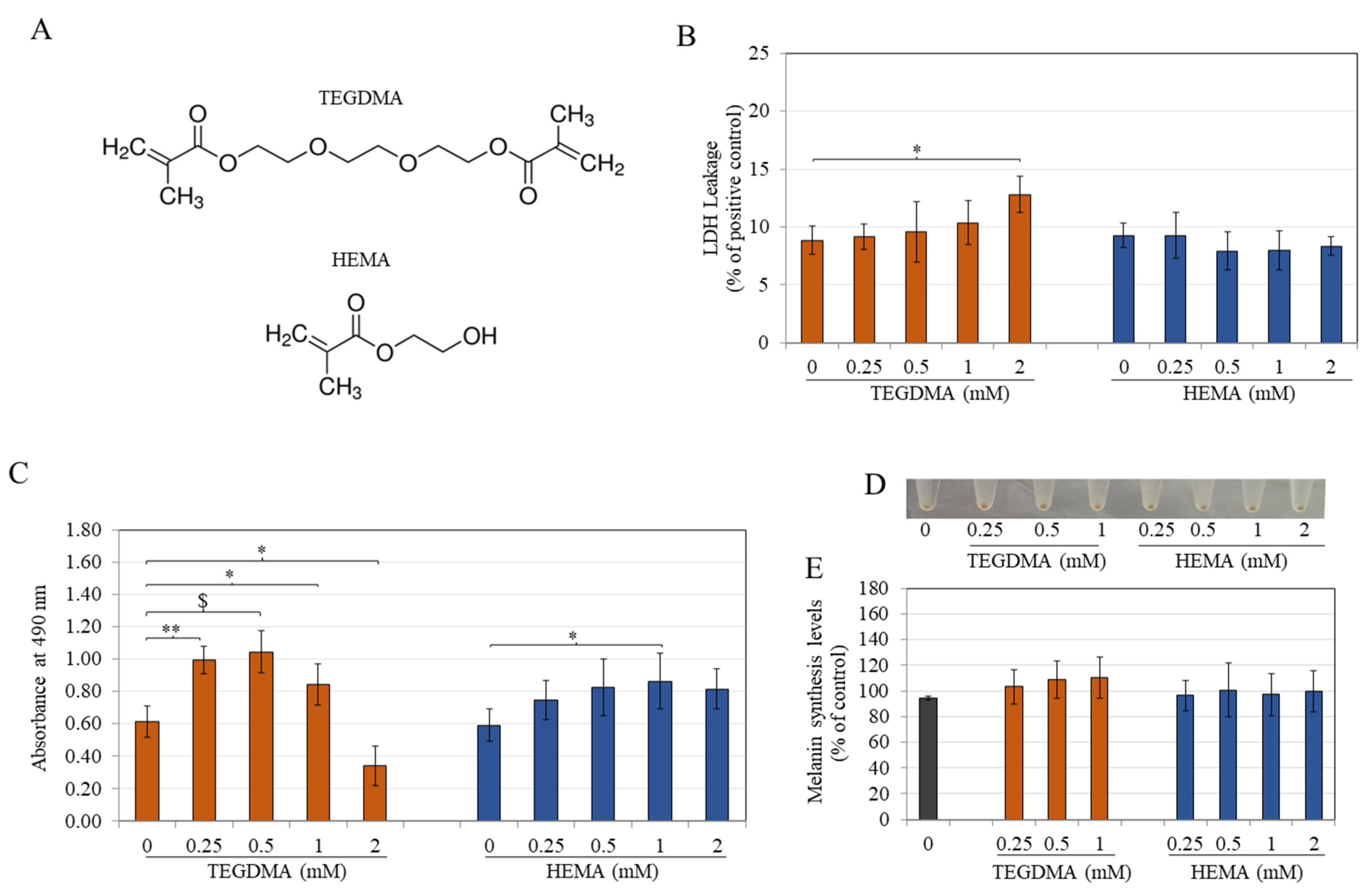
3.1. TEGDMA Is More Cytotoxic Than HEMA to Melanocytes

The two monomers TEGDMA and HEMA (the chemical structures shown in Figure 1A) were examined for cytotoxicity over a 0.25–2 mM concentration range. Results revealed that TEGDMA at 2 mM significantly increased LDH leakage by 1.45-fold with no change at lower concentrations. At the same time, there was no change in LDH leakage levels at any concentration of HEMA (Figure 1B).
Next, results from the MTS assay showed that TEGDMA at 2 mM significantly lowered cell viability (reduction in MTS absorbance by 44.26%) compared to the control group. Intriguingly, it significantly increased MTS absorbances at lower concentrations of 0.25, 0.5, and 1 mM (Figure 1C). HEMA displayed no change in cell metabolic activity over the concentration range of 0.25–2 mM. However, it significantly increased absorbance at 1 mM, which was similar to TEGDMA at 1 mM (Figure 1C). Collectively, these results indicate that TEGDMA induces cytotoxicity in melanocytes by diminishing the metabolic activity of cells with significant damage to cell membranes at concentrations > 1 mM. Based on these results, nontoxic concentrations of both monomers (TEGDMA: 0.25–1 mM and HEMA: 0.25–2 mM) were used for further experiments with melanocytes.

3.2. TEGDMA and HEMA Do Not Affect Intracellular Melanin Levels

Visual inspection of cell pellets showed no appreciable change in the monomer-treated groups’ pellet color compared to the control (Figure 1D). Next, melanin levels of cells treated with 0.5 and 1 mM TEGDMA were 109.12 ± 14.45 and 110.44 ± 16.07, respectively, which were not significantly different from the control group, although they appeared somewhat increased (Figure 1E). At the same time, HEMA also had no significant effect on melanin production at any concentration (Figure 1E). These data showed that both monomers do not affect cellular melanin production.

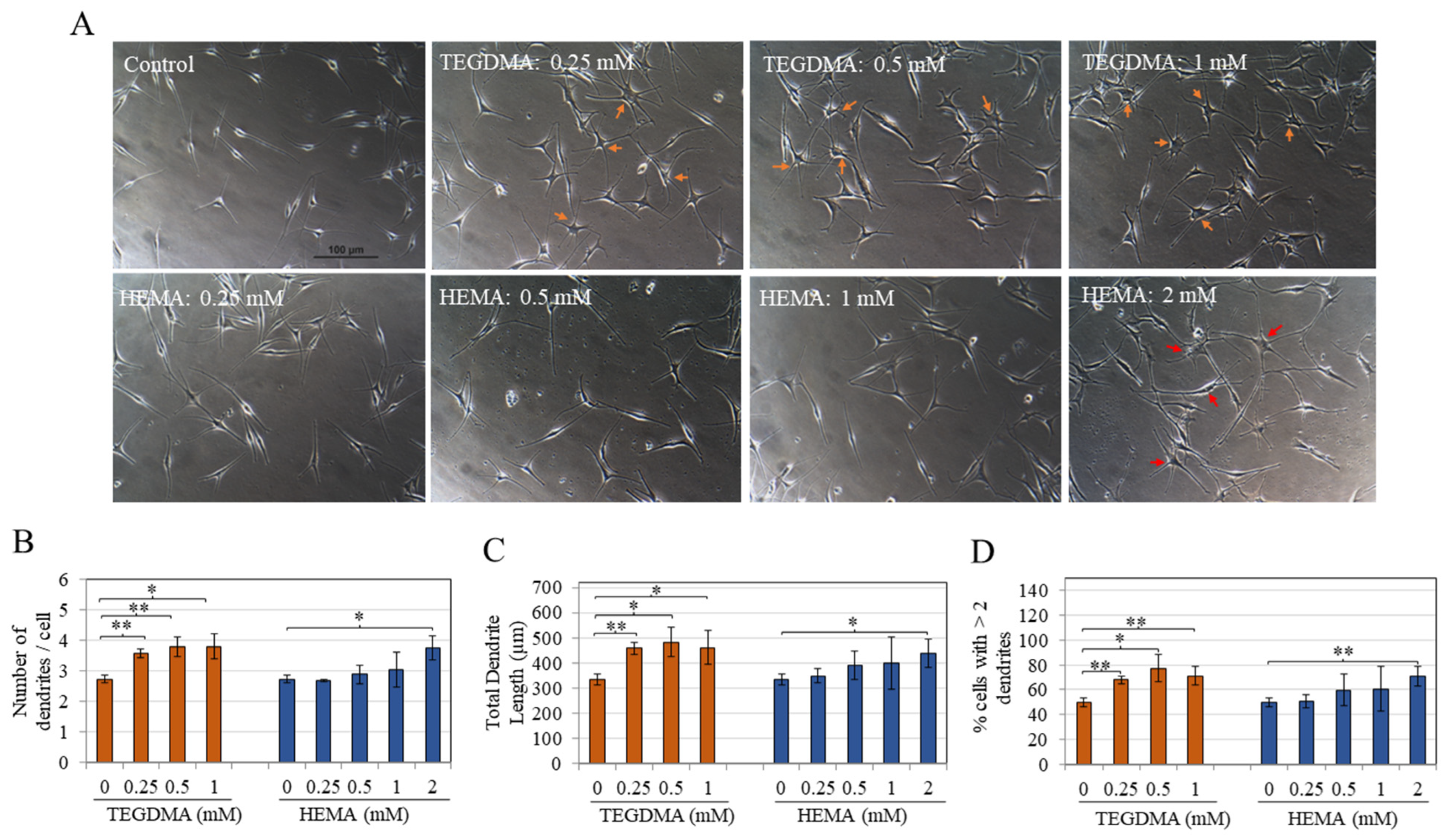
3.3. TEGDMA and HEMA Markedly Stimulate Melanocyte Dendricity

As the resin monomers did not affect melanin biosynthesis, it was evaluated whether they may affect the subsequent steps of melanogenesis, which involve the export of melanosomes from the tips of melanocyte dendrites. Results of qualitative examination of images of monomer-treated cells showed that TEGDMA significantly stimulated melanocyte dendricity from the lowest concentration onwards; cells displayed an arborized morphology in 0.25, 0.5, and 1 mM TEGDMA groups in contrast to the untreated group, which had thin and fewer dendrites (Figure 2A). In contrast, HEMA did not alter morphology up to concentrations of 1 mM. In comparison, at 2 mM, there was a significant enhancement of dendricity, as seen by elongated lengths and a greater number of dendrites per cell (Figure 2A).
Treatment with TEGDMA at 0.25, 0.5, and 1 mM significantly increased the number of dendrites per cell by 31.05%, 38.37%, and 39.14%. In comparison, treatment with HEMA did not affect dendrite number in the 0.25–1 mM concentration range but induced a significant increase of 37.15% at the highest concentration of 2 mM (Figure 2B).
Next, results of total dendrite length displayed a similar trend to TEGDMA, with a significantly greater increase in total dendrite length by 37.55%, 44.92%, and 38.41% at 0.25, 0.5, and 1 mM, respectively. In comparison, HEMA showed a significant increase of 31.63% only at 2 mM (Figure 2C).
Lastly, TEGDMA significantly stimulated the percentage of cells with >2 dendrites of the untreated control group (49.63%) to 67.91%, 77.11%, and 71.30% at 0.25, 0.5, and 1 mM, respectively. At the same time, HEMA showed a significant increase to 70.94% at 2 mM (Figure 2D).
Collectively, these results demonstrate that TEGDMA displays a greater sensitivity in stimulating dendricity parameters that were achieved over a wide concentration range (0.25–2 mM). Moreover, the stimulatory effects on dendrite number and length by TEGDMA at 0.25 mM were also achieved by HEMA, albeit at an 8-fold higher concentration of 2 mM.

3.4. TEGDMA Has No Effect, While HEMA Inhibits Intracellular Tyrosinase Activity

TEGDMA did not affect the intracellular tyrosinase activity at any concentration (Figure 3A). However, HEMA was shown to suppress tyrosinase activity in a concentration-dependent manner; significant inhibitions of 17.18% and 36.26% were found by HEMA at 1 mM and 2 mM concentrations, respectively (Figure 3A).

3.5. TEGDMA and HEMA Do Not Affect Tyrosinase Activity in a Cell-Free System

The two monomers, TEGDMA and HEMA, were examined over a concentration range of 0.25–2 mM using a standard in vitro assay with purified mushroom tyrosinase to assess if they might exhibit any direct effect on tyrosinase enzyme activity. Our results showed that none of the monomers affected tyrosinase enzyme activity in a cell-free system (Figure 3B).

3.6. TEGDMA and HEMA Do Not Alter Intracellular ROS Levels

Exposure to either TEGDMA or HEMA at different concentrations did not affect the ROS levels in cells compared to the control (Figure 3C). This suggests that in melanocytes, the monomers do not lead to the induction of ROS-mediated oxidative stress.

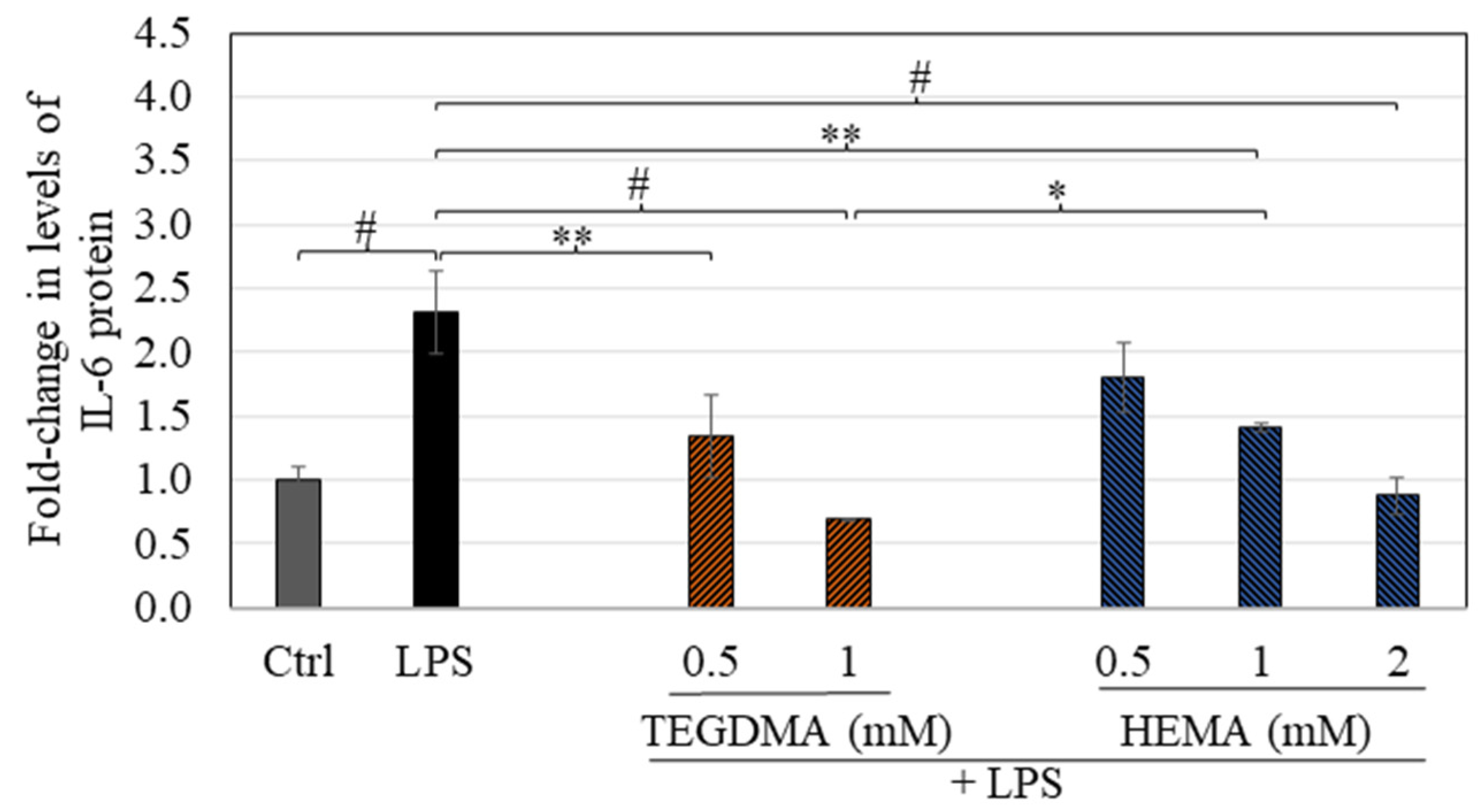
3.7. TEGDMA and HEMA Suppress LPS-Stimulated Cytokine Secretion
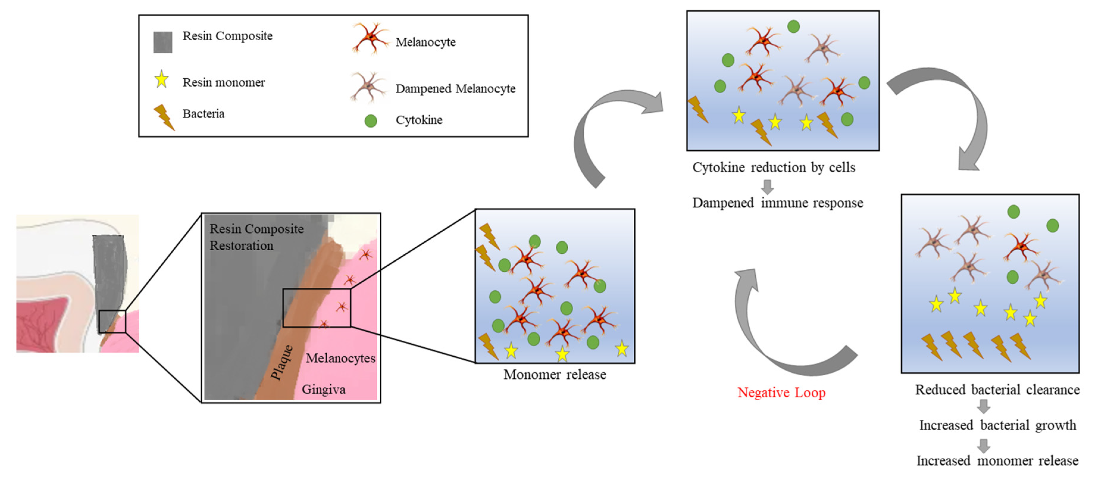
Treatment with LPS induced a 2.32-fold (p < 0.0001) greater secretion of IL-6 from cells as compared to untreated cells (Figure 4). Co-treatment with TEGDMA at 0.5 mM and 1 mM induced a 1.34-fold (p < 0.01) and 0.69-fold (p < 0.0001) IL-6 secretion, respectively, that was significantly lower as compared to the LPS-stimulated group (Figure 4). On the other hand, co-treatment with HEMA at 0.5, 1, and 2 mM induced a 1.80-fold (p > 0.05), 1.41-fold (p < 0.01), and 0.87-fold (p < 0.0001) IL-6 secretion, respectively, that was significantly lower than the LPS-stimulated group (Figure 4). At a concentration of 1 mM, TEGDMA exhibited a considerably higher degree of suppression (p < 0.05) on IL-6 levels compared to the levels produced by HEMA (Figure 4).
Based on these data, it appears that both monomers, TEGDMA (0.5–1 mM) and HEMA (1–2 mM), possess the capacity to significantly dampen the immune response of LPS-stimulated (inflamed) melanocytes, with TEGDMA exhibiting a higher potency than HEMA in suppressing IL-6 secretion from LPS-stimulated cells.

4. Discussion

To our knowledge, there are no published reports on the impact of monomer(s) or RBCs on human melanocytes. Thus, we cannot directly compare our findings. TEGDMA and HEMA were selected in this study as these monomers have been extensively used in commercial dental resin materials, either in single or mixture form. Studies have shown the systemic effects of monomers, where they may migrate with the bloodstream into other tissues causing a greater local concentration to induce adverse effects [63]. A concentration range of 0.25–2 mM was selected as these were shown to be clinically relevant [64]. HEMA was also tested over a higher concentration range in HEMn-LP cells and found to be significantly cytotoxic at concentrations of 4, 6, and 8 mM as determined by LDH assay (Figure S1A) and MTS assay (Figure S1B). The mean IC50 values based on MTS assays were calculated and found to be 4.04 mM and 2.07 mM for HEMA and TEGDMA monomers in HEMn-LP cells, respectively. Both monomers were thus evaluated at concentration ranges that were below their IC50. HEMn-LP cells have been selected in this study as they are metabolically less active in the synthesis and secretion of melanin than darkly-pigmented melanocytes and thus more closely mimic oral melanocytes, which are similarly known to be metabolically less active than epidermal melanocytes [49]. Our results of higher cytotoxicity of TEGDMA compared to HEMA agree with previous reports that have evaluated these monomers in other cells, such as pulp cells and fibroblasts [65] or macrophages [66]. HEMA is a hydrophilic monomer, while TEGDMA is hydrophobic. Hence, the higher cytotoxicity obtained by TEGDMA may be ascribed to its higher cellular uptake and permeability, which is consistent with a prior report that showed that the mechanism of monomer cytotoxicity is attributed to alterations in cellular permeability caused by changes in cellular membrane lipid layers [67]. Our results of hormetic-like effects on cellular viability in MTS assay by HEMA and TEGDMA are similar to previous studies, where a similar effect was noted in tetrazolium-based viability assay by HEMA [68] and TEGDMA [68,69]. A previous study reported that gingival fibroblasts from three different donors displayed differences in IC50 values for TEGDMA toxicity [70]. In contrast, another study showed different sensitivities of gingival fibroblasts from different donors on glutathione determination assay [71].
Primary cells used in this study were obtained from a single donor, and there might be donor-specific responses to the two monomers, which should be tested in further studies. However, preliminary cytotoxicity evaluation (Figure S2) of both monomers in human melanocytes from a different donor showed more significant cytotoxicity of HEMA, while TEGDMA did not show any change in cytotoxicity. It should be noted that melanocytes from another donor differed in pigmentation phenotype (darkly pigmented). Hence the possibility that HEMA alone (and not TEGDMA) showed melanin pigment-dependent cytotoxicity to melanocytes cannot be ruled out. TEGDMA was also evaluated at a higher concentration (3 mM) and was shown to induce significant cell membrane damage by 50% as evaluated by the LDH assay (Figure S3A) and markedly diminish cellular viability by 92.62% compared to the control (Figure S3B). Examination of the effects of monomers on apoptosis or its related pathways was not the focus of this study and hence was not expanded. However, HEMA and TEGDMA have been shown to activate apoptosis-related pathways in other cells [72,73,74].
Our results of increased melanocyte differentiation and dendricity without any changes in cellular melanin content by the monomers are reminiscent of previous studies that also showed a similar finding. For example, the natural product olive leaf extract stimulated dendricity without any changes in intracellular melanin in HEMn-LP cells [75]. Another study showed that treatment with butyrate led to marked differentiation and phenotypic alterations of B16F10 melanoma cells in the absence of changes in intracellular melanin [76]. Our results indicate that TEGDMA induced significant morphological changes in melanocytes at nontoxic concentrations; these changes reflect a higher capacity of cells to export melanin due to an increase in dendrite number and length, implying activation of cells by this monomer at concentrations 8-fold lower than that of HEMA. This implication is supported by the clinical observation of melanoblasts that showed elongated dendrites that reached into epithelium surface layers in the case of pregnancy-induced gingivitis, while the dendrites were short and reduced in number in the case of leukoplakia [77]. A prior study reported changes in the morphology of human gingival fibroblasts after a 96 h exposure to 3 mM HEMA and described that fibroblasts lost their spindle-like morphology and showed irregular surfaces [78]. Another study showed that TEGDMA suppressed filopodia and lamellipodia formation in dental pulp cells [79]. Since there has yet to be a study on the effects of these compounds on melanocytes, our results cannot be directly compared to any study and differ from the results of morphological changes of the studies above. Although filopodia have been implicated in dendrite extension and melanosome export [80], our results showed that TEGDMA might induce dendrite elongation by increasing filopodia length. Although the underlying mechanisms that lead to the stimulation of melanocyte differentiation by TEGDMA were not elucidated, it can be speculated that TEGDMA might increase levels of the melanogenesis protein, microphthalmia transcription factor (MITF), that regulates melanosome export [81].
Tyrosinase is the rate-limiting enzyme in the melanin production process [82]. Although HEMA attenuated tyrosinase activity in cells, it was ineffective at inhibiting the tyrosinase activity of a soluble tyrosinase enzyme from mushrooms, which agrees with previous reports that have discussed the discrepancy between mushroom tyrosinase used in cell-free assays and human tyrosinases present in the cellular system [83,84]. HEMA binds to macromolecules due to its α,β-unsaturated group and is known to form thiol-complex [85]. As thiols are known to inhibit tyrosinase activity [86], the possibility that suppression of tyrosinase activity occurred due to thiol complex formation cannot be ruled out. A natural compound derived from melanin, allomelanin, was shown to dye HEMA polymer typically used for soft contact lenses, indicating the possibility of HEMA binding to melanin [87].
The absence of changes in ROS generation of HEMn-LP cells for both monomers at nontoxic concentrations contrasts studies showing that HEMA [88,89] and TEGDMA [90,91] can induce ROS production in different cells. However, there is no study yet on ROS levels in melanocytes. Also, it should be noted that previous studies that reported ROS generation analyzed ROS after short periods (4 h) and evaluated higher concentrations up to 4 mM [88]. As the goal of this study was focused on changes in melanocyte functions and ROS levels at concentrations where no considerable cytotoxicity was induced, higher concentrations were not examined for ROS levels. Both monomers have previously shown glutathione (GSH) depletion effects in a cell-free system [92] as well as in cell cultures [93,94,95,96,97]. Higher cytotoxicity of TEGDMA was shown to be due to its greater GSH depletion than HEMA in gingival fibroblasts [98]. However, this study did not examine the effects of monomers on melanocyte GSH depletion. The effects of these monomers on GSH depletion and antioxidant enzymes catalase and superoxide dismutase (SOD), should be probed in future investigations.
Higher cytokine release by TEGDMA might be attributed, in part, to its higher lipophilicity, which allows it to permeate the melanocyte cell membrane and induce markedly greater secretion of IL-6. A previous study also showed that TEGDMA penetrates cellular membranes and oral mucosal epithelium to induce inflammatory responses [99]. We speculate that the suppression of IL-6 secretion by monomers in LPS-stimulated melanocytes might involve, at least, in part, a downregulation of relevant mitogen-activated protein kinases (MAP-kinases) that might activate the signal transduction cascades [100]. The mechanisms for suppressing IL-6 secretion by monomers were not elucidated as it was beyond the scope of this study. A previous study demonstrated elevated levels of nuclear factor erythroid 2-related factor 2 (Nrf2) by HEMA monomer in macrophages [101]. Similar to macrophages, melanocytes are also immunocompetent cells [102,103]. Hence, HEMA might cause increased Nrf2 expression in melanocytes, which will need to be investigated in future studies. Although only one cytokine (IL-6) was tested under LPS-stimulated conditions in this study, TEGDMA and HEMA were also shown to suppress TNF-α in macrophages after LPS stimulation [104]. Hence it is likely that apart from IL-6, other pro-inflammatory cytokines might also be downregulated and warrant future investigations. One of the limitations of this study was that IL-6 release at monomer concentrations <0.5 mM and in the absence of inflammation (basal conditions) was not examined and should be a subject of future investigations. Regardless, LPS is released from cariogenic microorganisms in deep carious lesions.
We used a model of co-exposure of cells to LPS and monomers, as this scenario mimics realistic clinical situations where both LPS and monomer will be simultaneously present after the removal of carious dentin and restoration with dental composite [105]. Prior studies have documented that both the leached resin monomers and RBCs can increase the growth of oral bacteria [106,107]. It was also demonstrated that the degradation of RBCs is augmented by the biofilms of oral bacteria [108]. Considering the results of our study, it is plausible to speculate that insufficient clearance of bacteria might follow due to the dampening of melanocyte immune response by both monomers under LPS stimulation; this, in turn, might induce a negative cycle which can cause impaired clearance of bacteria thus causing a further increase in composite degradation and release of higher amounts of monomer, which in turn would dampen immune response further (Figure 5). Also, we chose to use LPS from E. coli, although LPS from periodontopathic bacteria P. gingivalis might have been more clinically relevant; however, in our previous study [109], LPS from P. gingivalis failed to induce any significant IL-6 release from HEMn-LP cells. Hence, we selected LPS from E coli. Multiple prior reports have also studied the effects of monomers in the presence of E. coli-derived LPS in macrophages [104,110,111,112].
Previous studies have demonstrated that using dental bleaching treatments augments the release of resin monomers [113,114,115]. For example, TEGDMA was released in greater amounts (as compared to Bis-GMA) as the concentration of bleaching agent (hydrogen peroxide) was increased [115], and another study showed that nanofilled dental composites released more TEGDMA than microfilled composites [114]. Moreover, hydrogen peroxide in bleaching gels was shown to augment the cytotoxicity of TEGDMA in oral cells [116]. Given the reports above, our findings of this study of higher cytotoxicity of TEGDMA to melanocytes suggest greater chances of exposure to higher amounts of TEGDMA, which might augment melanocytotoxicity. As RBCs and dentin bonding agents (DBAs) typically comprise comonomers HEMA and TEGDMA in combination with base monomers UDMA and Bis-GMA, the evaluation of biological effects of the mixtures of both monomers might be more relevant as the combination might produce synergistic, additive, or antagonistic effects on melanocyte functions. A separate study tested the combination of TEGDMA with UDMA in CHO cells and found no significant difference in cytotoxicity of the combination compared to single monomers [117]. Despite the significance of testing mixtures, the activity of single monomer(s) was only examined in the present study.
Both HEMA and TEGDMA are methacrylic acid (MAA)-based monomers that possess ester groups, which can undergo degradation by salivary esterases [118] or metabolic transformation by hydrolysis/enzymatic catalysis [119]. MAA is the chief metabolic product of HEMA degradation; it exhibited cytotoxicity similar to that of HEMA, leading to the conclusion that the cytotoxicity and genotoxicity of HEMA might be attributable to this degradation product [120]. The degradation products of TEGDMA have also been reported and comprise MAA in addition to other products such as 2,3-epoxymethacrylic acid (2,3-EMA) and triethylene glycol, with 2,3-EMA reported to have cytotoxicity similar to that of TEGDMA [121]. Future studies to examine the biocompatibility of TEGDMA and HEMA degradation products to melanocytes would also be significant. Moreover, alternative monomers for replacing TEGDMA have also been reported [122]; further studies will also be necessary to identify whether these alternative monomers might impair melanocyte function.
Increased pigmentation in oral mucosa manifests as dark brown gums and poses aesthetic concerns for patients. Active consumption of tobacco and cigarette smoking is known to stimulate a higher level of gingival pigmentation [123], partly attributed to the binding of benzopyrene and nicotine in tobacco smoke to melanin pigment. Interestingly, TEGDMA is a matrix monomer in one of the commonly used gingiva-colored composites, Amaris Gingiva (VOCO America Inc., Fort Mill, SC, USA) [124,125]. Our results thus point out that TEGDMA might elevate gingival pigmentation due to its robust capacity to stimulate melanogenic differentiation characterized by a multidendritic phenotype. Because the demand for gingival depigmenting modalities is rising owing to patients’ esthetic needs, if indeed it can be shown that TEGDMA or HEMA leached from restorations increase oral pigmentation, it would further pose esthetic concerns necessitating cosmetic rehabilitation to correct gingival melanin pigmentation. Whether a mixture of TEGDMA with other monomers or tobacco smoke might exhibit interactive effects or synergistically increase melanin production and export will be worthy of future investigations.
There are several limitations to the study that must be considered. A fundamental limitation is that our study does not recapitulate the oral cavity environment with the presence of saliva, pellicle layer, biofilm, or other oral cells. Moreover, as this study was conducted with melanocytes of epidermal origin, the extrapolation of results to oral melanocytes needs to be done with caution, as it has been shown that oral melanocytes exhibit less metabolic activity compared to epidermal melanocytes under physiological circumstances [49]. Nevertheless, oral melanocytes can increase their metabolic activity under injury or inflammatory triggers. Another limitation is that the measurement of dendritic parameters is only a surrogate for melanosome export. Hence, the increase in melanosome export by monomers has to be validated by additional research employing a physiological system made up of melanocyte cocultures with gingival keratinocytes to validate our findings. Furthermore, this study evaluated the response of melanocytes to a continuous exposure scenario with monomers; thus, whether melanocytes might recover their morphology and function after removal of the compounds or if intermittent exposure might lead to permanent changes in melanocyte dendricity or tyrosinase activity would be interesting for future studies, especially since some of the monomers can persist in the oral cavity and cause a sustained exposure for months. For instance, HEMA was shown to leach continuously from composites up until 52 weeks after initial immersion, thus indicative of a sustained chronic exposure [126]. The mechanisms of cytotoxicity (apoptosis or necrosis) by the monomers were not delineated in this study. As our current study was focused on the assessment of the cytotoxicity of monomers in direct contact with melanocytes, we did not study the effects of melanocytes in contact with eluants of dental composites (that contained leached monomers) that have been used in previous studies and are more representative of clinical situations [127]. Additionally, a prior study that examined cytotoxicity to neural cells used insert membranes with a composite that simulated dentin tubules and enabled real-time diffusion of monomers mimicking in vivo scenarios [40]. As 90% of the material is leached over the first 24 h [128], the use of such a membrane would allow a more realistic exposure assessment. Hence, a follow-up study incorporating such a system will be interesting, especially as neural cells are closely related to melanocytes. Lastly, whether these monomers might induce genotoxicity in melanocytes by forming DNA adducts will be worthy of future investigations.

5. Conclusions

In summary, this pilot study’s findings show that the two commonly used resin monomers, TEGDMA and HEMA, have differing effects on primary human melanocytes, with TEGDMA being more cytotoxic than HEMA. Our results validated TEGDMA-induced changes in melanocyte homeostasis, including a greater capacity to impair melanocyte’s immune response than HEMA. In addition, TEGDMA increased melanocyte dendricity over a wide concentration range of 0.25–1 mM, as compared to HEMA, which only showed these effects at a single high concentration of 2 mM. This effect might also be a causative factor in the induction of gingival pigmentation leading to aesthetic concerns with TEGDMA or HEMA-releasing restorations. Further investigation using a physiological system consisting of melanocyte cocultures with gingival keratinocytes is needed to validate the increase in melanosome export to corroborate our findings. Future studies are essential to clarify the clinical implications of TEGDMA- and HEMA-associated melanocytic differentiation and immune function.

Supplementary Materials

The following supporting information can be downloaded at: https://www.mdpi.com/article/10.3390/oral3030029/s1, Figure S1: (A) Cytotoxicity (LDH leakage) and (B) Viability (absorbance at 490 nm) of melanocytes treated with HEMA monomer over a higher concentration range of 2–8 mM for 72 h; * p < 0.05, ** p < 0.01, $ p < 0.001, and # p < 0.0001 vs. control (0 mM group); one-way ANOVA with Dunnett’s post hoc test; Data are mean ± SD of one representative experiment in triplicates of two separate experiments. Figure S2: Viability of melanocytes from a different donor (HEMn-DP cells) treated with monomers TEGDMA and HEMA over a concentration range of 0.25–3 mM for 72 h; $ p < 0.001 and # p < 0.0001 vs. control group (Ctrl); one-way ANOVA with Dunnett’s post-hoc test; Data are mean ± SD of quadruplicate determinations (n = 4 per group). Figure S3: (A) Cytotoxicity (LDH leakage) and (B) Viability of melanocytes treated with TEGDMA and HEMA monomers each at a concentration of 3 mM for 72 h; * p < 0.05, ** p < 0.01, and # p < 0.0001; one-way ANOVA with Tukey’s post-hoc test; Data are mean ± SD of three independent experiments for (A) and four independent experiments for (B).

Funding

This work was supported, in part, by funds from the Stony Brook Foundation. The funder had no role in study design, data collection, analysis, publication decision, or manuscript preparation.

Institutional Review Board Statement

Not applicable.

Informed Consent Statement

Not applicable.

Data Availability Statement

Data are available from the corresponding author upon reasonable request.

Acknowledgments

The author acknowledges Sanford R. Simon (Stony Brook University) for access to facilities.

Conflicts of Interest

The author declares no conflict of interest.

References

  1. Mulligan, S.; Hatton, P.V.; Martin, N. Resin-Based Composite Materials: Elution and Pollution. Br. Dent. J. 2022, 232, 644–652. [Google Scholar] [CrossRef]
  2. Cho, K.; Rajan, G.; Farrar, P.; Prentice, L.; Prusty, B.G. Dental Resin Composites: A Review on Materials to Product Realizations. Compos. Part B Eng. 2022, 230, 109495. [Google Scholar]
  3. Yadav, R.; Lee, H.H. Fabrication, Characterization, and Selection Using Fahp-Topsis Technique of Zirconia, Titanium Oxide, and Marble Dust Powder Filled Dental Restorative Composite Materials. Polym. Adv. Technol. 2022, 33, 3286–3295. [Google Scholar] [CrossRef]
  4. Ali, S.; Sangi, L.; Kumar, N.; Kumar, B.; Khurshid, Z.; Zafar, M.S. Evaluating Antibacterial and Surface Mechanical Properties of Chitosan Modified Dental Resin Composites. Technol. Health Care 2020, 28, 165–173. [Google Scholar] [CrossRef] [PubMed]
  5. Dahri, W.M.; Kumar, N.; Altaf, N.; Mughal, W.; Zafar, M.S. Mechanical and Biomimetic Characteristics of Bulk-Fill Resin Dental Composites Following Exposure in A Simulated Acidic Oral Environment. Biomimetics 2023, 8, 19. [Google Scholar] [CrossRef]
  6. Kumar, N.; Sangi, L. Water Sorption, Solubility, and Resultant Change in Strength Among Three Resin-Based Dental Composites. J. Investig. Clin. Dent. 2014, 5, 144–150. [Google Scholar] [CrossRef]
  7. Yadav, R.; Lee, H.; Lee, J.-H.; Singh, R.K.; Lee, H.-H. A Comprehensive Review: Physical, Mechanical, and Tribological Characterization of Dental Resin Composite Materials. Tribol. Int. 2023, 179, 108102. [Google Scholar]
  8. Ferracane, J. Elution of Leachable Components from Composites. J. Oral Rehabil. 1994, 21, 441–452. [Google Scholar] [CrossRef]
  9. Wataha, J.; Rueggeberg, F.; Lapp, C.; Lewis, J.; Lockwood, P.; Ergle, J.; Mettenburg, D.J. In Vitro Cytotoxicity of Resin-Containing Restorative Materials After Aging in Artificial Saliva. Clin. Oral Investig. 1999, 3, 144–149. [Google Scholar] [CrossRef]
  10. Bakkal, M.; Yılmaz, B.; Durmus, A.; Durmus, Z.; Ozalp, S. Polymerization Characteristics of Colored Compomers Cured with Different Led Units. J. Appl. Biomater. Funct. Mater. 2019, 17, 2280800019827805. [Google Scholar] [CrossRef] [Green Version]
  11. Bayindir, Y.Z.; Yildiz, M.; Bayindir, F. The Effect of “Soft-Start Polymerization” on Surface Hardness of Two Packable Composites. Dent. Mater. J. 2003, 22, 610–616. [Google Scholar] [CrossRef] [PubMed] [Green Version]
  12. Vandenbulcke, J.D.; Marks, L.A.; Martens, L.C.; Verbeeck, R.M. Comparison of Curing Depth of A Colored Polyacid-Modified Composite Resin with Different Light-Curing Units. Quintessence Int. 2010, 41, 787–794. [Google Scholar] [PubMed]
  13. Germain, H.S.; Swartz, M.; Phillips, R.; Moore, B.; Roberts, T. Properties of Microfilled Composite Resins As Influenced By Filler Content. J. Dent. Res. 1985, 64, 155–160. [Google Scholar] [CrossRef]
  14. Van Landuyt, K.; Nawrot, T.; Geebelen, B.; De Munck, J.; Snauwaert, J.; Yoshihara, K.; Scheers, H.; Godderis, L.; Hoet, P.; Van Meerbeek, B. How Much Do Resin-Based Dental Materials Release? A Meta-Analytical Approach. Dent. Mater. 2011, 27, 723–747. [Google Scholar] [CrossRef] [PubMed]
  15. Jaffer, F.; Finer, Y.; Santerre, J. Interactions Between Resin Monomers and Commercial Composite Resins with Human Saliva Derived Esterases. Biomaterials 2002, 23, 1707–1719. [Google Scholar] [CrossRef]
  16. Delima, A.J.; Van Dyke, T.E. Origin and Function of the Cellular Components in Gingival Crevice Fluid. Periodontol. 2000 2003, 31, 55–76. [Google Scholar] [CrossRef]
  17. Gitalis, R.; Zhou, L.; Marashdeh, M.Q.; Sun, C.; Glogauer, M.; Finer, Y. Human Neutrophils Degrade Methacrylate Resin Composites and Tooth Dentin. Acta Biomater. 2019, 88, 325–331. [Google Scholar] [CrossRef]
  18. Almeida, G.S.; Poskus, L.T.; Guimarães, J.G.A.; Silva, E.M. The Effect of Mouthrinses on Salivary Sorption, Solubility and Surface Degradation of A Nanofilled and A Hybrid Resin Composite. Oper. Dent. 2010, 35, 105–111. [Google Scholar] [CrossRef] [Green Version]
  19. Puckett, A.D.; Fitchie, J.G.; Kirk, P.C.; Gamblin, J. Direct Composite Restorative Materials. Dent. Clin. N. Am. 2007, 51, 659–675. [Google Scholar] [CrossRef]
  20. Abdulmajeed, A.A.; Kokkari, A.K.; Käpylä, J.; Massera, J.; Hupa, L.; Vallittu, P.K.; Närhi, T.O. In Vitro Blood and Fibroblast Responses to Bisgma–Tegdma/Bioactive Glass Composite Implants. J. Mater. Sci. Mater. Med. 2014, 25, 151–162. [Google Scholar] [CrossRef]
  21. Tou, G.A.D.A.; Gomes, J.M.; Rinco, L.S.D.O.; Yamauti, M.; Diniz, I.M.A.; Pires, F.; Schmidt, M.E.P.; Menezes, H.C.; Cardeal, Z.D.L.; Bottoli, C.B.G. Release of Leachable Products from Resinous Compounds in the Saliva of Children with Anterior Open Bite Treated with Spur. J. Appl. Oral Sci. 2023, 30, e20220227. [Google Scholar] [CrossRef] [PubMed]
  22. Szczesio-Wlodarczyk, A.; Polikowski, A.; Krasowski, M.; Fronczek, M.; Sokolowski, J.; Bociong, K. The Influence of Low-Molecular-Weight Monomers (Tegdma, Hddma, Hema) on the Properties of Selected Matrices and Composites Based on Bis-Gma and Udma. Materials 2022, 15, 2649. [Google Scholar] [CrossRef] [PubMed]
  23. Tauscher, S.; Angermann, J.; Catel, Y.; Moszner, N. Evaluation of Alternative Monomers to Hema for Dental Applications. Dent. Mater. 2017, 33, 857–865. [Google Scholar] [CrossRef] [PubMed]
  24. Wang, T.; Nakabayashi, N. Effect of 2-(Methacryloxy) Ethyl Phenyl Hydrogen Phosphate on Adhesion to Dentin. J. Dent. Res. 1991, 70, 59–66. [Google Scholar] [CrossRef] [PubMed]
  25. Arcís, R.W.; López-Macipe, A.; Toledano, M.; Osorio, E.; Rodríguez-Clemente, R.; Murtra, J.; Fanovich, M.A.; Pascual, C.D. Mechanical Properties of Visible Light-Cured Resins Reinforced with Hydroxyapatite for Dental Restoration. Dent. Mater. 2002, 18, 49–57. [Google Scholar] [CrossRef]
  26. Michelsen, V.B.; Moe, G.; Skålevik, R.; Jensen, E.; Lygre, H. Quantification of Organic Eluates from Polymerized Resin-Based Dental Restorative Materials By Use of Gc/Ms. J. Chromatogr. B 2007, 850, 83–91. [Google Scholar] [CrossRef]
  27. Goda, T.; Ishihara, K. Soft Contact Lens Biomaterials from Bioinspired Phospholipid Polymers. Expert Rev. Med. Devices 2006, 3, 167–174. [Google Scholar] [CrossRef]
  28. Lord, M.S.; Stenzel, M.H.; Simmons, A.; Milthorpe, B.K. The Effect of Charged Groups on Protein Interactions with Poly (Hema) Hydrogels. Biomaterials 2006, 27, 567–575. [Google Scholar] [CrossRef]
  29. Montheard, J.-P.; Chatzopoulos, M.; Chappard, D. 2-Hydroxyethyl Methacrylate (Hema): Chemical Properties and Applications in Biomedical Fields. J. Macromol. Sci. Part C Polym. Rev. 1992, 32, 1–34. [Google Scholar]
  30. Sideridou, I.; Tserki, V.; Papanastasiou, G. Effect of Chemical Structure on Degree of Conversion in Light-Cured Dimethacrylate-Based Dental Resins. Biomaterials 2002, 23, 1819–1829. [Google Scholar] [CrossRef]
  31. Geurtsen, W.; Leyhausen, G. Chemical-Biological Interactions of the Resin Monomer Triethyleneglycol-Dimethacrylate (Tegdma). J. Dent. Res. 2001, 80, 2046. [Google Scholar] [CrossRef] [PubMed]
  32. Cebe, M.A.; Cebe, F.; Cengiz, M.F.; Cetin, A.R.; Arpag, O.F.; Ozturk, B. Elution of Monomer from Different Bulk Fill Dental Composite Resins. Dent. Mater. 2015, 31, E141–E149. [Google Scholar] [CrossRef] [PubMed]
  33. Örtengren, U.; Wellendorf, H.; Karlsson, S.; Ruyter, I. Water Sorption and Solubility of Dental Composites and Identification of Monomers Released in An Aqueous Environment. J. Oral Rehabil. 2001, 28, 1106–1115. [Google Scholar] [CrossRef] [PubMed]
  34. Venz, S.; Dickens, B. Nir-Spectroscopic Investigation of Water Sorption Characteristics of Dental Resins and Composites. J. Biomed. Mater. Res. 1991, 25, 1231–1248. [Google Scholar] [CrossRef]
  35. Sideridou, I.; Tserki, V.; Papanastasiou, G. Study of Water Sorption, Solubility and Modulus of Elasticity of Light-Cured Dimethacrylate-Based Dental Resins. Biomaterials 2003, 24, 655–665. [Google Scholar] [CrossRef]
  36. Nakamura, M.; Oshima, H.; Hashimoto, Y. Monomer Permeability of Disposable Dental Gloves. J. Prosthet. Dent. 2003, 90, 81–85. [Google Scholar] [CrossRef]
  37. Kaufman, G.; Skrtic, D. Morphological and Kinetic Study of Oral Keratinocytes Assembly on Reconstituted Basement Membrane: Effect of Tegdma. Arch. Oral Biol. 2019, 104, 103–111. [Google Scholar] [CrossRef]
  38. Mahdhaoui, K.; Fournier, B.; Derbanne, M.A. Unbound Monomers Do Diffuse Through the Dentin Barrier. Dent. Mater. 2017, 33, 743–751. [Google Scholar] [CrossRef]
  39. Rashid, H.; Sheikh, Z.; Vohra, F. Allergic Effects of the Residual Monomer Used in Denture Base Acrylic Resins. Eur. J. Dent. 2015, 9, 614–619. [Google Scholar]
  40. Kamalak, H.; Kamalak, A.; Taghizadehghalehjoughi, A.; Hacimuftuoglu, A.; Nalci, K.A. Cytotoxic and Biological Effects of Bulk Fill Composites on Rat Cortical Neuron Cells. Odontology 2018, 106, 377–388. [Google Scholar] [CrossRef] [Green Version]
  41. Sharma, S.; Khan, Q.; Schreurs, O.J.F.; Sapkota, D.; Samuelsen, J.T. Investigation of Biological Effects of Hema in 3d-Organotypic Co-Culture Models of Normal and Malignant Oral Keratinocytes. Biomater. Investig. Dent. 2023, 10, 2234400. [Google Scholar] [CrossRef]
  42. Jiang, E.-S.; Moon, W.; Lim, B.-S.; Chang, J.; Chung, S.H. Cytotoxicity and Reactive Oxygen Species Production Induced By Different Co-Monomer Eluted from Nanohybrid Dental Composites. BMC Oral Health 2023, 23, 55. [Google Scholar] [CrossRef] [PubMed]
  43. Thomas, A.J.; Erickson, C.A. The Making of A Melanocyte: The Specification of Melanoblasts from the Neural Crest. Pigment Cell Melanoma Res. 2008, 21, 598–610. [Google Scholar] [CrossRef] [PubMed]
  44. Elobeid, A.S.; Kamal-Eldin, A.; Abdelhalim, M.A.K.; Haseeb, A.M. Pharmacological Properties of Melanin and Its Function in Health. Basic Clin. Pharmacol. Toxicol. 2017, 120, 515–522. [Google Scholar] [CrossRef] [Green Version]
  45. Nilima, S.; Vandana, K.L. Melanin: A Scavenger in Gingival Inflammation. Indian J. Dent. Res. 2011, 22, 38–43. [Google Scholar] [CrossRef]
  46. Del Marmol, V.; Beermann, F. Tyrosinase and Related Proteins in Mammalian Pigmentation. Febs Lett. 1996, 381, 165–168. [Google Scholar] [CrossRef] [PubMed]
  47. Tripathi, R.K.; Hearing, V.J.; Urabe, K.; Aroca, P.; Spritz, R.A. Mutational Mapping of the Catalytic Activities of Human Tyrosinase. J. Biol. Chem. 1992, 267, 23707–23712. [Google Scholar] [CrossRef]
  48. Hearing, V.J.; Tsukamoto, K. Enzymatic Control of Pigmentation in Mammals. Faseb J. 1991, 5, 2902–2909. [Google Scholar] [CrossRef] [PubMed]
  49. Barrett, A.; Scully, C. Human Oral Mucosal Melanocytes: A Review. J. Oral Pathol. Med. 1994, 23, 97–103. [Google Scholar] [CrossRef]
  50. Eisen, D. Disorders of Pigmentation in the Oral Cavity. Clin. Dermatol. 2000, 18, 579–587. [Google Scholar] [CrossRef]
  51. Baala Vignesh, A.; Thenmozhi, M. Effect of Smoking on Oral Pigmentation-A Survey. Drug Invent. Today 2018, 10, 2348–2350. [Google Scholar]
  52. Gulati, N.; Dutt, P.; Gupta, N.; Tyagi, P. Gingival Pigmentation: Revisited. J. Adv. Med. Dent. Sci. Res. 2016, 4, 48. [Google Scholar]
  53. Anders Hedin And, C.; Larsson, Å. The Ultrastructure of the Gingival Epithelium in Smokers’ Melanosis. J. Periodontal. Res. 1984, 19, 177–190. [Google Scholar] [CrossRef] [PubMed]
  54. Fruet-Arruda, R.T.; Anselmo, G.G.; Tortamano, A.C.A.; Rossi, A.L.; Biffi, M.B.; Marco, R.L.; Kato, I.T.; Nuñez, S.C.; Prates, R.A. Melanin Pigmented Gingival Tissue Impairs Red-Light Lateral Scattering for Antimicrobial Photodynamic Therapy. Photodiagnosis Photodyn. Ther. 2021, 33, 102135. [Google Scholar] [CrossRef]
  55. Goenka, S. Biological Impact of the Ratio of E-Cigarette Liquid Base Constituents, Propylene Glycol and Vegetable Glycerin, on Primary Human Melanocytes. Oral 2023, 3, 40–56. [Google Scholar] [CrossRef]
  56. Goenka, S.; Simon, S.R. Effects of Fluoride Exposure on Primary Human Melanocytes from Dark and Light Skin. Toxics 2020, 8, 114. [Google Scholar] [CrossRef]
  57. Goenka, S.R. Simon, S. Asoprisnil, a Selective Progesterone Receptor Modulator (Sprm), Inhibits Melanosome Export in B16f10 Cells and Hemn-Dp Melanocytes. Molecules 2020, 25, 3581. [Google Scholar] [CrossRef]
  58. Kim, H.J.; Kim, J.S.; Woo, J.-T.; Lee, I.-S.; Cha, B.-Y. Hyperpigmentation Mechanism of Methyl 3, 5-Di-Caffeoylquinate Through Activation of P38 and Mitf Induction of Tyrosinase. Acta Biochim. Biophys. Sin. 2015, 47, 548–556. [Google Scholar] [CrossRef]
  59. Goenka, S.; Simon, S.R. Organogold Drug Auranofin Exhibits Anti-Melanogenic Activity in B16f10 and Mnt-1 Melanoma Cells. Arch. Dermatol. Res. 2020, 312, 213–221. [Google Scholar] [CrossRef]
  60. Jian, Q.; An, Q.; Zhu, D.; Hui, K.; Liu, Y.; Chi, S.; Li, C. Microrna 340 Is Involved in Uvb-Induced Dendrite Formation Through the Regulation of Rhoa Expression in Melanocytes. Mol. Cell. Biol. 2014, 34, 3407–3420. [Google Scholar] [CrossRef] [Green Version]
  61. Hempel, S.L.; Buettner, G.R.; O’malley, Y.Q.; Wessels, D.A.; Flaherty, D.M. Dihydrofluorescein Diacetate Is Superior for Detecting Intracellular Oxidants: Comparison with 2′,7′-Dichlorodihydrofluorescein Diacetate, 5 (and 6)-Carboxy-2′,7′-Dichlorodihydrofluorescein Diacetate, and Dihydrorhodamine 123. Free Radic. Biol. Med. 1999, 27, 146–159. [Google Scholar] [CrossRef] [PubMed]
  62. Endo, K.; Mizutani, T.; Okano, Y.; Masaki, H. A Red Pumpkin Seed Extract Reduces Melanosome Transfer to Keratinocytes by Activation of Nrf2 Signaling. J. Cosmet. Dermatol. 2019, 18, 827–834. [Google Scholar] [CrossRef] [PubMed]
  63. Schweikl, H.; Spagnuolo, G.; Schmalz, G. Genetic and Cellular Toxicology of Dental Resin Monomers. J. Dent. Res. 2006, 85, 870–877. [Google Scholar] [CrossRef]
  64. Noda, M.; Wataha, J.; Kaga, M.; Lockwood, P.; Volkmann, K.; Sano, H. Components of Dentinal Adhesives Modulate Heat Shock Protein 72 Expression in Heat-Stressed Thp-1 Human Monocytes At Sublethal Concentrations. J. Dent. Res. 2002, 81, 265–269. [Google Scholar] [CrossRef]
  65. Lee, D.H.; Lim, B.-S.; Lee, Y.-K.; Ahn, S.-J.; Yang, H.-C. Involvement of Oxidative Stress in Mutagenicity and Apoptosis Caused By Dental Resin Monomers in Cell Cultures. Dent. Mater. 2006, 22, 1086–1092. [Google Scholar] [CrossRef]
  66. Becher, R.; Kopperud, H.M.; Al, R.H.; Samuelsen, J.T.; Morisbak, E.; Dahlman, H.J.; Lilleaas, E.M.; Dahl, J.E. Pattern of Cell Death After in Vitro Exposure to Gdma, Tegdma, Hema and Two Compomer Extracts. Dent. Mater. 2006, 22, 630–640. [Google Scholar] [CrossRef]
  67. Lefebvre, C.A.; Schuster, G.; Rueggeberg, F.; Tamare-Selvy, K.; Knoernschild, K. Responses of Oral Epithelial Cells to Dental Resin Components. J. Biomater. Sci. Polym. Ed. 1996, 7, 965–976. [Google Scholar] [CrossRef]
  68. Inamitsu, H.; Okamoto, K.; Sakai, E.; Nishishita, K.; Murata, H.; Tsukuba, T. The Dental Resin Monomers Hema and Tegdma Have Inhibitory Effects on Osteoclast Differentiation with Low Cytotoxicity. J. Appl. Toxicol. 2017, 37, 817–824. [Google Scholar] [CrossRef] [PubMed]
  69. Walters, N.J.; Xia, W.; Salih, V.; Ashley, P.F.; Young, A.M. Poly (Propylene Glycol) and Urethane Dimethacrylates Improve Conversion of Dental Composites and Reveal Complexity of Cytocompatibility Testing. Dent. Mater. 2016, 32, 264–277. [Google Scholar] [CrossRef] [Green Version]
  70. Moharamzadeh, K.; Van Noort, R.; Brook, I.M.; Scutt, A.M. Cytotoxicity of Resin Monomers on Human Gingival Fibroblasts and Hacat Keratinocytes. Dent. Mater. 2007, 23, 40–44. [Google Scholar] [CrossRef]
  71. Engelmann, J.; Leyhausen, G.; Leibfritz, D.; Geurtsen, W. Effect of Tegdma on the Intracellular Glutathione Concentration of Human Gingival Fibroblasts. J. Biomed. Mater. Res. Off. J. Soc. Biomater. Jpn. Soc. Biomater. Aust. Soc. Biomater. Korean Soc. Biomater. 2002, 63, 746–751. [Google Scholar] [CrossRef] [PubMed]
  72. Huang, F.M.; Kuan, Y.H.; Lee, S.S.; Chang, Y.C. Cytotoxicity and Genotoxicity of Triethyleneglycol-Dimethacrylate in Macrophages Involved in Dna Damage and Caspases Activation. Environ. Toxicol. 2015, 30, 581–588. [Google Scholar] [CrossRef] [PubMed]
  73. Batarseh, G.; Windsor, L.; Labban, N.; Liu, Y.; Gregson, K. Triethylene Glycol Dimethacrylate Induction of Apoptotic Proteins in Pulp Fibroblasts. Oper. Dent. 2014, 39, E1–E8. [Google Scholar] [CrossRef] [PubMed] [Green Version]
  74. Paranjpe, A.; Bordador, L.; Wang, M.-Y.; Hume, W.; Jewett, A. Resin Monomer 2-Hydroxyethyl Methacrylate (Hema) Is A Potent Inducer of Apoptotic Cell Death in Human and Mouse Cells. J. Dent. Res. 2005, 84, 172–177. [Google Scholar] [CrossRef] [PubMed]
  75. Goenka, S.; Simon, S.R. A Novel Pro-Melanogenic Effect of Standardized Dry Olive Leaf Extract on Primary Human Melanocytes from Lightly Pigmented and Moderately Pigmented Skin. Pharmaceuticals 2021, 14, 252. [Google Scholar] [CrossRef]
  76. Nordenberg, J.; Wasserman, L.; Beery, E.; Aloni, D.; Malik, H.; Stenzel, K.H.; Novogrodsky, A. Growth Inhibition of Murine Melanoma By Butyric Acid and Dimethylsulfoxide. Exp. Cell Res. 1986, 162, 77–85. [Google Scholar] [CrossRef]
  77. Lauand, F.; Lia, R.C.; Marcantonio, E.; Acetoze, P.; Neto, C. Dendritic Melanoblasts in Pathologic Gingiva. Bull. Group. Int. Rech. Sci. Stomatol. Odontol. 1981, 24, 193–207. [Google Scholar]
  78. Falconi, M.; Teti, G.; Zago, M.; Pelotti, S.; Breschi, L.; Mazzotti, G. Effects of Hema on Type I Collagen Protein in Human Gingival Fibroblasts. Cell Biol. Toxicol. 2007, 23, 313–322. [Google Scholar] [CrossRef]
  79. Chang, H.H.; Chang, M.C.; Huang, G.F.; Wang, Y.L.; Chan, C.P.; Wang, T.M.; Lin, P.S.; Jeng, J.H. Effect of Triethylene Glycol Dimethacrylate on the Cytotoxicity, Cyclooxygenase-2 Expression and Prostanoids Production in Human Dental Pulp Cells. Int. Endod. J. 2012, 45, 848–858. [Google Scholar] [CrossRef]
  80. Scott, G.; Leopardi, S.; Printup, S.; Madden, B.C. Filopodia Are Conduits for Melanosome Transfer to Keratinocytes. J. Cell Sci. 2002, 115, 1441–1451. [Google Scholar] [CrossRef]
  81. Chiaverini, C.; Beuret, L.; Flori, E.; Abbe, P.; Bille, K.; Bahadoran, P.; Ortonne, J.-P.; Bertolotto, C.; Ballotti, R. Microphthalmia-Associated Transcription Factor Regulates Rab27a Gene Expression and Controls Melanosome Transport. J. Biol. Chem. 2008, 283, 12635–12642. [Google Scholar] [CrossRef] [Green Version]
  82. Bae-Harboe, Y.S.; Park, H.Y. Tyrosinase: A Central Regulatory Protein for Cutaneous Pigmentation. J. Investig. Dermatol. 2012, 132, 2678–2680. [Google Scholar] [CrossRef] [PubMed] [Green Version]
  83. Mann, T.; Gerwat, W.; Batzer, J.; Eggers, K.; Scherner, C.; Wenck, H.; Stäb, F.; Hearing, V.J.; Röhm, K.-H.; Kolbe, L. Inhibition of Human Tyrosinase Requires Molecular Motifs Distinctively Different from Mushroom Tyrosinase. J. Investig. Dermatol. 2018, 138, 1601–1608. [Google Scholar] [CrossRef] [PubMed] [Green Version]
  84. Oyama, T.; Yoshimori, A.; Ogawa, H.; Shirai, Y.; Abe, H.; Kamiya, T.; Tanuma, S.-I. The Structural Differences Between Mushroom and Human Tyrosinase Cleared By Investigating the Inhibitory Activities of Stilbenes. J. Mol. Struct. 2023, 1272, 134180. [Google Scholar] [CrossRef]
  85. Samuelsen, J.T.; Kopperud, H.M.; Holme, J.A.; Dragland, I.S.; Christensen, T.; Dahl, J.E. Role of Thiol-Complex Formation in 2-Hydroxyethyl- Methacrylate-Induced Toxicity in Vitro. J. Biomed. Mater. Res. A 2011, 96, 395–401. [Google Scholar] [CrossRef] [PubMed]
  86. Qiu, L.; Zhang, M.; Tonks, I.; Kay, G.; Parsons, P.G.; Sturm, R.A.; Gardiner, B. Inhibition of Melanin Synthesis By Cystamine in Human Melanoma Cells. J. Investig. Dermatol. 2000, 114, 21–27. [Google Scholar] [CrossRef] [PubMed] [Green Version]
  87. Ahn, S.-Y.; Choi, M.; Jeong, D.-W.; Park, S.; Park, H.; Jang, K.-S.; Choi, K.-Y. Synthesis and Chemical Composition Analysis of Protocatechualdehyde-Based Novel Melanin Dye By 15t Ft-Icr: High Dyeing Performance on Soft Contact Lens. Dye. Pigment. 2019, 160, 546–554. [Google Scholar]
  88. Samuelsen, J.T.; Michelsen, V.B.; Bruun, J.A.; Dahl, J.E.; Jensen, E.; Ortengren, U. The Dental Monomer Hema Causes Proteome Changes in Human Thp-1 Monocytes. J. Biomed. Mater. Res. A 2019, 107, 851–859. [Google Scholar] [CrossRef] [PubMed]
  89. Morisbak, E.; Ansteinsson, V.; Samuelsen, J.T. Cell Toxicity of 2-Hydroxyethyl Methacrylate (Hema): The Role of Oxidative Stress. Eur. J. Oral Sci. 2015, 123, 282–287. [Google Scholar] [CrossRef]
  90. Lefeuvre, M.; Amjaad, W.; Goldberg, M.; Stanislawski, L. Tegdma Induces Mitochondrial Damage and Oxidative Stress in Human Gingival Fibroblasts. Biomaterials 2005, 26, 5130–5137. [Google Scholar] [CrossRef]
  91. Yeh, C.-C.; Chang, J.Z.-C.; Yang, W.-H.; Chang, H.-H.; Lai, E.H.-H.; Kuo, M.Y.-P. Nadph Oxidase 4 Is Involved in the Triethylene Glycol Dimethacrylate-Induced Reactive Oxygen Species and Apoptosis in Human Embryonic Palatal Mesenchymal and Dental Pulp Cells. Clin. Oral Investig. 2015, 19, 1463–1471. [Google Scholar] [CrossRef] [PubMed]
  92. Ansteinsson, V.; Kopperud, H.B.; Morisbak, E.; Samuelsen, J.T. Cell Toxicity of Methacrylate Monomers-The Role of Glutathione Adduct Formation. J. Biomed. Mater. Res. A 2013, 101, 3504–3510. [Google Scholar] [CrossRef] [PubMed]
  93. Stanislawski, L.; Soheili–Majd, E.; Perianin, A.; Goldberg, M. Dental Restorative Biomaterials Induce Glutathione Depletion in Cultured Human Gingival Fibroblast: Protective Effect of N-Acetyl Cysteine. J. Biomed. Mater. Res. Off. J. Soc. Biomater. Jpn. Soc. Biomater. Aust. Soc. Biomater. Korean Soc. Biomater. 2000, 51, 469–474. [Google Scholar]
  94. Engelmann, J.; Leyhausen, G.; Leibfritz, D.; Geurtsen, W. Metabolic Effects of Dental Resin Components in Vitro Detected By Nmr Spectroscopy. J. Dent. Res. 2001, 80, 869–875. [Google Scholar] [CrossRef]
  95. Majd, E.S.; Goldberg, M.; Stanislawski, L. In Vitro Effects of Ascorbate and Trolox on the Biocompatibility of Dental Restorative Materials. Biomaterials 2003, 24, 3–9. [Google Scholar] [CrossRef]
  96. Stanislawski, L.; Lefeuvre, M.; Bourd, K.; Soheili-Majd, E.; Goldberg, M.; Périanin, A. Tegdma-Induced Toxicity in Human Fibroblasts Is Associated with Early and Drastic Glutathione Depletion with Subsequent Production of Oxygen Reactive Species. J. Biomed. Mater. Res. Pt. A Off. J. Soc. Biomater. Jpn. Soc. Biomater. Aust. Soc. Biomater. Korean Soc. Biomater. 2003, 66, 476–482. [Google Scholar] [CrossRef] [PubMed]
  97. Engelmann, J.; Janke, V.; Volk, J.; Leyhausen, G.; Von Neuhoff, N.; Schlegelberger, B.; Geurtsen, W. Effects of Bisgma on Glutathione Metabolism and Apoptosis in Human Gingival Fibroblasts in Vitro. Biomaterials 2004, 25, 4573–4580. [Google Scholar] [CrossRef] [PubMed]
  98. Volk, J.; Engelmann, J.; Leyhausen, G.; Geurtsen, W. Effects of Three Resin Monomers on the Cellular Glutathione Concentration of Cultured Human Gingival Fibroblasts. Dent. Mater. 2006, 22, 499–505. [Google Scholar] [CrossRef]
  99. Bakopoulou, A.; Papadopoulos, T.; Garefis, P. Molecular Toxicology of Substances Released from Resin–Based Dental Restorative Materials. Int. J. Mol. Sci. 2009, 10, 3861–3899. [Google Scholar] [CrossRef] [Green Version]
  100. Schmalz, G.; Krifka, S.; Schweikl, H. Toll-Like Receptors, Lps, and Dental Monomers. Adv. Dent. Res. 2011, 23, 302–306. [Google Scholar] [CrossRef]
  101. Gallorini, M.; Petzel, C.; Bolay, C.; Hiller, K.-A.; Cataldi, A.; Buchalla, W.; Krifka, S.; Schweikl, H. Activation of the Nrf2-Regulated Antioxidant Cell Response Inhibits Hema-Induced Oxidative Stress and Supports Cell Viability. Biomaterials 2015, 56, 114–128. [Google Scholar] [CrossRef] [PubMed]
  102. Gasque, P.; Jaffar-Bandjee, M.C. The Immunology and Inflammatory Responses of Human Melanocytes in Infectious Diseases. J. Infect. 2015, 71, 413–421. [Google Scholar] [CrossRef] [PubMed]
  103. Lu, Y.; Zhu, W.Y.; Tan, C.; Yu, G.H.; Gu, J.X. Melanocytes Are Potential Immunocompetent Cells: Evidence from Recognition of Immunological Characteristics of Cultured Human Melanocytes. Pigment Cell Res. 2002, 15, 454–460. [Google Scholar] [CrossRef]
  104. Bolling, A.K.; Samuelsen, J.T.; Morisbak, E.; Ansteinsson, V.; Becher, R.; Dahl, J.E.; Mathisen, G.H. Dental Monomers Inhibit Lps-Induced Cytokine Release from the Macrophage Cell Line Raw264.7. Toxicol. Lett. 2013, 216, 130–138. [Google Scholar] [CrossRef]
  105. Schweikl, H.; Buchalla, W.; Krifka, S. Cell Responses to Cariogenic Microorganisms and Dental Resin Materials—Crosstalk At the Dentin-Pulp Interface? Dent. Mater. 2017, 33, 514–524. [Google Scholar] [CrossRef]
  106. Matalon, S.; Slutzky, H.; Weiss, E.I. Surface Antibacterial Properties of Packable Resin Composites: Part I. Quintessence Int. 2004, 35, 189–193. [Google Scholar]
  107. Hansel, C.; Leyhausen, G.; Mai, U.; Geurtsen, W. Effects of Various Resin Composite (Co) Monomers and Extracts on Two Caries-Associated Micro-Organisms in Vitro. J. Dent. Res. 1998, 77, 60–67. [Google Scholar] [CrossRef]
  108. Beyth, N.; Bahir, R.; Matalon, S.; Domb, A.J.; Weiss, E.I. Streptococcus Mutans Biofilm Changes Surface-Topography of Resin Composites. Dent. Mater. 2008, 24, 732–736. [Google Scholar] [CrossRef]
  109. Goenka, S. Effects of Serotype and Species Dependency of Bacterial Lipopolysaccharides in Human Melanocytes from Lightly and Darkly-Pigmented Skin. BBA Adv. 2022, 2, 100042. [Google Scholar] [CrossRef]
  110. Eckhardt, A.; Harorli, T.; Limtanyakul, J.; Hiller, K.-A.; Bosl, C.; Bolay, C.; Reichl, F.-X.; Schmalz, G.; Schweikl, H. Inhibition of Cytokine and Surface Antigen Expression in Lps-Stimulated Murine Macrophages By Triethylene Glycol Dimethacrylate. Biomaterials 2009, 30, 1665–1674. [Google Scholar] [CrossRef]
  111. Bolling, A.K.; Solhaug, A.; Morisbak, E.; Holme, J.A.; Samuelsen, J.T. The Dental Monomer Hydroxyethyl Methacrylate (Hema) Counteracts Lipopolysaccharide-Induced Il-1β Release—Possible Role of Glutathione. Toxicol. Lett. 2017, 270, 25–33. [Google Scholar] [CrossRef]
  112. Krifka, S.; Petzel, C.; Hiller, K.-A.; Frank, E.-M.; Bosl, C.; Spagnuolo, G.; Reichl, F.-X.; Schmalz, G.; Schweikl, H. Resin Monomer-Induced Differential Activation of Map Kinases and Apoptosis in Mouse Macrophages and Human Pulp Cells. Biomaterials 2010, 31, 2964–2975. [Google Scholar] [CrossRef] [PubMed]
  113. Schuster, L.; Rothmund, L.; He, X.; Van Landuyt, K.L.; Schweikl, H.; Hellwig, E.; Carell, T.; Hickel, R.; Reichl, F.-X.; Högg, C. Effect of Opalescence® Bleaching Gels on the Elution of Dental Composite Components. Dent. Mater. 2015, 31, 745–757. [Google Scholar] [CrossRef] [PubMed]
  114. Tabatabaee, M.H.; Arami, S.; Ghavam, M.; Rezaii, A. Monomer Release from Nanofilled and Microhybrid Dental Composites After Bleaching. J. Dent. 2014, 11, 56. [Google Scholar]
  115. Durner, J.; Stojanovic, M.; Urcan, E.; Spahl, W.; Haertel, U.; Hickel, R.; Reichl, F.-X. Effect of Hydrogen Peroxide on the Three-Dimensional Polymer Network in Composites. Dent. Mater. 2011, 27, 573–580. [Google Scholar] [CrossRef] [PubMed]
  116. Reichl, F.-X.; Seiss, M.; Marquardt, W.; Kleinsasser, N.; Schweikl, H.; Kehe, K.; Hickel, R. Toxicity Potentiation By H2o2 with Components of Dental Restorative Materials on Human Oral Cells. Arch. Toxicol. 2008, 82, 21–28. [Google Scholar] [CrossRef] [PubMed]
  117. Wisniewska-Jarosinska, M.; Poplawski, T.; Chojnacki, C.J.; Pawlowska, E.; Krupa, R.; Szczepanska, J.; Blasiak, J. Independent and Combined Cytotoxicity and Genotoxicity of Triethylene Glycol Dimethacrylate and Urethane Dimethacrylate. Mol. Biol. Rep. 2011, 38, 4603–4611. [Google Scholar] [CrossRef] [Green Version]
  118. Jung, Y.-J.; Hyun, H.-K.; Kim, Y.-J.; Jang, K.-T. Effect of Collagenase and Esterase on Resin-Dentin Interface: A Comparative Study Between A Total-Etch Adhesive and A Self-Etch Adhesive. Am. J. Dent. 2009, 22, 295. [Google Scholar]
  119. Santerre, J.; Shajii, L.; Leung, B. Relation of Dental Composite Formulations to Their Degradation and the Release of Hydrolyzed Polymeric-Resin-Derived Products. Crit. Rev. Oral Biol. Med. 2001, 12, 136–151. [Google Scholar] [CrossRef] [Green Version]
  120. Szczepanska, J.; Poplawski, T.; Synowiec, E.; Pawlowska, E.; Chojnacki, C.J.; Chojnacki, J.; Blasiak, J. 2-Hydroxylethyl Methacrylate (Hema), A Tooth Restoration Component, Exerts Its Genotoxic Effects in Human Gingival Fibroblasts trough Methacrylic Acid, An Immediate Product of Its Degradation. Mol. Biol. Rep. 2012, 39, 1561–1574. [Google Scholar]
  121. Emmler, J.; Seiss, M.; Kreppel, H.; Reichl, F.X.; Hickel, R.; Kehe, K. Cytotoxicity of the Dental Composite Component Tegdma and Selected Metabolic By-Products in Human Pulmonary Cells. Dent. Mater. 2008, 24, 1670–1675. [Google Scholar] [CrossRef]
  122. Pérez-Mondragón, A.A.; Cuevas-Suárez, C.E.; Castillo, O.R.S.; González-López, J.A.; Herrera-González, A.M. Evaluation of Biocompatible Monomers As Substitutes for Tegdma in Resin-Based Dental Composites. Mater. Sci. Eng. C 2018, 93, 80–87. [Google Scholar] [CrossRef]
  123. Axeix, T.; Hedin, C.A. Epidemiologic Study of Excessive Oral Melanin Pigmentation with Special Reference to the Influence of Tobacco Habits. Eur. J. Oral Sci. 1982, 90, 434–442. [Google Scholar] [CrossRef] [PubMed]
  124. Paryag, A.; Lowe, J.; Rafeek, R. Colored Gingiva Composite Used for the Rehabilitation of Gingiva Recessions and Non-Carious Cervical Lesions. Dent. J. 2017, 5, 33. [Google Scholar] [CrossRef] [PubMed] [Green Version]
  125. Paryag, A.A.; Rafeek, R.N.; Mankee, M.S.; Lowe, J. Exploring the Versatility of Gingiva-Colored Composite. Clin. Cosmet. Investig. Dent. 2016, 8, 63. [Google Scholar] [PubMed] [Green Version]
  126. Putzeys, E.; De Nys, S.; Cokic, S.M.; Duca, R.C.; Vanoirbeek, J.; Godderis, L.; Van Meerbeek, B.; Van Landuyt, K.L. Long-Term Elution of Monomers from Resin-Based Dental Composites. Dent. Mater. 2019, 35, 477–485. [Google Scholar] [CrossRef]
  127. Hampe, T.; Wiessner, A.; Frauendorf, H.; Alhussein, M.; Karlovsky, P.; Bürgers, R.; Krohn, S. Monomer Release from Dental Resins: The Current Status on Study Setup, Detection and Quantification for in Vitro Testing. Polymers 2022, 14, 1790. [Google Scholar] [CrossRef]
  128. Ferracane, J.; Condon, J. Rate of Elution of Leachable Components from Composite. Dent. Mater. 1990, 6, 282–287. [Google Scholar] [CrossRef]
Figure 1. (A) Chemical structures of the two resin monomers TEGDMA and HEMA; (B) Lactate dehydrogenase (LDH) membrane damage assay for cytotoxicity; (* p < 0.05 vs. control; Student’s t-test) and (C) MTS cell viability (absorbance at 490 nm) for melanocytes treated with both monomers (0–2 mM) for 72 h. * p < 0.05; ** p < 0.01; $ p < 0.001 vs. control; one-way ANOVA with Dunnett’s test; data are mean ± SD of 3–4 independent experiments for (B) and 4–5 independent experiments for (C). (D) Photos of cell pellets of monomer-treated and control groups from a representative experiment; (E) Cellular melanin contents after treatment with the two monomers for 72 h; data are mean ± SD based on three separate experiments.
Oral 03 00029 g001
Figure 2. (A) Representative phase-contrast images of cells treated with TEGDMA (0.25–1 mM) or HEMA (0.25–2 mM) for a duration of 72 h; arrows (orange) denote the cells of TEGDMA groups that had greater number and lengths of dendrites; similar effects seen in HEMA (2 mM) shown by red arrows; Quantitation of dendricity of cells by using parameters: (B) Number of dendrites per cell; (C) Total dendrite length and (D) % cells with >2 dendrites; * p < 0.05 and ** p < 0.01 vs. control group; Student’s unpaired t-test; All data are mean ± SD of at least three independent experiments.
Oral 03 00029 g002
Figure 3. (A) Tyrosinase activity of HEMn-LP cells that were treated with TEGDMA and HEMA for 72 h, * p < 0.05 and ** p < 0.01 vs. control by unpaired t-test, data are mean ± SD of three independent experiments; (B) Mushroom tyrosinase activity assay for both monomers over the concentration range 0.25–2 mM; KA at 0.5 mM was used as a positive control; # p < 0.05 vs. Ctrl (control) by one-way ANOVA with Dunnett’s test; data are mean ± SD of triplicates; (C) Intracellular ROS levels in HEMn-LP cells treated with monomers for 72 h; One-way ANOVA with Dunnett’s test, Data are mean ± SD of at least three independent experiments.
Oral 03 00029 g003
Figure 4. IL-6 cytokine protein levels measured in HEMn-LP cell culture supernatants after treatment with resin monomers with or without 10 µg/mL LPS for 48 h; * p < 0.05; ** p < 0.01 and # p < 0.0001; one-way ANOVA with Tukey’s test; Data are mean ± SD of triplicates.
Oral 03 00029 g004
Figure 5. Schematic illustrating the negative loop after impairment of melanocyte immune response by monomer release from restorations; dampened melanocyte indicates immune-impaired melanocyte in the schematic.
Oral 03 00029 g005
Disclaimer/Publisher’s Note: The statements, opinions and data contained in all publications are solely those of the individual author(s) and contributor(s) and not of MDPI and/or the editor(s). MDPI and/or the editor(s) disclaim responsibility for any injury to people or property resulting from any ideas, methods, instructions or products referred to in the content.

