
Article

Zn2+-Cross-Linked Polyelectrolyte Complexes Based on Diethylaminoethyl Chitosan and Dextran Sulfate for Sustained Delivery of Dexamethasone Phosphate


by
Anton N. Bokatyi
1,
Natallia V. Dubashynskaya
1,
Andrey Y. Borovskoy
1,
Valentina A. Petrova
1,
Igor V. Kudryavtsev
2,
Andrey S. Trulioff
2,
Artem A. Rubinstein
2,
Tatiana S. Sall
2,
Yuliya A. Nashchekina
3,
Alexey V. Malkov
4 and
Yury A. Skorik
1,*
1
Branch of Petersburg Nuclear Physics Institute Named by B.P. Konstantinov of National Research Centre “Kurchatov Institute”—Institute of Macromolecular Compounds, Bolshoi VO 31, Saint Petersburg 199004, Russia
2
Institute of Experimental Medicine, Akademika Pavlova st. 12, Saint Petersburg 197022, Russia
3
Institute of Cytology of the Russian Academy of Sciences, Tikhoretsky 4, Saint Petersburg 194064, Russia
4
Core Facility Center “Arktika”, Northern (Arctic) Federal University, Northern Dvina Embankment 17, Arkhangelsk 163000, Russia
*
Author to whom correspondence should be addressed.
Macromol 2026, 6(3), 61; https://doi.org/10.3390/macromol6030061
Submission received: 25 June 2026 / Revised: 30 July 2026 / Accepted: 6 August 2026 / Published: 11 August 2026

Abstract

Dexamethasone phosphate (DexP) is a potent glucocorticoid limited by rapid clearance and a short half-life. To enable sustained DexP delivery, we developed polyelectrolyte complexes based on chitosan (CS) or diethylaminoethyl chitosan (DeaeCS) cross-linked with dextran sulfate (DS) via Zn2+ ions, which are proposed to form coordination bonds with sulfate, phosphate, and amino groups, creating a denser hybrid matrix. The resulting spherical particles (132–180 nm) exhibited tunable surface charge (−23.0 to +28.3 mV) and high encapsulation efficiency (up to 100%). XRD indicated amorphization of DexP within the matrix. In vitro release in simulated tear fluid showed that both Zn2+ cross-linking and DeaeCS significantly prolonged release. The optimized DexP-DS-DeaeCS-Zn-10 formulation achieved 1.5-fold higher corneal permeability (Papp = 14.28 × 10⁻⁶ cm/s) compared to free DexP while maintaining low cytotoxicity in SIRC and THP-1 cells. Encapsulated DexP effectively suppressed TNF-α-induced CD54 expression in macrophages, confirming preserved anti-inflammatory activity. These hybrid particles combine sustained release, enhanced corneal penetration, and Zn2+-mediated anti-inflammatory effects, representing a promising platform for improved DexP delivery.

1. Introduction

Effective drug delivery remains a central challenge for glucocorticoid therapy using dexamethasone phosphate (DexP). Due to its potent anti-inflammatory and immunosuppressive effects, DexP is widely used in ophthalmology, otology, rheumatology, and oncology [1,2,3]. However, its clinical potential is limited by rapid elimination, degradation by endogenous phosphatases, and a short half-life [4]. Achieving therapeutic concentrations often requires high doses and frequent administration, leading to systemic accumulation and severe side effects.
To address these limitations, polymer-based drug delivery systems have emerged as a promising strategy. The use of multifunctional polymer matrices as drug carriers modulates their interaction with cell membranes, thereby enhancing therapeutic outcomes [5,6,7]. The development of improved delivery systems based on polymer particles primarily ensures prolonged drug release, which allows for maintaining a stable concentration of the active substance within the therapeutic window over an extended period. This also facilitates targeted DexP action at the lesion, minimizing off-target drug accumulation in healthy organs and tissues. Furthermore, encapsulation in a polymer matrix and subsequent controlled release protects the DexP molecule from premature metabolism in biological environments, enhancing its stability and facilitating dosage reduction. As a result, DexP bioavailability is increased, and its systemic toxicity is significantly reduced [8,9,10].
For the production of polymer systems with improved DexP delivery, the use of hybrid particles based on natural polysaccharides is particularly promising [11]. In this study, we used chitosan (CS), a cationic polyelectrolyte with intrinsic bioactivity, biocompatibility, biodegradability, and good mucoadhesive properties [12,13,14]. Its characterization, however, is complicated by polydispersity and aggregation, as discussed in a recent critical review [15]. The use of CS derivatives, such as diethylaminoethyl chitosan (DeaeCS), improves polymer solubility and increases the positive charge density, which facilitates more effective interactions with both anionic biomolecules and drugs [16,17]. The stability of CS/DeaeCS-based polyelectrolyte complexes (PECs) depends critically on the pKa of the cationic component. Native CS (pKa ~6.0–6.5) deprotonates at physiological pH (7.4), weakening electrostatic interactions and causing swelling and premature dissociation. In contrast, DeaeCS contains tertiary amino groups with higher pKa values (~9–10), maintaining a high positive charge density at pH 7.4, which preserves structural integrity and prevents uncontrolled burst release [18].
As the polyanion, we selected dextran sulfate (DS), an accessible, non-toxic, and biocompatible polysaccharide containing a large number of sulfate groups capable of forming strong bonds with the protonated amino groups of CS, ensuring the strength and stability of the formed PECs [19]. DS is a branched polyanionic polysaccharide with a sulfur content of approximately 17%, which is approximately equivalent to ~2.3 sulfate groups per monomer unit [20,21]. In addition, DS can be used as a targeting ligand for the delivery of anti-inflammatory agents, as it is specifically recognized by scavenger receptor class A, which is overexpressed on activated macrophages at sites of inflammation [22,23,24].
Polyelectrolyte self-assembly in aqueous solutions is a gentle “green” method for PEC formation [25,26]. Introducing Zn2+ ions as additional cross-linkers promotes a denser matrix, enhancing stability, encapsulation, and sustained release [27,28]. Zn2+ is chosen for its coordination capacity and bioactivity. It forms stable bonds with amino groups of CS/DeaeCS [29,30,31], sulfate groups of DS [32,33], and phosphate groups of DexP [34,35], creating a compact hybrid matrix that prevents burst release. At pH 7.4, Zn2+ compensates for CS deprotonation, maintaining particle integrity in vivo. Zn2+ also possesses intrinsic anti-inflammatory and antioxidant properties, modulating immune cell responses to complement glucocorticoid therapy [36,37].
CS- and DS-based PECs are known for prolonged drug release and enhanced permeability across biological barriers. Chavan et al. [38] developed CS-DS nanoparticles (50–120 nm) loaded with ciprofloxacin (EE 83%, ζ-potential +4 mV) that sustained drug release for 21 h with stable antimicrobial activity. Chaiyasan et al. [39] reported that CS-DS particles (~400 nm, ζ-potential +48 mV) adhered to the porcine corneal surface for >4 h and partially penetrated the epithelium, enabling controlled and prolonged ocular delivery.
Previous work from our group demonstrated that polyanion architecture modulates the properties of PECs based on CS and DeaeCS with Zn2+ cross-linking [27,28]. Hyaluronic acid (carboxyl groups) formed looser, swollen matrices with faster DexP release (~70% in 10 h), whereas chondroitin sulfate (sulfate and carboxyl groups) yielded smaller, denser particles with slower release (15–25% in 8 h). These differences arise from varying electrostatic interaction strengths and pKa values between amino and sulfate/carboxylate groups. Introduction of Deae groups improved solubility and affinity for anions, producing stable particles (210–360 nm, ζ-potential 22–25 mV) at ~5-fold polycation excess. Overall, sulfated polyanions combined with Zn2+ cross-linking increased PEC stability, delayed drug release, and preserved anti-inflammatory activity with low cytotoxicity.
However, in those earlier systems, the use of carboxylated or less sulfated polyanions either led to too rapid drug release (hyaluronic acid) or failed to enhance corneal permeability (chondroitin sulfate). Here, we introduce DS—a highly sulfated polyanion with ~2.3 sulfate groups per monomer unit—to create a much denser electrostatic and coordination network. We hypothesize that this, combined with the paracellular transport-promoting effect of DeaeCS, will achieve for the first time a dual function: sustained drug retention and significantly improved corneal penetration. This dual advantage has not been reported previously for Zn2+-cross-linked polysaccharide-based drug delivery systems.
Accordingly, the aim of this study was to develop stable hybrid systems based on CS/DeaeCS and DS, cross-linked with Zn2+ ions, and to demonstrate that the use of highly sulfated DS, together with the appropriate cationic polymer and Zn2+, ensures delayed release kinetics of DexP, thereby improving its cellular permeability and anti-inflammatory effect.

2. Materials and Methods

2.1. Materials and Reagents

The following biopolymers were used in this study: CS from crab shells (Bioprogress, Shchelkovo, Russia) with a viscosity average MW of 37,000 and a DDA of 74% [40]; DeaeCS with a degree of substitution of 83% and a degree of quaternization of 14% (synthesized based on the aforementioned CS) according to [41]; DS (Loba Biotech GmbH, Fischamend, Austria) with a characteristic viscosity of 0.54 dL/g measured in a 0.3 M sodium chloride solution at 25 °C using an Ubbelohde viscometer (Design Bureau Pushchino, Pushchino, Russia). The sulfur content in DS was determined to be 17% by inductively coupled plasma atomic emission spectroscopy.
DexP, zinc sulfate, sodium chloride, sodium bicarbonate, potassium chloride, calcium chloride, sodium hydroxide, glucose, and albumin were purchased from Sigma-Aldrich (St. Louis, MI, USA). Glacial acetic acid (AcOH) and 1 M hydrochloric acid were obtained from Acros Organics (Waltham, MA, USA).

2.2. Preparation of DexP-Loaded Polyelectrolyte Complexes

Stock solutions were prepared as follows: DexP (0.025 mg/mL), DS (0.125 mg/mL), and ZnSO4 (0.1 mg/mL) in distilled water; CS (0.6 mg/mL) in 0.1 M acetic acid (pH ~3); and DeaeCS (0.6 mg/mL) in both distilled water (pH ~6–7) and 0.1 M AcOH (pH ~3).
PECs were fabricated by sequentially adding polymer solutions (polyanion followed by polycation) and zinc sulfate solution to the DexP solution according to the mass ratios outlined in Table S1. The 1:5 mass ratio of DexP to DS was chosen based on preliminary screening that indicated optimal particle formation and encapsulation efficiency. Sample codes reflect the composition: DexP (drug), DS (polyanion), CS or DeaeCS (polycation), Zn (cross-linker), and the trailing number indicates the polycation mass ratio. Mixtures were subjected to probe sonication (20 W, 3 s pulse on, 7 s pulse off, total duration 180 s) using a Bandelin Sonopuls Mini 20 device (Bandelin Electronics, Berlin, Germany). Solutions were added dropwise via a 23 G needle. The resulting dispersions were incubated overnight to ensure particle stabilization. To remove unbound low-molecular-weight components (free DexP and zinc sulfate), dispersions were centrifuged at 4500 rpm for 15 min at 20 °C using JetSpin™ centrifugal filters (30,000 MWCO, Jet Bio-Filtration, Guangzhou, China). Finally, samples were lyophilized using a 10 N freeze dryer (Fanbolun Ltd., Guangzhou, China). Upon reconstitution in distilled water, the particle dispersions exhibited a pH of 6.5–7.0, which is within the physiologically acceptable range.

2.3. Physicochemical Characterization

Hydrodynamic diameter (Dh) and ζ-potential were measured via dynamic light scattering (DLS) and electrophoretic light scattering (ELS), respectively. Measurements were performed at a 90° scattering angle using a Compact-Z analyzer (Fotocor, Moscow, Russia) equipped with a 659.7 nm helium–neon laser. The autocorrelation function of the scattered light was analyzed using a multi-exponential decay with distribution analysis (Photocor, DynaLS software v.1.0). This method is suitable for polydisperse and non-unimodal samples and provides a full size distribution.
Crystalline structures were analyzed by X-ray diffraction (XRD) using an XRD-7000 S diffractometer (Shimadzu, Kyoto, Japan). Diffractograms were recorded in reflection mode (θ-θ goniometer geometry) using a non-reflective silicon holder rotating at 30 rpm. Cu-Kα radiation (40 kV, 40 mA) was employed, and signals were detected using a scintillation detector with a counter monochromator. Data were collected over a 2θ range of 10–150° with a step size of 0.02° and a scanning rate of 1°/min.
Bound Zn2+ content was quantified by ICP-AES on an ICPE-9820 spectrometer (Shimadzu, Kyoto, Japan).
Surface morphology was examined by scanning electron microscopy (SEM) using a Tescan Mira 3 microscope (Tescan, Brno, Czech Republic). Dispersion samples were deposited on double-sided carbon tape, vacuum-dried for 24 h, and carbon-coated to minimize charging effects. Images were acquired in secondary electron mode at an accelerating voltage of 3 kV with a working distance of approximately 5 mm.

2.4. Encapsulation Efficiency and Drug Content Determination

Encapsulation efficiency (EE) and DexP content were determined indirectly by quantifying the non-incorporated drug. Following PEC synthesis, free DexP was separated from the dispersion using 30,000 MWCO JetSpin™ centrifugal filter units (Guangzhou Jet Bio-Filtration Co., Ltd., Guangzhou, China). The filtrate was analyzed by UV-Vis spectroscopy (UV-1700 PharmaSpec, Shimadzu, Kyoto, Japan) at 242 nm against a blank consisting of the corresponding empty PEC dispersion (without DexP) subjected to the same centrifugal filtration. EE and DexP content were calculated using the following equations:
EE   ( % )   =   ( D e x P   m a s s   t o t a l D e x P   m a s s   i n   t h e   f i l t r a t e ) × 100 D e x P   m a s s   t o t a l
DexP   content   ( μ g / mg ) = ( D e x P   m a s s   t o t a l D e x P   m a s s   i n   t h e   f i l t r a t e ) × 1000 P E C   m a s s

2.5. In Vitro Release Study

DexP release kinetics were evaluated in tear fluid simulant medium (TFS). The composition of TFS was as follows: NaCl (0.67 g/L), NaHCO3 (0.20 g/L), KCl (0.14 g/L), CaCl2·2H2O (0.08 g/L), glucose (0.50 g/L), and albumin (0.10 g/L) [42,43]. The pH of the TFS was 7.4, and its osmolarity was approximately 300 mOsm/L, mimicking physiological tear fluid. Samples (10 mg) were suspended in 2 mL of TFS at 32 °C, which is within the physiologically relevant ocular surface temperature range (32–35 °C) reported for the human cornea [44,45]. At predetermined intervals, the mixture was ultrafiltered using 30,000 MWCO JetSpin™ filters (Guangzhou Jet Bio-Filtration Co., Ltd., Guangzhou, China), and the medium was replenished. Released DexP was quantified by UV-Vis spectrophotometry at 242 nm.
To characterise the release kinetics, the experimental data were fitted to the Weibull mathematical model:
Q = 1 − exp(−atb)
where Q is the cumulative fraction of DexP released; a is a time constant characterising the release rate; b is the shape factor of the release curve; and t is the release time [46]. The kinetic parameters were estimated by non-linear least squares regression using Microsoft Excel with the Solver add-in. The best-fit values were obtained by minimising the sum of squared residuals between the experimental and predicted release fractions.

2.6. Anti-Inflammatory Activity and Cell Viability Assays

The human monocytic cell line THP-1 (Institute of Cytology of the Russian Academy of Sciences, St. Petersburg, Russia) was used to evaluate macrophage-mediated inflammatory responses. Cells were cultured in RPMI-1640 medium (Biolot, St. Petersburg, Russia) supplemented with 10% fetal bovine serum (FBS; Gibco Inc., Waltham, MA, USA), 50 μg/mL gentamicin (Biolot, St. Petersburg, Russia), and 2 mM L-glutamine (Biolot, St. Petersburg, Russia) at 37 °C in a 5% CO2 atmosphere.
For viability assays, 200 µL of cell suspension at a concentration of 2 × 10⁶ cells/mL (resulting in 4 × 10⁵ cells/well) was seeded into 96-well plates (Sarstedt, Nümbrecht, Germany) and exposed to PECs for 24 h. Viability was assessed via flow cytometry using YO-PRO-1/PI double staining. YO-PRO-1 iodide (250 nM) and propidium iodide (1 μM) (Thermo Fisher Scientific Inc., Waltham, MA, USA) were used to distinguish viable, apoptotic, and necrotic populations. Data were acquired on a Navios™ flow cytometer (Beckman Coulter, Brea, CA, USA) (≥10,000 events/sample) and analyzed using Kaluza™ software v.2.0 (Beckman Coulter, Brea, CA, USA). Results are expressed as the percentage of cells (n ≥ 8).
To assess inflammatory activation, 200 µL of THP-1 cell suspension at 2 × 10⁶ cells/mL (4 × 10⁵ cells/well) was stimulated with 10 ng/mL recombinant human tumor necrosis factor-alpha (TNF-α) (BioLegend Inc., San Diego, CA, USA) for 24 h. Intact cells served as a negative control. Cells were subsequently washed and labeled with PE-conjugated mouse anti-human CD54 antibodies (clone HA58) (BioLegend Inc., San Diego, CA, USA) and 4′,6-diamidino-2-phenylindole (1 μg/mL) (BioLegend Inc., San Diego, CA, USA) for dead cell exclusion. CD54 expression was quantified as Mean Fluorescence Intensity (MFI) based on at least 10,000 single viable cells per sample.

2.7. Corneal Cytotoxicity Assessment

Cytotoxicity was evaluated using the Statens Seruminstitut Rabbit Cornea (SIRC) cell line (Russian Cell Culture Collection, Institute of Cytology of the Russian Academy of Sciences, St. Petersburg, Russia). Cells were maintained in Eagle’s Minimum Essential Medium (EMEM; Gibco) supplemented with 10% (v/v) heat-inactivated FBS (HyClone, Logan, UT, USA), 1% non-essential amino acids, 1% L-glutamine, and an antibiotic mixture (50 U/mL penicillin, 50 μg/mL streptomycin) at 37 °C in a humidified 5% CO2 atmosphere.
For assays, SIRC cells were seeded into 96-well plates (5.0 × 10³ cells/100 μL/well) and allowed to adhere for 12 h. The medium was then replaced with fresh medium containing samples (equivalent to DexP concentrations of 0.4, 0.1, and 0.01 mg/mL) and incubated for 72 h. Cell viability was quantified via the 3-(4,5-dimethylthiazol-2-yl)-2,5-diphenyltetrazolium bromide (MTT) assay. Supernatants were removed, and 50 μL/well of MTT solution (0.1 mg/mL in EMEM) was added. After 2 h at 37 °C, formazan crystals were dissolved in dimethyl sulfoxide (50 μL/well). Optical density was measured at 570 nm using a microplate spectrophotometer.

2.8. In Vitro Corneal Permeability Study

An in vitro permeability model was established using SIRC cells grown on microporous polycarbonate membranes (Costar Transwell, Corning, NY, USA) [47]. Cells (1.0 × 10⁵ cells/500 μL/well) were seeded on collagen-coated permeable filter inserts with a transparent PET membrane (1 μm pore diameter, nominal pore density 1.6 × 10⁶ pores/cm2, 0.3 cm2 growth surface, Falcon® 353104, Corning Inc., Corning, NY, USA) in 24-well plates. This pore size was chosen because it supports the formation of a tight cell monolayer while allowing sufficient permeability for small molecules, as previously established for SIRC cell cultures [47,48].
The basolateral chamber contained 1 mL of EMEM. SIRC cell monolayers were grown for 10 days prior to the transport experiments, with a medium change performed on day 7. As previously reported, SIRC cells form multiple epithelioid cell layers, and the number of layers increases with culture time [48]. This characteristic makes SIRC cells a valuable in vitro model for corneal epithelium permeability and toxicity studies. Barrier integrity was assessed by measuring transepithelial electrical resistance (TEER) using a voltammeter [49]. On day 10, TEER values reached 732 ± 23 Ω·cm2, consistent with previously reported values for immortalized corneal epithelium monolayers (400–800 Ω·cm2) [50].
Inserts and wells were rinsed with Dulbecco’s phosphate-buffered saline (DPBS). Test formulations (DexP, DexP-DS-DeaeCS-Zn-2.5, and DexP-DS-DeaeCS-Zn-10, each prepared in distilled water) were added to the apical chamber (0.5 mL, equivalent to 1 mg/mL DexP), while the basolateral chamber received 1 mL of DPBS. DexP added to cell-free inserts served as a 100% permeability control. Plates were incubated under humidified conditions at 37 °C in a 5% CO2 atmosphere. Every 30 min for 3 h, 1 mL of basolateral DPBS was replaced with fresh DPBS. DexP content was determined by UV-Vis spectrophotometry at 242 nm.
The apparent permeability coefficient (Papp) was calculated using the following equation:
P app   =   d Q d t × 1 A × C 0
where Papp is the apparent permeability coefficient (cm/s), dQ/dt is the permeation rate (μg/s), A is the monolayer surface area (0.33 cm2), and C0 is the initial concentration of the test substance in the apical chamber (μg/mL).

2.9. Statistical Analysis

Statistical analysis was performed using Microsoft Excel (with the Analysis ToolPak) and GraphPad Prism 9 (GraphPad Software, San Diego, CA, USA). For physicochemical data (hydrodynamic diameter, ζ-potential, encapsulation efficiency, drug content, and bound Zn2+ content), data are expressed as mean ± standard deviation (SD) of at least three independent preparations (n = 3). For corneal cytotoxicity (MTT assay in SIRC cells), data are presented as mean ± SD (n = 5) and were analyzed using Student’s t-test. For biological data obtained via flow cytometry, non-parametric tests were applied. THP-1 cell viability data are presented as median and interquartile range [Me (Q25; Q75)] with n = 8 independent replicates. Anti-inflammatory activity (CD54 expression) is also presented as median and interquartile range [Me (Q25; Q75)] with n = 6 independent replicates. These datasets were analyzed using the non-parametric Mann–Whitney U test for pairwise comparisons. Statistical significance was defined as p < 0.05, p < 0.01, or p < 0.001, as indicated in the respective figure legends and tables.

3. Results and Discussion

3.1. Formation and Physicochemical Characterization of DexP-PECs

Complexes between oppositely charged polyelectrolytes form predominantly through electrostatic interactions, supplemented by hydrophobic and van der Waals forces. Within a degree of deacetylation range of 75–100%, CS exhibits a high cationic charge density due to protonated amino groups (pKa ~6.0–6.5), behaving as a strong polyelectrolyte characterized by high intrinsic viscosity and a high second virial coefficient [51,52,53]. DeaeCS is a water-soluble cationic derivative of CS grafted with aminoalkyl radicals containing tertiary amines (pKa ~9–10). These groups impart a high positive charge density to the macromolecule, ensuring high PEC stability at physiological pH. Both the degree of substitution and the degree of quaternization significantly affect the ability of DeaeCS to form colloidally stable PECs with polyanions [41].
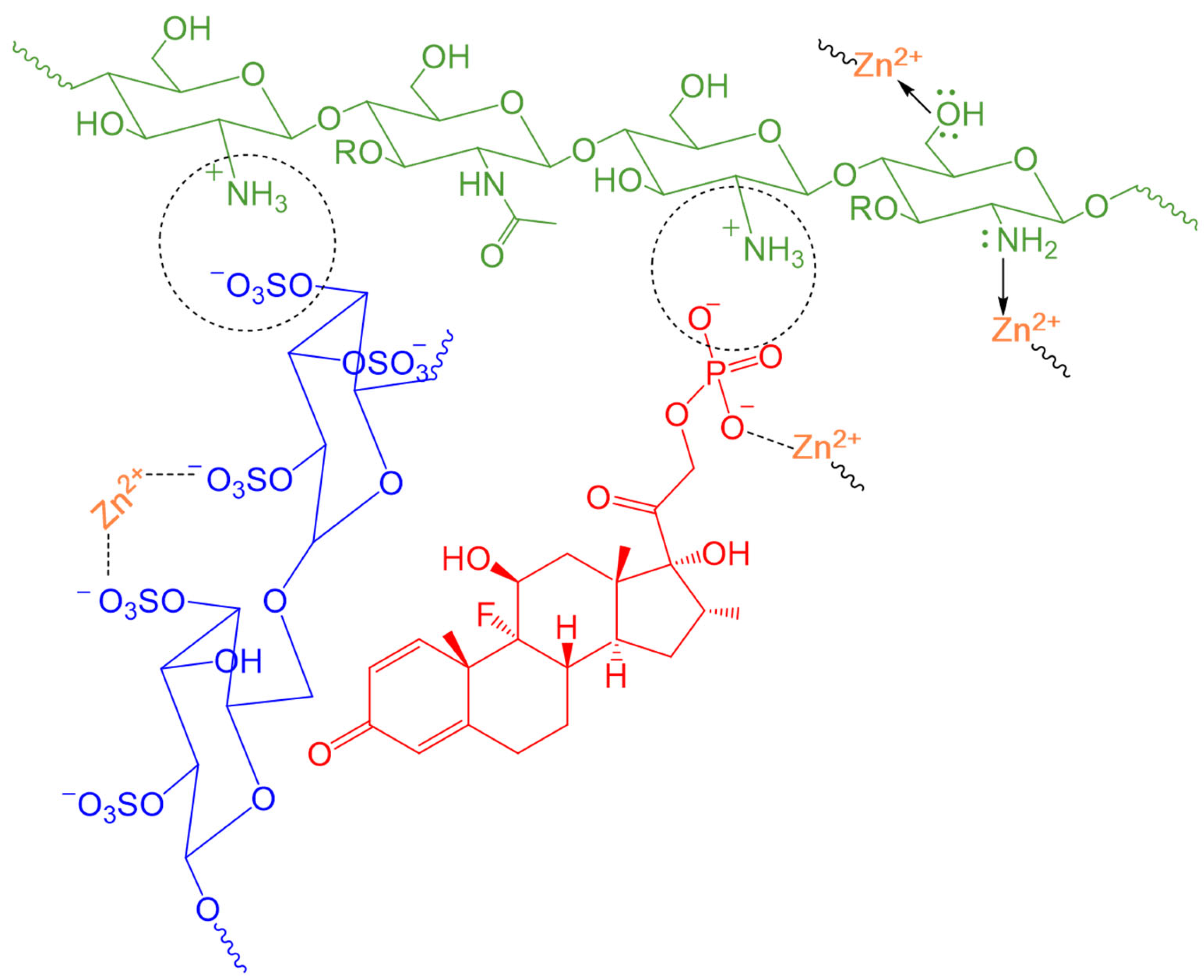
Hybrid (polymer-ionic) particle formation involves not only direct electrostatic attraction between oppositely charged polyelectrolytes but also the coordination of Zn2+ ions as an additional inorganic cross-linker. Zn2+ ions, possessing a high coordination number (typically 4 or 6), are expected to act as coordination bridges between multiple system components simultaneously. Due to their divalent nature, Zn2+ ions are hypothesized to interact with negatively charged sulfate groups of DS, where the coordination of Zn2+ ions to two sulfate groups belonging to different polysaccharide chains leads to additional densification of the polymer network and a reduction in particle size [27,54]. A critical aspect is the direct coordination of Zn2+ ions to the phosphate group of DexP. The formation of stable Zn2+ complexes with DexP phosphate groups facilitates the retention of DexP molecules within the polymer matrix. This significantly enhances EE, as the drug is retained not only by weak physical forces but also by strong coordination bonds [34]. Furthermore, Zn2+ can potentially interact with the tertiary amino groups of Deae-fragments and the primary amino groups of CS, which contain lone electron pairs capable of donor-acceptor interactions with Zn2+ ions [28,54,55]. The suggested network of possible interactions is illustrated in Figure 1.
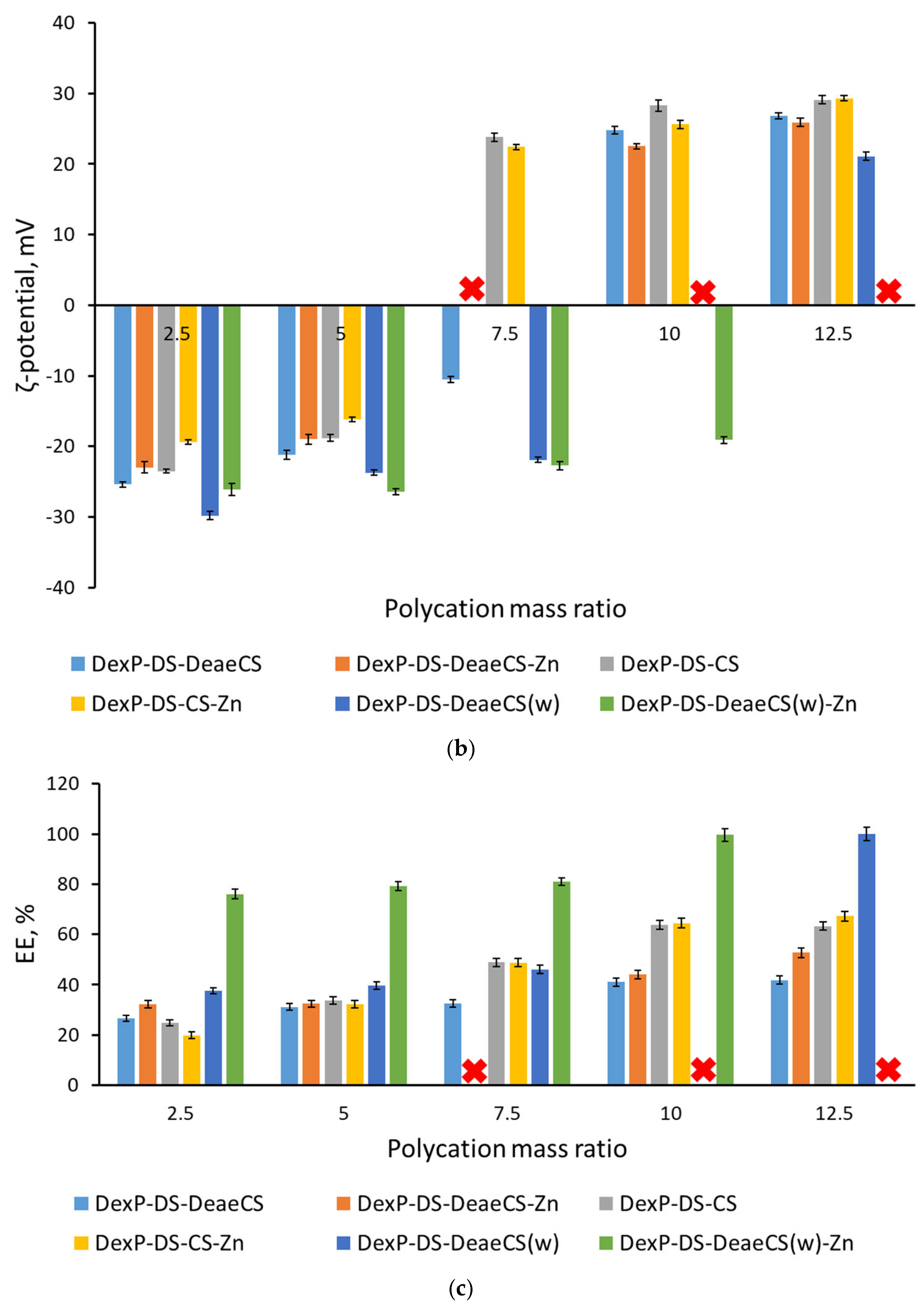
PEC synthesis involved maintaining a fixed 1:5 mass ratio of DexP to DS, while the polycationic component (DeaeCS/CS) content was varied. Zinc sulfate was added in a mass fraction equivalent to that of DexP across all samples. The assembly patterns of these DexP-loaded complexes, based on DS and varying polycation levels, were analyzed using DLS and ELS; results are summarized in Figure 2.
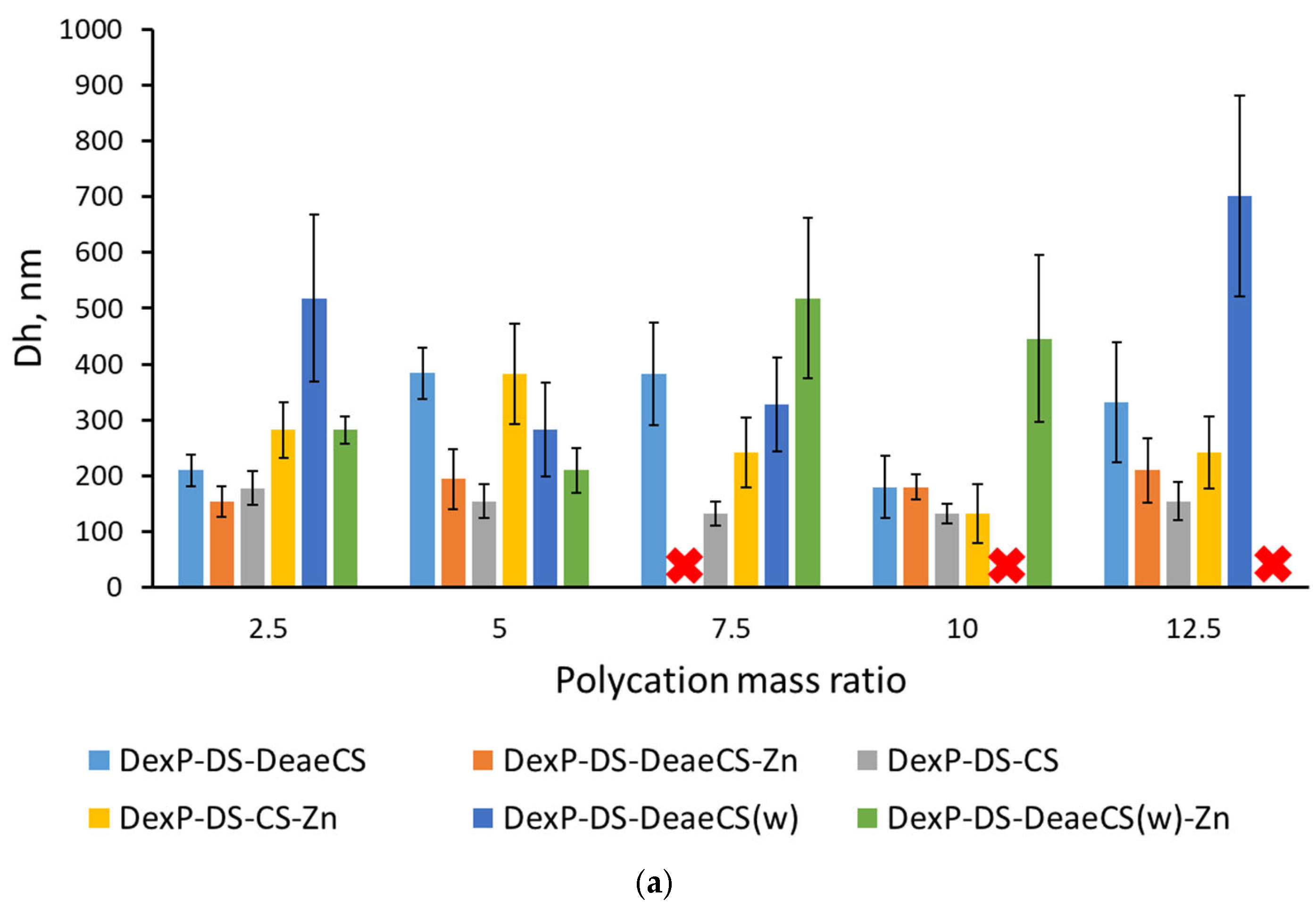
Analysis of hydrodynamic diameters revealed significant differences when using an aqueous solution of DeaeCS compared to its AcOH solution, with a tendency toward increased PEC size in aqueous media (Figure 2a). DeaeCS-based nanoparticles in an acidic medium were found to be larger than CS-based ones. This may be explained by the fact that Deae groups create steric hindrance to denser packing and maintain a high degree of hydration and electrostatic tension within the matrix, whereas native CS forms more compact collapsed structures due to simpler and closer ionic interactions with DS. The lack of significant influence of Zn2+ ions on particle size trends is attributed to specific polyelectrolyte interactions between the amino groups of CS/DeaeCS and the sulfate groups of DS, which lead to the formation of strong and compact complexes [19].
ζ-Potential values varied from −19.4 to +29.3 mV for both Zn2+-containing and Zn2+-free complexes prepared using 0.1 M acetic acid solutions of CS or DeaeCS (pH ≈ 3.0). In contrast, PECs obtained with aqueous DeaeCS solutions (pH ≈ 6–7) exhibited a negative ζ-potential (from −26.1 to −19.1 mV) until the cationic component reached 10 pbw (Figure 2b). Data analysis indicated that the surface charge of nanoparticles formed in acidic medium strongly depended on the polycation fraction, whereas only negatively charged nanoparticles were formed in aqueous medium unless a large excess of DeaeCS was used.
The charge state of the polyelectrolytes is pH-dependent. CS contains primary amino groups with pKa ≈ 6.5. DeaeCS is more complex: it retains residual primary amino groups; secondary amino groups (monosubstituted) and tertiary amino groups (disubstituted) on the CS backbone, both with pKa ≈ 6.5–7.0; tertiary amino groups on the diethylaminoethyl substituents (pKa ≈ 9–10); and a small fraction (14%) of quaternized amino groups that carry a permanent positive charge regardless of pH. DS has sulfate groups (pKa ≈ 1–2), which are fully deprotonated under all experimental conditions. DexP contains two phosphate groups with pKa values of approximately 1.9 and 6.4; thus, at pH 3.0 it carries a net charge of about −1 (the second phosphate is largely protonated), while at pH 6–7 it is fully deprotonated (−2).
The reversal of ζ-potential with increasing polycation content is explained by the sequential neutralization of anionic groups (DS and DexP) by the polycation. Complex formation stoichiometry is governed not only by charge neutralization but also by polymer flexibility and the nature of the ionic groups (weakly basic amino groups vs. strongly acidic sulfate groups) [19]. Depending on the assembly conditions, the effective stoichiometry can require up to six amino groups per one sulfate group [56]. A point of electroneutrality occurs when the total positive charge (from protonated amino groups, quaternized ammonium groups, and Zn2+ ions) balances the total negative charge (from DS, DexP, and any free sulfate). Near this point, particles often become unstable and may aggregate. Given that DS contains approximately three anionic groups per disaccharide unit and the degree of deacetylation of CS is 74%, a substantial excess of amino groups is necessary to obtain positively charged particles.
Notably, a significant increase in DexP encapsulation efficiency was observed for these systems, reaching up to 100% with the introduction of Zn2+ ions (Figure 2c). The high binding efficiency of DexP when using water instead of AcOH as a solvent for DeaeCS can be explained by the absence of competition from acetate ions for binding to the positively charged centers of DeaeCS. Furthermore, given the pKa values of the DexP phosphate groups (~1.9 and ~6.4), it can be assumed that in 0.1 M AcOH (pH ≈ 3.0), most DexP molecules carry a net charge of −1, which reduces their electrostatic incorporation into the hybrid matrix. In contrast, in an aqueous solution of DeaeCS (pH ≈ 6–7), the phosphate groups are predominantly deprotonated, giving DexP a charge of −2. This allows the DexP molecule to interact cooperatively with multiple binding sites (e.g., amino groups and Zn2+ ions), thereby acting as an additional cross-linking agent and enhancing encapsulation efficiency.
The permeability of anionic particles through the cell membrane is a complex process, generally less efficient than for cationic analogs due to the electrostatic barrier (the cell surface carries a negative charge, causing negatively charged particles to accumulate in the intercellular space). However, anionic particles can be internalized via mechanisms such as pinocytosis, interaction with positively charged domains of cellular receptors and transport proteins, and the affinity of hydrophobic fragments (in this case, the hydrophobic steroid backbone of the DexP molecule) for membrane lipids. Negatively charged particles are suitable for systemic administration and exhibit reduced cytotoxicity (whereas cationic particles can actively bind to cell membranes, causing lysis and cell death). Furthermore, in systemic circulation, anionic particles are less likely to be taken up by macrophages, increasing circulation time and prolonging drug action. Although this study focused on producing cationic particles for local DexP delivery, we utilized the resulting anionic PECs to compare their cytotoxicity and cellular permeability with cationic counterparts. A detailed description of the PECs used in biological testing is presented in Table 1.
According to XRD data, pristine DexP and ZnSO4 exhibit distinct crystalline patterns. In contrast, diffractograms of the synthesized DexP/Zn2+-containing PECs are dominated by a broad amorphous halo typical of the polymeric framework. The absence of sharp reflections associated with DexP and zinc sulfate suggests that these components are molecularly dispersed or converted into an amorphous state within the polymer particles (Figure S1). The transition of DexP from a crystalline to an amorphous state within the PEC matrix indicates the formation of a single nanostructured system (rather than a simple mixture of components) and is a crucial factor for enhancing therapeutic efficacy. Molecular dispersion of the drug eliminates the lattice energy barrier, thereby significantly increasing its dissolution rate and ensuring higher bioavailability. Furthermore, the absence of crystalline aggregates facilitates more uniform drug distribution, supporting the controlled and sustained release profile observed in kinetic studies (see Section 3.2).
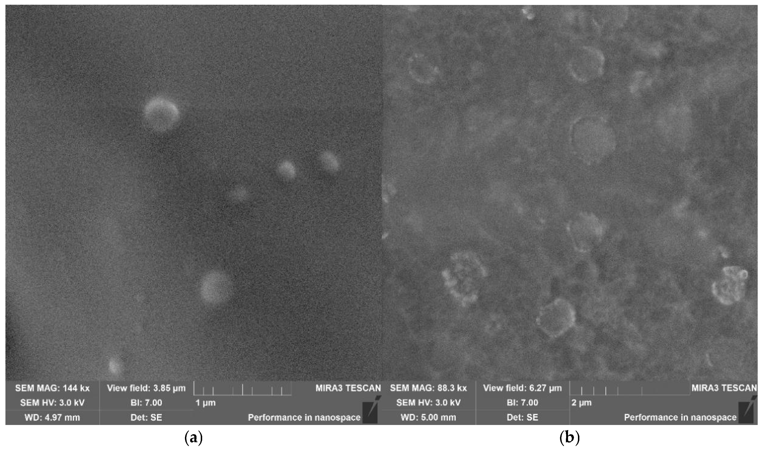
SEM analysis of the DexP-DS-DeaeCS-10 and DexP-DS-DeaeCS-Zn-10 formulations (Figure 3) revealed a predominantly spherical morphology. The average size of these polyelectrolyte complexes in the solid state was approximately 200 nm, which is close to their hydrodynamic diameters measured by DLS (180 ± 56 nm and 180 ± 22 nm, respectively). However, SEM images also showed some degree of polydispersity and particles appearing larger than the DLS-derived mean. This discrepancy is likely due to drying artefacts and particle aggregation during sample preparation for electron microscopy. The close agreement between DLS and SEM values for the Zn2+-cross-linked formulation suggests that the particles are sufficiently rigid to limit swelling in aqueous medium [57], although the loss of the hydration shell upon drying may contribute to size reduction. CS-based formulations exhibited similar morphology in preliminary imaging; the focus of SEM analysis was on the optimized DeaeCS-based systems used in biological studies.

3.2. In Vitro Release Kinetics of DexP from PECs

The in vitro release kinetics of DexP from the developed PECs were evaluated in TFS at pH 7.4 and 32 °C to mimic physiological ocular surface conditions. The assembly of these systems relies on the electrostatic coupling of DeaeCS/CS polycations with anionic DexP and DS, resulting in the formation of intra- and intermolecular complexes. The structural integrity of the matrix is further reinforced through complex formation with Zn2+ ions, which act as secondary cross-linkers. Nevertheless, when exposed to the salinity and pH levels typical of tear fluid, these ionic networks undergo relaxation. This leads to matrix swelling and accelerated diffusion, which serves as the primary mechanism for the controlled release of encapsulated therapeutic agents [27,58]. The release profile is governed by a combination of diffusion through the polymer matrix, polymer chain relaxation (swelling), and ion exchange between the counterions in the release medium and the charged groups within the PECs.
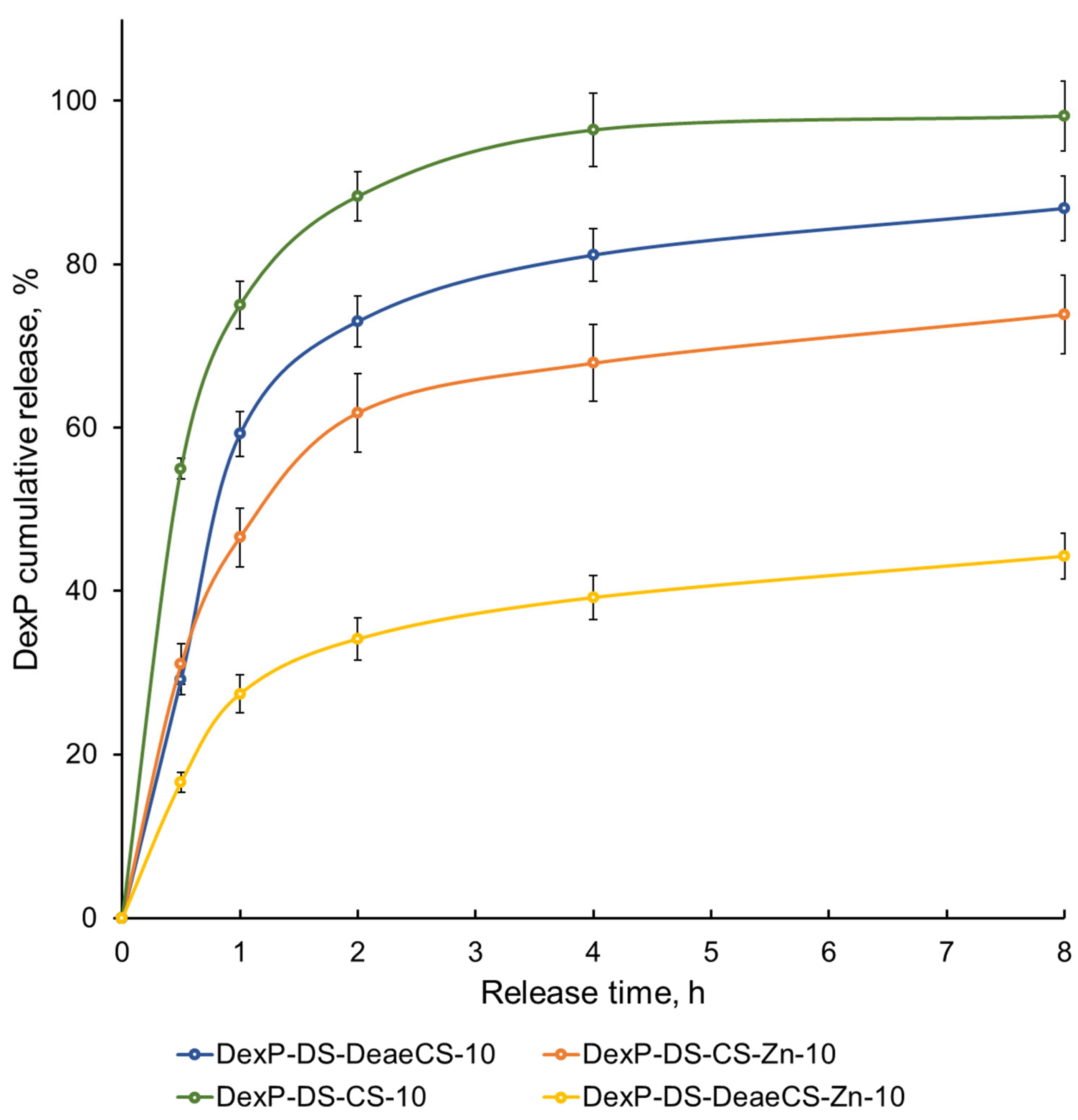
We investigated the effect of polycation nature and quantity as well as the presence of the cross-linking agent (Zn2+ ions) on DexP release kinetics from the developed polyelectrolyte particles. The in vitro release profiles of DexP in TFS are shown in Figure 4. Modifying CS with Deae groups alleviates the main drawback of the native polymer: the loss of aggregation stability at neutral pH. Due to the high pKa value of the tertiary amino groups of the Deae-fragment (~9–10), the resulting polyelectrolyte bonds are characterized by increased resistance to hydrolytic cleavage under physiological conditions (pH 7.4), which is a critical requirement for the creation of stable DexP transport systems. In contrast, native CS (pKa ~6.5) undergoes partial deprotonation at pH 7.4, weakening electrostatic interactions and leading to faster matrix disintegration and drug release. Consequently, complexes based on DeaeCS as the polycationic component were characterized by a slower drug release compared to native CS.
The presence of Zn2+ ions significantly reduced the release rate in both CS and DeaeCS-based systems. This attenuation is attributed to the formation of additional coordination bonds between Zn2+ and the phosphate groups of DexP, as well as the sulfate groups of DS. These coordination interactions are stronger than simple electrostatic attractions and create a denser hybrid matrix that impedes drug diffusion. The synergistic effect of Zn2+ cross-linking and polycationic charge density results in a more sustained release profile, which is particularly advantageous for ocular applications where rapid precorneal clearance typically limits drug bioavailability. By sustaining the release phase, the developed formulations could potentially reduce the frequency of instillation required to maintain therapeutic concentrations.
The observed release kinetics suggest a diffusion-controlled mechanism modulated by matrix erosion and swelling. The slower release from Zn2+-containing formulations indicates that the cross-linking density effectively prevents the burst-release effect often observed in physically cross-linked PECs. This finding is consistent with previous reports on Zn2+-stabilized polyelectrolyte systems for ophthalmic delivery [27,28]. Furthermore, the stability of the release profile at physiological pH confirms that the DeaeCS-based matrix maintains its integrity in the ocular environment, unlike native CS systems, which may prematurely dissociate. Compared to CS-DS nanoparticles loaded with ciprofloxacin, which demonstrated sustained release for 21 h [38], the DexP-DS-DeaeCS-Zn systems exhibit comparable or improved release characteristics while offering the additional benefit of Zn2+-mediated anti-inflammatory activity.
To evaluate the mass transport kinetics of DexP over the 8 h release period, the experimental release data were fitted to the Weibull model described in Section 2.5. The derived kinetic parameters are summarised in Table 2.
The Weibull equation adequately described the release profiles, with high correlation coefficients (R2 up to 0.982). For the non-Zn2+-cross-linked formulations (DexP-DS-DeaeCS-10 and DexP-DS-CS-10), the shape factors (b) were 0.983 and 0.832, respectively. Values approaching unity (0.75–1.00) in the Weibull model are indicative of Case-II transport, where polymer chain relaxation and matrix erosion predominantly govern the release kinetics [46]. This is consistent with the pronounced initial burst release observed for these systems.
In contrast, the Zn2+-cross-linked formulations (DexP-DS-DeaeCS-Zn-10 and DexP-DS-CS-Zn-10) exhibited markedly lower b values of 0.2605 and 0.3569, respectively. A shape factor below 0.35 mathematically confirms a diffusion-controlled release mechanism through a highly tortuous and restricted pathway within a dense, extensively cross-linked polyelectrolyte matrix. These results validate that Zn2+-mediated coordination successfully establishes a robust structural barrier that effectively retards drug transport, consistent with the sustained release profiles observed experimentally (Figure 4).
Collectively, these data demonstrate that the release kinetics can be finely tuned by varying the polycation type and Zn2+ content, offering a versatile platform for optimizing therapeutic regimens for inflammatory conditions. The sustained release profile, combined with the enhanced permeability characteristics described in Section 3.4, positions these particles as promising candidates for prolonged DexP delivery.

3.3. Anti-Inflammatory Activity and Cytocompatibility in THP-1 Cells

For biological evaluations, we selected formulations with a polycation content of 10 pbw (CS or DeaeCS) as these provided optimal colloidal stability and drug encapsulation (Table 1). Additionally, the anionic formulation DexP-DS-DeaeCS-Zn-2.5 (ζ-potential −23.0 mV) was included in permeability studies (Section 3.4) as a representative negatively charged particle to compare the effect of surface charge on corneal transport.

3.3.1. Cytocompatibility of DexP-PECs in THP-1 Cells

To evaluate the cytocompatibility of the developed DexP-PECs, we employed the human monocytic cell line THP-1, which serves as a well-established in vitro model for assessing macrophage-mediated inflammatory responses and drug-induced cytotoxicity [59,60].
Initial cytotoxicity screening was performed at a DexP concentration of 0.1 μg/mL, representing a therapeutically relevant dose for anti-inflammatory applications. Cell viability was assessed via flow cytometry using YO-PRO-1/propidium iodide (PI) double staining, which enables simultaneous discrimination of viable (YO-PRO-1PI), early apoptotic (YO-PRO-1+PI), and late apoptotic/necrotic (YO-PRO-1+PI+) cell populations (Table S2).
Notably, DeaeCS alone demonstrated a statistically significant reduction in the viable cell population (93.73% vs. 95.31% in control, p < 0.05) accompanied by a concomitant increase in late apoptotic/necrotic cells (4.04% vs. 2.95% in control, p < 0.05). However, it is important to emphasize that when DeaeCS was incorporated into the PEC formulation (DexP-DS-DeaeCS-10 and DexP-DS-DeaeCS-Zn-10), this cytotoxic effect was completely abolished, with viability levels comparable to the negative control (95.99% and 95.86%, respectively). This finding suggests that the electrostatic complexation of DeaeCS with anionic DS effectively neutralizes the positive charge density, thereby minimizing potential cytotoxic interactions with cell membranes.
Free DexP, individual polymer components (CS·HCl, DS), and zinc sulfate did not exhibit significant cytotoxicity at this concentration, confirming the biocompatibility of individual formulation components. To evaluate the safety margin of the developed formulations, cytotoxicity was further assessed at a ten-fold higher DexP concentration (1 μg/mL), representing a worst-case scenario for local drug accumulation (Table S3). At this elevated concentration, all DexP-PEC formulations maintained excellent cytocompatibility, with viable cell populations exceeding 95% across all tested samples. Importantly, no concentration-dependent cytotoxicity was observed for the encapsulated DexP formulations, in contrast to what might be expected with free drug administration. This finding underscores the protective role of the polymer matrix, which not only modulates drug release kinetics but also shields cells from potential high-dose exposure.

3.3.2. Suppression of THP-1 Cell Activation by DexP-PECs

To evaluate the therapeutic potential of the developed DexP-PECs, we assessed their ability to suppress inflammatory activation in THP-1 monocytic cells. CD54 (intercellular adhesion molecule-1) was selected as the primary marker of inflammatory activation, as its upregulation on monocyte/macrophage surfaces is a well-established indicator of pro-inflammatory signaling and plays a critical role in leukocyte recruitment to sites of inflammation. TNF-α stimulation is known to induce robust CD54 expression through activation of pro-inflammatory signaling cascades, making it a reliable model for evaluating anti-inflammatory drug efficacy [61,62].
In the absence of inflammatory stimuli, THP-1 cells exhibited low baseline CD54 expression (0.436 MFI units; Q25–Q75: 0.396–0.457), consistent with the resting monocytic phenotype (Table S4). Stimulation with TNF-α (10 ng/mL) for 24 h resulted in a dramatic 27-fold increase in CD54 expression (11.911 MFI units; Q25–Q75: 11.218–12.598; p < 0.001 vs. unstimulated control), confirming successful inflammatory activation of the cell line.
Analysis of individual formulation components revealed distinct immunomodulatory profiles. Free DexP significantly suppressed TNF-α-induced CD54 upregulation (3.286 MFI units vs. 11.911 MFI units in TNF-α control; p < 0.01), demonstrating approximately 72% reduction in inflammatory marker expression. This finding confirms the preserved biological activity of DexP following the encapsulation process.
Notably, DeaeCS alone demonstrated a pro-inflammatory effect, significantly increasing CD54 expression in both unstimulated (0.559 MFI units; p < 0.05 vs. control) and TNF-α-stimulated conditions (17.613 MFI units; p < 0.01 vs. TNF-α control). This observation is consistent with the cytotoxicity data from Section 3.3.1 and may be attributed to the high cationic charge density of DeaeCS, which can activate monocytes and up-regulate pro-inflammatory cytokine production through membrane perturbation and associated inflammatory pathways [63,64]. However, this pro-inflammatory effect was completely abolished when DeaeCS was incorporated into PEC formulations (DexP-DS-DeaeCS-10 and DexP-DS-DeaeCS-Zn-10), suggesting that electrostatic complexation with anionic DS effectively neutralizes the immunostimulatory properties of the cationic polymer. In addition to charge neutralization, DS acts as a shielding agent that physically prevents direct contact between the cationic groups of DeaeCS and the macrophage membrane, thereby avoiding the activation of pro-inflammatory signaling pathways.
Native CS·HCl and DS did not significantly modulate CD54 expression, indicating their immunological inertness at the concentrations tested. Zinc sulfate demonstrated a modest but statistically significant reduction in TNF-α-induced CD54 expression (10.457 MFI units; p < 0.05 vs. TNF-α control), supporting previous reports of the intrinsic anti-inflammatory properties of Zn2+ ions [36,37].
All DexP-containing PEC formulations effectively suppressed TNF-α-induced CD54 upregulation, with CD54 expression levels comparable to or lower than free DexP (Table S4). These findings demonstrate that encapsulation does not compromise the anti-inflammatory activity of DexP. Furthermore, the comparable efficacy between Zn2+-containing and Zn2+-free formulations suggests that the cross-linking agent does not interfere with drug bioactivity.
To evaluate the concentration-response relationship, CD54 suppression was assessed at both 0.1 μg/mL and 1 μg/mL DexP equivalents (Table S5). At the higher concentration (1 μg/mL), free DexP demonstrated enhanced suppression (2.542 MFI units; 79% reduction), while PEC formulations maintained comparable efficacy to the 0.1 μg/mL dose. Interestingly, only DexP-DS-DeaeCS-Zn-10 showed a statistically significant dose-dependent improvement in CD54 suppression (p = 0.025, Mann–Whitney U test), suggesting that the combination of DeaeCS and Zn2+ may facilitate more efficient intracellular drug delivery or sustained release at the target site (Table 3).
Several mechanisms may contribute to the observed anti-inflammatory efficacy of DexP-PECs. Primarily, the amorphous state of DexP within the PEC matrix, as confirmed by XRD analysis, ensures rapid dissolution upon cellular uptake, allowing the drug to effectively engage its intracellular glucocorticoid receptor target. Concurrently, the cationic surface charge of the PECs (+22.5 to +28.3 mV) facilitates electrostatic interactions with negatively charged cell membranes, potentially enhancing internalization via endocytosis. Furthermore, the Zn2+-cross-linked matrix likely provides sustained intracellular drug release, maintaining therapeutic concentrations over extended periods and reducing the need for frequent dosing. The intrinsic anti-inflammatory properties of Zn2+ may also complement DexP activity, as evidenced by the modest CD54 suppression observed with ZnSO4 alone. Crucially, the electrostatic complexation of DeaeCS with DS eliminates the pro-inflammatory effects observed with free DeaeCS, ensuring the biocompatibility of the final formulation. Notably, all PEC formulations suppressed CD54 expression comparably to free DexP, indicating that encapsulation preserves anti-inflammatory bioactivity, while the dose-dependent improvement observed specifically for DexP-DS-DeaeCS-Zn-10 suggests an optimal formulation for concentration-responsive therapy. Collectively, these data demonstrate that the developed DexP-PECs maintain potent anti-inflammatory activity while offering the advantages of sustained release and enhanced biocompatibility. The ability to suppress CD54 upregulation—a critical mediator of leukocyte recruitment—positions these formulations as promising candidates for treating inflammatory conditions where macrophage activation plays a central pathogenic role.

3.4. Corneal Cytotoxicity and Permeability of DexP-PECs

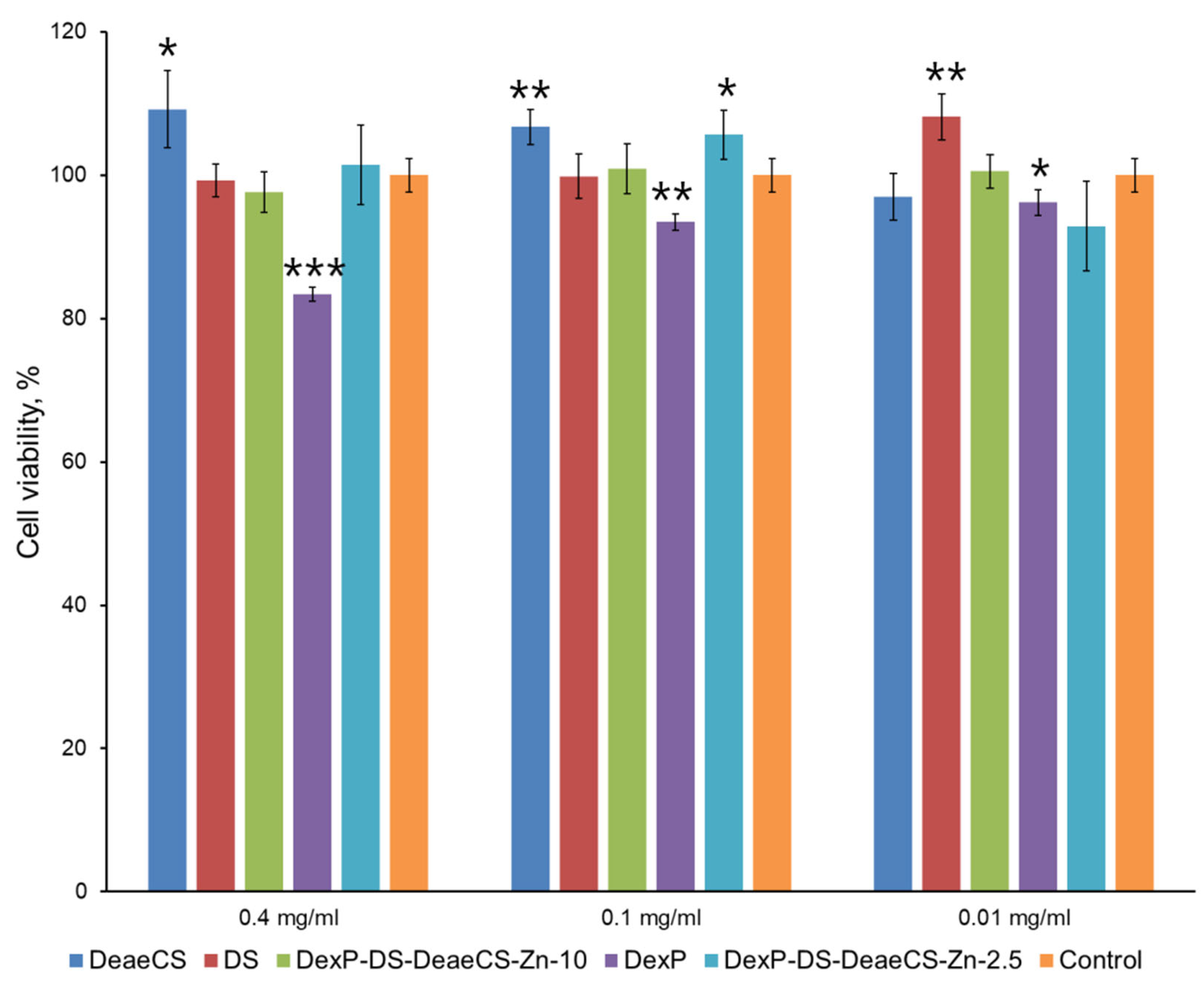
To evaluate the safety profile of the developed formulations for ocular applications, cytotoxicity was assessed using the SIRC rabbit corneal epithelial cell line. The cytotoxicity of free DexP and DexP-loaded PECs was evaluated over a concentration range equivalent to 0.01–0.4 mg/mL DexP following 72 h exposure, and cell viability was quantified using the MTT assay (Figure 5).
Free DexP exhibited a concentration-dependent cytotoxic effect, with cell viability decreasing progressively at higher concentrations. This observation is consistent with previous reports on glucocorticoid-induced cytotoxicity in corneal epithelial cells, where prolonged exposure to high drug concentrations can compromise cell membrane integrity and metabolic activity [8,9,10]. In contrast, all DexP-containing polymer particles demonstrated excellent biocompatibility across the entire concentration range tested, with cell viability exceeding 90% even at the highest concentration (0.4 mg/mL DexP equivalent). This marked difference in cytotoxicity profiles underscores the protective role of the polymer matrix, which shields corneal cells from direct exposure to high local drug concentrations while maintaining therapeutic efficacy through sustained release.
The absence of cytotoxicity in PEC formulations can be attributed to several factors. First, the encapsulation of DexP within the polyelectrolyte matrix reduces the free drug concentration available for immediate cellular uptake, thereby minimizing potential toxic effects. Second, the biocompatible nature of the polymer components (CS, DeaeCS, DS) and Zn2+ ions at the concentrations employed in this study contributes to the favorable safety profile. Notably, the Zn2+-cross-linked formulations did not exhibit increased cytotoxicity compared to their non-cross-linked counterparts, confirming that the coordination cross-linking strategy does not compromise biocompatibility. These findings establish the cytocompatibility of the developed DexP-PECs and support their further evaluation in permeability studies.
Permeability studies were conducted using the SIRC cell monolayer model described in Section 2.7. Barrier integrity was confirmed prior to transport experiments, with TEER values reaching 732 ± 23 Ω·cm2 on day 10, consistent with previously reported values for functional corneal epithelial monolayers [50]. The apparent permeability coefficient (Papp) and lag time values for free DexP and selected PEC formulations are summarized in Table 4.
As summarized in Table 4, the formation of PECs effectively delayed the initial release, with the lag time increasing proportionally to the DeaeCS content. Remarkably, the optimized formulation (10 pbw DeaeCS) exhibited the longest lag time but simultaneously demonstrated the highest apparent permeability, nearly 1.5-fold greater than that of free DexP. Importantly, the calculated Papp reflects the steady-state flux of DexP that has already been released from the particles. Therefore, the higher Papp value for the 10 pbw formulation, despite its longer lag time, indicates that once released, DexP molecules traverse the corneal epithelium significantly more efficiently than free DexP. This enhanced permeability may be attributed to the paracellular transport-promoting effect of DeaeCS, which is proposed to transiently modulate tight junctions, rather than to faster drug release. Thus, the formulation combines a sustained release phase (prolonged residence) with a subsequent phase of improved drug penetration, offering a dual advantage over free DexP.
This apparent paradox, where sustained release coexists with enhanced permeability, can be explained by the dual functionality of the DeaeCS polycation. While the PEC matrix effectively prevents the rapid wash-out of DexP through sustained retention, the cationic DeaeCS chains act as potent permeation enhancers by temporarily opening the tight junctions between corneal epithelial cells. The electrostatic interaction between the positively charged amino groups of DeaeCS and the negatively charged epithelial surface facilitates this paracellular transport mechanism. This interaction may induce transient reorganization of tight junction proteins (such as zonula occludens), allowing increased paracellular flux of the drug-loaded particles or released drug molecules. Additionally, the mucoadhesive properties of DeaeCS may prolong the residence time of the formulation on the corneal surface, further enhancing the opportunity for drug absorption.
The combination of sustained release (evidenced by extended lag time) and enhanced permeability (evidenced by increased Papp) positions the DexP-DS-DeaeCS-Zn-10 formulation as a promising candidate for ocular drug delivery. Compared to CS-DS nanoparticles loaded with ciprofloxacin, which demonstrated sustained release for 21 h but did not report permeability enhancement [38], the current system offers the additional benefit of improved corneal penetration. Similarly, while Chaiyasan et al. [39] reported that CS-DS nanoparticles stabilized with polyethyleneglycol 400 ensured long-term adhesion to the porcine corneal surface (more than 4 h) and partial penetration into the corneal epithelium, the Zn2+-cross-linked DeaeCS system described here achieves comparable or superior permeability without the need for additional surface stabilizers.
The dual advantage provided by the synthesized PECs—protecting the drug from rapid washout through a sustained lag period and enhancing its bioavailability via improved paracellular DexP penetration—addresses two critical limitations of conventional ophthalmic formulations. By extending the lag time and enhancing permeability simultaneously, the developed system could potentially reduce the frequency of instillation required to maintain therapeutic concentrations while improving drug bioavailability at the target site. These findings, combined with the preserved anti-inflammatory activity demonstrated in Section 3.3 and the favorable cytotoxicity profile established in this section, support the further development of these hybrid particles for clinical applications in ocular inflammatory conditions.

4. Conclusions and Future Perspectives

In this study, we developed Zn2+-cross-linked polyelectrolyte complexes based on DS and DeaeCS for sustained DexP delivery. The DeaeCS polycation ensures matrix stability at physiological pH, while Zn2+ coordination with sulfate and phosphate groups enhances drug retention and minimizes burst release. XRD analysis confirmed that both DexP and ZnSO4 lose their crystalline nature upon incorporation into the polymer matrix, transitioning into a molecularly dispersed amorphous state.
The optimized formulation (DexP-DS-DeaeCS-Zn-10) extended the lag time to 0.37 h and achieved a 1.5-fold increase in apparent corneal permeability (Papp = 14.28 × 10⁻⁶ cm/s) compared to free DexP (9.58 × 10⁻⁶ cm/s). Thus, the synthesized PECs offer a potential dual advantage: (i) protecting the drug from rapid clearance at the application site through sustained retention, and (ii) enhancing its bioavailability via improved paracellular penetration across epithelial barriers. Encapsulated DexP effectively suppressed TNF-α-induced CD54 expression in macrophages, confirming preserved anti-inflammatory activity, while maintaining low cytotoxicity in both corneal (SIRC) and macrophage (THP-1) cell lines. Collectively, these hybrid particles represent a promising platform for improved DexP delivery, combining prolonged residence, enhanced corneal penetration, and the potential for Zn2+-mediated anti-inflammatory effects. Although the present study focused on ocular administration as a primary model, the versatility of the system suggests potential applicability to other inflammatory conditions requiring sustained drug delivery.
The present study provides a solid proof-of-concept for this dual-action delivery system. Further work will focus on direct spectroscopic characterization of the coordination interactions, evaluation of colloidal and storage stability, assessment of mucoadhesive properties using mucin interaction assays, and optimization of pH, osmolarity, and sterilization methods for clinical translation. For clinical translation, aseptic manufacturing by sterile filtration (0.22 µm) of the individual polymer and drug solutions, followed by aseptic self-assembly under laminar flow, is proposed. Importantly, ex vivo penetration studies using relevant epithelial models and in vivo pharmacokinetic and efficacy evaluations in appropriate inflammation models are required to confirm the translational potential of these formulations.

Supplementary Materials

The following supporting information can be downloaded at https://www.mdpi.com/article/10.3390/macromol6030061/s1, Table S1: Component mass ratios used for the fabrication of DexP-PECs; Table S2: Viability of THP-1 cells after 24 h incubation in the presence of DexP-PECs (based on 0.1 μg DexP). Results (n = 8) are presented as median and interquartile range, Me (Q25; Q75); Table S3: Viability of THP-1 cells after 24 h incubation in the presence of DexP-PECs (based on 1 μg of DexP). Results (n = 8) are presented as median and interquartile range, Me (Q25; Q75); Table S4: Effect of DexP-PECs (based on 0.1 μg/mL dexamethasone) on THP-1 cell line activation. Results (n = 6) are presented as arbitrary CD54 fluorescence units (MFI units) and are given as median and interquartile range, Me (Q25; Q75); Table S5: Effect of DexP-PECs (at a concentration of 1 μg/mL DexP) on the level of THP-1 cell line activation. Results (n = 6) are presented as arbitrary units of CD54 fluorescence (MFI units) and are given as median and interquartile range, Me (Q25; Q75); Figure S1: XRD patterns of pristine DexP and ZnSO4 compared to their incorporated forms within the polymer complexes.

Author Contributions

Conceptualization, N.V.D. and Y.A.S.; methodology, N.V.D., A.N.B., I.V.K. and T.S.S.; investigation, A.N.B., N.V.D., A.Y.B., V.A.P., A.S.T., A.A.R., T.S.S., Y.A.N. and A.V.M.; writing—original draft preparation, A.N.B., N.V.D., I.V.K. and Y.A.S.; writing—review and editing, Y.A.S.; supervision, Y.A.S.; project administration, N.V.D.; funding acquisition, N.V.D. All authors have read and agreed to the published version of the manuscript.

Funding

This research was funded by the Russian Science Foundation (grant No. 25-75-20013).

Institutional Review Board Statement

Not applicable.

Data Availability Statement

The raw data supporting the conclusions of this article will be made available by the authors on request.

Acknowledgments

The authors are grateful to the Core Facility Center “Arktika” of the Northern (Arctic) Federal University for providing the equipment for X-ray diffraction analysis.

Conflicts of Interest

The authors declare no conflicts of interest.

References

  1. Madamsetty, V.S.; Mohammadinejad, R.; Uzieliene, I.; Nabavi, N.; Dehshahri, A.; Garcia-Couce, J.; Tavakol, S.; Moghassemi, S.; Dadashzadeh, A.; Makvandi, P.; et al. Dexamethasone: Insights into pharmacological aspects, therapeutic mechanisms, and delivery systems. ACS Biomater. Sci. Eng. 2022, 8, 1763–1790. [Google Scholar] [CrossRef] [PubMed]
  2. Placha, D.; Jampilek, J. Chronic inflammatory diseases, anti-inflammatory agents and their delivery nanosystems. Pharmaceutics 2021, 13, 64. [Google Scholar] [CrossRef] [PubMed]
  3. Reichardt, S.D.; Amouret, A.; Muzzi, C.; Vettorazzi, S.; Tuckermann, J.P.; Lühder, F.; Reichardt, H.M. The role of glucocorticoids in inflammatory diseases. Cells 2021, 10, 2921. [Google Scholar] [CrossRef] [PubMed]
  4. Fulmali, A.; Bharate, S.S. Phosphate moiety in FDA-approved pharmaceutical salts and prodrugs. Drug Dev. Res. 2022, 83, 1059–1074. [Google Scholar] [CrossRef] [PubMed]
  5. Urbańska, J.; Karewicz, A.; Nowakowska, M. Polymeric delivery systems for dexamethasone. Life Sci. 2014, 96, 1–6. [Google Scholar] [CrossRef] [PubMed]
  6. Pourmadadi, M.; Maleki, M.; Rahdar, A.; Aboudzadeh, M.A. Innovative polymeric and non-polymeric nanocarrier systems for dexamethasone: Enhancing precision and reducing side effects. J. Nanoparticle Res. 2025, 27, 167. [Google Scholar] [CrossRef]
  7. Hoseini, Z.S.; Rezayat, S.M.; Rahdar, A.; Pandey, S.; Jaafari, M.R. Multifunctional nanocarriers for prednisolone delivery: A new frontier in cancer and inflammation therapy. BioNanoScience 2026, 16, 33. [Google Scholar] [CrossRef]
  8. Luo, L.; Yang, J.; Oh, Y.; Hartsock, M.J.; Xia, S.; Kim, Y.-C.; Ding, Z.; Meng, T.; Eberhart, C.G.; Ensign, L.M.; et al. Controlled release of corticosteroid with biodegradable nanoparticles for treating experimental autoimmune uveitis. J. Control. Release 2019, 296, 68–80. [Google Scholar] [CrossRef] [PubMed]
  9. Begines, B.; Ortiz, T.; Pérez-Aranda, M.; Martínez, G.; Merinero, M.; Argüelles-Arias, F.; Alcudia, A. Polymeric nanoparticles for drug delivery: Recent developments and future prospects. Nanomaterials 2020, 10, 1403. [Google Scholar] [CrossRef] [PubMed]
  10. Lühder, F.; Reichardt, H.M. Novel drug delivery systems tailored for improved administration of glucocorticoids. Int. J. Mol. Sci. 2017, 18, 1836. [Google Scholar] [CrossRef] [PubMed]
  11. Shahbaz, A.; Hussain, N.; Basra, M.A.R.; Bilal, M. Polysaccharides-based nano-hybrid biomaterial platforms for tissue engineering, drug delivery, and food packaging applications. Starch-Stärke 2022, 74, 2200023. [Google Scholar] [CrossRef]
  12. Cazorla-Luna, R.; Martín-Illana, A.; Notario-Pérez, F.; Ruiz-Caro, R.; Veiga, M.-D. Naturally occurring polyelectrolytes and their use for the development of complex-based mucoadhesive drug delivery systems: An overview. Polymers 2021, 13, 2241. [Google Scholar] [CrossRef] [PubMed]
  13. Wani, T.U.; Pandith, A.H.; Sheikh, F.A. Polyelectrolytic nature of chitosan: Influence on physicochemical properties and synthesis of nanoparticles. J. Drug Deliv. Sci. Technol. 2021, 65, 102730. [Google Scholar] [CrossRef]
  14. Desai, N.; Rana, D.; Salave, S.; Gupta, R.; Patel, P.; Karunakaran, B.; Sharma, A.; Giri, J.; Benival, D.; Kommineni, N. Chitosan: A potential biopolymer in drug delivery and biomedical applications. Pharmaceutics 2023, 15, 1313. [Google Scholar] [CrossRef] [PubMed]
  15. Zubareva, A.A.; Gasilova, E.R.; Poshina, D.N.; Skorik, Y.A. Molecular weight of chitosan: From structural complexity to analytical reliability. Carbohydr. Polym. 2026, 385, 125348. [Google Scholar] [CrossRef] [PubMed]
  16. de Souza, R.H.F.V.; Dalla Picola, I.P.; Shi, Q.; Petrônio, M.S.; Benderdour, M.; Fernandes, J.C.; Lima, A.M.F.; Martins, G.O.; Junior, A.M.M.; de Oliveira Tiera, V.A.; et al. Diethylaminoethyl-chitosan as an efficient carrier for sirna delivery: Improving the condensation process and the nanoparticles properties. Int. J. Biol. Macromol. 2018, 119, 186–197. [Google Scholar] [CrossRef] [PubMed]
  17. de Oliveira Pedro, R.; Goycoolea, F.M.; Pereira, S.; Schmitt, C.C.; Neumann, M.G. Synergistic effect of quercetin and ph-responsive deae-chitosan carriers as drug delivery system for breast cancer treatment. Int. J. Biol. Macromol. 2018, 106, 579–586. [Google Scholar] [CrossRef] [PubMed]
  18. Ardean, C.; Davidescu, C.M.; Nemeş, N.S.; Negrea, A.; Ciopec, M.; Duteanu, N.; Negrea, P.; Duda-Seiman, D.; Musta, V. Factors influencing the antibacterial activity of chitosan and chitosan modified by functionalization. Int. J. Mol. Sci. 2021, 22, 7449. [Google Scholar] [CrossRef] [PubMed]
  19. Delair, T. Colloidal polyelectrolyte complexes of chitosan and dextran sulfate towards versatile nanocarriers of bioactive molecules. Eur. J. Pharm. Biopharm. 2011, 78, 10–18. [Google Scholar] [CrossRef] [PubMed]
  20. Ramasundaram, S.; Saravanakumar, G.; Sobha, S.; Oh, T.H. Dextran sulfate nanocarriers: Design, strategies and biomedical applications. Int. J. Mol. Sci. 2022, 24, 355. [Google Scholar] [CrossRef] [PubMed]
  21. Yu, M.; Every, H.A.; Jiskoot, W.; Witkamp, G.-J.; Buijs, W. Molecular structure of dextran sulphate sodium in aqueous environment. J. Mol. Struct. 2018, 1156, 320–329. [Google Scholar] [CrossRef]
  22. Platt, N.; Suzuki, H.; Kurihara, Y.; Kodama, T.; Gordon, S. Role for the class a macrophage scavenger receptor in the phagocytosis of apoptotic thymocytes in vitro. Proc. Natl. Acad. Sci. USA 1996, 93, 12456–12460. [Google Scholar] [CrossRef] [PubMed]
  23. Law, S.T.; Taylor, P.C. Role of biological agents in treatment of rheumatoid arthritis. Pharmacol. Res. 2019, 150, 104497. [Google Scholar] [CrossRef] [PubMed]
  24. Yu, C.; Liu, H.; Guo, C.; Chen, Q.; Su, Y.; Guo, H.; Hou, X.; Zhao, F.; Fan, H.; Xu, H.; et al. Dextran sulfate-based mmp-2 enzyme-sensitive sr-a receptor targeting nanomicelles for the treatment of rheumatoid arthritis. Drug Deliv. 2022, 29, 454–465. [Google Scholar] [CrossRef] [PubMed]
  25. Buriuli, M.; Verma, D. Polyelectrolyte complexes (pecs) for biomedical applications. In Advances in Biomaterials for Biomedical Applications; Springer: Berlin/Heidelberg, Germany, 2017; pp. 45–93. [Google Scholar]
  26. Lin, J.; Jiao, G.; Scott, A.J.; Xu, C.C.; Gagnon, G.; Kermanshahi-Pour, A. Green synthesis of self-assembly, self-healing, and injectable polyelectrolyte complex hydrogels using chitosan, sulphated polysaccharides, hydrolyzed collagen and nanocellulose. Int. J. Biol. Macromol. 2025, 288, 138566. [Google Scholar] [CrossRef] [PubMed]
  27. Dubashynskaya, N.V.; Bokatyi, A.N.; Trulioff, A.S.; Rubinstein, A.A.; Novikova, V.P.; Petrova, V.A.; Vlasova, E.N.; Malkov, A.V.; Kudryavtsev, I.V.; Skorik, Y.A. Delivery system for dexamethasone phosphate based on a zn2+-crosslinked polyelectrolyte complex of diethylaminoethyl chitosan and chondroitin sulfate. Carbohydr. Polym. 2025, 348, 122899. [Google Scholar] [CrossRef] [PubMed]
  28. Dubashynskaya, N.V.; Bokatyi, A.N.; Trulioff, A.S.; Rubinstein, A.A.; Kudryavtsev, I.V.; Skorik, Y.A. Development and bioactivity of zinc sulfate cross-linked polysaccharide delivery system of dexamethasone phosphate. Pharmaceutics 2023, 15, 2396. [Google Scholar] [CrossRef] [PubMed]
  29. Rogina, A.; Lončarević, A.; Antunović, M.; Marijanović, I.; Ivanković, M.; Ivanković, H. Tuning physicochemical and biological properties of chitosan through complexation with transition metal ions. Int. J. Biol. Macromol. 2019, 129, 645–652. [Google Scholar] [CrossRef] [PubMed]
  30. Lima, I.S.; Airoldi, C. A thermodynamic investigation on chitosan–divalent cation interactions. Thermochim. Acta 2004, 421, 133–139. [Google Scholar] [CrossRef]
  31. Pestov, A.; Bratskaya, S. Chitosan and its derivatives as highly efficient polymer ligands. Molecules 2016, 21, 330. [Google Scholar] [CrossRef] [PubMed]
  32. Wu, D.; Ensinas, A.; Verrier, B.; Primard, C.; Cuvillier, A.; Champier, G.; Paul, S.; Delair, T. Zinc-stabilized chitosan-chondroitin sulfate nanocomplexes for hiv-1 infection inhibition application. Mol. Pharm. 2016, 13, 3279–3291. [Google Scholar] [CrossRef] [PubMed]
  33. Tiyaboonchai, W.; Woiszwillo, J.; Sims, R.C.; Middaugh, C.R. Insulin containing polyethylenimine–dextran sulfate nanoparticles. Int. J. Pharm. 2003, 255, 139–151. [Google Scholar] [CrossRef] [PubMed]
  34. Saparbayeva, A.; Lee, J.; Hlaing, S.P.; Kim, J.; Kwak, D.; Kim, H.; Lee, E.H.; Hwang, S.; Kim, M.-S.; Moon, H.R.; et al. Ionically bridged dexamethasone sodium phosphate–zinc–plga nanocomplex in alginate microgel for the local treatment of ulcerative colitis. Arch. Pharmacal Res. 2023, 46, 646–658. [Google Scholar] [CrossRef] [PubMed]
  35. Ishihara, T.; Izumo, N.; Higaki, M.; Shimada, E.; Hagi, T.; Mine, L.; Ogawa, Y.; Mizushima, Y. Role of zinc in formulation of plga/pla nanoparticles encapsulating betamethasone phosphate and its release profile. J. Control. Release 2005, 105, 68–76. [Google Scholar] [CrossRef] [PubMed]
  36. Prasad, A.S. Zinc: An antioxidant and anti-inflammatory agent: Role of zinc in degenerative disorders of aging. J. Trace Elem. Med. Biol. 2014, 28, 364–371. [Google Scholar] [CrossRef] [PubMed]
  37. Kim, B.; Lee, W.-W. Regulatory role of zinc in immune cell signaling. Mol. Cells 2021, 44, 335–341. [Google Scholar] [CrossRef] [PubMed]
  38. Chavan, C.; Bala, P.; Pal, K.; Kale, S. Cross-linked chitosan-dextran sulphate vehicle system for controlled release of ciprofloxaxin drug: An ophthalmic application. OpenNano 2017, 2, 28–36. [Google Scholar] [CrossRef]
  39. Chaiyasan, W.; Praputbut, S.; Kompella, U.B.; Srinivas, S.P.; Tiyaboonchai, W. Penetration of mucoadhesive chitosan-dextran sulfate nanoparticles into the porcine cornea. Colloids Surf. B Biointerfaces 2017, 149, 288–296. [Google Scholar] [CrossRef] [PubMed]
  40. Raik, S.V.; Poshina, D.N.; Lyalina, T.A.; Polyakov, D.S.; Vasilyev, V.B.; Kritchenkov, A.S.; Skorik, Y.A. N-[4-(n, n, n-trimethylammonium) benzyl] chitosan chloride: Synthesis, interaction with DNA and evaluation of transfection efficiency. Carbohydr. Polym. 2018, 181, 693–700. [Google Scholar] [CrossRef] [PubMed]
  41. Raik, S.V.; Gasilova, E.R.; Dubashynskaya, N.V.; Dobrodumov, A.V.; Skorik, Y.A. Diethylaminoethyl chitosan–hyaluronic acid polyelectrolyte complexes. Int. J. Biol. Macromol. 2020, 146, 1161–1168. [Google Scholar] [CrossRef] [PubMed]
  42. Fotaki, N.; Klein, S. In Vitro Drug Release Testing of Special Dosage Forms; John Wiley & Sons: Hoboken, NJ, USA, 2019. [Google Scholar]
  43. Hagigit, T.; Nassar, T.; Behar-Cohen, F.; Lambert, G.; Benita, S. The influence of cationic lipid type on in-vitro release kinetic profiles of antisense oligonucleotide from cationic nanoemulsions. Eur. J. Pharm. Biopharm. 2008, 70, 248–259. [Google Scholar] [CrossRef] [PubMed]
  44. Efron, N.; Young, G.; Brennan, N.A. Ocular surface temperature. Curr. Eye Res. 1989, 8, 901–906. [Google Scholar] [PubMed]
  45. Destruel, P.L.; Zeng, N.; Maury, M.; Mignet, N.; Boudy, V. In vitro and in vivo evaluation of in situ gelling systems for sustained topical ophthalmic delivery: State of the art and beyond. Drug Discov. Today 2017, 22, 638–651. [Google Scholar] [CrossRef] [PubMed]
  46. Papadopoulou, V.; Kosmidis, K.; Vlachou, M.; Macheras, P. On the use of the weibull function for the discernment of drug release mechanisms. Int. J. Pharm. 2006, 309, 44–50. [Google Scholar] [CrossRef] [PubMed]
  47. Račić, A.; Čalija, B.; Milić, J.; Dukovski, B.J.; Lovrić, J.; Dobričić, V.; Micov, A.; Vuković, M.; Stepanović-Petrović, R.; Krajišnik, D. Formulation of olopatadine hydrochloride viscous eye drops–physicochemical, biopharmaceutical and efficacy assessment using in vitro and in vivo approaches. Eur. J. Pharm. Sci. 2021, 166, 105906. [Google Scholar] [CrossRef] [PubMed]
  48. Hutak, C.M.; Kavanagh, M.E.; Reddy, I.K.; Barletta, M.A. Growth pattern of sirc rabbit corneal cells in microwell inserts. J. Toxicol. Cutan. Ocul. Toxicol. 1997, 16, 145–156. [Google Scholar] [CrossRef]
  49. Voronkina, I.V.; Smagina, L.V.; Ivanova, A.A.; Sall, T.S.; Adamyan, Y.E. A low-cost device for measuring teer at cultivation of epithelial/endothelial cells on inserts. Med. Acad. J. 2024, 24, 35–44. [Google Scholar] [CrossRef]
  50. Hornof, M.; Toropainen, E.; Urtti, A. Cell culture models of the ocular barriers. Eur. J. Pharm. Biopharm. 2005, 60, 207–225. [Google Scholar] [CrossRef] [PubMed]
  51. Lamarque, G.; Lucas, J.-M.; Viton, C.; Domard, A. Physicochemical behavior of homogeneous series of acetylated chitosans in aqueous solution: Role of various structural parameters. Biomacromolecules 2005, 6, 131–142. [Google Scholar] [CrossRef] [PubMed]
  52. Montembault, A.; Viton, C.; Domard, A. Rheometric study of the gelation of chitosan in aqueous solution without cross-linking agent. Biomacromolecules 2005, 6, 653–662. [Google Scholar] [CrossRef] [PubMed]
  53. Schatz, C.; Viton, C.; Delair, T.; Pichot, C.; Domard, A. Typical physicochemical behaviors of chitosan in aqueous solution. Biomacromolecules 2003, 4, 641–648. [Google Scholar] [CrossRef] [PubMed]
  54. Wu, G.; Ma, F.; Xue, Y.; Peng, Y.; Hu, L.; Kang, X.; Sun, Q.; Ouyang, D.F.; Tang, B.; Lin, L. Chondroitin sulfate zinc with antibacterial properties and anti-inflammatory effects for skin wound healing. Carbohydr. Polym. 2022, 278, 118996. [Google Scholar] [CrossRef] [PubMed]
  55. Cao, J.; Naeem, M.; Noh, J.-K.; Lee, E.H.; Yoo, J.-W. Dexamethasone phosphate-loaded folate-conjugated polymeric nanoparticles for selective delivery to activated macrophages and suppression of inflammatory responses. Macromol. Res. 2015, 23, 485–492. [Google Scholar] [CrossRef]
  56. Drogoz, A.; David, L.; Rochas, C.; Domard, A.; Delair, T. Polyelectrolyte complexes from polysaccharides: Formation and stoichiometry monitoring. Langmuir 2007, 23, 10950–10958. [Google Scholar] [CrossRef] [PubMed]
  57. Dubashynskaya, N.V.; Bokatyi, A.N.; Gasilova, E.R.; Dobrodumov, A.V.; Dubrovskii, Y.A.; Knyazeva, E.S.; Nashchekina, Y.A.; Demyanova, E.V.; Skorik, Y.A. Hyaluronan-colistin conjugates: Synthesis, characterization, and prospects for medical applications. Int. J. Biol. Macromol. 2022, 215, 243–252. [Google Scholar] [CrossRef] [PubMed]
  58. Dubashynskaya, N.V.; Raik, S.V.; Dubrovskii, Y.A.; Shcherbakova, E.S.; Demyanova, E.V.; Shasherina, A.Y.; Anufrikov, Y.A.; Poshina, D.N.; Dobrodumov, A.V.; Skorik, Y.A. Hyaluronan/colistin polyelectrolyte complexes: Promising antiinfective drug delivery systems. Int. J. Biol. Macromol. 2021, 187, 157–165. [Google Scholar] [CrossRef] [PubMed]
  59. Chanput, W.; Mes, J.J.; Wichers, H.J. Thp-1 cell line: An in vitro cell model for immune modulation approach. Int. Immunopharmacol. 2014, 23, 37–45. [Google Scholar] [CrossRef] [PubMed]
  60. Shah, P.T.; Tufail, M.; Wu, C.; Xing, L. Thp-1 cell line model for tuberculosis: A platform for in vitro macrophage manipulation. Tuberculosis 2022, 136, 102243. [Google Scholar] [CrossRef] [PubMed]
  61. Miyazawa, M.; Ito, Y.; Kosaka, N.; Nukada, Y.; Sakaguchi, H.; Suzuki, H.; Nishiyama, N. Role of tnf-alpha and extracellular atp in thp-1 cell activation following allergen exposure. J. Toxicol. Sci. 2008, 33, 71–83. [Google Scholar] [CrossRef] [PubMed]
  62. Mitachi, T.; Kouzui, M.; Maruyama, R.; Yamashita, K.; Ogata, S.; Kojima, H.; Itagaki, H. Some non-sensitizers upregulate cd54 expression by activation of the nlrp3 inflammasome in thp-1 cells. J. Toxicol. Sci. 2019, 44, 213–224. [Google Scholar] [CrossRef] [PubMed]
  63. Andrade Del Olmo, J.; Alonso, J.M.; Saez-Martinez, V.; Benito-Cid, S.; Moreno-Benitez, I.; Bengoa-Larrauri, M.; Perez-Gonzalez, R.; Vilas-Vilela, J.L.; Perez-Alvarez, L. Self-healing, antibacterial and anti-inflammatory chitosan-peg hydrogels for ulcerated skin wound healing and drug delivery. Biomater. Adv. 2022, 139, 212992. [Google Scholar] [CrossRef] [PubMed]
  64. Ngo, D.-H.; Vo, T.-S.; Ngo, D.-N.; Kang, K.-H.; Je, J.-Y.; Pham, H.N.-D.; Byun, H.-G.; Kim, S.-K. Biological effects of chitosan and its derivatives. Food Hydrocoll. 2015, 51, 200–216. [Google Scholar] [CrossRef]
Figure 1. Network of possible interactions in multicomponent polyelectrolyte complexes stabilized by Zn2+.
Macromol 06 00061 g001
Figure 2. Physicochemical characterization of the synthesized PECs: (a) mean hydrodynamic diameter, (b) surface charge (ζ-potential), and (c) drug encapsulation efficiency. Data are presented as mean ± SD (n = 3). Formulations that resulted in precipitation are marked with a red cross.
Macromol 06 00061 g002aMacromol 06 00061 g002b
Figure 3. SEM images of DexP-DS-DeaeCS-10 (a) and DexP-DS-DeaeCS-Zn-10 (b).
Macromol 06 00061 g003
Figure 4. Impact of polycation type and Zn2+ ions on DexP release kinetics. The experiment was performed using TFS at 32 °C. Each point is presented as mean ± SD (n = 3).
Macromol 06 00061 g004
Figure 5. Cytotoxicity of DexP-PECs against rabbit corneal cells. All data are presented as mean ± SD (n = 5). * p < 0.05, ** p < 0.01, and *** p < 0.001 indicate statistically significant differences compared to the control group (determined by Student’s t-test).
Macromol 06 00061 g005
Table 1. Properties of the PECs (mean ± SD, n = 3).
FormulationSolvent for PolycationDh
(nm)
ζ-potential
(mV)
EE
(%)
DexP Content
(μg/mg)
Zn2+ Content
(%)
DexP-DS-DeaeCS-100.1 M AcOH180 ± 56
192 ± 84 *
24.8 ± 0.4
25.0 ± 0.6 *
41.1 ± 1.226.7 ± 1.1-
DexP-DS-DeaeCS-Zn-100.1 M AcOH180 ± 22
172 ± 16 *
22.5 ± 0.4
22.2 ± 0.4 *
44.0 ± 1.528.5 ± 1.31.56 ± 0.14
DexP-DS-CS-100.1 M AcOH132 ± 18
120 ± 22 *
28.3 ± 0.6
29.1 ± 0.9 *
63.7 ± 2.140.8 ± 1.5-
DexP-DS-CS-Zn-100.1 M AcOH132 ± 52
144 ± 56 *
25.6 ± 0.4
24.9 ± 0.8 *
64.4 ± 1.841.2 ± 1.21.47 ± 0.11
DexP-DS-DeaeCS-Zn-2.50.1 M AcOH154 ± 28
162 ± 30 *
−23.0 ± 0.8
−22.7 ± 0.6 *
32.3 ± 2.341.3 ± 1.41.31 ± 0.10
* Dh and the ζ-potential after 24 h of storage at room temperature (20 ± 2 °C).
Table 2. Kinetic parameters of DexP release from the developed PECs according to the Weibull model (mean ± SD, n = 3).
ParameterDexP-DS-DeaeCS-10DexP-DS-DeaeCS-Zn-10DexP-DS-CS-10DexP-DS-CS-Zn-10
a0.9620.7911.6560.295
b0.9830.2610.8320.357
R20.9460.8630.9820.933
Table 3. Dose-dependent effects of DexP-PECs (based on 0.1 and 1 μg/mL DexP) on the level of THP-1 cell line activation. Results (n = 6) are presented as arbitrary CD54 fluorescence units (MFI units) and are given as median and interquartile range, Me (Q25; Q75).
Sample0.1 μg of DexP1 μg of DexPMann–Whitney
U Test
DexP3.286 (3.021; 3.584)2.542 (2.432; 2.910)0.010
DexP-DS-DeaeCS-103.197 (3.123; 3.261)3.173 (2.519; 3.331)0.631
DexP-DS-DeaeCS-Zn-104.100 (3.948; 4.163)3.502 (2.855; 3.576)0.025
DexP-DS-CS-103.491 (3.138; 3.688)3.237 (2.723; 3.447)0.337
DexP-DS-CS-Zn-104.384 (3.150; 4.653)3.403 (3.279; 3.639)0.337
Table 4. Characterization of DexP permeability through rabbit corneal cells.
SamplePapp × 106
(cm/s)
Relative Permeability LevelLag Time
(h)
Characteristics of PECs
DexP9.58Basic level0.06Free drug diffusion from an aqueous solution
DexP-DS-DeaeCS-Zn-2.57.31Basic level0.15Formation of a primary polymer barrier for drug diffusion involving polymers and Zn2+
DexP-DS-DeaeCS-Zn-1014.28Improved level0.37Formation of a denser, structured matrix, significantly prolonging the drug diffusion
Disclaimer/Publisher’s Note: The statements, opinions and data contained in all publications are solely those of the individual author(s) and contributor(s) and not of MDPI and/or the editor(s). MDPI and/or the editor(s) disclaim responsibility for any injury to people or property resulting from any ideas, methods, instructions or products referred to in the content.