


Abstract
Polysaccharide-based films are widely studied as topical systems due to their biocompatibility and tunable structural properties. In this study, composite films based on agar–chitosan (A-series) and gellan–chitosan (G-series) were developed with naringin as a bioactive component. The effects of polymer composition and naringin loading on structural organization, swelling behavior, antibacterial activity, and biocompatibility were evaluated. The results show that agar-based systems exhibited significantly higher swelling (~1370%), indicating a more open and highly hydrated structure, whereas gellan-based films formed more compact networks with moderate swelling (~347%). The incorporation of naringin (0.25 g per formulation) led to a pronounced increase in swelling in gellan-based systems (~777%), suggesting reduced network density, while only a slight effect was observed for agar-based films (~1444%). Antimicrobial studies against Staphylococcus aureus confirmed activity in both systems. The gellan-based formulation showed increased antibacterial activity with higher naringin loading (up to 30.0 ± 0.0 mm), whereas agar-based systems demonstrated maximum activity at lower naringin content (27.67 ± 0.58 mm). These findings indicate that antibacterial performance is influenced not only by the amount of bioactive compound but also by matrix structure and release characteristics. In vivo studies of selected A-series samples confirmed good tolerability of the naringin-loaded film (A1N1), with no signs of systemic toxicity or skin irritation, while anti-inflammatory activity under acute conditions was limited. Overall, the developed polysaccharide-based films show potential as topical systems; however, further optimization of polymer composition and formulation parameters is required to achieve a balance between structural stability and biological performance.
1. Introduction
The skin is the largest organ of the human body and serves as a critical barrier against environmental threats, including microbial invasion, mechanical injury, and chemical exposure. Disruption of skin integrity initiates a complex healing process involving hemostasis, inflammation, proliferation, and remodeling. Impaired wound healing, particularly in chronic and non-healing wounds, presents a substantial clinical challenge due to the risk of infection, prolonged inflammation, and delayed tissue regeneration. Conventional dressings such as gauze primarily offer physical coverage but often fail to maintain a moist healing environment, support cellular activity, or protect against microbial contamination, which are essential for efficient tissue repair [1,2,3].
Chitosan is a linear natural polysaccharide derived from the deacetylation of chitin, a structural component of crustacean exoskeletons and fungal cell walls. It consists of randomly arranged D-glucosamine and N-acetyl-D-glucosamine units linked by β-(1→4) glycosidic bonds. Due to its biocompatibility, biodegradability, and low toxicity, chitosan has garnered significant attention in biomedical applications. In wound care, it is extensively investigated because of its intrinsic antibacterial properties, hemostatic activity, and ability to stimulate fibroblast proliferation and extracellular matrix synthesis. Chitosan can be processed into various material forms, such as hydrogels, films, membranes, and nanocomposites, enabling diverse wound dressing designs. However, its restricted mechanical strength, pH-dependent solubility, and relatively rapid degradation limit its standalone use, prompting the development of composite systems with enhanced structural and functional characteristics [4,5,6,7,8,9].
Blending chitosan with other natural polysaccharides such as gellan gum or agar is a promising strategy to overcome these limitations. Gellan gum, an anionic microbial polysaccharide, forms robust hydrogels with well-defined gelation behavior, while agar contributes structural stability and water retention when incorporated into composite matrices. Polysaccharide composites combining chitosan with gellan or agar have demonstrated improved mechanical integrity, moisture management, and biocompatibility, offering potential advantages over single-component systems for wound care applications [10]. Nonetheless, comparative studies systematically evaluating the influence of distinct polysaccharide partners on the physicochemical and structural properties of chitosan-based composites remain limited, highlighting the need for further investigation.
In addition to material composition, the functionality of wound dressings can be enhanced through the incorporation of bioactive compounds. Flavonoids such as naringin, commonly found in citrus fruits, exhibit antioxidant, anti-inflammatory, and antimicrobial activities that may support tissue repair and collagen synthesis in wound environments. However, the poor aqueous solubility and rapid metabolic clearance of naringin hinder its direct therapeutic use, necessitating encapsulation or controlled release from polymer matrices to sustain local bioavailability [11,12,13,14].
Building upon previous studies on chitosan-based polysaccharide systems [15], the present work focuses on the development of agar–chitosan (A-series) and gellan–chitosan (G-series) film-forming systems incorporating naringin as a bioactive component. The study aimed to evaluate the influence of polymer composition on film formation, physicochemical properties, antimicrobial activity, and preliminary biocompatibility. Special attention was given to comparing agar- and gellan-based systems in order to assess the role of the polysaccharide matrix in determining the functional properties of the materials. The obtained results provide a basis for further optimization of these biopolymer systems and their potential application as topical formulations.
2. Materials and Methods
2.1. Materials
Agar (Sigma-Aldrich, Cat. No. 01916, CAS 9002-18-0, Darmstadt, Germany) and gellan gum (Gelrite®, Sigma-Aldrich, Cat. No. G1910, CAS 71010-52-1, Darmstadt, Germany) were used as polysaccharide components. Chitosan (low molecular weight, 150–250 kDa, degree of deacetylation 75–85%; Sigma-Aldrich, Cat. No. 448869, CAS 9012-76-4, Darmstadt, Germany) was employed as a cationic polymer. Glutaraldehyde solution (25% (v/v), aqueous; Sigma-Aldrich, Darmstadt, Germany) was used as a chemical crosslinking agent. Naringin (Dr. Ehrenstorfer, LGC Standards, Teddington, UK) was used as a bioactive compound, and sodium benzoate (Sigma-Aldrich, CAS 532-32-1, Darmstadt, Germany) was added as a preservative. Sodium chloride (analytical grade, Mikhailovsky Plant of Chemical Reagents, Mikhailovka, Russia) and calcium chloride (analytical grade) were used for ionic crosslinking. Mueller–Hinton broth and Mueller–Hinton agar (HiMedia Laboratories, Mumbai, India) were used for antimicrobial testing. Ethanol (96%, Talgar Spirit, Talgar, Kazakhstan) and purified water were used as solvents. All reagents were of analytical grade and used as received without further purification.
2.2. Preparation of Gellan–Chitosan Films
After dispersing 1.0 g of gellan gum in 50 mL of distilled water with constant magnetic swirling, the mixture was heated to almost boiling until it was homogenous. In a separate experiment, 0.5 g of chitosan was heated and continuously stirred in 50 mL of distilled water. To guarantee that the polymer system was homogenized, the two polysaccharide dispersions were then blended and heated [16,17,18].
As a preservative, sodium benzoate (0.25 g diluted in 5 mL of distilled water) was added. Naringin was added to naringin-containing films at two distinct loadings (0.125 and 0.25 g per formulation), dispersed in 25 mL of distilled water, and stirred continuously. An equivalent volume of distilled water (25 mL) was added to control films without naringin in order to keep the total volume and composition of all formulations constant. 5.0 mL of a 25% (v/v) aqueous glutaraldehyde solution was added while stirring continuously to start the chemical crosslinking process [16]. Until a visibly homogenous and viscous polymer composition was achieved, the mixture was kept under stirring and heating. To finish the first gelation and network development, the resultant liquid was poured into Petri plates and left to remain at room temperature for a full day.
To improve structural integrity, the resulting hydrogels were then immersed in 0.05 M CaCl2 or 0.1 M NaCl solutions for 30 min to undergo secondary ionic crosslinking [16]. All samples were thoroughly cleaned with distilled water following crosslinking in order to eliminate any remaining unreacted glutaraldehyde and other soluble low-molecular-weight components. After being gently air-dried, the samples were wrapped in polyethylene bags and kept in a refrigerator at 4 °C until needed again.
2.3. Preparation of Agar–Chitosan Films
Under constant magnetic swirling, 1.0 g of agar was dissolved in 50 mL of distilled water and heated to almost boiling until it was completely dissolved. In a separate experiment, 0.5 g of chitosan was heated and continuously stirred in 50 mL of distilled water. A homogenous polymer composition was achieved by combining and heating the agar and chitosan dispersions.
As a preservative, sodium benzoate (0.25 g diluted in 5 mL of distilled water) was added. Naringin was added to naringin-containing films at two distinct loadings (0.125 and 0.25 g per formulation), dispersed in 25 mL of distilled water, and stirred continuously. An equivalent volume of distilled water (25 mL) was added to control films without naringin in order to keep the total volume and composition of all formulations constant.
5.0 mL of a 25% (v/v) aqueous glutaraldehyde solution was added while stirring continuously to start the chemical crosslinking process. Until visually consistent viscous compositions were achieved, the mixtures were heated and stirred continuously. To finish the first gelation and network development, the resultant solutions were poured into Petri dishes and left to remain at room temperature for a full day.
The structural characteristics of A-series systems were not significantly improved by ionic crosslinking with NaCl and CaCl2 in preliminary tests. It was discovered that sufficient homogenization and gel formation could be achieved with prolonged heating. Consequently, the agar-based films did not undergo any additional ionic crosslinking with inorganic salts, in contrast to the G-series systems. This method replicates the distinct gelation behavior of agar systems, where sufficient structural integrity was obtained following film production and drying through the combination of chemical crosslinking and thermal treatment. All samples were thoroughly cleaned with distilled water following crosslinking in order to eliminate any remaining unreacted glutaraldehyde and other soluble low-molecular-weight components. After being gently air-dried, the samples were wrapped in polyethylene bags and kept in a refrigerator at 4 °C until needed again.
2.4. Analysis by Fourier Transform Infrared (FTIR) Spectroscopy
To confirm the chemical composition and identify intermolecular interactions in the studied samples, Fourier-transform infrared (FTIR) spectroscopy was performed using a Shimadzu IRSpirit spectrometer (Shimadzu, Kyoto, Japan) equipped with an attenuated total reflectance (ATR) accessory.
The analyzed samples included representative films from both A-series (agar–chitosan) and G-series (gellan–chitosan), as well as the corresponding individual components. For each system, samples with and without naringin (0.25 g per formulation) were investigated in order to evaluate the effect of the bioactive additive on the polymer structure.
Measurements were carried out in the spectral range of 400–4000 cm−1 with a resolution of 4 cm−1. For each sample, at least 32 scans were recorded and averaged.
Spectral processing was performed using the instrument software, including baseline correction and intensity normalization. The resulting spectra were used for comparative analysis of characteristic absorption bands associated with functional groups and structural changes in the polymer matrices.
All samples were analyzed in the solid state using the ATR mode without additional preparation.
2.5. Thermal Analysis
The thermal properties of the primary polymeric components, the bioactive additive, and representative crosslinked polymer films were evaluated using thermogravimetric analysis (TG, Shimadzu, Kyoto, Japan) and differential scanning calorimetry (DSC, Shimadzu, Kyoto, Japan). The analyzed samples included pure agar, gellan gum, chitosan, naringin, as well as representative crosslinked films from both A-series (agar–chitosan) and G-series (gellan–chitosan) systems. These samples were selected to assess the intrinsic thermal behavior of the individual components and to evaluate the effect of crosslinking and polymer composition on the thermal stability of the resulting materials.
Approximately 5–10 mg of each sample was placed in an aluminum crucible and heated from room temperature to 600 °C at a rate of 20 °C/min under a nitrogen atmosphere. TG curves were used to analyze weight loss and decomposition stages, while DSC curves were employed to identify thermal transitions such as moisture loss and other thermally induced processes.
2.6. Swelling Behavior Analysis
The swelling behavior of the developed biopolymer films was evaluated to assess the structural characteristics and integrity of the polymer network. Pre-weighed dry samples (Wd) were immersed in distilled water at room temperature for 24 h to reach equilibrium swelling. After incubation, the samples were carefully removed, and excess surface water was gently blotted using filter paper. The swollen samples were then weighed (Ws). All measurements were performed in triplicate.
The swelling ratio (SR) was calculated using the following equation:
where Wd is the initial dry weight and Ws is the weight of the swollen sample.
2.7. In Vitro Antimicrobial Activity of Synthesized Films
The antimicrobial activity of the selected samples from both A-series (agar–chitosan) and G-series (gellan–chitosan) was evaluated against Staphylococcus aureus ATCC 33591 using broth microdilution and agar diffusion methods in accordance with CLSI guidelines.
Stock solutions were prepared by dissolving the samples in dimethyl sulfoxide (DMSO) to obtain a concentration of 100 mg/mL. Due to the absence of precise data on the content of active components, the activity of the samples was expressed based on the total mass of the material.
For determination of the minimum inhibitory concentration (MIC), two-fold serial dilutions of the stock solutions were prepared in Mueller–Hinton broth using sterile 96-well microplates. Each well contained 100 µL of the medium and the corresponding dilution of the sample. A bacterial suspension (~1.5 × 10⁵ CFU/mL) was added to each well. The plates were incubated at (37 ± 1) °C for 18–24 h.
Following incubation, 50 µL of 0.05% resazurin solution was added to each well as a viability indicator, and the plates were further incubated for 30 min. The MIC was defined as the lowest concentration of the sample that inhibited visible bacterial growth.
For the agar diffusion assay, Mueller–Hinton agar plates were inoculated with Staphylococcus aureus ATCC 33591 (1.5 × 10⁸ CFU/mL). Wells were created in the agar, and 150 µL of each sample solution was added. All experiments were performed in triplicate (n = 3).
The plates were incubated at (37 ± 1) °C for 18–24 h, after which the diameters of the growth inhibition zones were measured to assess antimicrobial activity.
2.8. In Vivo Skin Irritation Assessment
In compliance with OECD Test Guideline 404, the cutaneous irritation potential of specific naringin-loaded film samples from the A-series (agar–chitosan) was assessed. The negative control was 1.0 mL of sterile distilled water. In this investigation, the G-series samples were not evaluated in vivo.
The study comprised two healthy adult Chinchilla rabbits (Oryctolagus cuniculus; body weight 2.5–3.0 kg) of either sex. To prevent skin harm, the dorsal fur was carefully trimmed using electric clippers before application. Aseptic conditions were used to make film specimens (1 cm × 1 cm), which were then placed onto intact dorsal skin. To guarantee constant and consistent skin contact, the films were fastened using hypoallergenic occlusive dressing. The films were removed after each application was left in place for a maximum of 24 h. After a single application, skin reactions were assessed at predetermined intervals (2, 4, 6, 24, 48, and 72 h). All observations were conducted on the same application site, and the films were not reapplied at each time point.
Individual animals were kept in normal laboratory cages with unlimited access to food and water under regulated environmental conditions (temperature 22 ± 3 °C, relative humidity 50–60%, 12 h light/dark cycle). Before the experiment, the animals were acclimated, and their clinical health was verified at baseline. Erythema, edema, ulceration, and necrosis were among the dermal reactions that were assessed by qualified staff in a blinded fashion using a standardized scoring system. Throughout the trial, body weight and overall clinical condition were tracked, and no humane endpoints were met during the monitoring period.
2.9. In Vivo Anti-Inflammatory Activity
The λ-carrageenan-induced paw edema paradigm was used to assess the anti-inflammatory efficacy in adult outbred rats (Rattus norvegicus; male; 8–10 weeks old; body weight 180–250 g; total n = 50). The animals were divided into two treatment groups (n = 25) at random: an experimental group that received the naringin-loaded film (A1-N) and a reference group that received diclofenac. The animals were kept in regulated conditions (temperature 22 ± 3 °C, relative humidity 50–60%, 12 h light/dark cycle) with free access to food and water for at least five days prior to the experiment. A subplantar injection of 0.1 mL of a 1% λ-carrageenan solution into the right hind paw caused acute inflammation. In accordance with the experimental technique, test samples were administered before inflammation was induced. A calibrated digital caliper was used to assess paw thickness at baseline and 1, 2, 3, 4, and 5 h after carrageenan was administered. An investigator who was blind to group assignment conducted all measurements. The reduction of paw edema in comparison to the reference treatment was the main outcome. One-way analysis of variance (ANOVA) and Tukey’s post hoc test were used for statistical analysis.
The Institutional Review Board (Ethics Committee) of Kazakh National Medical University authorized the experimental protocol (approval number No. 6; approval date: 16 June 2025). Every technique was carried out in compliance with globally recognized standards for the use and care of laboratory animals as well as the ARRIVE recommendations.
3. Results and Discussion
3.1. Visual Evaluation of Polymeric Films
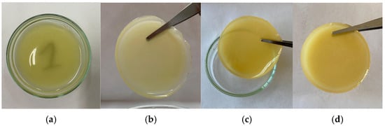
Transdermal biopolymer systems must create homogenous and mechanically stable polymer films because their physicochemical characteristics, drug-loading capacity, and biological performance are substantially determined by their gelation behavior and crosslinking efficiency [19,20]. Two polysaccharide-based composite systems, gellan gum–chitosan (G1) and agar–chitosan (A1), including formulations containing the bioactive substance naringin, were created in this investigation (Table 1, Figure 1). The chitosan component was chemically crosslinked using glutaraldehyde, and gellan gum and agar helped create networks by intermolecular interactions; in the case of gellan, this process was further aided by ion-mediated gelation.
Table 1.
Composition of formulations with different feed ratios.
Figure 1.
Photographs of the prepared films: (a) gellan–chitosan film (simple G1); (b) gellan–chitosan film containing 0.185% (w/v) naringin (simple G1-N); (c) agar–chitosan film (simple A1); (d) agar–chitosan film containing 0.185% (w/v) naringin (simple A1-N).
Effective gelation and the creation of a stable three-dimensional network were demonstrated by the G1 system’s generation of homogenous, continuous sheets with smooth surfaces and uniform thickness. This behavior is in line with other studies showing that, when combined with cationic polysaccharides like chitosan, gellan gum creates mechanically strong hydrogel matrices, mainly as a result of hydrogen bonds and electrostatic interactions between carboxyl and amino groups [21,22,23,24].
In contrast, the A1 system initially showed limited film-forming ability, remaining largely liquid and structurally unstable. This behavior may be associated with the thermo-reversible nature of agar gelation, which requires careful control of temperature and polymer concentration for effective network formation. After adjustment of the synthesis conditions, including increased temperature and continuous agitation, the mixture became more homogeneous and produced a uniform, mechanically stable film without visible phase separation or residual fluidity. These findings indicate that controlled thermal processing can improve gelation and promote intermolecular interactions in agar-based composite systems [25,26,27].
Overall, the observed differences in film formation suggest that the structural organization and crosslinking density of the polymer network strongly depend on the nature of the polysaccharide matrix and processing conditions. To further evaluate these structural features, swelling analysis was performed as an indirect method for assessing network density and integrity.
3.2. Infrared Spectroscopic Evaluation of Polysaccharide-Based Composite Films
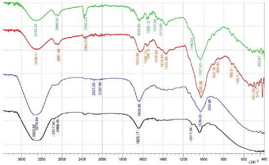
In all samples, a broad absorption band is observed in the region of 3300–3350 cm−1, attributed to overlapping stretching vibrations of hydroxyl (O–H) and amino (N–H) groups (Figure 2). For chitosan, this band corresponds to intra- and intermolecular hydrogen bonds involving –OH and –NH2 groups, whereas for gellan and agar, it reflects the presence of numerous hydroxyl groups of polysaccharide nature. In composite samples containing naringin, a slight broadening of this band and a weak shift toward lower wavenumbers are observed, indicating enhanced hydrogen bonding interactions between the components of the system. This behavior is consistent with the involvement of naringin hydroxyl groups in the formation of an additional hydrogen-bonding network with the polymer matrix.
Figure 2.
FTIR spectra of the prepared biopolymer films: A1-N (agar–chitosan film containing naringin), G1-N (gellan–chitosan film containing naringin), G1 (gellan–chitosan film), and A1 (agar–chitosan film), arranged from top to bottom.
In the region of 2920–2850 cm−1, all spectra exhibit bands corresponding to the stretching vibrations of aliphatic C–H bonds characteristic of the polysaccharide backbone. No significant shifts in the positions of these bands are observed in the composites; however, slight variations in their intensity may be associated with changes in the local environment of macromolecules and the packing density of the chains during the formation of the composite structure.
Characteristic bands of chitosan corresponding to amide I and amide II are observed at ~1650–1655 cm−1 and ~1540–1555 cm−1, respectively. The amide I band is mainly attributed to the stretching vibrations of C=O of residual acetyl groups, whereas the amide II band is associated with N–H bending vibrations and C–N stretching vibrations. In the composite samples, slight changes in the intensity and shape of the band in the ~1650 cm−1 region are observed, which are related to the overlap of signals from chitosan amide groups and carboxylate groups of gellan gum, as well as to changes in their chemical environment. At the same time, no pronounced appearance of a new band characteristic of covalent bonds (e.g., C=N) is observed, indicating the predominantly non-covalent nature of interactions in the system [28,29].
In the spectra of composites containing naringin, additional weak bands are observed in the regions of ~1510–1520 cm−1 and 1450–1470 cm−1, which can be attributed to vibrations of the aromatic backbone of the flavonoid. The presence of these bands confirms the successful incorporation of naringin into the polymer matrix without structural degradation.
In the region of 1400–1450 cm−1, bands corresponding to the bending vibrations of CH2 groups, as well as symmetric vibrations of carboxylate groups of gellan, are observed. In the composites, the shape and intensity of these bands change slightly, which may be associated with electrostatic interactions between the protonated amino groups of chitosan and the carboxylate groups of gellan gum.
The most intense bands in all spectra are recorded in the region of 1150–1000 cm−1 and correspond to the stretching vibrations of C–O–C and C–O glycosidic bonds. In the composite samples, some broadening and redistribution of intensity are observed in this region (in particular, near ~1080 and ~1020 cm−1), indicating changes in the conformation of the polysaccharide network and redistribution of hydrogen bonds between the components. The absence of new distinct bands, along with the preservation of characteristic polysaccharide signals, suggests that the primary structure of the matrix remains intact.
In the region below 1000 cm−1 (the “fingerprint region”), bands corresponding to vibrations of saccharide rings and bending vibrations of C–H bonds are observed. Peaks near ~930–950 cm−1 and ~720–770 cm−1 are associated with vibrations of polysaccharide structures. In the composites, slight changes in their intensity are noted, indicating the involvement of these fragments in the formation of intermolecular interactions.
Thus, the analysis of FTIR spectra shows that during the formation of composite systems based on chitosan, gellan, and agar with the addition of naringin, the main structural elements of the initial components are preserved. At the same time, the observed broadening of the O–H/N–H band, changes in the amide vibration region, and redistribution of the intensity of glycosidic bands indicate the formation of a developed network of intermolecular interactions, predominantly of a hydrogen-bonding and electrostatic nature. The absence of new characteristic bands of covalent bonds suggests that the incorporation of naringin occurs without chemical modification of the polymer matrix, through physical incorporation and non-covalent interactions. These features may contribute to enhanced structural integrity, stability, and functional properties of the resulting composite materials [30,31].
3.3. Thermogravimetric (TG) and Differential Scanning Calorimetry (DSC) Analysis
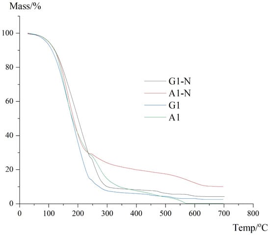
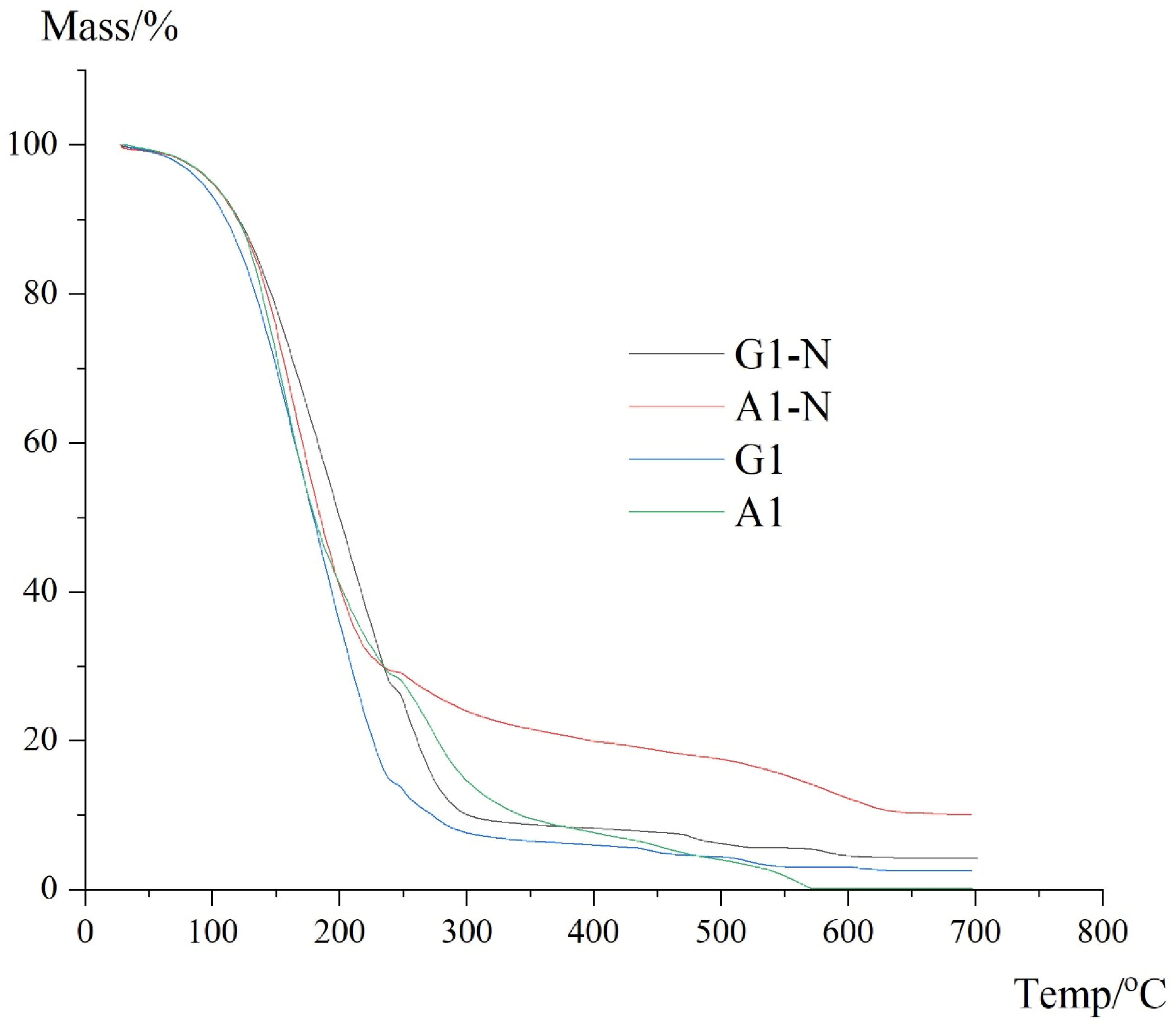
Thermal stability of the prepared biopolymer films was evaluated using thermogravimetric analysis (TGA), and the results are presented in Figure 3. The analysis was performed for both gellan- and agar-based systems, with and without naringin incorporation (G1, G1-N, A1, and A1-N), allowing for a direct comparison of their thermal behavior.
Figure 3.
TGA curves of gellan- and agar-based biopolymer films with and without naringin.
All samples exhibited a similar multi-stage degradation profile. The initial mass loss observed below approximately 120 °C is attributed to the removal of physically adsorbed and bound water. This stage is characteristic of hydrophilic polysaccharide-based materials and reflects differences in water retention associated with network structure and composition.
The main degradation stage occurred in the temperature range of approximately 200–350 °C, corresponding to the thermal decomposition of the polysaccharide backbone. In this region, cleavage of glycosidic linkages and decomposition of functional groups within chitosan, gellan gum, and agar take place. The position and slope of the degradation curves indicate variations in thermal stability among the samples.
The G1 sample demonstrated a relatively rapid mass loss in this region, suggesting a more compact but less thermally resistant structure compared to the modified systems. In contrast, the G1-N sample showed a slightly shifted degradation profile with a more gradual mass loss, which may be associated with the involvement of naringin in intermolecular interactions within the polymer matrix.
A similar trend was observed for agar-based systems. The A1 sample exhibited typical polysaccharide degradation behavior, while the A1-N sample retained a higher residual mass at elevated temperatures. This behavior may be attributed to the presence of naringin, which undergoes multi-stage thermal decomposition and contributes to the formation of thermally stable residues.
At temperatures above 400 °C, all samples showed gradual mass loss associated with further decomposition of carbonaceous structures. Notably, the A1-N sample demonstrated the highest residual mass at 700 °C, indicating comparatively enhanced thermal stability among the studied systems.
Overall, the TGA results indicate that the incorporation of naringin does not lead to a significant decrease in thermal stability and, in some cases, contributes to a more gradual degradation process. These findings are consistent with FTIR analysis, which suggests that naringin is incorporated into the polymer matrix through non-covalent interactions, leading to subtle modifications in the structural organization without disrupting the integrity of the polymer backbone.
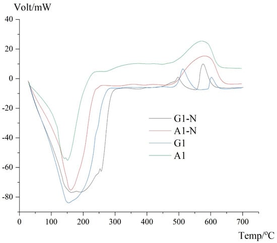
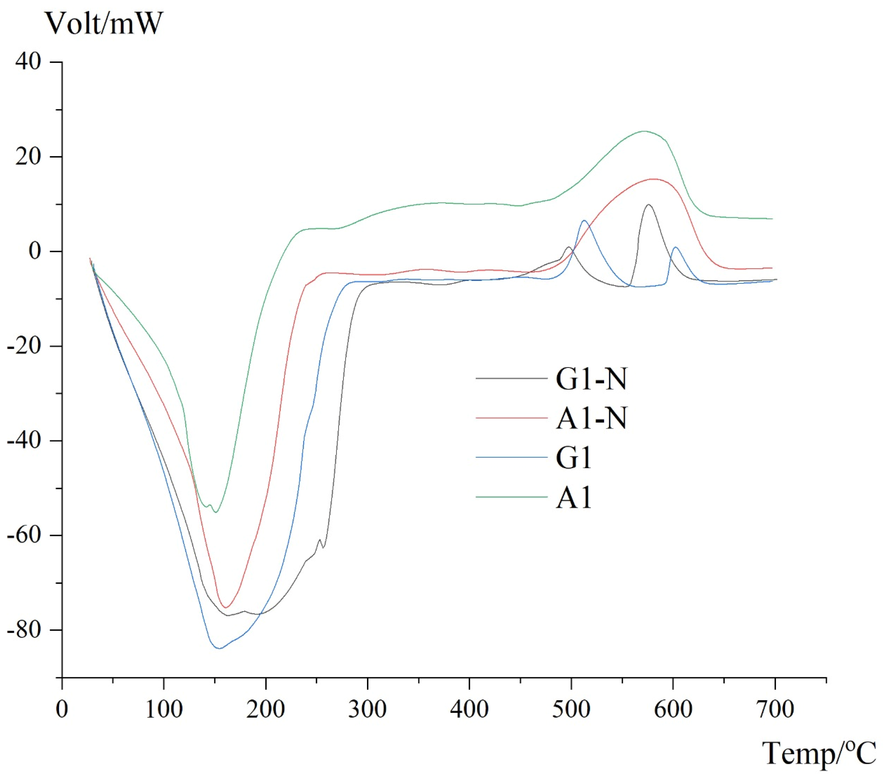
The thermal transitions of the prepared biopolymer films were further analyzed using differential scanning calorimetry (DSC), and the results are presented in Figure 4. The analysis was carried out for both gellan- and agar-based systems, with and without naringin incorporation, allowing a comparative evaluation of their thermal behavior.
Figure 4.
DSC curves of gellan- and agar-based biopolymer films with and without naringin.
All samples exhibit a pronounced endothermic peak in the temperature range of approximately 120–200 °C, which is associated with the removal of bound water and relaxation of the polymer network. The position and intensity of this peak vary among the samples, reflecting differences in hydration level and intermolecular interactions within the polymer matrices.
The G1 sample shows a well-defined endothermic minimum, indicating a relatively compact structure with limited mobility of polymer chains. Upon incorporation of naringin (G1-N), the peak becomes broader and slightly shifted, suggesting changes in the local environment and a redistribution of intermolecular interactions. This behavior is consistent with the formation of additional hydrogen bonding interactions involving naringin, leading to a more heterogeneous thermal response.
A similar trend is observed for agar-based systems. The A1 sample exhibits a broader and less intense endothermic transition compared to G1, which is indicative of a more loosely organized structure with higher water retention. In the A1-N sample, the transition becomes slightly more pronounced, suggesting a moderate influence of naringin on the thermal behavior of the agar matrix.
At higher temperatures (above ~450 °C), weak thermal effects are observed for all samples, corresponding to the decomposition of residual polymer fragments and carbonaceous structures. Notably, the A1 sample shows the most intense thermal response in this region, while the naringin-containing samples exhibit smoother transitions, indicating a more gradual degradation process.
Overall, the DSC results suggest that the incorporation of naringin leads to subtle changes in the thermal behavior of the composite films, primarily associated with modifications in intermolecular interactions and network organization. These observations are in agreement with FTIR and TGA analyses, which indicate that naringin is incorporated into the polymer matrix without altering the fundamental structure of the polymers, while influencing their physicochemical properties.
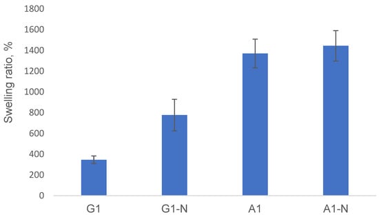
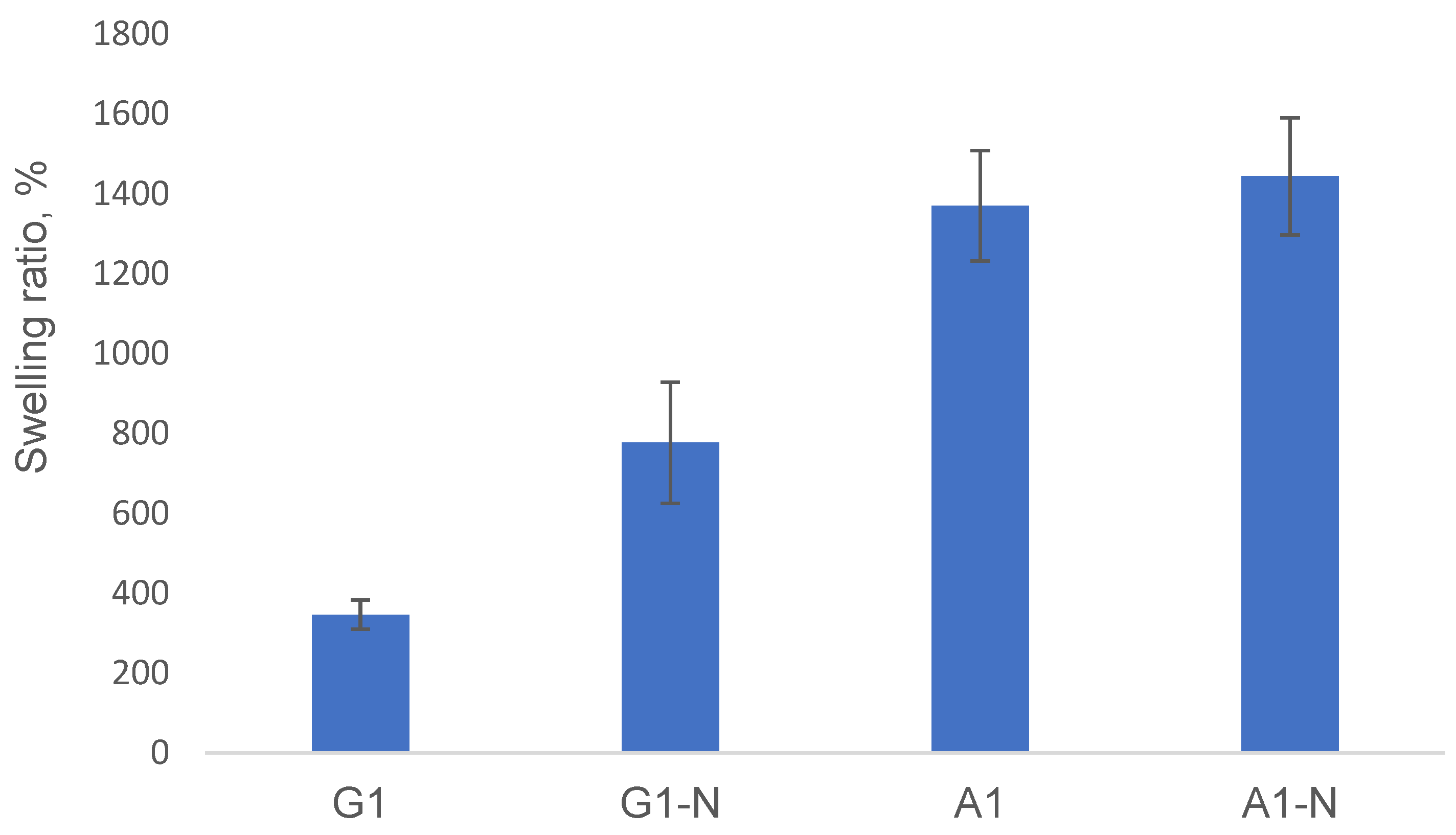
In order to assess the impact of polymer composition, naringin incorporation, and ionic treatment on the structural organization of the polymer networks, the swelling behavior of the developed films was further investigated. The results of the swelling study are presented in Figure 5.
Figure 5.
Effect of naringin on the swelling behavior of gellan- and agar-based biopolymer films (mean ± SD, n = 3).
As shown in Figure 5, the gellan-based system (G1) exhibited a moderate swelling ratio (~347%), indicating the formation of a relatively dense polymer network. Upon incorporation of naringin (G1-N), the swelling ratio increased significantly (~777%), suggesting a decrease in network density and enhanced water uptake. This behavior may be attributed to the disruption of intermolecular interactions within the polymer matrix caused by the presence of the bioactive compound.
In contrast, the agar-based system (A1) demonstrated a substantially higher swelling ratio (~1370%), reflecting a more open and highly hydrated network structure. The addition of naringin (A1-N) resulted in a slight further increase in swelling (~1444%), indicating that the agar-based matrix is less sensitive to structural changes induced by the additive compared to the gellan system.
The observed differences in swelling behavior can be explained by variations in network density and ionic crosslinking efficiency. In CaCl2-treated systems, divalent Ca2+ ions promote the formation of ionic bridges between polymer chains, leading to a more compact and ordered network with reduced free volume and limited water uptake [32]. In contrast, NaCl-treated systems retain a comparatively looser structure, as monovalent Na+ ions mainly provide electrostatic screening without forming stable crosslinking junctions.
The effect of ionic treatment was particularly pronounced in the gellan-based films, where ion-mediated gelation plays a key role in network formation [21,22,23,24,32]. In agar-based systems, the effect was less significant due to the thermally driven gelation mechanism [25,26,27].
Overall, these findings demonstrate that swelling behavior is strongly influenced by polymer composition and the structural organization of the network. Changes in swelling reflect variations in crosslinking density and chain mobility, highlighting the importance of optimizing formulation parameters to achieve the desired balance between hydration, mechanical stability, and structural integrity in biomedical applications [32].
3.4. Antimicrobial Activity (In Vitro)
The antimicrobial activity of the developed hydrogel systems was evaluated against Staphylococcus aureus using the agar diffusion method. The results are presented in Table 2.
Table 2.
Antimicrobial activity of the samples.
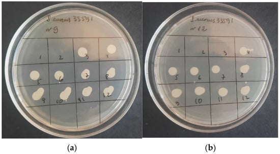
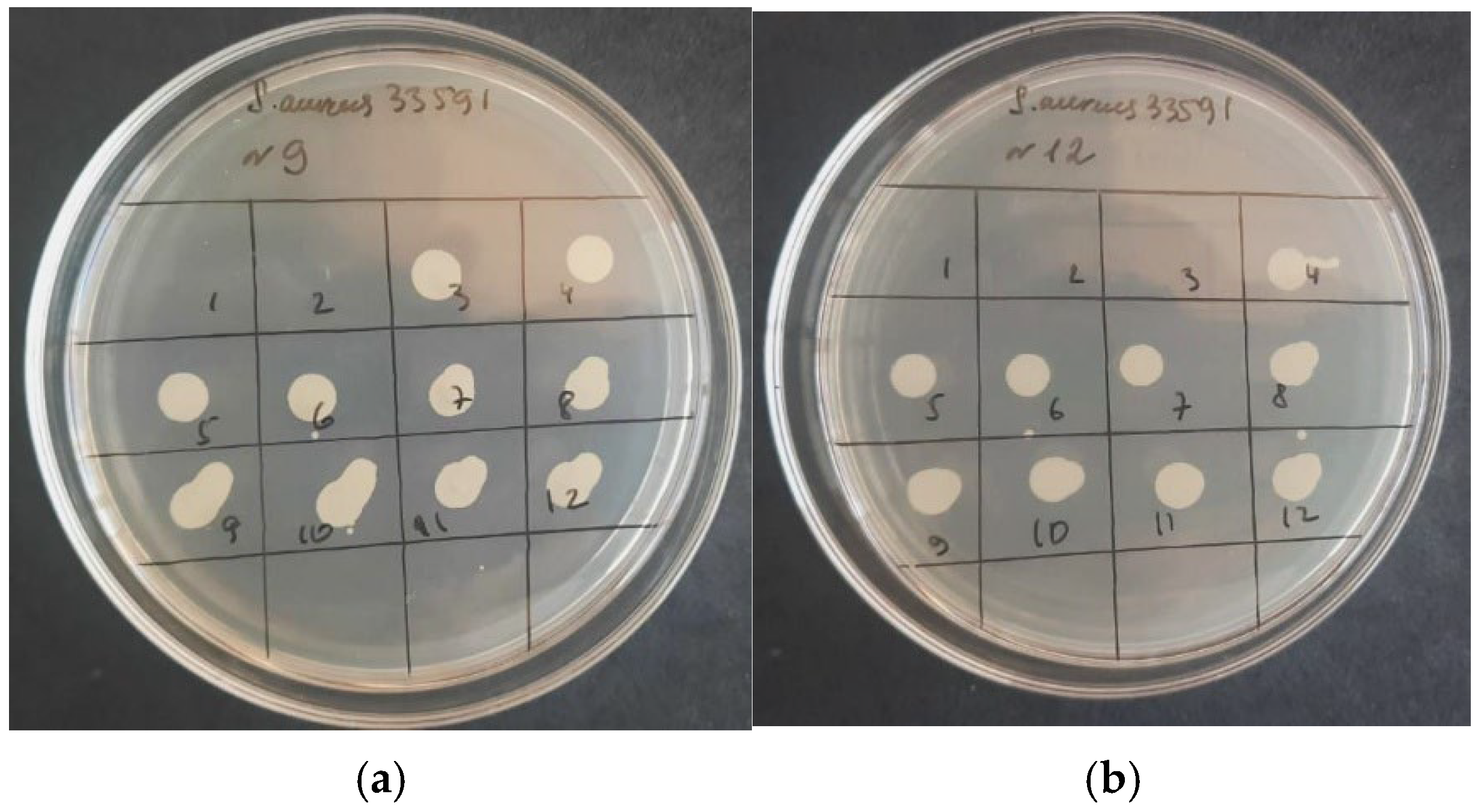
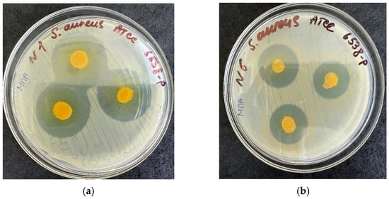
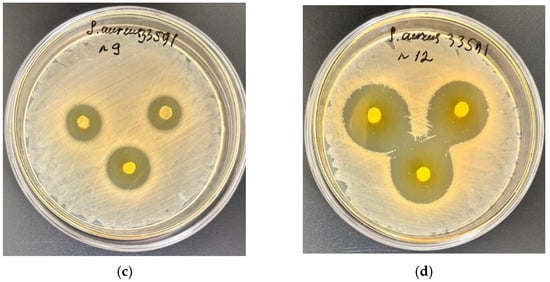
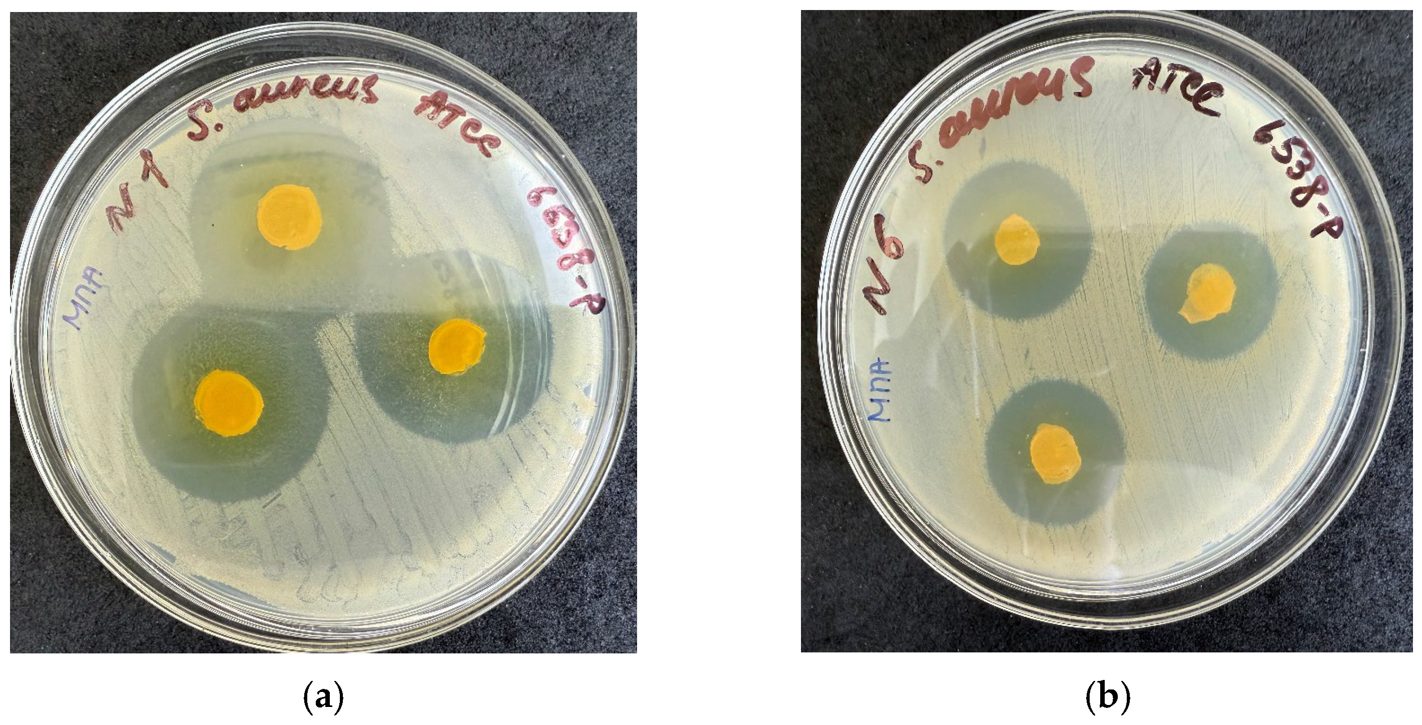
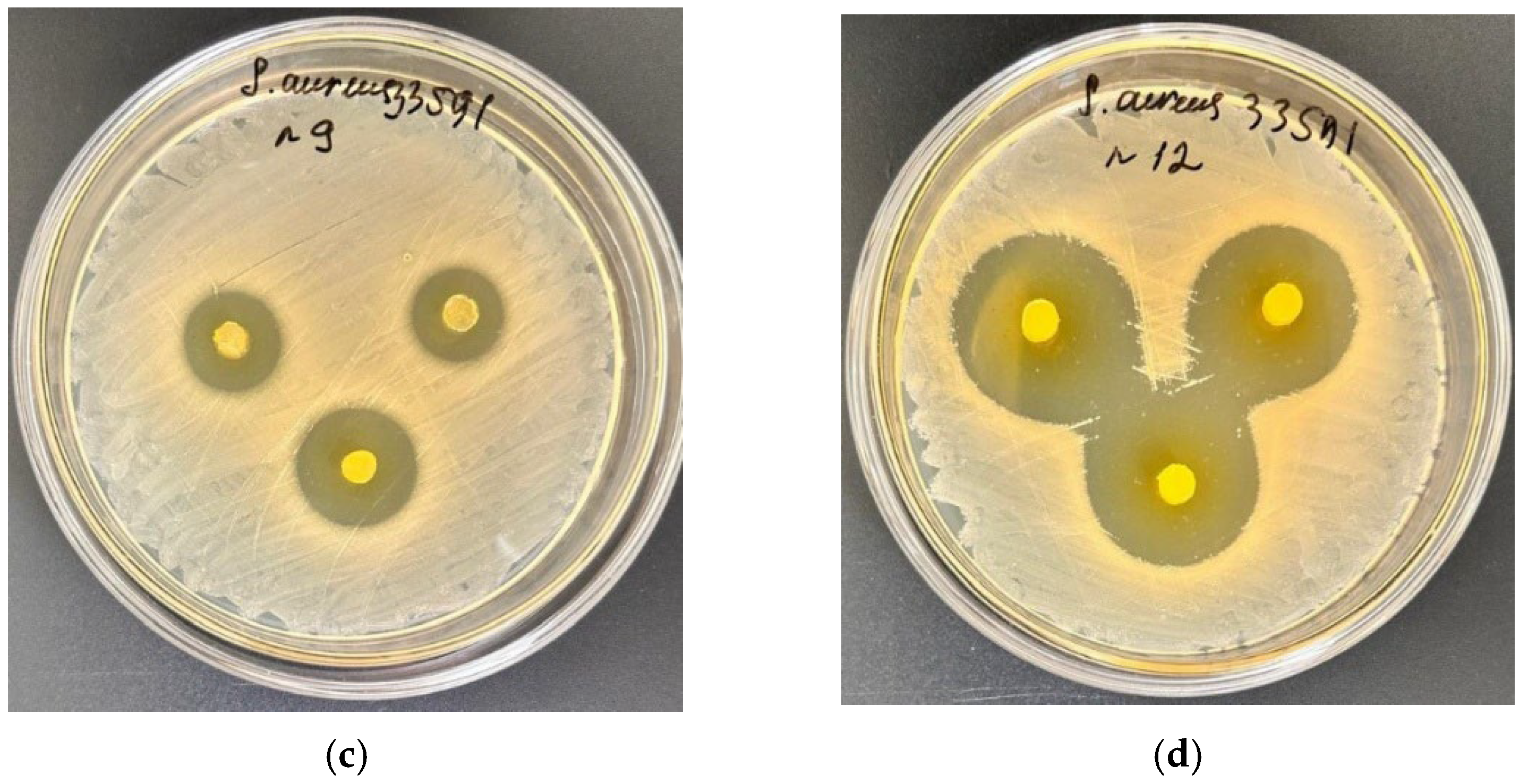
The antibacterial activity of the agar- and gellan-based formulations is presented in Figure 6 and Figure 7, respectively.
Figure 6.
Antimicrobial activity of A1-based hydrogel samples containing naringin against Staphylococcus aureus ATCC 33591 determined by the agar diffusion method: (a) A1-N1 (0.093% w/v naringin), (b) A1-N2 (0.185% w/v naringin).
Figure 7.
Representative inhibition zones of hydrogel samples containing naringin against Staphylococcus aureus ATCC 33591 determined by the agar diffusion method: (a,b) G1-N1 (0.185% w/v naringin) and G1-N2 (0.093% w/v naringin); (c,d) A1-N1 (0.185% w/v naringin) and A1-N2 (0.093% w/v naringin).
The gellan-based formulation demonstrated a dependence on naringin loading (Table 3). The sample with higher naringin concentration (G1-N1, 0.185% w/v naringin) exhibited a pronounced antibacterial effect, with a growth inhibition zone of 30.0 ± 0.0 mm. In contrast, the sample with lower naringin concentration (G1-N2, 0.093% w/v naringin) showed a reduced inhibition zone of 22.7 ± 1.2 mm (Figure 7a,b).
Table 3.
Growth inhibition zones of S. aureus ATCC 33591 by the samples (mm).
A different trend was observed for the agar-based system (Figure 6). The sample with lower naringin concentration (A1-N2, 0.093% w/v naringin) demonstrated a larger inhibition zone (27.67 ± 0.58 mm), whereas the sample with higher loading (A1-N1, 0.185% w/v naringin) exhibited a smaller inhibition zone of 16.0 ± 1.73 mm (Figure 7c,d).
These results suggest that antibacterial activity is influenced not only by the amount of incorporated bioactive compound but also by the composition and structural characteristics of the hydrogel systems, which may affect the release and availability of active components. Similar observations have been reported for antimicrobial hydrogel systems, where matrix composition plays a key role in regulating the diffusion and activity of incorporated agents [33]. It should be noted that sodium benzoate was incorporated into all formulations primarily as a preservative to ensure microbiological stability during storage. Therefore, although sodium benzoate is known to possess antimicrobial properties, its contribution to the observed antibacterial effects is considered secondary. Since sodium benzoate was present in all formulations, the differences in antibacterial activity are more likely associated with the presence and release behavior of naringin within the polymer matrix. However, further studies are required to elucidate the mechanisms underlying these observations.
3.5. Safety and Biocompatibility
The safety and biocompatibility of the developed films were evaluated through subchronic toxicity and dermal irritation studies using A-series samples. No signs of systemic toxicity or local irritation were observed under the experimental conditions, indicating good overall tolerance of the tested formulations.
3.5.1. Subchronic (Sub-Acute) Toxicity of the Naringin Transdermal Delivery System
The subchronic toxicity of the naringin-loaded film (A1-N1, 0.185% w/v naringin) was evaluated in outbred male mice over a 28-day period under repeated topical application conditions.
No significant changes in body weight were observed in the treated group (27.93 ± 4.76 g) compared to the control group (p > 0.05). Similarly, the masses of internal organs, including the liver (2.03 ± 0.49 g) and spleen (0.35 ± 0.27 g), remained within normal physiological ranges.
No signs of systemic toxicity or abnormal clinical behavior were detected throughout the study. These results suggest that repeated topical application of the tested formulation does not induce systemic toxicity under the studied conditions. These findings are consistent with previous reports demonstrating the favorable biocompatibility and safety profile of chitosan-based materials intended for wound-healing applications [34,35].
3.5.2. Localized Irritation of the Naringin Film Sample
The dermal irritation potential of the naringin-loaded film (A1-N1, 0.185% w/v naringin) was evaluated in Chinchilla rabbits (Oryctolagus cuniculus; body weight 2.5–3.0 kg) in accordance with OECD Test Guideline 404.
Prior to application, the dorsal skin was carefully shaved without causing damage. Film specimens (1 cm × 1 cm) were applied to intact skin and secured with occlusive dressing for up to 72 h. Skin reactions were evaluated at 2, 4, 6, 24, 48, and 72 h following application.
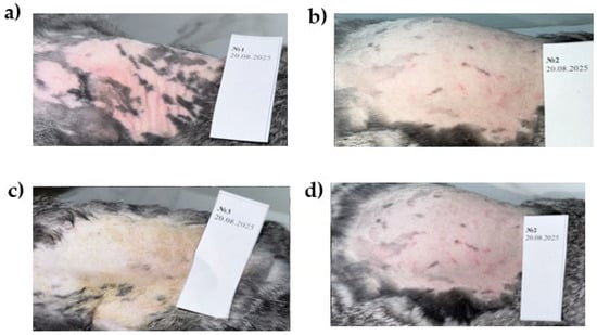
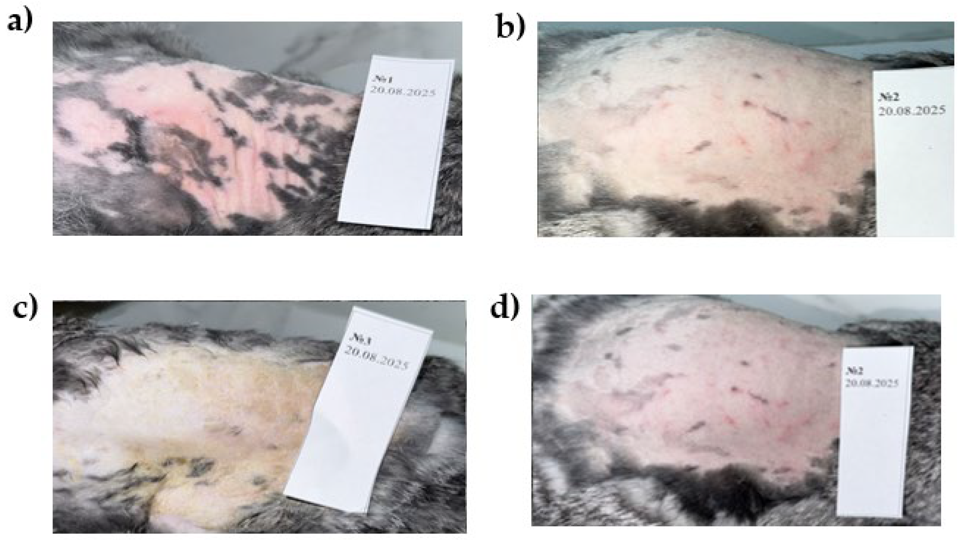
Visual inspection of the application sites (Figure 8) showed no visible signs of irritation compared to baseline observations. Throughout the observation period, no changes in general condition or body weight were recorded.
Figure 8.
Representative images of rabbit dorsal skin 72 h after a single topical application of the naringin-loaded film (0.185% w/v) under occlusive conditions. The dorsal skin was shaved prior to treatment. Images represent (a) Animal 1, (b) Animal 2, (c) Animal 3, and (d) an additional application site from Animal 2. No visible signs of erythema, edema, ulceration, necrosis, or other dermal irritation were observed at any application site.
Representative images of rabbit dorsal skin are presented in Figure 8. No visible signs of erythema, edema, ulceration, necrosis, or other dermal irritation were observed at any application site 72 h after a single topical application of the naringin-loaded film. These findings are consistent with a primary irritation index of 0, indicating that the formulation did not induce skin irritation.
No local dermal reactions, including erythema, edema, ulceration, or necrosis, were observed. The primary irritation index was calculated as 0, indicating the absence of irritation after a single topical application.
3.6. Anti-Inflammatory Activity
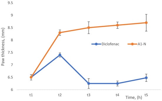
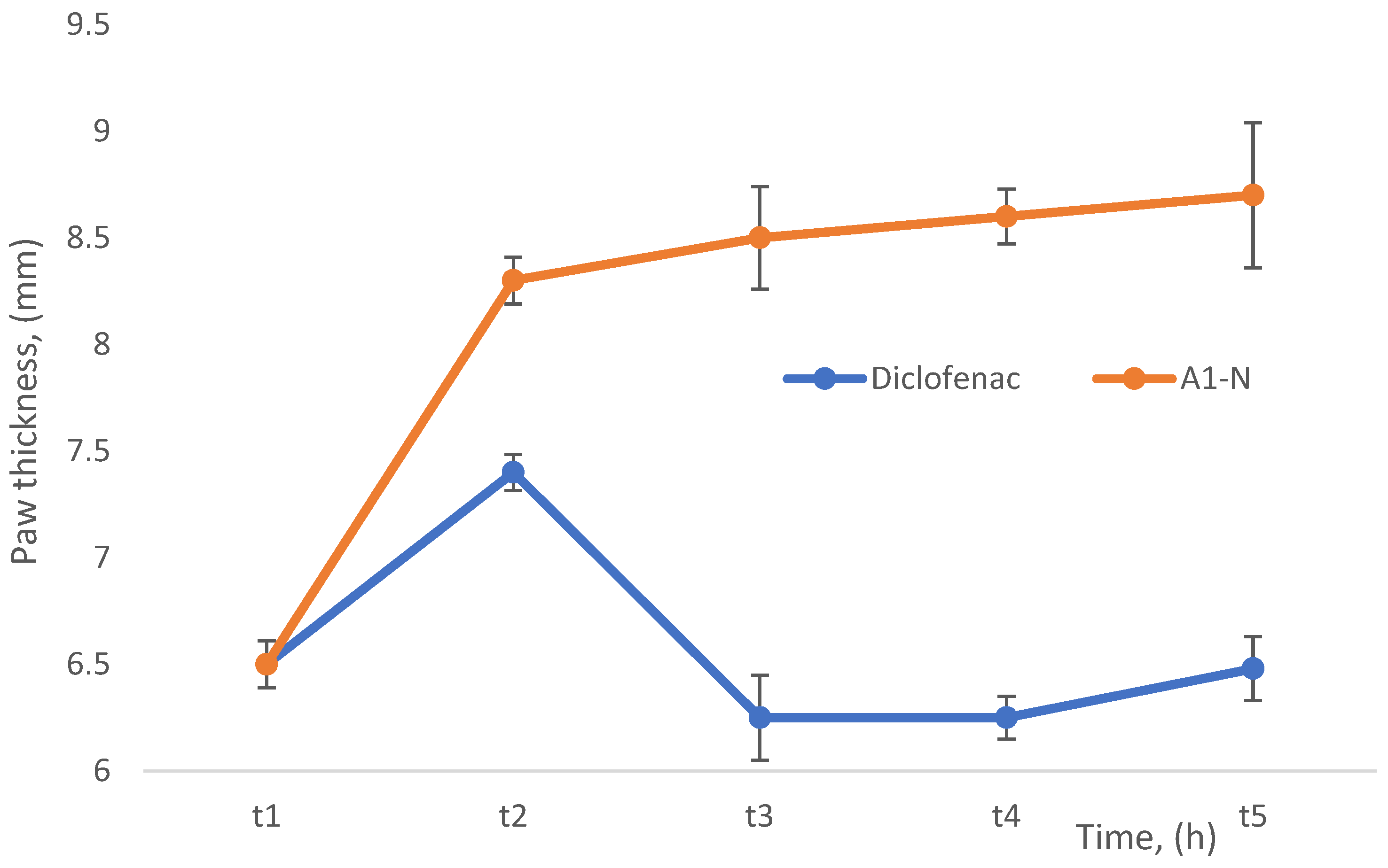
At 1 h (t1), paw thickness was comparable across all groups (6–7 mm), confirming consistent induction of inflammation. At 2 h (t2), the diclofenac-treated group showed a reduction in paw edema (6–7 mm), whereas the naringin-treated group exhibited higher values (7–9 mm), indicating a limited anti-inflammatory response. This trend persisted at later time points (t3–t5), with diclofenac maintaining lower and more stable edema levels, while the naringin-treated group showed consistently elevated paw thickness (8–9 mm).
Statistical analysis (one-way ANOVA followed by Tukey’s post hoc test) revealed significant differences between groups from t2 onward (p < 0.000001), with diclofenac demonstrating significantly higher efficacy compared to the naringin-loaded film (A1-N) (Figure 9).
Figure 9.
Effect of diclofenac and naringin-loaded film (A1-N) on paw thickness in the λ-carrageenan-induced edema model. Data are presented as mean ± SD (n = 3).
The anti-inflammatory effect was evaluated using the λ-carrageenan-induced paw edema model, with paw thickness measured at predefined time intervals following injection.
Overall, the results indicate that the tested formulation exhibited a limited anti-inflammatory effect under the experimental conditions. This may be associated with restricted bioavailability of naringin from the film matrix or an insufficient dose for acute inflammation suppression. Similar limitations related to drug release behavior have been reported for hydrogel-based delivery systems, where matrix properties can significantly influence therapeutic efficacy [36]. Further studies are required to evaluate dose-dependent effects and to optimize the formulation.
4. Conclusions
In this work, polysaccharide-based film systems were developed using agar–chitosan (A-series) and gellan–chitosan (G-series) compositions with naringin incorporated as a bioactive component.
The results demonstrate that the physicochemical properties, structural organization, and swelling behavior of the developed systems are strongly influenced by the type of polysaccharide matrix and preparation conditions. Gellan-based films formed more compact networks with moderate swelling, whereas agar-based systems exhibited higher swelling capacity, indicating a more open and highly hydrated structure.
The incorporation of naringin affected the structural properties of the polymer networks, particularly in gellan-based systems, where a significant increase in swelling suggested a decrease in network density. In contrast, agar-based systems showed a less pronounced response to naringin incorporation.
Antimicrobial evaluation confirmed that both A- and G-series samples exhibited activity against Staphylococcus aureus. The gellan-based formulations demonstrated increased antibacterial activity with higher naringin loading, whereas the agar-based systems showed an opposite trend. These findings suggest that antimicrobial performance is governed not only by the amount of the bioactive compound but also by the structure of the polymer matrix and the release behavior.
In vivo studies performed on selected A-series samples indicated good tolerability of the naringin-loaded film (A1-N1), with no signs of systemic toxicity or skin irritation under the tested conditions.
At the same time, the anti-inflammatory assessment demonstrated limited activity under acute conditions, indicating the need for further optimization of formulation parameters and dosing strategies.
Overall, the obtained results highlight the potential of polysaccharide-based composite films as topical systems. At the same time, they emphasize the importance of optimizing polymer composition and structure to achieve a balanced combination of mechanical stability, controlled swelling, and biological activity.
Author Contributions
Conceptualization, G.B.; methodology, G.B. and A.K.; investigation, validation and data curation, I.K. and N.C.; formal analysis, N.C.; resources and funding acquisition, G.B. and N.C.; writing—original draft, G.B. and A.K.; writing—review and editing, G.B.; supervision and project administration, G.B. All authors have read and agreed to the published version of the manuscript.
Funding
This research is supported by the Ministry of Science and Higher Education of the Republic of Kazakhstan (Grant No. AP19679386 “Development of a thermoregulated film sample with antibacterial and anti-inflammatory action”).
Institutional Review Board Statement
The animal experimental protocol was approved by the Institutional Review Board of Kazakh National Medical University (approval code No. 6; approval date: 16 June 2025). All procedures were conducted in accordance with internationally accepted guidelines for the care and use of laboratory animals.
Data Availability Statement
The datasets generated and/or analyzed during the current study are not publicly available due to ethical restrictions related to the use of animal models but can be obtained from the corresponding author upon reasonable request.
Acknowledgments
During manuscript preparation, AI-based tools were used solely for language editing (translation and phrasing) and for preparing a schematic illustration of the polymer synthesis (graphic drawing). All scientific concepts, experimental design, data interpretation, and conclusions were developed by the authors.
Conflicts of Interest
The authors declare no conflicts of interest. The funders had no role in the study design, data collection, analysis, interpretation, manuscript writing, or decision to publish the results.
References
- Boateng, J.S.; Matthews, K.H.; Stevens, H.N.E.; Eccleston, G.M. Wound Healing Dressings and Drug Delivery Systems: A Review. J. Pharm. Sci. 2008, 97, 2892–2923. [Google Scholar] [CrossRef] [Scilit] [PubMed]
- Pereira, R.F.; Bártolo, P.J. Traditional Therapies for Skin Wound Healing. Adv. Wound Care 2016, 5, 208–229. [Google Scholar] [CrossRef] [Scilit] [PubMed]
- Khattak, S.; Ullah, I.; Yousaf, M.T.; Ullah, S.; Yousaf, H.; Li, Y.; Jin, H.; Shen, J.; Xu, H.-T. Advancements in Hydrogels: A Comprehensive Review of Natural, Synthetic, and Hybrid Innovations for Wound Healing. Int. J. Biol. Macromol. 2025, 327, 147270. [Google Scholar] [CrossRef] [Scilit] [PubMed]
- Rinaudo, M. Chitin and Chitosan: Properties and Applications. Prog. Polym. Sci. 2006, 31, 603–632. [Google Scholar] [CrossRef] [Scilit]
- Wang, J.; Zhuang, S. Chitosan-Based Materials: Preparation, Modification and Application. J. Clean. Prod. 2022, 355, 131825. [Google Scholar] [CrossRef] [Scilit]
- Jayakumar, R.; Prabaharan, M.; Sudheesh Kumar, P.T.; Nair, S.V.; Furuike, T.; Tamur, H. Novel Chitin and Chitosan Materials in Wound Dressing. In Biomedical Engineering, Trends in Materials Science; Laskovski, A., Ed.; InTech: Rijeka, Croatia, 2011. [Google Scholar] [CrossRef] [Scilit] [PubMed]
- Moeini, A.; Pedram, P.; Makvandi, P.; Malinconico, M.; Gomez d’Ayala, G. Wound Healing and Antimicrobial Effect of Active Secondary Metabolites in Chitosan-Based Wound Dressings: A Review. Carbohydr. Polym. 2020, 233, 115839. [Google Scholar] [CrossRef] [Scilit] [PubMed]
- Deng, C.; Li, F.; Griffith, M.; Ruel, M.; Suuronen, E.J. Application of Chitosan-Based Biomaterials for Blood Vessel Regeneration. Macromol. Symp. 2010, 297, 138–146. [Google Scholar] [CrossRef] [Scilit]
- Ul-Islam, M.; Alabbosh, K.F.; Manan, S.; Khan, S.; Ahmad, F.; Ullah, M.W. Chitosan-Based Nanostructured Biomaterials: Synthesis, Properties, and Biomedical Applications. Adv. Ind. Eng. Polym. Res. 2024, 7, 79–99. [Google Scholar] [CrossRef] [Scilit]
- Bayrami, M.; Fathi, M.; Dalir Abdolahinia, E.; Adibkia, K. Enhanced Ocular Delivery of Timolol Maleate through Thiolated Chitosan Nanoparticles/Gellan Gum-Based Hydrogels. Carbohydr. Res. 2026, 563, 109865. [Google Scholar] [CrossRef] [Scilit] [PubMed]
- Alsakhawy, M.A.; Abdelmonsif, D.A.; Haroun, M.; Sabra, S.A. Naringin-Loaded Arabic Gum/Pectin Hydrogel as a Potential Wound Healing Material. Int. J. Biol. Macromol. 2022, 222, 701–714. [Google Scholar] [CrossRef] [Scilit] [PubMed]
- Shilpa, V.; Shams, R.; Dash, K.K.; Pandey, V.K.; Dar, A.H.; Ayaz Mukarram, S.; Harsányi, E.; Kovács, B. Phytochemical Properties, Extraction, and Pharmacological Benefits of Naringin: A Review. Molecules 2023, 28, 5623. [Google Scholar] [CrossRef] [Scilit] [PubMed]
- Baishal, S.; Prakash, J.; Marvaan, M.S.; Sundar, M.; Pannerselvam, B.; Venkatasubbu, G.D. Naringin and Graphene Oxide Incorporated Moringa Oleifera Gum/Poly(Vinyl) Alcohol Patch for Enhanced Wound Healing. Int. J. Biol. Macromol. 2024, 259, 129198. [Google Scholar] [CrossRef] [Scilit] [PubMed]
- Bian, D.; Pilehvar, Y.; Kousha, S.; Bi, J. Bioactive Wound Healing 3D Structure Based on Chitosan Hydrogel Loaded with Naringin/Cyclodextrin Inclusion Nanocomplex. ACS Omega 2024, 9, 10566–10576. [Google Scholar] [CrossRef] [Scilit] [PubMed]
- Begimova, G.; Kuldanova, A.; Smailova, K.; Kurmanbayeva, I. Development of a Biodegradable Patch Based on Polysaccharides. Polymers 2025, 17, 2908. [Google Scholar] [CrossRef] [Scilit] [PubMed]
- Gadziński, P.; Froelich, A.; Jadach, B.; Wojtyłko, M.; Tatarek, A.; Białek, A.; Krysztofiak, J.; Gackowski, M.; Otto, F.; Osmałek, T. Ionotropic Gelation and Chemical Crosslinking as Methods for Fabrication of Modified-Release Gellan Gum-Based Drug Delivery Systems. Pharmaceutics 2023, 15, 108. [Google Scholar] [CrossRef] [Scilit] [PubMed]
- De Oliveira, A.C.; Sabino, R.M.; Souza, P.R.; Muniz, E.C.; Popat, K.C.; Kipper, M.J.; Zola, R.S.; Martins, A.F. Chitosan/Gellan Gum Ratio Content into Blends Modulates the Scaffolding Capacity of Hydrogels on Bone Mesenchymal Stem Cells. Mater. Sci. Eng. C 2020, 106, 110258. [Google Scholar] [CrossRef] [Scilit] [PubMed]
- Hu, Z.; Hong, P.; Liao, M.; Kong, S.; Huang, N.; Ou, C.; Li, S. Preparation and Characterization of Chitosan—Agarose Composite Films. Materials 2016, 9, 816. [Google Scholar] [CrossRef] [Scilit] [PubMed]
- Elhefian, E.A.; Nasef, M.M.; Yahaya, A.H. Preparation and Characterization of Chitosan/Agar Blended Films: Part 2. Thermal, Mechanical, and Surface Properties. J. Chem. 2012, 9, 510–516. [Google Scholar] [CrossRef] [Scilit]
- Kazeminava, F.; Javanbakht, S.; Nouri, M.; Gholizadeh, P.; Nezhad-Mokhtari, P.; Ganbarov, K.; Tanomand, A.; Kafil, H.S. Gentamicin-Loaded Chitosan/Folic Acid-Based Carbon Quantum Dots Nanocomposite Hydrogel Films as Potential Antimicrobial Wound Dressing. J. Biol. Eng. 2022, 16, 36. [Google Scholar] [CrossRef] [Scilit] [PubMed]
- Dai, H.; Cheng, X.; Guo, N.; Zhang, F.; Xu, Z.; Wang, S.; Zhu, G. A Novel Gellan Gum-Chitosan Composite Film Functionalized with Melatonin for Enhanced Preservation of Fresh Blueberries. Foods 2026, 15, 745. [Google Scholar] [CrossRef] [Scilit] [PubMed]
- Liu, D.; Zhang, L.; Shao, Z.; Liu, C.; Gao, T.; Liu, J.; Zhu, Y.; Miyazaki, T. Fabrication of a Schiff-Base Crosslinked Hydrogel with Tunable Dynamic Properties and Cytocompatibility Based on Oxidized Gellan Gum and Aminated Carboxymethyl Chitosan for Wound Dressing. J. Polym. Res. 2026, 33, 79. [Google Scholar] [CrossRef] [Scilit]
- Jeyachandran, S.; Sekar, S. Natural Anti-Biofilm Agents: A Comprehensive Review and Future Perspectives. Curr. Microbiol. 2026, 83, 115. [Google Scholar] [CrossRef] [Scilit] [PubMed]
- Yang, Z.; Guan, C.; Zhou, C.; Pan, Q.; He, Z.; Wang, C.; Liu, Y.; Song, S.; Yu, L.; Qu, Y.; et al. Amphiphilic Chitosan/Carboxymethyl Gellan Gum Composite Films Enriched with Mustard Essential Oil for Mango Preservation. Carbohydr. Polym. 2023, 300, 120290. [Google Scholar] [CrossRef] [Scilit] [PubMed]
- Chouni, K.; Assekouri, A.; Elhatimi, W.; Messak, Y.; Lahkale, R.; Sabbar, E. Tailoring Optical, Electrical, Dielectric and Mechanical Properties of Agar-Agar Biocomposite Films Using Potassium Dihydrogen Phosphate as a Reinforcing Agent. Mater. Chem. Phys. 2026, 347, 131481. [Google Scholar] [CrossRef] [Scilit]
- Akal, G.Y.; Akbaş, P. Functional Biodegradable Agar Based Films from Polyphenol-Rich Matcha Extracts via Deep Eutectic Solvents: From Microstructure to Bioactivity. Int. J. Biol. Macromol. 2025, 332, 148710. [Google Scholar] [CrossRef] [Scilit] [PubMed]
- Madian, N.G.; El-Ashmanty, B.A.; Abdel-Rahim, H.K. Improvement of Chitosan Films Properties by Blending with Cellulose, Honey and Curcumin. Polymers 2023, 15, 2587. [Google Scholar] [CrossRef] [Scilit] [PubMed]
- Socrates, G. Infrared and Raman Characteristics Group Frequencies; John Wiley & Sons: Hoboken, NJ, USA, 2001. [Google Scholar]
- Kumar, A.; Jha, A. Drug Development Strategies. In Anticandidal Agents; Elsevier: Amsterdam, The Netherlands, 2017; pp. 63–71. [Google Scholar] [CrossRef] [Scilit]
- Chen, Y.; Meng, W.; Yu, H.; He, C.; Yu, M.; Zhou, Y.; Jiang, Y.; Bian, L.; Peng, X. Solvent Volatilization Annealing-Prepared Janus Film with Asymmetric Bioadhesion and Inherent Biological Functions to Expedite Oral Ulcer Healing. Biomaterials 2025, 318, 123131. [Google Scholar] [CrossRef] [Scilit] [PubMed]
- Salehi, M.; Ehterami, A.; Farzamfar, S.; Vaez, A.; Ebrahimi-Barough, S. Accelerating Healing of Excisional Wound with Alginate Hydrogel Containing Naringenin in Rat Model. Drug Deliv. Transl. Res. 2021, 11, 142–153. [Google Scholar] [CrossRef] [Scilit] [PubMed]
- Groult, S.; Budtova, T. Tuning Structure and Properties of Pectin Aerogels. Eur. Polym. J. 2018, 108, 250–261. [Google Scholar] [CrossRef] [Scilit]
- Malmsten, M. Antimicrobial and Antiviral Hydrogels. Soft Matter 2011, 7, 8725. [Google Scholar] [CrossRef] [Scilit]
- Feng, P.; Luo, Y.; Ke, C.; Qiu, H.; Wang, W.; Zhu, Y.; Hou, R.; Xu, L.; Wu, S. Chitosan-Based Functional Materials for Skin Wound Repair: Mechanisms and Applications. Front. Bioeng. Biotechnol. 2021, 9, 650598. [Google Scholar] [CrossRef] [Scilit] [PubMed]
- Sionkowska, A.; Lewandowska, K.; Kurzawa, M. Chitosan-Based Films Containing Rutin for Potential Cosmetic Applications. Polymers 2023, 15, 3224. [Google Scholar] [CrossRef] [Scilit] [PubMed]
- Gustafson, C.T.; Boakye-Agyeman, F.; Brinkman, C.L.; Reid, J.M.; Patel, R.; Bajzer, Z.; Dadsetan, M.; Yaszemski, M.J. Controlled Delivery of Vancomycin via Charged Hydrogels. PLoS ONE 2016, 11, e0146401. [Google Scholar] [CrossRef] [Scilit] [PubMed]

Disclaimer/Publisher’s Note: The statements, opinions and data contained in all publications are solely those of the individual author(s) and contributor(s) and not of MDPI and/or the editor(s). MDPI and/or the editor(s) disclaim responsibility for any injury to people or property resulting from any ideas, methods, instructions or products referred to in the content. |
© 2026 by the authors. Licensee MDPI, Basel, Switzerland. This article is an open access article distributed under the terms and conditions of the Creative Commons Attribution (CC BY) license.