
Article

Limited PLA Mineralization Under Mesophilic Amycolatopsis orientalis Bioaugmentation and Skimmed Milk Powder Biostimulation


ECLORE Research Unit, UniLaSalle, Normandy University, 76130 Mont Saint Aignan, France
*
Author to whom correspondence should be addressed.
Macromol 2026, 6(3), 47; https://doi.org/10.3390/macromol6030047 (registering DOI)
Submission received: 24 June 2026 / Revised: 8 July 2026 / Accepted: 14 July 2026 / Published: 16 July 2026

Abstract

Polylactic acid (PLA) remains poorly mineralized under mesophilic conditions relevant to home and decentralized composting. This study assessed whether bioaugmentation with Amycolatopsis orientalis, protein-based biostimulation with skimmed milk powder, or their combined application could enhance the mineralization of compression-molded amorphous PLA fragments at 28 °C in activated vermiculite. Closed respirometric bioreactors were monitored for 90 days, and the PLA mineralization extent was calculated from the cumulative CO2 evolution after correction using treatment-specific blanks. The recovered PLA fragments were further analyzed by FTIR-ATR and DSC to provide complementary physicochemical monitoring. The final mineralization remained low, reaching 1.19 ± 1.88% for bioaugmentation, 3.49 ± 1.82% for biostimulation, and 8.75 ± 4.31% for the combined treatment. The combined treatment gave the highest mean value, which was significantly higher than bioaugmentation alone, but the individual biological replicates followed heterogeneous trajectories. In particular, BABS-3 reached 13.19% mineralization, indicating that higher responses can occur at the individual bioreactor level, although they were not consistently reproduced. FTIR-ATR and DSC revealed treatment- and replicate-dependent physicochemical changes but did not provide evidence of extensive bulk PLA transformation. These results contrast those of previous reports of higher PLA mineralization under warmer, mature compost conditions, emphasizing the complexity of the combined influence of temperature and matrix. Overall, the tested strategies were insufficient to achieve effective home compostability of PLA at 28 °C.

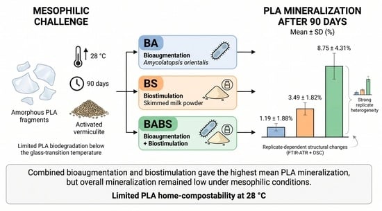
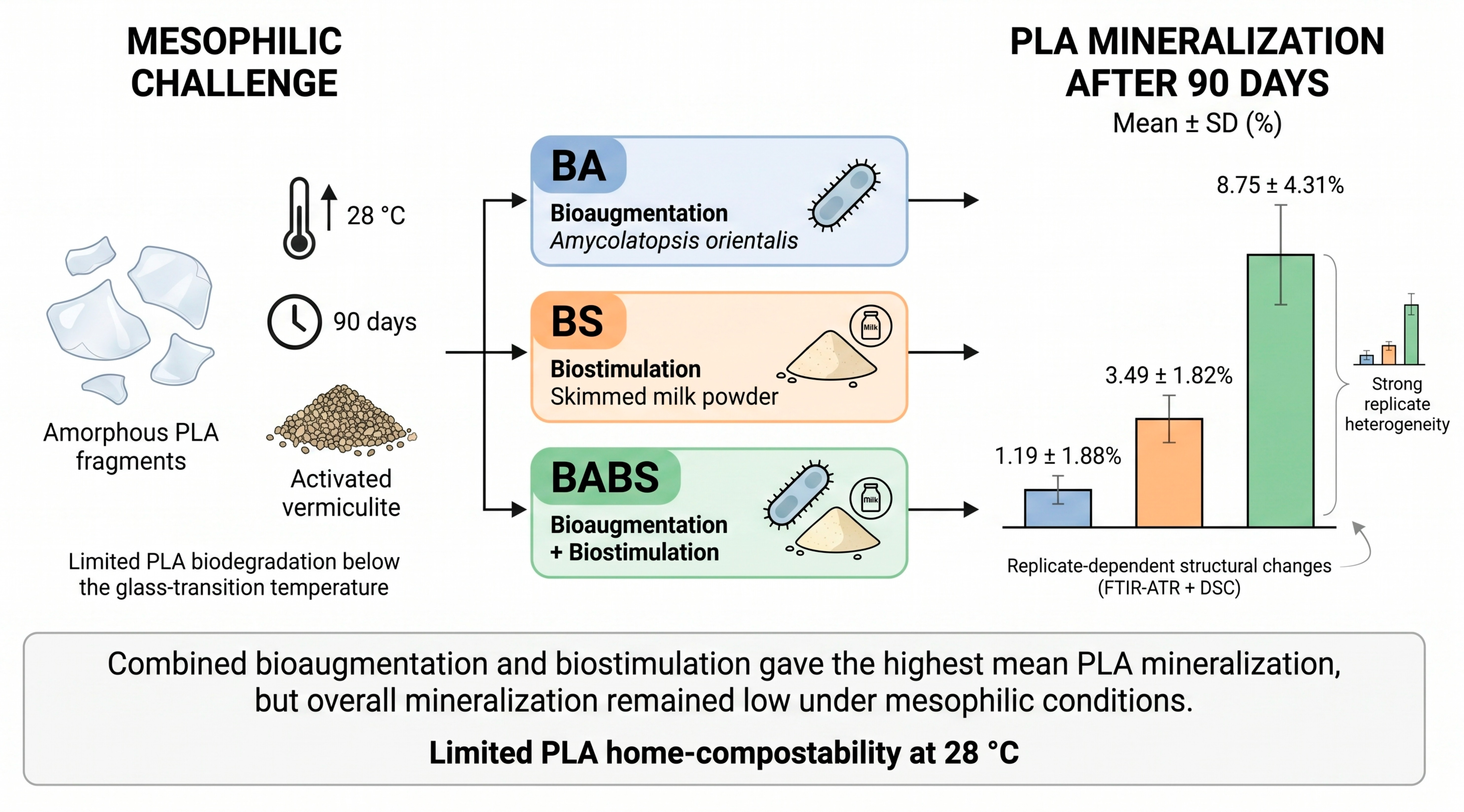
Graphical Abstract

1. Introduction

Polylactic acid (PLA) is one of the most widely used biobased aliphatic polyesters for short-life applications. Although PLA is commonly described as biodegradable, its mineralization is highly dependent on environmental and material-related factors, including temperature, humidity, sample geometry, crystallinity, molar mass, and the structure and activity of the microbial community [1,2,3,4]. PLA degradation is generally favored under thermophilic composting conditions, where hydrolysis and microbial conversion proceed more readily than under mesophilic conditions [4,5,6,7].
This temperature dependence is a major limitation for home and decentralized composting conditions, which are characterized by lower and more variable temperatures than industrial composting [3,8]. As reported previously for PLA, PBS, and PBAT materials under home-composting-like conditions, PLA frequently shows slow disintegration and limited mineralization under conditions close to those of home composting [9]. At 28 °C, PLA remains far below its glass transition temperature, which restricts chain mobility and can limit the accessibility of ester bonds to hydrolytic and enzymatic attack [4,10,11].
Bioaugmentation and biostimulation are potential strategies for improving polyester biodegradation under suboptimal conditions, but their effectiveness remains uncertain. Bioaugmentation requires the introduced strain to survive, compete with resident microorganisms, colonize the polymer surface, and express active extracellular enzymes in the target matrix. Biostimulation may enhance microbial metabolism or enzyme production, but it can also introduce readily biodegradable carbon and nitrogen sources, thereby complicating the interpretation of CO2 evolution [12,13,14,15].
Amycolatopsis orientalis has been identified as a promising PLA-degrading actinobacterium under controlled laboratory conditions, mainly because of its ability to produce extracellular enzymes active toward PLA or related ester substrates [16,17,18,19,20,21,22,23]. Proteinaceous substrates have also been reported to induce enzymatic activities associated with PLA degradation [18,19,20,21,22]. Skimmed milk powder was therefore selected in the present study as a practical protein-based biostimulant.
Activated vermiculite was used as a controlled low-carbon medium to reduce interference from background organic matter and to enable treatment-specific blank correction [24,25,26,27]. This system does not reproduce all features of real home compost, particularly its organic-matter content and microbial complexity. However, it provides a useful framework for testing whether bioaugmentation and biostimulation can improve PLA mineralization under strictly mesophilic conditions.
The objective of this study was to compare bioaugmentation with A. orientalis, protein-based biostimulation with skimmed milk powder, and their combined application for PLA mineralization at 28 °C. Blank-corrected respirometry was combined with FTIR-ATR and DSC monitoring of recovered PLA fragments to assess mineralization behavior and supporting physicochemical changes.

2. Materials and Methods

2.1. PLA Material and Film Preparation

Commercial amorphous-grade PLA (PLE 005-A; MFI: 3 g/10 min at 190 °C/2.16 kg; D-lactide content: 4%; Tm ≈ 160 °C; density = 1.24 g·cm−3) was supplied in pellet form by Natureplast (Mondeville, France). Pellets were oven-dried at 60 °C overnight prior to processing. Thin films were fabricated by compression molding using a using a 15 T hydraulic press (SCAMEX, Isques, France). For each film, 15 g of polymer was placed between two sheets of parchment paper and subjected to a two-step pressing protocol: natural closing pressure for 90 s, followed by 200 bar for 90 s. Plate temperatures were maintained at 165 °C. Films were cut into approximately 2 × 2 cm fragments. Film thickness was assessed from 50 measurements with a digital caliper. Although this geometry provides an estimate of the exposed surface area (i.e., ~77 cm−1), the true accessible surface also depends on roughness and surface topography, which were not measured in this study.

2.2. Initial PLA Characterization

Initial PLA fragments were characterized by CHN elemental analysis, size exclusion chromatography (SEC), DSC, and water contact angle measurements (Table 1). These analyses were used to define the initial carbon content for mineralization calculations and to describe material properties known to influence biodegradation.
Total carbon content of cryogenically ground plastic fragments was measured in triplicate using a FlashSmart CHNS/O elemental analyzer (Thermo Fisher Scientific, Waltham, MA, USA). Samples of 2–6 mg were oven-dried at 40 °C and weighed using a precision balance (ME36S, Sartorius AG, Göttingen, Germany). Initial DSC was performed in triplicate using a Netzsch DSC 214 Polyma instrument (Selb, Germany). Approximately 8–12 mg of each sample was analyzed under argon at 40 mL·min−1. Samples were heated to 180 °C, cooled to 0 °C, and reheated to 180 °C at 10 °C/min. SEC was carried out using an ACQUITY APC UHPLC system (Waters Corporation, Milford, MA, USA) equipped with a refractive index detector and calibrated with PMMA standards. Samples were dissolved overnight in hexafluoroisopropanol, filtered through a 0.2 µm PTFE membrane, and injected at 35 °C. Static water contact angle measurements were performed at 22 ± 1 °C using the sessile drop method on a DSA25 goniometer (KRÜSS GmbH, Hamburg, Germany).

2.3. Compost Extract and Activated Vermiculite Medium

A biotic compost extract was prepared by suspending 15 g of mature compost in 150 mL of Ringer solution, followed by homogenization for 1 h, filtration through a 1 mm sieve, and centrifugation at 10,000 rpm for 20 min. The resulting pellet was resuspended in 150 mL of fresh Ringer solution. The mature compost originated from an industrial composting facility processing lignocellulosic green waste and was compliant with NF U44-051 [28]. Each bioreactor received 15 mL of compost extract. Vermiculite was used as an inert, low-carbon support to limit background organic carbon and enable treatment-specific blank correction.

2.4. Amycolatopsis orientalis Inoculum

Amycolatopsis orientalis CIP 107113 (Institut Pasteur, Paris, France) was precultured in ISP2 broth at 28 °C and 150 rpm. The strain was initially incubated 48 h in 5 mL of ISP2 medium in a sterile 50 mL tube. Subsequently, 2 mL of this preculture was transferred into 75 mL of ISP2 medium in 500 mL Erlenmeyer flasks and incubated for an additional 48 h under the same conditions. Viable cell counts were determined by serial dilution and plating on tryptic soy agar. Bioaugmented reactors received 30 mL of culture. Based on a mean viable count of (1.73 ± 0.13) × 109 CFU·mL−1, the inoculum corresponded to approximately 5.19 × 10¹⁰ CFU of A. orientalis per bioreactor.

2.5. Biostimulation Protocol

Commercial skimmed milk powder (Régilait, Saint-Martin-Belle-Roche, France) was added to BS and BABS reactors using a sequential exploratory supplementation protocol: 5 g on days 0, 14, and 68, and 10 g on day 42. This protocol was designed as an exploratory staggered supplementation strategy and should not be interpreted as a true dose–response experiment because dose and timing were not independently varied.
Skimmed milk powder was selected as a protein-rich biostimulant for both scientific and practical reasons. Mayekar and Auras reported enhanced PLA mineralization in mature compost at 37 °C using skimmed milk powder as a biostimulant [5,6]. In addition, proteinaceous substrates have been reported to stimulate PLA-degrading activity in Amycolatopsis orientalis, supporting the rationale for testing a combined bioaugmentation–biostimulation strategy [18,19,20,21,22]. Finally, because the present study targeted decentralized composting conditions, skimmed milk powder was considered a more realistic and practically accessible biostimulant than more specific chemical inducers.

2.6. Respirometric Biodegradation Assay

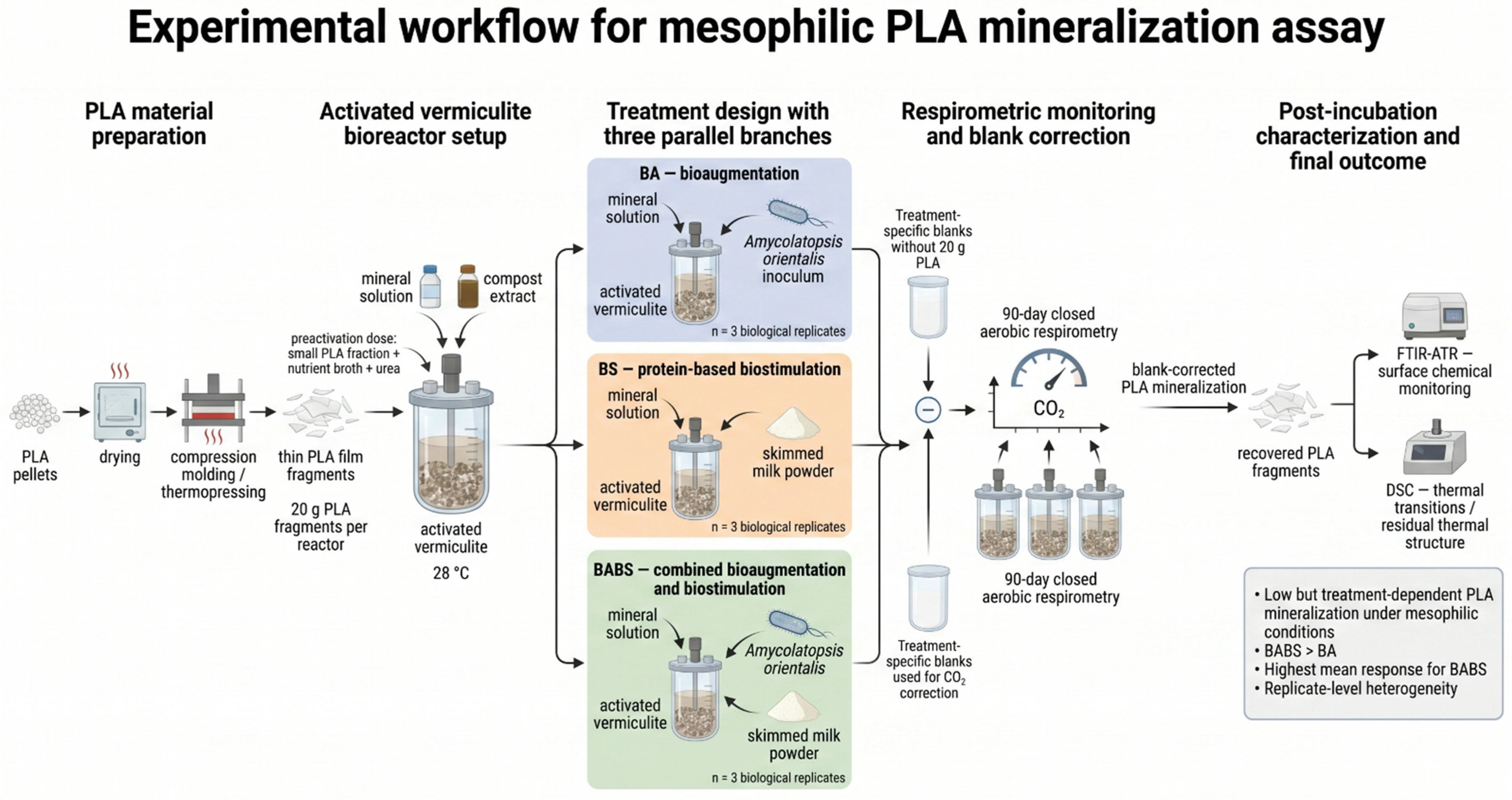
Closed aerobic respirometric bioreactors (ECHO Instruments d.o.o., Slovenske Konjice, Slovenia) were incubated for 90 days at 28 ± 1 °C in the dark, following ISO 14855-1 principles [29] and adapted from NF T51-800 (Table 1; Figure 1) [30]. The incubation temperature of 28 °C was chosen to comply with the NF T51-800 home compostability framework, which requires testing below 30 °C, while accounting for chamber temperature fluctuations.
Each PLA-containing reactor included 20 g of thermopressed PLA fragments, 200 g of vermiculite (2–4 mm), 440 mL of pH 7.2 buffered mineral solution, and the treatment-specific inoculum and/or biostimulant. The mineral solution composition per 5 L was 0.650 g CaCl2·2H2O, 34.850 g Na2HPO4·2H2O, 18.750 g KH2PO4, 20 g (NH4)2SO4, 1 g MgSO4·7H2O, 0.0135 g FeSO4·7H2O, 0.005 g MnSO4·7H2O, 0.005 g ZnSO4·7H2O, 0.005 g H3BO3, 0.005 g KI, and 0.005 g (NH4)6Mo7O24·4H2O [25]. Substrate moisture was maintained by weekly addition of ultrapure water into the designated lower compartment of the reactors and by two additions of 30 mL of mineral solution on days 30 and 60.
Each treatment had a corresponding blank without the 20 g PLA fragments but receiving the same compost extract, activation dose, inoculum, and/or biostimulant additions. These treatment-specific blanks were used to correct CO2 production not attributable to the main PLA fragments. All treatments were conducted in three biological replicates.

2.7. Microbial Preactivation

A 7-day preactivation phase was implemented before the addition of the 20 g of PLA fragments. Each reactor received an activation dose composed of 0.5 g of finely fragmented PLA, 1 g of nutrient broth, and 65 mg of urea to approach a C/N ratio of 25:1. This activation mixture was applied to both treatment reactors and their corresponding blanks, thereby allowing treatment-specific blank correction.

2.8. Mineralization Calculation

PLA mineralization extent (%) was calculated from cumulative CO2 production in PLA-containing reactors after subtraction of corresponding treatment-specific blank and normalization to theoretical CO2 yield calculated from initial PLA carbon content. The calculation was performed as follows:
M i n e r a l i z a t i o n   extent   % = C O 2 , t e s t C O 2 , b l a n k T h C O 2 , P L A × 100
where CO2,test is the cumulative CO2 evolved in the PLA-containing reactor, CO2,blank is the cumulative CO2 evolved in the corresponding treatment-specific blank, and ThCO2,PLA is the theoretical CO2 production corresponding to complete aerobic mineralization of the 20 g PLA sample based on the CHN carbon content. Final percentages are reported as mineralization extent.

2.9. Post-Incubation FTIR-ATR and DSC Monitoring

Recovered PLA fragments collected after the 90-day experiment were gently rinsed with ultrapure demineralized water to remove residual mineral particles and surface deposits, then oven-dried at 40 °C before physicochemical characterization. FTIR-ATR and DSC were used as complementary post-incubation monitoring techniques.
FTIR-ATR spectra were acquired using a Nicolet iS10 FTIR spectrometer equipped with a Smart iTX ATR accessory (Thermo Fisher Scientific, Singapore). Spectra were recorded from 4000 to 500 cm−1 using 32 scans at a resolution of 4 cm−1, then exported and processed using a dedicated Python (version 3.14.5; Python Software Foundation, Wilmington, DE, USA) workflow including asymmetric least-squares baseline correction.
DSC analyses were performed in triplicate using a DSC 214 Polyma calorimeter (NETZSCH-Gerätebau GmbH, Selb, Germany). Approximately 8–12 mg of each sample was sealed in DSC pans and subjected to a first heating scan to 180 °C, cooling to 0 °C, and a second heating scan to 180 °C. Heating and cooling rates were 10 °C min−1 under argon purge at 40 mL min−1.

2.10. Statistical Analysis

Final mineralization extents were analyzed using one-way ANOVA, followed by Tukey’s HSD post hoc tests at α = 0.05. Normality was assessed using the Shapiro–Wilk test on model residuals and, descriptively, within each treatment group. Homogeneity of variances was evaluated using Levene’s test. Because only three biological replicates were available per treatment, these assumption tests were interpreted cautiously.

3. Results and Discussion

3.1. Initial PLA Properties

The compression-molded PLA fragments were thin amorphous films with a mean thickness of 266 ± 57 µm and a carbon content of 49.0 ± 0.29% (Table 2). Initial SEC gave Mw = 89.9 kDa, Mn = 43.2 kDa, and a dispersity index of 2.08, indicating a non-degraded PLA with a molar mass range consistent with processed commercial materials. This point is important because molar mass directly affects thermomechanical behavior, chain mobility and biodegradation sensitivity; an excessively low initial molar mass could artificially increase hydrolysis, oligomer release and microbial assimilation. The initial crystallinity was negligible (<1%), which is consistent with the amorphous-grade PLA used here. Its 4% D-lactide content reduces stereoregularity and limits crystallization during processing; therefore, the cold crystallization peak observed during DSC heating reflects the crystallization occurring during the scan rather than the high initial crystallinity. The glass transition temperature was 65.9 ± 1.0 °C, and the water contact angle was 73.8 ± 4.1°. These properties indicate that the PLA was incubated well below its glass transition temperature during the 28 °C respirometric assay. Overall, matrix structure, crystallinity, molar mass and surface/water interaction properties are relevant because they can influence the behavior of biodegradable polyesters during environmental exposure and biodegradation [2,10,11,31].

3.2. Treatment-Level Mineralization Extent

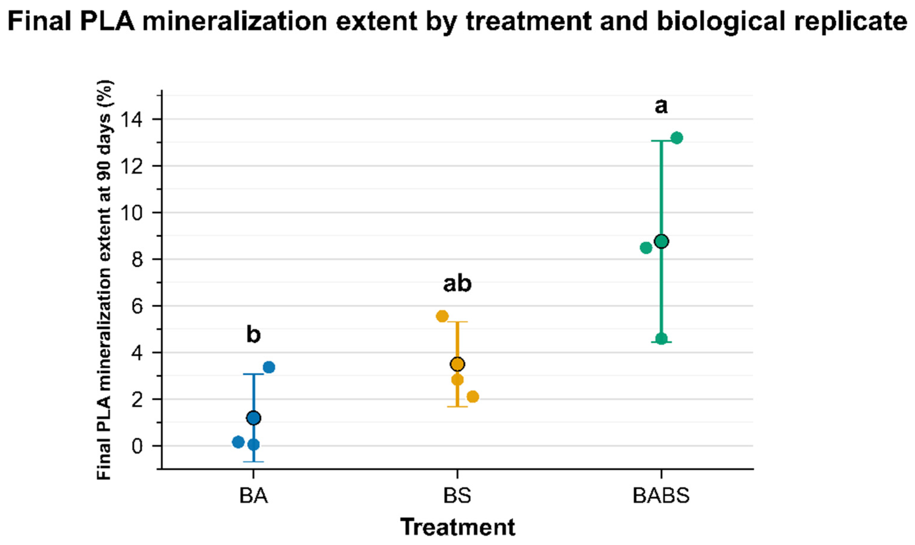
After 90 days at 28 °C, blank-corrected PLA mineralization remained low in all treatments. Mean final mineralization extents were 1.19 ± 1.88% for bioaugmentation with A. orientalis (BA), 3.49 ± 1.82% for protein-based biostimulation with skimmed milk powder (BS), and 8.75 ± 4.31% for the combined bioaugmentation–biostimulation treatment (BABS) (Table 3; Figure 2). Thus, BABS gave the highest mean final mineralization extent. However, the absolute mineralization remained far below the level expected for effective home compostability (i.e., 90%), confirming that PLA remained poorly mineralized under the tested mesophilic conditions.
Because the highest mean mineralization extent was only 8.75%, most of the initial PLA carbon was not converted into CO2 during the 90-day incubation. This non-mineralized carbon was likely still mainly present in the recovered PLA fragments, although minor fractions may have been incorporated into microbial biomass or released as soluble organic compounds or low-molecular-weight degradation products.
One-way ANOVA indicated a treatment effect on final mineralization extent (F = 5.308, p = 0.047). Tukey’s HSD post hoc comparisons showed that BABS was significantly higher than BA (p = 0.043), whereas BA and BS were not significantly different (p = 0.588), and BABS was not significantly higher than BS (p = 0.157). Therefore, the combined treatment should be interpreted as the highest mean response and as significantly higher than bioaugmentation alone, which suggests a weak synergistic tendency. However, because BABS was not significantly higher than biostimulation alone and showed marked replicate-level heterogeneity, this pattern should not be considered a uniformly superior treatment.
The assumptions of the parametric analysis were evaluated but should be interpreted cautiously because of the limited sample size. Although n = 3 is consistent with the minimum replicate number commonly used in standardized compostability and respirometric protocols, including NF T51-800-like approaches, it remains limited for capturing reactor-level variability, especially in the heterogeneous BABS treatment. Shapiro–Wilk tests did not reject normality for the ANOVA residuals (W = 0.944, p = 0.626), and group-wise tests gave p values of 0.056, 0.386 and 0.895 for BA, BS and BABS, respectively. Levene’s test did not reject homogeneity of variances (F = 0.783, p = 0.499).
This is why individual reactor trajectories are discussed alongside treatment-level means.
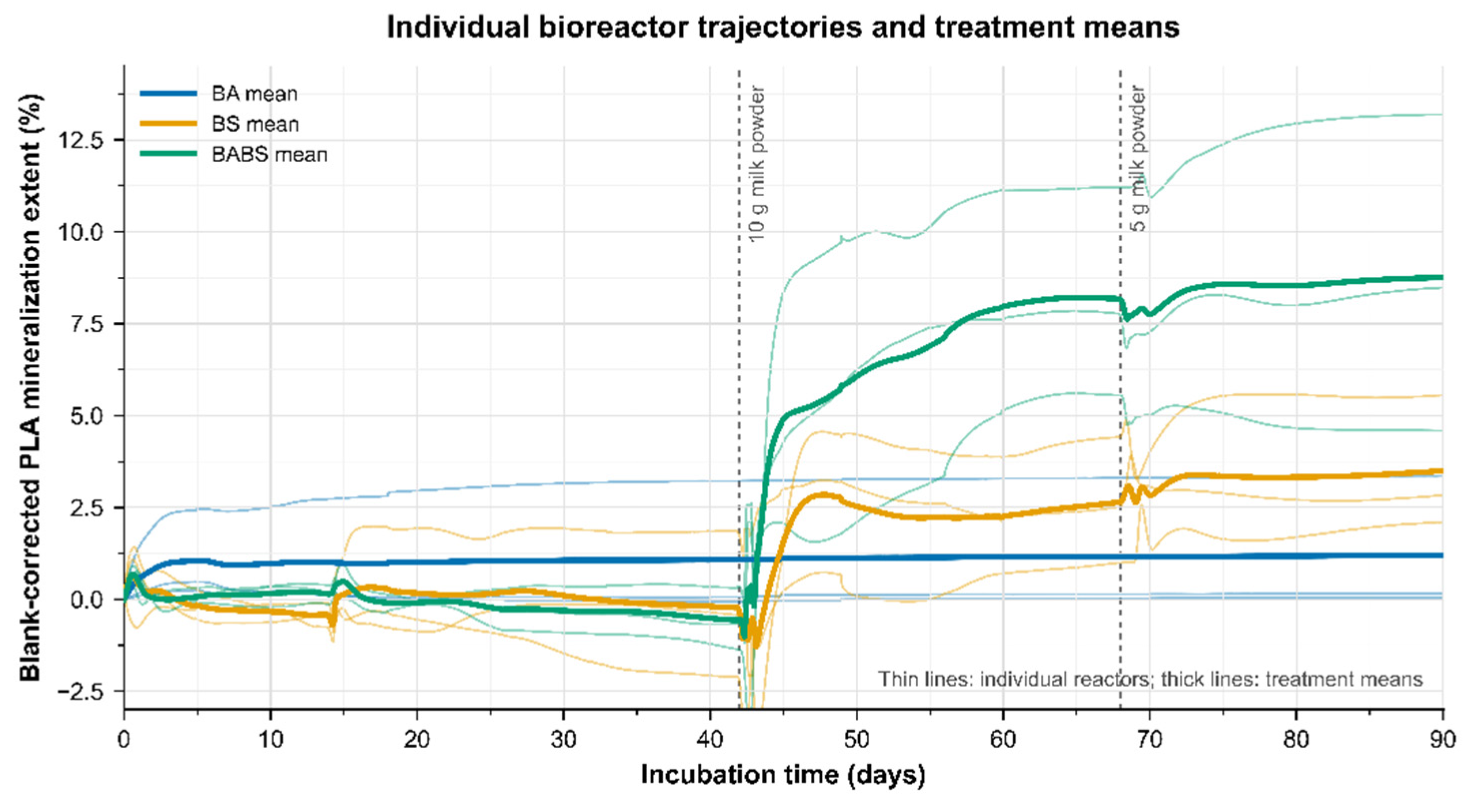
The individual trajectories revealed substantial replicate-level heterogeneity (Figure 3; Table 4). In the BA treatment, BA-1 and BA-2 remained close to zero throughout the experiment, whereas BA-3 reached 3.36% mineralization at day 90. Overall, bioaugmentation with A. orientalis alone was insufficient to produce a reproducible increase in PLA mineralization.
The BS treatment also showed heterogeneous trajectories. BS-1 reached 5.55% mineralization at day 90 and contributed strongly to the BS mean, whereas BS-2 and BS-3 reached 2.83% and 2.10%, respectively. This indicates that the resident compost-derived microbiota was able to respond to protein-based supplementation in some bioreactors, but that this response remained limited and poorly reproducible. The BABS treatment produced the highest individual and mean responses, but it was also heterogeneous: BABS-3 reached 13.19%, BABS-1 reached 8.48%, and BABS-2 reached 4.59%. Therefore, BABS should not be described as a uniformly reproduced treatment response. Instead, it generated a higher mean mineralization extent driven partly by one strongly responding bioreactor.
In the biostimulated treatments, the main increase in mineralization occurred after day 42, which coincided with the 10 g skimmed-milk-powder addition. However, this observation should not be interpreted as evidence of a true dose threshold, because dose and timing were confounded in the sequential supplementation protocol. The response may reflect the higher protein input, changes in microbial community structure over time, progressive colonization of PLA surfaces, or a combination of these factors. A dedicated dose–response or timing-controlled design would be required to separate these effects.
Although proteinaceous substrates have been reported to stimulate enzymatic activities associated with PLA degradation, no extracellular enzyme activity was measured in the present study. Therefore, protein-induced enzyme production remains a plausible but unverified explanation for the limited increase observed in the biostimulated treatments.
The strong response of BABS-3 remains noteworthy, as this replicate reached 13.19% mineralization. This shows that values above 10% can occur under combined bioaugmentation and biostimulation. However, BABS-3 should not be considered representative of the whole BABS treatment. Indeed, BABS-1, BABS-2 and BABS-3 followed divergent trajectories. This heterogeneity may reflect reactor-level variability in microbial community assembly and in the establishment of the bioaugmented strain. Such variability is consistent with stochastic assembly processes in bioreactor microbial communities [32] and with previous work showing that bioaugmentation efficiency depends strongly on the polymer–matrix system [33]. In addition, successful bioaugmentation requires the introduced strain to remain active and persistent in the target habitat [34].
A limitation of this study is that the fate of the bioaugmented Amycolatopsis orientalis strain was not monitored after inoculation. Although the initial inoculum was quantified before addition to the reactors, no post-incubation qPCR or selective plating assay was performed. Therefore, the weak BA response cannot be attributed specifically to poor strain survival, limited PLA colonization, competition with the compost-derived microbiota, insufficient extracellular activity at 28 °C, or intrinsic inefficiency of the strain under the tested conditions. Accordingly, the results should be interpreted as treatment-level mineralization extents derived from respirometric monitoring rather than as direct evidence of A. orientalis persistence, activity or inactivity.
Taken together, the mineralization results show that the tested strategies were insufficient to achieve effective PLA mineralization at 28 °C. Protein-based biostimulation produced only a limited increase, while BABS gave the highest mean and individual responses but remained heterogeneous and not significantly higher than biostimulation alone.
Despite the well-known difficulty of mineralizing PLA at 28 °C, these strategies warranted experimental evaluation because Mayekar and Auras [5,6] reported approximately 25% PLA mineralization after 180 days in mature compost supplemented with skimmed milk powder at 37 °C, still below the Tg of PLA. Moreover, A. orientalis had been reported as a PLA-degrading candidate under simplified mesophilic laboratory conditions. These findings justified testing whether protein-based biostimulation, A. orientalis bioaugmentation, or their combination could produce a measurable response under stricter conditions below 30 °C.
The much lower mineralization observed here likely reflects both matrix and temperature effects. Activated vermiculite was deliberately selected as a low-organic-carbon support to reduce background respiration, allow treatment-specific blank correction and facilitate recovery of residual polymer [35]. However, unlike mature compost, this medium does not reproduce the full organic and biological complexity of compost, including organic matter, humic-like substances, nutrients and diverse microbial communities [36]. This lower complexity may have limited microbial succession, PLA surface colonization, extracellular activity and establishment of A. orientalis under the present conditions. In addition, the higher incubation temperature used by Mayekar and Auras, i.e., 37 °C, likely intensified biological activity and enzymatic processes relative to 28 °C [37]. BABS should therefore be interpreted as a limited, reactor-dependent response under stricter mesophilic and low-carbon conditions rather than as a robust strategy for achieving PLA home compostability.

3.3. DSC Monitoring of Recovered Fragments

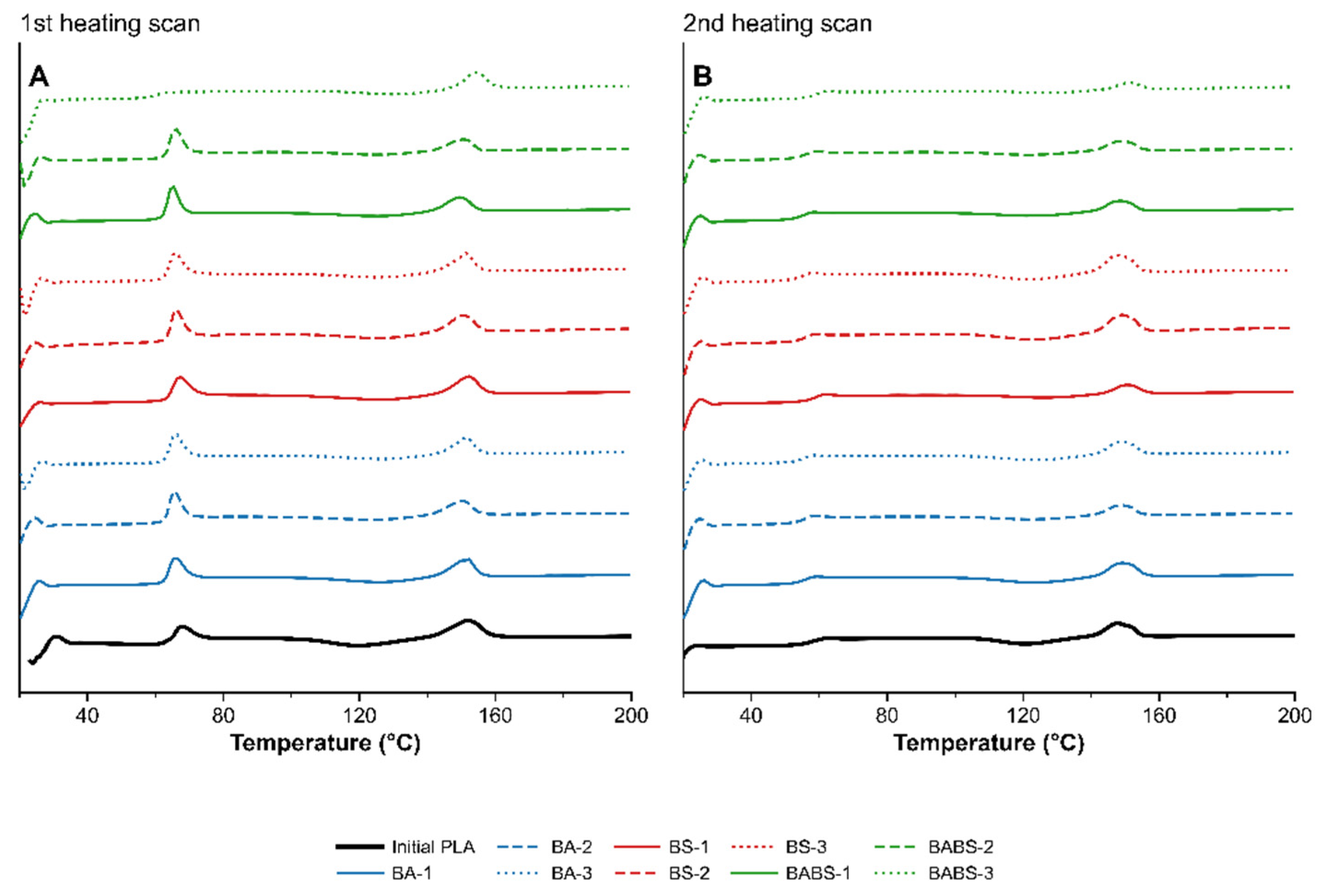
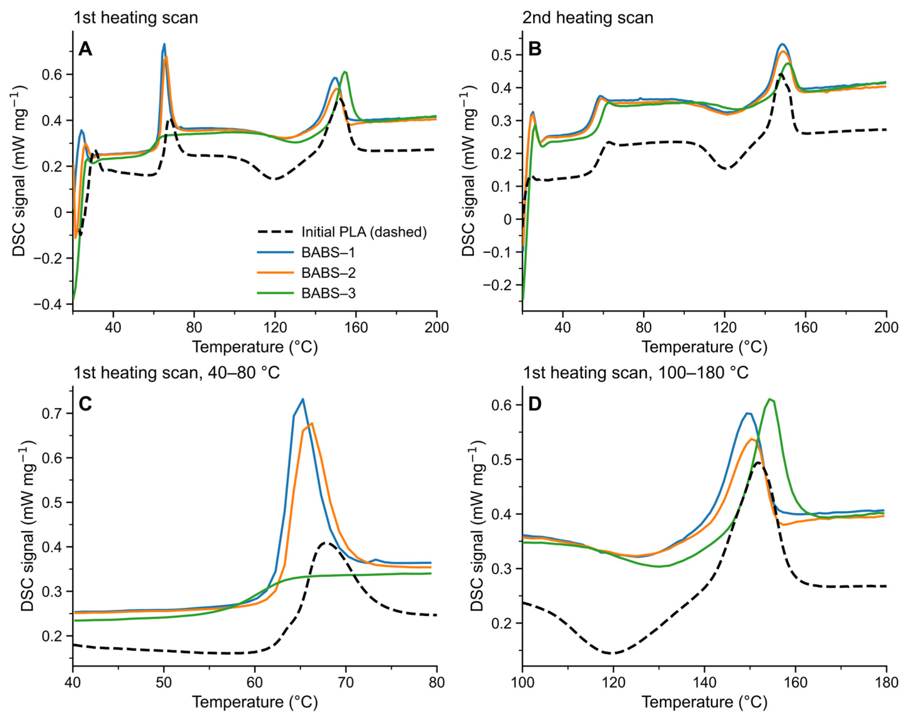
DSC was used as a complementary technique to compare the residual thermal behavior of PLA fragments recovered after 90 days of incubation. The stacked thermograms including the initial PLA, BA, BS and BABS replicates are shown in Figure 4 and Figure 5 for both the first and second heating scans. Overall, all recovered samples retained the main thermal signature of PLA, with a glass transition/enthalpic relaxation region and cold crystallization and melting events still visible. However, the first heating scan revealed replicate-dependent differences in peak shape, relaxation intensity and transition temperatures, indicating that incubation did not produce a uniform thermal response across reactors. Because the combined bioaugmentation–biostimulation treatment (BABS) produced the highest mean mineralization extent at day 90 while also showing the strongest replicate-level heterogeneity, this condition was examined in more detail.
In the first heating scan, which preserved the thermal and environmental history of the recovered specimens, the initial PLA displayed an apparent Tg inflection at approximately 65.9 °C, an enthalpic relaxation maximum at 67.8 °C, a cold crystallization event centered around 119.6 °C, and a melting peak at 151.8 °C. The BABS replicates showed markedly different responses. BABS-1 exhibited an apparent Tg inflection at 63.3 °C, a relaxation maximum at 65.3 °C, Tcc at 125.3 °C and Tm at 149.3 °C. BABS-2 showed a similar relaxation behavior, with Tg at 64.3 °C, relaxation at 66.3 °C, Tcc at 125.3 °C and Tm at 150.3 °C. By contrast, BABS-3 displayed a more atypical profile, with an apparent Tg inflection at 60.3 °C, no sharp enthalpic relaxation peak comparable to BABS-1 and BABS-2, Tcc shifted to 130.3 °C, corresponding to an increase of approximately 9% relative to the initial PLA, and the highest apparent Tm at 154.3 °C.
A cautious interpretation is that the BABS-3 residue may have been enriched in domains comparatively less prone to cold crystallization and more thermally stable. Preferential alteration or mineralization of more accessible, less ordered, or surface-exposed PLA fractions could leave behind residual material biased toward more stable domains. This residue selection effect is consistent with the known influence of PLA morphology and chain mobility on biodegradation, where amorphous and more mobile regions are generally more accessible to hydrolytic and enzymatic attack than more ordered domains [2,4,10,11].
The second heating scan provided a complementary view because the first heating scan erased the previous thermal history of the specimens. For BABS-1 and BABS-2, the upward shift of Tcc observed in the first heating scan almost disappeared after thermal erasure, decreasing from 125.3 °C in S1 to approximately 120.3–121.3 °C in S3. Their Tm values also converged toward the initial PLA after thermal erasure. This suggests that their first-scan differences were largely governed by physical aging, relaxation state and thermal history rather than by irreversible bulk transformation. BABS-3, however, retained comparatively higher Tcc and Tm values during the second heating scan, although the difference was attenuated relative to the first heating scan. This persistence suggests that BABS-3 underwent a more pronounced selection or reorganization of the residual PLA fraction.

3.4. FTIR-ATR Monitoring at Treatment and Replicate Levels

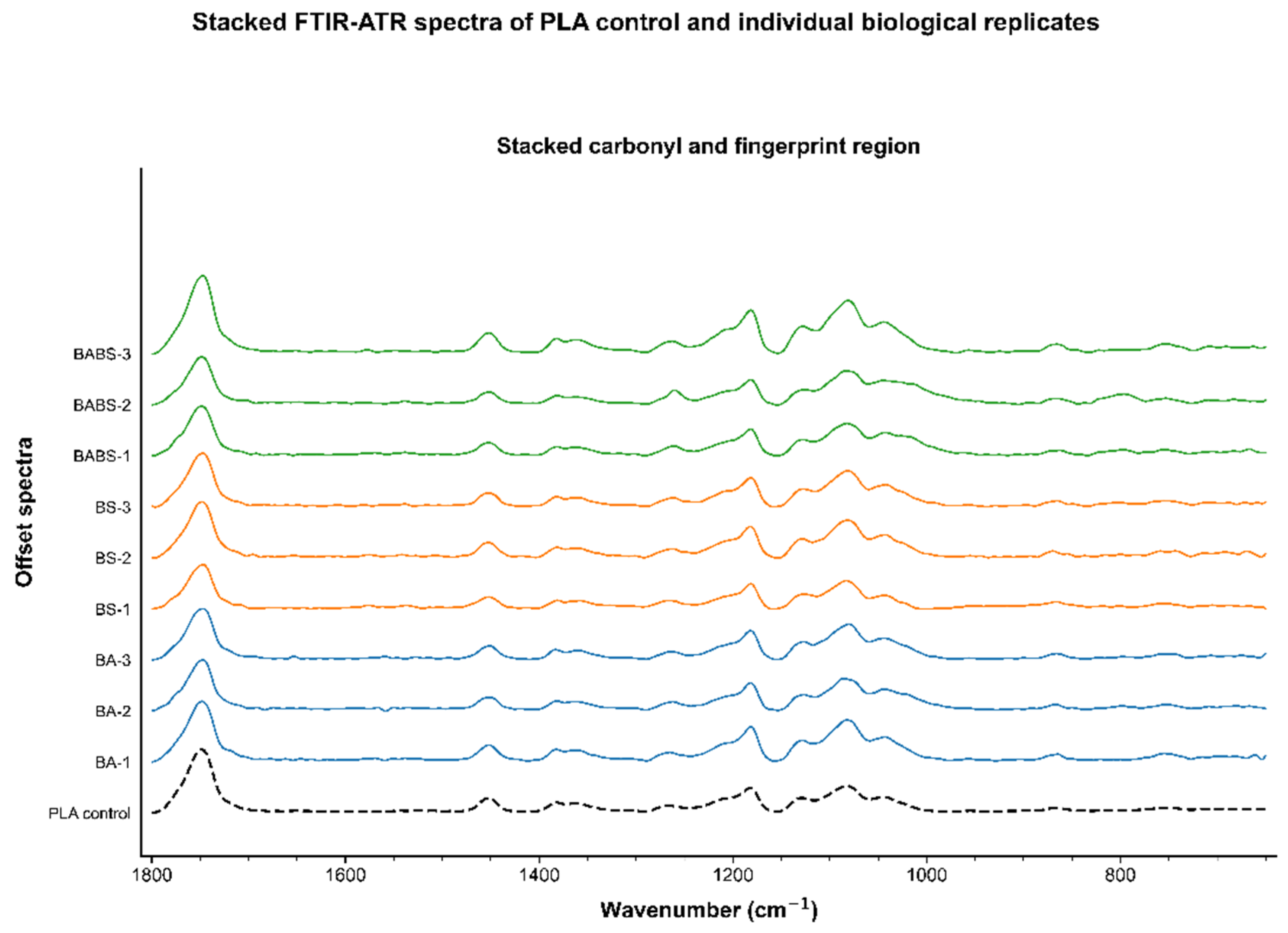
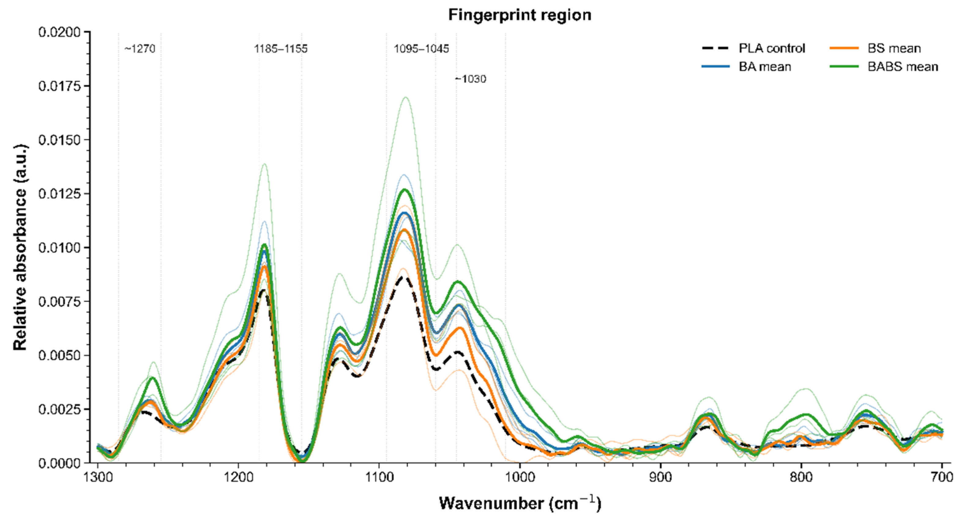
The replicate-level FTIR-ATR analysis showed that all incubated PLA samples retained the characteristic PLA fingerprint, including the ester carbonyl band at 1750 cm−1 and the C–O–C/C–O stretching region between 1200 and 1000 cm−1. After normalization to the carbonyl band, the BABS treatment displayed the most pronounced modifications in the relative shape of the fingerprint region, particularly around the C–O–C/C–O stretching bands (Figure 6 and Figure 7). These changes may reflect subtle surface-level modifications associated with microbial colonization, enzymatic attack of ester bonds, surface reorganization, or residual biological material. This interpretation is consistent with the higher mean mineralization observed for BABS, since PLA biodegradation under compost-like conditions is generally initiated by microbial attachment and extracellular enzymatic cleavage of ester bonds. However, the preservation of the main PLA bands and the absence of a strong broad O–H contribution indicate that FTIR-ATR did not evidence extensive surface hydrolysis or advanced chemical degradation. Therefore, the FTIR results should be interpreted as complementary evidence of heterogeneous surface alteration rather than as direct proof of extensive PLA depolymerization.

4. Conclusions

Under strictly mesophilic conditions at 28 °C, PLA mineralization remained very limited in activated vermiculite. Bioaugmentation with Amycolatopsis orientalis alone produced near-zero mineralization after 90 days, reaching only 1.19 ± 1.88%. Protein-based biostimulation with skimmed milk powder led to a limited increase, with a final mineralization extent of 3.49 ± 1.82%. The combined bioaugmentation–biostimulation treatment produced the highest final mineralization extent, reaching 8.75 ± 4.31%, and was significantly higher than bioaugmentation alone. However, it was not significantly higher than biostimulation alone, and the response was heterogeneous among biological replicates. Therefore, the combined treatment should be interpreted as a limited, reactor-dependent enhancement rather than as a robust or consistently reproducible strategy.
These mineralization extents remained far below the level required for effective home compostability. The results also contrast those of previous studies by Mayekar and Auras, who reported enhanced PLA biodegradation using biostimulants in mature compost at 37 °C over 180 days [5,6]. This discrepancy likely reflects the combined effects of higher incubation temperature and the greater biological and organic matter complexity of mature compost compared with the carbon-limited activated vermiculite system used here.
FTIR-ATR and DSC provided useful complementary monitoring of recovered PLA fragments and revealed treatment- and replicate-dependent physicochemical changes. However, these analyses did not demonstrate extensive bulk polymer transformation. The main contribution of this study is therefore defining the limitations of simple bioaugmentation and protein-based biostimulation strategies for PLA under mesophilic composting-like conditions, while emphasizing the importance of examining individual bioreactor trajectories in addition to treatment-level means.

Author Contributions

Conceptualization, J.B.; methodology, J.B.; formal analysis, J.B.; investigation, J.B.; writing—original draft preparation, J.B.; writing—review and editing, J.B.; supervision, F.B. and R.G. All authors have read and agreed to the published version of the manuscript.

Funding

This work was supported by the French region of Normandy Projet emergent.

Institutional Review Board Statement

Not applicable.

Data Availability Statement

The raw data supporting the conclusions of this article will be made available by the authors on request.

Acknowledgments

The authors thank UniLaSalle and the ECLORE research unit for technical and scientific support throughout this work. The Agrobiotech research platform (GIS IBISA) is gratefully acknowledged for access to facilities and technical support. The authors also thank Grace Ifembo-Sikumbili and Rebecca Djamba for technical assistance in carrying out experimental measurements. During the preparation of the Graphical Abstract, the authors used the BioRender website. Created in BioRender. Bellon, J. (2026). https://BioRender.com/jq1eg8y. The authors have reviewed and edited the output and take full responsibility for the content of this publication.

Conflicts of Interest

The authors declare no conflicts of interest.

References

  1. MacLeod, M.; Arp, H.P.H.; Tekman, M.B.; Jahnke, A. The global threat from plastic pollution. Science 2021, 373, 61–65. [Google Scholar] [CrossRef] [PubMed]
  2. Rosli, N.A.; Karamanlioglu, M.; Kargarzadeh, H.; Ahmad, I. Comprehensive exploration of natural degradation of poly(lactic acid) blends in various degradation media: A review. Int. J. Biol. Macromol. 2021, 187, 732–741. [Google Scholar] [CrossRef] [PubMed]
  3. Vaverkova, M.; Adamcova, D.; Zloch, J. How do degradable/biodegradable plastic materials decompose in home composting environment? J. Ecol. Eng. 2014, 15, 82–89. [Google Scholar]
  4. Ho, K.L.G.; Pometto, A.L., III; Hinz, P.N. Effects of temperature and relative humidity on polylactic acid plastic degradation. J. Environ. Polym. Degrad. 1999, 7, 83–92. [Google Scholar] [CrossRef]
  5. Mayekar, P.C.; Auras, R. Accelerating biodegradation: Enhancing poly(lactic acid) breakdown at mesophilic environmental conditions with biostimulants. Macromol. Rapid Commun. 2024, 45, 2300641. [Google Scholar] [CrossRef]
  6. Mayekar, P.C.; Auras, R. Speeding it up: Dual effects of biostimulants and iron on the biodegradation of poly(lactic acid) at mesophilic conditions. Environ. Sci. Processes Impacts 2024, 26, 530–539. [Google Scholar] [CrossRef] [PubMed]
  7. Castro-Aguirre, E.; Auras, R.; Selke, S.; Rubino, M.; Marsh, T. Enhancing the biodegradation rate of poly(lactic acid) films and PLA bio-nanocomposites in simulated composting through bioaugmentation. Polym. Degrad. Stab. 2018, 154, 46–54. [Google Scholar] [CrossRef]
  8. Bellon, J.; Bacoup, F.; Gattin, R. Challenges and Opportunities in Home-Composting of Biodegradable Plastics: A Comprehensive Review. Results Eng. 2025, 28, 107535. [Google Scholar] [CrossRef]
  9. Bellon, J.; Bacoup, F.; Gattin, R. Home Compostability of PLA, PBS, and PBAT Biocomposites Incorporating Orange Peel and Brewer’s Spent Grain: Mineralization Kinetics, Structural Evolution, Phytotoxicity, and Bacterial Isolation. J. Hazard. Mater. Adv. 2026, 22, 101197. [Google Scholar] [CrossRef]
  10. Pantani, R.; Sorrentino, A. Influence of crystallinity on the biodegradation rate of injection-moulded poly(lactic acid) samples in controlled composting conditions. Polym. Degrad. Stab. 2013, 98, 1089–1096. [Google Scholar] [CrossRef]
  11. Chinaglia, S.; Tosin, M.; Degli-Innocenti, F. Biodegradation rate of biodegradable plastics at molecular level. Polym. Degrad. Stab. 2018, 147, 237–244. [Google Scholar] [CrossRef]
  12. Shahnawaz, M.; Sangale, M.K.; Ade, A.B. Bioremediation Technology for Plastic Waste; Springer: Singapore, 2019. [Google Scholar]
  13. Baskaran, S.; Sathiavelu, M. Bioaugmentation and biostimulation of dumpsites for plastic degradation. In Cost Effective Technologies for Solid Waste and Wastewater Treatment; Elsevier: Amsterdam, The Netherlands, 2022; pp. 9–23. [Google Scholar]
  14. Shen, J.; Bartha, R. Priming effect of substrate addition in soil-based biodegradation tests. Appl. Environ. Microbiol. 1996, 62, 1428–1430. [Google Scholar] [CrossRef] [PubMed]
  15. d’Errico, G.; Giovannelli, D.; Montano, C.; Milanovic, V.; Ciani, M.; Manini, E. Bioremediation of high organic load lagoon sediments: Compost addition and priming effects. Chemosphere 2013, 91, 99–104. [Google Scholar] [CrossRef] [PubMed]
  16. Butbunchu, N.; Pathom-Aree, W. Actinobacteria as promising candidates for polylactic acid type bioplastic degradation. Front. Microbiol. 2019, 10, 2834. [Google Scholar] [CrossRef] [PubMed]
  17. Yasin, N.M.; Polanska, M.; Verbeken, K.; Van Impe, J.F.; Akkermans, S. Optimizing thermal and thermal-alkaline pretreatments for polylactic acid biodegradation by Amycolatopsis orientalis and Amycolatopsis thailandensis. Bioresour. Technol. 2025, 420, 132114. [Google Scholar] [CrossRef] [PubMed]
  18. Jarerat, A.; Tokiwa, Y.; Tanaka, H. Microbial poly(L-lactide)-degrading enzyme induced by amino acids, peptides, and poly(L-amino acids). J. Polym. Environ. 2004, 12, 139–146. [Google Scholar] [CrossRef]
  19. Jarerat, A.; Tokiwa, Y.; Tanaka, H. Production of poly(L-lactide)-degrading enzyme by Amycolatopsis orientalis for biological recycling of poly(L-lactide). Appl. Microbiol. Biotechnol. 2006, 72, 726–731. [Google Scholar] [CrossRef] [PubMed]
  20. Tokiwa, Y.; Calabia, B.P. Biodegradability and biodegradation of poly(lactide). Appl. Microbiol. Biotechnol. 2006, 72, 244–251. [Google Scholar] [CrossRef] [PubMed]
  21. Qi, X.; Ren, Y.; Wang, X. New advances in the biodegradation of poly(lactic) acid. Int. Biodeterior. Biodegrad. 2017, 117, 215–223. [Google Scholar] [CrossRef]
  22. Li, F.; Zhan, Y.; Liu, D.; Liu, D.; Xia, H.; Chen, S. Production of poly(L-lactide)-degrading enzyme by Amycolatopsis orientalis ssp. orientalis and its catalytic ability in biological recycling of poly(L-lactide). J. Polym. Environ. 2013, 21, 1143–1149. [Google Scholar] [CrossRef]
  23. Zhang, W.; Li, C.; Liang, H.; Wang, Z. Enzymatic degradation of polylactic acid using a serine protease from Amycolatopsis orientalis: Characterization and degradation mechanism. Int. J. Biol. Macromol. 2025, 314, 144410. [Google Scholar] [CrossRef] [PubMed]
  24. Bellia, G.; Tosin, M.; Degli-Innocenti, F. The test method of composting in vermiculite is unaffected by the priming effect. Polym. Degrad. Stab. 2000, 69, 113–120. [Google Scholar] [CrossRef]
  25. Copinet, A.; Legin-Copinet, E.; Erre, D. Compostability of co-extruded starch/poly(lactic acid) polymeric material degradation in an activated inert solid medium. Materials 2009, 2, 749–764. [Google Scholar] [CrossRef]
  26. Longieras, A.; Copinet, A.; Bureau, G.; Tighzert, L. An inert solid medium for simulation of material biodegradation in compost and achievement of carbon balance. Polym. Degrad. Stab. 2004, 83, 187–194. [Google Scholar] [CrossRef]
  27. Gattin, R.; Copinet, A.; Bertrand, C.; Couturier, Y. Biodegradation study of a starch and poly(lactic acid) co-extruded material in liquid, composting and inert mineral media. Int. Biodeterior. Biodegrad. 2002, 50, 25–31. [Google Scholar] [CrossRef]
  28. NF U44-051:2006; Organic Soil Improvers—Designations, Specifications and Marking. Association Française de Normalisation (AFNOR): La Plaine Saint-Denis, France, 2006.
  29. ISO 14855-1:2012; Determination of the Ultimate Aerobic Biodegradability of Plastic Materials Under Controlled Composting Conditions—Method by Analysis of Evolved Carbon Dioxide—Part 1: General Method. International Organization for Standardization: Geneva, Switzerland, 2012.
  30. NF T51-800:2015; Plastics—Specifications for Plastics Suitable for Home Composting. Association Française de Normalisation (AFNOR): La Plaine Saint-Denis, France, 2015.
  31. Bellon, J.; Bacoup, F.; Marais, S.; Gattin, R. PLA, PBS, and PBAT Biocomposites-Part A: Matrix-Filler Interactions with Agro-Industrial Waste Fillers (Brewer’s Spent Grain, Orange Peel) and Their Influence on Thermal, Mechanical, and Water Sorption Properties. Materials 2025, 18, 3867. [Google Scholar] [CrossRef] [PubMed]
  32. Zhou, J.; Liu, W.; Deng, Y.; Jiang, Y.H.; Xue, K.; He, Z.; Van Nostrand, J.D.; Wu, L.; Yang, Y.; Wang, A. Stochastic assembly leads to alternative communities with distinct functions in a bioreactor microbial community. mBio 2013, 4, e00584-12. [Google Scholar] [CrossRef] [PubMed]
  33. Bellon, J.; Bacoup, F.; Gattin, R. Enhancing the Biodegradation of PBS, PBAT, and Their Biocomposites under Decentralized Mesophilic Composting Conditions through Bioaugmentation. J. Environ. Chem. Eng. 2026, 14, 121600. [Google Scholar] [CrossRef]
  34. Thompson, I.P.; van der Gast, C.J.; Ciric, L.; Singer, A.C. Bioaugmentation for bioremediation: The challenge of strain selection. Environ. Microbiol. 2005, 7, 909–915. [Google Scholar] [CrossRef] [PubMed]
  35. Bellia, G.; Tosin, M.; Floridi, G.; Degli-Innocenti, F. Activated vermiculite, a solid bed for testing biodegradability under composting conditions. Polym. Degrad. Stab. 1999, 66, 65–79. [Google Scholar] [CrossRef]
  36. Aguilar-Paredes, A.; Valdés, G.; Araneda, N.; Valdebenito, E.; Hansen, F.; Nuti, M. Microbial Community in the Composting Process and Its Positive Impact on the Soil Biota in Sustainable Agriculture. Agronomy 2023, 13, 542. [Google Scholar] [CrossRef]
  37. Miyatake, F.; Iwabuchi, K. Effect of compost temperature on oxygen uptake rate, specific growth rate and enzymatic activity of microorganisms in dairy cattle manure. Bioresour. Technol. 2006, 97, 961–965. [Google Scholar] [CrossRef] [PubMed]
Figure 1. Experimental workflow of mesophilic PLA mineralization assay. Created in BioRender. Bellon, J. (2026) https://BioRender.com/sq7h6rf.
Macromol 06 00047 g001
Figure 2. Final PLA mineralization extent at 90 days by treatment and biological replicate. Points represent individual reactors; larger symbols and error bars represent mean ± standard deviation (n = 3). Different letters denote significant differences between treatments according to Tukey’s HSD test at 5% significance level.
Macromol 06 00047 g002
Figure 3. Individual blank-corrected PLA mineralization trajectories for BA, BS and BABS reactors. Thin lines represent individual reactor trajectories, whereas the thicker superimposed lines represent treatment-level mean curves. Vertical dashed lines indicate the 10 g milk-powder addition at day 42 and the final 5 g addition at day 68 in the biostimulated treatments. Negative intermediate values reflect blank-corrected noise around zero and were not physically interpreted.
Macromol 06 00047 g003
Figure 4. Stacked DSC thermograms of initial PLA and recovered PLA fragments after 90 days of incubation under BA, BS and BABS treatments. (A) First heating scan (S1), preserving thermal and environmental history of recovered fragments. (B) Second heating scan (S3), after thermal erasure.
Macromol 06 00047 g004
Figure 5. DSC thermograms of initial PLA and BABS replicates. (A) Full first heating scan. (B) Full second heating scan. (C) Magnification of glass transition/enthalpic relaxation region during first heating scan. (D) Magnification of cold crystallization and melting region during first heating scan.
Macromol 06 00047 g005
Figure 6. FTIR-ATR spectra of initial PLA and recovered PLA fragments after 90 days under BA, BS and BABS treatments.
Macromol 06 00047 g006
Figure 7. FTIR-ATR fingerprint-region analysis of initial PLA and recovered fragments. The dashed line represents the initial PLA spectrum, whereas the solid lines represent the mean spectra of the BA, BS and BABS treatments, each calculated from three biological replicates. Shaded areas, where shown, represent ±1 standard deviation. The comparison highlights replicate-dependent changes in the C–O–C/C–O stretching region, especially for BABS samples.
Macromol 06 00047 g007
Table 1. Experimental design used for respirometric biodegradation assay. All reactors also received mineral solution and same preactivation dose.
TreatmentCompost ExtractA. orientalis InoculumSkimmed Milk PowderPLA FragmentsBlank CorrectionBiological Replicates
BA15 mL30 mL culture; 5.19 × 10¹⁰ CFU/reactorNo20 gBA blank without 20 g PLAn = 3
BS15 mLNo5 g at days 0, 14 and 68; 10 g at day 4220 gBS blank without 20 g PLAn = 3
BABS15 mL30 mL culture; 5.19 × 10¹⁰ CFU/reactor5 g at days 0, 14 and 68; 10 g at day 4220 gBABS blank without 20 g PLAn = 3
Table 2. Initial physicochemical, molecular, thermal and surface properties of thermopressed PLA fragments.
PropertyValue
Carbon content (%)49.0 ± 0.29
Thickness (µm)266 ± 57
Mw (kDa)89.9
Mn (kDa)43.2
Dispersity index2.08
Crystallinity (%)<1
Tg (°C)65.9 ± 1.0
Water contact angle (°)73.8 ± 4.1
Table 3. Final blank-corrected PLA mineralization extent after 90 days. Tukey letters indicate significant differences at 5% significance level.
TreatmentM90 Mean ± SD (%)nTukey Grouping
BA1.19 ± 1.883b
BS3.49 ± 1.823ab
BABS8.75 ± 4.313a
Table 4. Individual blank-corrected mineralization extents by bioreactor.
TreatmentReactorM42 (%)M68 (%)M90 (%)M42–M90 Increase (%)
BABA-10.070.140.160.09
BABA-2−0.050.030.050.10
BABA-33.223.303.360.14
BSBS-1−2.324.445.557.86
BSBS-21.722.542.831.12
BSBS-3−0.580.962.102.68
BABSBABS-1−0.587.708.489.06
BABSBABS-20.375.534.594.22
BABSBABS-3−1.2711.2213.1914.46
Disclaimer/Publisher’s Note: The statements, opinions and data contained in all publications are solely those of the individual author(s) and contributor(s) and not of MDPI and/or the editor(s). MDPI and/or the editor(s) disclaim responsibility for any injury to people or property resulting from any ideas, methods, instructions or products referred to in the content.