
Article

Dietary Escherichia coli Nissle 1917 Modulates Gut Microbiota and Inflammatory Cytokines in Hybrid Grouper in a Recirculating Aquarium System

1
College of Animal Science and Veterinary Medicine, Tianjin Agricultural University, Tianjin 300392, China
2
College of Life Sciences, Tianjin Normal University, Tianjin 300387, China
*
Authors to whom correspondence should be addressed.
J. Zool. Bot. Gard. 2026, 7(2), 23; https://doi.org/10.3390/jzbg7020023
Submission received: 20 April 2026 / Revised: 27 May 2026 / Accepted: 5 June 2026 / Published: 12 June 2026

Abstract

Probiotics are widely studied as antibiotic alternatives in commercial aquaculture, yet their effects on fish maintained under long-term aquarium conditions remain poorly understood. This study addressed this gap by evaluating dietary Escherichia coli Nissle 1917 (EcN) supplementation on gut microbiota and inflammatory cytokine expression in hybrid grouper (Epinephelus fuscoguttatus♀ × E. lanceolatus♂) from a recirculating aquarium system. In this study, hybrid grouper were maintained in triplicate tanks under long-term aquarium environments, and fed a basal diet with 1 × 10⁸ CFU/g EcN (SS group) or a control diet (CS group) for 28 consecutive days. Based on 16S rRNA high-throughput sequencing and qPCR, the intestinal microbiota and expression levels of IL-4, TNF-α, and IL-1β were measured. At the phylum level, the relative abundance of Firmicutes increased from 15.63% (CS) to 66.70% (SS), while Proteobacteria decreased from 76.77% to 30.61%. At the genus level, Exiguobacterium became the dominant taxon in the SS group. Furthermore, EcN supplementation significantly upregulated IL-4 expression and downregulated TNF-α and IL-1β expression. EcN supplementation significantly altered gut microbiota composition, with marked changes in community structure and notable shifts in dominant taxa. Thus, this study provides one of the investigations into EcN-mediated restructuring of intestinal bacterial communities and modulation of host immune transcriptional responses in hybrid grouper maintained under controlled aquarium settings. These findings offer a foundation for designing microbiome-targeted interventions in captive marine fish systems.

1. Introduction

Fish rearing in public aquarium recirculating systems differs fundamentally from conventional aquaculture and natural environments, yet this distinction has received disproportionately little attention in the context of probiotic research. In commercial aquaculture, probiotics have been extensively evaluated as growth promoters, immunostimulants, and alternatives to antibiotic prophylaxis across a range of economically important species [1]. Unlike pond- or cage-reared fish, aquarium-housed fish are concurrently exposed to multiple, sustained stressors inherent to captive display environments [2]. In aquarium settings, stressors including confinement stress, transport-related disturbance, and potential water quality fluctuations can compromise fish immunity, thereby creating favorable conditions for opportunistic pathogens. Antimicrobial therapy has served as the primary strategy for managing bacterial pathogen challenges in both commercial aquaculture operations and ornamental fish husbandry, encompassing prophylactic application and disease treatment protocols [3]. Nevertheless, indiscriminate and excessive deployment of antimicrobials has triggered substantial concerns [4].
Probiotics have attracted increasing interest as feed additives in aquaculture and aquarium husbandry owing to their beneficial effects on host health and disease resistance [5,6,7]. Among them, Escherichia coli Nissle 1917 (EcN) stands out as a well-characterized probiotic strain with a favorable safety profile [8]. EcN exerts its probiotic functions through multiple strain-specific mechanisms, including the production of antibacterial microcins, efficient colonization of the intestinal mucus layer via its iron acquisition systems, and reinforcement of epithelial barrier integrity through the regulation of tight junction-related pathways [9]. Given the limitations of antibiotic use in both production and exhibit settings, EcN offers a particularly attractive alternative for mitigating bacterial diseases without the drawbacks associated with chemical treatments.
Compared with its extensive application in mammalian systems [10,11,12], the use of EcN in aquarium-housed species remains limited. To date, only several studies have directly evaluated EcN in fish species. Nofouzi et al. reported that dietary EcN supplementation improved growth performance, enhanced immune responses, and increased resistance to pathogenic Aeromonas hydrophila in goldfish (Carassius auratus) [13]. Mohammed et al. reported that EcN exhibited considerable therapeutic potential in zebrafish by attenuating antibiotic-induced neurotoxicity, possibly through promoting intestinal commensal bacterial survival and thereby modulating neurochemical homeostasis via the gut–brain axis [14]. Nag et al. found that EcN administration in a zebrafish model of adherent-invasive E. coli infection led to reduced AIEC colonization [15]. In contrast, ZeinEddine et al. reported that dietary EcN had no significant effect on growth or survival in Nile tilapia (Oreochromis niloticus), but could alter its hematological parameters, indicating a regulatory effect on the immune system [16].
The hybrid grouper (Epinephelus fuscoguttatus♀ × E. lanceolatus♂) is one of the most valuable species in China, and is also a popular exhibit in public aquariums worldwide in closed recirculating systems for educational purposes [17]. Accumulating data over recent years have illuminated the mechanistic links between intestinal bacterial consortia and probiotic-driven immunomodulation in marine fish, with hybrid grouper systems providing particularly valuable insights. Song et al. analyzed 936 sequencing profiles from six grouper species and demonstrated that hybrid groupers possess more stable and competitive gut microbial networks, and that probiotics can enhance microbial diversity and stability [18]. Liu et al. showed that under overcrowding stress, the gut microbiota of hybrid grouper remodels itself to enhance stress adaptation [19]. These inconsistent findings suggest that the probiotic efficacy may be species-specific and dependent on host physiological context. Therefore, the probiotic potential of EcN should be evaluated on a case-by-case basis for each target species [20]. To date, most existing EcN research in fish has focused on freshwater species (e.g., goldfish, zebrafish) under open or semi-open systems, leaving a substantial knowledge gap regarding marine aquarium-based systems [21]. In particular, little is known about whether EcN can effectively modulate intestinal microbiota composition or influence the microbial response to pathogen infection in hybrid grouper maintained in closed recirculating aquarium systems. These systems represent a unique and increasingly important model, so understanding the effects of EcN under such conditions is of both scientific and practical importance.
The present study aimed to investigate the effects of dietary EcN supplementation on the intestinal microbiota composition and diversity of hybrid grouper (Epinephelus fuscoguttatus♀ × E. lanceolatus♂) maintained under public aquarium recirculating conditions, using 16S rRNA high-throughput sequencing. Specifically, we sought to characterize EcN-induced shifts in intestinal bacterial community structure and evaluate its potential role in shaping the intestinal microbial ecosystem of this species under long-term captive aquarium settings. This study will provide preliminary microbiota data on EcN effects in marine aquarium systems, thereby addressing a current knowledge gap. However, it is important to recognize that compositional changes in the microbiota alone do not constitute evidence of probiotic efficacy; future work must include direct assessments of health outcomes, immune parameters, and the functional consequences of the observed microbial shifts.

2. Materials and Methods

2.1. Diet Preparation

Escherichia coli Nissle 1917 (EcN) was obtained and confirmed by 16S rRNA gene sequencing prior to use. EcN was cultured in Luria–Bertani (LB) and harvested by centrifugation at 5000× g for 10 min. The basal diet was a commercially available marine carnivorous fish pellet (Hai Da Brand, Tianjin Haida Feed Co., Ltd., Tianjin, China) (Supplementary Materials, Table S1). The complete ingredient composition and inclusion levels were provided in Table S2.
The basal diet was uniformly sprayed with the E. coli Nissle 1917 bacterial suspension, ensuring even coverage. The feed was then air-dried in a cool, well-ventilated area to achieve a final concentration of 1 × 10⁸ CFU/g feed. The dose of 1 × 10⁸ CFU/g was selected based on previous studies in hybrid grouper and other fish species, which demonstrated safety and efficacy at this concentration [14,17]. To maintain bacterial viability, the experimental diet was prepared fresh weekly and stored at 4 °C until use. All experimental procedures were conducted and approved by the Institutional Animal Care and Use Committee of Tianjin Normal University (No. 2026051401).

2.2. Sample Collection

Hybrid groupers (Epinephelus fuscoguttatus♀ × E. lanceolatus♂), with an average initial body weight of 50.32 ± 3.56 g, were obtained from the recirculating aquaculture system of a public aquarium in Tianjin, China. After a 1-week acclimation period, fish were randomly distributed into two groups: the control group (CS), which received the basal diet, and the EcN treatment group (SS), which received the EcN-supplemented diet. Each group contained three replicate tanks, yielding a total of six experimental units. Each tank housed 15 fish. Daily monitoring and 30% water renewal maintained optimal conditions: temperature 26–28 °C, salinity 28–30‰, pH 7.8–8.2, and dissolved oxygen >6.0 mg/L. Throughout the 28-day trial, fish were provided feed at 12:00 and 18:00 h daily until apparent satiation.
At the end of the feeding trial, three fish were randomly sampled from each tank, deeply anesthetized and then euthanized. After euthanasia, the abdominal cavity was opened under sterile conditions. For gut microbiota analysis, the intestinal contents were aseptically expressed from the whole intestine by gentle squeezing with sterile forceps and collected into sterile cryotubes. A mid-intestine segment (approximately 2 cm) was then excised for RNA extraction. To avoid cross-contamination, all instruments were sterilized between fish, and dissections were performed on a clean surface using disposable gloves changed per sample. The intestinal contents of the three fish from the same tank were pooled to generate a total of six composite samples: CS-1, CS-2, and CS-3 from the three control tanks, and SS-1, SS-2, and SS-3 from the three EcN-treated tanks (Table S3). Each pooled sample thus represented the microbiota of three individual fish from one tank and served as one biological replicate for subsequent 16S rRNA gene sequencing and microbial community analysis.

2.3. DNA Extraction and 16S rRNA Gene Sequencing

Total genomic DNA was extracted from the intestinal content samples using the TIANamp Stool DNA Kit (Tiangen Biotech, Beijing, China) according to the manufacturer’s instructions. DNA concentration and purity were assessed using a NanoDrop 2000 spectrophotometer (Thermo Fisher Scientific, Waltham, MA, USA), and DNA integrity was verified by 1% agarose gel electrophoresis. The V3–V4 hypervariable region of the bacterial 16S rRNA gene was amplified using primers 341F (5′-CCTACGGGNGGCWGCAG-3′) and 806R (5′-GACTACHVGGGTATCTAATCC-3′) [22]. Each PCR reaction was established with a final volume of 25 µL, comprising 12.5 µL of 2× Phusion High-Fidelity PCR Master Mix (Thermo Fisher Scientific), forward and reverse primers each at a concentration of 0.2 µM, and 10 ng of template genomic DNA. Thermal cycling was performed as follows: initial denaturation at 98 °C for 1 min, followed by 30 amplification cycles of 98 °C for 10 s, 58 °C for 30 s, and 72 °C for 30 s, with a final extension phase at 72 °C for 5 min. Successfully amplified fragments were subsequently isolated using magnetic bead-based purification, quantified via fluorometric measurement, and prepared for high-throughput sequencing performed on an Illumina MiSeq platform (Illumina, San Diego, CA, USA) by Majorbio Bio-Pharm Technology Co., Ltd. (Shanghai, China).

2.4. RNA Extraction and Quantitative Real-Time PCR

Total RNA was isolated from mid-intestine tissue using TRIzol® Reagent (Thermo Fisher Scientific, Waltham, MA, USA). Spectrophotometric analysis (NanoDrop 2000) confirmed adequate RNA purity (A260/280 = 1.8–2.0) and concentration, while gel electrophoresis (1.5% agarose) verified RNA integrity. The PrimeScript™ RT Reagent Kit with gDNA Eraser (Takara Bio, Shiga, Japan) was employed to generate first-strand cDNA from 1 μg RNA, with the gDNA Eraser step eliminating genomic DNA contamination. Synthesized cDNA was diluted fivefold and archived at −20 °C pending qPCR analysis.
Transcript abundance of IL-4, TNF-α, and IL-1β was measured by quantitative PCR on an ABI StepOne Plus platform. Oligonucleotide primers are detailed in Table S4. Amplification reactions (20 μL total volume) were assembled with 10 μL TB Green Premix Ex Taq II, 0.3 μM each primer, and 2 μL cDNA template. Product specificity was validated through dissociation curve analysis. Technical replicates (n = 3) were performed for each biological sample. Expression data were normalized against β-actin reference transcripts and analyzed using the 2^−ΔΔCt method with β-actin as the internal reference gene [23].

2.5. Bioinformatics Analysis and Statistical Analysis

Illumina MiSeq-generated paired-end sequences underwent merging at overlapping regions via FLASH, with subsequent quality control performed through Trimmomatic [24] to discard substandard reads. Quality-filtered sequences were binned into operational taxonomic units (OTUs) applying a 97% similarity cutoff within the UPARSE framework. Representative sequences from each OTU received taxonomic annotation through the RDP Classifier, queried against the Silva 138/16S reference library [25]. Within-sample diversity was characterized using richness estimators (Ace, Chao1) and evenness indices (Shannon, Simpson) computed via Mothur (v1.48.3) [26]. Between-sample community dissimilarity was quantified through weighted and unweighted UniFrac phylogenetic distance metrics. Ordination by principal coordinate analysis (PCoA) depicted compositional variation patterns among samples. Taxa exhibiting differential abundance between control (CS) and treatment (SS) groups were identified through Student’s t-test comparisons. Multiple comparison adjustments applied Benjamini–Hochberg false discovery rate control. Three-tier significance thresholds were denoted: * p < 0.05, ** p < 0.01, *** p < 0.001 [27]. Analytical procedures were executed in SPSS Statistics (v26.0), while visualization was employed in the R programming environment.

3. Results

3.1. Basic Sequencing Information

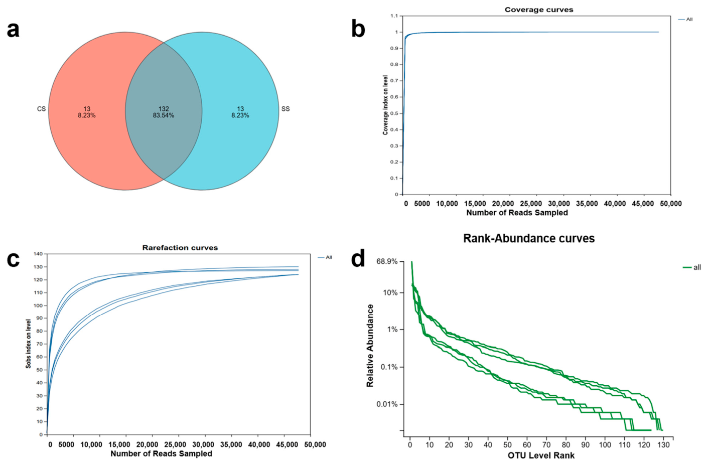
Clustering of quality-filtered sequences at a 97% sequence similarity threshold yielded 158 operational taxonomic units, among which 132 were shared between CS and SS groups (Figure 1a, Table S5). Analysis of shared and unique taxa using Venn diagrams showed a high degree of overlap in the bacterial community structure (Figure S1). At the phylum taxonomic level, no phylum was unique to the CS group, whereas the SS group exhibited two unique phyla; nine phyla were shared between the two groups. At the genus taxonomic level, the CS group harbored 7 unique genera, the SS group harbored 8 unique genera, and 97 genera were shared.
Evaluation of sequencing depth adequacy employed three complementary saturation metrics. Good’s coverage estimator values exceeded 99% across all samples, providing quantitative confirmation of comprehensive taxon sampling (Figure 1b). Rarefaction curves displaying observed species richness (Sobs) demonstrated asymptotic behavior in both control (CS) and treatment (SS) groups, indicating plateau-phase saturation (Figure 1c). Rank-abundance curve analysis revealed gradually declining distribution slopes (Figure 1d), characteristic of communities with high species equitability distributed evenly across constituent taxa. These analysis indicators of sequencing work were sufficient to accurately characterize the intestinal bacterial communities within both experimental groups, validating the biological reliability of subsequent comparative analyses.

3.2. Species Composition and Differences in Gut Microbiota

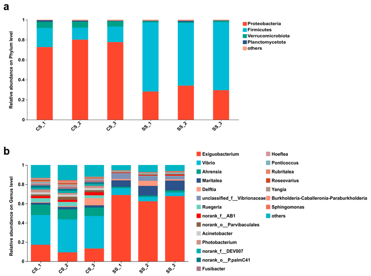
At the phylum level, the CS group was dominated by Proteobacteria (76.77%), Firmicutes (15.63%), Verrucomicrobiota (5.80%), Planctomycetota (1.08%), and Bacteroidota (0.65%), whereas the SS group exhibited a marked shift, with Firmicutes becoming the most abundant phylum (66.70%), followed by Proteobacteria (30.61%), others (1.44%), and Verrucomicrobiota (1.25%) (Figure 2a). At the genus level, the top five genera in the CS group were Maritalea (19.84%), others (15.30%), Ruegeria (10.27%), unclassified_f__Rhodobacteraceae (10.01%), and Rubritalea (9.05%); in the SS group, Exiguobacterium (66.34%) was overwhelmingly dominant, followed by others (10.51%), Maritalea (8.99%), Vibrio (5.15%), and unclassified_f__Vibrionaceae (4.63%) (Figure 2b).
Microbial community composition was characterized across three hierarchical taxonomic levels to provide a multi-resolution visualization of bacterial abundances. The proportional abundances of bacterial taxa at the class, order, and family levels are shown in Figure S2.

3.3. Alpha and Beta Diversity Analysis

Analysis of gut microbiota richness and diversity was performed using Ace, Chao, Shannon, and Simpson indices, revealing differential treatment effects on richness and community structure (Table 1 and Table S6). The Ace and Chao indices estimate community richness, while the Shannon and Simpson indices reflect both richness and evenness, thereby providing a comprehensive assessment of community diversity. Good’s coverage values indicate the sequencing depth and the probability of capturing all species present in a sample.
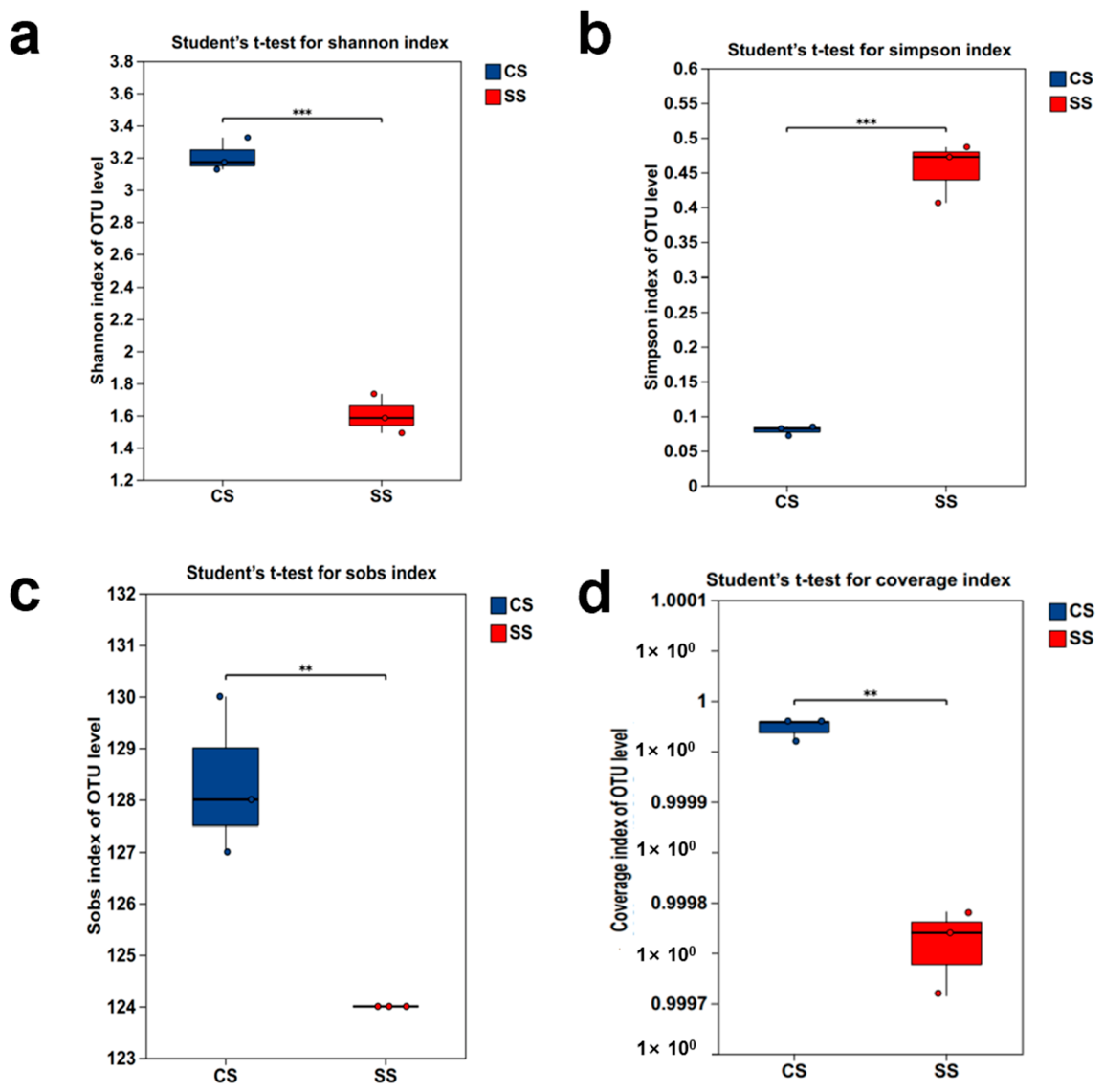
Observed species richness (Sobs) was significantly lower in SS (124.00 ± 0.00) versus CS (128.33 ± 1.53) (p = 0.008); however, coverage-based richness estimators (Ace, Chao1) showed no significant differences between groups (p > 0.3). Community diversity metrics revealed substantial treatment effects. Shannon diversity index was significantly higher in CS (3.207 ± 0.104) than in SS (1.604 ± 0.122) (p < 0.001), approximately twofold higher than in the treatment group. Simpson index exhibited significantly elevated values in SS (0.455 ± 0.043) compared to CS (0.080 ± 0.007) (p < 0.001), along with corresponding reductions in evenness indices (Shannon evenness: 0.661 vs. 0.333; Simpson evenness: 0.098 vs. 0.018; p < 0.001 for both). Good’s coverage values exceeded 99.9% in both groups (CS: 99.997%; SS: 99.976%), confirming adequate sequencing depth (Figure 3).
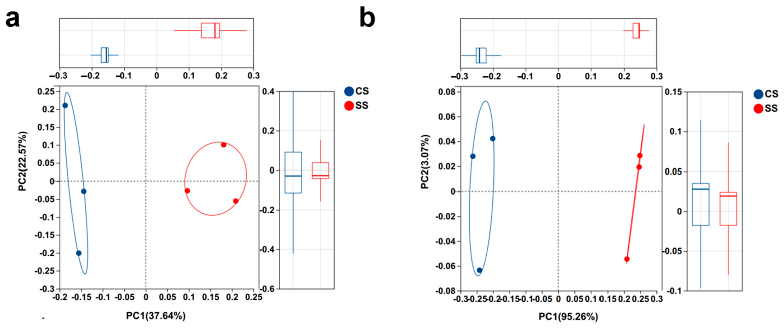
Beta diversity analysis revealed structured partitioning of bacterial community composition according to treatment group. PCoA analysis using Weighted and Unweighted UniFrac distances revealed pronounced compositional separation between treatment groups, with control (CS) and supplemented (SS) samples forming spatially discrete clusters (Figure 4). Strong between-group separation exceeded within-group variation, indicating that EcN supplementation substantially altered community composition while maintaining within-group consistency.

3.4. The Intestinal Microbiota Composition Between the CS and SS Groups

The 16S rRNA gene sequencing revealed significant treatment-dependent restructuring of the intestinal bacterial community. Compared to the control group (CS), the EcN-supplemented group (SS) exhibited pronounced shifts in both dominant phyla and genus-level composition.
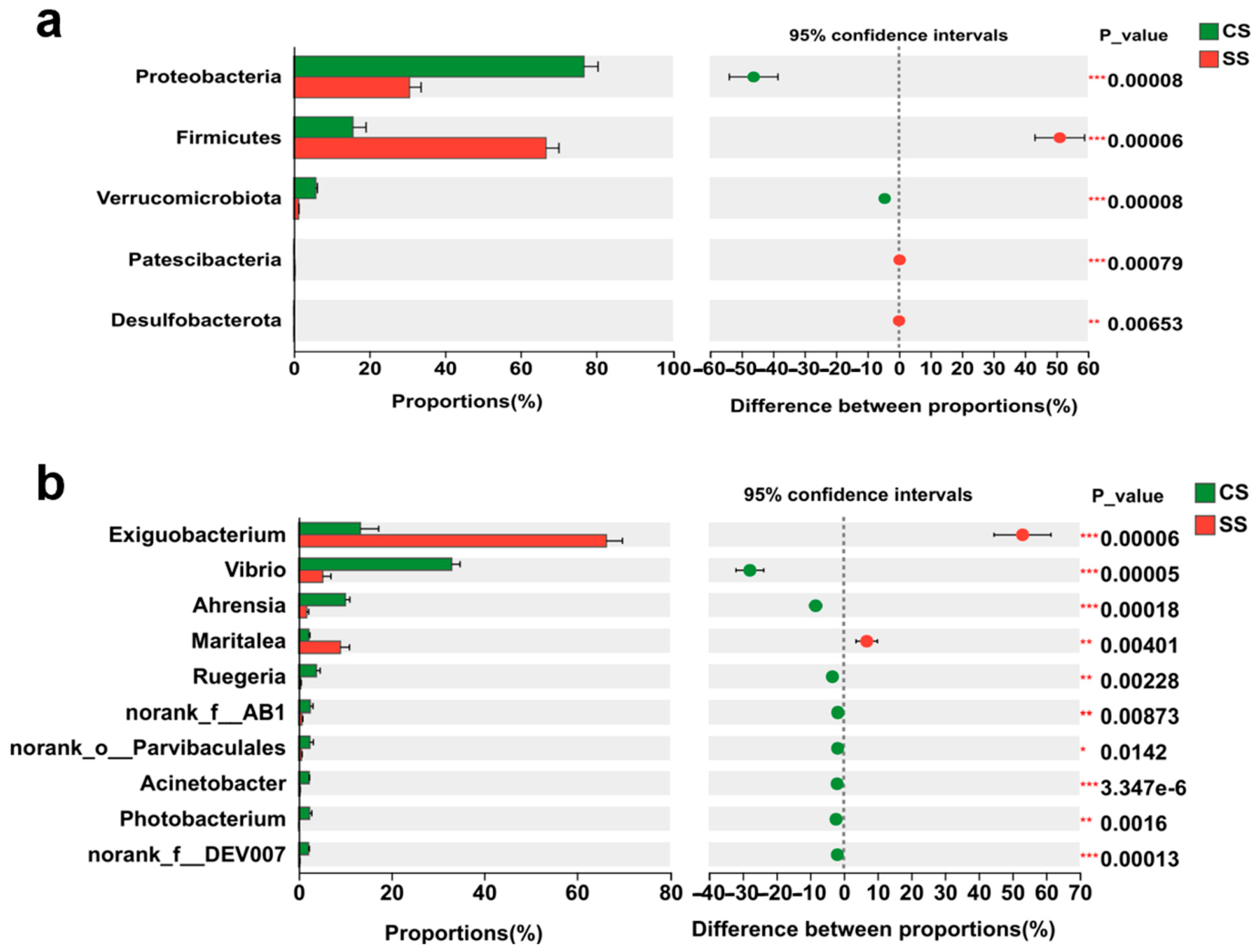
Phylum-level taxonomic analysis revealed coordinated directional shifts in response to EcN treatment (Figure 5a). Firmicutes increased significantly in the SS group (p = 0.00006 vs. CS), while Proteobacteria decreased proportionally (p = 0.00008). Verrucomicrobia followed a similar declining trend (p = 0.00008), and Patescibacteria and Desulfobacterota showed modest reductions (p = 0.000079 and p = 0.00653, respectively) (Table S7). These opposing trajectories reflect EcN-induced community reorganization favoring Firmicutes over Proteobacteria at the phylum level (Figure 5a).
Genus-level data identified Exiguobacterium as the primary expanding taxon in the SS group (p = 0.00006), with proportions substantially exceeding CS values. Eight genera declined in the SS group: Vibrio, Ahrensia, Maritalea, Ruegeria, norank_f_AB1, norank_o_Parvibaculales, Photobacterium, and norank_f_DEV007 (all p < 0.001, Table S8). This suggests EcN-mediated competitive displacement or antimicrobial antagonism restructures the genus-level community architecture (Figure 5b).

3.5. Immunomodulatory Effects of EcN Supplementation

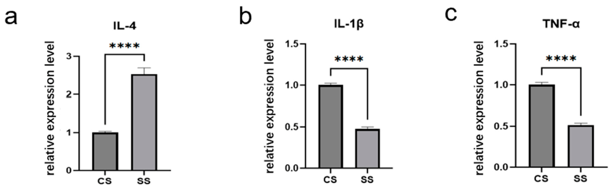
Quantification of intestinal cytokine transcripts revealed distinct anti-inflammatory immune polarization in response to EcN. SS treatment induced upregulation of the Th2-associated marker IL-4 (p < 0.001) concurrent with suppression of both TNF-α (p < 0.001) and IL-1β (p < 0.001), reflecting a shift from pro-inflammatory to anti-inflammatory immune status (Figure 6).

4. Discussion

Given the limited knowledge of gut microbiota modulation by probiotics in marine fish, particularly in grouper, the present study aimed to investigate the effects of EcN on intestinal microbial composition and host immunity.
In this study, the results demonstrated that EcN supplementation substantially remodeled the intestinal microbiota structure of grouper. At the phylum level, Firmicutes enrichment following EcN supplementation represents a notable shift in community composition. As primary metabolic contributors to fish intestinal environments, Firmicutes members encode pathways for complex carbohydrate degradation and short-chain fatty acid production [28,29,30]. In herbivorous fish, Firmicutes carry genes encoding enzymes related to cellulose, hemicellulose, and starch degradation, and can cooperate with other microbiota to decompose complex polysaccharides, compensating for the lack of endogenous digestive enzymes [31,32]. Therefore, the enrichment of Firmicutes suggests that EcN may have contributed to a gut microbial structure potentially favorable for intestinal homeostasis and nutrient metabolism in grouper. This shift could potentially enhance nutrient bioavailability; however, functional validation is required to confirm metabolic benefits.
At the genus level, the relative abundance of Exiguobacterium in the SS group reached 66.34%, which was extremely significantly higher than that in the CS group (p < 0.001), whereas the dominant genera in the CS group, such as Ruegeria and Ahrensia, were significantly reduced in the SS group. Exiguobacterium strains have previously been reported to possess probiotic potential in aquatic animals. For example, Exiguobacterium strains have been associated with growth promotion, immune enhancement, and improved resistance to bacterial infection in hybrid grouper [33,34]. Although the present study did not directly verify the functional role of Exiguobacterium, its marked enrichment following EcN supplementation suggests that it may represent an important component of the microbial restructuring induced by EcN. It is possible that EcN altered the intestinal environment in a way that favored the expansion of specific Firmicutes members, including Exiguobacterium, although the underlying mechanism remains unclear and requires further investigation.
Notably, alpha diversity analysis showed that the Shannon and Sobs indices of the SS group were significantly lower than those of the CS group (p < 0.001), indicating that probiotic intervention reduced gut microbiota diversity. This phenomenon is common when a single dominant genus proliferates extensively and does not necessarily imply functional deterioration of the microbiota [35]. The observed reduction in microbial diversity suggests that the community structure may have become less complex under the studied condition. Such a decrease may reflect selective pressure favoring specific taxa, reduced ecological stability, or a shift toward a more specialized microbial assemblage. However, changes in diversity alone do not necessarily indicate whether microbial alteration is beneficial or harmful and should therefore be interpreted alongside taxonomic composition, dominant genera, and the biological context of the host or environment. Given the altered cytokine profile (upregulated IL-4 and downregulated TNF-α/IL-1β) in the SS group, we suggested that the dominant colonization of Exiguobacterium exerted beneficial immunomodulatory effects on the host, and the temporary decline in diversity is an accompanying phenomenon during microbiota remodeling.
Gene expression analysis of cytokine markers demonstrated reciprocal modulation patterns under EcN treatment. The anti-inflammatory cytokine IL-4 underwent significant transcriptional upregulation in the SS group (p < 0.001), whereas pro-inflammatory mediators TNF-α and IL-1β showed pronounced mRNA downregulation in the SS group (both p < 0.001). This indicates that EcN may induce an anti-inflammatory phenotype by modulating intestinal immune homeostasis. IL-4 is typically associated with Th2-type immune responses, and its upregulation helps restrain excessive inflammatory reactions [36,37]. TNF-α and IL-1β are key mediators of inflammation, and their downregulation is closely associated with improved intestinal barrier function and reduced susceptibility to pathogens [38,39,40]. Considering the enrichment of Firmicutes (especially Exiguobacterium) in the gut microbiota, we speculate that the probiotic may promote the production of short-chain fatty acids and other metabolites through gut microbiota remodeling, thereby contributing to the regulation of intestinal immune responses via the gut-immune axis [41,42,43]. While Firmicutes enrichment can be associated with metabolic benefits or metabolic disorders depending on context, causality cannot be inferred from compositional data alone. Metabolomic profiling of SCFA, growth performance trials, and pathogen resistance assays would definitively establish whether Firmicutes enrichment represents a beneficial metabolic restructuring.
Thus, we proposed that EcN supplementation was associated with shifts in intestinal microbial composition and immune-related gene expression in grouper, suggesting a potential modulatory role in intestinal health that warrants further functional investigation. EcN creates a favorable niche for Firmicutes members such as Exiguobacterium through competitive exclusion, microcin production, or alterations in the intestinal microenvironment, allowing their massive colonization. As a dominant genus, Exiguobacterium produces digestive enzymes, thereby potentially improving host nutrient utilization efficiency. The high relative abundance of Exiguobacterium should be interpreted with caution. While this taxon may be genuinely enriched in the analyzed samples, the possibility of technical bias or contamination cannot be fully ruled out. Therefore, additional validation would be required to confirm the robustness of this observation.
Our findings suggested a potential association between EcN-induced microbial remodeling and altered intestinal immune responses. Although the present study does not establish a causal relationship, these results support the idea that changes in gut microbial composition may be linked to the modulation of host intestinal immunity [44,45,46,47]. In aquaculture, probiotics can influence fish growth performance and the structure of the intestinal microbiota [48], while the gut microbiota and its metabolites (e.g., short-chain fatty acids) assist probiotics in remodeling the intestinal microbiota by modulating molecular pathways involved in host immune responses [49]. In addition, EcN itself may directly interact with host immune cells and exert immunomodulatory functions. It is possible that microbial metabolites or microbe-host interactions contributed to these effects [50,51]; however, such mechanisms were not examined in this study and should be clarified in future work using metabolomics, bacterial isolation, and functional validation approaches. Another important limitation is the use of 97% OTU clustering rather than ASV-based methods, which limits strain-level resolution and may underestimate diversity.
This study provides, to our knowledge, one of the first systematic evaluations of EcN application in grouper under aquarium management. The results indicate that EcN has the potential to reshape the intestinal microbiota and modulate immune-related gene expression in this species. From an aquaculture perspective, these findings support the possibility that EcN may serve as a promising probiotic candidate for the regulation of intestinal health in marine fish. Nevertheless, additional studies are still needed to evaluate its long-term effects, optimal dosage, colonization characteristics, and practical efficacy under farming conditions.

5. Conclusions

This study demonstrates that dietary EcN supplementation profoundly restructures intestinal microbiota in hybrid grouper maintained under recirculating aquarium conditions. EcN treatment shifted community composition from Proteobacteria dominance to Firmicutes enrichment, with Exiguobacterium emerging as the predominant genus. Simultaneously, EcN amplified anti-inflammatory IL-4 transcription while attenuating pro-inflammatory TNF-α and IL-1β expression, reflecting coordinated modulation across the microbiota–immunity interface. These findings address a critical gap regarding probiotic efficacy in captive marine fish systems. Future research should validate functional outcomes through pathogen challenge trials, metabolomic analysis, and barrier integrity assays, and elucidate mechanisms. This work establishes EcN as a promising candidate for microbiome-targeted interventions in aquarium-based grouper culture, providing a foundation for evidence-based probiotic strategies in recirculating systems and intensive aquaculture.

Supplementary Materials

The following supporting information can be downloaded at https://www.mdpi.com/article/10.3390/jzbg7020023/s1: Figure S1: Phylum level (A) and genus level (B) species Venn diagram analysis of intestinal flora, Figure S2: Species composition diagram of intestinal micro-biota (A: class level, B: Order level, C: family level). Table S1. Ingredient composition and inclusion levels of the basal diet used in the feeding trial (% dry matter basis). Table S2. Proximate composition of the basal diet. Table S3. Experimental grouping information. Table S4. Primers used for qPCR analysis. Table S5. The sample sequence information. Table S6. The α-diversity of the gut microbiota. Table S7. The abundance analysis at the Phylum-level based on FDR. Table S8. The abundance analysis at the genus-level based on FDR.

Author Contributions

Formal analysis, Q.C., Y.M., Y.Y. and Y.S.; investigation, Q.C. and Y.M.; methodology, Q.C., Y.M. and Y.Y.; resources, Q.C., Y.M., Y.Y. and X.F.; software, Y.Y. and Y.S.; writing—original draft, Q.C., X.F. and H.W.; writing—review and editing, Q.C. and H.W. All authors have read and agreed to the published version of the manuscript.

Funding

This research was funded by Tianjin Enterprise Science and Technology Commissioner Program (24YDTPJC00210).

Institutional Review Board Statement

The animal study protocol was approved by the Institutional Animal Care and Use Committee of Tianjin Normal University (approval number: 2026051401).

Data Availability Statement

The datasets for this study can be found in the NCBI Sequence Read Archive (SRA) under accession number PRJNA1447178.

Acknowledgments

We also thank the editors and reviewers for their professional suggestions to improve the manuscript.

Conflicts of Interest

The authors declare no conflicts of interest.

References

  1. Hoseinifar, S.H.; Faheem, M.; Liaqat, I.; Doan, H.V.; Ghosh, K.; Ringø, E. Promising probiotic candidates for sustainable aquaculture: An updated review. Animals 2024, 14, 3644. [Google Scholar] [CrossRef] [PubMed]
  2. Turlybek, N.; Nurbekova, Z.; Mukhamejanova, A.; Baimurzina, B.; Kulatayeva, M.; Aubakirova, K.M.; Alikulov, Z. Sustainable aquaculture systems and their impact on fish nutritional quality. Fishes 2025, 10, 206. [Google Scholar] [CrossRef]
  3. Edwards, P. Aquaculture environment interactions: Past, present and likely future trends. Aquaculture 2015, 447, 2–14. [Google Scholar] [CrossRef]
  4. Rector, M.E.; Filgueira, R.; Bailey, M.; Walker, T.R.; Grant, J. Sustainability outcomes of aquaculture eco-certification: Challenges and opportunities. Rev. Aquac. 2022, 15, 840–852. [Google Scholar] [CrossRef]
  5. Ou, W.; Yu, G.; Zhang, Y.; Mai, K. Recent progress in the understanding of the gut microbiota of marine fishes. Mar. Life Sci. Technol. 2021, 3, 434–448. [Google Scholar] [CrossRef]
  6. El Saadony, M.T.; Alagawany, M.; Patra, A.K.; Kar, I.; Tiwari, R.; Dawood, M.A.O.; Dhama, K.; Abdel Latif, H.M.R. The functionality of probiotics in aquaculture: An overview. Fish Shellfish Immunol. 2021, 117, 36–52. [Google Scholar] [CrossRef]
  7. Ringø, E.; Ramasamy, H.; Soltani, M.; Ghosh, K. The effect of gut microbiota and probiotics on metabolism in fish and shrimp. Animals 2022, 12, 3016. [Google Scholar] [CrossRef]
  8. Gronbach, K.; Eberle, U.; Müller, M.; Olschläger, T.A.; Dobrindt, U.; Leithäuser, F.; Niess, J.H.; Döring, G.; Reimann, J.; Autenrieth, I.B.; et al. Safety of probiotic Escherichia coli strain Nissle 1917 depends on intestinal microbiota and adaptive immunity of the host. Infect. Immun. 2010, 78, 3036–3046. [Google Scholar] [CrossRef]
  9. Falzone, L.; Lavoro, A.; Candido, S.; Salmeri, M.; Zanghì, A.; Libra, M. Benefits and concerns of probiotics: An overview of the potential genotoxicity of the colibactin-producing Escherichia coli Nissle 1917 strain. Gut Microbes 2024, 16, 2397874. [Google Scholar] [CrossRef] [PubMed]
  10. Wang, C.; Chuprom, J.; Wang, Y.; Fu, L. Beneficial bacteria for aquaculture: Nutrition, bacteriostasis and immunoregulation. J. Appl. Microbiol. 2020, 128, 28–40. [Google Scholar] [CrossRef]
  11. Große, C.; Scherer, J.; Koch, D.; Otto, M.; Taudte, N.; Grass, G. A new ferrous iron-uptake transporter, EfeU (YcdN), from Escherichia coli. Mol. Microbiol. 2006, 62, 120–131. [Google Scholar] [CrossRef]
  12. Zhao, Z.J.; Xu, S.M.; Zhang, W.Y.; Wu, D.J.; Yang, G.S. Probiotic Escherichia coli Nissle 1917 for inflammatory bowel disease applications. Food Funct. 2022, 13, 5914–5924. [Google Scholar] [CrossRef] [PubMed]
  13. Nofouzi, K.; Sheikhzadeh, N.; Hamidian, G. Growth performance, mucosal immunity and disease resistance in goldfish (Carassius auratus) orally administered with Escherichia coli Strain Nissle 1917. Fish. Physiol. Biochem. 2024, 50, 1731–1743. [Google Scholar] [CrossRef]
  14. Mohammed, V.; Shanmugaraja, M. E. Coli Nissle 1917 Attenuates antibiotic mediated neurotoxicity possibly through Gut-Brain Axis in zebrafish. Appl. Biochem. Biotechnol. 2026, 198, 3768–3789. [Google Scholar] [CrossRef] [PubMed]
  15. Nag, D.; Farr, D.; Raychaudhuri, S.; Withey, J.H. An adult zebrafish model for adherent-invasive Escherichia coli indicates protection from AIEC infection by probiotic E. coli Nissle. iScience 2022, 25, 104572. [Google Scholar] [CrossRef]
  16. ZeinEddine, R.; Nasser, N.; Kassem, I.; El-Haroun, E.; Dawood, M.A.O. Effect of the human probiotic bacterium Escherichia coli Nissle (1917) on performance and immune response of Nile tilapia Oreochromis niloticus. J. Appl. Aquac. 2022, 34, 527–541. [Google Scholar] [CrossRef]
  17. Rimmer, M.A.; Glamuzina, B. A review of grouper (Family Serranidae: Subfamily Epinephelinae) aquaculture from a sustainability science perspective. Rev. Aquac. 2019, 11, 58–87. [Google Scholar] [CrossRef]
  18. Song, W.; Chen, C.; Du, X.; Zhang, R.; Zhao, J. Patterns and influencing factors of intestinal microbiota from different groupers. Appl. Microbiol. 2025, 7, 96. [Google Scholar] [CrossRef]
  19. Liu, Z.; Wang, P.; Wei, J.; Li, J.; Luo, X.; Huang, X.; Zhang, X.; Li, W.; Qin, Q. Effect of intestinal microbiota on adaptation to overcrowding stress in grouper (Epinephelus fuscoguttatus♀×E. lanceolatus♂). Fish. Shellfish Immunol. 2025, 159, 110165. [Google Scholar] [CrossRef] [PubMed]
  20. Wang, A.R.; Ran, C.; Ringø, E. Progress in fish gastrointestinal microbiota research. Rev. Aquac. 2018, 10, 626–640. [Google Scholar] [CrossRef]
  21. Fernández, G.M.; Alcantara, J.M.O.; Lagunes, C.D.; Asuar, L.E.; Sosa, M.B.R.; Ortiz, M.L.A. Composition and structure of gut microbiota of wild and captive Epinephelus morio via 16S rRNA analysis and functional prediction. Microorganisms 2025, 13, 1792. [Google Scholar] [CrossRef]
  22. Langille, M.G.I.; Zaneveld, J.; Caporaso, J.G.; McDonald, D.; Knights, D.; Reyes, J.A.; Clemente, J.C.; Burkepile, D.E.; Vega Thurber, R.L.; Knight, R.; et al. Predictive functional profiling of microbial communities using 16S rRNA marker gene sequences. Nat. Biotechnol. 2013, 31, 814–821. [Google Scholar] [CrossRef]
  23. Livak, J.K.; Schmittgen, T.D. Analysis of relative gene expression data using real-time quantitative PCR and the 2-ΔΔCT method. Methods 2001, 25, 402–408. [Google Scholar] [CrossRef] [PubMed]
  24. Magoč, T.; Salzberg, S.L. FLASH: Fast length adjustment of short reads to improve genome assemblies. Bioinformatics 2011, 27, 2957–2963. [Google Scholar] [CrossRef]
  25. Conway, J.R.; Lex, A.; Gehlenborg, N. UpSetR: An R package for the visualization of intersecting sets and their properties. Bioinformatics 2017, 33, 2938–2940. [Google Scholar] [CrossRef] [PubMed]
  26. Schloss, P.D.; Westcott, S.L.; Ryabin, T.; Hall, J.R.; Hartmann, M.; Hollister, E.B.; Lesniewski, R.A.; Weber, C.F. Introducing mothur: Open-source, platform-independent, community-supported software for describing and comparing microbial communities. Appl. Environ. Microbiol. 2009, 75, 7537–7541. [Google Scholar] [CrossRef] [PubMed]
  27. Wickham, H.; Averick, M.; Bryan, J.; Chang, W.; McGowan, L.D.; François, R.; Grolemund, G.; Hayes, A.; Henry, L.; Hester, J.; et al. R: A language and environment for statistical computing. J. Open Source Softw. 2020, 8, 1–20. [Google Scholar] [CrossRef]
  28. Cui, X.; Zhang, Q.; Zhang, Q.; Zhang, Y.; Chen, H.; Liu, G.; Zhu, L. Research progress of the gut microbiome in hybrid fish. Microorganisms 2022, 10, 891. [Google Scholar] [CrossRef]
  29. Kanika, H.N.; Liaqat, N.; Chen, H.; Ke, J.; Lu, G.; Wang, J.; Wang, C.H. Fish gut microbiome and its application in aquaculture and biological conservation. Front. Microbiol. 2025, 15, 1521048. [Google Scholar] [CrossRef]
  30. Bernard, W.P.; Elle, L.; James, C.P.; Brodie, C.; Kazlauskaite, R. The role of the gut microbiome in sustainable teleost aquaculture. Proc. Biol. Sci. 2020, 287, 20200184. [Google Scholar] [CrossRef]
  31. Wu, S.; Yi, R.; Peng, C.; Hao, Y.; Xiong, F.; Wang, G.; Li, W.; Zou, H.; Angert, E.R. Metatranscriptomic discovery of plant biomass-degrading capacity from grass carp intestinal microbiomes. FEMS Microbiol. Ecol. 2015, 91, fiv107. [Google Scholar] [CrossRef]
  32. Li, M.; Hui, L.; Hongwei, Y.; Qianwen, D.; Rui, X.; Jie, C.; Wenhao, Z.; Yalin, Y.; Zhen, Z.; Yuanyuan, Y.; et al. Deciphering the gut microbiome of grass carp through multi-omics approach. Microbiome 2024, 12, 2. [Google Scholar] [CrossRef]
  33. Xie, G.; Chen, X.; Feng, Y.; Yu, Z.; Lu, Q.; Li, M.; Ye, Z.; Lin, H.; Yu, W.; Shu, H. Effects of Dietary Multi-Strain Probiotics on Growth Performance, Antioxidant Status, Immune Response, and Intestinal Microbiota of Hybrid Groupers (Epinephelus fuscoguttatus ♀ × E. lanceolatus ♂). Microorganisms 2024, 12, 1358. [Google Scholar] [CrossRef] [PubMed]
  34. Zhang, M.; Feng, Y.; Zhong, Z.; Du, Q.; Yu, W.; Wu, J.; Huang, X.; Huang, Z.; Xie, G.; Shu, H. Host Gut-Derived Probiotic, Exiguobacterium acetylicum G1-33, Improves Growth, Immunity, and Resistance to Vibrio harveyi in Hybrid Grouper (Epinephelus fuscoguttatus ♀ × Epinephelus lanceolatus ♂). Microorganisms 2024, 12, 1688. [Google Scholar] [CrossRef]
  35. Kokou, F.; Goor, S.; Friedman, J.; Eyal, S.; Ovadia, O.; Harpaz, S.; Cnaani, A.; Mizrahi, I. Core gut microbial communities are maintained by beneficial interactions and strain variability in fish. Nat. Microbiol. 2019, 4, 2456–2465. [Google Scholar] [CrossRef] [PubMed]
  36. Wang, T.; Johansson, P.; Abós, B.; Holt, A.; Tafalla, C.; Jiang, Y.; Wang, A.; Xu, Q.; Qi, Z.; Huang, W.; et al. First in-depth analysis of the novel Th2-type cytokines in salmonid fish reveals distinct patterns of expression and modulation but overlapping bioactivities. Oncotarget 2016, 7, 10917–10946. [Google Scholar] [CrossRef]
  37. Bottiglione, F.; Dee, C.T.; Lea, R.; Zeef, L.A.H.; Badrock, A.P.; Wane, M.; Bugeon, L.; Dallman, M.J.; Allen, J.E.; Hurlstone, A.F.L. Zebrafish IL-4-like cytokines and IL-10 suppress inflammation but only IL-10 is essential for gill homeostasis. J. Immunol. 2020, 205, 994–1008. [Google Scholar] [CrossRef]
  38. Huo, H.; Liu, B.; Wang, Z.; Zhou, Q. Identification and expression analysis of Th2 immune-related gene IL4/13A in turbot (Scophthalmus maximus). Front. Immunol. 2024, 15, 1500840. [Google Scholar] [CrossRef]
  39. Al-Sadi, R.M.; Ma, T.Y. IL-1β causes an increase in intestinal epithelial tight junction permeability. J. Immunol. 2007, 178, 4641–4649. [Google Scholar] [CrossRef]
  40. Zou, J.; Secombes, C.J. The function of fish cytokines. Biology 2016, 5, 23. [Google Scholar] [CrossRef] [PubMed]
  41. Figueiredo, S.L.; Aparecida, D.P.A.; Yasmin, K.; Agnoletto, R.V.; Frandoloso, R.; Kreutz, L.C. Modulation of expression of proinflammatory genes and humoral immune response following immunization or infection with Aeromonas hydrophila in silver catfish (Rhamdia quelen). Fish. Shellfish Immunol. Rep. 2022, 3, 100053. [Google Scholar] [CrossRef]
  42. Kalaiselvan, P.; Malarvizhi, K.; Ranjan, A. Probing into the Impacts of Endogenous and Exogenous Short-Chain Fatty Acids (SCFAS) in Fish Health and Growth—A Review. Ann. Anim. Sci. 2025, 25, 119–137. [Google Scholar] [CrossRef]
  43. Fontinha, F.; Martins, N.; Campos, G.; Peres, H.; Teles, A.O. The effects of Short-Chain Fatty acids in gut immune and oxidative responses of European Sea Bass (Dicentrarchus labrax): An Ex Vivo Approach. Animals 2024, 14, 1360. [Google Scholar] [CrossRef]
  44. Turnbaugh, P.J.; Ley, R.E.; Mahowald, M.A.; Magrini, V.; Mardis, E.R.; Gordon, J.I. An obesity-associated gut microbiome with increased capacity for energy harvest. Nature 2006, 444, 1027–1031. [Google Scholar] [CrossRef] [PubMed]
  45. Ringø, E. Probiotics in shellfish aquaculture. Aquac. Fish. 2020, 5, 1–27. [Google Scholar] [CrossRef]
  46. Ley, R.E.; Turnbaugh, P.J.; Klein, S.; Gordon, J.I. Microbial ecology: Human gut microbes associated with obesity. Nature 2006, 444, 1022–1023. [Google Scholar] [CrossRef]
  47. De Schryver, P.; Vadstein, O. Ecological theory as a foundation to control pathogenic invasion in aquaculture. ISME J. 2014, 8, 2360–2368. [Google Scholar] [CrossRef] [PubMed]
  48. Venegas, P.D.; De la Fuente, M.K.; Landskron, G.; González, M.J.; Quera, R.; Dijkstra, G.; Harmsen, H.J.M.; Faber, K.N.; Hermoso, M.A. Short Chain Fatty Acids (SCFAs)-Mediated gut epithelial and immune regulation and its relevance for inflammatory bowel diseases. Front. Immunol. 2019, 10, 277. [Google Scholar] [CrossRef]
  49. Zhu, Y.; Li, W.; Zhang, M.; Zhong, Z.; Zhou, Z.; Han, J.; Zhang, C.; Yang, J.; Wu, Y.; Shu, H. Screening of host gut-derived probiotics and effects of feeding probiotics on growth, immunity, and antioxidant enzyme activity of hybrid grouper (Epinephelus fuscoguttatus♀ × E. lanceolatus♂). Fish. Shellfish Immunol. 2023, 136, 108700. [Google Scholar] [CrossRef]
  50. Mohammed, E.A.H.; Ahmed, A.E.M.; Kovács, B.; Pál, K. The significance of probiotics in aquaculture: A review of research trend and latest scientific findings. Antibiotics 2025, 14, 242. [Google Scholar] [CrossRef] [PubMed]
  51. Cani, P.D.; Gérard, C. Gut microbiota, metabolites and host immunity. Nat. Rev. Immunol. 2016, 16, 341–352. [Google Scholar] [CrossRef]
Figure 1. Microbial community characteristics of fecal samples from hybrid grouper. (a) Venn diagram showing the shared and unique OTUs between CS and SS groups. (b) Good’s coverage curves indicating sequencing depth; coverage >99.8% for all samples. (c) Rarefaction curves of observed species (Sobs) showing that sequencing depth was sufficient to capture microbial diversity. (d) Rank-abundance distribution illustrating species richness and evenness.
Jzbg 07 00023 g001
Figure 2. Relative abundance of gut microbiota from hybrid grouper at the phylum level (a) and genus level (b). (a) Stacked bar plots showing the relative abundance (%) of the dominant bacterial phyla across all samples in the CS and SS groups; (b) Stacked bar plots showing the relative abundance (%) of the dominant bacterial genera across all samples in the CS and SS groups. Only phyla/genera taxa with a relative abundance ≥1% are shown; all remaining taxa are aggregated as “Others”.
Jzbg 07 00023 g002
Figure 3. Analysis of α-diversity between the CS and SS groups. Alpha diversity based on (a) Shannon index, (b) Simpson index, (c) Sobs index, (d) Good’s coverage. Statistical comparisons were performed using Student’s t-test. Significance levels are indicated as: ** p < 0.01, *** p < 0.001.
Jzbg 07 00023 g003
Figure 4. Beta diversity analysis between CS and SS groups. Principal coordinate analysis (PCoA) based on (a) Unweighted UniFrac distances and (b) Weighted UniFrac distances. Each point represents an individual sample.
Jzbg 07 00023 g004
Figure 5. The gut microflora comparison of relative abundance at the phylum-level and genus-level between different groups. The gut microflora comparison of relative abundance at the phylum-level (a) and genus-level (b) between the CS and SS groups. Data are expressed as mean ± SEM. Statistical significance was determined by Student’s t-test. Significance indicators: * p < 0.05, ** p < 0.01, *** p < 0.001.
Jzbg 07 00023 g005
Figure 6. The mRNA expression levels of IL-4 (a), IL-1β (b) and TNF-α (c). (a) IL-4, (b) IL-1β, (c) TNF-α. Gene expression levels were normalized to β-actin as the internal reference gene and are shown relative to the CS group. Significance indicators: **** p < 0.0001.
Jzbg 07 00023 g006
Table 1. Analysis of α-diversity in the gut microbiota from hybrid grouper.
SampleSobsShannonSimpsonAceChao
SS-11241.491990.48667131.6682130.5
SS-21241.734820.40611128.5878127.75
SS-31241.585330.47225128.928133.1667
CS-11283.171090.08475128.3134128
CS-21273.324420.07185127.168127
CS-31303.126360.08225130.5434130.2
Disclaimer/Publisher’s Note: The statements, opinions and data contained in all publications are solely those of the individual author(s) and contributor(s) and not of MDPI and/or the editor(s). MDPI and/or the editor(s) disclaim responsibility for any injury to people or property resulting from any ideas, methods, instructions or products referred to in the content.