
Review

Molecular Parallels: Innate Immunity and Pathogen Strategies in Plants and Animals
by
Lesly Cristel Jiménez Cabrera
1,†,
Pablo Alejandro Gamas-Trujillo
1,†,
César De los Santos-Briones
1,
Luis Sáenz-Carbonell
1,
Ignacio Islas-Flores
2,
Karla Gisel Carreón-Anguiano
3,
Roberto Vázquez-Euan
4,
Nuvia Kantún-Moreno
5 and
Blondy Canto-Canché
1,*
1
Unidad de Biotecnología, Centro de Investigación Científica de Yucatán, A.C., Calle 43 No. 130 x 32 y 34, Colonia Chuburná de Hidalgo, Mérida C.P. 97205, Yucatán, Mexico
2
Unidad de Biología Integrativa, Centro de Investigación Científica de Yucatán, A.C., Calle 43 No. 130 x 32 y 34, Colonia Chuburná de Hidalgo, Mérida C.P. 97205, Yucatán, Mexico
3
Universidad Autónoma de Durango Unidad Laguna, Cto. Industrial Durango #5001 ex. Ejido Emiliano Zapata, Gómez Palacio C.P. 35140, Durango, Mexico
4
Secihti-Facultad de Medicina, Universidad Autónoma de Yucatán, Avenida Itzáes # 498 x 59 y 59A, Col. Centro, Mérida C.P. 97000, Yucatán, Mexico
5
Laboratorio de Virología, Centro de Investigaciones Regionales Dr. Hideyo Noguchi, Universidad Autónoma de Yucatán, Mérida C.P. 97000, Yucatán, Mexico
*
Author to whom correspondence should be addressed.
These authors contributed equally to this work.
Immuno 2026, 6(2), 27; https://doi.org/10.3390/immuno6020027
Submission received: 21 February 2026 / Revised: 28 March 2026 / Accepted: 9 April 2026 / Published: 15 April 2026

Abstract

Both plants and animals have developed a sophisticated two-tiered innate immune system. This involves an initial recognition of microbial patterns conserved on the cell surface (PAMP-triggered immunity) and a subsequent more specific intracellular recognition of pathogenic effectors or their activities (effector-triggered immunity). A common fundamental feature is the use of NLR-like intracellular receptors to detect insider threats. Both plant NLRs (receptors containing nucleotide-binding domains and leucine-rich repeats) and animal NLRs (NOD-like receptors) share a modular tripartite architecture, typically featuring a central nucleotide-binding domain (NBD/NOD) and C-terminal leucine-rich repeats (LRRs). The NBD/NOD is crucial for facilitating the exchange of ADP/ATP, acting as a molecular switch to promote oligomerization and activation of NLRs in both kingdoms. In this review, we summarize the similarities and differences between plant and animal molecular perception and immunity mechanisms. Additionally, we highlight the fact that some human pathogens can infect plants, and crucially, some plant pathogens are capable of causing disease in humans. This suggests conserved molecular strategies to invade and manipulate host cells belonging to different biological kingdoms, uncovering that plant and human pathology may benefit from future investigations in their respective fields.

1. Introduction

Plant and mammal hosts possess innate immunity, which serves as the first line of defense, acting within minutes to hours to detect and neutralize threats [1,2]. Most life forms on Earth, including archaea, bacteria, protists, fungi, oomycetes, invertebrates and plants, rely solely on this system. From a phylogenetic point of view, it represents the oldest immune system in living beings [3]. This system includes physical barriers, antimicrobial peptides, and in the case of mammals, active anti-inflammatory cascades. It also prepares the adaptive response to prevent infections.
Innate immunity Pattern Recognition Receptors (PRRs) are capable of recognizing molecules or parts of molecules with unique structures in microbes, allowing them to identify exogenous threats. The structures they detect are known as Pathogen-Associated Molecular Patterns (PAMPs) or Microorganism-Associated Molecular Patterns (MAMPs) [4]. Additionally, they can recognize Damage-Associated Molecular Patterns (DAMPs), which are generated in response to stress or cellular damage [5,6]. In plants and vertebrates, the recognition of PAMPs and/or DAMPs by membrane-associated PRRs mainly leads to the activation of extensive transcriptional programs. These programs boost the production of antimicrobial molecules and orchestrate a broader adaptive host response [2].
Over the years, research has shown that there are both exclusive and shared PRRs in the plant and animal kingdoms. Among the shared receptors are Toll-like receptors (TLRs) [7], C-type lectin-like receptors (CLRs) [8], and NOD-like receptors (NLRs) [9].
In mammals, NLRs are responsible for activating the inflammasome. These are multiprotein complexes formed after PRRs detect PAMPs or DAMPs [10]. Despite the distinct evolutionary origins of their NLRs, both kingdoms independently evolved fundamentally similar strategies for intracellular pathogen detection and defense. These strategies include the use of modular NLR-like receptors, the formation of large oligomeric signaling platforms, and the activation of regulated cell death [2]. In 2019, a functionally similar structure to the inflammasome, called the “resistosome”, was described in plants and is also activated by NLRs [1]. These structures are specialized in defending plants and animals, enabling them to resist and survive against attempts by pathogens to colonize.
Our objective is to highlight the similarities between plant and animal molecular perception and innate immunity mechanisms. We also aim to emphasize that some human pathogens, such as Pseudomonas aeruginosa and Salmonella enterica, can infect plants, while some plant pathogens such as Alternaria infectoria and nematode Xiphinema brevicollum can cause disease in humans. While rare, these cases raise concerns about the ability of plant-pathogens to adapt to human body temperatures and evade immune responses, acting as “crossover pathogens”. This suggests conserved molecular strategies for invading and manipulating host cells across different biological kingdoms. These findings provide great opportunities for future research perspectives and novel discoveries in pathology.

2. Innate Immunity

Plants and animals have developed a complex defense system, the full extent of which is not yet fully known or understood. Studies have identified new proteins that regulate defense and other essential processes for their survival. The system, known as “Innate Immunity”, is activated when it detects pathogens. Host cells act as a “surveillance system”, responsible for detecting signals from various sources, including microorganisms and the environment. This detection activates their defense mechanisms and initiates a signaling cascade [11].
Pattern recognition receptors (PRRs) are evolutionarily conserved proteins that serve as “gatekeepers” of innate immunity. They detect pathogen-associated molecular patterns (PAMPs) and damage-associated molecular patterns (DAMPs) from pathogens and damaged cells, triggering defense responses [12,13]. By recognizing and binding to these ligands, PRRs can prevent host colonization. They typically consist of ligand recognition domains, intermediate domains and effector domains. These receptors identify and bind to specific ligands, attracting adaptor molecules with similar structures through their effector domains. This activation of different signaling pathways leads to antimicrobial defenses and cellular repair mechanisms, all of which are crucial for host protection [12].
Although PAMPs have been largely associated with plants, in animals, the PRRs expressed in epithelial cells, tissue-resident macrophages and dendritic cells mediate defense responses to pathogens. This can result in inflammation and the production of cytokines [14,15]. In plants, recognition of PAMPs and DAMPs leads to the production of jasmonic acid or salicylic acid. This recognition activates the first level of innate immunity, known as pattern-triggered immunity (PTI), which plays a crucial role in reducing pathogen invasion and limiting infection. PTI provides rapid defense responses such as the production of reactive oxygen species (ROS) [13].
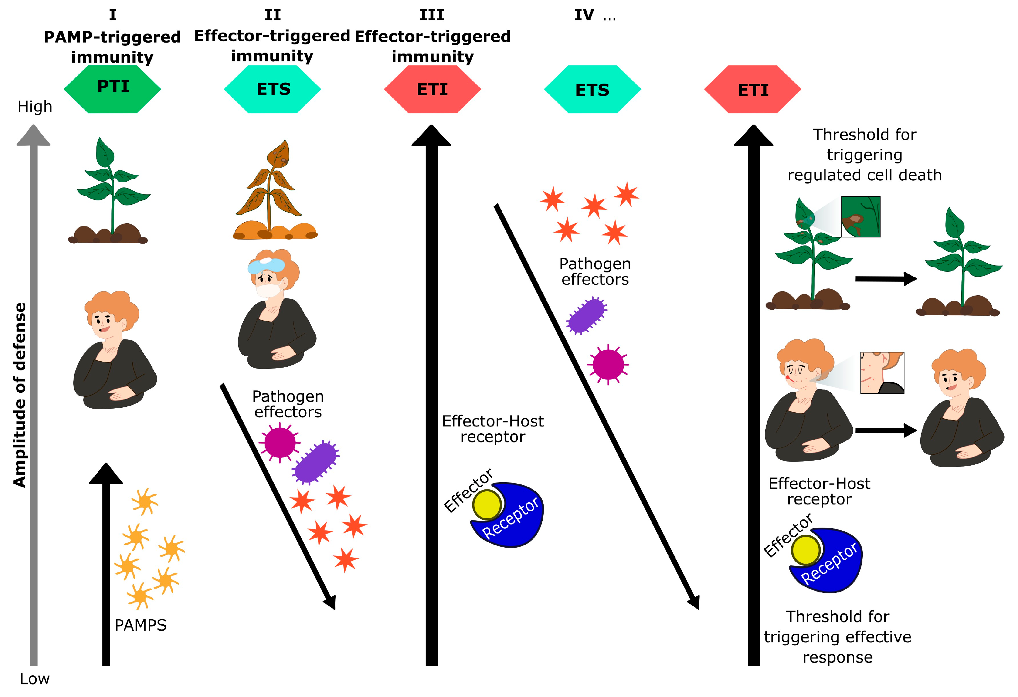
However, pathogens have evolved effector proteins over time to manipulate the host, bypass the initial plant defense level, and ultimately colonize and infect the host. This infection process is known as Effector Triggered Susceptibility (ETS) [16,17]. Similarly, more information about ETS has been reported in plants [18,19]. In mammals, bacterial pathogens inject effector proteins to suppress innate immunity and modulate inflammatory cell death [20].
In response, hosts have developed receptors capable of recognizing these effectors. This recognition is primarily through intracellular leucine-rich repeat domain (LRR) and nucleotide-binding (NB) receptors, known as NLRs. NLRs trigger a more intense immune response that aims to halt pathogen growth, known as Effector Triggered Immunity (ETI), which constitutes the second level of innate immunity [13,21]. ETI is a robust and intense form of defense, often accompanied by programmed cell death or hypersensitivity response (HR) [22]. In animal ETI, the host senses the consequences of the effector’s activity on cellular targets rather than the effectors themselves, often triggering pathways like NF-kB, pyroptosis and inflammasomes that release DAMPs.
A critical distinction of ETI from PTI is its specificity: unlike PTI, which can be triggered by harmless or commensal microbes, ETI is a specific response to true pathogens, as only pathogens encode and deploy these virulence-associated effectors. This specificity allows hosts to accurately distinguish pathogenic from non-pathogenic microbes and thus scale the magnitude and type of immune responses in proportion to the actual threat [23]. PTI and ETI work together to enhance the defense response. ETI can enhance PTI signaling, and reciprocally, PTI enhances ETI and is functionally essential for the response of ETI, suggesting a dynamic continuum rather than a strict division [24,25].
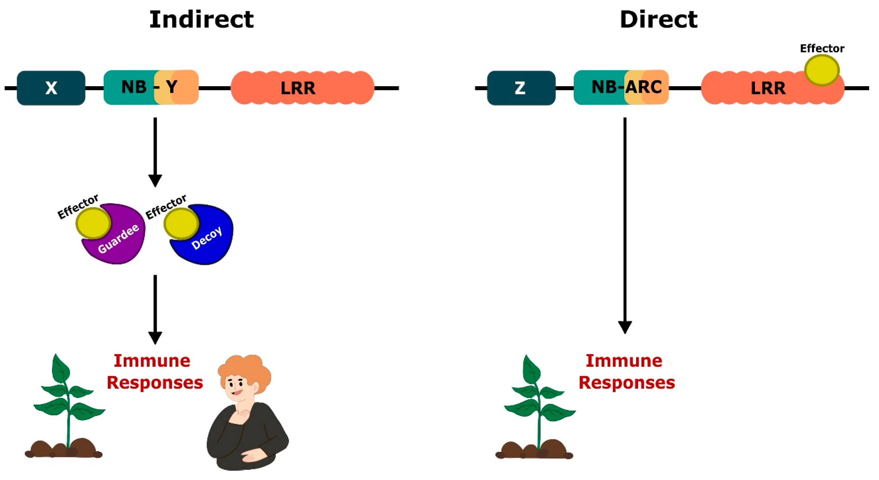
Plant and animal NLRs have evolved sophisticated and diverse mechanisms for recognizing pathogenic effectors, often through indirect means. The mechanism involves guardee proteins that monitor host proteins with anti-pathogen functions [26]. In plants, effectors may be directly detected by NLRs through their LRR domains, leading to receptor activation, or indirectly sensed through guard and decoy proteins [27] (Figure 1).
For example, Arabidopsis RPM1 or RPS2 NLRs are activated when the host protein RIN4 (a guardee) is modified by effector proteins of Pseudomonas syringae such as AvrRpm1 or AvrRpt2 [28]. Another example of a guardee NLR is RPS5, which monitors the protein kinase PBS1. Degradation of PBS1 by the effector protease AvrPphB triggers RPS5-mediated immunity [29].
From the guard model, the “decoy model” emerges, in which host proteins recognize effector-mediated modifications of a “decoy” protein in plants and animals. These decoys structurally resemble the actual targets of the host but have little intrinsic function beyond serving as bait for pathogenic effectors [27,30].
A particularly ingenious strategy is the “integrated decoy model” (NLR-IDs) also known as NLRs with non-canonical domains, where some NLRs have evolved to directly integrate a decoy domain (ID) into their own structure [30,31]. These integrated domains act as targets for pathogenic effectors, and their modification directly triggers NLR-mediated immunity [32]. A classic example is the RRS1/RPS4 NLR pair, where RRS1 contains a WRKY decoy domain that is attacked by effectors such as PopP2 and AvrRps4. Atypical NLRs with multiple IDs, such as WRKY, kinase, heavy metal-associated (AMF) and zinc-finger BED (zf-BED) domains, function as integrated decoys [33].
The existence of multiple, increasingly complex reconnaissance models, such as direct, guard, decoy and integrated decoy, indicates a dynamic co-evolutionary arms race. Pathogens are continuously evolving their effectors to suppress host immunity, which in turn forces hosts to devise new detection strategies. The “guard model” and “decoy model” are particularly ingenious adaptations of plants and animals to safeguard their survival since pathogens under selection to evade effector recognition cannot easily circumvent detection when NLRs monitor host protein integrity.
A significant difference between plant and animal ETI is the primary form of regulated cell death. Plants mainly use HR, a localized necrosis. HR effectively stops the spread and colonization of the pathogen by limiting its access to plant metabolites [2]. In contrast, animals have diversified their cell death pathways, including inflammatory pyroptosis/necroptosis and “silent” apoptosis [34]. Morphologically, HR appears as plant-regulated necrosis, exhibiting characteristics similar to mammalian necroptosis or pyroptosis, including early plasma membrane rupture, cytoplasm contraction, chromatin condensation, core disruption, tonoplast dismantling, and vacuolar collapse [2]. ETI also activates multiple signaling pathways in host cells, similar to PTI, but often with a much longer duration and amplitude.
The evolutionary divergence in the primary forms of regulated cell death in plants and animals reflects distinct physiological needs and evolutionary pressures. Plants prioritize immediate, localized containment through a necrosis-like response, while animals have developed more sophisticated cell death programs to balance effective pathogen removal with the need to maintain tissue integrity and control systemic inflammation [35].
This interaction reflects the coevolution between host and pathogen, leading to the diversification of both effectors and receptors in their colonization and defense efforts, respectively. Jones & Dangl [21] proposed this evolutionary dynamic in the Zigzag model, where pathogens progress from PTI to ETS, and then the host responds with ETI. While originally defined in plant pathology, this concept may be updated and also be applicable to animals (Figure 2).
Although both plants and animals share the basic concept of pathogen detection through PRR- and NLR-mediated mechanisms, receptor families have evolved differently to address kingdom-specific challenges. However, there are some PRRs that are present in both kingdoms, serving similar functions and sharing structural characteristics. In the next section, we will describe the PRRs and NLRs found in each realm and explore their similarities and differences.

3. PRRs in Plants

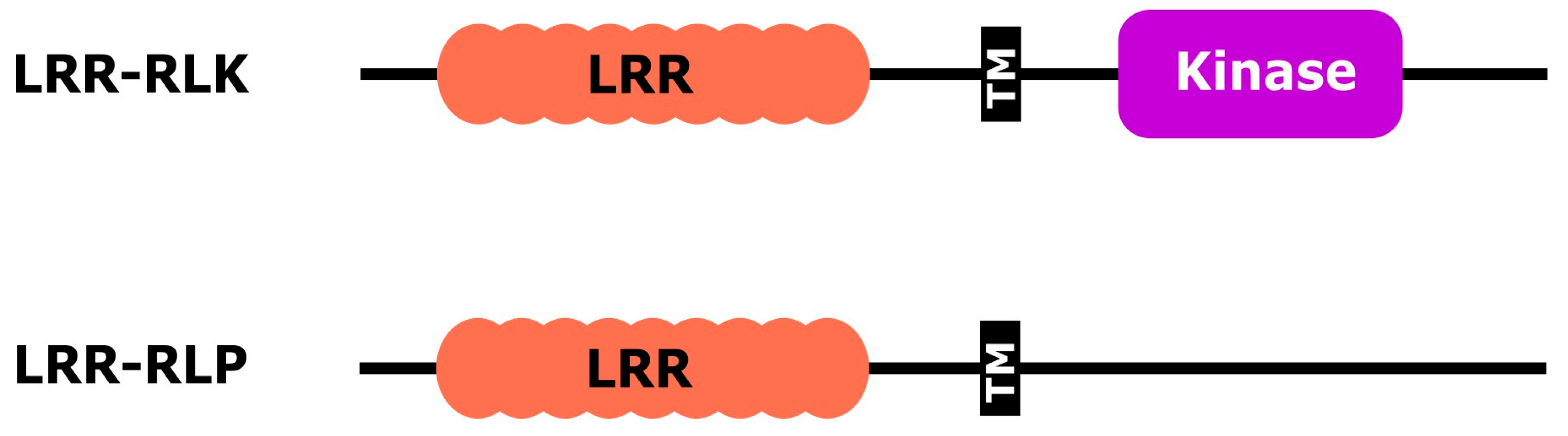
In plants, PRRs mainly consist of two structural types: receptor-like kinases (RLKs) and receptor-like proteins (RLPs). Both RLKs and RLPs are composed of an extracellular ligand-binding domain (ECD) and either a transmembrane α-helix or a glycosylphosphatidylinositol (GPI) anchorage domain. RLKs, like animal tyrosine kinase receptors [36], contain an intracellular kinase domain. In contrast, RLPs do not have the intracellular kinase domain but instead have a short cytoplasmic region [37,38]. PRRs can identify specific ligands using specialized ECDs, such as leucine-rich repeats (LRRs), lysine motifs (LysM), lectin, wall-associated kinases (WAK), S-locus domain, malectin-like, proline-rich, and cysteine-rich repeat [39,40,41]. ECDs exhibit high structural variability, allowing them to recognize a wide range of ligands, including peptides, polysaccharides, lipopolysaccharides, and steroids. RLKs and RLPs can be classified into sub-families according to the type of ECDs they contain, with the most common ECD found in RLKs and RLPs being the leucine-rich repeat (LRR): LRR-RLKs and LRR-RLPs [38] (Figure 3).
Receptor-like kinases (RLKs) are present in a variety of organisms, including Plasmodium, Toxoplasma, plants, and animals. However, they have not been found in fungi [42], nor in Dictyostelium, the model organism of Amoebozoa, nor in Leishmania major, Giardia intestinalis and the choanoflagellate Monosiga brevicollis [43]. Table 1 shows some RLKs involved in plant immunity.
A well-known example of LRR-RLK is FLS2 (Flagellin-Sensitive 2), a receptor from Arabidopsis thaliana. This receptor recognizes a highly conserved epitope of 22 amino acids (known as flg22), corresponding to the N-terminal end of bacterial flagellin. However, this recognition is not carried out individually; FLS2 acts in collaboration with the co-receptor BAK1 (BRI1-Associated Receptor Kinase 1), leading to the formation of a signaling complex that activates plant defense responses, including the production of reactive oxygen species (ROS), the activation of mitogen-dependent kinases (MAPK), and defense-related gene expression [44]. The FLS2 receptor plays an essential role in resistance against pathogenic bacteria, particularly after foliar inoculation, supporting its central role as a PRR in plant immunity. Interestingly, in humans, the Toll-like receptor TLR5 is also capable of identifying bacterial flagellin, although the identification is done for a different reason than that identified by FLS2 [45]. This functional convergence in organisms from different kingdoms highlights the evolutionary importance of recognizing conserved microbial patterns.
Another example of LRR-RLK is the XA21 kinase in rice (Oryza sativa), which confers resistance to most strains of the Gram-negative bacterium Xanthomonas oryzae (the causative agent of a major agronomic disease of rice) by recognizing RaxX, a protein highly conserved in this species. To trigger an effective immune response, XA21 must bind with its specific ligand, RaxX21-sY, a 21-amino acid sulfated peptide derived from RaxX [46].
Although many RLKs and RLPs act as PRRs, some of these proteins have also been shown to be involved in fundamental physiological processes such as plant growth and development, reproduction, symbiosis, and tolerance to abiotic stress [47]. Readers may refer to the review by Liu et al. [41] to see current lists of RLKs involved in different plant processes.
Table 1. Plant RLKs involved in innate immunity.
RLKsFamilyPlantLigandLigand OriginReference
DORN1Lectin-RLKArabidopsiseATPPlants[48]
EFRLRR-RLKArabidopsiself18Bacteria[49]
FLS2LRR-RLKArabidopsisflg22Bacteria[44]
FLS3LRR-RLKTomatoflgll-28Bacteria[49]
LORELectin-RLKArabidopsisLPSBacteria[50]
OsCERK1LysM-RLKRiceChitinBacteria fungi insects[51]
PEPR1/2LysM-RLKArabidopsisPepsPlants[52]
WAK1EGF-Like-RLKArabidopsisOGsPlants[53]
XA21LRR-RLKRiceRaxX21-sYBacteria[52]
HSL3LRR-RLKArabidopsisSCREW proteinsPlants[54]
RLPs lack intracellular kinase domains, thus requiring LRR-RLK co-receptors to activate downstream signaling. LRR-RLPs constitute the largest family of RLPs. The LRR ECD of NLPs has additional domains known as N-loop outs (NLs) and island domains (IDs) interspersed between the LRR motifs. NLs are present in most immunity-related LRR-RLPs, while IDs are present in all immunity-related LRR-RLPs. Usually, NLs are located closer to the N-termini, while IDs are located closer to the C-termini of the LRR motif. While LRR-RLKs regulate plant processes through their cytosolic kinase domains, LRR-RLPs initiate immune responses through their juxtamembrane and transmembrane regions [55,56].
Plant species possess different numbers of RLP and RLK genes, and this expansion is believed to have occurred throughout evolution as an adaptive response to evolving pathogens during land plant conquest [57]. For example, 226 LRR-RLKs and 59 LRR-RLPs have been identified in A. thaliana, whereas 81 LRR-RLKs and 8 LRR-RLPs have been found in Selaginella moellendorfii [38].
Examples of NLPs include RLP23, Cf2, Cf4, Cf5, Cf9, EIX2, Ve1, Hrc9-4E, I, and I-3 [56]. In Arabidopsis, the RLP23 receptor recognizes the nlp20 peptide, which shares high conservation with the necrosis-inducing ethylene peptide (NLP) found in bacteria, fungi, and oomycetes [55]. For proper function, RLP23 relies on multiple co-receptors, including BAK1, SOBIR1 and various RLCKs such as PBL19/20/30/31/32 [58]. This signaling pathway leads to various immune responses including the production of ROS, phosphorylation of BIK1 and MAPK proteins, callose deposition, and the synthesis of salicylic acid (SA) and ethylene (ET) [59]. These responses are similar to those induced by FLS2, indicating conserved signaling modules in different plant PRRs.
In tomato (Solanum lycopersicum), the LRR-RLPs Cf-2, Cf-4, and Cf-9 confer resistance to the fungus Cladosporium fulvum by recognizing the effectors Avr2, Avr4 and Avr9, respectively, triggering HR [60]. Both RLKs and RLPs trigger highly overlapping immune responses, attributed to the activation of signaling pathways that share downstream key components such as PAD4, EDS1, and ADR1 [61,62]. Table 2 shows a list of plant RLPs.

4. PRRs in Animals

Similar to plants, animals have highly specialized and complex innate mechanisms for detecting and responding to pathogen attacks. In both invertebrates and vertebrates, innate immunity acts as the first line of defense against infections. In vertebrates, it also plays a crucial role in activating adaptive immunity by producing costimulatory molecules, cytokines, and chemokines that regulate T cell activation and differentiation [77].
The majority of PRRs in vertebrates can be classified into five main groups based on the homology of their protein domains: Toll-like receptors (TLRs), NOD-like receptors (NLRs), retinoic acid-inducible gene I (RIG-I) receptors (RLRs), C-type lectin receptors (CLRs), and AIM2-like receptors (ALRs) also known as “absent in melanoma 2” [12].
Analogous to plant PRRs, these animal receptors are structured with ligand recognition domains, intermediate regions, and effector domains. When they detect the corresponding ligand, PRRs recruit adaptor molecules with structurally compatible domains, enabling the activation of various intracellular signaling pathways [2].
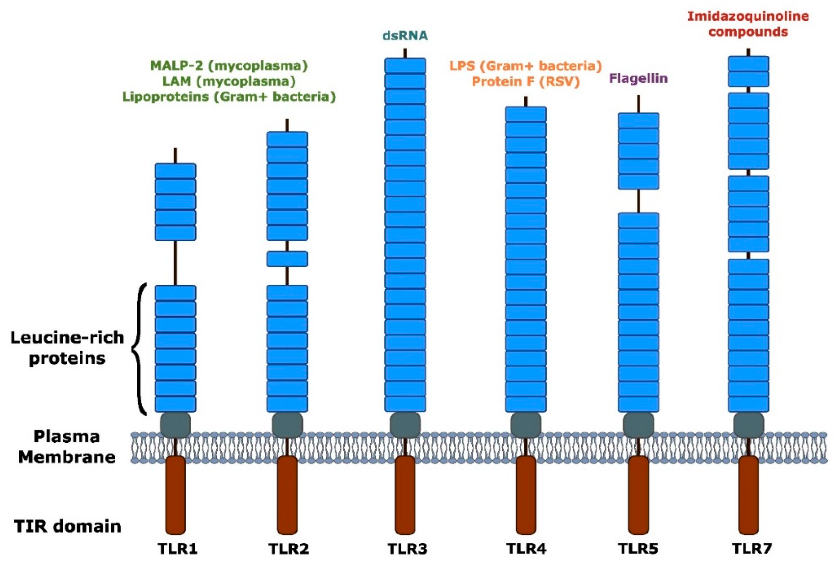
Toll receptors (TLRs) were among the first PRRs identified in the innate immune system and play a crucial role in activating inflammatory responses [78]. These receptors, anchored to the plasma membrane, serve two main functions: the specific recognition of microbial ligands and the transduction of intracellular signals; TLRs may also connect to adaptive immune responses. TLRs have an amino-terminal domain with leucine-rich repeats (LRRs) and an intracellular carboxy-terminal region containing a conserved domain known as the Toll/interleukin-1 homology domain (TIR). The extracellular domain has variable LRRs (Figure 4), which are mainly associated with ligand recognition but may also participate in the dimerization process necessary for the functional activation of the receptor [79]. Table 3 shows a list of TLRs.
The activation of TLR triggers a signaling cascade that leads to the activation of transcription factors, enabling the expression of genes related to inflammation. Consequently, various pro-inflammatory mediators are produced and secreted to combat infection [80]. Similarly, in plants, receptors like FLS2 activate signaling pathways that lead to the expression of defense genes, highlighting a conserved recognition strategy and the critical role of activating signals for survival.
Table 3. Animal TLRs in innate immunity.
TLRFamilyClassificationDAMPReference
TLR1TLR1Non-viral Triacyl-lipopeptide and peptoglycans[81]
TLR2TLR1Non-viral Heat shock proteins[82]
TLR3TLR3ViralmRNA, siRNA, and tRNA[83]
TLR4TLR4Non-viral Heat shock proteins, amyloid-β peptides, oxidized low-density lipoproteins[84]
TLR5TLR5Non-viral High-mobility group box 1 (HMGB1)[85]
TLR6TLR1Non-viral Amyloid-β peptides, oxidized low-density lipoproteins, versican[86]
TLR7TLR7ViralImmune complexes, self RNA[87]
TLR8TLR8ViralImmune complexes, self RNA[88]
TLR9TLR9ViralDNA[89]
TLR10TLR1, TLR6, TLR10 Lipopolysaccharide, flagellin, ARNdc[90]
In antiviral innate immunity, viral nucleic acids are recognized by Toll-like receptors TLR7 and TLR9. Most other cell types detect these viral nucleic acids through the RIG-I-like receptors (RLR), which trigger antiviral responses [91].
The RIG-I receptor was initially identified in retinoic acid-induced acute promyelocytic leukemia cells. Subsequently, in 2004, it was demonstrated that this receptor could induce the expression of a reporter gene under the control of the IFN-β promoter, confirming its role in detecting viral infections and activating innate immune responses [92].
RIG-I receptors consist of three functional regions. The N-terminal end contains two serial caspase activation and recruitment (CARD) domains, arranged in tandem, responsible for initiating signal transduction [93,94]. The middle region contains a DexD/H-type helicase domain, characteristic of the RLR families, with ATPase and helicase activity [95]. Finally, the C-terminal end consists of two domains: the repressor domain (RD) responsible for maintaining the receptor in a basal state, and the C-terminal domain (CTD) capable of recognizing viral RNA and regulating the receptor’s conformation [96,97].
While Toll-like receptors recognize nucleic acid molecules derived from viruses only on the endosomal membrane, restricting their activity to specific intracellular compartments, RLRs can be expressed in cells infected by viruses and detect viral particles in the cytosol leading to a rapid and localized response to foreign genetic material [98]. This distinction in receptor subcellular localization is also observed in plants, where some PRRs are located in the plasma membrane to detect external signals, while others act intracellularly (NLR), indicating a shared functional organization between kingdoms.
C-type lectin receptors (CLRs) are another class of PRRs that recognize carbohydrates on the surface of pathogenic microorganisms depending on calcium ions (Ca2+). This recognition is facilitated by a specialized domain called the carbohydrate recognition domain (CRD), which helps differentiate between self and foreign structures [2].
CLRs can be categorized based on cell localization, into transmembrane receptors and soluble (secretory) receptors [99]. Transmembrane receptors are further classified as type I and type II, depending on the orientation of the N-terminal ends. In type I, the N-terminal end faces the extracellular space and contains multiple CRDs, while in type II, the N-terminal end faces the cytosol and contains a single CRD [100,101].
The last group of receptors discussed in this section are AIM2-like receptors (ALRs), which are specialized in detecting intracellular DNA [102]. ALRs consist of a HIN-200-like DNA-binding domain at the C-terminal end, which binds specifically to double-stranded DNA, and a PYD at the N-terminal end [102,103]. The PYD interacts with the PYD of the ASC (apoptosis-associated speck-like protein containing a CARD domain), promoting the assembly of the inflammasome. This complex formation facilitates the maturation and release of the pro-inflammatory cytokines IL-1β and IL-18 [14]. In addition to their role in innate immunity, ALRs also regulate apoptosis, a process linked to the development and progression of various types of cancer [104].
In addition to membrane-associated PRRs, intracellular receptors such as animal nucleotide-binding and oligomerization domain (NOD)-like receptors or plant nucleotide-binding domain (NBD)-containing leucine rich repeat immune receptors likely detect effectors injected into the host cell by the pathogen to hijack the immune signaling cascade. These intracellular receptors are abbreviated as NLRs and will be described in the next section.

5. Plant NLRs and Resistosome Assembly

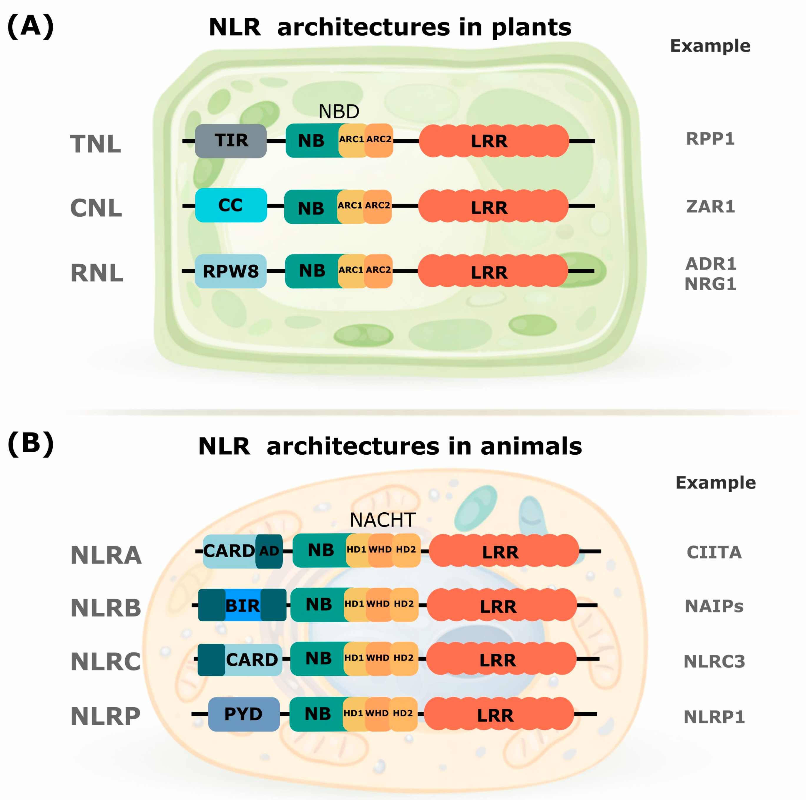
In plants, NLRs are encoded by R genes, and there are three types of NLRs (Figure 5A) distinguished by their N-terminal domain: CC domain NLRs (coiled coil), TIR (Toll receptor/interleukin-1), and RPW8-like coiled domain NLRs (powdery mildew resistance 8). CC-NLRs (CNLs) and TIR-NLRs (TNLs) generally act as pathogen-secreted effector sensors, while RPW8-NLRs (RNLs) signal downstream of many NLR sensors and are called auxiliary NLRs [105].
The central domain, NB-ARC, is the most conserved NLR domain, belonging to the superfamily of signal transduction ATPases (STAND). It works as an ADP-ATP molecular switch, regulating the ON/OFF state of NLRs and is characteristic of plants [29]. The C-terminal domain (LRR), composed of repeated units, functions primarily as a ligand-binding platform and often possesses autoinhibitory properties, preventing premature activation. It is generally responsible for the direct or indirect recognition of effectors [106]. Several NLRs are found in all terrestrial plants, including mosses, liverworts, conifers, and flowering plants, in addition to charophytic algae [107,108,109].
A classic example of a plant NLR is the pseudokinase HopZ1-ETI Deficient 1 (ZED1), which is essential for activating the CNL-like receptor HopZ-activated Resistance 1 (ZAR1). ZAR1 enables the indirect recognition of several effectors, such as AvrAC from Xanthomonas campestris pv. campestris and HopZ1a from Pseudomonas syringae pv. syringae A2 and 7B40 [110].
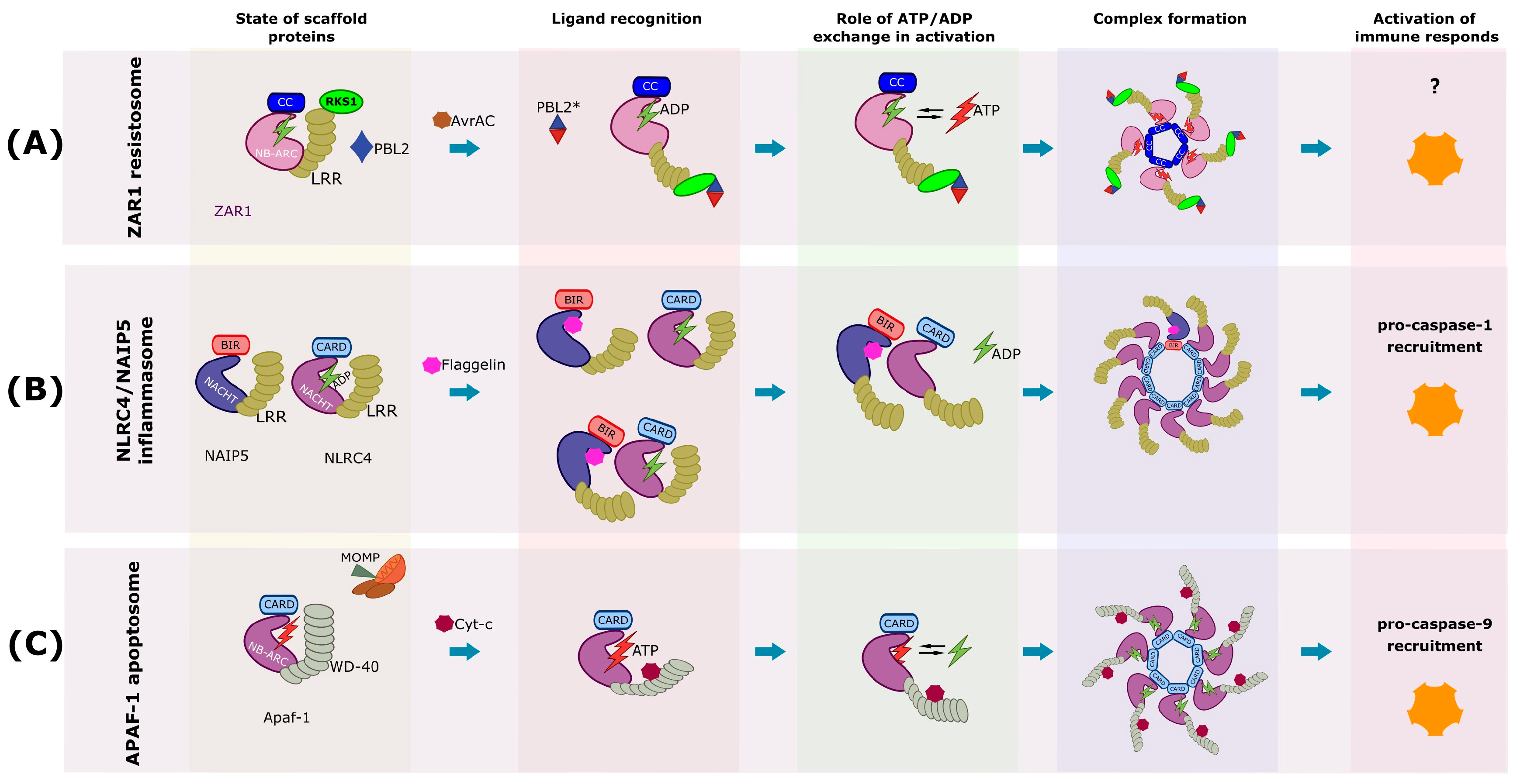
Both AvrAC and HopZ1a modify receptor-like cytoplasmic kinases (RLCKs) to enhance virulence. AvrAC uridylates RLCK PBL2, producing PBL2^UMP, which then interacts with RLCK RKS1, already associated in a preformed complex with ZAR1 [111]. This interaction induces the assembly of the ternary complex ZAR1-RKS1-PBL2^UMP [112]. On the other hand, HopZ1a acetylates the pseudokinase ZED1, also part of a preassembled complex with ZAR1, leading to the formation of a higher order complex [113,114] (Figure 6A). Additionally, other RLCKs are targeted by HopZ1a and recognized by the ZAR1-ZED1 complex, where ZED1 acts as a functional adapter, analogous to RKS1 [115]. Uridylation of PBL2 does not enhance the virulence induced by AvrAC, and ZED1 lacks catalytic activity. Therefore, both AvrAC and HopZ1a are considered decoys rather than gatekeepers [111,114].
The pseudokinase RKS1 acts as a molecular intermediary, connecting ZAR1 with its modified substrate [116]. In the absence of an effector, the ZAR1-RKS1 complex remains monomeric with ADP bound to the WHD and NBD, a configuration that prevents spontaneous activation. The LRR domain physically hinders dorsoventral stacking of NBDs, preventing premature oligomerization. This molecular safety mechanism ensures that ZAR1 only activates in the presence of the correct effector [116,117]. Incorporating of PBL2-UMP into the ZAR1-RKS1 complex causes a significant conformational change: the NBD rotates approximately 60°, facilitating ADP release [103]; however, the complex remains monomeric. Binding ATP to the nucleotide-free ZAR1 serves as the final activation signal for the primed monomeric ZAR1-RKS1-PBL2-UMP complex [112]. Cryo-EM analysis revealed a wheel-like structure composed of five protomers (Figure 6A). Each ZAR1 domain contributes to pentamer formation by interacting with adjacent protomers, while the PBL2-UMP-RKS1 complex interacts with the LRR domain to form the wheel spokes, although it does not participate in oligomer formation. Oligomerization of plant NLRs has been observed in both CC-NLRs and TIR-NLRs [116].
The pentameric CC domain oligomerization creates a funnel-shaped structure with variable charge distribution along its inner surface, resembling pore-forming toxins. A critical structural change during activation involves the N-terminal α-helix rotating 130° and projecting outward into the solvent. This α-helix is essential for ZAR1’s plasma membrane association and biological activity [117]. Bi et al. [118] demonstrated that reconstituted ZAR1 resistosomes incorporate into planar lipid bilayers and function as cation-selective channels with a preference for Ca2+. In plant cells expressing activated ZAR1, calcium influx depends on the glutamate residue at position 11 (Glu11), indicating that the channel’s selectivity filter involves specific charged residues. Calcium elevation is among the earliest measurable responses during pathogen recognition [119,120] and dysregulated calcium accumulation can trigger hypersensitive cell death even without pathogens [121]. The temporal gap between resistosome formation and cell death, coupled with Glu11’s requirement for both calcium entry and cell death, suggests that ZAR1 initiates a calcium-dependent signaling cascade rather than causing immediate membrane collapse.
Crucially, these resistosomes exhibit direct enzymatic activities: CNL/RNL resistosomes function as Ca2+-permeable channels [33], and the TIR domains of TNL resistosomes act as NADases [122]. This means that the activated complex is not simply a signal transducer; it is an effector molecule itself, directly disrupting cellular homeostasis. This dual activity provides plants with a fast and powerful defense. Direct enzyme activity, such as membrane permeabilization by ZAR1 or metabolic disruption by TIR NADase activity, offers immediate, high-impact disruption of cellular processes essential for pathogen survival [122]. This, along with signal amplification through oligomerization, ensures a coordinated and highly effective defense, highlighting the sophistication of plants’ innate immunity.
Downstream of NLR activation, certain “helper” NLRs are essential for cell death and signal transduction [123]. These include members of the ACTIVATED DISEASE RESISTANCE PROTEIN 1 (ADR1) family and the N REQUIREMENT GENE 1 (NRG1) family, both RNLs [122]. While most TNLs utilize the NRG1 family for signal transduction, some prefer the ADR1 family, and in some cases, both families show complete redundancy. RNLs and proteins of the EDS1 (enhanced disease susceptibility 1) family, such as EDS1, PAD4, and SAG101, form crucial complexes that regulate TNL signaling [124]. Specifically, the second messengers generated by the TIR domains of activated TNLs (e.g., pRib-AMP/ADP and ADPr-ATP/ADPr-ADPR) bind to the EDS1-PAD4 or EDS1-SAG101 complexes, promoting their interaction with ADR1 or NRG1 [125].
The ZAR1-RKS1-PBL2-UMP complex provides molecular evidence for the guard/decoy model. Instead of directly binding pathogen molecules, ZAR1 detects post-translational modifications that effectors impose on host proteins [116]. Structural analysis reveals that RKS1’s N- and C-terminal regions contact two uridylated residues on PBL2, creating a molecular signature for modified-self recognition. RKS1 simultaneously interacts with ZAR1’s LRR domain, bridging the modified PBL2 to the receptor [117].

6. Mammals NLRs and Inflammasome Assembly

Traditionally, innate immunity in mammals was thought to be based on the recognition of PAMPs by receptors like TLRs and other PRRs. However, it has been discovered that there is a parallel response known as effector-triggered immunity (ETI), which is activated by detecting perturbations in cellular homeostasis caused by effector proteins secreted by pathogens. These alterations are also referred as processes that alter homeostasis (HAMPs) [126]. Like plants, animals have sophisticated innate immune systems that rely on intracellular immune receptors to detect pathogens. Animal NLRs specialize in detecting cytosolic Pathogen-Associated Molecular Patterns, such as toxins secreted by bacteria or viral proteins, as well as indicators of intracellular stress [127]. Although functionally analogous to plant NLRs, animal NLRs have a different evolutionary origin; with a NACHT subtype (NAIP, CIITE, HET-E, and TP1) of nucleotide-binding domain (NBD), in contrast to the NB-ARC subtype found in plants. The N-terminal domains of animal NLRs are part of the superfamily of death domains, including the pyrin effector domain (PYD), the caspase recruitment and activation domains (CARD), the acid transactivation domain (NLRA), and the baculovirus inhibitory repeat (BIR) domain [128].
Plant and animal immune systems, despite being evolutionarily distant, share surprisingly similar molecular architectures (Figure 5A,B). In mammals, NLR proteins have undergone significant functional specialization, resulting in the formation of distinct multiprotein complexes: inflammasomes and apoptosomes (Figure 6B,C) [129]. While both are crucial for host defense and cell fate, they have distinct primary functions and downstream outcomes.
This suggests functional compensation linked to the physiology of the organism and its ecological niche. Plants, being sessile, could benefit more from rapid, localized, and somewhat aggressive cell death to arrest the infection directly at the site, as they cannot physically escape. Animals, being motile, with immune cells and more complex and sensitive tissue structures, could benefit from both inflammatory responses (to recruit systemic immune cells) and silent cell death (to eliminate damaged or infected cells without causing excessive tissue damage or systemic inflammation).
The inflammasome is a macromolecular structure of the animal innate immune system used for defense against infected cells [130,131]. Most inflammasomes studied so far contain an NLR scaffold protein, such as NLRP1, NLRP3, and NLRC4 [1].
NLRC4 is an example of how animal innate immune systems detect specific bacterial components. This NLR receptor contains CARD (N-terminal), NBD/NACHT (Central) and LRR (C-terminal) domains (Figure 6B). Its activation requires cooperation with NAIP proteins that act as primary sensors [1].
NAIP proteins determine ligand specificity. In humans, a single NAIP gene encodes at least two isoforms, while mice possess four distinct NAIP genes. The different NAIP/NLRC4 interactions determine ligand specificity [132].
In its inactive form, NLRC4 resides in the cytosol in an auto-inhibited conformation, as revealed by the crystal structure of truncated mouse NLRC4 lacking the CARD domain [133]. Similar to APAF-1, NLRC4 auto-inhibition is partially achieved through interdomain interactions among NBD, HD1, winged helix domain (WHD), and HD2, which are stabilized by binding to an ADP molecule. Interestingly, the relative domain organization of NBD/HD1/WHD is the same between APAF-1 and NLRC4. Another NLRC4 domain that contributes to the protein’s auto-inhibitory state is the C-terminal LRR domain [132,134].
NAIP5, for example, recognizes bacterial flagellin through its NB domain. Flagellin binding relaxes NAIP5’s closed conformation, exposing the NBD and WHD that subsequently recruit NLRC4. The oligomerization site of NLRC4 (WHD) becomes exposed, releasing the bound ADP, thus leading to NLRC4 polymerization and inflammasome formation [135].
Cryo-EM data reveals that the NLRC4/NAIP5 inflammasome structure has a disk-like organization (Figure 6B), with a single NAIP5–flagellin complex initiating the assembly of ten NLRC4 protomers [136]. This 1:10 stoichiometry represents an incredibly sensitive signal amplification strategy where just one ligand molecule can induce the formation of the entire complex [1,137].

7. Apoptotic Pathways

The apoptosome is a macromolecular structure that forms during intrinsic apoptotic pathway activation, while the extrinsic pathway leads to the formation of another macromolecular structure known as the death-inducing signaling complex (DISC). Both structures serve as docking platforms for the activation of apoptotic caspases. The intrinsic pathway is also referred to as the mitochondria-mediated apoptotic pathway because of the critical role mitochondria plays in apoptosis. In mammals, the release of mitochondrial cytochrome c initiates apoptosome formation [1].
Apoptosis is the most extensively studied form of animal cell death, characterized by the orderly disassembly of the dying cell within the boundaries of an intact plasma membrane. This highly regulated process is controlled by caspases, a family of cysteine-dependent aspartate-specific proteases that, except for caspase-1 (enriched in monocytes/macrophages) and caspase-14 (restricted to keratinocytes), are widely expressed as cytosolic/nucleoplasmic zymogens [138]. Upon activation by upstream stimuli, caspases perform limited proteolysis resulting in the inactivation or activation of distinct downstream signaling cascades that allow for the controlled demolition of cells. Within the caspase family, apoptotic caspases are functionally distinguished as initiator or executioner caspases. Initiator caspases translate upstream death signals into proteolytic action upon association with multicomponent signaling complexes that generate active proteases [139]. The two main pathways for caspase cascade activation are the extrinsic and intrinsic pathways. In the extrinsic pathway, activation of cell surface death receptors leads to the formation of a protein complex containing the initiator caspase-8. In the intrinsic/mitochondrial pathway, initiator caspase-9 is activated following mitochondrial outer membrane permeabilization (MOMP) and the subsequent release of cytochrome c into the cytosol, where it induces the formation of the apoptosome—a cytosolic protein complex consisting of apoptotic protease activating factor 1 (Apaf-1) and caspase-9 (Figure 6C) [139]. Both caspase-8 and caspase-9 proteolytically activate executioner caspases-3 and -7, which ultimately drive the characteristic morphological features of apoptosis including membrane blebbing, chromosomal DNA fragmentation, packaging of cellular constituents into apoptotic bodies, and ultimately cell death.
APAF-1, the human homolog of Caenorhabditis elegans ced-4, plays a crucial role in organizing the central molecular platform of intrinsic apoptosis in mammals. Its modular architecture consists of an N-terminal CARD domain, a central ATPase domain (NB-ARC), short helical domains, and C-terminal WD-40 repeats [1,139].
In its inactive state, APAF-1 adopts a compact monomeric conformation where the WD-40 domain interacts with the N-terminal region, keeping the complex closed. When cytochrome c is released from mitochondria, it binds to WD-40, triggering conformational opening. The active apoptosome then forms a heptamer with seven-fold symmetry, resembling a wheel with seven spokes. Caspase-9 molecules attach to the central CARDs, forming a dome where proteolytic activation occurs [1,139].
Interestingly, in non-mammalian organisms, cytochrome c does not always play a role in assembly. The Drosophila system (Dark) forms octameric rings, while C. elegans CED-4 also forms octameric structures but is considerably smaller and lacks cytochrome c binding sites [1,139].

8. GSDM-Mediated Pyroptosis

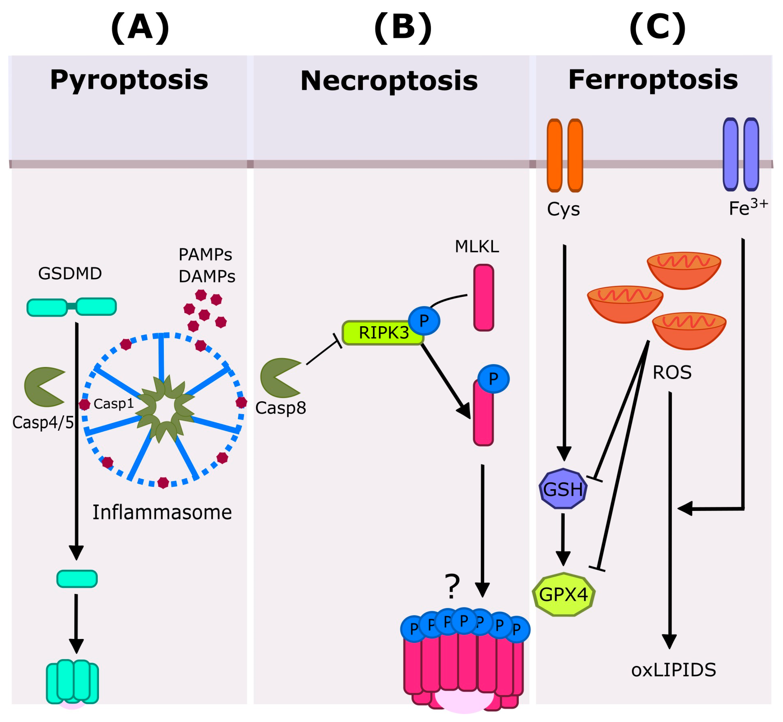
Pyroptosis is a lytic form of cell death in which caspase-1 mediates the proteolytic maturation of gasdermin D (GSDMD) to release its pore-forming domain (Figure 7A). Pyroptosis can also be triggered by caspase-4, caspase-5, and their murine orthologue caspase-11, all capable of cleaving GSDMD and inducing membrane pore formation [140]. Canonical inflammasome activation requires pattern recognition by various sensors including NLRs (NLRP3, NLRP1b, NLRC4), the cytosolic DNA sensor AIM2, and Pyrin, which recruit caspase-1 either directly or through the adaptor protein ASC (apoptosis-associated speck-like protein containing a CARD) [131]. In contrast, caspase-4, -5, and -11 are activated by cytosolic lipopolysacharide (LPS) detection independently of canonical inflammasome formation, although they cannot process IL-1β and IL-18 [141].
Gasdermin-like proteins have been identified in fungi and bacteria, in addition to animals. They share functional features with mammalian GSDMs, including protease-mediated activation, pore-forming capacity, and plasma membrane permeabilization. This leads to cell death during allorecognition and phage infection [140,142]. These discoveries establish gasdermins as an evolutionarily ancient mechanism of cell death and membrane pore formation shedding light on the evolution of regulated necrosis in immunity. Notably, no GSDM-like proteins have been identified in plants to date [10], underscoring the importance of further structural and functional analyses to determine if analogous systems exist in plant immunity.

9. MLKL-Mediated Necroptosis

Necroptosis is a form of regulated necrotic cell death in animals orchestrated by receptor-interacting protein kinase 3 (RIPK3) and its substrate mixed lineage kinase-like (MLKL) (Figure 7B) [143]. Activation of RIPK3 occurs through protein–protein interactions with three RHIM-containing proteins: RIPK1 (connecting RIPK3 to death receptor signaling), TRIF (mediating RIPK3 activation downstream of TLR3 and TLR4), and ZBP1/DAI (mediating RIPK3 activation in response to certain viruses and endogenous Z-form nucleic acids) [144,145,146]. Caspase-8 inhibits RIPK3 activation and necroptosis induction, by cleaving RIPK1/RIPK3 complex components. Necroptosis execution requires RIPK3-dependent phosphorylation of MLKL, which then induces plasma membrane damage through mechanisms that are not fully understood [145,147].
Pathogens use various strategies to evade necroptosis. Bacterial peptidases from enteropathogenic Escherichia coli and Shigella degrade components of the necroptotic machinery [148,149], while viral pathogens have sophisticated inhibition mechanisms. For example, MCMV expresses viral inhibitor of RIP activation (vIRA) proteins (M45) that interact with TRIF, ZBP1, RIPK1, and RIPK3 to impair necroptosis [150], and HCMV UL45 acts downstream of RIPK3 and MLKL [151]. Cowpox virus produces viral inducer of RIPK3 degradation (vIRD), leading to the degradation of RIPK3 [152], while other poxviruses express viral MLKL-like proteins (vMLKL) that sequester RIPK3 [153]. Additionally, poxvirus crmA inhibits caspase-8-mediated apoptosis without affecting its inhibition of necroptosis [154], and HSV-1/HSV-2 ribonucleotide reductase subunits (ICP6/ICP10) prevent both apoptosis and necroptosis [155].
The evolutionary conservation of necroptotic machinery is evident in the HeLo domain, a characteristic N-terminal four-helix bundle structure found in MLKLs and analogous cell death-inducing proteins across animals, fungi, and plants [156,157,158,159,160]. In fungi, Het-S/Het-s proteins trigger hyphal death during heterokaryon incompatibility [156], while in plants, the N-terminal RPW8-like coiled-coil domains of helper NLRs (ADR1 and NRG1 families) are structurally similar to HeLo domains and can induce HR cell death [156,161]. A recent discovery identified a conserved MLKL-like protein family across seed plants [160]. Genetic analyses using combinatorial Arabidopsis MLKL (AtMLKL) mutants show redundant function in conferring TNL-mediated disease resistance [160]. HeLo domain-containing helper NLRs are also essential for TNL-mediated HR and immunity [159], indicating that two distinct HeLo domain-containing protein families—plant MLKLs and RNLs—participate cooperatively in TNL-mediated immunity. Furthermore, the mobility of AtMLKL on microtubules is associated with immune response [160], similar to the microtubule-dependent translocation of animal MLKLs from cytoplasmic necrosomes to plasma membranes [162], suggesting comparable biochemical mechanisms for plant and animal MLKL-mediated cell death and immunity.

10. Fe3+-Mediated Ferroptosis

Host organisms utilize a variety of genetically encoded cell death programs in response to pathogen challenges. Ferroptosis is a well-defined cell death program in animals, alongside apoptosis, necroptosis, and pyroptosis. Ferroptosis is a form of necrotic cell death that relies on iron and oxidative stress (Figure 7C). It is characterized by iron-dependent lipid peroxidation leading to the loss of plasma membrane integrity. Unlike apoptosis, pyroptosis, or necroptosis, ferroptosis does not seem to involve predefined molecular cascades or scaffolds [10].
The structural similarities between NLR platforms, such as the ZAR1 resistosome functioning as a Ca2+ permeable channel, and animal immune systems highlight a common principle: the alteration of endomembrane integrity is a crucial step in immunogenic cell death signaling in both animals and plants. The transient or permanent loss of endomembrane integrity is a key step in many animal cell death forms (pyroptosis, necroptosis, ferroptosis, apoptosis) and plant HR [118].
A process similar to ferroptosis has been observed in plants associated with HR. In rice, when the fungal pathogen Magnaporthe oryzae is recognized by NLR-mediated mechanisms, ferric ions and ROS accumulate in cells undergoing HR. It is important to note that the use of ferroptosis inhibitors, such as deferoxamine and ferrostatin-1, has been shown to reduce HR levels. Despite these similarities, the extent of conservation of the ferroptosis mechanism between plants and animals is yet to be determined [163].
The convergence of complex, multi-component mechanisms suggests significant and consistent evolutionary pressures to combat intracellular threats. This phenomenon underscores the critical importance and effectiveness of immune strategies for the survival of multicellular organisms against microbial pathogens. It implies that these mechanisms represent highly robust and possibly “optimal” solutions for detecting and responding to intracellular invaders, regardless of the specific molecular actors involved. This understanding can provide insights into general principles of innate immunity that go beyond specific biological kingdoms.

11. Cross-Kingdom Pathogenicity

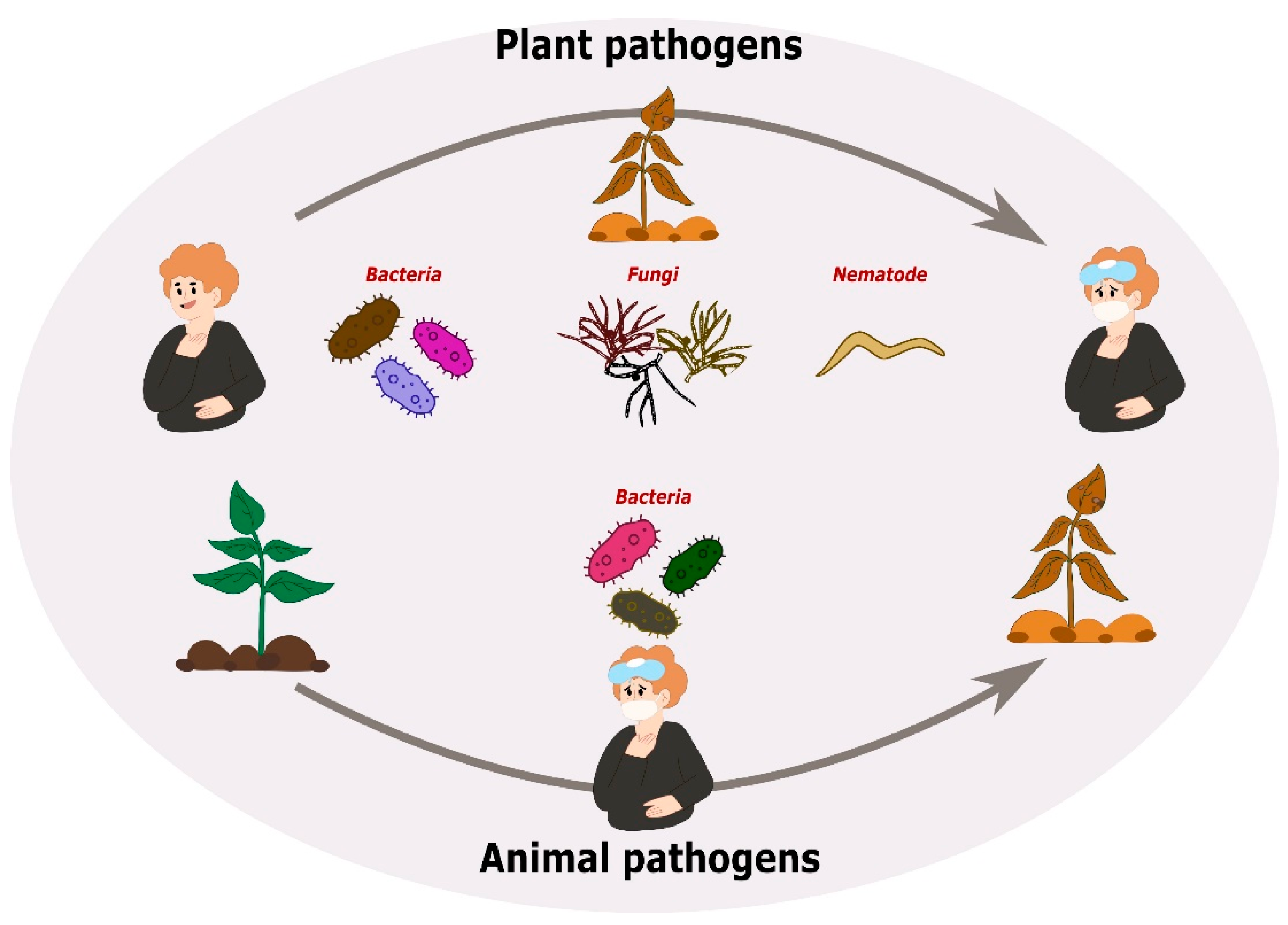
The fields of plant pathology and medical pathology have traditionally operated under the assumption of strict host specificity from pathogens. Plant pathogens (phytopathogens) have evolved highly specialized mechanisms to overcome plant-specific barriers, such as the rigid cell wall, through enzymes and plant hormone analogues. Similarly, human pathogens have evolved virulence determinants to exploit mammalian physiology and evade the complex adaptive immune system [164]. However, emerging research has challenged this dichotomy, highlighting the ability of microbes seemingly dedicated to one realm to cause disease in the other. It has been documented that human pathogens can infect plants and, crucially, that specialized plant pathogens can cause disease in humans (Figure 8) [165].

11.1. Plant Pathogens Infecting Humans and Animals

Some plant pathogens, such as fungi, oomycetes, bacteria, viruses, and nematodes, have evolved the capability to cross boundaries between kingdoms and infect humans and animals, particularly those with compromised immune systems [164].

11.1.1. Bacterial Infections

A well-known example is Rhizobium radiobacter, formerly known as Agrobacterium tumefaciens, which is the causative agent of crown gall disease in plants. In humans, this bacterium has been isolated as responsible for catheter-related bacteremia in neutropenic and immunocompromised patients, as well as cases of contact lens-related infectious keratitis [166,167].
The Burkholderia cepacia Complex (Bcc), which includes more than 20 species, is a phytopathogen known to cause onion rot. In human pathology, Bcc has caused fatal lung infections in patients with Cystic Fibrosis (CF) since the 1980s. Among the cases reported in recent years is one that occurred in February 2018 in the United States, where 60 patients were reported in various hospitals in California, New Jersey, Pennsylvania, Maine, Nevada and Ohio who contracted a Bcc infection. These infections were linked to the use of batches of rinse-free foaming cleansers that were contaminated with these microorganisms, as determined by comparing a clinical isolate with isolates from samples of the pharmaceutical product through pulsed-field gel electrophoresis (PFGE) [168]. Another case reported was in September 2019, where an outbreak of Bcc was detected in four patients on dialysis at Queen Mary Hospital in Hong Kong, China. Patients showed serous or bloody secretions at the exit ports of their dialysis catheters. The reservoir was identified in aqueous chlorhexidine solutions that had been contaminated during dilution in production plants of two different brands [169]. Other related species such as B. cenocepacia have caused sepsis, while B. gladioli and B. glumae have caused pneumonia infections in patients with chronic granulomatous disease [170].
Another group known for cross-kingdom infections is the Pantoea species. P. agglomerans, a phytopathogen, causes leaf blight and bulb rot in onions, blight and vascular wilt in maize and sorghum crops and is responsible for leaf necrosis. As an opportunistic pathogen it was responsible for 12 cases of infection in patients with malignant tumors in hospitals; this infection was attributed to contamination of a hospital sink [171]. In the early 1970s, it was responsible for a major outbreak of sepsis in the United States and Canada, affecting 25 hospitals. This outbreak was due to contaminated caps in infusion fluid bottles, with 378 cases reported, and has also been isolated from the synovial fluid of patients with arthritis, peritonitis, or osteomyelitis [172].
One amazing example of cross-kingdom pathogenicity is Pseudomonas aeruginosa.
P. aeruginosa is a highly versatile, opportunistic Gram-negative bacterium that can colonize and cause diseases in a wide variety of hosts, ranging from plants to humans.
Research in 1999 demonstrated that the human opportunistic pathogen P. aeruginosa strain PA14 kills C. elegans using the same virulence factors required for maximum virulence in mouse pathogenicity, such as gacA, lasR, pstP, and mtrR, among other genes [173].
In agriculture, P. aeruginosa is well-known for its capacity to improve plant growth and reduce a variety of plant diseases. P. aeruginosa possesses aggressive rhizosphere competence, quick colonization, formation of several root exudates, and bioactive substances (such as vitamins, siderophores, and antibiotics), making it serve as a plant growth promoter and a biocontrol agent [174].
However, P. aeruginosa is also a major nosocomial (hospital-acquired) pathogen, particularly notorious in intensive care units (ICUs). It causes bacteremia and sepsis in neutropenic cancer patients undergoing chemotherapy, and it is the number one cause of hospital-acquired pneumonia and respiratory failure. It also affects diabetic ulcers, burn wounds, corneal ulcers, surgical wounds, and patients with cystic fibrosis or AIDS, resulting in higher case fatality rates than other bacterial causes [175,176].
While the plant–pathogen interaction is often studied in agricultural contexts, plants in the hospital environment (e.g., flowers, potted plants, vegetables) can act as reservoirs for this bacterium, allowing it to multiply before being transmitted to immunocompromised patients [177,178]. This knowledge has led to improved control of nosocomial infections.

11.1.2. Fungal Infections

However, there is also growing and troubling evidence of the ability of plant-associated fungi and oomycetes to infect humans and animals. Some of these organisms can infect both plants and humans, posing a danger to public health and agriculture. A specific and well-documented case of a plant fungal pathogen that causes infection in humans is Alternaria infectoria, a fungus responsible for flower blight in guayule and iris plants [179]. In humans, A. infectoria has been identified as a causative agent of phaeohyphomycosis and keratitis. It is also considered one of the most abundant and potent sources of sensitizing allergens present in the air, causing allergic respiratory diseases, such as severe asthma [180]. A. infectoria caused skin lesions in a heart transplant patient treated with isavuconazole [181] and rhinosinusitis in a 28-year-old woman with no relevant medical history [182].
Other genera of phytopathological importance isolated from human clinical specimens include Bipolaris species (such as B. spicifera and B. hawaiiensis), which cause leaf spots and blights in plants. In humans, they are opportunistic and infections have been reported in immunocompromised individuals. A recent case involved a patient with diabetes mellitus in which B. spicifera was found on the surface of an ulcer, with minimal inflammation [183]. The genus Cladosporium, which includes numerous plant pathogenic species, has been isolated from various human infection samples, with species like C. herbarum and C. sphaerospermum causing respiratory diseases [184]. C. halotolerans has been localized in the skin, nails, and maxillofacial tissue of immunocompromised patients with a history of trauma [185].
In 2013, in the United States, 751 cases and 64 deaths (8% mortality rate) were reported, associated with contaminated methylprednisolone acetate (MPA) used for epidural and intra-articular injections. Clinical manifestations include meningitis and isolated paraspinal infections such as epidural abscess, discitis, vertebral osteomyelitis, arachnoiditis, and phlegmon. The causal agent was identified as Exserohilum rostratum, a fungal phytopathogen of sugar cane, maize, wheat and sorghum [186]. This example shows that fungi capable of crossing kingdoms to humans may cause outbreaks.
Recently, in India a 61-year-old mycologist became the first documented human case of infection by Chondrostereum purpureum, a plant fungus causing silver leaf disease in plants, particularly in the rose family. C. purpureum was confirmed as the cause of infection via DNA sequencing. The patient was treated, highlighting the importance of identifying such rare occurrences in those with high exposure to plant pathogens [187].

11.1.3. Parasitic Infections

Phytopathogenic nematodes typically do not directly threaten humans or animals, as they are highly specialized in parasitizing plants [164]. However, in September 2022, Haouchine et al. [188] reported the presence of the phytopathogenic nematode Xiphinema brevicollum in a 55-year-old woman who presented with severe abdominal pain. Clinical examinations revealed hypereosinophilia and the presence of X. brevicollum eggs and larvae in her feces. Molecular analyses, including mitochondrial DNA sequencing, confirmed the species. This was the first evidence of an X. brevicollum infection in humans [188].
Another example is zoonotic trematode infections, primarily caused by Fasciola hepatica and F. gigantica. These infections involve freshwater snails as intermediate hosts and mammals (such as sheep, cattle, and humans) as definitive hosts. Humans become infected by ingesting the infective metacercarial stage, which encysts on aquatic vegetation like raw watercress and morning glory. Once ingested, the metacercariae excyst in the duodenum and penetrate the intestinal wall, migrating through the abdominal cavity into the liver parenchyma, causing fascioliasis, or liver rot [189,190].
Ascaris lumbricoides (roundworm), Trichuris trichiura (whipworm), and hookworms are soil-transmitted helminths. Eggs present in human feces contaminate the soil. Infection occurs when soil/eggs are ingested from contaminated vegetables, or when hookworm larvae in the soil penetrate the skin. These parasites are prevalent in tropical and subtropical areas where they are associated with substantial morbidity [191].

11.2. Human/Animal Pathogens Infecting Plants

Cross-kingdom infection is not limited to the transfer from plants to humans. Some microbes, considered animal or human pathogens, have been shown to be equally capable of colonizing and causing infections in plant hosts.

11.2.1. Bacterial Infections

Salmonella enterica is a Gram-negative bacterium that causes salmonellosis with an estimated 94 million infections in humans [192]. In 2012, Üstün et al. [193] reported that effectors of S. enterica induce symptoms similar to the hypersensitive response in leaves of Nicotiana benthamiana. Chlorosis and leaf wilt caused by S. enterica have also been described in Arabidopsis thaliana [194].
Another example is the bacterium Enterococcus faecalis, which has been reported to infect and cause disease in plants, specifically A. thaliana. Three strains (FA-2-2, V583 and OG1RF) were able to colonize both leaves and roots, ultimately resulting in the death of the plant seven days after inoculation [195].
Shigella is a bacterium that causes a high global burden of diarrheal diseases in humans. A study published in 2019 revealed that it can also colonize and proliferate in plants, particularly A. thaliana. Four strains (S. boydii, S. sonnei, S. flexneri 2a and S. flexneri 5a) were examined, demonstrating different levels of growth within plant tissues. Microscopic observations confirmed the presence of Shigella in leaves and in damaged cells [196].
Bacteria can manipulate diseases in plants by introducing effector proteins using the type III secretion system (T3SS).

11.2.2. Fungal Infections

Aspergillus fumigatus is a well-known and devastating causal agent of human mycoses. This fungus is not only a plant pathogen but also a saprotrophic, soil-borne fungus typically found on decaying plant material such as compost and flower bulb waste. Another example is Cryptococcus neoformans, a basidiomycetous yeast that causes infections known as cryptococcosis, including life-threatening meningoencephalitis. C. neoformans is also not a plant pathogen but is often isolated from decaying wood material [197]. Examples of human/animal fungal pathogens infecting plants are still lacking.
In fungi and oomycetes, the mechanisms of protein translocation are less understood compared to bacteria. Studies have identified an N-terminal motif called RXLR-dEER in oomycetes, present in several Avr proteins, which is crucial for their entry into plant cells. This RXLR-dEER motif bears a strong resemblance to the RXLX E/Q/D motif, known as the PEXEL motif, used by the human parasite Plasmodium falciparum, which causes malaria, to introduce its effector proteins into erythrocytes. Interestingly, a fusion protein containing the effector core domain of AVR3a from P. infestans coupled with the PEXEL sequence of HRPII from P. falciparum was successfully delivered into the plant by P. infestans [198]. A similar experiment in Plasmodium, delivering HRPII with the RXLR leader of AVR3a, was also successful in P. falciparum [199], confirming the importance of these motifs in effector translocation. These experiments show that the translocation machinery in Phytophthora can recognize Plasmodium signals, and vice versa. Recent studies reveal that the RXLR and PEXEL motifs are cleaved off within 40 amino acids after the signal peptide cleavage site, and then both types of cleaved proteins are acetylated, a crucial modification for effector export [200,201,202]. Despite these similarities, it remains to be determined whether these motifs are involved in cross-kingdom pathogenicity.

11.2.3. Parasitic Infections

Much effort has been expended on diseases jumping to humans from plants. To date, there are no reports of human or animal parasites infecting plants. However, parasites have evolved convergent strategies to counter host defenses, including mechanisms of active penetration and evading the host’s innate and/or adaptive immune responses, resulting in striking similarities. Plant-parasitic nematodes (PPNs), particularly sedentary endoparasites like root-knot (Meloidogyne spp.) and cyst (Heterodera spp.) nematodes, employ sophisticated strategies to hijack host plant root cells that are strikingly similar to the mechanisms used by animal-parasitic nematodes such as Trichinella spiralis. PPNs use a stylet to inject effector proteins, which are often produced in the esophageal glands, directly into the cytoplasm of plant cells or the apoplast to modify cell structure and suppress plant immunity. T. spiralis similarly secretes effectors that regulate host immune responses, such as inhibiting nucleotide-mediated mast cell activation. Both plant-parasitic nematodes and T. spiralis act as master regulators of their host’s biology. PPNs force the plant cells to undergo dramatic re-differentiation into large, metabolically active “giant cells” or “syncytia”. Similarly, T. spiralis infects muscle cells and reprograms them into a nurse cell complex, which acts as a “home” and source of nutrition for the parasite [203,204].
Trypanosomatids are a group of parasitic single-celled eukaryotes within the order Kinetoplastea. Among the trypanosomatids are important human pathogens that cause serious diseases such as Leishmania spp. (etiological agent of distinct forms of leishmaniasis), Trypanosoma cruzi (etiological agent of Chagas’ disease), and Trypanosoma brucei (etiological agent of African sleeping sickness). Some Trypanosomatids can infect insects and vertebrates from different orders [205]. The virulence mechanisms include adhesion, host cell invasion and complement system evasion, which are mediated by a group of different parasite molecules that eventually achieve the same result in different hosts, making it difficult to determine the proteins responsible for the progression of each evasion process [206]. On the contrary, plant-infecting trypanosomatids known as Phytomonas are relatively poorly understood. Based on genome sequence, Phytomonas comprises a phylogenetic sister group with Leishmania. To date, few molecular components of the Phytomonas cell surface have been characterized. Those that have been identified so far are conserved in other trypanosomatids. One strain, P. serpens 9T, was isolated from a tomato fruit in South America. Interestingly, immunogenic similarities between P. serpens and T. cruzi were found, and oral exposure to P. serpens has been observed to attenuate the symptoms of Chagas disease in C57BL/6 mice. Additionally, although naturally occurring Phytomonas infections in mammals have not been described, inoculation of mice with Phytomonas-infected latex has been reported to produce infections [207].
These examples support the concept of a cross-kingdom pathogenesis mechanism, where various pathogenic organisms use similar molecular strategies to invade and control host cells from different biological kingdoms. This suggests an evolutionary convergence in infection and adaptation processes.

12. Conclusions and Perspectives

A structural comparison among resistosomes, apoptosomes, and inflammasomes reveals remarkable evolutionary convergence. All three systems utilize NB-ARC/NBDs for nucleotide-dependent oligomerization. The evolutionary conservation of cell death mechanisms extends beyond these oligomeric platforms. The identification of HeLo domain-containing proteins across animals, fungi, and plants, including plant MLKLs and helper NLRs, suggests ancient origins for regulated necrosis in immunity [156,160]. Similarly, the observation of ferroptosis-like processes in plant HR [163], involving iron-dependent lipid peroxidation comparable to animal ferroptosis [10], indicates that endomembrane integrity disruption represents a conserved principle in immunogenic cell death across kingdoms. Studying the function of animal NLR proteins has led to a better understanding of the role of plant NLRs [157], leading to engineering plant NLRs for crop protection [208]. On the other hand, research on plant immunity may help to better understand inflammation processes in humans to alleviate serious diseases, including autoimmune disorders, chronic inflammatory conditions, and malignant diseases. Autoimmunity, a term usually related to animals, has also been described in plants, where inappropriate activation of NLRs results in the spontaneous initiation of plant defense responses and cell death [209], reinforcing evolutionary convergence of plant and animal processes.
Likewise, plant ETI has been extensively investigated, and this knowledge may serve as a foundational model that benefits research into animal ETI. To date, hundreds of effectors have been experimentally validated from phytopathogens, and they may improve effector prediction in animal pathogens. Agroinfiltration in Nicotiana benthamiana can be used for functional validation of effectors in animal pathogens [193], aiding in the faster development of effectoromics in animal pathogens. This is important because some effectors have been identified as targets for developing vaccines [210,211]. In summary, further cross-kingdom comparisons may be beneficial for therapeutic possibilities in medicine and agriculture, bridging millions of years of evolution with shared molecular solutions to pathogen defense.

Author Contributions

Conceptualization, B.C.-C., L.C.J.C. and P.A.G.-T.; methodology, L.C.J.C., P.A.G.-T. and K.G.C.-A.; resources, B.C.-C.; writing—original draft preparation, B.C.-C., L.C.J.C. and P.A.G.-T.; writing—review and editing, C.D.l.S.-B., L.S.-C., I.I.-F., K.G.C.-A., R.V.-E. and N.K.-M.; supervision, B.C.-C.; project administration, C.D.l.S.-B.; funding acquisition, B.C.-C. All authors have read and agreed to the published version of the manuscript.

Funding

This research received funding from the SECIHTI-Mexico project FOP16-2021-01 No. 320993, and Fellowships 4016973 for L.C.J.-C., and 4002386 for P.A.G.-T.

Institutional Review Board Statement

Not applicable.

Informed Consent Statement

Not applicable.

Data Availability Statement

The original contributions presented in this study are included in the article.

Conflicts of Interest

The authors declare no conflicts of interest.

References

  1. Mermigka, G.; Amprazi, M.; Mentzelopoulou, A.; Amartolou, A.; Sarris, P.F. Plant and Animal Innate Immunity Complexes: Fighting Different Enemies with Similar Weapons. Trends Plant Sci. 2020, 25, 80–91. [Google Scholar] [CrossRef] [PubMed]
  2. Roudaire, T.; Héloir, M.-C.; Wendehenne, D.; Zadoroznyj, A.; Dubrez, L.; Poinssot, B. Cross Kingdom Immunity: The Role of Immune Receptors and Downstream Signaling in Animal and Plant Cell Death. Front. Immunol. 2021, 11, 612452. [Google Scholar] [CrossRef]
  3. Pradeu, T.; Thomma, B.P.H.J.; Girardin, S.E.; Lemaitre, B. The Conceptual Foundations of Innate Immunity: Taking Stock 30 Years Later. Immunity 2024, 57, 613–631. [Google Scholar] [CrossRef]
  4. Sellge, G.; Kufer, T.A. PRR-Signaling Pathways: Learning from Microbial Tactics. Semin. Immunol. 2015, 27, 75–84. [Google Scholar] [CrossRef]
  5. Cicchinelli, S.; Pignataro, G.; Gemma, S.; Piccioni, A.; Picozzi, D.; Ojetti, V.; Franceschi, F.; Candelli, M. PAMPs and DAMPs in Sepsis: A Review of Their Molecular Features and Potential Clinical Implications. Int. J. Mol. Sci. 2024, 25, 962. [Google Scholar] [CrossRef]
  6. De Lorenzo, G.; Ferrari, S.; Cervone, F.; Okun, E. Extracellular DAMPs in Plants and Mammals: Immunity, Tissue Damage and Repair. Trends Immunol. 2018, 39, 937–950. [Google Scholar] [CrossRef]
  7. Li, S.; Manik, M.K.; Shi, Y.; Kobe, B.; Ve, T. Toll/Interleukin-1 Receptor Domains in Bacterial and Plant Immunity. Curr. Opin. Microbiol. 2023, 74, 102316. [Google Scholar] [CrossRef]
  8. Naithani, S.; Komath, S.S.; Nonomura, A.; Govindjee, G. Plant Lectins and Their Many Roles: Carbohydrate-Binding and Beyond. J. Plant Physiol. 2021, 266, 153531. [Google Scholar] [CrossRef] [PubMed]
  9. Danial, N.N.; Korsmeyer, S.J. Cell Death. Cell 2004, 116, 205–219. [Google Scholar] [CrossRef] [PubMed]
  10. Maekawa, T.; Kashkar, H.; Coll, N.S. Dying in Self-Defence: A Comparative Overview of Immunogenic Cell Death Signalling in Animals and Plants. Cell Death Differ. 2023, 30, 258–268. [Google Scholar] [CrossRef]
  11. Ding, L.-N.; Li, Y.-T.; Wu, Y.-Z.; Li, T.; Geng, R.; Cao, J.; Zhang, W.; Tan, X.-L. Plant Disease Resistance-Related Signaling Pathways: Recent Progress and Future Prospects. Int. J. Mol. Sci. 2022, 23, 16200. [Google Scholar] [CrossRef]
  12. Li, D.; Wu, M. Pattern Recognition Receptors in Health and Diseases. Sig. Transduct. Target. Ther. 2021, 6, 291. [Google Scholar] [CrossRef]
  13. Saijo, Y.; Loo, E.P.; Yasuda, S. Pattern Recognition Receptors and Signaling in Plant–Microbe Interactions. Plant J. 2018, 93, 592–613. [Google Scholar] [CrossRef]
  14. Chen, R.; Zou, J.; Chen, J.; Zhong, X.; Kang, R.; Tang, D. Pattern Recognition Receptors: Function, Regulation and Therapeutic Potential. Sig. Transduct. Target. Ther. 2025, 10, 216. [Google Scholar] [CrossRef]
  15. Kurtz, J.; Andino, R.; Boraschi, D.; Contreras-Garduño, J.; Kachroo, A.; Khan, I.; Lanz Mendoza, H.; Mukherjee, K.; Peuß, R.; Ton, J. Trained Immunity and Immune Priming in Plants and Invertebrates. eLife 2025, 14, e106597. [Google Scholar] [CrossRef] [PubMed]
  16. Göhre, V.; Robatzek, S. Breaking the Barriers: Microbial Effector Molecules Subvert Plant Immunity. Annu. Rev. Phytopathol. 2008, 46, 189–215. [Google Scholar] [CrossRef]
  17. Yu, X.; Niu, H.; Liu, C.; Wang, H.; Yin, W.; Xia, X. PTI-ETI Synergistic Signal Mechanisms in Plant Immunity. Plant Biotechnol. J. 2024, 22, 2113–2128. [Google Scholar] [CrossRef] [PubMed]
  18. Hatsugai, N.; Igarashi, D.; Mase, K.; Lu, Y.; Tsuda, Y.; Chakravarthy, S.; Wei, H.; Foley, J.W.; Collmer, A.; Glazebrook, J.; et al. A Plant Effector-triggered Immunity Signaling Sector Is Inhibited by Pattern-triggered Immunity. EMBO J. 2017, 36, 2758–2769. [Google Scholar] [CrossRef]
  19. Martel, A.; Ruiz-Bedoya, T.; Breit-McNally, C.; Laflamme, B.; Desveaux, D.; Guttman, D.S. The ETS-ETI Cycle: Evolutionary Processes and Metapopulation Dynamics Driving the Diversification of Pathogen Effectors and Host Immune Factors. Curr. Opin. Plant Biol. 2021, 62, 102011. [Google Scholar] [CrossRef] [PubMed]
  20. Stuart, L.M.; Paquette, N.; Boyer, L. Effector-Triggered versus Pattern-Triggered Immunity: How Animals Sense Pathogens. Nat. Rev. Immunol. 2013, 13, 199–206. [Google Scholar] [CrossRef]
  21. Jones, J.D.G.; Dangl, J.L. The Plant Immune System. Nature 2006, 444, 323–329. [Google Scholar] [CrossRef]
  22. Yuan, M.; Ngou, B.P.M.; Ding, P.; Xin, X.-F. PTI-ETI Crosstalk: An Integrative View of Plant Immunity. Curr. Opin. Plant Biol. 2021, 62, 102030. [Google Scholar] [CrossRef]
  23. Remick, B.C.; Gaidt, M.M.; Vance, R.E. Effector-Triggered Immunity. Annu. Rev. Immunol. 2023, 41, 453–481. [Google Scholar] [CrossRef]
  24. Naveed, Z.A.; Wei, X.; Chen, J.; Mubeen, H.; Ali, G.S. The PTI to ETI Continuum in Phytophthora-Plant Interactions. Front. Plant Sci. 2020, 11, 593905. [Google Scholar] [CrossRef]
  25. Tena, G. PTI and ETI Are One. Nat. Plants 2021, 7, 1527. [Google Scholar] [CrossRef] [PubMed]
  26. Kufer, T.A.; Creagh, E.M.; Bryant, C.E. Guardians of the Cell: Effector-Triggered Immunity Steers Mammalian Immune Defense. Trends Immunol. 2019, 40, 939–951. [Google Scholar] [CrossRef] [PubMed]
  27. Van Der Hoorn, R.A.L.; Kamoun, S. From Guard to Decoy: A New Model for Perception of Plant Pathogen Effectors. Plant Cell 2008, 20, 2009–2017. [Google Scholar] [CrossRef] [PubMed]
  28. Ray, S.K.; Macoy, D.M.; Kim, W.-Y.; Lee, S.Y.; Kim, M.G. Role of RIN4 in Regulating PAMP-Triggered Immunity and Effector-Triggered Immunity: Current Status and Future Perspectives. Mol. Cells 2019, 42, 503–511. [Google Scholar] [CrossRef]
  29. Contreras, M.P.; Lüdke, D.; Pai, H.; Toghani, A.; Kamoun, S. NLR Receptors in Plant Immunity: Making Sense of the Alphabet Soup. EMBO Rep. 2023, 24, e57495. [Google Scholar] [CrossRef]
  30. Malik, H.S.; Bliska, J.B. Guards and Decoys: RIPoptosome and Inflammasome Pathway Regulators of Bacterial Effector-Triggered Immunity. PLoS Pathog. 2025, 21, e1012884. [Google Scholar] [CrossRef]
  31. Kroj, T.; Chanclud, E.; Michel-Romiti, C.; Grand, X.; Morel, J. Integration of Decoy Domains Derived from Protein Targets of Pathogen Effectors into Plant Immune Receptors Is Widespread. New Phytol. 2016, 210, 618–626. [Google Scholar] [CrossRef] [PubMed]
  32. Duxbury, Z.; Wu, C.; Ding, P. A Comparative Overview of the Intracellular Guardians of Plants and Animals: NLRs in Innate Immunity and Beyond. Annu. Rev. Plant Biol. 2021, 72, 155–184. [Google Scholar] [CrossRef]
  33. Zhang, B.; Liu, M.; Wang, Y.; Yuan, W.; Zhang, H. Plant NLRs: Evolving with Pathogen Effectors and Engineerable to Improve Resistance. Front. Microbiol. 2022, 13, 1018504. [Google Scholar] [CrossRef] [PubMed]
  34. Zhuang, J.; Xie, L.; Zheng, L. A Glimpse of Programmed Cell Death Among Bacteria, Animals, and Plants. Front. Cell Dev. Biol. 2022, 9, 790117. [Google Scholar] [CrossRef]
  35. Salguero-Linares, J.; Coll, N.S. Cell Death as a Defense Strategy against Pathogens in Plants and Animals. PLoS Pathog. 2023, 19, e1011253. [Google Scholar] [CrossRef] [PubMed]
  36. Tang, D.; Wang, G.; Zhou, J.-M. Receptor Kinases in Plant-Pathogen Interactions: More Than Pattern Recognition. Plant Cell 2017, 29, 618–637. [Google Scholar] [CrossRef]
  37. Couto, D.; Zipfel, C. Regulation of Pattern Recognition Receptor Signalling in Plants. Nat. Rev. Immunol. 2016, 16, 537–552. [Google Scholar] [CrossRef]
  38. Ramírez-Zavaleta, C.Y.; García-Barrera, L.J.; Rodríguez-Verástegui, L.L.; Arrieta-Flores, D.; Gregorio-Jorge, J. An Overview of PRR- and NLR-Mediated Immunities: Conserved Signaling Components across the Plant Kingdom That Communicate Both Pathways. Int. J. Mol. Sci. 2022, 23, 12974. [Google Scholar] [CrossRef]
  39. Boutrot, F.; Zipfel, C. Function, Discovery, and Exploitation of Plant Pattern Recognition Receptors for Broad-Spectrum Disease Resistance. Annu. Rev. Phytopathol. 2017, 55, 257–286. [Google Scholar] [CrossRef]
  40. Escocard De Azevedo Manhães, A.M.; Ortiz-Morea, F.A.; He, P.; Shan, L. Plant Plasma Membrane-resident Receptors: Surveillance for Infections and Coordination for Growth and Development. J. Integr. Plant Biol. 2021, 63, 79–101. [Google Scholar] [CrossRef]
  41. Liu, J.; Li, W.; Wu, G.; Ali, K. An Update on Evolutionary, Structural, and Functional Studies of Receptor-like Kinases in Plants. Front. Plant Sci. 2024, 15, 1305599. [Google Scholar] [CrossRef] [PubMed]
  42. Han, G. Origin and Evolution of the Plant Immune System. New Phytol. 2019, 222, 70–83. [Google Scholar] [CrossRef]
  43. Dievart, A.; Gottin, C.; Périn, C.; Ranwez, V.; Chantret, N. Origin and Diversity of Plant Receptor-Like Kinases. Annu. Rev. Plant Biol. 2020, 71, 131–156. [Google Scholar] [CrossRef]
  44. Zhao, Q.; Bao, J.; Li, H.; Hu, W.; Kong, Y.; Zhong, Y.; Fu, Q.; Xu, G.; Liu, F.; Jiao, X.; et al. Structural and Biochemical Basis of FLS2-Mediated Signal Activation and Transduction in Rice. Plant Commun. 2024, 5, 100785. [Google Scholar] [CrossRef]
  45. Yoon, S.; Kurnasov, O.; Natarajan, V.; Hong, M.; Gudkov, A.V.; Osterman, A.L.; Wilson, I.A. Structural Basis of TLR5-Flagellin Recognition and Signaling. Science 2012, 335, 859–864. [Google Scholar] [CrossRef]
  46. Ercoli, M.F.; Luu, D.D.; Rim, E.Y.; Shigenaga, A.; Teixeira De Araujo, A.; Chern, M.; Jain, R.; Ruan, R.; Joe, A.; Stewart, V.; et al. Plant Immunity: Rice XA21-Mediated Resistance to Bacterial Infection. Proc. Natl. Acad. Sci. USA 2022, 119, e2121568119. [Google Scholar] [CrossRef]
  47. Breiden, M.; Simon, R. Q&A: How Does Peptide Signaling Direct Plant Development? BMC Biol. 2016, 14, 58. [Google Scholar] [CrossRef] [PubMed][Green Version]
  48. Choi, J.; Tanaka, K.; Cao, Y.; Qi, Y.; Qiu, J.; Liang, Y.; Lee, S.Y.; Stacey, G. Identification of a Plant Receptor for Extracellular ATP. Science 2014, 343, 290–294. [Google Scholar] [CrossRef] [PubMed]
  49. Hind, S.R.; Strickler, S.R.; Boyle, P.C.; Dunham, D.M.; Bao, Z.; O’Doherty, I.M.; Baccile, J.A.; Hoki, J.S.; Viox, E.G.; Clarke, C.R.; et al. Tomato Receptor FLAGELLIN-SENSING 3 Binds flgII-28 and Activates the Plant Immune System. Nat. Plants 2016, 2, 16128. [Google Scholar] [CrossRef]
  50. Ranf, S.; Gisch, N.; Schäffer, M.; Illig, T.; Westphal, L.; Knirel, Y.A.; Sánchez-Carballo, P.M.; Zähringer, U.; Hückelhoven, R.; Lee, J.; et al. A Lectin S-Domain Receptor Kinase Mediates Lipopolysaccharide Sensing in Arabidopsis thaliana. Nat Immunol. 2015, 16, 426–433. [Google Scholar] [CrossRef]
  51. Yang, C.; Liu, R.; Pang, J.; Ren, B.; Zhou, H.; Wang, G.; Wang, E.; Liu, J. Poaceae-Specific Cell Wall-Derived Oligosaccharides Activate Plant Immunity via OsCERK1 during Magnaporthe oryzae Infection in Rice. Nat. Commun. 2021, 12, 2178. [Google Scholar] [CrossRef] [PubMed]
  52. Pruitt, R.N.; Schwessinger, B.; Joe, A.; Thomas, N.; Liu, F.; Albert, M.; Robinson, M.R.; Chan, L.J.G.; Luu, D.D.; Chen, H.; et al. The Rice Immune Receptor XA21 Recognizes a Tyrosine-Sulfated Protein from a Gram-Negative Bacterium. Sci. Adv. 2015, 1, e1500245. [Google Scholar] [CrossRef]
  53. Zuo, C.; Liu, Y.; Guo, Z.; Mao, J.; Chu, M.; Chen, B. Genome-Wide Annotation and Expression Responses to Biotic Stresses of the WALL-ASSOCIATED KINASE—RECEPTOR-LIKE KINASE (WAK-RLK) Gene Family in Apple (Malus domestica). Eur J Plant Pathol. 2019, 153, 771–785. [Google Scholar] [CrossRef]
  54. Liu, X.; Liang, C.; Hou, S.; Wang, X.; Chen, D.; Shen, J.; Zhang, W.; Wang, M. The LRR-RLK Protein HSL3 Regulates Stomatal Closure and the Drought Stress Response by Modulating Hydrogen Peroxide Homeostasis. Front. Plant Sci. 2020, 11, 548034. [Google Scholar] [CrossRef]
  55. Albert, I.; Böhm, H.; Albert, M.; Feiler, C.E.; Imkampe, J.; Wallmeroth, N.; Brancato, C.; Raaymakers, T.M.; Oome, S.; Zhang, H.; et al. An RLP23–SOBIR1–BAK1 Complex Mediates NLP-Triggered Immunity. Nat. Plants 2015, 1, 15140. [Google Scholar] [CrossRef]
  56. Ngou, B.P.M.; Ding, P.; Jones, J.D.G. Thirty Years of Resistance: Zig-Zag through the Plant Immune System. Plant Cell 2022, 34, 1447–1478. [Google Scholar] [CrossRef]
  57. Ngou, B.P.M.; Heal, R.; Wyler, M.; Schmid, M.W.; Jones, J.D.G. Concerted Expansion and Contraction of Immune Receptor Gene Repertoires in Plant Genomes. Nat. Plants 2022, 8, 1146–1152. [Google Scholar] [CrossRef]
  58. Pruitt, R.N.; Zhang, L.; Saile, S.C.; Karelina, D.; Fröhlich, K.; Wan, W.-L.; Rao, S.; Gust, A.A.; Locci, F.; Joosten, M.H.A.J.; et al. Arabidopsis Cell Surface LRR Immune Receptor Signaling through the EDS1-PAD4-ADR1 Node 2020. bioRxiv 2020. [Google Scholar]
  59. Wan, W.; Zhang, L.; Pruitt, R.; Zaidem, M.; Brugman, R.; Ma, X.; Krol, E.; Perraki, A.; Kilian, J.; Grossmann, G.; et al. Comparing Arabidopsis Receptor Kinase and Receptor Protein-mediated Immune Signaling Reveals BIK1-dependent Differences. New Phytol. 2019, 221, 2080–2095. [Google Scholar] [CrossRef] [PubMed]
  60. Rooney, H.C.E.; Van’T Klooster, J.W.; Van Der Hoorn, R.A.L.; Joosten, M.H.A.J.; Jones, J.D.G.; De Wit, P.J.G.M. Cladosporium Avr2 Inhibits Tomato Rcr3 Protease Required for Cf-2 -Dependent Disease Resistance. Science 2005, 308, 1783–1786. [Google Scholar] [CrossRef]
  61. Pruitt, R.N.; Locci, F.; Wanke, F.; Zhang, L.; Saile, S.C.; Joe, A.; Karelina, D.; Hua, C.; Fröhlich, K.; Wan, W.-L.; et al. The EDS1–PAD4–ADR1 Node Mediates Arabidopsis Pattern-Triggered Immunity. Nature 2021, 598, 495–499. [Google Scholar] [CrossRef] [PubMed]
  62. Tian, H.; Wu, Z.; Chen, S.; Ao, K.; Huang, W.; Yaghmaiean, H.; Sun, T.; Xu, F.; Zhang, Y.; Wang, S.; et al. Activation of TIR Signalling Boosts Pattern-Triggered Immunity. Nature 2021, 598, 500–503. [Google Scholar] [CrossRef]
  63. Cai, M.; Yu, H.; Sun, E.; Zuo, C. Receptor-like Proteins: Decision-Makers of Plant Immunity. Phytopathol. Res. 2024, 6, 58. [Google Scholar] [CrossRef]
  64. Thomas, C.M.; Jones, D.A.; Parniske, M.; Harrison, K.; Balint-Kurti, P.J.; Hatzixanthis, K.; Jones, J.D. Characterization of the Tomato Cf-4 Gene for Resistance to Cladosporium fulvum Identifies Sequences That Determine Recognitional Specificity in Cf-4 and Cf-9. Plant Cell 1997, 9, 2209–2224. [Google Scholar] [CrossRef] [PubMed][Green Version]
  65. Huang, W.R.H.; Braam, C.; Kretschmer, C.; Villanueva, S.L.; Liu, H.; Ferik, F.; Van Der Burgh, A.M.; Boeren, S.; Wu, J.; Zhang, L.; et al. Receptor-like Cytoplasmic Kinases of Different Subfamilies Differentially Regulate SOBIR1/BAK1-Mediated Immune Responses in Nicotiana benthamiana. Nat. Commun. 2024, 15, 4339. [Google Scholar] [CrossRef]
  66. Dixon, M.S.; Hatzixanthis, K.; Jones, D.A.; Harrison, K.; Jones, J.D.G. The Tomato Cf-5 Disease Resistance Gene and Six Homologs Show Pronounced Allelic Variation in Leucine-Rich Repeat Copy Number. Plant Cell 1998, 10, 1915–1925. [Google Scholar] [CrossRef] [PubMed]
  67. Jones, D.A.; Thomas, C.M.; Hammond-Kosack, K.E.; Balint-Kurti, P.J.; Jones, J.D.G. Isolation of the Tomato Cf-9 Gene for Resistance to Cladosporium fulvum by Transposon Tagging. Science 1994, 266, 789–793. [Google Scholar] [CrossRef]
  68. Zheng, Q.; Bao, J.; Li, L.; Shen, Z.; Wang, J.; Daskalov, A.; Zhu, X.; Lin, F. Integrated Transcriptomic and Metabolomic Analysis Reveals the Regulation Network of CEBiP in Rice Defense Against Magnaporthe oryzae. Int. J. Mol. Sci. 2025, 26, 5194. [Google Scholar] [CrossRef]
  69. Du, J.; Verzaux, E.; Chaparro-Garcia, A.; Bijsterbosch, G.; Keizer, L.C.P.; Zhou, J.; Liebrand, T.W.H.; Xie, C.; Govers, F.; Robatzek, S.; et al. Elicitin Recognition Confers Enhanced Resistance to Phytophthora infestans in Potato. Nat. Plants 2015, 1, 15034. [Google Scholar] [CrossRef]
  70. Sussholz, O.; Pizarro, L.; Schuster, S.; Avni, A. SlRLK-like Is a Malectin-like Domain Protein Affecting Localization and Abundance of LeEIX2 Receptor Resulting in Suppression of EIX-induced Immune Responses. Plant J. 2020, 104, 1369–1381. [Google Scholar] [CrossRef]
  71. Willmann, R.; Lajunen, H.M.; Erbs, G.; Newman, M.-A.; Kolb, D.; Tsuda, K.; Katagiri, F.; Fliegmann, J.; Bono, J.-J.; Cullimore, J.V.; et al. Arabidopsis Lysin-Motif Proteins LYM1 LYM3 CERK1 Mediate Bacterial Peptidoglycan Sensing and Immunity to Bacterial Infection. Proc. Natl. Acad. Sci. USA 2011, 108, 19824–19829. [Google Scholar] [CrossRef]
  72. Liu, B.; Li, J.-F.; Ao, Y.; Qu, J.; Li, Z.; Su, J.; Zhang, Y.; Liu, J.; Feng, D.; Qi, K.; et al. Lysin Motif–Containing Proteins LYP4 and LYP6 Play Dual Roles in Peptidoglycan and Chitin Perception in Rice Innate Immunity. Plant Cell 2012, 24, 3406–3419. [Google Scholar] [CrossRef]
  73. Saur, I.M.L.; Kadota, Y.; Sklenar, J.; Holton, N.J.; Smakowska, E.; Belkhadir, Y.; Zipfel, C.; Rathjen, J.P. NbCSPR Underlies Age-Dependent Immune Responses to Bacterial Cold Shock Protein in Nicotiana benthamiana. Proc. Natl. Acad. Sci. USA 2016, 113, 3389–3394. [Google Scholar] [CrossRef]
  74. Zhang, W.; Fraiture, M.; Kolb, D.; Löffelhardt, B.; Desaki, Y.; Boutrot, F.F.G.; Tör, M.; Zipfel, C.; Gust, A.A.; Brunner, F. Arabidopsis RECEPTOR-LIKE PROTEIN30 and Receptor-Like Kinase SUPPRESSOR OF BIR1-1/EVERSHED Mediate Innate Immunity to Necrotrophic Fungi. Plant Cell 2013, 25, 4227–4241. [Google Scholar] [CrossRef]
  75. Jehle, A.K.; Lipschis, M.; Albert, M.; Fallahzadeh-Mamaghani, V.; Fürst, U.; Mueller, K.; Felix, G. The Receptor-Like Protein ReMAX of Arabidopsis Detects the Microbe-Associated Molecular Pattern eMax from Xanthomonas. Plant Cell 2013, 25, 2330–2340. [Google Scholar] [CrossRef]
  76. De Jonge, R.; Peter Van Esse, H.; Maruthachalam, K.; Bolton, M.D.; Santhanam, P.; Saber, M.K.; Zhang, Z.; Usami, T.; Lievens, B.; Subbarao, K.V.; et al. Tomato Immune Receptor Ve1 Recognizes Effector of Multiple Fungal Pathogens Uncovered by Genome and RNA Sequencing. Proc. Natl. Acad. Sci. USA 2012, 109, 5110–5115. [Google Scholar] [CrossRef] [PubMed]
  77. Avelino, C.D.; Montenegro, Y.H.A. The Role of Innate Immune System: A Crosstalk between Invertebrates and Humans. J. Cell. Immunol. 2025, 7, 116–121. [Google Scholar] [CrossRef]
  78. Sonnenberg, G.F.; Hepworth, M.R. Functional Interactions between Innate Lymphoid Cells and Adaptive Immunity. Nat. Rev. Immunol. 2019, 19, 599–613. [Google Scholar] [CrossRef]
  79. Chen, Y.-H.; Wu, K.-H.; Wu, H.-P. Unraveling the Complexities of Toll-like Receptors: From Molecular Mechanisms to Clinical Applications. Int. J. Mol. Sci. 2024, 25, 5037. [Google Scholar] [CrossRef]
  80. Lacagnina, M.J.; Watkins, L.R.; Grace, P.M. Toll-like Receptors and Their Role in Persistent Pain. Pharmacol. Ther. 2018, 184, 145–158. [Google Scholar] [CrossRef] [PubMed]
  81. Liu, G.; Zhang, H.; Zhao, C.; Zhang, H. Evolutionary History of the Toll-Like Receptor Gene Family across Vertebrates. Genome Biol. Evol. 2020, 12, 3615–3634. [Google Scholar] [CrossRef]
  82. Brubaker, S.W.; Bonham, K.S.; Zanoni, I.; Kagan, J.C. Innate Immune Pattern Recognition: A Cell Biological Perspective. Annu. Rev. Immunol. 2015, 33, 257–290. [Google Scholar] [CrossRef] [PubMed]
  83. Bortolotti, D.; Gentili, V.; Rizzo, S.; Schiuma, G.; Beltrami, S.; Strazzabosco, G.; Fernandez, M.; Caccuri, F.; Caruso, A.; Rizzo, R. TLR3 and TLR7 RNA Sensor Activation during SARS-CoV-2 Infection. Microorganisms 2021, 9, 1820. [Google Scholar] [CrossRef]
  84. Zamyatina, A.; Heine, H. Lipopolysaccharide Recognition in the Crossroads of TLR4 and Caspase-4/11 Mediated Inflammatory Pathways. Front. Immunol. 2020, 11, 585146. [Google Scholar] [CrossRef]
  85. Feng, S.; Zhang, C.; Chen, S.; He, R.; Chao, G.; Zhang, S. TLR5 Signaling in the Regulation of Intestinal Mucosal Immunity. J. Inflamm. Res. 2023, Volume 16, 2491–2501. [Google Scholar] [CrossRef]
  86. De Nardo, D. Toll-like Receptors: Activation, Signalling and Transcriptional Modulation. Cytokine 2015, 74, 181–189. [Google Scholar] [CrossRef]
  87. Ignacio, B.J.; Albin, T.J.; Esser-Kahn, A.P.; Verdoes, M. Toll-like Receptor Agonist Conjugation: A Chemical Perspective. Bioconjugate Chem. 2018, 29, 587–603. [Google Scholar] [CrossRef]
  88. Wu, H.; Wang, H.; Jiang, W.; Lian, Z. The Evolutionary Characteristics and Structural Biology of GALLUS Toll-like Receptor 21. J. Mol. Recognit. 2018, 31, e2696. [Google Scholar] [CrossRef]
  89. Shimizu, T. RNA Recognition in Toll-like Receptor Signaling. Curr. Opin. Struct. Biol. 2024, 88, 102913. [Google Scholar] [CrossRef] [PubMed]
  90. Su, S.; Tao, L.; Deng, Z.; Chen, W.; Qin, S.; Jiang, H. TLR10: Insights, Controversies and Potential Utility as a Therapeutic Target. Scand. J. Immunol. 2021, 93, e12988. [Google Scholar] [CrossRef] [PubMed]
  91. Rehwinkel, J.; Gack, M.U. RIG-I-like Receptors: Their Regulation and Roles in RNA Sensing. Nat. Rev. Immunol. 2020, 20, 537–551. [Google Scholar] [CrossRef]
  92. Yoneyama, M.; Kikuchi, M.; Natsukawa, T.; Shinobu, N.; Imaizumi, T.; Miyagishi, M.; Taira, K.; Akira, S.; Fujita, T. The RNA Helicase RIG-I Has an Essential Function in Double-Stranded RNA-Induced Innate Antiviral Responses. Nat. Immunol. 2004, 5, 730–737. [Google Scholar] [CrossRef]
  93. Jiang, F.; Ramanathan, A.; Miller, M.T.; Tang, G.-Q.; Gale, M.; Patel, S.S.; Marcotrigiano, J. Structural Basis of RNA Recognition and Activation by Innate Immune Receptor RIG-I. Nature 2011, 479, 423–427. [Google Scholar] [CrossRef]
  94. Chan, Y.K.; Gack, M.U. RIG-I-like Receptor Regulation in Virus Infection and Immunity. Curr. Opin. Virol. 2015, 12, 7–14. [Google Scholar] [CrossRef]
  95. Leung, D.W.; Amarasinghe, G.K. Structural Insights into RNA Recognition and Activation of RIG-I-like Receptors. Curr. Opin. Struct. Biol. 2012, 22, 297–303. [Google Scholar] [CrossRef]
  96. Luo, D.; Ding, S.C.; Vela, A.; Kohlway, A.; Lindenbach, B.D.; Pyle, A.M. Structural Insights into RNA Recognition by RIG-I. Cell 2011, 147, 409–422. [Google Scholar] [CrossRef] [PubMed]
  97. Xu, X.; Wan, H.; Nie, L.; Shao, T.; Xiang, L.; Shao, J. RIG-I: A Multifunctional Protein beyond a Pattern Recognition Receptor. Protein Cell 2018, 9, 246–253. [Google Scholar] [CrossRef] [PubMed]
  98. Chan, Y.K.; Gack, M.U. Viral Evasion of Intracellular DNA and RNA Sensing. Nat. Rev. Microbiol. 2016, 14, 360–373. [Google Scholar] [CrossRef]
  99. Sancho, D.; Reis, E.; Sousa, C. Signaling by Myeloid C-Type Lectin Receptors in Immunity and Homeostasis. Annu. Rev. Immunol. 2012, 30, 491–529. [Google Scholar] [CrossRef]
  100. Brown, G.D.; Willment, J.A.; Whitehead, L. C-Type Lectins in Immunity and Homeostasis. Nat. Rev. Immunol. 2018, 18, 374–389. [Google Scholar] [CrossRef] [PubMed]
  101. Li, K.; Underhill, D.M. C-Type Lectin Receptors in Phagocytosis. In C-Type Lectins in Immune Homeostasis; Yamasaki, S., Ed.; Current Topics in Microbiology and Immunology; Springer International Publishing: Cham, Switzerland, 2020; Volume 429, pp. 1–18. [Google Scholar]
  102. Feng, S.; Chen, T.; Lei, G.; Hou, F.; Jiang, J.; Huang, Q.; Peng, Y.; Ye, C.; Hu, D.-L.; Fang, R. Absent in Melanoma 2 Inflammasome Is Required for Host Defence against Streptococcus pneumoniae Infection. Innate Immun. 2019, 25, 412–419. [Google Scholar] [CrossRef] [PubMed]
  103. Wang, B.; Tian, Y.; Yin, Q. AIM2 Inflammasome Assembly and Signaling. In Structural Immunology; Jin, T., Yin, Q., Eds.; Advances in Experimental Medicine and Biology; Springer: Singapore, 2019; Volume 1172, pp. 143–155. [Google Scholar]
  104. Chen, P.A.; Shrivastava, G.; Balcom, E.F.; McKenzie, B.A.; Fernandes, J.; Branton, W.G.; Wheatley, B.M.; Petruk, K.; Van Landeghem, F.K.H.; Power, C. Absent in Melanoma 2 Regulates Tumor Cell Proliferation in Glioblastoma Multiforme. J. Neuro-Oncol. 2019, 144, 265–273. [Google Scholar] [CrossRef] [PubMed]
  105. Maruta, N.; Burdett, H.; Lim, B.Y.J.; Hu, X.; Desa, S.; Manik, M.K.; Kobe, B. Structural Basis of NLR Activation and Innate Immune Signalling in Plants. Immunogenetics 2022, 74, 5–26. [Google Scholar] [CrossRef]
  106. Barragan, A.C.; Weigel, D. Plant NLR Diversity: The Known Unknowns of Pan-NLRomes. Plant Cell 2021, 33, 814–831. [Google Scholar] [CrossRef]
  107. Gao, Y.; Wang, W.; Zhang, T.; Gong, Z.; Zhao, H.; Han, G.-Z. Out of Water: The Origin and Early Diversification of Plant R -Genes. Plant Physiol. 2018, 177, 82–89. [Google Scholar] [CrossRef]
  108. Yue, J.; Meyers, B.C.; Chen, J.; Tian, D.; Yang, S. Tracing the Origin and Evolutionary History of Plant Nucleotide-binding Site–Leucine-rich Repeat (NBS-LRR) Genes. New Phytol. 2012, 193, 1049–1063. [Google Scholar] [CrossRef] [PubMed]
  109. Zhong, Y.; Cheng, Z.-M. A Unique RPW8-Encoding Class of Genes That Originated in Early Land Plants and Evolved through Domain Fission, Fusion, and Duplication. Sci. Rep. 2016, 6, 32923. [Google Scholar] [CrossRef]
  110. Laflamme, B.; Dillon, M.M.; Martel, A.; Almeida, R.N.D.; Desveaux, D.; Guttman, D.S. The Pan-Genome Effector-Triggered Immunity Landscape of a Host-Pathogen Interaction. Science 2020, 367, 763–768. [Google Scholar] [CrossRef]
  111. Wang, G.; Roux, B.; Feng, F.; Guy, E.; Li, L.; Li, N.; Zhang, X.; Lautier, M.; Jardinaud, M.-F.; Chabannes, M.; et al. The Decoy Substrate of a Pathogen Effector and a Pseudokinase Specify Pathogen-Induced Modified-Self Recognition and Immunity in Plants. Cell Host Microbe 2015, 18, 285–295. [Google Scholar] [CrossRef]
  112. Wang, J.; Hu, M.; Wang, J.; Qi, J.; Han, Z.; Wang, G.; Qi, Y.; Wang, H.-W.; Zhou, J.-M.; Chai, J. Reconstitution and Structure of a Plant NLR Resistosome Conferring Immunity. Science 2019, 364, eaav5870. [Google Scholar] [CrossRef]
  113. Hu, M.; Qi, J.; Bi, G.; Zhou, J.-M. Bacterial Effectors Induce Oligomerization of Immune Receptor ZAR1 In Vivo. Mol. Plant 2020, 13, 793–801. [Google Scholar] [CrossRef]
  114. Lewis, J.D.; Lee, A.H.-Y.; Hassan, J.A.; Wan, J.; Hurley, B.; Jhingree, J.R.; Wang, P.W.; Lo, T.; Youn, J.-Y.; Guttman, D.S.; et al. The Arabidopsis ZED1 Pseudokinase Is Required for ZAR1-Mediated Immunity Induced by the Pseudomonas Syringae Type III Effector HopZ1a. Proc. Natl. Acad. Sci. USA 2013, 110, 18722–18727. [Google Scholar] [CrossRef]
  115. Bastedo, D.P.; Khan, M.; Martel, A.; Seto, D.; Kireeva, I.; Zhang, J.; Masud, W.; Millar, D.; Lee, J.Y.; Lee, A.H.-Y.; et al. Perturbations of the ZED1 Pseudokinase Activate Plant Immunity. PLoS Pathog. 2019, 15, e1007900. [Google Scholar] [CrossRef]
  116. Feng, B.; Tang, D. Mechanism of Plant Immune Activation and Signaling: Insight from the First Solved Plant Resistosome Structure. J. Integr. Plant Biol. 2019, 61, 902–907. [Google Scholar] [CrossRef]
  117. Wang, J.; Wang, J.; Hu, M.; Wu, S.; Qi, J.; Wang, G.; Han, Z.; Qi, Y.; Gao, N.; Wang, H.-W.; et al. Ligand-Triggered Allosteric ADP Release Primes a Plant NLR Complex. Science 2019, 364, eaav5868. [Google Scholar] [CrossRef]
  118. Bi, G.; Su, M.; Li, N.; Liang, Y.; Dang, S.; Xu, J.; Hu, M.; Wang, J.; Zou, M.; Deng, Y.; et al. The ZAR1 Resistosome Is a Calcium-Permeable Channel Triggering Plant Immune Signaling. Cell 2021, 184, 3528–3541.e12. [Google Scholar] [CrossRef] [PubMed]
  119. Grant, M.; Brown, I.; Adams, S.; Knight, M.; Ainslie, A.; Mansfield, J. The RPM1 Plant Disease Resistance Gene Facilitates a Rapid and Sustained Increase in Cytosolic Calcium That Is Necessary for the Oxidative Burst and Hypersensitive Cell Death. Plant J. 2000, 23, 441–450. [Google Scholar] [CrossRef] [PubMed]
  120. Lecourieux, D.; Ranjeva, R.; Pugin, A. Calcium in Plant Defence-signalling Pathways. New Phytol. 2006, 171, 249–269. [Google Scholar] [CrossRef]
  121. Yu, X.; Xu, G.; Li, B.; De Souza Vespoli, L.; Liu, H.; Moeder, W.; Chen, S.; De Oliveira, M.V.V.; Ariádina De Souza, S.; Shao, W.; et al. The Receptor Kinases BAK1/SERK4 Regulate Ca2+ Channel-Mediated Cellular Homeostasis for Cell Death Containment. Curr. Biol. 2019, 29, 3778–3790.e8. [Google Scholar] [CrossRef]
  122. Nguyen, Q.-M.; Iswanto, A.B.B.; Son, G.H.; Kim, S.H. Recent Advances in Effector-Triggered Immunity in Plants: New Pieces in the Puzzle Create a Different Paradigm. Int. J. Mol. Sci. 2021, 22, 4709. [Google Scholar] [CrossRef] [PubMed]
  123. Wang, Z.; Liu, X.; Yu, J.; Yin, S.; Cai, W.; Kim, N.H.; El Kasmi, F.; Dangl, J.L.; Wan, L. Plasma Membrane Association and Resistosome Formation of Plant Helper Immune Receptors. Proc. Natl. Acad. Sci. USA 2023, 120, e2222036120. [Google Scholar] [CrossRef]
  124. Sun, X.; Lapin, D.; Feehan, J.M.; Stolze, S.C.; Kramer, K.; Dongus, J.A.; Rzemieniewski, J.; Blanvillain-Baufumé, S.; Harzen, A.; Bautor, J.; et al. Pathogen Effector Recognition-Dependent Association of NRG1 with EDS1 and SAG101 in TNL Receptor Immunity. Nat. Commun. 2021, 12, 3335. [Google Scholar] [CrossRef]
  125. Wu, Z. TIR Innovations in Plant Immunity. New Phytol. 2026, 249, 758–763. [Google Scholar] [CrossRef]
  126. Maekawa, T.; Kufer, T.A.; Schulze-Lefert, P. NLR Functions in Plant and Animal Immune Systems: So Far and yet so Close. Nat. Immunol. 2011, 12, 817–826. [Google Scholar] [CrossRef] [PubMed]
  127. Sundaram, B.; Tweedell, R.E.; Prasanth Kumar, S.; Kanneganti, T.-D. The NLR Family of Innate Immune and Cell Death Sensors. Immunity 2024, 57, 674–699. [Google Scholar] [CrossRef] [PubMed]
  128. Kim, Y.K.; Shin, J.-S.; Nahm, M.H. NOD-Like Receptors in Infection, Immunity, and Diseases. Yonsei Med. J. 2016, 57, 5. [Google Scholar] [CrossRef] [PubMed]
  129. Platnich, J.M.; Muruve, D.A. NOD-like Receptors and Inflammasomes: A Review of Their Canonical and Non-Canonical Signaling Pathways. Arch. Biochem. Biophys. 2019, 670, 4–14. [Google Scholar] [CrossRef]
  130. Broz, P.; Dixit, V.M. Inflammasomes: Mechanism of Assembly, Regulation and Signalling. Nat. Rev. Immunol. 2016, 16, 407–420. [Google Scholar] [CrossRef]
  131. Zheng, D.; Liwinski, T.; Elinav, E. Inflammasome Activation and Regulation: Toward a Better Understanding of Complex Mechanisms. Cell Discov. 2020, 6, 36. [Google Scholar] [CrossRef]
  132. Matico, R.E.; Yu, X.; Miller, R.; Somani, S.; Ricketts, M.D.; Kumar, N.; Steele, R.A.; Medley, Q.; Berger, S.; Faustin, B.; et al. Structural Basis of the Human NAIP/NLRC4 Inflammasome Assembly and Pathogen Sensing. Nat. Struct. Mol. Biol. 2024, 31, 82–91. [Google Scholar] [CrossRef]
  133. Hu, Z.; Yan, C.; Liu, P.; Huang, Z.; Ma, R.; Zhang, C.; Wang, R.; Zhang, Y.; Martinon, F.; Miao, D.; et al. Crystal Structure of NLRC4 Reveals Its Autoinhibition Mechanism. Science 2013, 341, 172–175. [Google Scholar] [CrossRef] [PubMed]
  134. Egan, M.S.; Zhang, J.; Shin, S. Human and Mouse NAIP/NLRC4 Inflammasome Responses to Bacterial Infection. Curr. Opin. Microbiol. 2023, 73, 102298. [Google Scholar] [CrossRef]
  135. Paidimuddala, B.; Cao, J.; Zhang, L. Structural Basis for Flagellin-Induced NAIP5 Activation. Sci. Adv. 2023, 9, eadi8539. [Google Scholar] [CrossRef]
  136. Tenthorey, J.L.; Haloupek, N.; López-Blanco, J.R.; Grob, P.; Adamson, E.; Hartenian, E.; Lind, N.A.; Bourgeois, N.M.; Chacón, P.; Nogales, E.; et al. The Structural Basis of Flagellin Detection by NAIP5: A Strategy to Limit Pathogen Immune Evasion. Science 2017, 358, 888–893. [Google Scholar] [CrossRef] [PubMed]
  137. Fusco, W.G.; Duncan, J.A. Novel Aspects of the Assembly and Activation of Inflammasomes with Focus on the NLRC4 Inflammasome. Int. Immunol. 2018, 30, 183–193. [Google Scholar] [CrossRef]
  138. Ramirez, M.L.G.; Salvesen, G.S. A Primer on Caspase Mechanisms. Semin. Cell Dev. Biol. 2018, 82, 79–85. [Google Scholar] [CrossRef]
  139. Nadendla, E.K.; Tweedell, R.E.; Kasof, G.; Kanneganti, T.-D. Caspases: Structural and Molecular Mechanisms and Functions in Cell Death, Innate Immunity, and Disease. Cell Discov. 2025, 11, 42. [Google Scholar] [CrossRef]
  140. Dai, Z.; Liu, W.-C.; Chen, X.-Y.; Wang, X.; Li, J.-L.; Zhang, X. Gasdermin D-Mediated Pyroptosis: Mechanisms, Diseases, and Inhibitors. Front. Immunol. 2023, 14, 1178662. [Google Scholar] [CrossRef]
  141. Downs, K.P.; Nguyen, H.; Dorfleutner, A.; Stehlik, C. An Overview of the Non-Canonical Inflammasome. Mol. Asp. Med. 2020, 76, 100924. [Google Scholar] [CrossRef] [PubMed]
  142. Lei, X.; Zhang, Z.; Xiao, X.; Qi, J.; He, B.; Wang, J. Enterovirus 71 Inhibits Pyroptosis through Cleavage of Gasdermin D. J. Virol. 2017, 91, e01069-17. [Google Scholar] [CrossRef]
  143. Wu, X.; Nagy, L.E.; Gautheron, J. Mediators of Necroptosis: From Cell Death to Metabolic Regulation. EMBO Mol. Med. 2024, 16, 219–237. [Google Scholar] [CrossRef]
  144. Pasparakis, M.; Vandenabeele, P. Necroptosis and Its Role in Inflammation. Nature 2015, 517, 311–320. [Google Scholar] [CrossRef]
  145. Grootjans, S.; Vanden Berghe, T.; Vandenabeele, P. Initiation and Execution Mechanisms of Necroptosis: An Overview. Cell Death Differ. 2017, 24, 1184–1195. [Google Scholar] [CrossRef]
  146. Jiao, H.; Wachsmuth, L.; Kumari, S.; Schwarzer, R.; Lin, J.; Eren, R.O.; Fisher, A.; Lane, R.; Young, G.R.; Kassiotis, G.; et al. Z-Nucleic-Acid Sensing Triggers ZBP1-Dependent Necroptosis and Inflammation. Nature 2020, 580, 391–395. [Google Scholar] [CrossRef] [PubMed]
  147. Flores-Romero, H.; Ros, U.; Garcia-Saez, A.J. Pore Formation in Regulated Cell Death. EMBO J. 2020, 39, e105753. [Google Scholar] [CrossRef] [PubMed]
  148. Pearson, J.S.; Giogha, C.; Mühlen, S.; Nachbur, U.; Pham, C.L.L.; Zhang, Y.; Hildebrand, J.M.; Oates, C.V.; Lung, T.W.F.; Ingle, D.; et al. EspL Is a Bacterial Cysteine Protease Effector That Cleaves RHIM Proteins to Block Necroptosis and Inflammation. Nat. Microbiol. 2017, 2, 16258. [Google Scholar] [CrossRef] [PubMed]
  149. Ashida, H.; Sasakawa, C.; Suzuki, T. A Unique Bacterial Tactic to Circumvent the Cell Death Crosstalk Induced by Blockade of Caspase-8. EMBO J. 2020, 39, e104469. [Google Scholar] [CrossRef]
  150. Upton, J.W.; Kaiser, W.J.; Mocarski, E.S. DAI/ZBP1/DLM-1 Complexes with RIP3 to Mediate Virus-Induced Programmed Necrosis That Is Targeted by Murine Cytomegalovirus vIRA. Cell Host Microbe 2012, 11, 290–297. [Google Scholar] [CrossRef]
  151. Omoto, S.; Guo, H.; Talekar, G.R.; Roback, L.; Kaiser, W.J.; Mocarski, E.S. Suppression of RIP3-Dependent Necroptosis by Human Cytomegalovirus. J. Biol. Chem. 2015, 290, 11635–11648. [Google Scholar] [CrossRef]
  152. Liu, Z.; Nailwal, H.; Rector, J.; Rahman, M.M.; Sam, R.; McFadden, G.; Chan, F.K.-M. A Class of Viral Inducer of Degradation of the Necroptosis Adaptor RIPK3 Regulates Virus-Induced Inflammation. Immunity 2021, 54, 247–258.e7. [Google Scholar] [CrossRef]
  153. Petrie, E.J.; Sandow, J.J.; Lehmann, W.I.L.; Liang, L.-Y.; Coursier, D.; Young, S.N.; Kersten, W.J.A.; Fitzgibbon, C.; Samson, A.L.; Jacobsen, A.V.; et al. Viral MLKL Homologs Subvert Necroptotic Cell Death by Sequestering Cellular RIPK3. Cell Rep. 2019, 28, 3309–3319.e5. [Google Scholar] [CrossRef]
  154. Nichols, D.; De Martini, W.; Cottrell, J. Poxviruses Utilize Multiple Strategies to Inhibit Apoptosis. Viruses 2017, 9, 215. [Google Scholar] [CrossRef]
  155. Guo, H.; Omoto, S.; Harris, P.A.; Finger, J.N.; Bertin, J.; Gough, P.J.; Kaiser, W.J.; Mocarski, E.S. Herpes Simplex Virus Suppresses Necroptosis in Human Cells. Cell Host Microbe 2015, 17, 243–251. [Google Scholar] [CrossRef]
  156. Daskalov, A.; Habenstein, B.; Sabaté, R.; Berbon, M.; Martinez, D.; Chaignepain, S.; Coulary-Salin, B.; Hofmann, K.; Loquet, A.; Saupe, S.J. Identification of a Novel Cell Death-Inducing Domain Reveals That Fungal Amyloid-Controlled Programmed Cell Death Is Related to Necroptosis. Proc. Natl. Acad. Sci. USA 2016, 113, 2720–2725. [Google Scholar] [CrossRef]
  157. Bentham, A.; Burdett, H.; Anderson, P.A.; Williams, S.J.; Kobe, B. Animal NLRs Provide Structural Insights into Plant NLR Function. Ann. Bot. 2016, 119, 698–702. [Google Scholar] [CrossRef]
  158. Barragan, C.A.; Wu, R.; Kim, S.-T.; Xi, W.; Habring, A.; Hagmann, J.; Van De Weyer, A.-L.; Zaidem, M.; Ho, W.W.H.; Wang, G.; et al. RPW8/HR Repeats Control NLR Activation in Arabidopsis thaliana. PLoS Genet. 2019, 15, e1008313. [Google Scholar] [CrossRef] [PubMed]
  159. Jubic, L.M.; Saile, S.; Furzer, O.J.; El Kasmi, F.; Dangl, J.L. Help Wanted: Helper NLRs and Plant Immune Responses. Curr. Opin. Plant Biol. 2019, 50, 82–94. [Google Scholar] [CrossRef] [PubMed]
  160. Mahdi, L.K.; Huang, M.; Zhang, X.; Nakano, R.T.; Kopp, L.B.; Saur, I.M.L.; Jacob, F.; Kovacova, V.; Lapin, D.; Parker, J.E.; et al. Discovery of a Family of Mixed Lineage Kinase Domain-like Proteins in Plants and Their Role in Innate Immune Signaling. Cell Host Microbe 2020, 28, 813–824.e6. [Google Scholar] [CrossRef] [PubMed]
  161. Collier, S.M.; Hamel, L.-P.; Moffett, P. Cell Death Mediated by the N-Terminal Domains of a Unique and Highly Conserved Class of NB-LRR Protein. Mol. Plant-Microbe Interact. 2011, 24, 918–931. [Google Scholar] [CrossRef]
  162. Samson, A.L.; Zhang, Y.; Geoghegan, N.D.; Gavin, X.J.; Davies, K.A.; Mlodzianoski, M.J.; Whitehead, L.W.; Frank, D.; Garnish, S.E.; Fitzgibbon, C.; et al. MLKL Trafficking and Accumulation at the Plasma Membrane Control the Kinetics and Threshold for Necroptosis. Nat. Commun. 2020, 11, 3151. [Google Scholar] [CrossRef]
  163. Dangol, S.; Chen, Y.; Hwang, B.K.; Jwa, N.-S. Iron- and Reactive Oxygen Species-Dependent Ferroptotic Cell Death in Rice- Magnaporthe oryzae Interactions. Plant Cell 2019, 31, 189–209. [Google Scholar] [CrossRef]
  164. Hossain, M.M.; Sultana, F.; Mostafa, M.; Ferdus, H.; Kundu, M.; Adhikary, S.; Akter, N.; Saha, A.; Sabbir, M.A.A. Stress Responses and Mechanisms of Phytopathogens Infecting Humans: Threats, Drivers, and Recommendations. Stresses 2025, 5, 28. [Google Scholar] [CrossRef]
  165. Kim, S.; Kim, C.-Y.; Park, S.-Y.; Kim, K.-T.; Jeon, J.; Chung, H.; Choi, G.; Kwon, S.; Choi, J.; Jeon, J.; et al. Two Nuclear Effectors of the Rice Blast Fungus Modulate Host Immunity via Transcriptional Reprogramming. Nat. Commun. 2020, 11, 5845. [Google Scholar] [CrossRef]
  166. Fenner, B.J.; Kumar, A.; Tan, N.Y.Q.; Ang, M. Case of Isolated Rhizobium radiobacter Contact Lens-Related Infectious Keratitis: A Plant Microbe Now Emerging as a Human Pathogen. Am. J. Ophthalmol. Case Rep. 2019, 15, 100476. [Google Scholar] [CrossRef]
  167. İyigün, F.; Közler, S.H.; Kılıç, Ü.; Yüksek, S.K. Rhizobium radiobacter Infection in a Preterm Infant and Review of the Literature. Ümraniye Pediatri Derg. 2024, 4, 47–52. [Google Scholar] [CrossRef]
  168. Gutiérrez Santana, J.C.; Coria Jiménez, V.R. Burkholderia cepacia Complex in Cystic Fibrosis: Critical Gaps in Diagnosis and Therapy. Ann. Med. 2024, 56, 2307503. [Google Scholar] [CrossRef] [PubMed]
  169. Wong, S.C.Y.; Wong, S.-C.; Chen, J.H.K.; Poon, R.W.S.; Hung, D.L.L.; Chiu, K.H.Y.; So, S.Y.C.; Leung, W.S.; Chan, T.M.; Yap, D.Y.H.; et al. Polyclonal Burkholderia cepacia Complex Outbreak in Peritoneal Dialysis Patients Caused by Contaminated Aqueous Chlorhexidine. Emerg. Infect. Dis. 2020, 26, 1987–1997. [Google Scholar] [CrossRef] [PubMed]
  170. Rajendraprasad, S.; Creech, Z.A.; Truong, G.T.D.; Nguyen, T.; Addula, M.; Mendoza, N.; Velagapudi, M. Fatal Case of Burkholderia gladioli Pneumonia in a Patient with COVID-19. Ochsner J. 2022, 22, 349–352. [Google Scholar] [CrossRef]
  171. Yablon, B.R.; Dantes, R.; Tsai, V.; Lim, R.; Moulton-Meissner, H.; Arduino, M.; Jensen, B.; Patel, M.T.; Vernon, M.O.; Grant-Greene, Y.; et al. Outbreak of Pantoea agglomerans Bloodstream Infections at an Oncology Clinic—Illinois, 2012-2013. Infect. Control Hosp. Epidemiol. 2017, 38, 314–319. [Google Scholar] [CrossRef]
  172. Lorenzi, A.S.; Bonatelli, M.L.; Chia, M.A.; Peressim, L.; Quecine, M.C. Opposite Sides of Pantoea agglomerans and Its Associated Commercial Outlook. Microorganisms 2022, 10, 2072. [Google Scholar] [CrossRef]
  173. Tan, M.-W.; Rahme, L.G.; Sternberg, J.A.; Tompkins, R.G.; Ausubel, F.M. Pseudomonas aeruginosa Killing of Caenorhabditis elegans Used to Identify P. aeruginosa Virulence Factors. Proc. Natl. Acad. Sci. USA 1999, 96, 2408–2413. [Google Scholar] [CrossRef]
  174. Mehmood, N.; Saeed, M.; Zafarullah, S.; Hyder, S.; Rizvi, Z.F.; Gondal, A.S.; Jamil, N.; Iqbal, R.; Ali, B.; Ercisli, S.; et al. Multifaceted Impacts of Plant-Beneficial Pseudomonas Spp. in Managing Various Plant Diseases and Crop Yield Improvement. ACS Omega 2023, 8, 22296–22315. [Google Scholar] [CrossRef] [PubMed]
  175. Elfadadny, A.; Ragab, R.F.; AlHarbi, M.; Badshah, F.; Ibáñez-Arancibia, E.; Farag, A.; Hendawy, A.O.; De Los Ríos-Escalante, P.R.; Aboubakr, M.; Zakai, S.A.; et al. Antimicrobial Resistance of Pseudomonas aeruginosa: Navigating Clinical Impacts, Current Resistance Trends, and Innovations in Breaking Therapies. Front. Microbiol. 2024, 15, 1374466. [Google Scholar] [CrossRef]
  176. Wood, S.J.; Kuzel, T.M.; Shafikhani, S.H. Pseudomonas aeruginosa: Infections, Animal Modeling, and Therapeutics. Cells 2023, 12, 199. [Google Scholar] [CrossRef] [PubMed]
  177. Surial, B.; Vázquez, M.; Steiger, W.; Rolli, E.; Brand, S.; Mühlethaler, K.; Marschall, J. Assessing the Infection Risk of a Vertical Garden in a Hospital Setting. Infect. Control Hosp. Epidemiol. 2022, 43, 273–275. [Google Scholar] [CrossRef]
  178. Taplin, D.; Mertz, P. Flower vases in hospitals as reservoirs of pathogens. Lancet 1973, 302, 1279–1281. [Google Scholar] [CrossRef] [PubMed]
  179. Gou, Y.-N.; Aung, S.L.L.; Htun, A.A.; Huang, C.-X.; Deng, J.-X. Alternaria Species in Section Alternaria Associated with Iris Plants in China. Front. Microbiol. 2022, 13, 1036950. [Google Scholar] [CrossRef]
  180. Fernandes, C.; Casadevall, A.; Gonçalves, T. Mechanisms of Alternaria Pathogenesis in Animals and Plants. FEMS Microbiol. Rev. 2023, 47, fuad061. [Google Scholar] [CrossRef]
  181. Maisons, V.; Desoubeaux, G.; Coustillères, F.; Lemaignen, A.; Chesnay, A.; Doman, M.; Lanternier, F.; Bernard, L.; Lacasse, M. Intricate Isavuconazole Therapy of a Subcutaneous Nodule Caused by Alternaria infectoria in a Heart Transplant Recipient. J. Med. Mycol. 2022, 32, 101235. [Google Scholar] [CrossRef]
  182. Paccoud, O.; Vignier, N.; Boui, M.; Migaud, M.; Vironneau, P.; Kania, R.; Méchaï, F.; Brun, S.; Alanio, A.; Tauziède-Espariat, A.; et al. Invasive Rhinosinusitis Caused by Alternaria infectoria in a Patient with Autosomal Recessive CARD9 Deficiency and a Review of the Literature. J. Fungi 2022, 8, 446. [Google Scholar] [CrossRef]
  183. Goto, M.; Goto-umeki, M.; Uehara, M.; Shimizu, F.; Futatsuya, T.; Nishida, H.; Mochizuki, T.; Hatano, Y. A Case of Cutaneous Colonization of Bipolaris spicifera Associated with Radiation Dermatitis. Eur. J. Dermatol. 2021, 31, 572–574. [Google Scholar] [CrossRef]
  184. Sandoval-Denis, M.; Sutton, D.A.; Martin-Vicente, A.; Cano-Lira, J.F.; Wiederhold, N.; Guarro, J.; Gené, J. Cladosporium Species Recovered from Clinical Samples in the United States. J. Clin. Microbiol. 2015, 53, 2990–3000. [Google Scholar] [CrossRef]
  185. Kaur, H.; Gupta, P.; Ahmad, H.; Shankarnarayan, S.A.; Srivastava, S.; Sahu, S.; Karuna, T.; Narang, T.; Gupta, S.; Ghosh, A.; et al. Cladosporium halotolerans: Exploring an Unheeded Human Pathogen. Mycopathologia 2023, 188, 1027–1040. [Google Scholar] [CrossRef]
  186. Abbas, K.M.; Dorratoltaj, N.; O’Dell, M.L.; Bordwine, P.; Kerkering, T.M.; Redican, K.J. Clinical Response, Outbreak Investigation, and Epidemiology of the Fungal Meningitis Epidemic in the United States: Systematic Review. Disaster Med. Public Health Prep. 2016, 10, 145–151. [Google Scholar] [CrossRef]
  187. Dutta, S.; Ray, U. Paratracheal Abscess by Plant Fungus Chondrostereum purpureum- First Case Report of Human Infection. Med. Mycol. Case Rep. 2023, 40, 30–32. [Google Scholar] [CrossRef]
  188. Haouchine, D.; Mantelet, S.; Marteau, A.; Brun, S.; Dabi, P.; Abbas, M.; Izri, A.; Akhoundi, M. First Evidence of Human Infection by Xiphinema brevicollum (Nematoda: Longidoridae). Int. J. Infect. Dis. 2022, 122, 609–611. [Google Scholar] [CrossRef] [PubMed]
  189. Cwiklinski, K.; O’Neill, S.M.; Donnelly, S.; Dalton, J.P. A Prospective View of Animal and Human Fasciolosis. Parasite Immunol. 2016, 38, 558–568. [Google Scholar] [CrossRef] [PubMed]
  190. Kumar, P.; Sunita, K.; Singh, R.N.; Singh, D.K. Fascioliasis: A fluke infection is food-borne parasitic zoonoses and controls their vectors. Int. J. Biol. Med. Res. 2020, 11, 6982–6989. [Google Scholar]
  191. Else, K.J.; Keiser, J.; Holland, C.V.; Grencis, R.K.; Sattelle, D.B.; Fujiwara, R.T.; Bueno, L.L.; Asaolu, S.O.; Sowemimo, O.A.; Cooper, P.J. Whipworm and Roundworm Infections. Nat. Rev. Dis. Primers 2020, 6, 44. [Google Scholar] [CrossRef]
  192. Alvarez, D.M.; Barrón-Montenegro, R.; Conejeros, J.; Rivera, D.; Undurraga, E.A.; Moreno-Switt, A.I. A Review of the Global Emergence of Multidrug-Resistant Salmonella enterica Subsp. Enterica Serovar Infantis. Int. J. Food Microbiol. 2023, 403, 110297. [Google Scholar] [CrossRef] [PubMed]
  193. Üstün, Ş.; Müller, P.; Palmisano, R.; Hensel, M.; Börnke, F. SseF, a Type III Effector Protein from the Mammalian Pathogen Salmonella enterica, Requires Resistance-gene-mediated Signalling to Activate Cell Death in the Model Plant Nicotiana Benthamiana. New Phytol. 2012, 194, 1046–1060. [Google Scholar] [CrossRef] [PubMed]
  194. Berger, C.N.; Brown, D.J.; Shaw, R.K.; Minuzzi, F.; Feys, B.; Frankel, G. Salmonella enterica Strains Belonging to O Serogroup 1,3,19 Induce Chlorosis and Wilting of Arabidopsis thaliana Leaves. Environ. Microbiol. 2011, 13, 1299–1308. [Google Scholar] [CrossRef]
  195. Jha, A.K.; Bais, H.P.; Vivanco, J.M. Enterococcus faecalis Mammalian Virulence-Related Factors Exhibit Potent Pathogenicity in the Arabidopsis thaliana Plant Model. Infect. Immun. 2005, 73, 464–475. [Google Scholar] [CrossRef]
  196. Jo, S.H.; Lee, J.; Park, E.; Kim, D.W.; Lee, D.H.; Ryu, C.M.; Choi, D.; Park, J.M. A Human Pathogenic Bacterium Shigella Proliferates in Plants through Adoption of Type III Effectors for Shigellosis. Plant Cell Environ. 2019, 42, 2962–2978. [Google Scholar] [CrossRef]
  197. Pintye, A.; Bacsó, R.; Kovács, G.M. Trans-Kingdom Fungal Pathogens Infecting Both Plants and Humans, and the Problem of Azole Fungicide Resistance. Front. Microbiol. 2024, 15, 1354757. [Google Scholar] [CrossRef]
  198. Grouffaud, S.; Van West, P.; Avrova, A.O.; Birch, P.R.J.; Whisson, S.C. Plasmodium falciparum and Hyaloperonospora parasitica Effector Translocation Motifs Are Functional in Phytophthora infestans. Microbiology 2008, 154, 3743–3751. [Google Scholar] [CrossRef] [PubMed][Green Version]
  199. Bhattacharjee, S.; Hiller, N.L.; Liolios, K.; Win, J.; Kanneganti, T.-D.; Young, C.; Kamoun, S.; Haldar, K. The Malarial Host-Targeting Signal Is Conserved in the Irish Potato Famine Pathogen. PLoS Pathog. 2006, 2, e50. [Google Scholar] [CrossRef]
  200. Gabriela, M.; Barnes, C.B.G.; Leong, D.; Sleebs, B.E.; Schneider, M.P.; Littler, D.R.; Crabb, B.S.; De Koning-Ward, T.F.; Gilson, P.R. Sequence Elements within the PEXEL Motif and Its Downstream Region Modulate PTEX-DEPENDENT Protein Export in Plasmodium falciparum. Traffic 2024, 25, e12922. [Google Scholar] [CrossRef]
  201. Meisrimler, C.; Eccersall, S. Piecing Together Oomycete Effector Processing and Host Translocation. New Phytol. 2025, 245, 1338–1340. [Google Scholar] [CrossRef]
  202. Xu, L.; Wang, S.; Wang, W.; Wang, H.; Welsh, L.; Boevink, P.C.; Whisson, S.C.; Birch, P.R.J. Proteolytic Processing of Both RXLR and EER Motifs in Oomycete Effectors. New Phytol. 2025, 245, 1640–1654. [Google Scholar] [CrossRef]
  203. Maule, A.G.; Curtis, R. Parallels Between Plant and Animal Parasitic Nematodes. In Genomics and Molecular Genetics of Plant-Nematode Interactions; Jones, J., Gheysen, G., Fenoll, C., Eds.; Springer: Dordrecht, The Netherlands, 2011; pp. 221–251. [Google Scholar]
  204. Perlin, M.H.; Poulin, R.; De Bekker, C. Invasion of the Four Kingdoms: The Parasite Journey across Plant and Non-plant Hosts. Biol. Rev. 2025, 100, 936–968. [Google Scholar] [CrossRef]
  205. Al-Ghafli, H.; Barribeau, S.M. Double Trouble: Trypanosomatids with Two Hosts Have Lower Infection Prevalence than Single Host Trypanosomatids. Evol. Med. Public Health 2023, 11, 202–218. [Google Scholar] [CrossRef]
  206. De Castro Neto, A.L.; Da Silveira, J.F.; Mortara, R.A. Comparative Analysis of Virulence Mechanisms of Trypanosomatids Pathogenic to Humans. Front. Cell. Infect. Microbiol. 2021, 11, 669079. [Google Scholar] [CrossRef] [PubMed]
  207. Jaskowska, E.; Butler, C.; Preston, G.; Kelly, S. Correction: Phytomonas: Trypanosomatids Adapted to Plant Environments. PLoS Pathog. 2015, 11, e1004927. [Google Scholar] [CrossRef]
  208. Zdrzałek, R.; Stone, C.; De La Concepcion, J.C.; Banfield, M.J.; Bentham, A.R. Pathways to Engineering Plant Intracellular NLR Immune Receptors. Curr. Opin. Plant Biol. 2023, 74, 102380. [Google Scholar] [CrossRef] [PubMed]
  209. Freh, M.; Gao, J.; Petersen, M.; Panstruga, R. Plant Autoimmunity—Fresh Insights into an Old Phenomenon. Plant Physiol. 2022, 188, 1419–1434. [Google Scholar] [CrossRef]
  210. Rüter, C.; Hardwidge, P.R. ‘Drugs from Bugs’: Bacterial Effector Proteins as Promising Biological (Immune-) Therapeutics. FEMS Microbiol. Lett. 2014, 351, 126–132. [Google Scholar] [CrossRef] [PubMed]
  211. Yang, J.; Nie, L.; Song, Y.; Yang, Z.; Yang, L.; Ren, H.; Li, W.; Mahmmod, Y.; Zhang, X.-X.; Yuan, Z.; et al. Immunogenicity and Protective Efficacy of a Recombinant Toxoplasma gondii GRA12 Vaccine in Domestic Cats. Vaccines 2025, 13, 851. [Google Scholar] [CrossRef]
Figure 1. NLR-mediated immune signaling occurs in both plants and animals. Immune activation can be triggered by the direct recognition of pathogen effectors in plants, or indirectly through guard or decoy proteins in both plants and animals. In indirect recognition, X represents the TIR, CC, or RPW8-like domain in plant NLRs, or the CARD-AD, BIR, CARD, or PYD in animal NLRs, while Y represents the ARC domain in plant NLRs or the HD-WHD in animal NLRs. In direct recognition, Z represents the TIR, CC, or RPW8-like domain in plant NLRs.
Immuno 06 00027 g001
Figure 2. Evolution of host–pathogen interactions. The Zigzag model, originally proposed for plants, is has now been extended to animals. In Phase 1, pathogen-associated molecular patterns (PAMPs) are recognized by pattern recognition receptors (PRRs), leading to the activation of pattern-triggered immunity (PTI) that can prevent further colonization of host cells. In Phase 2, successful pathogens release virulence-contributing effectors that disrupt PTI and lead to disease development. In Phase 3, the effectors are detected by resistance proteins (R) in plants, or analogous systems in mammals, triggering effector-triggered immunity (ETI), which results in disease resistance and often programmed cell death. In Phase 4, the pathogen attempts to evade ETI by altering or diversifying its effectors. This, in turn, leads to the coevolution of new specificities of host proteins through natural selection, reactivating ETI. The ellipsis indicates that it is not the end, but the evolution continues.
Immuno 06 00027 g002
Figure 3. Plant PRRs consists of two main types. LRR-RLK and LRR-RLP both have LRR and TM domains, but only RLK possesses a kinase domain.
Immuno 06 00027 g003
Figure 4. Structural characteristics of the human Toll-like receptor (TLR) protein family. Members of the Toll/TLR family are defined by an extracellular leucine-rich repeat (LRR) domain at the amino terminus, which is primarily involved in ligand recognition, and a cytoplasmic Toll/interleukin-1 receptor (TIR) domain essential for signal transduction. The figure illustrates the overall structural organization of TLRs and highlights the specific ligands recognized by each receptor. Abbreviations: MALP-2, macrophage-activating lipopeptide-2; LAM, lipoarabinomannan.
Immuno 06 00027 g004
Figure 5. Domain architectures and effector recognition models of NLRs in plants (A) and animals (B). In plants, NLR domain architectures can be categorized into three main groups: those with an N-terminal TIR (TNL), CC (CNL), or RPW8-like domain (RNL) fused to the NBD and LRR domains. The NBD itself can be further divided into the NB, ARC1, and ARC2 subdomains. On the other hand, animal NLR domain architectures are classified into four major classes: NLRA, which has an N-terminal CARD followed by an AD; NLRB, which contains three tandem BIR domains; NLRC, which carries a CARD; and NLRP, which includes an N-terminal PYD.
Immuno 06 00027 g005
Figure 6. Macromolecular structures in innate immunity systems include the following: (A) pentameric resistosome in plants, (B) decameric inflammasome, and (C) heptameric apoptosome in animals. The interaction of the C-terminal domain (LRR or WD-40) with the N-terminal end of the proteins helps maintain the folded structure of scaffold proteins. AvrAC acts as a uridylyltransferase that modifies PBL2 into uridylated-PBL2 (PBL2*). Ligand recognition, such as cytochrome c and uridylation of PBL2, is mediated through the C-terminal domain of Apaf-1 and ZAR1, while NLRC4 senses stimuli indirectly by detecting changes in NAIP after stimuli binding (for example, flagellin). In the case of apoptosome and resistosome oligomerization, activation of protomers occurs after ATP/ADP exchange in Apaf-1 and ZAR1. For the inflammasome, the release of ADP seems sufficient for NLRC4 oligomerization. Once assembled, apoptosome and inflammasome recruit pro-caspases and their activation triggers apoptosis and inflammation, respectively. The signaling mechanism for the resistosome remains unknown (indicated with “?”).
Immuno 06 00027 g006
Figure 7. Three kinds of regulated necrosis (Pyroptosis, Necroptosis, and Ferroptosis). (A) Caspase-1 or caspase-4, 5 (in humans), can be induced by upstream initiators such as DAMPs, PAMPs, and LPS, leading to GSDMD cleavage, membrane rupture, and ultimately pyroptosis. (B) Death receptors sense viruses, bacteria, and other pathogens, activating downstream factors that lead to RIPK3/MLKL activation. Phosphorylated MLKL induces plasma membrane damage through a mechanism that is not fully understood (indicated with “?”), resulting in necroptosis. Caspase-8, activated through the extrinsic pathway of apoptosis, can deactivate the phosphorylation of RIPK3/MLKL. (C) Ferroptosis, an iron-dependent form of regulated necrosis, is generally induced by lipid peroxidation. Ferroptosis can be triggered by either the inhibition of GPX4 by ROS or the depletion of the amino acid cysteine.
Immuno 06 00027 g007
Figure 8. Cross-kingdom Pathogenicity. Examples of phytopathogens capable of infecting and causing diseases in immunocompromised human patients include Agrobacterium tumefaciens, Burkholderia cepacia, and Pantoea agglomerans (bacteria); Alternaria infectoria, Bipolaris spicifera, Cladosporium herbarum, C. sphaerospermum, and C. halotolerans (fungi); and Xiphinema brevicollum (nematode). On the other hand, Salmonella enterica, Enterococcus faecalis, Shigella boydii, S. sonnei, and S. flexneri (bacteria) can infect Nicotiana benthamiana and Arabidopsis thaliana (plants), causing disease symptoms.
Immuno 06 00027 g008
Table 2. Plant RLPs involved in innate immunity.
RLPsFamilyPlantLigandLigand OriginReference
Cf-2LRR-RLPTomatoAvr2Fungus[63]
Cf-4LRR-RLPTomatoAvr4Fungus[64]
Cf-4eLRR-RLPTomatoAvr4eFungus[65]
Cf-5LRR-RLPTomatoUnknownFungus[66]
Cf-9LRR-RLPTomatoAvr9Fungus[67]
CEBiPLysM-RLPRiceChitin Fungus[68]
ELRLRR-RLPPotatoElicitin Oomycetes[69]
LeEix1LRR-RLPTomatoeix Fungus[70]
LeEix2LRR-RLPTomatoeixFungus[70]
LYM1/3LysM-RLPArabidopsisPGNsBacteria[71]
LYP4/6LysM-RLPRicePGNs/chitinBacteria Fungus[72]
NbCSPRLRR-RLPN. benthamianacsp22Bacteria[73]
RLP23LRR-RLPArabidopsisNlp20Bacteria Fungus Oomycete[55]
RLP30LRR-RLPArabidopsisSCFE1Fungus[74]
ReMax/RLP1LRR-RLPArabidopsiseMaxbacteria[75]
Ve1LRR-RLPTomatoAve1Fungus[76]
Disclaimer/Publisher’s Note: The statements, opinions and data contained in all publications are solely those of the individual author(s) and contributor(s) and not of MDPI and/or the editor(s). MDPI and/or the editor(s) disclaim responsibility for any injury to people or property resulting from any ideas, methods, instructions or products referred to in the content.