Abstract
Exosomes, extracellular vesicles known for their stability, low immunogenicity, and excellent tissue penetration, are employed as delivery vehicles. These exosomes can traverse the tumor barrier and deliver therapeutic agents directly into pancreatic cancer cells. Targeted exosome vectors containing gene fragments to inhibit Kirsten rat sarcoma viral oncogene homolog (KRAS) activity are crucial for treating pancreatic tumors. Therefore, the content of the exosomes is critical. This study aims to compare the function of exosomes released by HEK-293T cells when exposed to ultraviolet A/B and ultraviolet C irradiation to determine its impact. HEK-293T cells were irradiated with ultraviolet A/B, and ultraviolet C for various indicated times, after which the cell count and exosome secretion were measured. Exosomes derived from HEK-293T cells were isolated through differential centrifugation and identified using four methods: cell counting, Bradford assay, nanoparticle tracking analysis (NTA), and Western blot analysis. Preliminary studies demonstrated that the cell count and Bradford assay expression were reduced in ultraviolet C compared to the control, with similar levels observed for ultraviolet A/B and the control. Exosome expression in Western blot analysis showed ultraviolet C, but a higher amount of ultraviolet A/B compared to the control. We introduce a comprehensive approach to ultraviolet irradiation, including ultraviolet A/B and ultraviolet C, which enhanced the secretion of exosomes by HEK-293T targeted vectors for KRAS inhibition in pancreatic cancer.
1. Introduction
Pancreatic cancer exhibits high mortality and resistance to chemotherapy, primarily due to high metastasis, drug resistance, and the dense desmoplastic matrix [1], which is often driven by Kirsten rat sarcoma viral oncogene homolog (KRAS)-activating mutations. Exosomes are small membrane-bound vesicles that are secreted by various cells into the extracellular environment; extracellular vesicles known for their stability, low immunogenicity, and excellent tissue penetration, are employed as delivery vehicles. These exosomes can traverse the tumor barrier and deliver therapeutic agents directly into pancreatic cancer cells [2]. Targeted exosome vectors containing gene fragments to inhibit KRAS activity are crucial for treating pancreatic tumors. Therefore, the content of the exosomes is critical.
This study aims to compare the function of exosomes released by HEK-293T cells when exposed to ultraviolet A/B and ultraviolet C irradiation to determine its impact. HEK-293T cells were irradiated with ultraviolet A/B and ultraviolet C for various indicated times [3], after which the cell count and exosome secretion were measured. Exosomes derived from HEK-293T cells were isolated through differential centrifugation and identified using four methods: cell counting, Bradford assay, Nanoparticle tracking analysis (NTA), and Western blot analysis. Exosome expression in Western blot analysis showed extremely small amount in ultraviolet C, but a higher amount in ultraviolet A/B compared to the control [4]. A comprehensive approach is used for ultraviolet irradiation, including ultraviolet A/B and ultraviolet C, to enhance the secretion of exosomes by HEK-293T. The findings open a new direction for the mass production of exosome-targeted vectors for KRAS inhibition in pancreatic cancer [5].
2. Materials and Method
Exosomes derived from HEK-293T cells were isolated through differential centrifugation and identified using four methods (Figure 1): cell counting, Bradford assay, NTA, and Western blot analysis.
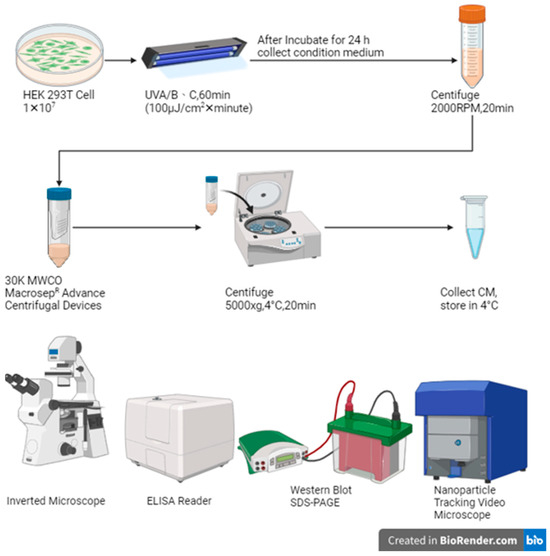
Figure 1.
Materials and method of this study.
2.1. Cell Culture
The protocol for culturing the human embryonic kidney epithelial cell line HEK-293T was propagated in Dulbecco’s modified Eagle’s medium (DMEM, Thermo Fisher Scientific, Waltham, MA, USA) supplemented with 10% fetal bovine serum (FBS, Gibco, Miami, FL, USA) and 1% penicillin/streptomycin (P/S) medium and passaging to another new plate was performed when the cells reach 80% confluence.
- Day 1: Cells were planted at a density of 1 × 10⁷ cells/mL in 10 cm dishes using DMEM medium supplemented with penicillin/streptomycin (P/S) and 10% fetal bovine serum (FBS); triple-plates cells were reserved for the control (Ct).
- Day 2: Irradiating the HEK-293T cells with ultraviolet A/B (UVP, CL-1000 Ultraviolet Crosslinker, Upland, CA, USA) and ultraviolet C (UVP, CL-1000 Ultraviolet Crosslinker, USA) light for 10/30/60 min; triple repeat. Then, the dishes were transferred to a cell culture incubator set at 37 °C with 5% CO2 for 24 h.
- Day 3: The conditioned medium was collected from the dishes.
2.2. Cell Count and Cell Morphology
An electric inverted fluorescence microscope (Zeiss, Axio Vert.A1, Oberkochen, Germany) was used to evaluate cell morphology and calculate the remaining cell count and the viability of each experimental group using a hemocytometer for cell counting under different ultraviolet A/B and ultraviolet C irradiation doses.
2.3. Exosome Concentration
For all the different irradiation doses condition medium was centrifuged at 2000 RPM at 4 °C for 20 min to preliminarily separate cell debris. The debris free sample subsequently transfer to filtration was performed using a 0.45 μm syringe filter. The sample was concentrated 20-fold in a 30K concentrator tube at 5000× g at 4 °C for 20 min and stored in a refrigerator at 4 °C.
2.4. Bradford Assay
A series of protein standards with known concentrations (0.05, 0.1, 0.2, and 0.4 mg/mL) were prepared. The different irradiation ultraviolet A/B doses were irradiated for 0, 10, 30, and 60 min. The Bradford reagent of 20% Coomassie Brilliant Blue dye (Bio-Rad, Thermo Fisher Scientific, Waltham, MA, USA) was added to both standards and concentration exosomes. The mixtures were incubated were incubated for a brief period to allow dye–protein binding. Then, the absorbance of each sample was measured at a specific wavelength using a multifunctional full-spectrum analyzer (Bio Tek, Synergy 2, Winooski, VT, USA). A standard curve was plotted using the absorbance values of the standards to determine the protein concentration of the concentration exosomes.
2.5. Nanoparticle Tracking Analysis (NTA)
The nanoparticle tracking analyzer (Particle Metrix, ZetaView, Inning am Ammersee, Germany) was used to analyze the size distribution and concentration of exosomes to verify the purity of the material in the conditioned medium (CM).
2.6. Western Blot
Western blot analysis was performed to detect the expression of CD9, CD63, and CD81 in the samples extracted from HEK-293T cells, using a high-performance chemiluminescence analyzer (Chemi Doc Touch Imaging System, Bio-Rad, Hercules, CA, USA).
3. Results
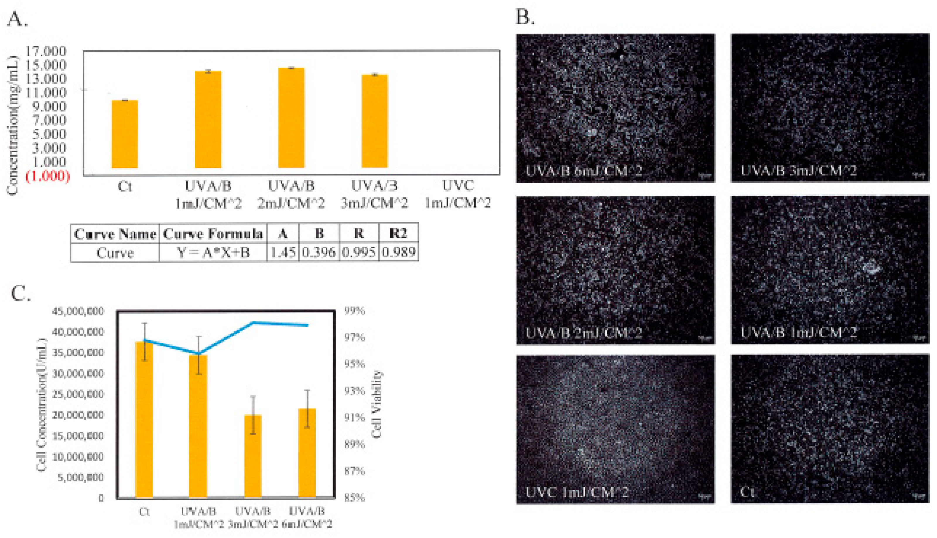
The results were compared to the control with similar levels observed in previous studies, including ultraviolet A/B and ultraviolet C, which enhanced the secretion of exosomes by HEK-293T-targeted vectors for KRAS inhibition in pancreatic cancer. A comparison of ultraviolet A/B and ultraviolet C irradiation is shown in Figure 2. A multifunctional full-spectrum analyzer measures the protein concentrations recovered from lysates by Bradford assay(A). Cell numbers increased with increasing doses of ultraviolet A/B within a certain range, but cell numbers dropped rapidly with 1 mJ/cm2 doses of ultraviolet C irradiation. An inverted fluorescent microscope was used to see the cell morphology and alive/dead cell(B), that was exposed to varying doses of ultraviolet A/B and ultraviolet C irradiation, but the cells that were exposed to 1 mJ/cm2 doses of ultraviolet C irradiation were all dead. Using a hemocytometer grid cell counter, the remaining cells and viability were measured. For varying doses of ultraviolet A/B, the viability was higher than 95% (C).
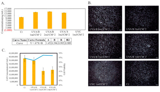
Figure 2.
Comparison of UVA/B and UVC. (A) Using a series of protein standards with known concentrations to measure the protein concentration for different irradiation ultraviolet A/B and ultraviolet C of the concentration exosomes. (B) Using an electric inverted fluorescence microscope to evaluate cell morphology and alive/dead cell. (C) Using a hemocytometer for cell counting and under different ultraviolet A/B and ultraviolet C irradiation doses and calculate the remaining cell count and the viability of each experimental group.
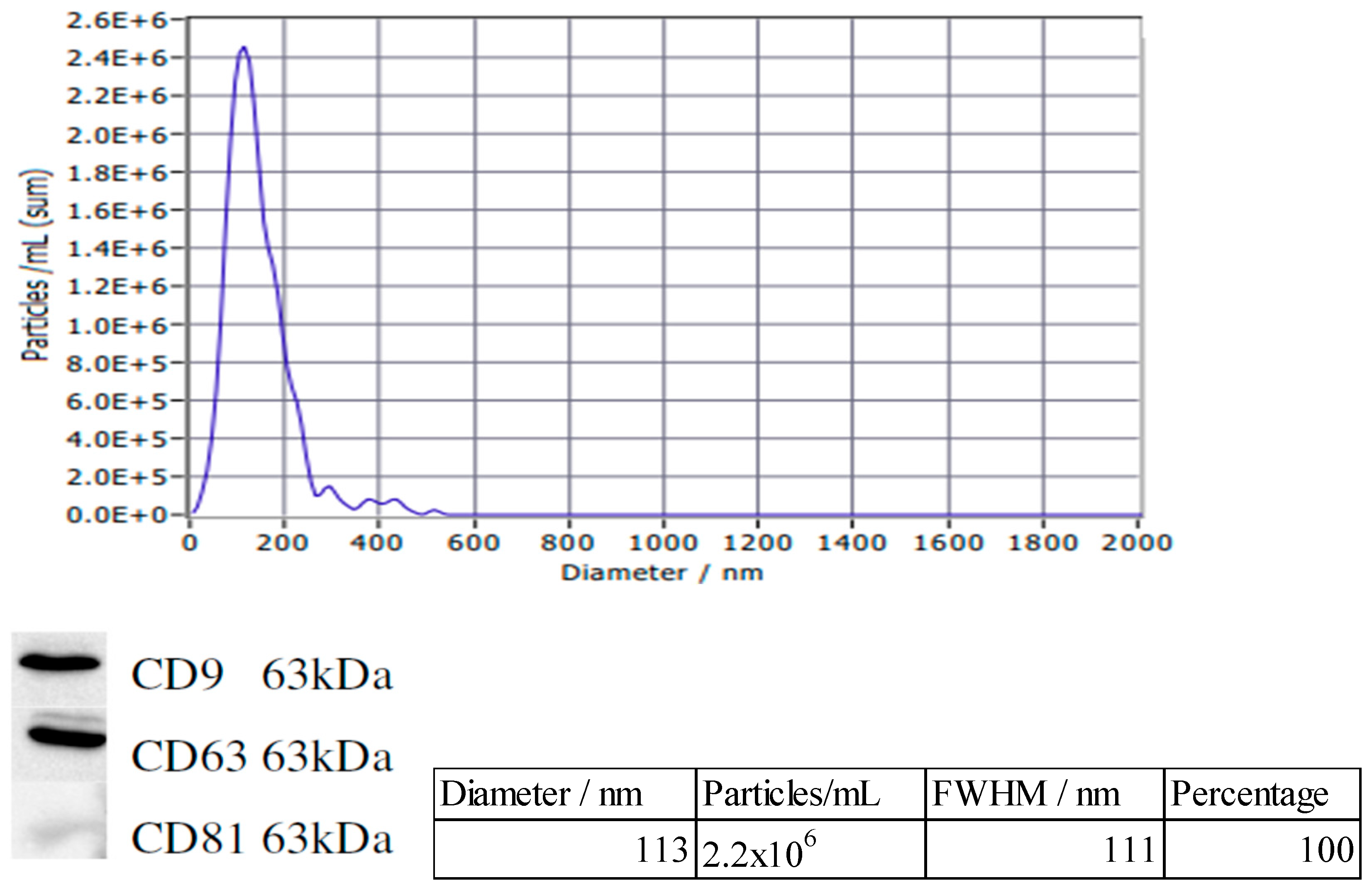
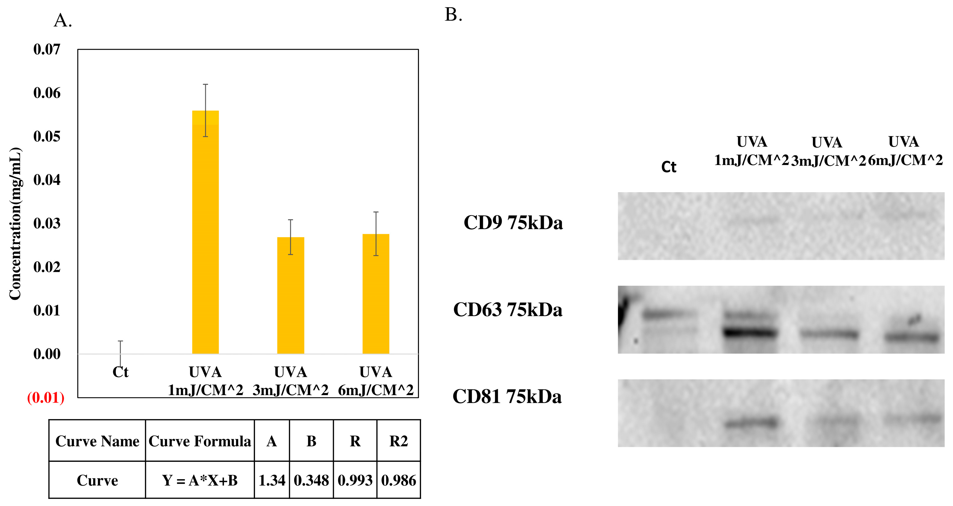
The nanoparticle tracking analysis results are shown in Figure 3 in order to analyzing the size of concentration exosomes, the peak value are 113 nm and the full width at half maximum (FWHM) value are 111 nm, typically have a size distribution ranging from approximately 50 to 150 nm in diameter, the nanoparticle tracking analysis result, we can ensured the purityof the substances present in the conditioned medium (CM). The outer membrane of the exosomes secreted by HEK-293T cells expresses the proteins CD9, CD63, and CD81. The effect of varying doses of ultraviolet A/B irradiation on exosome production is shown in Figure 4. Using the Bradford assay to measure exosome concentration (A), the expression levels of CD9, CD63, and CD81 in Western blot analysis were analyzed (B), with the results aligning with the Bradford assay findings.
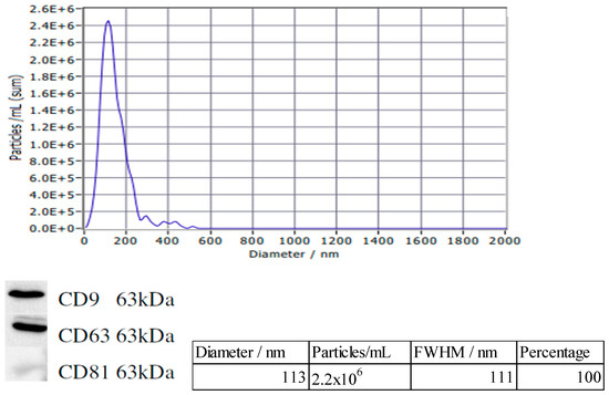
Figure 3.
Characterization of exosomes.

Figure 4.
Comparison of UVA/B and UVC. (A) Using a series of protein standards with known concentrations to measure the protein concentration for different irradiation ultraviolet A/B of the concentration exosomes. (B) The expression levels of CD9, CD63, and CD81 in Western blot analysis.
4. Conclusions
Preliminary studies demonstrated that cell count and Bradford assay expression were drop rapidly with 1 mJ/CM2 doses of ultraviolet C irradiation, and the same result exposed to another doses. Using the Bradford Assay to measure the protein concentration of the concentration exosomes, and with the Western blot analysis to analyze the expression levels of CD9, CD63, and CD81. The results of comparing the two measurement methods both reach the maximum at the 1 mJ/CM2 doses of ultraviolet A/B irradiation. This study introduces a comprehensive approach to ultraviolet A/B irradiation at the 1 mJ/CM2 dose, which can maximum enhancement the secretion of exosomes by HEK-293T targeted vectors for KRAS inhibition in pancreatic cancer.
Author Contributions
Conceptualization, C.-C.C., P.L., R.-H.C., Y.X. and Y.-J.L.; methodology, C.-C.C., P.L., R.-H.C. and Y.-J.L.; software, C.-C.C., P.L. and R.-H.C.; validation, C.-C.C., P.L., R.-H.C. and Y.-J.L.; formal analysis, C.-C.C. and P.L.; investigation, C.-C.C. and P.L.; resources, C.-C.C., P.L., R.-H.C. and Y.-J.L.; data curation, C.-C.C. and P.L.; writing—original draft preparation, C.-C.C.; writing—review and editing, C.-C.C.; visualization, C.-C.C.; supervision, C.-C.C.; project administration, C.-C.C.; funding acquisition, C.-C.C. All authors have read and agreed to the published version of the manuscript.
Funding
This research received no external funding.
Institutional Review Board Statement
Not applicable.
Informed Consent Statement
Not applicable.
Data Availability Statement
Data are contained within the article.
Conflicts of Interest
The authors declare no conflict of interest.
References
- Henry, B.M.; Skinningsrud, B.; Saganiak, K.; Pękala, P.A.; Walocha, J.A.; Tomaszewski, K.A. Development of the human pancreas and its vasculature—An integrated review covering anatomical, embryological, histological, and molecular aspects. Ann. Anat. 2019, 221, 115–124. [Google Scholar] [CrossRef] [PubMed]
- Baert, A.L.; Delorme, G.; Van Hoe, L. (Eds.) The pancreas: Normal radiological anatomy and variants. In Radiology of the Pancreas; Springer: Berlin/Heidelberg, Germany, 1999; pp. 19–68. [Google Scholar]
- Liu, J.; Ye, Z.; Xiang, M.; Chang, B.; Cui, J.; Ji, T.; Zhao, L.; Li, Q.; Deng, Y.; Xu, L.; et al. Functional extracellular vesicles engineered with lipid-grafted hyaluronic acid effectively reverse cancer drug resistance. Biomaterials 2019, 223, 119475. [Google Scholar] [CrossRef] [PubMed]
- Shen, Z.; Sun, J.; Shao, J.; Xu, J. Ultraviolet B irradiation enhances the secretion of exosomes by human primary melanocytes and changes their exosomal miRNA profile. PLoS ONE 2020, 15, e0237023. [Google Scholar] [CrossRef] [PubMed]
- Wang, C.; Li, N.; Li, Y.; Hou, S.; Zhang, W.; Meng, Z.; Wang, S.; Jia, Q.; Tan, J.; Wang, R.; et al. Engineering a HEK-293T exosome-based delivery platform for efficient tumor-targeting chemotherapy/internal irradiation combination therapy. J. Nanobiotechnol. 2022, 20, 247. [Google Scholar] [CrossRef] [PubMed]
Disclaimer/Publisher’s Note: The statements, opinions and data contained in all publications are solely those of the individual author(s) and contributor(s) and not of MDPI and/or the editor(s). MDPI and/or the editor(s) disclaim responsibility for any injury to people or property resulting from any ideas, methods, instructions or products referred to in the content. |
© 2025 by the authors. Licensee MDPI, Basel, Switzerland. This article is an open access article distributed under the terms and conditions of the Creative Commons Attribution (CC BY) license (https://creativecommons.org/licenses/by/4.0/).