Proceeding Paper

Optimization of the Spinning Parameters for Wire-Based Electrospinning of Casein–PEO Nanofiber Mats †

1
Faculty of Engineering and Mathematics, Bielefeld University of Applied Sciences and Arts, 33619 Bielefeld, Germany
2
Chemical and Biological Department, Science School, Universidad de las Américas Puebla, San Andrés Cholula, Puebla 72810, Mexico
3
Department of Physical and Biophysical Chemistry (PC III), Faculty of Chemistry, Bielefeld University, 33615 Bielefeld, Germany
*
Author to whom correspondence should be addressed.
Presented at the 1st International Online Conference on Bioengineering, 16–18 October 2024; Available online: https://sciforum.net/event/IOCBE2024.
Eng. Proc. 2024, 81(1), 7; https://doi.org/10.3390/engproc2024081007
Published: 14 February 2025
(This article belongs to the Proceedings of The 1st International Online Conference on Bioengineering)

Abstract

:
Casein is a natural milk protein that has been investigated for various applications. Casein nanofibers are of great interest for tissue engineering. Spinning casein alone has proven difficult due to its unsuitable viscoelasticity and extensive intermolecular interactions. Our study aims at optimizing casein/poly(ethylene oxide) (PEO) spinning solutions for needleless electrospinning. For this purpose, PEO with different molecular weights was mixed with different caseins in different ratios and processed with different spinning parameters. Scanning electron microscopy (SEM) images show the impact of these parameters on the diameter distribution and morphology of the nanofibers. The paper gives the first overview of the optimum spinning parameters for wire-based electrospinning of casein/PEO nanofiber mats that are mostly standard values (maximum electrode–electrode distance, carriage speed of 100 mm/s, and PEO with 300 kDa molecular weight), combined with the addition of beeswax to improve fiber morphology and reduce beads.

1. Introduction

The natural milk protein casein is used in a wide range of applications, such as in drug delivery, the food industry, cartilage tissue engineering, and other biotechnological and biomedical applications [1,2,3]. Casein nanofibers in particular are intensively investigated for tissue engineering [4,5,6]. Electrospinning of casein is usually performed as co-electrospinning with a spinning agent, such as poly(vinyl alcohol), poly(caprolactone) (PCL), or poly(ethylene oxide) (PEO) [7,8], as its insufficient viscoelasticity and extensive intermolecular interactions usually prevent sole spinning [9,10,11]. Only recently, electrospinning of pure casein from solutions with a pH > 9 and a high ethanol content of >40%, ideally 60%, has been reported [12]. Spinning from flammable solvents, however, is problematic for spinning in a strong electric field.
Here, we report the results of wire-based electrospinning of aqueous casein–PEO solutions with different molecular weights (6 kDa–1 MDa), mixed in various ratios with casein of different quality. While a previous study investigated PAN–casein nanofiber mats electrospun by a wire-based technique [13], no previous reports of wire-based electrospinning of casein–PEO were found in the literature. Our paper thus provides the first overview of the optimal spinning parameters for wire-based electrospinning of casein–PEO nanofiber mats.

2. Materials and Methods

Spinning solutions were prepared from casein (according to Hammersten), PEO (Sigma-Aldrich, Saint Louis, MO, USA), and distilled water in different ratios. The molecular weight of PEO ranged from 6 kDa to 1 MDa, as specified in Table 1, where the mass ratios of the different components are also described. Solutions were stirred with a magnetic stirrer at 600–800 rpm for a minimum of 1 day at room temperature. In general, a PEO–water solution was first prepared and stirred before casein was added.
Electrospinning was performed with the wire-based device “Nanospider Lab” (Elmarco Ltd., Liberec, Czech Republic). The following spinning parameters were kept constant: substrate speed of 0 mm/min, substrate–counter electrode distance of 50 mm, voltage of 75–80 kV, nozzle of 0.9 mm, and duration of 30 min. The varying spinning parameters are given in Table 2.
The samples were characterized by confocal laser scanning microscopy (CLSM) using a VK-8710 (Keyence, Neu-Isenburg, Germany). Higher resolution images were obtained using a scanning electron microscope (SEM) Phenom ProX G3 Desktop SEM (Thermo Fisher Scientific, Waltham, MA, USA).

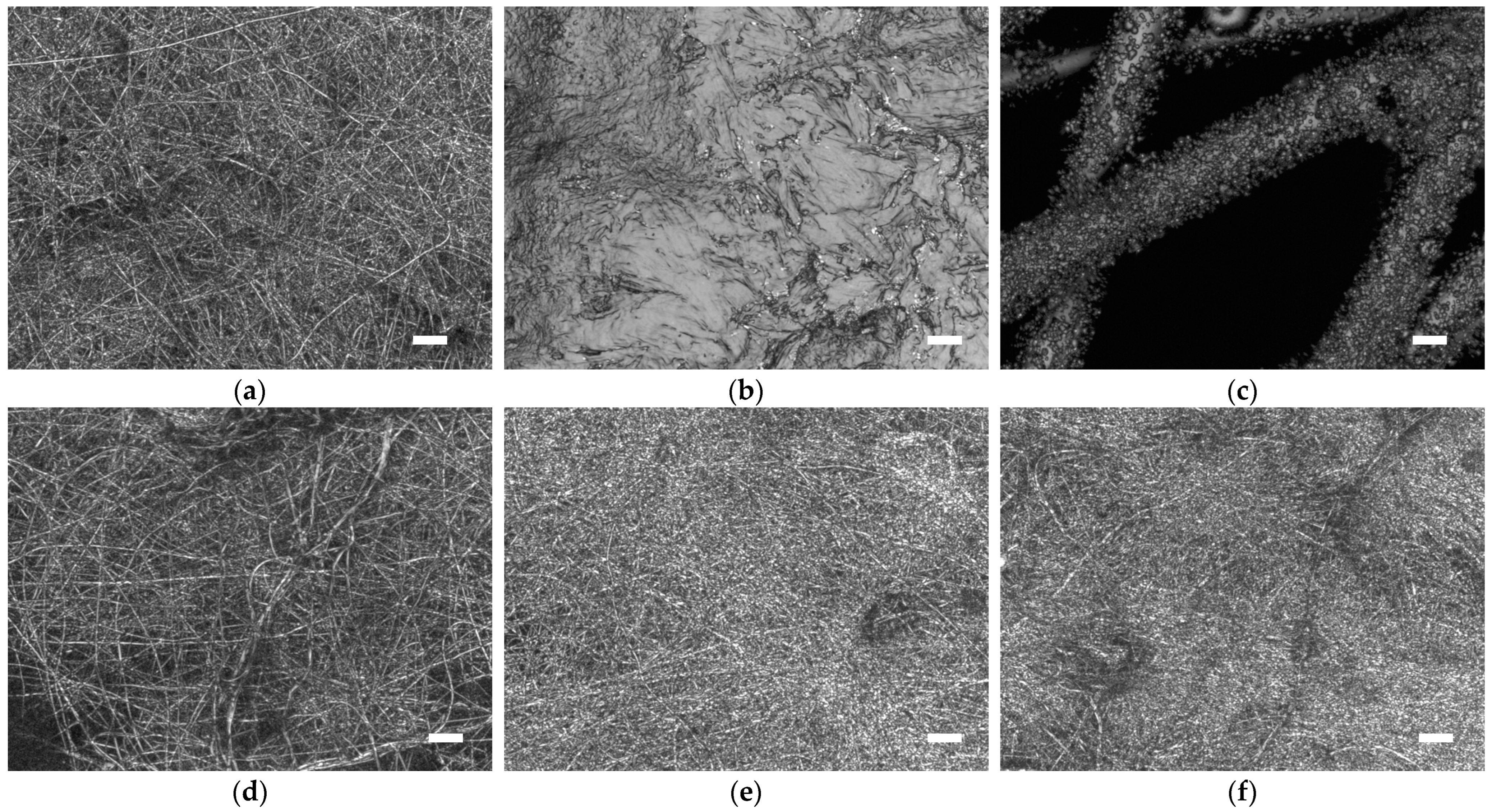
3. Results and Discussion

The different solution and spinning parameters led to quite different spinning results, with electrospun nanofiber mats being formed in most cases, but also with electrospraying (samples 5 and 6) occurring as well as the formation of small spheres on the substrate without a fibrous structure (samples 3 and 4). This leads to the first conclusion that electrospraying occurs for the PEO with the smallest molecular weights and the formation of non-fibrous spheres occurring for the samples including PEO with slightly higher molecular weight (cf. Table 1). An overview of the CLSM images of some samples is given in Figure 1.
In addition to the samples where no nanofibers have formed (Figure 1b,c), different fiber diameters and surface morphologies are visible in Figure 1a,d–f. This is why some of the fibrous samples were also investigated by SEM. Representative images are shown in Figure 2. While Figure 2a gives an overview of sample 1 with a nominal magnification of 1000×, the other images have nominal magnifications of 5000×. All SEM images show nanofiber mats with arbitrarily oriented fibers; however, there are clear differences between some of them. Sample 2 (Figure 2c), e.g., appears to contain molten areas in the large pores between the fibers. This is, on the one hand, unexpected since high molecular weight PEO with 1000 kDa is often used for electrospinning [14]; on the other hand, similar morphologies can be found not only after heat treatment of nanofiber mats [15], but also after the crosslinking of high molecular weight PEO [16]. In the future, atomic force microscopy (AFM) measurements will be performed to distinguish the different polymers in this nanofiber mat [17].
The other nanofiber mats show thicker or thinner nanofibers, sometimes relatively straight, especially in the case of sample 8 (Figure 2e) which is more wiggly. It should be mentioned that sample 8 belongs to the samples that were spun at higher humidity in the spinning chamber, and it is known from the literature that straighter PEO fibers can be spun at a lower relative humidity [18,19].
Another factor influenced by humidity, but also by the molecular weight and the solid content in the solution, is the fiber diameter. Figure 3 thus shows representative fiber diameters, calculated from the SEM images in Figure 2 using ImageJ 1.51j8 (National Institutes of Health, Bethesda, MD, USA).
Comparing Figure 3b–f, the widest diameter distribution is visible, as expected, for sample 2 (Figure 3c). The smallest diameters and the narrowest distributions were found for samples 1 and 11 (Figure 3b,f), as can also be seen from the comparison in Figure 3a, which is advantageous for many applications. Both these samples were spun with standard parameters (cf. Table 2)—with the addition of beeswax in sample 11—and 3.2 g PEO with 300 kDa in 36.8 g water with an addition of 1.5 g or 3.0 g casein for samples 1 and 11, respectively (cf. Table 1). The visual comparison of sample 1 (Figure 2b) and sample 11 (Figure 2f) shows that the latter has a more regular morphology, indicating that the higher casein content is preferable. Moreover, more tests with beeswax are required as sample 11 contains no visible beads or other irregularities of the fibers.

4. Conclusions

Electrospinning of PEO–casein nanofiber mats with various PEO molecular weights, blending ratios, and solid contents in the aqueous solution was performed using a wire-based device. To obtain fibrous samples instead of electrosprayed droplets or non-fibrous spheres on the substrate, PEO with a minimum molecular weight of 100 kDa was required. The standard spinning parameters were most useful, while higher relative humidity, a smaller distance between the electrode and substrate, etc., resulted in less homogeneous nanofiber mats. The smallest average fiber diameter in combination with the narrowest diameter distribution was achieved with 3.2 g PEO 300 kDa, 3.0 g casein, and 10 g beeswax in 36.8 g distilled water, suggesting further experiments with beeswax as a fiber coating as well as tests of the water resistance of these nanofiber mats depending on the amount of beeswax. Regular nanofibers with a certain amount of water resistance could be used in biotechnological and biomedical applications such as drug delivery and cartilage tissue engineering. Future investigations of the nanofiber mats’ mechanical properties as well as the growth of mammalian cells on such nanofibrous PEO–casein substrates are planned to evaluate their usability in tissue engineering.

Author Contributions

Conceptualization, T.G.; methodology, T.G. and A.E.; formal analysis, A.E.; investigation, H.G.R.C. and U.G.; writing—original draft preparation, A.E.; writing—review and editing, all authors; visualization, U.G. and A.E. All authors have read and agreed to the published version of the manuscript.

Funding

This research received no external funding.

Institutional Review Board Statement

Not applicable.

Informed Consent Statement

Not applicable.

Data Availability Statement

All data are included in this paper.

Acknowledgments

This project was supported by the students’ exchange from Universidad de las Américas Puebla (UDLAP), which has a several year-long trajectory of cooperation in student and staff exchange, and conjoint projects with HSBI.

Conflicts of Interest

The authors declare no conflicts of interest.

References

  1. Gandhi, S.; Roy, I. Drug delivery applications of casein nanostructures: A minireview. J. Drug Deliv. Sci. Technol. 2021, 66, 102843. [Google Scholar] [CrossRef]
  2. Biranje, S.S.; Sun, J.Z.; Cheng, L.; Cheng, Y.; Shi, Y.F.; Yu, S.J.; Jiao, H.X.; Zhang, M.; Lu, X.C.; Han, W.J.; et al. Development of Cellulose Nanofibril/Casein-Based 3D Composite Hemostasis Scaffold for Potential Wound-Healing Application. ACS Appl. Mater. Interfaces 2022, 14, 3792–3808. [Google Scholar] [CrossRef] [PubMed]
  3. Wu, X.C.; Hu, Q.Y.; Liang, X.R.; Fang, S. Fabrication of colloidal stable gliadin-casein nanoparticles for the encapsulation of natamycin: Molecular interactions and antifungal application on cherry tomato. Food Chem. 2022, 391, 133288. [Google Scholar] [CrossRef] [PubMed]
  4. Movahedi, M.; Salehi, A.O.M.; Hajipour, F.P.; Eternad, S. Casein release and characterization of electrospun nanofibres for cartilage tissue engineering. Bull. Mater. Sci. 2022, 45, 76. [Google Scholar] [CrossRef]
  5. Sendil, Ö.; Yilmaz, S.S.; Ozcelik, E.Y.; Uzuner, H.; Aytac, A. Cross-linked electrospun polyvinyl alcohol/sodium caseinate nanofibers for antibacterial applications. Vinyl Addit. Technol. 2023, 29, 48–65. [Google Scholar] [CrossRef]
  6. George, N.; Chakraborty, S.; Mary, N.L.; Suguna, L. Incorporating silver nanoparticles into electrospun nanofibers of casein/polyvinyl alcohol to develop scaffolds for tissue engineering. Int. J. Biol. Macromol. 2024, 267, 131501. [Google Scholar] [CrossRef] [PubMed]
  7. John, N. Electrospinning of Biofibers and their Applications. In Electrospun Nanofibers from Bioresources for High-Performance Applications, 1st ed.; CRC Press: Boca Raton, FL, USA, 2022; pp. 29–48. [Google Scholar]
  8. Golba, B.; Kalaoglu-Altan, O.I.; Sanyal, R.; Sanyal, A. Hydrophilic Cross-Linked Polymeric Nanofibers Using Electrospinning: Imparting Aqueous Stability to Enable Biomedical Applications. ACS Appl. Polym. Mater. 2022, 4, 1–17. [Google Scholar] [CrossRef]
  9. Selvaraj, S.; Thangam, R.; Fathima, N.N. Electrospinning of casein nanofibers with silver nanoparticles for potential biomedical applications. Electrospinning of casein nanofibers with silver nanoparticles for potential biomedical applications. Int. J. Biol. Macromol. 2018, 120, 1674–1681. [Google Scholar] [CrossRef] [PubMed]
  10. Dai, J.M.; Hu, W.; Yang, H.Y.; Li, C.Z.; Cui, H.Y.; Li, X.Z.; Lin, L. Controlled release and antibacterial properties of PEO/casein nanofibers loaded with Thymol/β-cyclodextrin inclusion complexes in beef preservation. Food Chem. 2022, 382, 132369. [Google Scholar] [CrossRef] [PubMed]
  11. Tomasula, P.M.; Sousa, A.M.M.; Liou, S.-C.; Li, R.; Bonnaillie, L.M.; Liu, L.S. Electrospinning of casein/pullulan blends for food-grade applications. J. Dairy Sci. 2016, 99, 1837–1845. [Google Scholar] [CrossRef] [PubMed]
  12. Sharma, D.; Ziegler, G.R.; Harte, F.M. Ethanol-mediated electrospinning of casein-only bead-free nanofibers. Food Hydrocoll. 2024, 148, 109503. [Google Scholar] [CrossRef]
  13. Diestelhorst, E.; Mance, F.; Mamun, A.; Ehrmann, A. Chemical and Morphological Modification of PAN Nanofibrous Mats with Addition of Casein after Electrospinning, Stabilisation and Carbonisation. Tekstilec 2020, 63, 38–49. [Google Scholar] [CrossRef]
  14. Grothe, T.; Großerhode, C.; Hauser, T.; Kern, P.; Stute, K.; Ehrmann, A. Needleless electrospinning of PEO nanofiber mats. Adv. Eng. Res. 2017, 102, 54–58. [Google Scholar]
  15. Wehlage, D.; Blattner, H.; Sabantina, L.; Böttjer, R.; Grothe, T.; Rattenholl, A.; Gudermann, F.; Lütkemeyer, D.; Ehrmann, A. Sterilization of PAN/Gelatine Nanofi brous Mats for Cell Growth. Tekstilec 2019, 62, 78–88. [Google Scholar] [CrossRef]
  16. Kianfar, P.; Vitale, A.; Dalle Vacche, S.; Bongiovanni, R. Enhancing properties and water resistance of PEO-based electrospun nanofibrous membranes by photo-crosslinking. J. Mater. Sci. 2021, 56, 1879–1896. [Google Scholar] [CrossRef]
  17. Werner, E.; Güth, U.; Brockhagen, B.; Döpke, C.; Ehrmann, A. Examination of Polymer Blends by AFM Phase Images. Technologies 2023, 11, 56. [Google Scholar] [CrossRef]
  18. Tripatanasuwan, S.; Zhong, Z.X.; Reneker, D.H. Effect of evaporation and solidification of the charged jet in electrospinning of poly(ethylene oxide) aqueous solution. Polymer 2007, 48, 5742–5746. [Google Scholar] [CrossRef]
  19. Pelipenko, J.; Kristl, J.; Jankovic, B.; Baumgartner, S.; Kocbek, P. The impact of relative humidity during electrospinning on the morphology and mechanical properties of nanofibers. Int. J. Pharm. 2013, 456, 125–134. [Google Scholar] [CrossRef] [PubMed]
Figure 1. Confocal laser scanning microscope (CLSM) images of samples (a) 1, (b) 4, (c) 5, (d) 7, (e) 8, and (f) 12. Scale bars correspond to 10 µm.
Engproc 81 00007 g001
Figure 2. Scanning electron microscopy (SEM) images of sample 1 (a) with 1000× nominal magnification and samples (b) 1, (c) 2, (d) 7, (e) 8, and (f) 11 with 5000× nominal magnification.
Engproc 81 00007 g002
Figure 3. (a) Fiber diameters for the samples under examination; diameter distributions for samples (b) 1, (c) 2, (d) 7, (e) 8, and (f) 11.
Engproc 81 00007 g003
Table 1. Spinning solutions for samples under investigation.
Sample No.Mol. Weight PEO (kDa)Mass PEO/gMass Casein/gMass Water/g
13003.21.536.8
210003.23.036.8
34010.03.030.0
41734.43.05.6
51032.03.08.0
6638.03.02.0
73003.23.036.8
83003.23.036.8
93003.23.036.8
103003.24.036.8
113003.23.036.8
123003.21.036.8
1310003.23.036.8
143003.23.036.8
156002.81.537.2
163003.21.036.8
173003.22.036.8
Table 2. Spinning parameters for samples under investigation. Parameters deviating from the standard ones are written in italics.
Sample No.Electrode–Electrode
Distance (mm)
Carriage Speed (mm/s)Rel. Humidity (%)Others
124010032–33
224010032–33
312010032–33
412010032–33
512010032–33
612010032–33
724010034–37
824010037–40
924010036–39
102405032–33
1124010032–33+10 g beeswax (Lembcke, Faulenrost, Germany)
1224010032–334 days stirring
132405032–33
1424010032–337 days at 40 °C
1524010032–33
1624010032–331 day at 40 °C
1724010032–337 days at 40 °C
Disclaimer/Publisher’s Note: The statements, opinions and data contained in all publications are solely those of the individual author(s) and contributor(s) and not of MDPI and/or the editor(s). MDPI and/or the editor(s) disclaim responsibility for any injury to people or property resulting from any ideas, methods, instructions or products referred to in the content.

