
Proceeding Paper

Optimizing Post-Curing Protocols for 3D-Printed Dental Models: A Long-Term Evaluation of Mechanical and Physical Properties †
by
Irina Besliu Bancescu
1,
Roxana Gheorghita
2,* and
Alexandru Nemtoi
2
1
Faculty of Mechanical Engineering, Automotive Engineering and Robotics, Stefan cel Mare University of Suceava, 720229 Suceava, Romania
2
Faculty of Medicine and Biological Sciences, Stefan cel Mare University of Suceava, 720229 Suceava, Romania
*
Author to whom correspondence should be addressed.
Presented at the International Conference on Electromagnetic Fields, Signals and BioMedical Engineering (ICEMS-BIOMED), Suceava, Romania, 7–9 May 2026.
Eng. Proc. 2026, 148(1), 2; https://doi.org/10.3390/engproc2026148002
Published: 30 June 2026

Abstract

This study evaluated the influence of post-polymerization duration and a 14-day aging period on the mechanical and physical properties of 3D-printed standard resin models. Methods: Specimens were printed using Anycubic resin (50 µm layers) and post-cured for 5, 7, and 10 min. Evaluations included mass variation, chromatic stability, confocal microscopy, and point-indentation tests using a Kistler 9257B dynamometer. Extended post-curing significantly enhanced structural stability. The 10-min aged group (O10) achieved the highest failure threshold at 1862.93 N (a 44% increase from baseline) and the most homogeneous microtopography (Z = 0.0826 mm). A 10-min post-curing cycle is the optimal threshold for maximizing mechanical resilience and minimizing volatile-induced mass loss in standard 3D-printed resins.

1. Introduction

3D printing represents a revolutionary innovation with vast applications in both medicine and dentistry. Continued technological advances are driving the development of advanced solutions, redefining medical practices and fostering a more efficient, personalized approach to dental care [1]. Specifically, resin-based 3D printing enables the fabrication of high-resolution structures with sharp edges and fine details, achieving resolutions up to 25 microns through technologies such as SLA (stereolithography) and DLP (digital light processing). This precision significantly exceeds the limits of conventional milling—often restricted by bur diameter—and traditional impressioning techniques, which are prone to dimensional instability [1,2]. Despite this rapid progress, systematic studies analyzing the long-term behavior of standard resins under aging factors remain surprisingly limited. Much of the current research focuses on immediate material characterization, often overlooking microstructural evolution and the subsequent decrease in impact or compression resilience as the polymer ages. This assessment is critical, as the mechanical properties and dimensional stability of light-cured resins are not static post-printing. For instance, Pérez et al. demonstrated that artificial aging performed according to ISO 4892-2:2013 (Plastics—Methods of Exposure to Laboratory Light Sources—Part 2: Xenon-Arc Lamps) [3] using the M2.1 exposure cycle induces significant changes in absorption and reflection coefficients, leading to color variations that exceed clinical acceptability thresholds [4]. However, optimizing the post-processing protocol can mitigate these effects. Lee et al. established that extending post-polymerization time to 20 min significantly improves the degree of conversion (DC) and chromatic stability, while reducing water sorption and solubility [5]. The impact of material composition and storage conditions is equally decisive. Reymus and Stawarczyk highlighted that while high-filler resins exhibit superior Martens hardness (HM) and elastic modulus, prolonged storage in distilled water (28 days at 37 °C) induces progressive degradation [6]. Furthermore, research by Mudhaffer et al. indicates that while printing orientation has a minimal effect on Martens parameters, standard resins remain more susceptible to aging than milled blocks, showing comparable hardness reduction in both distilled water and artificial saliva [7]. Comparative studies by Bento et al. further confirm that 3D-printed resins record lower microhardness and flexural strength values after 20,000 thermal cycles compared to CAD/CAM-milled resins, which perform similarly to conventional heat-polymerized PMMA [8]. Nevertheless, technology-specific differences exist; Topsakal et al. observed that thermocycling affects DLP-processed resins more significantly than SLA variants. Despite this, both technologies maintain sufficient structural integrity to withstand physiological occlusal forces [9]. Interestingly, from an aesthetic perspective, 3D-printed resins may exhibit superior chromatic stability compared to traditional PMMA, even with an increased susceptibility to surface degradation [10]. These findings underscore the significant clinical potential of additive manufacturing, provided that chemical compositions and post-processing protocols continue to be optimized for long-term durability.
The primary objective of this study was to evaluate the aging process of 3D-printed resin models and to identify subsequent alterations in their physical and mechanical properties. Furthermore, the research investigated the influence of post-polymerization treatment duration as a critical factor in stabilizing these characteristics. The working hypothesis posits that 3D-printed resin models exhibit a decelerated aging process when subjected to extended post-polymerization protocols, which may enhance their structural longevity. This focus aligns with broader trends in the field, as 3D printing represents a revolutionary innovation with vast applications in both medicine and dentistry.

2. Materials and Methods

The study utilized a standard white photopolymer resin produced by Anycubic (Anycubic, Shenzhen, China), characterized as a general-purpose 3D printing material compatible with light-curing systems operating within the 365–405 nm wavelength range. According to the manufacturer, this resin offers high rigidity, low shrinkage, and high anatomical fidelity. The dental models were generated from intraoral scans and exported as digital .stl files. For print preparation, Lychee Slicer Plus (30-day trial), version 7.6.5 (Mango 3D, Toulouse, France) (Mango 3D, Bordeaux, France) was employed to configure printing parameters, generate support structures, and slice the models. The specimens were fabricated using an Anycubic Photon 5 printer (Shenzhen Anycubic Technology Co., Ltd., Shenzhen, China), selected for its high precision in reproducing dental micro-details. The specific printing parameters were configured as follows: Layer thickness: 0.05 mm; Normal exposure time: 8 s; Off time: 1 s; Bottom exposure time: 60 s; Bottom layers: 6; Anti-aliasing level: 1; Z-axis lift distance: 6 mm; Z-axis lift speed: 3 mm/s; Z-axis retract speed: 3 mm/s. Following the printing process, all specimens were washed in isopropyl alcohol (IPA) to remove residual monomer. To investigate the effect of post-polymerization duration, the models were subjected to controlled UV/thermal exposure using a Nexa3D XiP curing system (Nexa3D, Ventura, CA, USA) for 5, 7, and 10 min, respectively. Mass variations during the study were monitored using a Kern precision balance (KERN & SOHN GmbH, Balingen, Germany; 140 g capacity, 0.001 g accuracy). Chromatic evaluation was performed using a Konica Minolta Chroma Meter CR-400 (Konica Minolta Sensing, Inc., Osaka, Japan) based on the CIE L*a*b* system. This method quantifies color perception through three coordinates: L*: luminosity (0 for absolute black to 100 for absolute white); a*: the green–red axis; b*: the blue–yellow axis. Measurements were conducted under standardized lighting to capture surface reflectance, with baseline values recorded at L* = 93.62, a* = −0.71, and b* = 4.15. The microtopography and surface roughness were analyzed using a MahrSurf CWM100 confocal microscope (Mahr GmbH, Göttingen, Germany). This non-invasive high-resolution technique allowed for a detailed investigation of the resin’s microstructure at the micrometric and submicrometric levels. The mechanical resistance was evaluated through a static point-indentation test. A controlled compressive force was applied using a 6 mm diameter high-speed steel (HSS) spherical punch to identify the material’s failure threshold under concentrated load. Force data were recorded dynamically using a Kistler 9257B piezoelectric dynamometer (Kistler Group, Winterthur, Switzerland), which captured forces (Fx, Fy, Fz) across the three Cartesian axes. This sensor was selected for its high sensitivity and rapid response time, ensuring precise measurement of the mechanical deformation process.

3. Results and Discussion

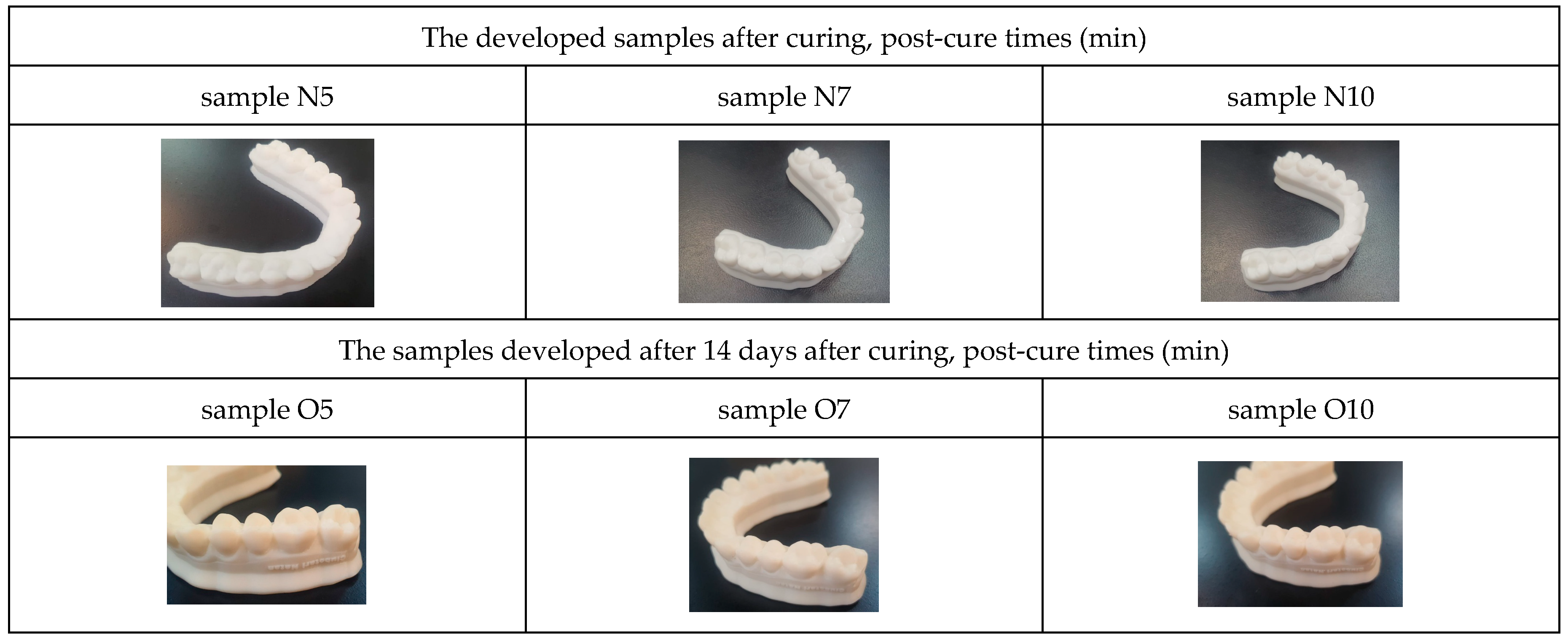
The specimens fabricated using the standard Anycubic resin (Shenzhen Anycubic Technology Co., Ltd., Shenzhen, China) under varying post-polymerization durations are presented in Figure 1. These models constitute the core experimental groups used for subsequent physical and mechanical assessments.
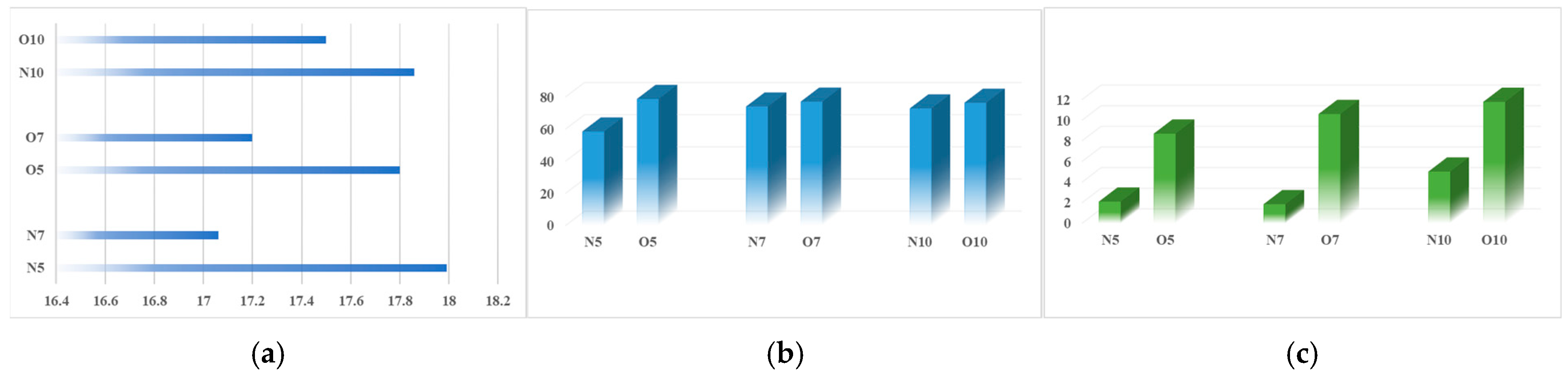
Visual inspection of the specimens revealed a distinct chromatic shift between the initial state and the post-aging phase (14 days). The baseline samples (N5, N7, N10) exhibited a high-luminosity white finish, consistent with the initial colorimetric values (L* = 93.62). After a 14-day aging period, the specimens (O5, O7, O10) displayed a perceptible yellowing effect, indicating chemical instability within the polymer matrix over time. Notably, the severity of the chromatic alteration appeared to be inversely proportional to the post-curing duration; samples subjected to longer UV exposure (10 min) showed a more stabilized surface texture compared to those with shorter curing cycles. The transition from the “N” series to the “O” series reflects the ongoing post-polymerization and oxidative processes occurring within the standard Anycubic resin. The yellowing observed in the 14-day aged samples is a well-documented phenomenon in 3D-printed photopolymers, often attributed to: residual photoinitiators, oxidation as previously discussed in the literature by Pérez et al. [4] and cross-linking density. The comparison between sample O5 and sample O10 suggests that the 10-min post-curing cycle achieves a higher degree of conversion (DC), which effectively “locks” the polymer network, making it more resilient to the environmental factors that trigger yellowing. The mass of the specimens exhibited slight fluctuations throughout the 14-day testing period. Increasing the post-polymerization duration initially enhanced the CIELab color coordinates; however, a significant chromatic shift occurred after the 14-day aging period, characterized by a decrease in the a* parameter and a concomitant increase in b*, indicating a dominant yellow–blue axis transformation over the green–red component. Although the total color change remained within clinically acceptable limits, the significant shift in the b* coordinate—moving toward the yellow spectrum—merits attention. In dental models, this chromatic drift can affect the visualization of fine anatomical details or the perception of fit. Our results indicate that while 10-min post-curing stabilizes the material, it also induces a baseline chromatic shift that practitioners must account for.
As illustrated in Figure 2, the reduction in specimen mass was inversely proportional to the post-curing duration. The most significant stability was recorded for group O10 (10-min treatment), which displayed the lowest mass loss. Conversely, the O5 group showed the highest degree of volatility. These results indicate that extended post-polymerization treatment directly enhances the material’s physical stability by facilitating a higher degree of cross-linking.
The inverse relationship between mass loss and post-curing time can be explained by the Degree of Conversion (DC). When the Anycubic resin is exposed to UV light for a longer interval (10 min vs. 5 min), a higher percentage of carbon–carbon double bonds are converted into single bonds, resulting in a more compact and stable polymer matrix.
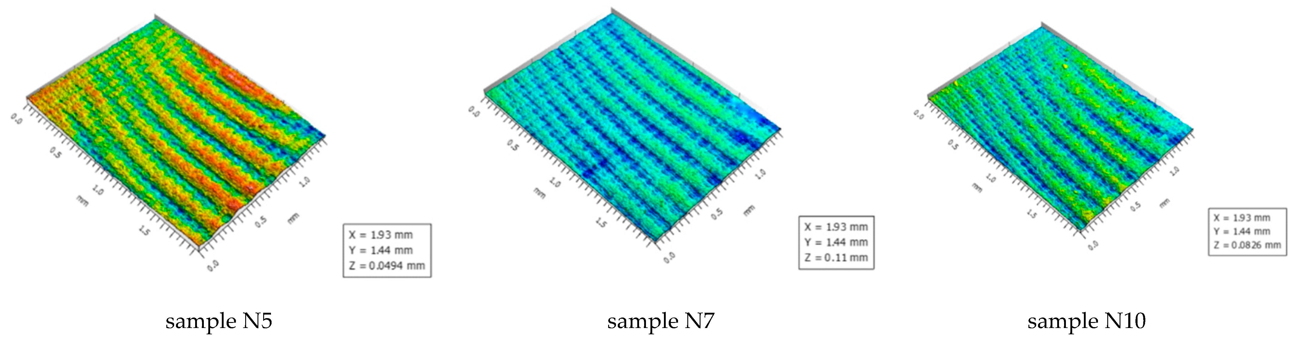
Confocal microscopy images (MahrSurf CWM100; Mahr GmbH, Göttingen, Germany) revealed distinct microtopographical variations across the specimens, directly correlated with the post-polymerization duration (Figure 3). While dimensional consistency was visually maintained, the Z-scale values (representing peak-to-valley distance) highlighted significant differences in surface texture.
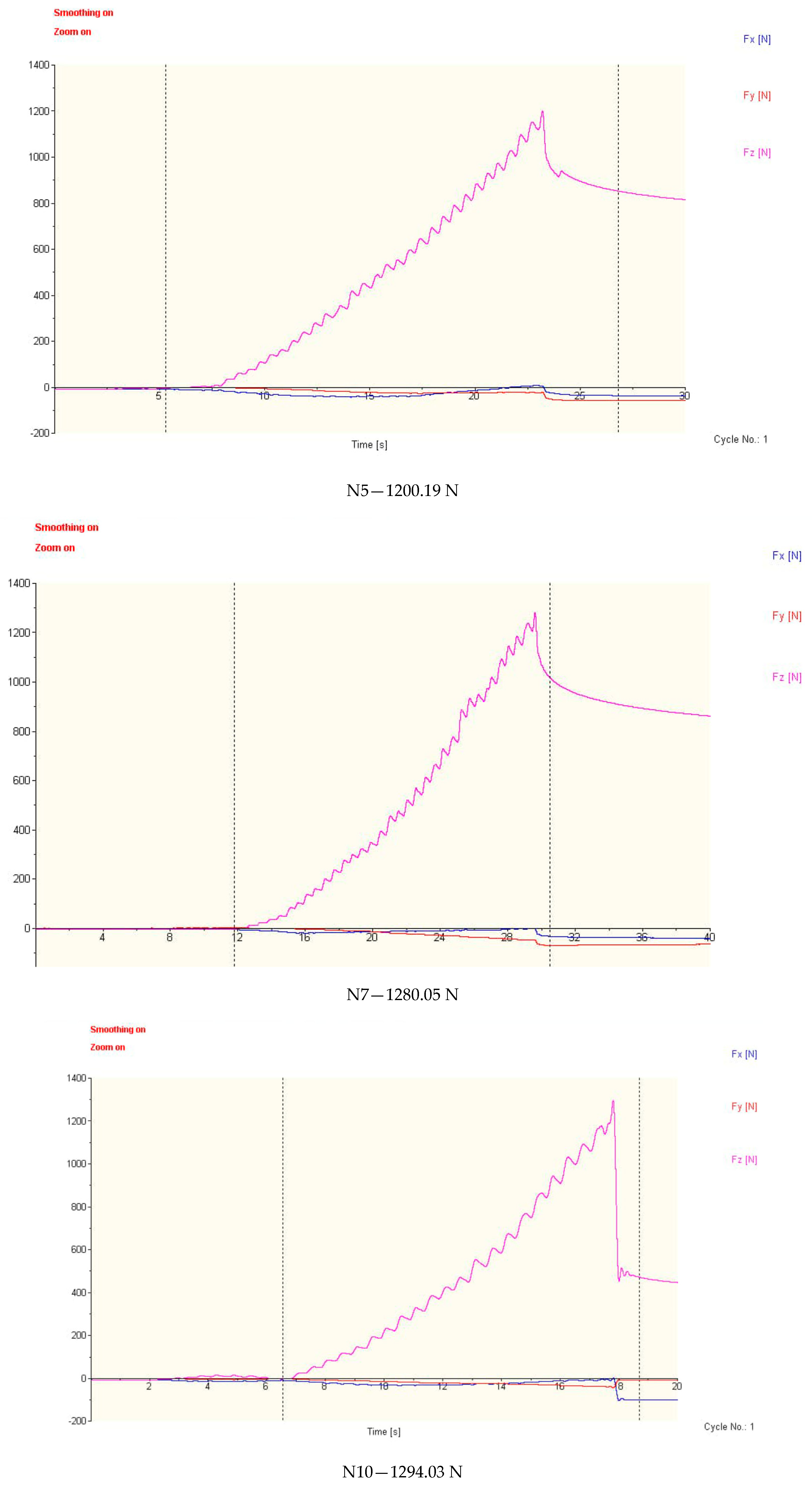
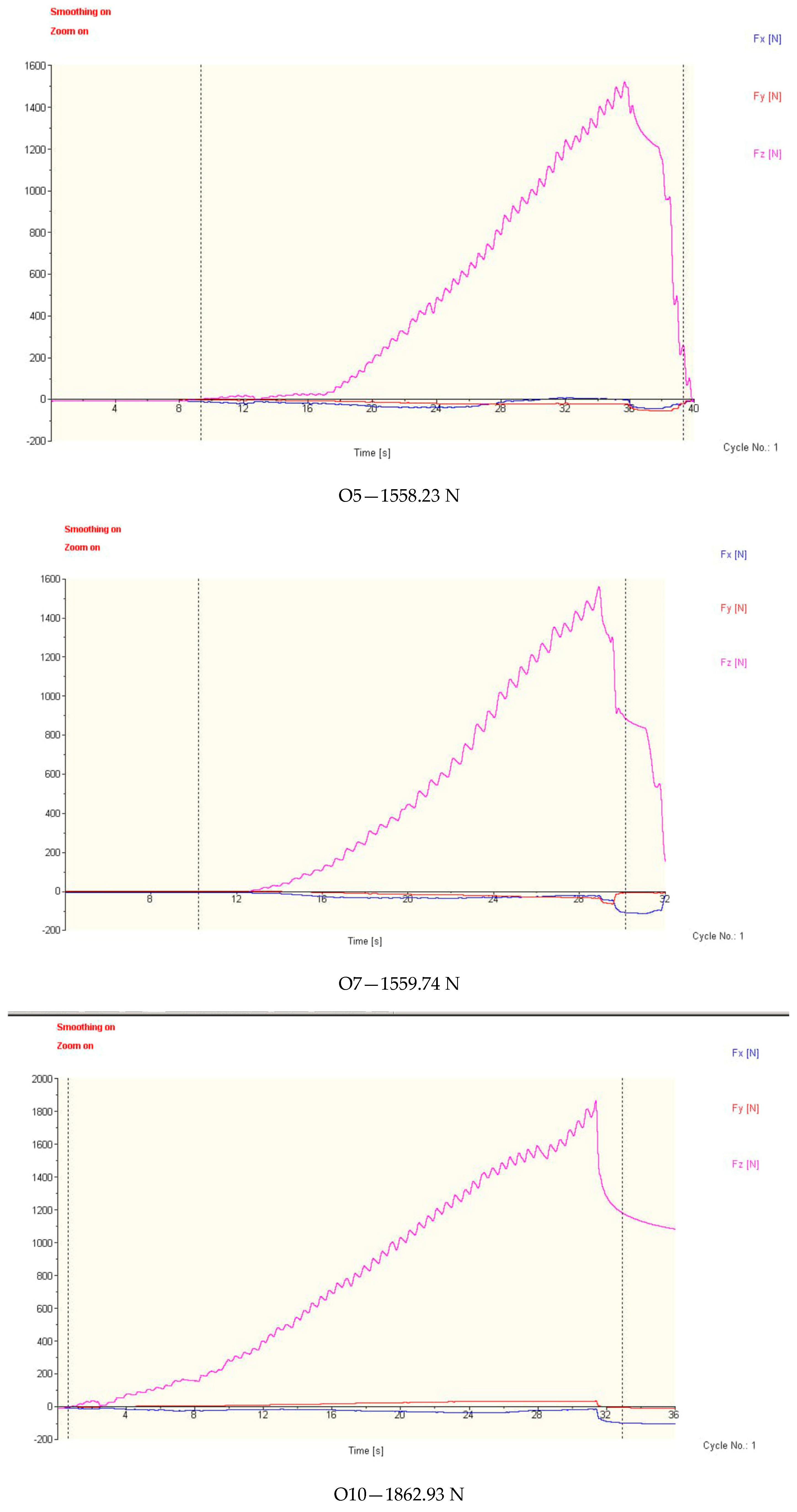
The 5-min curing group displayed the lowest Z-amplitude (0.0494 mm), indicating a relatively flat surface with minimal texture definition. In contrast, the 7-min group exhibited the highest degree of surface roughness, recorded as a maximum Z-scale of 0.11 mm, characterized by deep ridges and valleys. Specimens subjected to 10 min of post-curing showed an intermediate Z-value (0.0826 mm) and a more homogeneous microtopography compared to the 7-min group. The transition in surface morphology from 5 to 7 min reflects the localized thermal and chemical rearrangements occurring during the accelerated photopolymerization phase. As the degree of conversion (DC) increases, the polymer matrix undergoes non-uniform volumetric shrinkage, which can amplify the inherent stratification defects (0.05 mm layer thickness) and lead to the formation of micro-ridges. The subsequent reduction in Z-amplitude between 7 and 10 min (0.11 mm to 0.0826 mm) suggests that extended UV/thermal exposure (10 min) facilitates a more complete and more uniform polymerization across the entire specimen volume. This extended curing likely induces viscoelastic relaxation within the polymer network, partially planarizing the sharp micro-textures formed during the earlier stages. The static point-indentation tests conducted with the Kistler 9257B piezoelectric dynamometer (Kistler Group, Winterthur, Switzerland) identified significant variations in the failure threshold of the specimens. As illustrated in the force-time graphs, both the post-curing duration and the 14-day aging period substantially influenced the maximum force (F_max) required to reach material failure. In the initial state, the specimens exhibited a progressive increase in resistance correlated with post-curing time: 1200.19 N (N5), 1280.05 N (N7), and 1294.03 N (N10). This linear trend suggests that extended UV exposure initially provides a modest gain in structural rigidity. Following the 14-day aging period, all specimens showed a notable increase in peak force values (Figure 4). The O5 and O7 groups recorded similar failure thresholds, at 1558.23 N and 1559.74 N, respectively. The O10 group (10-min post-curing, aged 14 days) displayed the highest mechanical resistance, reaching a peak force of 1862.93 N. This represents an approximately 44% increase in strength compared to its baseline (N10) and a significantly higher resilience than the other aged groups.

4. Conclusions

A 10-min post-polymerization protocol is essential for reaching the peak mechanical failure threshold (1862.93 N), significantly exceeding the performance of 5- and 7-min curing cycles. The 14-day aging period induced a “dark-curing” effect, increasing material rigidity across all groups; however, the O10 group achieved the highest structural stability, showing a 44% increase in strength compared to its baseline. Specimen mass loss was inversely proportional to post-curing duration, with the 10-min group exhibiting the highest physical stability and the lowest evaporation of residual volatiles. Confocal microscopy confirmed that 10 min of UV exposure facilitates a more homogeneous surface (Z-amplitude of 0.0826 mm), reducing stress concentration points compared to the high-roughness 7-min threshold. While aging triggered visible yellowing in all specimens, the extended 10-min protocol provided better color stabilization, underscoring the necessity of rigorous post-processing for dental model fidelity.
It is important to note that this evaluation focused on two specific resin formulations from a single manufacturer (Anycubic). While this provides a controlled baseline for optimizing post-curing times, the findings serve as a benchmark rather than a universal rule. Variability in photo-initiator concentrations across different brands may lead to different optimal curing thresholds.

Author Contributions

Conceptualization, R.G., A.N. and I.B.B.; methodology, R.G., A.N. and I.B.B.; formal analysis, R.G., A.N. and I.B.B.; investigation, R.G., A.N. and I.B.B.; writing—original draft preparation, R.G., A.N. and I.B.B.; writing—review and editing, R.G., A.N. and I.B.B. All authors have read and agreed to the published version of the manuscript.

Funding

This work was supported by the project titled “Modern strategies for correcting dento-maxillary anomalies using personalized surgical guides”, financed by the North-East Regional Program 2021–2027, Priority PRNE_P1: A more competitive, more innovative region, under the contract number 743/31.07.2025, SMIS Code 338520.

Institutional Review Board Statement

Not applicable.

Informed Consent Statement

Not applicable.

Data Availability Statement

The data presented in this study are available from the corresponding author upon reasonable request.

Conflicts of Interest

The authors declare no conflicts of interest.

References

  1. Dawood, A.; Marti, B.M.; Sauret-Jackson, V.; Darwood, A. 3D Printing in Dentistry. Br. Dent. J. 2015, 219, 521–529. [Google Scholar] [CrossRef] [PubMed]
  2. Hazeveld, A.; Slater, J.J.H.; Ren, Y. Accuracy and Reproducibility of Dental Replica Models Reconstructed by Different Rapid Prototyping Techniques. Am. J. Orthod. Dentofac. Orthop. 2014, 145, 108–115. [Google Scholar] [CrossRef] [PubMed]
  3. ISO 4892-2:2013; Plastics—Methods of Exposure to Laboratory Light Sources. Part 2: Xenon-Arc Lamps. ISO: Geneva, Switzerland, 2013. Available online: https://www.iso.org/standard/55481.html (accessed on 25 March 2026).
  4. Pérez, M.M.; Espinar, C.; Pecho, O.E.; Tejada-Casado, M.; Della Bona, A. Effect of Aging on Optical Behavior and Color of 3D Printing Resin-Based Dental Restorative Materials. J. Dent. 2025, 157, 105734. [Google Scholar] [CrossRef] [PubMed]
  5. Lee, E.H.; Ahn, J.S.; Lim, Y.J.; Kwon, H.B.; Kim, M.J. Effect of Post-Curing Time on the Color Stability and Related Properties of a Tooth-Colored 3D-Printed Resin Material. J. Mech. Behav. Biomed. Mater. 2022, 126, 104993. [Google Scholar] [CrossRef] [PubMed]
  6. Reymus, M.; Stawarczyk, B. Influence of Different Postpolymerization Strategies and Artificial Aging on Hardness of 3D-Printed Resin Materials: An In Vitro Study. Int. J. Prosthodont. 2020, 33, 674–681. [Google Scholar] [CrossRef]
  7. Mudhaffer, S.; Haider, J.; Satterthwaite, J.; Silikas, N. Effects of Print Orientation and Artificial Aging on the Flexural Strength and Flexural Modulus of 3D Printed Restorative Resin Materials. J. Prosthet. Dent. 2025, 133, 1345–1357. [Google Scholar] [PubMed]
  8. Bento, V.A.; Gomes, J.M.; Oliveira-Limirio, J.P.; Rosa, C.D.D.R.D.; Lemos, C.A.; Santos, D.M.D.; Pellizzer, E.P. Effect of Aging on the Mechanical Properties of CAD/CAM-Milled and 3D-Printed Acrylic Resins for Denture Bases. Int. J. Prosthodont. 2024, 37, 5–11. [Google Scholar] [CrossRef] [PubMed]
  9. Topsakal, K.G.; Aksoy, M.; Duran, G.S. The Effect of Aging on the Mechanical Properties of 3-Dimensional Printed Biocompatible Resin Materials Used in Dental Applications: An In Vitro Study. Am. J. Orthod. Dentofac. Orthop. 2023, 164, 441–449. [Google Scholar] [CrossRef]
  10. Alfouzan, A.F.; Alotiabi, H.M.; Labban, N.; Al-Otaibi, H.N.; Al Taweel, S.M.; AlShehri, H.A. Color Stability of 3D-Printed Denture Resins: Effect of Aging, Mechanical Brushing and Immersion in Staining Medium. J. Adv. Prosthodont. 2021, 13, 160–170. [Google Scholar] [CrossRef] [PubMed]
Figure 1. The 3D-printed resin specimens tested.
Engproc 148 00002 g001
Figure 2. Mass variations (a) and CIE Lab* colorimetric analyses ((b): L*, (c): b*) for the tested resin samples.
Engproc 148 00002 g002
Figure 3. Confocal microscopy images of samples after post-curing (color scale represents surface height variations (Z-axis), with warmer colors (yellow–red) indicating higher surface regions and cooler colors (green–blue) indicating lower surface regions).
Engproc 148 00002 g003
Figure 4. Comparative force–time curves obtained via piezoelectric dynamometry during static point-indentation tests. The colored curves correspond to the force components measured along the X, Y, and Z axes, as indicated in the legend.
Engproc 148 00002 g004aEngproc 148 00002 g004b
Disclaimer/Publisher’s Note: The statements, opinions and data contained in all publications are solely those of the individual author(s) and contributor(s) and not of MDPI and/or the editor(s). MDPI and/or the editor(s) disclaim responsibility for any injury to people or property resulting from any ideas, methods, instructions or products referred to in the content.