
Proceeding Paper

Eco-Friendly Biosynthesis of Iron Oxide Nanoparticles Using Psidium guajava Leaf Extract for Photocatalytic Degradation of Methylene Blue †
by
Herry Purnama
*,
Fanni Kani Hanifa
and
Choirunisa Rurita Paldefi
Department of Chemical Engineering, Faculty of Engineering, Universitas Muhammadiyah Surakarta, Surakarta 57169, Indonesia
*
Author to whom correspondence should be addressed.
Presented at the 9th Mechanical Engineering, Science and Technology International Conference (MEST 2025), Samarinda, Indonesia, 11–12 December 2025.
Eng. Proc. 2026, 137(1), 13; https://doi.org/10.3390/engproc2026137013

Published: 25 May 2026

Abstract

Increasing volumes of dye-containing wastewater generated by the textile industry have become a serious environmental issue, particularly in Indonesia, where textile production contributes substantially to industrial activity. Among synthetic dyes, methylene blue (MB) is widely used because of its low cost and high solubility in water; however, its persistence, toxicity, and potential carcinogenicity make its removal from wastewater highly important. Conventional treatment methods are often limited by incomplete degradation and secondary waste generation. In this study, iron oxide nanoparticles (IONPs) were synthesized through a green route using Psidium guajava leaf extract as both a reducing and stabilizing agent. Characterization by PSA, UV-Vis, SEM-EDX, and XRD confirmed the formation of magnetite-like iron oxide particles with sizes ranging from 209.2 to 291.4 nm. Photocatalytic experiments showed high MB degradation efficiency (94.7–99.0%) under UV irradiation, highlighting the potential of guava leaf-mediated IONPs as low-cost, sustainable photocatalysts for wastewater treatment.

1. Introduction

Dyes are widely used in the textile, chemical, cosmetic, and laboratory industries for their color properties and as indicators. However, their discharge into water bodies without adequate treatment poses serious risks to both ecosystems and public health [1,2,3]. Dyes can be broadly classified into cationic (basic dyes), anionic (acid, reactive, direct dyes), and non-ionic (disperse dyes). Among these, methylene blue (MB) is a commonly used cationic dyes due to its low cost and high solubility in water [2,3].
Methylene blue belongs to the thiazine dye family and contains aromatic azo-type structures that are chemically stable, toxic, mutagenic, and potentially carcinogenic [2,3]. Its persistence in aquatic environments reduces light penetration, inhibits photosynthesis, and results in long-term ecological damage. In Indonesia, where the textile industry makes a substantial contribution to the national economy, the volume of dye-contaminated effluents is correspondingly high, making MB pollution an issue of significant environmental concern.
Conventional treatment processes, such as coagulation–flocculation, activated sludge, and chemical oxidation, often suffer from several limitation, including high energy demand, excessive chemical consumption, incomplete degradation, and the generation of secondary sludge. These drawbacks have encouraged the development of more sustainable alternatives, such as advanced oxidation processes, membrane technologies, and nanomaterial-assisted degradation.
Nanoparticles, typically defined as particles with dimensions of 1 to 100 nm, possess a high surface-area-to-volume ratio, tunable surface chemistry, and unique optical and electronic properties, making them promising materials for water treatment applications. Among these, iron oxide nanoparticles (IONPs) have attracted considerable interest because of their relatively low toxicity, magnetic separability, and ability to participate in Fenton and photo-Fenton reactions. Compared with TiO2, iron oxide has a narrower band gap, enabling more efficient photoactivation under visible light, with light absorption extending to approximately 600 nm [4].
Green synthesis of nanoparticles using plant extracts has emerged as an environmentally friendly alternative to conventional chemical routes. Plant-derived phytochemicals, such as polyphenols, flavonoids, and glycosides, can simultaneously serve as reducing and capping agents, eliminating the need for harsh chemicals and elevated temperatures [5,6,7,8,9,10,11,12,13]. Psidium guajava (guava) leaves are abundant in Indonesia and contain substantial amounts of polyphenols and flavonoids, making them a suitable candidate for green synthesis of nanoparticles [4,14].
In parallel, photocatalysis employing semiconductor or metal oxide nanoparticles has proven to be an effective method for degrading organic pollutants, including dyes, under light irradiation. Photocatalytic processes can operate at ambient conditions, generate minimal sludge, and achieve near-complete mineralization of pollutants [15,16].
Therefore, this study aims to develop and evaluate an eco-friendly method for synthesizing iron oxide nanoparticles using guava (P. guajava) leaf extract and to assess their performance in the photocatalytic degradation of methylene blue. Specifically, this study:
  • biosynthesizes IONPs via co-precipitation using P. guajava leaf extract;
  • characterizes the nanoparticles using PSA, UV–Vis, SEM–EDX, and XRD; and
  • investigates the effect of different irradiation conditions (sunlight with and without H2O2, and UV light) on the degradation efficiency of MB.

2. Materials and Methods

2.1. Materials

Fresh Psidium guajava leaves were collected locally. Iron(II) chloride tetrahydrate (FeCl2·4H2O), iron(III) chloride hexahydrate (FeCl3·6H2O), sodium hydroxide (NaOH, 1 M), hydrogen peroxide (H2O2, 30%), methylene blue, and distilled water were used as received (analytical grade). All glassware was cleaned thoroughly and dried before use.

2.2. Preparation of Guava Leaf Extract

Fresh, pest-free guava leaves were washed several times with distilled water to remove dust and contaminants. The leaves were shade-dried at room temperature for approximately one week to preserve their bioactive compounds and prevent polyphenol degradation. The dried leaves were then ground into a fine powder to increase the surface area.
For extraction, 10 g of leaf powder was mixed with 100 mL of distilled water and heated at 60 °C for 45 min under continuous stirring. The mixture was cooled to room temperature (≈28 °C) and filtered through Whatman No. 1 filter paper. The resulting aqueous extract was either used immediately or stored at 4 °C for later use.

2.3. Green Synthesis of Iron Oxide Nanoparticles

Iron oxide nanoparticles were synthesized via co-precipitation using guava leaf extract as the reducing and stabilizing agent. A series of precursor solutions containing FeCl2·4H2O and FeCl3·6H2O at a constant molar ratio of 1:2 was prepared with different total moles, yielding four samples (IONPs-1 to IONPs-4):
  • IONPs-1: 0.05 mol FeCl2·4H2O and 0.10 mol FeCl3·6H2O
  • IONPs-2: 0.10 mol FeCl2·4H2O and 0.20 mol FeCl3·6H2O
  • IONPs-3: 0.20 mol FeCl2·4H2O and 0.40 mol FeCl3·6H2O
  • IONPs-4: 0.40 mol FeCl2·4H2O and 0.80 mol FeCl3·6H2O
In each case, 50 mL of guava leaf extract was added dropwise to a 50 mL mixed iron salt solution under stirring at room temperature. The pH of the precursor solution was adjusted to 11 by slow addition of 1 M NaOH, inducing the formation of a dark precipitate. The suspension was stirred for 30 min using a magnetic stirrer.
The resulting black precipitate was separated by centrifugation at 2500 rpm for 20 min and washed with distilled water two to three times to remove unreacted species and residual ions. The purified nanoparticles were dried in an oven at 80 °C for 10 h and subsequently stored in airtight containers. The dried IONPs were weighed to determine the mass yield.

2.4. Characterization of Nanoparticles

  • Particle Size Analyzer (PSA)
The hydrodynamic diameter of the synthesized IONPs was measured using a Horiba SZ-100 (Horiba Ltd., Kyoto, Japan) particle size analyzer to confirm that the particles were in the nanometer range.
  • UV–Vis Spectrophotometry
UV–Vis spectra of methylene blue, guava leaf extract, and IONPs were obtained to identify characteristic absorption peaks and to monitor dye degradation. The MB solution was scanned over the range of 600–700 nm, while the guava extract and IONPs were scanned over the range of 200–300 nm using a Thermo Fisher Scientific Genesys 10 (Thermo Fisher Scientific Inc., Waltham, MA, USA) UV-Vis spectrophotometer.
  • SEM–EDX
Morphology and elemental composition of selected IONP samples (IONPs-1 and IONPs-3) were analyzed using Thermo Fisher Scientific Phenom ProX (Thermo Fisher Scientific Inc., Waltham, MA, USA) scanning electron microscopy coupled with energy dispersive X-ray spectroscopy.
  • XRD
X-ray diffraction (XRD) analysis was performed using a Bruker D2 Phaser (Bruker Co., Karlsruhe, Germany) diffractometer over a 2θ range of 10–80° to identify crystalline phases present in IONPs-1 and IONPs-3.

2.5. Photocatalytic Degradation Experiments

The photocatalytic activity of the IONPs was evaluated through the degradation of methylene blue in aqueous solution. A working MB solution with an initial concentration of 5 ppm was prepared. For each experiment, 1 mg of IONPs was added to 10 mL of MB solution (three aliquots were prepared to obtain a total volume of 30 mL).
For treatments involving H2O2, 0.5 mL of 30% H2O2 was added to each 10 mL reaction mixture. The mixtures were homogenized using an ultrasonic bath for 20 min at room temperature (≈28 °C).
Photocatalytic degradation was then conducted under three conditions:
  • Sunlight with H2O2;
  • Sunlight without H2O2;
  • UV lamp (13 W, type C, three lamps, ≈15 cm from the sample).
For sunlight experiments, reactions were performed outdoors between 10:00 and 13:00 local time (Sukoharjo, Indonesia, December), corresponding to a UV index of approximately 3 (moderate). UV lamp experiments were performed in an enclosed chamber designed to maximize UV exposure.
Aliquots were withdrawn at 30 min intervals up to 210 min and analyzed by UV–Vis spectrophotometer at 666 nm, which corresponds to the maximum absorbance wavelength of methylene blue.

2.6. Calculation of Degradation Efficiency

The degradation efficiency (%) of methylene blue was calculated using:
% Efficiency = C 0     C t C 0   × 100 %
where C0 is the initial absorbance of the MB solution, and Ct is the absorbance at time t.

3. Results and Discussion

3.1. Particle Size Distribution

PSA analysis showed that the synthesized IONPs had diameters of 209.2–291.4 nm. Although these values are slightly larger than the ideal 1–100 nm definition of nanoparticles, this scale is common in green synthesis due to the presence of organic capping layers. The guava leaf extract contains polyphenols and flavonoids that act as both reducing and stabilizing agents, forming an organic shell around the inorganic core [5,6,7]. This shell prevents agglomeration and imparts colloidal stability, resulting in hydrodynamic diameters larger than the crystallite size.
The observation confirms the successful formation of nano-sized iron oxide structures, sufficiently small to exhibit high surface area and reactivity for photocatalytic processes.

3.2. UV–Vis Spectral Characteristics

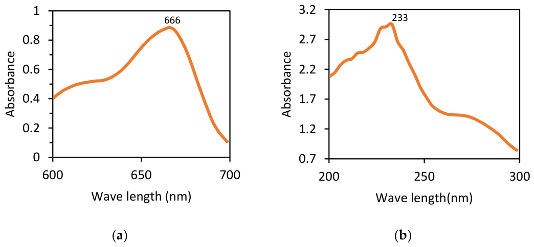
The UV–Vis spectrum of methylene blue exhibited a prominent absorption peak at 666 nm, consistent with literature values for this dye [2,3]. This wavelength was therefore chosen for monitoring MB concentration during degradation experiments.
The guava leaf extract showed a peak at around 233 nm, associated with π–π* transitions of aromatic rings in polyphenolic compounds. The IONPs displayed pronounced absorption in the UV region (200–300 nm), attributable to charge transfer transitions within the iron oxide lattice. Although the instrument’s lower wavelength limit prevented full observation of the absorption maxima for IONPs, the spectral features indicated successful formation of iron oxide species, as shown in Figure 1.

3.3. Photocatalytic Degradation Performance

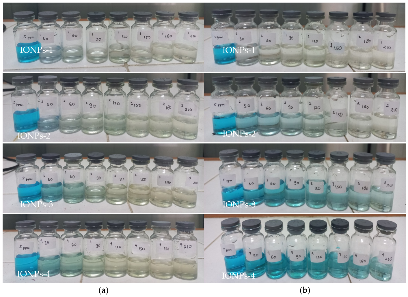
A consistent decrease in MB absorbance was observed over time under all photocatalytic conditions. Quantitatively, the degradation efficiencies after 210 min are summarized in Table 1 and the decolouration steps of MB are shown in Figure 2.
The results show that all IONP samples degraded methylene blue to a significant extent under UV irradiation, with efficiencies ranging from 94.7% to 99.0%. The best performance was observed for IONPs-1 under UV light. Sunlight combined with H2O2 also achieved high degradation efficiencies (>82.6%), whereas sunlight alone resulted in lower degradation (49.2–81.1%).
The superior performance of UV irradiation can be attributed to the higher photon energy and controlled exposure conditions. The dedicated UV chamber with three 13 W lamps at a fixed distance (≈15 cm) provided more uniform and intense irradiation than natural sunlight, which is inherently variable.

3.4. Role of H2O2 and Photo-Fenton Mechanism

The addition of H2O2 markedly enhanced dye degradation under both sunlight and UV irradiation. This can be explained by the classical Fenton and photo-Fenton reactions involving Fe2+/Fe3+ and H2O2 [15,16]:
Fe2+ + H2O2 → Fe3+ + OH + OH
Fe2+ + H2O2 → Fe3+ + H+ + OOH
The hydroxyl radicals (OH) and hydroperoxyl radicals (OOH) generated during these reactions are highly reactive oxidizing species capable of attacking the aromatic rings and chromophoric groups of methylene blue, leading to breakdown and eventual mineralization.
In the absence of H2O2, the degradation efficiency decreased significantly, especially under sunlight, confirming the crucial role of H2O2 as an oxidant and activator of the catalyst.

3.5. Effect of Precursor Concentration

Interestingly, increasing the molar amount of iron precursors from IONPs-1 to IONPs-4 did not improve photocatalytic performance; instead, a slight decline in degradation efficiency was observed at higher precursor loadings. Several factors may contribute to this behavior:
  • Particle aggregation: Higher precursor concentrations can promote the formation of larger aggregates, reducing the effective surface area available for catalysis.
  • Light scattering and shielding: Excessive nanoparticle concentration may lead to light scattering and shielding effects, limiting photon penetration into the suspension.
  • Suboptimal capping: For a fixed volume of leaf extract, increasing iron concentration may reduce the relative amount of phytochemicals available per iron ion, diminishing capping efficiency and causing partial aggregation.
These findings suggest that there is an optimal precursor concentration at which the balance between particle size, dispersion, and surface reactivity yields maximum photocatalytic performance. In this study, that condition corresponded to IONPs-1.

3.6. SEM-EDX Analysis

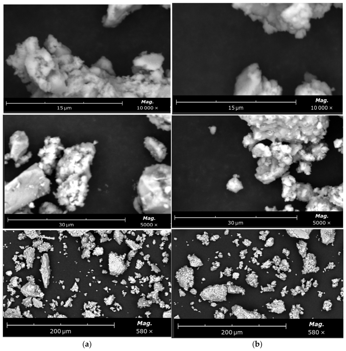
SEM images of IONPs-1 and IONPs-3 revealed crystalline, compact structures with a tendency to form aggregates, which is typical for magnetite-type nanoparticles due to magnetic dipole–dipole interactions [17]. The surface morphology was dense rather than porous, consistent with the expected structure of iron oxides, as shown in Figure 3.
IONPs exhibit a crystal-like morphology. This is because IONPs, which are a form of magnetite, are naturally non-porous. Their crystalline structure is dense and compact, formed by an ordered lattice of Fe2+ and Fe3+ ions. Ultrasonication helps disperse the aggregated IONPs, increasing their effective surface area and thereby enabling more efficient degradation of dye molecules.
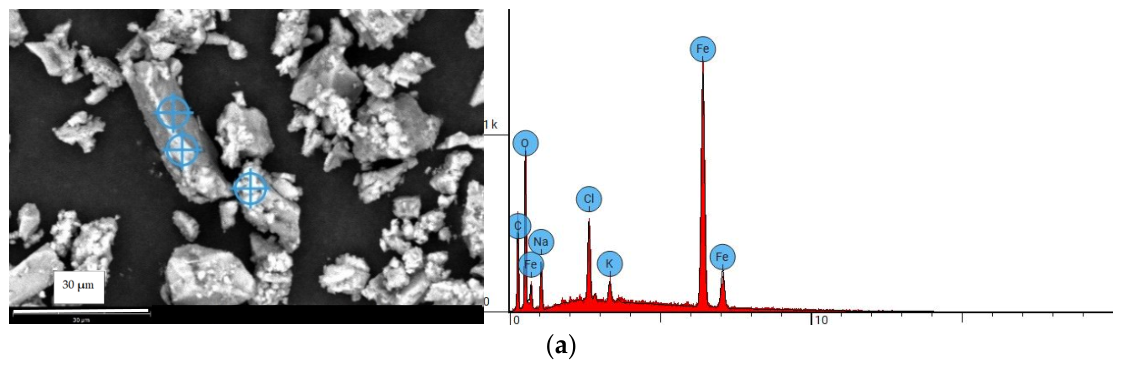
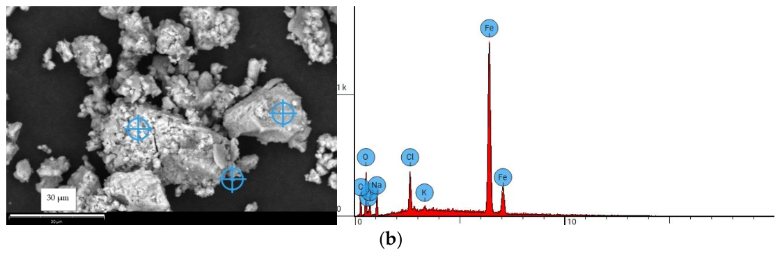
According to Figure 4 and Table 2, EDX analysis confirmed the presence of Fe and O as major elements, alongside C, Na, Cl, and K. The high oxygen content (≈31–35 at.%) supports the formation of iron oxide phases, possibly magnetite (Fe3O4) or related oxides [10]. Carbon originates from organic molecules in the guava extract acting as capping agents, which prevent excessive agglomeration by providing steric stabilization [6,7].
Sodium and chlorine derive from NaOH and FeCl2/FeCl3 precursors, indicating that small amounts of these ions remained after washing. Potassium likely originates from the plant material itself, as K is commonly stored in plant tissues. Notably, IONPs-3 showed higher Fe content compared with IONPs-1, consistent with the higher initial iron precursor concentration.

3.7. XRD Analysis

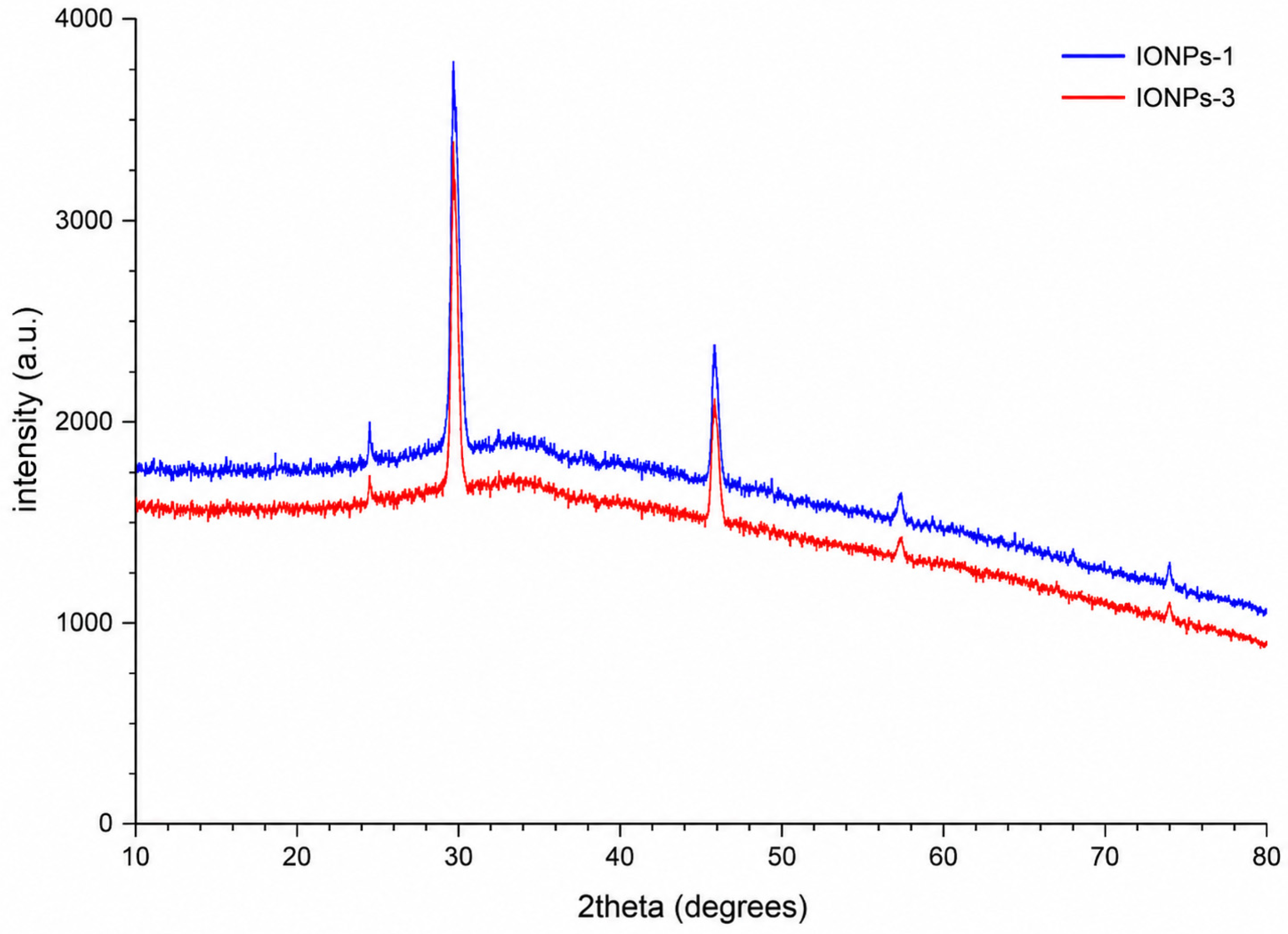
XRD patterns of IONPs-1 and IONPs-3, as shown in Figure 5, exhibited distinct diffraction peaks. For IONPs-1, peaks were observed at 2θ ≈ 17.4°, 27.4°, 31.7°, 45.4°, 48.5°, and 56.5°, whereas IONPs-3 displayed peaks at approximately 27.4°, 31.7°, 45.5°, 56.4°, and 75.3°. Phase analysis indicated the presence of multiple phases, including ferrihydrite, halite, hematite, magnesite, and magnetite. The dominant peak near 31.7° (100% relative intensity) is consistent with magnetite-like structures [17,18].
The coexistence of several phases can be attributed to the green synthesis conditions and the presence of plant-derived organics, which may influence nucleation and growth processes. Nonetheless, the presence of magnetite is advantageous for photocatalysis and magnetic recovery.

3.8. Proposed Degradation Mechanism

Based on the characterization results and degradation data, the following mechanism is proposed:
  • Under UV or sunlight, IONPs absorb photons and generate electron–hole pairs.
  • Electrons reduce Fe3+ to Fe2+, while holes oxidize water or hydroxide ions to form ·OH radicals.
  • In the presence of H2O2, Fe2+ catalyzes the decomposition of H2O2 into additional ·OH radicals via Fenton reactions.
  • Reactive ·OH radicals attack methylene blue molecules, breaking chromophores and aromatic rings, ultimately leading to decolorization and mineralization.
The magnetic nature of IONPs allows their rapid separation from the treated solution using an external magnet, enabling potential recovery and reuse in continuous or semi-continuous treatment systems.

4. Conclusions

Iron oxide nanoparticles were successfully synthesized via an eco-friendly route using Psidium guajava leaf extract as a natural reducing and capping agent. PSA confirmed particle diameters in the range of 209.2–291.4 nm. SEM–EDX and XRD analyses verified the formation of iron oxide phases, mainly magnetite-like structures, stabilized by plant-derived organic species.
Photocatalytic experiments demonstrated that the biosynthesized IONPs effectively degraded methylene blue under both sunlight and UV irradiation. The highest degradation efficiency (94.7–99%) was achieved with IONPs-1 (prepared from 0.05 mol FeCl2·4H2O and 0.01 mol FeCl3·6H2O) under UV light within 210 min. The presence of H2O2 significantly enhanced degradation by promoting Fenton-type reactions and generating hydroxyl radicals.
These results highlight the potential of guava leaf-mediated iron oxide nanoparticles as a low-cost, environmentally benign photocatalyst for dye wastewater treatment. Future work should explore catalyst reusability, performance in real textile effluents, optimization of extract-to-precursor ratios, and scale-up strategies.

Author Contributions

Conceptualization, H.P., F.K.H. and C.R.P.; methodology, F.K.H. and C.R.P.; validation, H.P.; formal analysis, F.K.H.; investigation, C.R.P.; resources, F.K.H.; data curation, C.R.P.; writing—original draft preparation, F.K.H. and C.R.P.; writing—review and editing, H.P.; visualization, F.K.H. and C.R.P.; supervision, H.P.; project administration, H.P. All authors have read and agreed to the published version of the manuscript.

Funding

This research received no external funding.

Institutional Review Board Statement

Not applicable.

Informed Consent Statement

Not applicable.

Data Availability Statement

Data are contained within the article.

Conflicts of Interest

The authors declare no conflict of interest.

References

  1. Purnama, H.; Dewi, D.E.; Jannah, N.M.; Kamaratih, A.A.; Hidayati, N. Liquid waste treatment containing Rhodamine B using emulsion liquid membrane from waste cooking oil. Results Eng. 2025, 25, 104066. [Google Scholar] [CrossRef]
  2. Rani, N.; Kumar, S.; Kumar, K. Green synthesis of N-doped-carbon dots/ZnO for enhanced photocatalytic degradation of methylene blue dye: Optimization of reaction parameters. Environ. Sci. Pollut. Res. 2024, 31, 63408–63425. [Google Scholar] [CrossRef] [PubMed]
  3. Bhujel, S.; Koju, S.; Yadav, S.K.; Khadka, D.; Sharma, M.L.; Parajuli, K. Green Synthesis of Iron Oxide Nanoparticles Using Melastoma malabathricum Leaf Extract for Photocatalytic Degradation of Methylene Blue and Antibacterial Activity. J. Nanotechnol. 2025, 2025, 7616178. [Google Scholar] [CrossRef]
  4. Adhikari, A.; Chhetri, K.; Acharya, D.; Pant, B.; Adhikari, A. Green Synthesis of Iron Oxide Nanoparticles Using Psidium guajava L. Leaves Extract for Degradation of Organic Dyes and Anti-Microbial Applications. Catalysts 2022, 12, 1188. [Google Scholar] [CrossRef]
  5. Islam, M.J.; Khatun, N.; Bhuiyan, R.H.; Sultana, S.; Shaikh, M.A.A.; Bitu, M.N.A.; Chowdhury, F.; Islam, S. Psidium guajava Leaf Extract-Mediated Green Synthesis of Silver Nanoparticles and Its Application in Antibacterial Coatings. RSC Adv. 2023, 13, 7227. [Google Scholar] [CrossRef] [PubMed]
  6. Mohamed, A.; Atta, R.R.; Kotp, A.A.; Abo El-Ela, F.I.; Abd El-Raheem, H.; Farghali, A.; Alkhalifah, D.H.M.; Hozzein, W.N.; Mahmoud, R. Green Synthesis and Characterization of Iron Oxide Nanoparticles for the Removal of Heavy Metals (Cd2+ and Ni2+) from Aqueous Solutions with Antimicrobial Investigation. Sci. Rep. 2023, 13, 4325. [Google Scholar] [CrossRef] [PubMed]
  7. Shahid-ul-Islam; Bairagi, S.; Kamali, M.R. Review on Green Biomass-Synthesized Metallic Nanoparticles and Composites and Their Photocatalytic Water Purification Applications: Progress and Perspectives. Chem. Eng. J. Adv. 2023, 14, 100460. [Google Scholar] [CrossRef]
  8. Khadka, D.; Gautam, P.; Dahal, R.; Ashie, M.D.; Paudyal, H.; Ghimire, K.N.; Pant, B.; Poudel, B.R.; Bastakoti, B.P.; Pokhrel, M.R. Evaluating the Photocatalytic Activity of Green Synthesized Iron Oxide Nanoparticles. Catalysts 2024, 14, 751. [Google Scholar] [CrossRef]
  9. Murari, K.; Vijayakumar, Y. CTAB-Assisted Synthesis of Iron Oxide Nanoparticles for Enhanced Photocatalytic Dye Degradation. Phys. Status Solidi (A) 2025, 223, e202500655. [Google Scholar] [CrossRef]
  10. Xu, Z.; Zada, N.; Habib, F.; Ullah, H.; Hussain, K.; Ullah, N.; Bibi, M.; Bibi, M.; Ghani, H.; Khan, S.; et al. Enhanced Photocatalytic Degradation of Malachite Green Dye Using Silver–Manganese Oxide Nanoparticles. Molecules 2023, 28, 6241. [Google Scholar] [CrossRef]
  11. Tirkey, A.; Upadhyay, L.S. Facile Synthesis of Nickel Oxide Nanoparticles and Its Application for Photocatalytic Degradation of Crystal Violet Dye. Plasmonics 2025, 21, 1241–1262. [Google Scholar] [CrossRef]
  12. Al-Zaqri, N.; Umamakeshvari, K.; Mohana, V.; Muthuvel, A.; Boshaala, A. Green synthesis of nickel oxide nanoparticles and its photocatalytic degradation and antibacterial activity. J. Mater. Sci. Mater. Electron. 2022, 33, 11864–11880. [Google Scholar] [CrossRef]
  13. Zamil, I.N.; AL-Mammar, D.E. Comparative study of the adsorption of Azo dye onto the surfaces of nano iron oxide prepared by the green method and commercial nano iron oxide. Nanotechnol. Environ. Eng. 2025, 10, 85. [Google Scholar] [CrossRef]
  14. Relhan, A.; Guleria, S.; Bhasin, A.; Mirza, A.; Zhou, J.L. Biosynthesized copper oxide nanoparticles by Psidium guajava plants with antibacterial, antidiabetic, antioxidant, and photocatalytic capacity. Biomass-Convers. Biorefin. 2024, 15, 26623–26640. [Google Scholar] [CrossRef]
  15. Egerton, T.A.; Purnama, H. Does hydrogen peroxide really accelerate TiO2 UV-C photocatalyzed decolouration of azo-dyes such as Reactive Orange 16? Dye. Pigment. 2014, 101, 280–285. [Google Scholar] [CrossRef]
  16. Fagier, M.A. Plant-Mediated Biosynthesis and Photocatalysis Activities of Zinc Oxide Nanoparticles: A Prospect towards Dyes Mineralization. J. Nanotechnol. 2021, 2021, 6629180. [Google Scholar] [CrossRef]
  17. Besenhard, M.O.; LaGrow, A.P.; Hodzic, A.; Kriechbaum, M.; Panariello, L.; Bais, G.; Loizou, K.; Damilos, S.; Cruz, M.M.; Thanh, N.T.K.; et al. Co-Precipitation Synthesis of Stable Iron Oxide Nanoparticles with NaOH: New Insights and Continuous Production via Flow Chemistry. Chem. Eng. J. 2020, 399, 125740. [Google Scholar] [CrossRef]
  18. Shilova, O.A.; Nikolaev, A.M.; Kovalenko, A.S.; Sinel’nikov, A.A.; Kopitsa, G.P.; Baranchikov, A.E. Synthesis of Magnetic Nanopowders of Iron Oxide: Magnetite and Maghemite. Russ. J. Inorg. Chem. 2020, 65, 426–430. [Google Scholar] [CrossRef]
Figure 1. Wavelength of (a) Methylene Blue; (b) Guava Leaf Extract.
Engproc 137 00013 g001
Figure 2. Degradation of methylene blue after 210 min with (a) UV lamps; (b) Sunlight.
Engproc 137 00013 g002
Figure 3. Morphology of IONPs with SEM (a) IONPs-1; (b) IONPs-3.
Engproc 137 00013 g003
Figure 4. Composition IONPs with SEM-EDX: (a) IONPs-1; (b) IONPs-3.
Engproc 137 00013 g004aEngproc 137 00013 g004b
Figure 5. XRD diffractogram of IONPs-1 and IONPs-3.
Engproc 137 00013 g005
Table 1. Degradation efficiency of methylene blue after 210 min for different irradiation conditions.
SampleSunlight + H2O2Sunlight OnlyUV Lamps
IONPs-197.5%81.1%99.0%
IONPs-297.2%74.3%97.6%
IONPs-392.7%54.4%96.1%
IONPs-482.6%49.2%94.7%
Table 2. SEM-EDX results for IONPs-1 and IONPs-3.
ElementSymbolsIONPs-1IONPs-3
Atomic ConcentrationMass ConcentrationAtomic ConcentrationMass Concentration
CarbonC0.46260.29060.40150.2222
OxigentO0.34610.28960.31630.2332
NatriumNa0.06580.07920.09920.1051
ChlorineCl0.01950.03610.03060.0501
PotassiumK0.00590.01200.00330.0060
IronFe0.10020.29260.14900.3834
Disclaimer/Publisher’s Note: The statements, opinions and data contained in all publications are solely those of the individual author(s) and contributor(s) and not of MDPI and/or the editor(s). MDPI and/or the editor(s) disclaim responsibility for any injury to people or property resulting from any ideas, methods, instructions or products referred to in the content.