
G Protein-Coupled Receptors in Pancreatic β-Cells: From Trafficking and Localization to Insulin Secretion and Diabetes

Abstract
1. Background
2. Synthesis, Trafficking and Localisation of GPCRs
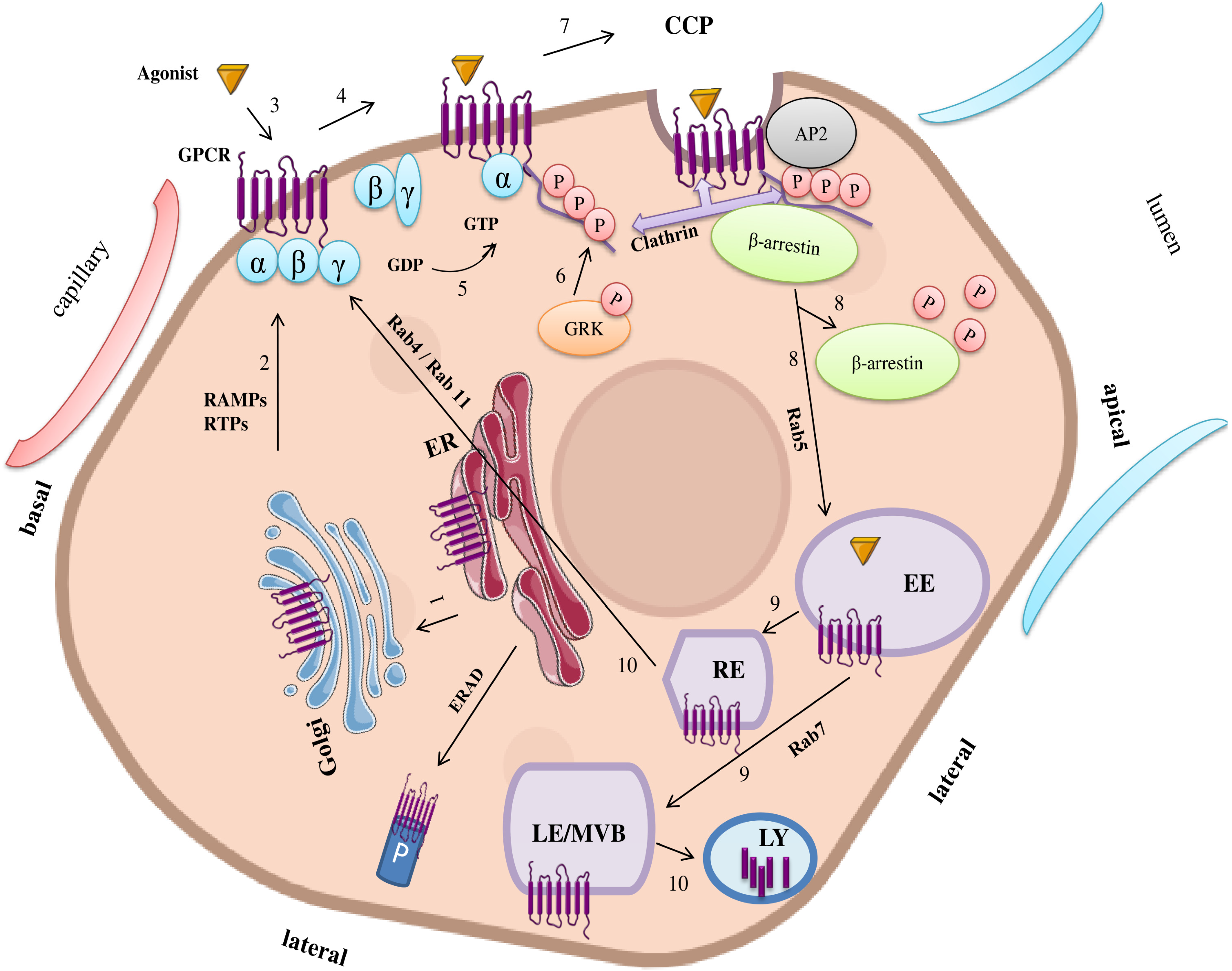
2.1. Trafficking from Endoplasmic Reticulum (ER)/Golgi to Plasma Membrane
2.2. Internalization of GPCRs Through Clathrin-Coated Pits (CCPs)
2.3. Trafficking at the Endosomal Compartment
2.4. Subcellular Localisation of GPCRs
3. Functional Roles in Pancreatic β-Cells
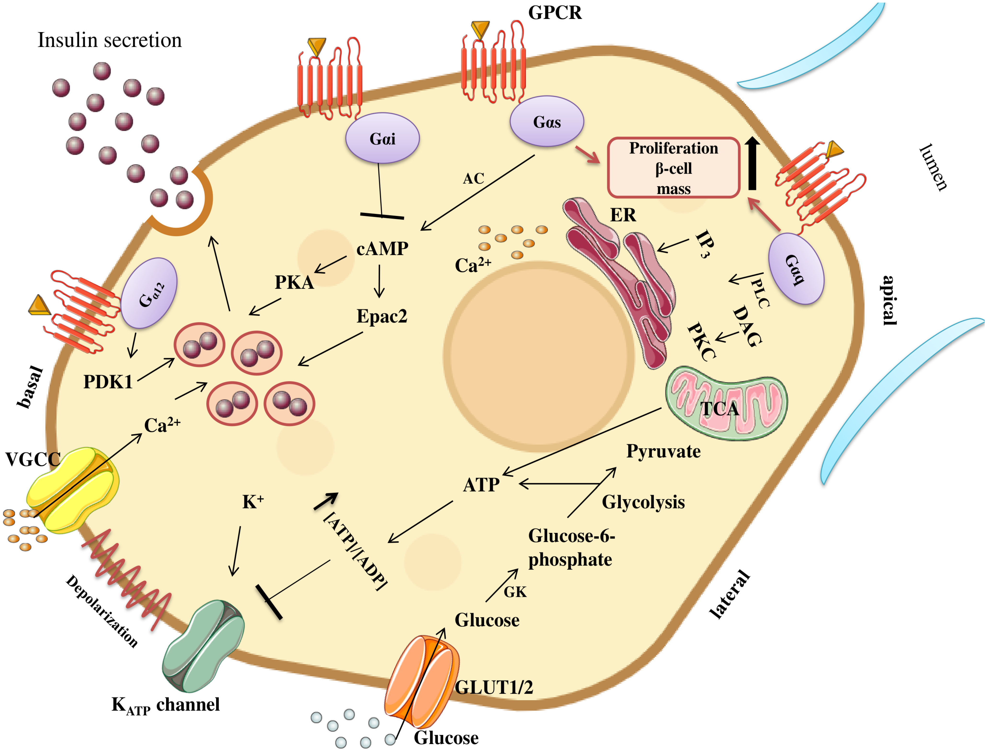
3.1. Regulation of β-Cell Survival, Proliferation, and Homeostasis
3.2. Regulation of Insulin Secretion
3.3. GPCR Regulation of Glucose-Stimulated Insulin Secretion (GSIS)
4. Conclusions
Author Contributions
Funding
Institutional Review Board Statement
Informed Consent Statement
Data Availability Statement
Conflicts of Interest
Abbreviations
| 5-HT2B | 5-Hydroxytryptamine recetor 2B |
| A2BAR | A2B adenosine receptor |
| AC | stimulates adenylyl cyclase |
| ADRA2A | Alpha-2A adrenergic receptor |
| AP2 | Adapter protein 2 |
| Bai3 | Brain angiogenesis inhibitor-3 |
| BiP | binding immunoglobulin protein |
| C1ql3 | Complement 1q-like-3 |
| cAMP | cyclic adenosine monophosphate |
| CB1/CB2 | Cannabinoid receptors 1 and 2 |
| CCL25 | C-C motif chemokine ligand 25 |
| CCL5 | C-C motif chemokine ligand 5 |
| CCPs | Clathrin-coated pits |
| CCR9 | C-C motif chemokine receptor 9 |
| CLR | Calcitonin receptor-like receptor |
| COPII | Coat protein complex II |
| DAG | diacylglycerol(DAG) |
| DUBs | Deubiquitnating enzymes |
| EE | Early endosomes |
| EP3, EP4 | Prostaglandin E2-subtypes |
| Epac2 | Exchange Protein Activated by cAMP 2 |
| ER | Endoplasmic reticulum |
| ERAD | ER-associated degradation |
| ERGIC | ER–Golgi intermediate compartment |
| ERp57 | Endoplasmic reticulum protein 57 |
| ESCRT | Endosomal sorting complexes required for transport |
| FEME | Fast endophilin-mediated endocytosis |
| FFAR2 | Free fatty acid receptor 2 |
| GABAB | Gamma-aminobutyric acid type B |
| GASP-1 | GPCR-associated sorting protein-1 |
| GIP-R | Gastric inhibitory polypeptide receptor |
| GK | Glucokinase |
| GLP-1R | GLP-1 receptor |
| GLUT2 | Glucose transporter type 2 |
| GPCRs | G protein–coupled receptors |
| GRKs | GPCR kinases |
| GSIS | Glucose-stimulated insulin secretion |
| HFD | High-fat diet |
| HNF-1α | Hepatocyte nuclear factor 1-alpha |
| Hrs | HGF regulated tyrosine kinase substrate |
| IP3 | inositol 1,4,5-trisphosphate |
| LE | Late endosomes |
| LGR4 | Leucine-rich repeat-containing receptor |
| LY | Lysosomes |
| M3R | Muscarinic receptor subtype 3 |
| mGluRs | Metabotropic glutamate receptors |
| MT1/2 | Melatonin receptors |
| MVBs | Multivesicular bodies |
| OEA | oleoylethanolamide |
| Olfr109 | Olfactory receptor 109 |
| P | Proteasome |
| PDK1 | 3-Phosphoinositide-dependent protein kinase-1 |
| Pdx1 | pancreatic and duodenal homeobox 1 |
| PGE2 | Prostaglandin E2 |
| PKA | Protein kinase A |
| PKC | Protein kinase C |
| PLCβ | phospholipase Cβ |
| PLC-γ1 | phospholipase C gamma 1 |
| PPARβ/δ | Peroxisome proliferator-activated receptor |
| RAMPs | Receptor activity–modifying proteins |
| RE | Recycling endosomes |
| REEPs | Receptor expression enhancing proteins |
| RhoGEFs | Rho guanine nucleotide exchange factors |
| RTPs | Receptor transport proteins |
| SSTR1/5 | Somatostatin receptors 1/5 |
| SUCNR1 | Succinate receptor 1 |
| T1D | Type 1 diabetes |
| T2D | Type 2 diabetes |
| TAAR1 | Trace-amine associated receptor 1 |
| TCA | Tricarboxylic acid |
| Ub | Ubiquitin |
| V2R | Vasopressin V2 receptor |
| Y1R | Neuropeptide Y receptor type 1 |
| β2ARs | β2-adrenergic receptors |
References
- Polakof, S. Diabetes Therapy: Novel Patents Targeting the Glucose-Induced Insulin Secretion. Recent Pat. DNA Gene Seq. 2010, 4, 1–9. [Google Scholar] [CrossRef] [PubMed]
- Campbell, J.E.; Newgard, C.B. Mechanisms controlling pancreatic islet cell function in insulin secretion. Nat. Rev. Mol. Cell Biol. 2021, 22, 142–158. [Google Scholar] [CrossRef] [PubMed]
- Fu, Z.; Gilbert, E.R.; Liu, D. Regulation of Insulin Synthesis and Secretion and Pancreatic Beta-Cell Dysfunction in Diabetes. Curr. Diabetes Rev. 2014, 9, 25–53. [Google Scholar]
- Keane, K.; Newsholme, P. Metabolic Regulation of Insulin Secretion. In Vitamins & Hormones; Academic Press: Cambridge, MA, USA, 2014; pp. 1–33. Available online: https://linkinghub.elsevier.com/retrieve/pii/B9780128001745000016 (accessed on 20 April 2025).
- Bouché, C.; Serdy, S.; Kahn, C.R.; Goldfine, A.B. The Cellular Fate of Glucose and Its Relevance in Type 2 Diabetes. Endocr. Rev. 2004, 25, 807–830. [Google Scholar] [CrossRef]
- GalGalicia-Garcia, U.; Benito-Vicente, A.; Jebari, S.; Larrea-Sebal, A.; Siddiqi, H.; Uribe, K.B.; Ostolaza, H.; Martín, C. Pathophysiology of Type 2 Diabetes Mellitus. Int. J. Mol. Sci. 2020, 21, 6275. [Google Scholar] [CrossRef] [PubMed]
- Ishihara, H. Metabolism-secretion coupling in glucose-stimulated insulin secretion. Diabetol. Int. 2022, 13, 463–470. [Google Scholar] [CrossRef]
- Tokarz, V.L.; MacDonald, P.E.; Klip, A. The cell biology of systemic insulin function. J. Cell Biol. 2018, 217, 2273–2289. [Google Scholar] [CrossRef]
- Amisten, S.; Atanes, P.; Hawkes, R.; Ruz-Maldonado, I.; Liu, B.; Parandeh, F.; Zhao, M.; Huang, G.C.; Salehi, A.; Persaud, S.J. A comparative analysis of human and mouse islet G-protein coupled receptor expression. Sci. Rep. 2017, 7, 46600. [Google Scholar] [CrossRef]
- Riddy, D.M.; Delerive, P.; Summers, R.J.; Sexton, P.M.; Langmead, C.J. G Protein–Coupled Receptors Targeting Insulin Resistance, Obesity, and Type 2 Diabetes Mellitus. Pharmacol. Rev. 2018, 70, 39–67. [Google Scholar] [CrossRef]
- Taneera, J.; Mohammed, I.; Mohammed, A.K.; Hachim, M.; Dhaiban, S.; Malek, A.; Dunér, P.; Elemam, N.M.; Sulaiman, N.; Hamad, M.; et al. Orphan G-protein coupled receptor 183 (GPR183) potentiates insulin secretion and prevents glucotoxicity-induced β-cell dysfunction. Mol. Cell. Endocrinol. 2020, 499, 110592. [Google Scholar] [CrossRef]
- Stäubert, C.; Wozniak, M.; Dupuis, N.; Laschet, C.; Pillaiyar, T.; Hanson, J. Superconserved receptors expressed in the brain: Expression, function, motifs and evolution of an orphan receptor family. Pharmacol. Ther. 2022, 240, 108217. [Google Scholar] [CrossRef]
- Öz-Arslan, D.; Yavuz, M.; Kan, B. Exploring orphan GPCRs in neurodegenerative diseases. Front. Pharmacol. 2024, 15, 1394516. [Google Scholar] [CrossRef]
- Wang, T.; Shao, J.; Kumar, S.; Alnouri, M.W.; Carvalho, J.; Günther, S.; Krasel, C.; Murphy, K.T.; Bünemann, M.; Offermanns, S.; et al. Orphan GPCR GPRC5C Facilitates Angiotensin II-Induced Smooth Muscle Contraction. Circ. Res. 2024, 134, 1259–1275. [Google Scholar] [CrossRef] [PubMed]
- Magalhaes, A.C.; Dunn, H.; Ferguson, S.S. Regulation of GPCR activity, trafficking and localization by GPCR-interacting proteins. Br. J. Pharmacol. 2012, 165, 1717–1736. [Google Scholar] [CrossRef]
- Rosenbaum, D.M.; Rasmussen, S.G.F.; Kobilka, B.K. The structure and function of G-protein-coupled receptors. Nature 2009, 459, 356–363. [Google Scholar] [CrossRef] [PubMed]
- Sposini, S.; Hanyaloglu, A.C. Evolving View of Membrane Trafficking and Signaling Systems for G Protein-Coupled Receptors. In Endocytosis and Signaling; Lamaze, C., Prior, I., Eds.; Progress in Molecular and Subcellular Biology, Vol. 57; Springer International Publishing: Cham, Switzerland, 2018; pp. 273–299. Available online: http://link.springer.com/10.1007/978-3-319-96704-2_10 (accessed on 20 April 2025).
- Nezhady, M.A.M.; Rivera, J.C.; Chemtob, S. Location Bias as Emerging Paradigm in GPCR Biology and Drug Discovery. iScience 2020, 23, 101643. [Google Scholar] [CrossRef]
- Pierce, K.L.; Premont, R.T.; Lefkowitz, R.J. Seven-transmembrane receptors. Nat. Rev. Mol. Cell Biol. 2002, 3, 639–650. [Google Scholar] [CrossRef]
- Wehbi, V.L.; Stevenson, H.P.; Feinstein, T.N.; Calero, G.; Romero, G.; Vilardaga, J.-P. Noncanonical GPCR signaling arising from a PTH receptor–arrestin–Gβγ complex. Proc. Natl. Acad. Sci. USA 2013, 110, 1530–1535. [Google Scholar] [PubMed]
- Godbole, A.; Lyga, S.; Lohse, M.J.; Calebiro, D. Internalized TSH receptors en route to the TGN induce local Gs-protein signaling and gene transcription. Nat. Commun. 2017, 8, 443. [Google Scholar] [CrossRef]
- Feinstein, T.N.; Yui, N.; Webber, M.J.; Wehbi, V.L.; Stevenson, H.P.; King, J.D.; Hallows, K.R.; Brown, D.; Bouley, R.; Vilardaga, J.-P. Noncanonical Control of Vasopressin Receptor Type 2 Signaling by Retromer and Arrestin. J. Biol. Chem. 2013, 288, 27849–27860. [Google Scholar] [CrossRef]
- Smith, J.S.; Pack, T.F.; Inoue, A.; Lee, C.; Zheng, K.; Choi, I.; Eiger, D.S.; Warman, A.; Xiong, X.; Ma, Z.; et al. Noncanonical scaffolding of Gαi and β-arrestin by G protein–coupled receptors. Science 2021, 371, eaay1833. [Google Scholar]
- Zhao, J.; DiGiacomo, V.; Ferreras-Gutierrez, M.; Dastjerdi, S.; de Opakua, A.I.; Park, J.-C.; Luebbers, A.; Chen, Q.; Beeler, A.; Blanco, F.J.; et al. Small-molecule targeting of GPCR-independent noncanonical G-protein signaling in cancer. Proc. Natl. Acad. Sci. USA 2023, 120, e2213140120. [Google Scholar] [CrossRef]
- Steiner, W.P.; Iverson, N.; Liu, G.; Venkatakrishnan, V.; Wu, J.; Stepniewski, T.M.; Michaelson, Z.; Bröckel, J.W.; Zhu, J.-F.; Bruystens, J.; et al. A Structural Mechanism for Noncanonical GPCR Signal Transduction in the Hedgehog Pathway. bioRxiv 2024. Available online: http://biorxiv.org/lookup/doi/10.1101/2024.10.31.621410 (accessed on 22 January 2026).
- Benincá, C.; Planagumà, J.; de Freitas Shuck, A.; Acín-Perez, R.; Muñoz, J.P.; de Almeida, M.M.; Brown, J.H.; Murphy, A.N.; Zorzano, A.; Enríquez, J.A.; et al. A new non-canonical pathway of Gαq protein regulating mitochondrial dynamics and bioenergetics. Cell. Signal. 2014, 26, 1135–1146. [Google Scholar] [CrossRef]
- Costa-Neto, C.M.; Parreiras-E-Silva, L.T. Deciphering complexity of GPCR signaling and modulation: Implications and perspectives for drug discovery. Clin. Sci. 2025, 139, 463–477. [Google Scholar] [CrossRef]
- Esseltine, J.L.; Ribeiro, F.M.; Ferguson, S.S.G. Rab8 Modulates Metabotropic Glutamate Receptor Subtype 1 Intracellular Trafficking and Signaling in a Protein Kinase C-Dependent Manner. J. Neurosci. 2012, 32, 16933–16942. [Google Scholar] [CrossRef] [PubMed]
- Singh, B.; Coffey, R.J. Trafficking of Epidermal Growth Factor Receptor Ligands in Polarized Epithelial Cells. Annu. Rev. Physiol. 2014, 76, 275–300. [Google Scholar] [CrossRef]
- Kunii, M.; Harada, A. Molecular mechanisms of polarized transport to the apical plasma membrane. Front. Cell Dev. Biol. 2024, 12, 1477173. [Google Scholar] [CrossRef]
- Lanctôt, P.M.; Leclerc, P.C.; Escher, E.; Leduc, R.; Guillemette, G. Role of N-Glycosylation in the Expression and Functional Properties of Human AT1Receptor. Biochemistry 1999, 38, 8621–8627. [Google Scholar] [CrossRef] [PubMed]
- Ritter, S.L.; Hall, R.A. Fine-tuning of GPCR activity by receptor-interacting proteins. Nat. Rev. Mol. Cell Biol. 2009, 10, 819–830. [Google Scholar] [CrossRef] [PubMed]
- Apaja, P.M.; Lukacs, G.L. Protein Homeostasis at the Plasma Membrane. Physiology 2014, 29, 265–277. [Google Scholar] [CrossRef]
- Bernier, V.; Lagacé, M.; Bichet, D.G.; Bouvier, M. Pharmacological chaperones: Potential treatment for conformational diseases. Trends Endocrinol. Metab. 2004, 15, 222–228. [Google Scholar] [CrossRef]
- Saito, H.; Kubota, M.; Roberts, R.W.; Chi, Q.; Matsunami, H. RTP Family Members Induce Functional Expression of Mammalian Odorant Receptors. Cell 2004, 119, 679–691. [Google Scholar] [CrossRef]
- McLatchie, L.M.; Fraser, N.J.; Main, M.J.; Wise, A.; Brown, J.; Thompson, N.; Solari, R.; Lee, M.G.; Foord, S.M. RAMPs regulate the transport and ligand specificity of the calcitonin-receptor-like receptor. Nature 1998, 393, 333–339. [Google Scholar] [CrossRef]
- Drake, M.T.; Shenoy, S.K.; Lefkowitz, R.J. Trafficking of G Protein—Coupled Receptors. Circ. Res. 2006, 99, 570–582. [Google Scholar] [CrossRef]
- Young, B.; Wertman, J.; Dupré, D.J. Regulation of GPCR Anterograde Trafficking by Molecular Chaperones and Motifs. In Progress in Molecular Biology and Translational Science; Elsevier: Amsterdam, The Netherlands, 2015; pp. 289–305. Available online: https://linkinghub.elsevier.com/retrieve/pii/S1877117315000538 (accessed on 20 April 2025).
- Dong, C.; Filipeanu, C.M.; Duvernay, M.T.; Wu, G. Regulation of G protein-coupled receptor export trafficking. Biochim. Biophys. Acta—Biomembr. 2007, 1768, 853–870. [Google Scholar] [CrossRef]
- Duvernay, M.T.; Zhou, F.; Wu, G. A Conserved Motif for the Transport of G Protein-coupled Receptors from the Endoplasmic Reticulum to the Cell Surface. J. Biol. Chem. 2004, 279, 30741–30750. [Google Scholar] [CrossRef]
- Bermak, J.C.; Li, M.; Bullock, C.; Zhou, Q.-Y. Regulation of transport of the dopamine D1 receptor by a new membrane-associated ER protein. Nat. Cell Biol. 2001, 3, 492–498. [Google Scholar] [CrossRef] [PubMed]
- Margeta-Mitrovic, M.; Jan, Y.N.; Jan, L.Y. A Trafficking Checkpoint Controls GABAB Receptor Heterodimerization. Neuron 2000, 27, 97–106. [Google Scholar] [CrossRef] [PubMed]
- Kaushal, S.; Khorana, H.G. Structure and Function in Rhodopsin. 7. Point Mutations Associated with Autosomal Dominant Retinitis Pigmentosa. Biochemistry 1994, 33, 6121–6128. [Google Scholar] [CrossRef] [PubMed]
- Morello, J.P.; Bichet, D.G. Nephrogenic Diabetes Insipidus. Annu. Rev. Physiol. 2001, 63, 607–630. [Google Scholar] [CrossRef]
- Pavlos, N.J.; Friedman, P.A. GPCR Signaling and Trafficking: The Long and Short of It. Trends Endocrinol. Metab. 2017, 28, 213–226. [Google Scholar] [CrossRef]
- Bomberger, J.M.; Parameswaran, N.; Spielman, W.S. Regulation of GPCR Trafficking by RAMPs. In RAMPs; Spielman, W.S., Parameswaran, N., Eds.; Advances in Experimental Medicine and Biology Vol. 744; Springer: New York, NY, USA, 2012; pp. 25–37. Available online: https://link.springer.com/10.1007/978-1-4614-2364-5_3 (accessed on 20 April 2025).
- Foster, S.R.; Bräuner-Osborne, H. Investigating Internalization and Intracellular Trafficking of GPCRs: New Techniques and Real-Time Experimental Approaches. In Targeting Trafficking in Drug Development; Ulloa-Aguirre, A., Tao, Y.X., Eds.; Handbook of Experimental Pharmacology Vol. 245; Springer International Publishing: Cham, Switzerland, 2017; pp. 41–61. Available online: http://link.springer.com/10.1007/164_2017_57 (accessed on 20 April 2025).
- Hanyaloglu, A.C. Advances in Membrane Trafficking and Endosomal Signaling of G Protein-Coupled Receptors. In International Review of Cell and Molecular Biology; Elsevier: Amsterdam, The Netherlands, 2018; pp. 93–131. Available online: https://linkinghub.elsevier.com/retrieve/pii/S1937644818300297 (accessed on 20 April 2025).
- Jean-Alphonse, F.; Hanyaloglu, A. Regulation of GPCR signal networks via membrane trafficking. Mol. Cell. Endocrinol. 2011, 331, 205–214. [Google Scholar] [CrossRef] [PubMed]
- Moore, C.A.; Milano, S.K.; Benovic, J.L. Regulation of Receptor Trafficking by GRKs and Arrestins. Annu. Rev. Physiol. 2007, 69, 451–482. [Google Scholar] [CrossRef]
- Apodaca, G.; Gallo, L.I.; Bryant, D.M. Role of membrane traffic in the generation of epithelial cell asymmetry. Nat. Cell Biol. 2012, 14, 1235–1243. [Google Scholar] [CrossRef]
- Kunselman, J.M.; Lott, J.; Puthenveedu, M.A. Mechanisms of selective G protein–coupled receptor localization and trafficking. Curr. Opin. Cell Biol. 2021, 71, 158–165. [Google Scholar] [CrossRef]
- Patwardhan, A.; Cheng, N.; Trejo, J. Post-Translational Modifications of G Protein–Coupled Receptors Control Cellular Signaling Dynamics in Space and Time. Pharmacol. Rev. 2021, 73, 120–151. [Google Scholar] [CrossRef]
- Sorkin, A.; Von Zastrow, M. Endocytosis and signalling: Intertwining molecular networks. Nat. Rev. Mol. Cell Biol. 2009, 10, 609–622. [Google Scholar] [CrossRef]
- Wu, C.-T.; Hilgendorf, K.I.; Bevacqua, R.J.; Hang, Y.; Demeter, J.; Kim, S.K.; Jackson, P.K. Discovery of ciliary G protein-coupled receptors regulating pancreatic islet insulin and glucagon secretion. Genes Dev. 2021, 35, 1243–1255. [Google Scholar] [CrossRef]
- Irannejad, R.; Von Zastrow, M. GPCR signaling along the endocytic pathway. Curr. Opin. Cell Biol. 2014, 27, 109–116. [Google Scholar] [CrossRef] [PubMed]
- Fölsch, H. Regulation of membrane trafficking in polarized epithelial cells. Curr. Opin. Cell Biol. 2008, 20, 208–213. [Google Scholar] [CrossRef] [PubMed]
- Fasciani, I.; Carli, M.; Petragnano, F.; Colaianni, F.; Aloisi, G.; Maggio, R.; Scarselli, M.; Rossi, M. GPCRs in Intracellular Compartments: New Targets for Drug Discovery. Biomolecules 2022, 12, 1343. [Google Scholar] [CrossRef] [PubMed]
- Filipowska, J.; Cisneros, Z.; Varghese, S.S.; Leon-Rivera, N.; Wang, P.; Kang, R.; Lu, G.; Yuan, Y.-C.; Shih, H.-P.; Bhattacharya, S.; et al. LGR4 is essential for maintaining β-cell homeostasis through suppression of RANK. Mol. Metab. 2025, 92, 102097. [Google Scholar] [CrossRef] [PubMed]
- Giudici, L.; Velic, A.; Daryadel, A.; Bettoni, C.; Mohebbi, N.; Suply, T.; Seuwen, K.; Ludwig, M.G.; Wagner, C.A. The Proton-Activated Receptor GPR4 Modulates Glucose Homeostasis by Increasing Insulin Sensitivity. Cell. Physiol. Biochem. 2013, 32, 1403–1416. [Google Scholar] [CrossRef]
- Buteau, J.; Roduit, R.; Susini, S.; Prentki, M. Glucagon-like peptide-1 promotes DNA synthesis, activates phosphatidylinositol 3-kinase and increases transcription factor pancreatic and duodenal homeobox gene 1 (PDX-1) DNA binding activity in beta (INS-1)-cells. Diabetologia 1999, 42, 856–864. [Google Scholar] [CrossRef]
- Edvell, A.; Lindström, P. Initiation of Increased Pancreatic Islet Growth in Young Normoglycemic Mice (Umeå +/?). Endocrinology 1999, 140, 778–783. [Google Scholar] [CrossRef][Green Version]
- Yang, Y.; Tong, Y.; Gong, M.; Lu, Y.; Wang, C.; Zhou, M.; Yang, Q.; Mao, T.; Tong, N. Activation of PPARβ/δ protects pancreatic β cells from palmitate-induced apoptosis by upregulating the expression of GLP-1 receptor. Cell. Signal. 2014, 26, 268–278. [Google Scholar] [CrossRef]
- Holst, B.; Egerod, K.L.; Jin, C.; Petersen, P.S.; Østergaard, M.V.; Hald, J.; Sprinkel, A.M.E.; Størling, J.; Mandrup-Poulsen, T.; Holst, J.J.; et al. G Protein-Coupled Receptor 39 Deficiency Is Associated with Pancreatic Islet Dysfunction. Endocrinology 2009, 150, 2577–2585. [Google Scholar] [CrossRef]
- Villa, S.R.; Priyadarshini, M.; Fuller, M.H.; Bhardwaj, T.; Brodsky, M.R.; Angueira, A.R.; Mosser, R.E.; Carboneau, B.A.; Tersey, S.A.; Mancebo, H.; et al. Loss of Free Fatty Acid Receptor 2 leads to impaired islet mass and beta cell survival. Sci. Rep. 2016, 6, 28159. [Google Scholar] [CrossRef] [PubMed]
- Dunér, P.; Al-Amily, I.M.; Soni, A.; Asplund, O.; Safi, F.; Storm, P.; Groop, L.; Amisten, S.; Salehi, A. Adhesion G Protein-Coupled Receptor G1 (ADGRG1/GPR56) and Pancreatic β-Cell Function. J. Clin. Endocrinol. Metab. 2016, 101, 4637–4645. [Google Scholar] [CrossRef]
- Olaniru, O.E.; Cheng, J.; Ast, J.; Arvaniti, A.; Atanes, P.; Huang, G.C.; King, A.J.; Jones, P.M.; Broichhagen, J.; Hodson, D.J.; et al. SNAP-tag-enabled super-resolution imaging reveals constitutive and agonist-dependent trafficking of GPR56 in pancreatic β-cells. Mol. Metab. 2021, 53, 101285. [Google Scholar] [CrossRef]
- Pi, M.; Chen, L.; Huang, M.-Z.; Zhu, W.; Ringhofer, B.; Luo, J.; Christenson, L.; Li, B.; Zhang, J.; Jackson, P.D.; et al. GPRC6A Null Mice Exhibit Osteopenia, Feminization and Metabolic Syndrome. PLoS ONE 2008, 3, e3858. [Google Scholar] [CrossRef]
- Pi, M.; Kapoor, K.; Ye, R.; Nishimoto, S.K.; Smith, J.C.; Baudry, J.; Quarles, L.D. Evidence for Osteocalcin Binding and Activation of GPRC6A in β-Cells. Endocrinology 2016, 157, 1866–1880. [Google Scholar] [CrossRef]
- Soni, A.; Amisten, S.; Rorsman, P.; Salehi, A. GPRC5B a putative glutamate-receptor candidate is negative modulator of insulin secretion. Biochem. Biophys. Res. Commun. 2013, 441, 643–648. [Google Scholar] [CrossRef] [PubMed]
- Kumar, R.; Balhuizen, A.; Amisten, S.; Lundquist, I.; Salehi, A. Insulinotropic and Antidiabetic Effects of 17β-Estradiol and the GPR30 Agonist G-1 on Human Pancreatic Islets. Endocrinology 2011, 152, 2568–2579. [Google Scholar] [CrossRef] [PubMed]
- Wang, A.; Luo, J.; Moore, W.; Alkhalidy, H.; Wu, L.; Zhang, J.; Zhen, W.; Wang, Y.; Clegg, D.J.; Xu, B.; et al. GPR30 regulates diet-induced adiposity in female mice and adipogenesis in vitro. Sci. Rep. 2016, 6, 34302. [Google Scholar] [CrossRef] [PubMed]
- Atanes, P.; Lee, V.; Huang, G.C.; Persaud, S.J. The role of the CCL25-CCR9 axis in beta-cell function: Potential for therapeutic intervention in type 2 diabetes. Metabolism 2020, 113, 154394. [Google Scholar] [CrossRef]
- Carboneau, B.A.; Allan, J.A.; Townsend, S.E.; Kimple, M.E.; Breyer, R.M.; Gannon, M. Opposing effects of prostaglandin E 2 receptors EP3 and EP4 on mouse and human β-cell survival and proliferation. Mol. Metab. 2017, 6, 548–559. [Google Scholar] [CrossRef]
- Röthe, J.; Kraft, R.; Ricken, A.; Kaczmarek, I.; Matz-Soja, M.; Winter, K.; Dietzsch, A.N.; Buchold, J.; Ludwig, M.-G.; Liebscher, I.; et al. The adhesion GPCR GPR116/ADGRF5 has a dual function in pancreatic islets regulating somatostatin release and islet development. Commun. Biol. 2024, 7, 104. [Google Scholar] [CrossRef]
- Ahlkvist, L.; Brown, K.; Ahrén, B. Upregulated insulin secretion in insulin-resistant mice: Evidence of increased islet GLP1 receptor levels and GPR119-activated GLP1 secretion. Endocr. Connect. 2013, 2, 69–78. [Google Scholar] [CrossRef]
- Chu, Z.-L.; Carroll, C.; Chen, R.; Alfonso, J.; Gutierrez, V.; He, H.; Lucman, A.; Xing, C.; Sebring, K.; Zhou, J.; et al. N-Oleoyldopamine Enhances Glucose Homeostasis through the Activation of GPR119. Mol. Endocrinol. 2010, 24, 161–170. [Google Scholar] [CrossRef]
- Moran, B.M.; Abdel-Wahab, Y.H.A.; Flatt, P.R.; McKillop, A.M. Evaluation of the insulin-releasing and glucose-lowering effects of GPR120 activation in pancreatic β-cells. Diabetes Obes. Metab. 2014, 16, 1128–1139. [Google Scholar] [PubMed]
- Hirasawa, A.; Tsumaya, K.; Awaji, T.; Katsuma, S.; Adachi, T.; Yamada, M.; Sugimoto, Y.; Miyazaki, S.; Tsujimoto, G. Free fatty acids regulate gut incretin glucagon-like peptide-1 secretion through GPR120. Nat. Med. 2005, 11, 90–94. [Google Scholar] [CrossRef] [PubMed]
- Zhang, D.; So, W.Y.; Wang, Y.; Wu, S.Y.; Cheng, Q.; Leung, P.S. Insulinotropic effects of GPR120 agonists are altered in obese diabetic and obese non-diabetic states. Clin. Sci. 2017, 131, 247–260. [Google Scholar] [CrossRef]
- Santulli, G.; Lombardi, A.; Sorriento, D.; Anastasio, A.; Del Giudice, C.; Formisano, P.; Béguinot, F.; Trimarco, B.; Miele, C.; Iaccarino, G. Age-Related Impairment in Insulin Release. Diabetes 2012, 61, 692–701. [Google Scholar] [CrossRef]
- Cheng, J.; Yang, Z.; Ge, X.-Y.; Gao, M.-X.; Meng, R.; Xu, X.; Zhang, Y.-Q.; Li, R.-Z.; Lin, J.-Y.; Tian, Z.-M.; et al. Autonomous sensing of the insulin peptide by an olfactory G protein-coupled receptor modulates glucose metabolism. Cell Metab. 2022, 34, 240–255.e10. [Google Scholar] [CrossRef] [PubMed]
- Gupta, R.; Nguyen, D.C.; Schaid, M.D.; Lei, X.; Balamurugan, A.N.; Wong, G.W.; Kim, J.-A.; Koltes, J.E.; Kimple, M.E.; Bhatnagar, S. Complement 1q-like-3 protein inhibits insulin secretion from pancreatic β-cells via the cell adhesion G protein–coupled receptor BAI3. J. Biol. Chem. 2018, 293, 18086–18098. [Google Scholar] [CrossRef]
- Gardner, J.; Wu, S.; Ling, L.; Danao, J.; Li, Y.; Yeh, W.-C.; Tian, H.; Baribault, H. G-protein-coupled receptor GPR21 knockout mice display improved glucose tolerance and increased insulin response. Biochem. Biophys. Res. Commun. 2012, 418, 1–5. [Google Scholar] [CrossRef]
- Osborn, O.; Oh, D.Y.; McNelis, J.; Sanchez-Alavez, M.; Talukdar, S.; Lu, M.; Li, P.; Thiede, L.; Morinaga, H.; Kim, J.J.; et al. G protein–coupled receptor 21 deletion improves insulin sensitivity in diet-induced obese mice. J. Clin. Investig. 2012, 122, 2444–2453. [Google Scholar] [CrossRef]
- Riddy, D.M.; Kammoun, H.L.; Murphy, A.J.; Bosnyak-Gladovic, S.; Gonzalez, R.D.l.F.; Merlin, J.; Ziemann, M.; Fabb, S.; Pierce, T.L.; Diepenhorst, N.; et al. Deletion of GPR21 improves glucose homeostasis and inhibits the CCL2-CCR2 axis by divergent mechanisms. BMJ Open Diabetes Res. Care 2021, 9, e002285. [Google Scholar] [CrossRef]
- Schwetz, T.A.; Ustione, A.; Piston, D.W. Neuropeptide Y and somatostatin inhibit insulin secretion through different mechanisms. Am. J. Physiol. Metab. 2013, 304, E211–E221. [Google Scholar] [CrossRef]
- Lorza-Gil, E.; Kaiser, G.; Ulven, E.R.; König, G.M.; Gerst, F.; Oquendo, M.B.; Birkenfeld, A.L.; Häring, H.-U.; Kostenis, E.; Ulven, T.; et al. FFA2-, but not FFA3-agonists inhibit GSIS of human pseudoislets: A comparative study with mouse islets and rat INS-1E cells. Sci. Rep. 2020, 10, 16497. [Google Scholar] [CrossRef]
- Priyadarshini, M.; Villa, S.R.; Fuller, M.; Wicksteed, B.; Mackay, C.R.; Alquier, T.; Poitout, V.; Mancebo, H.; Mirmira, R.G.; Gilchrist, A.; et al. An Acetate-Specific GPCR, FFAR2, Regulates Insulin Secretion. Mol. Endocrinol. 2015, 29, 1055–1066. [Google Scholar] [CrossRef]
- Tang, C.; Ahmed, K.; Gille, A.; Lu, S.; Gröne, H.-J.; Tunaru, S.; Offermanns, S. Loss of FFA2 and FFA3 increases insulin secretion and improves glucose tolerance in type 2 diabetes. Nat. Med. 2015, 21, 173–177. [Google Scholar] [CrossRef] [PubMed]
- Berger, M.; Scheel, D.W.; Macias, H.; Miyatsuka, T.; Kim, H.; Hoang, P.; Ku, G.M.; Honig, G.; Liou, A.; Tang, Y.; et al. Gαi/o -coupled receptor signaling restricts pancreatic β-cell expansion. Proc. Natl. Acad. Sci. USA 2015, 112, 2888–2893. [Google Scholar] [CrossRef] [PubMed]
- Rosengren, A.H.; Jokubka, R.; Tojjar, D.; Granhall, C.; Hansson, O.; Li, D.-Q.; Nagaraj, V.; Reinbothe, T.M.; Tuncel, J.; Eliasson, L.; et al. Overexpression of Alpha2A-Adrenergic Receptors Contributes to Type 2 Diabetes. Science 2010, 327, 217–220. [Google Scholar] [CrossRef] [PubMed]
- Gao, Z.-G.; Balasubramanian, R.; Kiselev, E.; Wei, Q.; Jacobson, K.A. Probing biased/partial agonism at the G protein-coupled A2B adenosine receptor. Biochem. Pharmacol. 2014, 90, 297–306. [Google Scholar] [CrossRef]
- Johnston-Cox, H.; Eisenstein, A.S.; Koupenova, M.; Carroll, S.; Ravid, K. The Macrophage A2b Adenosine Receptor Regulates Tissue Insulin Sensitivity. PLoS ONE 2014, 9, e98775. [Google Scholar] [CrossRef]
- Silvestre, R.A.; Rodríguez-Gallardo, J.; Egido, E.M.; Marco, J. Inhibition of Insulin Release by Urotensin II—A Study on the Perfused Rat Pancreas. Horm. Metab. Res. 2001, 33, 379–381. [Google Scholar]
- Silvestre, R.; Egido, E.; Hernandez, R.; Leprince, J.; Chatenet, D.; Tollemer, H.; Chartrel, N.; Vaudry, H.; Marco, J. Urotensin-II is present in pancreatic extracts and inhibits insulin release in the perfused rat pancreas. Eur. J. Endocrinol. 2004, 151, 803–809. [Google Scholar] [CrossRef]
- Li, H.M.; Zhang, M.; Xu, S.T.; Li, D.Z.; Zhu, L.Y.; Peng, S.W.; Chen, G.Q.; Martin, P.M.; Ganapathy, V.; Wei, C.J. Nicotinic Acid Inhibits Glucose-Stimulated Insulin Secretion via the G Protein—Coupled Receptor PUMA-G in Murine Islet β Cells. Pancreas 2011, 40, 615–621. [Google Scholar] [CrossRef]
- Lane, J.M.; Chang, A.-M.; Bjonnes, A.C.; Aeschbach, D.; Anderson, C.; Cade, B.E.; Cain, S.W.; Czeisler, C.A.; Gharib, S.A.; Gooley, J.J.; et al. Impact of Common Diabetes Risk Variant in MTNR1B on Sleep, Circadian, and Melatonin Physiology. Diabetes 2016, 65, 1741–1751. [Google Scholar] [CrossRef] [PubMed]
- Tuomi, T.; Nagorny, C.L.F.; Singh, P.; Bennet, H.; Yu, Q.; Alenkvist, I.; Isomaa, B.; Östman, B.; Söderström, J.; Pesonen, A.-K.; et al. Increased Melatonin Signaling Is a Risk Factor for Type 2 Diabetes. Cell Metab. 2016, 23, 1067–1077. [Google Scholar] [CrossRef] [PubMed]
- Olaniru, O.E.; Pingitore, A.; Giera, S.; Piao, X.; González, R.C.; Jones, P.M.; Persaud, S.J. The adhesion receptor GPR56 is activated by extracellular matrix collagen III to improve β-cell function. Cell. Mol. Life Sci. 2018, 75, 4007–4019. [Google Scholar] [CrossRef]
- McCreath, K.J.; Espada, S.; Gálvez, B.G.; Benito, M.; de Molina, A.; Sepúlveda, P.; Cervera, A.M. Targeted Disruption of the SUCNR1 Metabolic Receptor Leads to Dichotomous Effects on Obesity. Diabetes 2014, 64, 1154–1167. [Google Scholar] [CrossRef]
- Sabadell-Basallote, J.; Astiarraga, B.; Castaño, C.; Ejarque, M.; Repollés-De-Dalmau, M.; Quesada, I.; Blanco, J.; Núñez-Roa, C.; Rodríguez-Peña, M.-M.; Martínez, L.; et al. SUCNR1 regulates insulin secretion and glucose elevates the succinate response in people with prediabetes. J. Clin. Investig. 2024, 134, e173214. [Google Scholar] [CrossRef]
- Duttaroy, A.; Zimliki, C.L.; Gautam, D.; Cui, Y.; Mears, D.; Wess, J. Muscarinic Stimulation of Pancreatic Insulin and Glucagon Release Is Abolished in M3 Muscarinic Acetylcholine Receptor–Deficient Mice. Diabetes 2004, 53, 1714–1720. [Google Scholar] [CrossRef]
- Gautam, D.; Han, S.-J.; Hamdan, F.F.; Jeon, J.; Li, B.; Li, J.H.; Cui, Y.; Mears, D.; Lu, H.; Deng, C.; et al. A critical role for β cell M3 muscarinic acetylcholine receptors in regulating insulin release and blood glucose homeostasis in vivo. Cell Metab. 2006, 3, 449–461. [Google Scholar] [CrossRef]
- Yamada, M.; Miyakawa, T.; Duttaroy, A.; Yamanaka, A.; Moriguchi, T.; Makita, R.; Ogawa, M.; Chou, C.J.; Xia, B.; Crawley, J.N.; et al. Mice lacking the M3 muscarinic acetylcholine receptor are hypophagic and lean. Nature 2001, 410, 207–212. [Google Scholar] [CrossRef]
- Michael, E.S.; Covic, L.; Kuliopulos, A. Trace amine–associated receptor 1 (TAAR1) promotes anti-diabetic signaling in insulin-secreting cells. J. Biol. Chem. 2019, 294, 4401–4411. [Google Scholar] [CrossRef]
- Juan-Picó, P.; Fuentes, E.; Bermúdez-Silva, F.J.; Díaz-Molina, F.J.; Ripoll, C.; de Fonseca, F.R.; Nadal, A. Cannabinoid receptors regulate Ca2+ signals and insulin secretion in pancreatic β-cell. Cell Calcium 2006, 39, 155–162. [Google Scholar] [CrossRef] [PubMed]
- Nakata, M.; Yada, T. Cannabinoids inhibit insulin secretion and cytosolic Ca2+ oscillation in islet β-cells via CB1 receptors. Regul. Pept. 2008, 145, 49–53. [Google Scholar] [CrossRef]
- Amisten, S.; Al-Amily, I.M.; Soni, A.; Hawkes, R.; Atanes, P.; Persaud, S.J.; Rorsman, P.; Salehi, A. Anti-diabetic action of all-trans retinoic acid and the orphan G protein coupled receptor GPRC5C in pancreatic β-cells. Endocr. J. 2017, 64, 325–338. [Google Scholar] [CrossRef]
- Kim, Y.-J.; Sano, T.; Nabetani, T.; Asano, Y.; Hirabayashi, Y. GPRC5B Activates Obesity-Associated Inflammatory Signaling in Adipocytes. Sci. Signal. 2012, 5, ra85. Available online: https://www.science.org/doi/10.1126/scisignal.2003149 (accessed on 20 April 2025). [CrossRef]
- Pi, M.; Wu, Y.; Lenchik, N.I.; Gerling, I.; Quarles, L.D. GPRC6A Mediates the Effects of l-Arginine on Insulin Secretion in Mouse Pancreatic Islets. Endocrinology 2012, 153, 4608–4615. [Google Scholar] [CrossRef]
- Rueda, P.; Harley, E.; Lu, Y.; Stewart, G.D.; Fabb, S.; Diepenhorst, N.; Cremers, B.; Rouillon, M.-H.; Wehrle, I.; Geant, A.; et al. Murine GPRC6A Mediates Cellular Responses to L-Amino Acids, but Not Osteocalcin Variants. PLoS ONE 2016, 11, e0146846. [Google Scholar] [CrossRef] [PubMed]
- Lamont, B.J.; Li, Y.; Kwan, E.; Brown, T.J.; Gaisano, H.; Drucker, D.J. Pancreatic GLP-1 receptor activation is sufficient for incretin control of glucose metabolism in mice. J. Clin. Investig. 2012, 122, 388–402. [Google Scholar] [CrossRef]
- MacDonald, P.E.; El-Kholy, W.; Riedel, M.J.; Salapatek, A.M.F.; Light, P.E.; Wheeler, M.B. The Multiple Actions of GLP-1 on the Process of Glucose-Stimulated Insulin Secretion. Diabetes 2002, 51, S434–S442. [Google Scholar] [CrossRef]
- Schwetz, T.A.; Reissaus, C.A.; Piston, D.W. Differential Stimulation of Insulin Secretion by GLP-1 and Kisspeptin-10. PLoS ONE 2014, 9, e113020. [Google Scholar] [CrossRef] [PubMed]
- Zaïmia, N.; Obeid, J.; Varrault, A.; Sabatier, J.; Broca, C.; Gilon, P.; Costes, S.; Bertrand, G.; Ravier, M.A. GLP-1 and GIP receptors signal through distinct β-arrestin 2-dependent pathways to regulate pancreatic β cell function. Cell Rep. 2023, 42, 113326. [Google Scholar] [CrossRef]
- El-Azzouny, M.; Evans, C.R.; Treutelaar, M.K.; Kennedy, R.T.; Burant, C.F. Increased Glucose Metabolism and Glycerolipid Formation by Fatty Acids and GPR40 Receptor Signaling Underlies the Fatty Acid Potentiation of Insulin Secretion. J. Biol. Chem. 2014, 289, 13575–13588. [Google Scholar] [CrossRef] [PubMed]
- Yamada, H.; Yoshida, M.; Ito, K.; Dezaki, K.; Yada, T.; Ishikawa, S.-E.; Kakei, M. Potentiation of Glucose-stimulated Insulin Secretion by the GPR40–PLC–TRPC Pathway in Pancreatic β-Cells. Sci. Rep. 2016, 6, 25912. [Google Scholar] [CrossRef] [PubMed]
- Liu, B.; Song, S.; Ruz-Maldonado, I.; Pingitore, A.; Huang, G.C.; Baker, D.; Jones, P.M.; Persaud, S.J. GPR55 -dependent stimulation of insulin secretion from isolated mouse and human islets of L angerhans. Diabetes Obes. Metab. 2016, 18, 1263–1273. [Google Scholar] [CrossRef] [PubMed]
- Liu, B.; Ruz-Maldonado, I.; Persaud, S.J. Global deletion of G protein-coupled receptor 55 impairs glucose homeostasis during obesity by reducing insulin secretion and β-cell turnover. Diabetes Obes. Metab. 2024, 26, 4591–4601. [Google Scholar] [CrossRef]
- McKillop, A.M.; Moran, B.M.; Abdel-Wahab, Y.H.A.; Flatt, P.R. Evaluation of the insulin releasing and antihyperglycaemic activities of GPR55 lipid agonists using clonal beta-cells, isolated pancreatic islets and mice. Br. J. Pharmacol. 2013, 170, 978–990. [Google Scholar] [CrossRef]
- Bennet, H.; Mollet, I.G.; Balhuizen, A.; Medina, A.; Nagorny, C.; Bagge, A.; Fadista, J.; Ottosson-Laakso, E.; Vikman, P.; Dekker-Nitert, M.; et al. Serotonin (5-HT) receptor 2b activation augments glucose-stimulated insulin secretion in human and mouse islets of Langerhans. Diabetologia 2016, 59, 744–754. [Google Scholar] [CrossRef] [PubMed]
- Al-Amily, I.M.; Dunér, P.; Groop, L.; Salehi, A. The functional impact of G protein-coupled receptor 142 (Gpr142) on pancreatic β-cell in rodent. Pflug. Arch. Eur. J. Physiol. 2019, 471, 633–645. [Google Scholar] [CrossRef]
- Lin, H.V.; Wang, J.; Wang, J.; Li, W.; Wang, X.; Alston, J.T.; Thomas, M.K.; Briere, D.A.; Syed, S.K.; Efanov, A.M. GPR142 prompts glucagon-like Peptide-1 release from islets to improve β cell function. Mol. Metab. 2018, 11, 205–211. [Google Scholar] [CrossRef]
- Ueda, Y.; Iwakura, H.; Bando, M.; Doi, A.; Ariyasu, H.; Inaba, H.; Morita, S.; Akamizu, T. Differential role of GPR142 in tryptophan-mediated enhancement of insulin secretion in obese and lean mice. PLoS ONE 2018, 13, e0198762. [Google Scholar] [CrossRef]
- Balhuizen, A.; Kumar, R.; Amisten, S.; Lundquist, I.; Salehi, A. Activation of G protein-coupled receptor 30 modulates hormone secretion and counteracts cytokine-induced apoptosis in pancreatic islets of female mice. Mol. Cell. Endocrinol. 2010, 320, 16–24. [Google Scholar] [CrossRef]
- Sharma, G.; Hu, C.; Brigman, J.L.; Zhu, G.; Hathaway, H.J.; Prossnitz, E.R. GPER Deficiency in Male Mice Results in Insulin Resistance, Dyslipidemia, and a Proinflammatory State. Endocrinology 2013, 154, 4136–4145. [Google Scholar] [CrossRef]
- Liu, B.; Hassan, Z.; Amisten, S.; King, A.J.; Bowe, J.E.; Huang, G.C.; Jones, P.M.; Persaud, S.J. The novel chemokine receptor, G-protein-coupled receptor 75, is expressed by islets and is coupled to stimulation of insulin secretion and improved glucose homeostasis. Diabetologia 2013, 56, 2467–2476. [Google Scholar] [CrossRef]
- Akbari, P.; Gilani, A.; Sosina, O.; Kosmicki, J.A.; Khrimian, L.; Fang, Y.-Y.; Persaud, T.; Garcia, V.; Sun, D.; Li, A.; et al. Sequencing of 640,000 exomes identifies GPR75 variants associated with protection from obesity. Science 2021, 373, eabf8683. [Google Scholar] [CrossRef]
- Meister, J.; Le Duc, D.; Ricken, A.; Burkhardt, R.; Thiery, J.; Pfannkuche, H.; Polte, T.; Grosse, J.; Schöneberg, T.; Schulz, A. The G Protein-coupled Receptor P2Y14 Influences Insulin Release and Smooth Muscle Function in Mice. J. Biol. Chem. 2014, 289, 23353–23366. [Google Scholar] [CrossRef]
- Xu, J.; Morinaga, H.; Oh, D.; Li, P.; Chen, A.; Talukdar, S.; Mamane, Y.; Mancini, J.A.; Nawrocki, A.R.; Lazarowsk, E.; et al. GPR105 Ablation Prevents Inflammation and Improves Insulin Sensitivity in Mice with Diet-Induced Obesity. J. Immunol. 2012, 189, 1992–1999. [Google Scholar] [CrossRef] [PubMed]
- Chopra, D.G.; Yiv, N.; Hennings, T.G.; Zhang, Y.; Ku, G.M. Deletion of Gpr27 in vivo reduces insulin mRNA but does not result in diabetes. Sci. Rep. 2020, 10, 5629. [Google Scholar] [CrossRef] [PubMed]
- Ku, G.M.; Pappalardo, Z.; Luo, C.C.; German, M.S.; McManus, M.T. An siRNA Screen in Pancreatic Beta Cells Reveals a Role for Gpr27 in Insulin Production. PLoS Genet. 2012, 8, e1002449. [Google Scholar]
- Bowe, J.E.; King, A.J.; Kinsey-Jones, J.S.; Foot, V.L.; Li, X.F.; O’byrne, K.T.; Persaud, S.J.; Jones, P.M. Kisspeptin stimulation of insulin secretion: Mechanisms of action in mouse islets and rats. Diabetologia 2009, 52, 855–862. [Google Scholar] [CrossRef]
- Hauge-Evans, A.C.; Richardson, C.C.; Milne, H.M.; Christie, M.R.; Persaud, S.J.; Jones, P.M. A role for kisspeptin in islet function. Diabetologia 2006, 49, 2131–2135. [Google Scholar] [CrossRef]
- Silvestre, R.A.; Egido, E.M.; Hernández, R.; Marco, J. Kisspeptin-13 inhibits insulin secretion without affecting glucagon or somatostatin release: Study in the perfused rat pancreas. J. Endocrinol. 2008, 196, 283–290. [Google Scholar]
- Jahromi, M.M. Haplotype specific alteration of diabetes MHC risk by olfactory receptor gene polymorphism. Autoimmun. Rev. 2012, 12, 270–274. [Google Scholar] [CrossRef]
- Kichi, Z.A.; Natarelli, L.; Sadeghian, S.; Boroumand, M.A.; Behmanesh, M.; Weber, C. Orphan GPR26 Counteracts Early Phases of Hyperglycemia-Mediated Monocyte Activation and Is Suppressed in Diabetic Patients. Biomedicines 2022, 10, 1736. [Google Scholar] [CrossRef] [PubMed]
- Gao, W.; Jockers, R. Pharmacogenomics of GPCR genes in type 2 diabetes and obesity. Curr. Opin. Endocr. Metab. Res. 2021, 16, 128–135. [Google Scholar] [CrossRef]
- Liu, T.; Ji, R.-L.; Tao, Y.-X. Naturally occurring mutations in G protein-coupled receptors associated with obesity and type 2 diabetes mellitus. Pharmacol. Ther. 2022, 234, 108044. [Google Scholar] [CrossRef]
- Karamitri, A.; Plouffe, B.; Bonnefond, A.; Chen, M.; Gallion, J.; Guillaume, J.-L.; Hegron, A.; Boissel, M.; Canouil, M.; Langenberg, C.; et al. Type 2 diabetes–associated variants of the MT2 melatonin receptor affect distinct modes of signaling. Sci. Signal. 2018, 11, eaan6622. [Google Scholar] [CrossRef]
- Li, S.; Huang, N.; Wang, M.; Huang, W.; Luo, Y.; Huang, J. GLP-1R in diabetes mellitus: From basic discovery to therapeutics development. Front. Pharmacol. 2025, 16, 1610512. [Google Scholar] [CrossRef]
- Lee, S.; Kim, H.; Choi, I.; Lee, S.; Min, K.H. HG043, a potent thienopyrimidine GPR119 agonist, demonstrates enhanced anti-diabetic and anti-obesity effects in preclinical models. Biomed. Pharmacother. 2025, 187, 118102. [Google Scholar] [CrossRef]
- Carullo, G.; Mazzotta, S.; Vega-Holm, M.; Iglesias-Guerra, F.; Vega-Pérez, J.M.; Aiello, F.; Brizzi, A. GPR120/FFAR4 Pharmacology: Focus on Agonists in Type 2 Diabetes Mellitus Drug Discovery. J. Med. Chem. 2021, 64, 4312–4332. [Google Scholar] [CrossRef] [PubMed]
- Ito, R.; Tsujihata, Y.; Suzuki, M.; Miyawaki, K.; Matsuda, K.; Takeuchi, K. Fasiglifam/TAK-875, a Selective GPR40 Agonist, Improves Hyperglycemia in Rats Unresponsive to Sulfonylureas and Acts Additively with Sulfonylureas. J. Pharmacol. Exp. Ther. 2016, 357, 217–227. [Google Scholar] [CrossRef] [PubMed]



| Receptor Name | Class | Transduction Mechanism | Ligand | Function | Cell/Animal Model | Study Method | Reference |
|---|---|---|---|---|---|---|---|
| Glucagon-like peptide 1 receptor (GLP-1R) | B | Gs Gq/G11 | Glucagon-like peptide-1 (GLP1), Glucagon | Support β-cell survival and proliferation |
|
| [61,63,116,117] |
| GPR39 | A | Gq/G11 Gs G12/G13 | Zn2+ |
|
| [64] | |
| GPR56/ADGRG1 | Adhesion | Gq/G11 G12/G13 | Collagen III |
|
| [66,67,100] | |
| GPCR family C group 6 member A (GPRC6A) | C | Gq/G11 | Basic amino acids, divalent and trivalent cations, the bone-derived peptide osteocalcin, and the steroid hormone testosterone |
|
| [68,69,111,112] | |
| G protein-coupled receptor class C group 5 member B(GPRC5B) | C | Gi ? | Unknown |
|
| [70,110] | |
| G protein-coupled receptor class C group 5 member C (GPRC5C) | C | Gs ? | Unknown |
|
| [9] | |
| Estrogen-sensitive GPR30 (GPER1) | A | Gi/Go Gs | 17β-estradiol |
|
| [71,72] | |
| C-C motif chemokine receptor 9 (CCR9) | A | Gi/Go | C-C motif chemokine ligand 25 (CCL25) | Compromise β-cell integrity by promoting apoptosis or inhibiting growth |
|
| [73] |
| Prostaglandin E2-subtypes (EP3, EP4) | A | Gi/Go | Prostaglandin E2 (PGE2) |
|
| [74] | |
| GPR116 | Adhesion | Gq/G11 | Peptides derived from the Stachel sequence: TSFSILMSPDSPD | Stimulates insulin secretion/ Promote insulin secretion and metabolic balance |
|
| [75] |
| GPR119 | A | Gs | Oleoylethanolamide, monooleoylglycerol |
|
| [76,77] | |
| GPR120 (FFAR4) | A | Gq/G11 | Long-chain fatty acids |
|
| [78,79,80] | |
| β2-adrenergic receptors (β2ARs) | A | Gs Gi/Go | Adrenaline Noradrenaline |
|
| [81] | |
| Olfactory receptor 109 (Olfr109) | A | Gi/Go | Pancreatic peptide insB: 9–23 | Inhibits insulin secretion/ Restrict insulin output, often contributing to glucose intolerance and T2D |
|
| [82] |
| Brain angiogenesis inhibitor-3 (BAI3) | Adhesion | Gi/Go ? | Complement 1q-like-3 (C1ql3) |
|
| [83] | |
| GPR21 | A | Gq/G11 ? | Unknown |
|
| [84,85,86] | |
| Neuropeptide Y receptor type 1 (Y1R) Somatostatin receptors 1/5 (SSTR1/5) | A | Gi/Go | Neuropeptide Y Somatostatin |
| - | [87] | |
| Free fatty acid receptor 2/3 (FFAR2/3) | A | Gq/G11 Gi/Go | Short chain fatty acids (acetate) | Exert context-dependent or modulatory effects (positive/negative regulators of insulin secretion) |
|
| [65,88,89,90] |
| Alpha-2A adrenergic receptor (ADRA2A) | A | Gi/Go Gs | Adrenaline Noradrenaline |
|
| [91,92] | |
| A2B adenosine receptor (A2BAR) | A | Gs Gq/G11 | Adenosine |
|
| [93,94] | |
| Urotensin II receptor (GPR14) | A | Gq/G11 | Urotensin II |
| - | [95,96] | |
| GPR109A | A | Gi/Go | Nicotinic acid |
|
| [97] |
| Receptor Name | Class | Transduction Mechanism | Ligand | Function | Cell/Animal Model | Study Method | Reference |
|---|---|---|---|---|---|---|---|
| GPR56/ADGRG1 | Adhesion | Gq/G11 G12/G13 | Collagen III | Stimulate GSIS |
|
| [66,67,100] |
| Succinate receptor 1 (SUCNR1/GPR91) | A | Gi/Go Gq/G11 | Succinate |
|
| [101,102] | |
| Muscarinic receptor subtype 3 (M3R) | A | Gq/G11 | Acetylcholine |
|
| [103,104,105] | |
| Trace-amine associated receptor 1 (TAAR1) | A | Gs Gq/G11 | Trace amines, monoamine neurotransmitters, thyronamine 3-iodothyronamine |
|
| [106] | |
| Glucagon-like peptide 1 receptor (GLP-1R) | B | Gs Gq/G11 | Glucagon-like peptide-1 (GLP1), Glucagon |
|
| [61,63,96,116] | |
| Glucose-dependent insulinotropic polypeptide receptor (GIP-R) | B | Gs | Gastric inhibitory polypeptide (GIP) |
|
| [116] | |
| GPR40 (FFAR1) | A | Gq/G11 | Long-chain fatty acids |
| - | [117,118] | |
| GPR55 | A | Gq/G11 G12/G13 | Cannabinoid endogenous ligands (endocannabinoids) and other non-cannabinoid lipid transmitters |
|
| [119,120,121] | |
| 5-hydroxytryptamine 2B receptor (5-HT2B) | A | Gq/G11 | Serotonin |
|
| [122] | |
| GPR142 | A | Gs Gq/G11 | L-tryptophan |
|
| [123,124,125] | |
| Estrogen-sensitive GPR30 (GPER1) | A | Gi/Go Gs | 17β-estradiol |
|
| [71,72] | |
| Cannabinoid receptors 1 and 2 (CB1/CB2) | A | -Gi/Go -Gs | Cannabinoids | Inhibit GSIS |
|
| [107,108] |
| GPR75 | A | Gq/G11 ? | Unknown | Variable or context-dependent effects with sometimes conflicting results |
|
| [128,129] |
| GPR105 | A | Gi/Go | UDP and UDP sugars |
|
| [130,131] | |
| GPR27 | A | Gq/G11 ? | Unknown |
|
| [132,133] | |
| GPR54 | A | Gq/G11 | Kisspeptins |
|
| [134,135,136] |
Disclaimer/Publisher’s Note: The statements, opinions and data contained in all publications are solely those of the individual author(s) and contributor(s) and not of MDPI and/or the editor(s). MDPI and/or the editor(s) disclaim responsibility for any injury to people or property resulting from any ideas, methods, instructions or products referred to in the content. |
© 2026 by the authors. Licensee MDPI, Basel, Switzerland. This article is an open access article distributed under the terms and conditions of the Creative Commons Attribution (CC BY) license.
Share and Cite
Tecucianu, R.M.; Tunaru, S.; Petrescu, S.M. G Protein-Coupled Receptors in Pancreatic β-Cells: From Trafficking and Localization to Insulin Secretion and Diabetes. Diabetology 2026, 7, 68. https://doi.org/10.3390/diabetology7040068
Tecucianu RM, Tunaru S, Petrescu SM. G Protein-Coupled Receptors in Pancreatic β-Cells: From Trafficking and Localization to Insulin Secretion and Diabetes. Diabetology. 2026; 7(4):68. https://doi.org/10.3390/diabetology7040068
Chicago/Turabian StyleTecucianu, Ramona M., Sorin Tunaru, and Stefana M. Petrescu. 2026. "G Protein-Coupled Receptors in Pancreatic β-Cells: From Trafficking and Localization to Insulin Secretion and Diabetes" Diabetology 7, no. 4: 68. https://doi.org/10.3390/diabetology7040068
APA StyleTecucianu, R. M., Tunaru, S., & Petrescu, S. M. (2026). G Protein-Coupled Receptors in Pancreatic β-Cells: From Trafficking and Localization to Insulin Secretion and Diabetes. Diabetology, 7(4), 68. https://doi.org/10.3390/diabetology7040068