Review

The Role of Adenosine Signaling in Obesity-Driven Type 2 Diabetes: Revisiting Mechanisms and Implications for Metabolic Regulation

by
Giuseppe Faraco
1 and
Joana M. Gaspar
1,2,*
1
Laboratory of Neuroimmune-Metabolism, Federal University of Santa Catarina, Florianópolis 88037-000, SC, Brazil
2
Graduate Program in Biochemistry, School of Biological Sciences, Federal University of Santa Catarina, Florianópolis 88037-000, SC, Brazil
*
Author to whom correspondence should be addressed.
Diabetology 2025, 6(5), 43; https://doi.org/10.3390/diabetology6050043
Submission received: 10 April 2025 / Revised: 5 May 2025 / Accepted: 14 May 2025 / Published: 19 May 2025

Abstract

:
The global prevalence of obesity and type 2 diabetes has increased considerably in recent decades, primarily due to behavioral changes associated with societal progress, such as increased consumption of high-calorie foods and sedentary lifestyles. Obesity is a disease of the energy homeostasis system, not merely a passive accumulation of fat. The hypothalamus serves as the regulatory center for energy balance, and together with peripheral organs, such as liver, pancreas, muscle and adipose tissue, controls food intake, energy expenditure, and whole-body metabolism. Adenosine, a product of ATP catabolism, exerts its effects through various G-protein-coupled receptors: A1R, A2AR, A2BR, and A3R. It plays a key role in regulating peripheral metabolism, including glucose homeostasis, insulin sensitivity, fat beta-oxidation, and lipolysis in adipose tissue. Beyond its roles in the CNS, adenosine receptors are also crucial in metabolic tissues, where they regulate glucose and lipid homeostasis and contribute to overall metabolic function. Several studies have been analyzing the role of adenosine system, specifically the adenosine receptors in the regulation of whole-body metabolism, and the importance of adenosine receptors in context of metabolic diseases and obesity. In this review, we provide an overview of the adenosine signaling system, highlighting its role in metabolic regulation as well as the pathophysiological mechanisms underlying obesity and type 2 diabetes.

1. Introduction

Both obesity and T2D are rising worldwide. According to the Global Obesity Observatory (World Obesity Federation), approximately 2.6 billion people worldwide were classified as having obesity or overweight in 2020 [1]. Additionally, an estimated 462 million individuals globally are affected by T2D, accounting for 6.28% of the world’s population [2].
Obesity is one of the most common chronic metabolic disorders and has recently been defined as a condition characterized by excess adiposity, with or without abnormal distribution or function of adipose tissue [3]. Evidence suggests that obesity is a disorder of the energy homeostasis system rather than merely an accumulation of fat [4]. While obesity has multifactorial causes, its increasing prevalence is primarily driven by excessive consumption of hypercaloric foods and reduced physical activity. Obesity is associated with several comorbidities, including insulin resistance and type 2 diabetes (T2D). While obesity is a well-established risk factor for T2D, accumulating evidence indicates that not all individuals with obesity exhibit metabolic dysfunction, described as metabolically healthy obese individuals. The underlying protective mechanisms seems to include subcutaneous (vs. visceral) fat predominance, adipose tissue expandability, reduced ectopic fat deposition, and lower inflammatory tone [5,6]. However, emerging evidence shows that the lower risk of developing type 2 diabetes and cardiovascular disease in metabolically healthy obese individual, may be transient with a potential for progression toward metabolic dysfunction over time [7,8].
Diabetes mellitus comprises a group of metabolic disorders characterized by dysregulated glucose metabolism due to defects in insulin secretion, decreased insulin sensitivity, or both. T2D is strongly linked to lifestyle factors, particularly reduced physical activity and increased obesity. It is marked by a progressive decline in insulin secretion from pancreatic β-cells and heightened peripheral resistance to insulin in multiple target tissues [9].
Adenosine is a purine nucleoside that plays a diverse range of physiological roles across various systems, organs, and tissues. It functions as an extracellular signaling molecule that accumulates in response to metabolic stress, injury, and inflammation [10,11]. Adenosine exerts its effects through G-protein-coupled adenosine receptors (A1, A2A, A2B, and A3), which are widely distributed throughout the body, including the central nervous system, cardiovascular system, pancreas, liver, and immune system [12]. These receptor subtypes differ in their influences on cellular metabolism and their tissue-specific distribution. Multiple studies have highlighted the role of the adenosine system in oxidative stress, inflammatory and metabolic regulation, including insulin and glucose homeostasis, as well as its involvement in the pathophysiology of obesity and T2D.
In this review, we will explore the evidence supporting the role of the adenosine system in metabolic and immune regulation, with a particular focus on the tissues involved in the pathogenesis of obesity and T2D. We used the PubMed as the primary database for our bibliographic search. The selected keywords are as follows: “Adenosine System” Or “Adenosine” AND “Metabolic Regulation”; Adenosine System” Or “Adenosine” AND “Pancreas”; Adenosine System” Or “Adenosine” AND “liver”; Adenosine System” Or “Adenosine” AND “White Adipose Tissue”; Adenosine System” Or “Adenosine” AND “brown adipose tissue”; Adenosine System” Or “Adenosine” AND “hypothalamus”; Adenosine System” Or “Adenosine” AND “obesity”; Adenosine System” Or “Adenosine” AND “type 2 diabetes”.

2. Obesity and Type 2 Diabetes: The Role of Inflammation
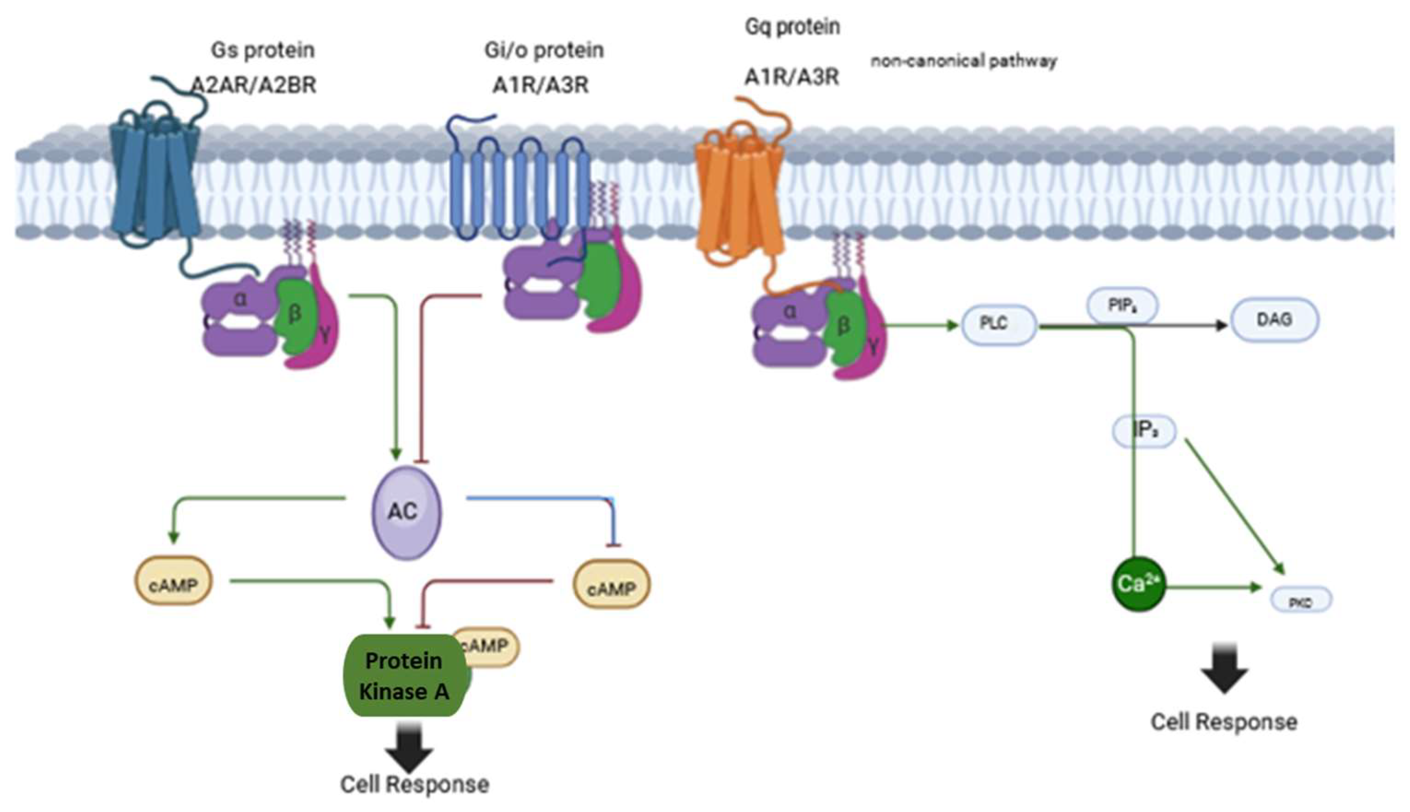
T2D is a pathological condition closely associated with obesity and low-grade inflammation. This inflammatory state leads to elevated levels of pro-inflammatory cytokines, free fatty acids, and glucose in circulation, which in turn contribute to dysfunction in multiple organs. Recent advances have redefined T2D not as a single disease entity but as a heterogeneous cluster of metabolic disorders with distinct pathophysiological mechanisms. Clustering analyses [13] have identified novel subgroups of T2D with differing degrees of insulin resistance, β-cell dysfunction, obesity, and other factors. These novels subgroups, are severe insulin-resistant diabetes, mild obesity-related diabetes, and mild age-related diabetes. These subtypes differ significantly in disease progression, risk of complications, and underlying pathophysiology, offering a refined framework for precision medicine approaches in diabetes care. However, in this manuscript, we will use the traditional definition of type 2 diabetes, and obesity as a risk factor for its development.
Obesity is a complex, chronic condition with significant health risks. The distribution of body fat, particularly ectopic fat deposition, plays a critical role in the development of chronic metabolic diseases, especially insulin resistance and T2D. During obesity progression, excessive calorie intake leads to fat accumulation primarily in visceral adipose tissue and ectopic sites, including the pancreas, heart, and muscle [14]. As visceral adipose tissue expands beyond its vascular supply, hypoxia and inflammatory responses are triggered, leading to the secretion of cytokines such as tumor necrosis factor-alpha (TNFα), interleukin-1beta (IL-1β), and interleukin-6 (IL-6), as well as chemokines [15,16]. This process is also associated with altered adipokine production, including increased levels of leptin and resistin and reduced adiponectin [17,18,19,20].
Consequently, cytokines and chemokines activate adipose tissue T-lymphocytes and resident macrophages, which in turn secrete higher levels of pro-inflammatory cytokines and recruit additional immune cells, including other T-lymphocytes, neutrophils, and monocytes [21,22,23]. In addition to changes in cytokine and adipokine release, adipose tissue expansion alters fatty acid flux and tissue metabolism [24,25].
The cause-and-effect relationships between hyperinsulinemia, insulin resistance, obesity, and type 2 diabetes remain to be clarified and unresolved. Although the insulin resistance–hyperinsulinemia axis has traditionally been viewed with insulin resistance as the primary defect, leading to compensatory hyperinsulinemia, in recent years, there is a growing body of evidence supporting a bidirectional model, where hyperinsulinemia itself may be the initiating or upstream factor that contributes to the development of insulin resistance, rather than simply being a consequence of it [26]. It has been proposed that environmental changes, such as chronic excess of nutrients, food additives or diabetogenic signals from the gut, result in beta cell overstimulation, exhibited as basal hyperinsulinaemia. The result is the development of obesity and insulin resistance, with these processes occurring either in series or in parallel [27]. Chronic hyperinsulinemia has been shown to desensitize insulin receptors, alter lipid metabolism, and promote adipogenesis, all of which feed into a cycle of worsening insulin sensitivity. The prevention of high-fat diet-induced hyperinsulinemia through partial ablation of the pancreas-specific Ins1 gene protects mice from diet-induced obesity and associated complications by downregulating genes required for differentiation in white adipose tissue [28].
Dysfunctional adipose tissue exhibits increased lipolysis, decreased adiponectin levels, and elevated influx of long-chain fatty acids, which, along with heightened pro-inflammatory cytokine activity, exacerbate systemic insulin resistance [29,30]. In obesity, fat accumulation also occurs in ectopic tissues, primarily the liver and muscle. Increased lipolysis in adipose tissue results in a higher influx of free fatty acids into these ectopic sites, further intensifying insulin resistance and inducing apoptosis. This sequence of events culminates in a “lipotoxic state”.
Chronic low-grade inflammation is a major contributor to insulin resistance, an early event in obesity that ultimately leads to T2D [31]. Among inflammatory mediators, TNFα plays a crucial role in insulin resistance. Overexpression of TNFα in adipose tissue is a common feature in animal models of obesity and is also observed in the adipose tissue of obese individuals, linking obesity to insulin resistance [16,32]. One proposed mechanism for TNFα-induced insulin resistance is its attenuation of insulin-stimulated tyrosine phosphorylation of the insulin receptor and IRS1 (insulin receptor substrate 1) in muscle and adipose tissue, thereby impairing insulin signaling [33]. TNFα is produced not only by adipocytes but also by macrophages [34,35]. The increased secretion of pro-inflammatory adipokines and decreased secretion of anti-inflammatory adipokines (such as adiponectin) promotes oxidative stress. Adipocytes under stress produce more ROS, which can cause damage to the cells and surrounding tissue. Oxidative stress in adipocytes can contribute to insulin resistance, inflammation, and the development of metabolic diseases. It can also affect other tissues and organs, leading to a systemic impact on health. ROS interfere with the insulin receptor signaling pathway by activating stress kinases, such as JNK (c-Jun N-terminal kinase) and IKKβ (IκB kinase), which inhibit the insulin signaling cascade. This leads to a reduced ability of cells to take up glucose, resulting in hyperglycemia and further metabolic disturbances.
Adipose tissue macrophages exist in two primary subtypes: resident M2 macrophages, which maintain tissue homeostasis and secrete anti-inflammatory cytokines like IL-10, and bone marrow-derived M1 macrophages, which migrate into adipose tissue and differentiate in response to fatty acids and cytokines [36,37]. Obesity exaggerates macrophage infiltration into omental adipose tissue—a specialized fat depot in the peritoneal cavity connected to the spleen, stomach, transverse colon, and pancreas. This infiltration is associated with increased monocyte chemoattractant protein-1 (MCP1) and colony-stimulating factor-1 (CSF1) mRNA levels, correlating with macrophage numbers. In severely obese women, higher MCP1 and CSF1 protein expression in omental fat has been linked to the number of metabolic syndrome parameters, particularly in cases where obesity is associated with impaired glucose homeostasis [21].
Even before significant visceral adipose tissue expansion occurs, high dietary fat intake induces inflammation in the brain, particularly in the hypothalamus [38,39], but also in other regions such as the hippocampus [40]. Saturated fat consumption rapidly increases pro-inflammatory cytokine expression in the hypothalamus through activation of resident microglia [38,39,41]. Persistent dietary fat intake leads to recruitment of peripheral myeloid cells to the hypothalamus, chronic inflammation, neuronal damage, and subsequent hypothalamic resistance to leptin and insulin [42,43]. Among hypothalamic neurons, POMC (Pro-opiomelanocortin) neurons are particularly susceptible to inflammation-induced damage. Over time, this neuronal imbalance disrupts the regulation of orexigenic and anorexigenic circuits, promoting body mass gain, impaired glucose tolerance, and insulin resistance (for more on the metabolic regulation of melanocortin circuits, see review [44].
Collectively, these findings highlight the intricate link between obesity, inflammation, and insulin resistance, which can progress to T2D and systemic metabolic dysfunction. Pro-inflammatory cytokines not only disrupt adipose tissue metabolism but also impair the function of distant organs involved in whole-body metabolism, including the brain, liver, and skeletal muscle. Given the growing societal burden of obesity and T2D, alongside lifestyle interventions, there is strong interest in developing new nutritional strategies to prevent disease progression and mitigate complications.

3. Adenosine System

Adenosine (C10H13N5O4) is a nucleoside ubiquitously present in the body, primarily derived from the metabolism of adenine nucleotides such as cAMP, ADP, and ATP, which are converted into 5′-AMP and subsequently hydrolyzed into adenosine by nucleosidases [45]. Adenosine can also be generated intracellularly through the hydrolysis of AMP or S-adenosylhomocysteine. It exerts a wide range of physiological effects across various tissues and plays critical roles in the immune system, oxidative stress, and pathological states associated with metabolic, inflammatory, and chronic diseases, including cardiovascular diseases, diabetes [46], neuroinflammation [47], and cancer, via its receptors or even through epigenetic mechanisms [48].
In 1979, van Calker et al. first proposed that adenosine mediates its effects via cell surface receptors rather than intracellular mechanisms [49]. Adenosine receptors are widely distributed throughout the body, present in nearly every human cell, and exert critical effects at both cellular and tissue levels. Adenosine signals through four distinct G-protein-coupled receptor subtypes: A1R, A2AR, A2BR, and A3R (Figure 1). Among these, A1R and A3R are coupled to inhibitory G-proteins (Gi/o), which suppress adenylate cyclase activity, leading to decreased intracellular cAMP levels. They can also activate phospholipase C (PLC) via Gq proteins. In contrast, A2AR and A2BR are coupled to stimulatory G-proteins (Gs), which increase intracellular cAMP levels by activating adenylate cyclase [50]. Additionally, A2BR can also couple with Gq proteins [51].
The affinity of these receptors for adenosine varies, adenosine has higher affinity for A1R, A2AR, and A3R have, whereas has low affinity for A2BR. This difference in affinity suggests that varying adenosine concentrations preferentially activate specific receptors, leading to distinct physiological effects. Under normal physiological conditions, adenosine primarily activates A1R, A2AR, and A3R, while A2BR is activated only when adenosine levels are significantly elevated [51].
The tissue distribution of adenosine receptors also differs. A1R and A2AR are the most extensively studied, particularly in the central nervous system (CNS), where they are expressed in neurons, astrocytes, and microglia. A1R is predominantly found in the cortex, cerebellum, hippocampus, and spinal cord, whereas A2AR is highly expressed in the striatum and olfactory bulb. These receptors are also found in peripheral tissues, including the heart, lungs, liver, blood vessels, and glands, with A2AR being particularly abundant in immune cells. The other subtypes, A2BR and A3R, are less well studied and are present at lower levels in the CNS but are found in the gut, lungs, adipose tissue, and glands [50].
In the brain, at physiological concentrations, adenosine primarily mediates its effects through A1R, which exerts an overall inhibitory action. In the hippocampus, this inhibition occurs mainly through presynaptic blockade of excitatory neurotransmission, reducing glutamate release [52]. In contrast, A2AR can have opposing effects depending on its location. For instance, in the striatum, A2AR stimulation enhances neurotransmitter release, counteracting the inhibitory effect of co-localized A1R [53]. The interaction between A2AR and dopamine D2 receptors in the striatum is being investigated as a potential therapeutic target for neuropsychiatric disorders, including Parkinson’s disease [54] and schizophrenia [55].
Recently, it has been described that A1R and A2R are also localized and are functionally active in mitochondrial fractions isolated from mouse brain and liver, and human brain, revealing that these receptors can be potential modulators of mitochondrial energy metabolism [56]. Beyond its roles in the CNS, adenosine receptors are also crucial in metabolic tissues, where they regulate glucose and lipid homeostasis and contribute to overall metabolic function. Given their widespread distribution and diverse physiological roles, adenosine receptors represent promising targets for therapeutic interventions in various diseases.

4. The Adenosine System in Metabolic Regulation

Extracellular adenosine plays a crucial role in preserving and restoring tissue homeostasis. Its receptors are widely distributed across metabolically active organs and the immune system. Notably, adenosine receptors are highly expressed in key metabolic-regulating organs, including the pancreas, liver, muscle, adipose tissue, and hypothalamus (Table 1) [57]. This widespread localization underscores their important roles in metabolism and their interdependencies in both physiological and pathological conditions (Figure 2).
Increasing evidence highlights the critical role of the adenosine system in regulating inflammation, immune responses, and oxidative stress—factors that are also central to metabolic disorders. The activation of A1R is often associated with antioxidant effects, especially in the brain, heart, and kidneys, through the reduction of reactive oxygen species (ROS) production and enhancing mitochondrial function. The A2AR has also potent anti-inflammatory and antioxidant effects via activation of pathways like Nrf2 (nuclear factor erythroid 2–related factor 2), which increases the expression of antioxidant enzymes like superoxide dismutase (SOD) and catalase (to understand the role of adenosine system in modulation of oxidative stress see the revision [58]).
An interesting aspect of this system is its interaction with caffeine, a well-known antagonist of the adenosine A1 receptor. Caffeine is widely used as a dietary supplement to promote weight loss; however, the precise mechanisms underlying its metabolic effects remain unclear.
In a mouse model of diet-induced obesity, elevated levels of adenosine have been detected in cerebrospinal fluid [59], suggesting that the adenosine system is dysregulated in obesity. This finding also indicates that adenosine signaling may directly influence insulin and glucose homeostasis, playing a role in the pathophysiology of T2D. Among adenosine receptors, A2A and A2B are particularly promising therapeutic targets for obesity and diabetes. Pharmacological modulation of these receptors through agonists or antagonists holds potential for improving metabolic disorders and associated complications.

4.1. The Role of Adenosine System in Pancreas

The endocrine pancreas plays a pivotal role in the regulation of glucose metabolism through the secretion of insulin and glucagon. These hormones are essential for maintaining glucose homeostasis. All four adenosine receptor subtypes (A1, A2A, A2B, and A3) are expressed in the pancreas, suggesting their involvement in pancreatic physiology and positioning them as potential therapeutic targets for diabetes mellitus.
In rat pancreatic islets, adenosine has been shown to inhibit glucose-induced insulin secretion in a concentration-dependent manner (0.1–10 μM) via activation of A1R. This effect is mediated through Gi-protein signaling, which inhibits adenylyl cyclase (AC), leading to a reduction in intracellular cAMP levels in β-cells [60,61,62]. However, at higher adenosine concentrations, insulin secretion may actually be enhanced due to a shift in receptor activation [63].
A1R-deficient (A1−/−) mice exhibit increased insulin secretion. Following glucose administration, these mice display sustained elevations in plasma insulin and glucagon levels, with a significantly enhanced second phase of insulin release compared to wild-type controls [61,64]. The enhanced insulin secretion in A1−/− mice is attributed, at least in part, to increased β-cell metabolic activity and elevated cAMP levels. Additionally, adenosine may regulate insulin secretion through modulation of autophagy [65]. Given these findings, A1 receptor antagonists—such as caffeine and 2(-benzylidene-1-indanone)—are being explored as potential therapeutics for enhancing insulin release in diabetes [66].
Conversely, at higher concentrations (100–300 μM), adenosine activates A2A receptors (A2AR), leading to increased insulin secretion [67]. This effect occurs via Gs-protein activation, which elevates intracellular cAMP levels, stimulates protein kinase A (PKA), and enhances insulin granule exocytosis. A2AR activation also promotes β-cell proliferation, further supporting its potential as a therapeutic target for diabetes [68].
Glucagon, secreted by pancreatic α-cells, plays a crucial role in maintaining blood glucose levels during fasting by stimulating hepatic glucose production. Additionally, glucagon acts in a paracrine manner on neighboring β-cells to promote insulin release under high glucose conditions [69].
Studies in perfused rat pancreas have shown that adenosine enhances glucagon secretion at low glucose levels through A2 receptor activation [70]. α-Cell-selective A2AR knockout (KO) mice have demonstrated that A2AR is essential for promoting glucagon release during hypoglycemia [71]. In T2D, elevated plasma glucagon levels contribute to hyperglycemia, a hallmark of the disease. Adenosine, through A2A receptor activation, exerts an autocrine/paracrine effect in promoting glucagon release, making it a potential target for modulating α-cell function therapeutically [71].
In A1R KO mice, glucagon and somatostatin pulses are prolonged, and their usual asynchronous pattern is disrupted. This suggests that endogenous adenosine, via A1R activation, plays a regulatory role in the pulsatile release of islet hormones, counteracting its inhibitory effect on insulin secretion [64]. A1R KO mice exhibit elevated plasma glucagon levels following glucose challenges or high-fat diet consumption, with prolonged glucagon release during hyperglycemia [61,72].
In non-obese diabetic (NOD) mice, a decrease in A1R expression in pancreatic α-cells is linked to abnormally high glucagon levels, suggesting a role for A1R dysfunction in the pathogenesis of type 1 diabetes (T1D) [73]. Moreover, A3 receptor activation has been implicated in β-cell survival, highlighting its potential as a therapeutic target [67].
In summary, adenosine through A1R inhibits insulin secretion, while at higher concentrations and by A2AR activation, adenosine increases insulin secretion. In pancreatic alpha-cells, at lower concentration adenosine increases the release of glucagon through A2AR. All of these studies point that the adenosine system plays a crucial role in pancreatic function by modulating insulin and glucagon secretion, positioning it as a key player in both normal physiology and the pathophysiology of diabetes. Targeting specific adenosine receptors offers a promising strategy for the treatment of diabetes and its associated metabolic dysregulations.

4.2. The Role of Adenosine System in the Liver

The liver plays a critical role in maintaining blood glucose levels in both fasting and postprandial states. Under physiological conditions, adenosine contributes to glucose homeostasis by counteracting insulin action in the liver, increasing hepatic glucose output through glycogenolysis and gluconeogenesis [74,75].
Studies using isolated hepatocytes treated with A1, A2A, and A3 receptor-selective agonists have demonstrated that adenosine promotes glycogenolysis, primarily via A1 receptor (A1R) activation through Ca2+-mediated pathways, while gluconeogenesis is predominantly regulated by A2AR activation [75]. These findings suggest that adenosine has a hyperglycemic effect in the liver.
The regulation of glycogenolysis by adenosine is mediated through adenylyl cyclase activation and is involved in the short-term regulation of hepatic glycogen phosphorylase, an enzyme that catalyzes the conversion of glycogen phosphorylase from its inactive (b) form to the active (a) form [74]. In isolated rat hepatocytes, adenosine and its agonists stimulate cAMP formation and regulate gluconeogenesis, likely via A2AR activation [75].
A2B receptor activation has also been implicated in hepatic glucose metabolism. In primary rat hepatocyte cultures, pharmacological stimulation of A2BR enhances both glycogenolysis and gluconeogenesis [76]. Furthermore, in mice treated with the nonselective adenosine receptor agonist 5′-N-ethylcarboxamidoadenosine (NECA), fasting blood glucose levels increased, and glucose disposal was delayed during glucose tolerance tests. These effects were inhibited by A2BR deletion or blockade, confirming the receptor’s role in modulating glucose metabolism [77].
In obesity, excessive hepatic glucose production through gluconeogenesis contributes to glucose intolerance. Interestingly, A2B receptor KO mice exhibit increased gluconeogenesis, suggesting that A2BR may play a protective role by limiting hepatic glucose production [78].
Adenosine also regulates liver cholesterol synthesis, which in turn influences plasma cholesterol and triglyceride levels [79,80]. Genetic ablation of A2BR leads to elevated liver and plasma cholesterol and triglycerides, as well as fatty liver pathology typical of hepatic steatosis. High-fat diet (HFD) consumption significantly upregulates A2BR gene expression, highlighting its role in lipid homeostasis. In A2BR-deficient mice fed an HFD, lipid accumulation in the liver is markedly increased, further supporting the receptor’s protective function [79]. Lipid regulation by A2BR is associated with negative regulation of sterol regulatory element-binding protein 1 (SREBP-1), a key transcription factor involved in lipid metabolism. This suggests that A2BR influences plasma lipid levels, at least in part, through SREBP-1 modulation [79].
In A2BR KO mice, elevated levels of pro-inflammatory cytokines have been linked to impaired insulin signaling due to the downregulation of insulin receptor substrate 2 (IRS-2). A2BR deletion also results in increased SREBP-1 levels, which not only suppresses IRS-2 but also promotes cholesterol synthesis, potentially explaining the observed hypercholesterolemia [81]. Pharmacological activation of A2BR has been shown to improve glucose tolerance, insulin signaling, and chronic inflammation in HFD-induced obesity models.
In a metabolic hepatic steatosis model, hepatic A1R activation has been found to reduce de novo lipogenesis by inhibiting SREBP-1, thereby ameliorating liver steatosis. Liver biopsies from patients with hepatic steatosis show positive A1R protein expression, whereas control patients do not exhibit this expression. A similar pattern has been observed in animal models of HFD-induced obesity. These findings suggest that the high expression of hepatic A1R in metabolic hepatic steatosis may serve as an adaptive protective response [82].
In conclusion, the adenosine system plays a crucial role in hepatic glucose and lipid metabolism. Adenosine activation of A1 receptor tends to suppress hepatic glucose output. However under inflammatory or insulin-resistant states, A2B receptor signaling can promote gluconeogenesis under certain inflammatory. While, by the activation of A2BR, adenosine decreased cholesterol and triglyceride levels. Adenosine also modulates fatty acid β-oxidation. A2A receptor activation has anti-inflammatory and protective effects on hepatocytes and may improve lipid handling. A2BR appears to have a protective function in both glucose and lipid homeostasis, and its activation could be a promising therapeutic strategy for improving insulin sensitivity and reducing hepatic steatosis in metabolic disorders, including diabetes and obesity-related liver dysfunction.

4.3. The Role of Adenosine System in Adipose Tissue

4.3.1. Brown Adipose Tissue

Brown adipose tissue (BAT) is an important organ in metabolic regulation due to its thermogenic properties, which are activated by cold exposure and sympathetic transmission. BAT activation increases energy expenditure and has become a growing area of interest in the regulation of obesity and metabolic disorders [83].
It has been demonstrated that adenosine activates cultured brown adipocytes at low nanomolar concentrations. Sympathetic activation of brown adipose tissue induces the release of adenosine, which primarily acts through the A2A receptor. Pharmacological blockade or genetic loss of A2A receptors in mice leads to a decrease in BAT-dependent thermogenesis, whereas treatment with A2A agonists significantly increases energy expenditure [84]. Additionally, stimulation of A2A receptors induces the browning of beige adipocytes. Mice fed a high-fat diet and treated with an A2A agonist are leaner and exhibit improved glucose tolerance, suggesting a protective effect of A2A receptor activation against HFD-induced obesity by increasing energy expenditure [84].
Importantly, KO mice for A2A receptors exhibit dysfunctional BAT, which may be correlated with heart function [85]. In this study, the authors also observed that BAT dysfunction caused by A2A receptor knockout leads to reduced production of Fibroblast growth factor 21 (FGF21), a factor shown to be important for cardioprotection against hypertensive cardiac remodeling [85].
In humans, adenosine administration has been shown to cause maximal perfusion effects in supraclavicular BAT, indicating that adenosine increases oxidative metabolism via A2A receptors [86]. This study also measured A2A receptor activation in BAT during cold exposure, demonstrating greater adenosine binding to A2A receptors, suggesting a cooperative effect of adenosine and noradrenaline in driving thermogenesis during cold exposure [86].
To further understand the pharmacological potential of adenosine receptors in BAT, a dual acting ligand- agonist of A2A and antagonist ofA3 receptors (LJ-4378), was investigated for its anti-obesity effects and underlying molecular mechanisms. In vitro treatment with the LJ-4378 increased the levels of brown adipocyte markers and mitochondrial proteins, including uncoupling protein 1 (UCP1), and induced lipolysis. In vivo, LJ-4378 treatment increased energy expenditure, and a 10-day treatment reduced body weight, fat content, and improved glucose tolerance in HFD-fed mice [87].
The A2B receptor is also abundantly expressed in BAT. Adipose tissue-specific ablation of the A2B receptor exacerbated age-related metabolic decline and reduced BAT energy expenditure. However, pharmacological stimulation of A2B improved obesity-related phenotypes. The expression of A2B receptors is higher in BAT biopsies from lean individuals, whereas in overweight subjects, A2B receptor expression is less abundant. In humans, A2B receptor expression in BAT is significantly inversely correlated with age and total body fat mass and positively correlated with UCP1 expression [88].
In conclusion, adenosine plays a key role in BAT metabolism and thermogenesis, as well as in the browning of beige adipose tissue. The A2AR activation, increases non-shivering thermogenesis, by the increase in the expression of UCP1 and mitochondrial activity, promoting an increase in energy expenditure and fat burning. Also promotes lipid mobilization and oxidation to fuel thermogenesis, as well as improves insulin sensitivity, supporting systemic glucose homeostasis. A2A receptor activation presents a promising pharmacological target for increasing BAT metabolism, which could be a potential strategy for combating obesity and related metabolic diseases.

4.3.2. White Adipose Tissue

White adipose tissue (WAT) is a key metabolic and endocrine organ that stores energy in the form of triglycerides. It plays a critical role in regulating energy balance and must rapidly shift its metabolism between fasting and feeding to maintain homeostasis. Abnormal and excessive fat accumulation leads to the expansion of WAT and contributes to the pathophysiology of obesity and T2D.
Adenosine is considered a major regulator of adipose tissue physiology. The most expressed adenosine receptor in WAT is A1R [89,90]. Treatment of adipocytes with an A1R antagonist (DPCPX) leads to an increase in both basal and noradrenaline-stimulated lipolysis, suggesting that endogenous adenosine acts at the adenosine A1 receptor. In A1R (−/−) KO mice, there is an increase in lipolysis and free fatty acid levels. Additionally, treatment with CPA (an A1R agonist) has no antilipolytic effect in A1R (−/−) mice [89]. Activation of A1R in adipocytes reduces cAMP levels and inhibits lipolysis by reducing the activity of hormone-sensitive lipase (HSL) and/or adipose triglyceride lipase (ATGL).
Pharmacological agents that reduce the release of free fatty acids from adipose tissue and decrease circulating FFA availability may be beneficial for insulin resistance and hyperlipidemia. Toward this goal, several selective and efficacious A1R agonists have been developed, with some entering early-phase clinical trials. However, none have yet received regulatory approval [91].
In animals with specific deletion of A1R in adipose tissue, fasting impairs insulin’s ability to suppress lipolysis when fed a normal chow diet, and glucose tolerance is impaired on a high-fat diet. These animals also exhibit a heightened lipolytic response to isoproterenol, though this effect disappears after a four-hour refeeding period [92]. This suggests that in WAT, feeding desensitizes A1 adenosine receptors. In obese Zucker rats A1 adenosine receptor signaling is more active in adipocytes. Gi alpha protein were found decreases in obese rats, however the sensitivity of the receptor was higher [93].
Adenosine, through A1R activation, has been found to increase leptin secretion from white adipocytes both in vitro and in vivo in rats following pharmacological stimulation [94]. However, in A1R (−/−) KO mice, serum leptin levels are higher compared to wild-type mice, while adiponectin levels remain similar between the groups. Free fatty acid concentrations are also higher in A1R (−/−) KO mice, with a similar trend observed for serum triglyceride concentrations [95].
Adenosine, through A1R, can increase lipogenesis both in vitro and in vivo in mice [89]. This effect is abolished in the absence of insulin, suggesting a crosstalk and cooperative effect between insulin and adenosine via A1R in promoting lipogenesis in WAT.
WAT also expresses A2AR, though in lower amounts than A1R. Activation of A2AR can attenuate lipid-induced adipocyte inflammation and improve insulin resistance. A2AR agonists enhance glucose uptake, suggesting a positive role for A2AR in insulin sensitivity in adipocytes [96]. When adipocytes are treated with an A2AR antagonist or subjected to siRNA knockdown of A2AR, the anti-inflammatory effects of A2AR agonists are prevented, and insulin resistance is induced. Furthermore, A2AR, which is widely expressed in leukocytes, can reduce inflammatory markers in diet-induced obesity mice and decrease macrophage infiltration in WAT [97]. Mice fed a high-fat diet for 12 weeks and treated with A2AR agonists for two weeks showed improvements in glucose homeostasis, along with reduced systemic inflammatory markers and decreased macrophage infiltration in visceral adipose tissue [97].
In diet-induced obesity, A2BR is upregulated in WAT. A2BR modulates cytokine secretion by macrophages in mouse adipose tissue, protecting against HFD-induced increases in TNF-α and IL-6, thereby improving insulin sensitivity in WAT [98]. Despite this, A2BR remains upregulated in diet-induced obesity mice.
In summary, adenosine as anti-lipolytic function and increases insulin sensitivity through A1R. However, A2B may have pro-lipolytic effects under some conditions, depending on receptor expression and metabolic state. The A2A and A2B receptors have anti-inflammatory roles, by suppressing macrophage activation and proinflammatory cytokine release. This is especially relevant in obesity-induced chronic inflammation in WAT. Since the adipose tissue of diabetic patients is characterized by impaired storage of triglycerides and increased lipolysis, targeting adenosine receptors in WAT could be a valuable strategy for modulating lipid metabolism and adipocyte inflammation, potentially offering therapeutic promise for managing insulin resistance and type 2 diabetes.

4.4. The Role of Adenosine in the Skeletal Muscle Metabolism

Skeletal muscle is an important metabolic organ that regulates whole-body energy metabolism. Dysregulation of muscle metabolism is linked to several pathologies, including insulin resistance, T2D, and obesity. Insulin resistance is associated with mitochondrial dysfunction, which is mediated by increased lipid accumulation in skeletal muscle, decreased oxidative metabolism, and inflammation [99].
In skeletal muscle, adenosine plays a role in the regulation of carbohydrate metabolism, which is influenced by both insulin and exercise. In human skeletal muscle, A2AR and A2BR are present in muscle fibers, while A1R is absent in these cells [100].
The stimulation of skeletal muscle cells (in vitro) with an adenosine analogue induces inflammatory mediators (such as IL-6) and nuclear receptors (NR4A) while significantly modulating metabolic gene expression. In this study, the authors demonstrate a crosstalk between the adenosine analogue and insulin, which potentiates glycolysis, fatty acid oxidation, and insulin sensitivity [101].
Skeletal muscle, similar to brown adipose tissue, exhibits a high expression of A2BR compared to other adenosine receptors and may be a target for counteracting sarcopenia and obesity. Mice with specific deletion of A2BR in skeletal muscle develop sarcopenia, experience diminished muscle strength, mitochondrial dysfunction, and reduced energy expenditure. Conversely, pharmacological activation of A2BR has the opposite effects, leading to increased muscle mass, enhanced mitochondrial oxidative metabolism, decreased senescence biomarkers, reduced lipid peroxidation, and restoration of age-related strength loss [88].
In obese Zucker rats, treatment with BWA1433 (an adenosine receptor antagonist) improves glucose uptake in skeletal muscle, thereby ameliorating glucose tolerance [100]. In a rat model of insulin resistance (induced by a high-sucrose diet), there is an increased expression of A1, A2A, and A2B adenosine receptors. In this model, treatment for two weeks with an A2A antagonist (SCH58261) and an A2B antagonist (MRS1754) improves insulin response and restores insulin signaling in skeletal muscle [102].
In conclusion, in skeletal muscle adenosine, increase insulin sensitivity and glucose utilization, by the activation of A1R. Also, the activation of A2R receptors play a crucial role in skeletal muscle by controlling energy metabolism and insulin sensitivity, providing protection against obesity, T2D, and age-related decline in muscle function.

4.5. The Role of Adenosine in the Hypothalamic Regulation of Whole-Body Energy Metabolism

In the nervous system, adenosine acts as a neuromodulator, primarily acting on A1 receptors to suppress neuronal activity. In contrast, activation of A2A receptors or A2B receptors stimulates neuronal activity. The physiological stimulation of adenosine receptors following modest increases in extracellular adenosine concentrations plays a crucial role in modulating various brain functions, including sleep and arousal, locomotion, anxiety, cognition, memory, and energy metabolism [10,59].
However, high levels of adenosine are released under metabolic stress associated with excessive neuronal activation, serving a protective function in controlling subsequent tissue damage [10]. The neuroprotective effects of adenosine are mainly mediated by the activation of A1 receptors.
The hypothalamus is a critical brain region involved in whole-body energy metabolism and homeostasis. It is also responsible for regulating temperature, hunger, thirst, hormone secretion, and the sleep-wake cycle [103]. While all adenosine receptors are expressed throughout the brain, only A1R and A2AR are present in the hypothalamus [59,104]. The expression of A1R in hypothalamic nuclei that regulate energy balance suggests its involvement in systemic energy homeostasis, feeding behavior, and glucose regulation. Specifically, A1R expression in paraventricular neurons of the hypothalamus appears to play an essential role in regulating food consumption and body weight.
In a high-fat diet (HFD)-induced obesity mouse model, increased adenosine levels were observed in plasma, CSF, and the hypothalamus. Additionally, A1R expression was elevated in the hypothalamus of these HFD-induced obese mice. Mice overexpressing A1R in the paraventricular hypothalamic neurons exhibited greater body weight gain, increased food consumption, and glucose intolerance compared to control mice (with basal A1R expression) when maintained on a standard chow diet. Notably, central caffeine treatment reduced body weight in HFD mice and improved glucose intolerance and plasma triglyceride levels [59].
Importantly, in the hypothalamus, astrocytes are implicated in regulating feeding behaviors by modulating orexigenic agouti-related peptide (AGRP) neuronal activity. In the medial basal hypothalamus, astrocyte stimulation reduces both basal and ghrelin-evoked food intake through an adenosine-mediated mechanism that inactivates AGRP neurons in the arcuate nucleus via A1R [105].
Hypothalamic neurons in the paraventricular nucleus also play a vital role in wakefulness. Adenosine, an endogenous regulator of sleep homeostasis, may serve as a crucial link between sleep and metabolism [106]. Under physiological conditions, adenosine promotes sleep induction. In the hypothalamus, the ventrolateral preoptic area, a key sleep center, expresses A2A receptors, whose activation induces non-rapid eye movement (NREM) sleep. Conversely, A1R antagonism in the paraventricular nucleus significantly reduces NREM sleep [107]. Proper sleep regulation is essential for maintaining metabolic health, as disturbances in sleep can contribute to obesity, diabetes, and metabolic disorders.
Table 1. Summary of the Role of Adenosine in Metabolic Regulation.
TissuePrimary Adenosine ReceptorsMain FunctionsMetabolic EffectsReferences
PancreasA1, A2A, A2B, A3Regulates insulin and glucagon secretion- Suppresses Glucagon release
- Inhibits insulin release under low glucose concentrations
[60,61,63,66,67,71]
LiverA1, A2A, A2BRegulates gluconeogenesis, glycogenolysis, lipid metabolism, and inflammation- Suppresses or promotes hepatic glucose production (context-dependent)
- Modulates insulin sensitivity and steatosis
[74,75,78,79]
White Adipose Tissue A1, A2A, A2BControls lipolysis, adipogenesis, and inflammation- A1R inhibits lipolysis and improves insulin action
- Increase leptin secretion
- A2AR/2BR reduce inflammation and promote M2 macrophages
[89,90,94,96]
Brown Adipose Tissue A2APromotes thermogenesis, lipid oxidation, and energy expenditure- A2AR activation enhances UCP1 expression and mitochondrial activity
- Increases energy dissipation via heat
[84,85,86,87]
Skeletal MuscleA1, A2A, A2B, A3Regulates insulin sensitivity, and glucose uptake- Enhances mitochondrial oxidative metabolism,
- Decreases senescence biomarkers
- Potentiates insulin sensitivity
[88,100,101]
HypothalamusA1, A2AControls appetite, sympathetic tone, and whole-body energy balance- A1R suppresses appetite via POMC/NPY modulation
- Inactivation of AgRP neurons
- Induces NREM sleep
[59,104,105,107]
In summary, the A1R activation in the arcuate nucleus of the hypothalamus, reduces food intake and promotes anorexigenic pathways enhancing satiety signals, pointing a potential anti-obesity action. Also sleep regulation by adenosine via A1R indirectly can affects energy metabolism, as sleep deprivation disrupts glucose regulation and appetite control.

4.6. The Role of Adenosine Receptors in Immune System Regulation

Under conditions of cellular stress, damage, metabolic stress, or inflammation, extracellular adenosine levels can increase up to 200-fold. Adenosine receptors, which are widely expressed on various immune cells, act as major regulators of inflammation and function as endogenous immune modulators. For example, adenosine has been reported to inhibit TNF-α production, acting as an anti-inflammatory molecule.
Monocytes and macrophages express A2B receptors that regulate a range of immune and inflammatory responses. In macrophages, activation of A2A receptors inhibits pro-inflammatory cytokines such as TNF-α and IL-12 [108,109]. Similarly, stimulation of A2B receptors appears to suppress pro-inflammatory cytokine production while increasing the expression of anti-inflammatory cytokines [110,111]. In human peripheral blood mononuclear cells, adenosine inhibits TNF-α production primarily via A2A receptor activation, whereas stimulation of A1R or A3R can enhance adenosine’s actions [112].
In lean adipose tissue, the presence of alternatively activated M2 macrophages is higher. In obesity, however, there is an infiltration and accumulation of pro-inflammatory M1 macrophages, and an increased M1/M2 ratio is associated with decreased insulin sensitivity. A2B receptor deficiency has been linked to reduced expression of transcription factors that drive M2 responses [78]. In adipose tissue of A2B-deficient mice, pro-inflammatory cytokines (CCL2, TNF-α, and IL-6) are elevated, while anti-inflammatory cytokine (IL-10) levels are decreased. These receptors also help preserve the M1 phenotype to maintain immune homeostasis in adipose tissue. In mice fed a HFD, liver A2B receptor expression is upregulated, whereas mice lacking A2B receptors show increased liver inflammation and insulin resistance [98].
Microglia—the resident immune cells of the brain—express A1, A2A, and A3 receptors. Under normal conditions, adenosine stimulates the proliferation of naïve microglial cells via the combined activation of A1 and A2 receptors [113]. In contrast, under pathological conditions, adenosine-mediated inhibition of activated microglial proliferation is primarily through A1 receptor activation [114]. Agonists of the A1 receptor can confer neuroprotection, at least partly by directly inhibiting microglial proliferation. Activation of adenosine A3R inhibits microglia reactivity, exerting anti-inflammatory and neuroprotective effects by regulating microglial phenotype polarization [115,116]. Therefore, the adenosine system may serve as a potential target for pharmacological intervention in diseases characterized by microglial activation.
In the hypothalamus, both animal models of obesity and human studies have shown activated microglia that secrete pro-inflammatory cytokines and promote further microglial recruitment [38,39,41]. Microglial activation in response to high saturated fat consumption or obesity can lead to neuronal damage, ultimately disrupting whole-body energy homeostasis, metabolism, and insulin sensitivity. Although the specific roles of adenosine in hypothalamic microglial cells have not yet been fully elucidated, the expression of adenosine receptors in these cells under metabolic conditions warrants further investigation.
In summary, adenosine is a powerful anti-inflammatory mediator, especially active during stress, or inflammation. It signals through A1, A2A, A2B, and A3 receptors to regulate immune responses. Adenosine suppresses inflammation by inhibiting cytokine release and is involved in to the macrophage shifts to M2 anti-inflammatory phenotype. In some contexts (e.g., A2B), may support chronic low-grade inflammation. Adenosine signaling in both macrophages and microglia may play a significant protective role in regulating metabolism and mitigating obesity-induced insulin resistance.

5. Conclusions

To summarize, adenosine plays a key role in the physiological metabolic regulation of metabolic tissues through its four G protein-coupled receptors: A1, A2A, A2B, and A3. These effects are tissue-specific and depend on both the extracellular concentration of adenosine and the receptor subtype activated. Understanding the downstream signaling pathways—whether Gs- or Gi-coupled—is essential for fully appreciating adenosine’s diverse roles under different physiological and pathological conditions.
Dysregulation of adenosine system seems to be involved in the pathophysiology of obesity, diabetes, and related metabolic disorders through its actions in brown adipose tissue (BAT), white adipose tissue (WAT), skeletal muscle, pancreas, liver, hypothalamus, and the immune system (Table 1). Adenosine is also an important modulator of oxidative stress, with diverse and receptor-specific effects that can be both protective and pathological. Dysregulation of the adenosine system in these metabolically active tissues further supports its relevance in such conditions, highlighting the therapeutic potential of targeting adenosine signaling pathways as a novel approach to managing metabolic diseases.
The heterogeneity of type 2 diabetes has been increasingly recognized, which presents an important opportunity for advancing precision medicine approaches in diabetes care. Highlighting the implications for precision medicine and the potential for pathway-specific interventions like adenosine signaling adds an important translational perspective. For instance, for insulin resistant diabetes, therapies that enhance adenosine signaling could be tailored to improve insulin sensitivity and reduce systemic inflammation. Personalized strategies could include using adenosine receptor agonists or interventions that enhance adenosine release, which could reduce the need for high doses of insulin or other medications. Also modulating adenosine pathways in obesity related diabetes patients could help address the root cause of insulin resistance—obesity—while improving metabolic function. This approach could complement weight loss interventions and reduce the need for more aggressive pharmaceutical treatments.
The clinical implications to modulating adenosine signaling may help to reduce WAT inflammation and improve insulin sensitivity in obesity and type 2 diabetes. It is also possible to modulate adenosine system to manage dyslipidemia, improving glucose metabolism as well as enhancing energy expenditure in metabolic disorders.

Author Contributions

G.F. and J.M.G. write, revised, and edited the manuscript. The final version was approved by the authors to be submitted, and took responsibility for the manuscript’s integrity and accuracy. All authors have read and agreed to the published version of the manuscript.

Funding

G.F. was financed by the Fundação de Amparo à Pesquisa e Inovação do Estado de Santa Catarina (FAPESC), project TO number: 2024TR001581 (EDITAL DE CHAMADA PÚBLICA FAPESC Nº 09/2024—Mulheres + Pesquisa 1º edição). J.M.G. was financed by Fundação de Amparo à Pesquisa e Inovação do Estado de Santa Catarina (FAPESC), project TO number: 2024TR001581 (EDITAL DE CHAMADA PÚBLICA FAPESC Nº 09/2024—Mulheres + Pesquisa 1º edição), and project TO number: 2024TR002262 (EDITAL DE CHAMADA PÚBLICA FAPESC Nº 21/2024—Programa de Pesquisa Universal); and Brazilian Federal Agency—Fundação Coordenação de Aperfeiçoamento de Pessoal de Nível Superior (CAPES).

Conflicts of Interest

The authors declare no conflicts of interest.

Abbreviations

The following abbreviations are used in this manuscript:
ACAdenylate cyclase
ADPAdenosine diphosphate
AMPAdenosine monophosphate
ATPAdenosine triphosphate
A1RAdenosine 1 Receptor
A2ARAdenosine 2A Receptor
A2BRAdenosine 2B Receptor
A3RAdenosine 3 Receptor
AGRPAgouti-related peptide
ATGLAdipose triglyceride lipase
BATBrown Adipose Tissue
cAMPCyclic Adenosine monophosphate
CCL2Monocyte chemoattractant protein-1
CNSCentral Nervous System
CSFcerebrospinal fluid
DAGDiacylglycerol
FGF21Fibroblast growth factor 21
HFDHigh Fat Diet
HSLHormone-sensitive lipase
IL1βInterleukin 1 beta
IL6Interleukin 6
IL10Interleukin 10
IP3inositol trisphosphate
IRS-1insulin receptor substrate 1
IRS-2insulin receptor substrate 2
JNKc-Jun N-terminal kinase
KOKnock Out
MCP-1Monocyte chemoattractant protein-1
NODNon-obese diabetic
Nrf2Nuclear factor erythroid 2–related factor 2
POMCPro-opiomelanocortin
PKAProtein kinase A
PLCPhospholipase C
ROSReactive oxygen species
SODSuperoxide dismutase
SREBP-1Sterol regulatory element-binding protein 1
T1DType 1 Diabetes
T2DType 2 Diabetes
TNF-αTumor Necrosis Factor-alpha
UCP-1Uncoupling protein 1
WATWhite adipose tissue

References

  1. World Obesity Federation. World Obesity Atlas 2023; World Obesity Federation: London, UK, 2023. [Google Scholar]
  2. Khan, M.A.B.; Hashim, M.J.; King, J.K.; Govender, R.D.; Mustafa, H.; Al Kaabi, J. Epidemiology of Type 2 Diabetes—Global Burden of Disease and Forecasted Trends. J. Epidemiol. Glob. Health 2020, 10, 107–111. [Google Scholar] [CrossRef] [PubMed]
  3. Rubino, F.; Cummings, D.E.; Eckel, R.H.; Cohen, R.V.; Wilding, J.P.H.; Brown, W.A.; Stanford, F.C.; Batterham, R.L.; Farooqi, I.S.; Farpour-Lambert, N.J.; et al. Definition and diagnostic criteria of clinical obesity. Lancet. Diabetes Endocrinol. 2025, 13, 221–262. [Google Scholar] [CrossRef]
  4. Schwartz, M.W.; Seeley, R.J.; Zeltser, L.M.; Drewnowski, A.; Ravussin, E.; Redman, L.M.; Leibel, R.L. Obesity Pathogenesis: An Endocrine Society Scientific Statement. Endocr. Rev. 2017, 38, 267–296. [Google Scholar] [CrossRef]
  5. Blüher, M. The distinction of metabolically ‘healthy’ from ‘unhealthy’ obese individuals. Curr. Opin. Lipidol. 2010, 21, 38–43. [Google Scholar] [CrossRef] [PubMed]
  6. Blüher, M. Are metabolically healthy obese individuals really healthy? Eur. J. Endocrinol. 2014, 171, R209–R219. [Google Scholar] [CrossRef] [PubMed]
  7. Mongraw-Chaffin, M.; Foster Meredith, C.; Anderson Cheryl, A.M.; Burke Gregory, L.; Haq, N.; Kalyani Rita, R.; Ouyang, P.; Sibley Christopher, T.; Tracy, R.; Woodward, M.; et al. Metabolically Healthy Obesity, Transition to Metabolic Syndrome, and Cardiovascular Risk. JACC 2018, 71, 1857–1865. [Google Scholar] [CrossRef]
  8. Bell, J.A.; Kivimaki, M.; Hamer, M. Metabolically healthy obesity and risk of incident type 2 diabetes: A meta-analysis of prospective cohort studies. Obes. Rev. 2014, 15, 504–515. [Google Scholar] [CrossRef]
  9. American Diabetes Association Professional Practice Committee. Summary of Revisions: Standards of Care in Diabetes—2024. Diabetes Care 2023, 47, S5–S10. [Google Scholar] [CrossRef]
  10. Fredholm, B.B.; Chen, J.F.; Masino, S.A.; Vaugeois, J.M. Actions of adenosine at its receptors in the CNS: Insights from knockouts and drugs. Annu. Rev. Pharmacol. Toxicol. 2005, 45, 385–412. [Google Scholar] [CrossRef]
  11. Haskó, G.; Cronstein, B.N. Adenosine: An endogenous regulator of innate immunity. Trends Immunol. 2004, 25, 33–39. [Google Scholar] [CrossRef]
  12. Vincenzi, F.; Pasquini, S.; Contri, C.; Cappello, M.; Nigro, M.; Travagli, A.; Merighi, S.; Gessi, S.; Borea, P.A.; Varani, K. Pharmacology of Adenosine Receptors: Recent Advancements. Biomolecules 2023, 13, 1387. [Google Scholar] [CrossRef] [PubMed]
  13. Ahlqvist, E.; Storm, P.; Käräjämäki, A.; Martinell, M.; Dorkhan, M.; Carlsson, A.; Vikman, P.; Prasad, R.B.; Aly, D.M.; Almgren, P.; et al. Novel subgroups of adult-onset diabetes and their association with outcomes: A data-driven cluster analysis of six variables. Lancet Diabetes Endocrinol. 2018, 6, 361–369. [Google Scholar] [CrossRef]
  14. Luo, J.; Wang, Y.; Mao, J.; Yuan, Y.; Luo, P.; Wang, G.; Zhou, S. Features, functions, and associated diseases of visceral and ectopic fat: A comprehensive review. Obesity 2025, 33, 825–838. [Google Scholar] [CrossRef]
  15. Wallenius, V.; Wallenius, K.; Ahrén, B.; Rudling, M.; Carlsten, H.; Dickson, S.L.; Ohlsson, C.; Jansson, J.O. Interleukin-6-deficient mice develop mature-onset obesity. Nat. Med. 2002, 8, 75–79. [Google Scholar] [CrossRef] [PubMed]
  16. Hotamisligil, G.S.; Arner, P.; Caro, J.F.; Atkinson, R.L.; Spiegelman, B.M. Increased adipose tissue expression of tumor necrosis factor-alpha in human obesity and insulin resistance. J. Clin. Investig. 1995, 95, 2409–2415. [Google Scholar] [CrossRef]
  17. Ioffe, E.; Moon, B.; Connolly, E.; Friedman, J.M. Abnormal regulation of the leptin gene in the pathogenesis of obesity. Proc. Natl. Acad. Sci. USA 1998, 95, 11852–11857. [Google Scholar] [CrossRef]
  18. Vaisse, C.; Halaas, J.L.; Horvath, C.M.; Darnell, J.E., Jr.; Stoffel, M.; Friedman, J.M. Leptin activation of Stat3 in the hypothalamus of wild-type and ob/ob mice but not db/db mice. Nat. Genet. 1996, 14, 95–97. [Google Scholar] [CrossRef]
  19. Steppan, C.M.; Bailey, S.T.; Bhat, S.; Brown, E.J.; Banerjee, R.R.; Wright, C.M.; Patel, H.R.; Ahima, R.S.; Lazar, M.A. The hormone resistin links obesity to diabetes. Nature 2001, 409, 307–312. [Google Scholar] [CrossRef] [PubMed]
  20. Côté, M.; Mauriège, P.; Bergeron, J.; Alméras, N.; Tremblay, A.; Lemieux, I.; Després, J.P. Adiponectinemia in visceral obesity: Impact on glucose tolerance and plasma lipoprotein and lipid levels in men. J. Clin. Endocrinol. Metab. 2005, 90, 1434–1439. [Google Scholar] [CrossRef]
  21. Harman-Boehm, I.; Blüher, M.; Redel, H.; Sion-Vardy, N.; Ovadia, S.; Avinoach, E.; Shai, I.; Klöting, N.; Stumvoll, M.; Bashan, N.; et al. Macrophage infiltration into omental versus subcutaneous fat across different populations: Effect of regional adiposity and the comorbidities of obesity. J. Clin. Endocrinol. Metab. 2007, 92, 2240–2247. [Google Scholar] [CrossRef]
  22. Barbosa, P.; Pinho, A.; Lázaro, A.; Paula, D.; Campos, J.C.; Tralhão, J.G.; Pereira, M.J.; Paiva, A.; Laranjeira, P.; Carvalho, E. High percentage of immune Th1 and Tc1 cells infiltrating visceral adipose tissue in people with obesity. Obes. Res. Clin. Pract. 2024, 18, 426–435. [Google Scholar] [CrossRef] [PubMed]
  23. Liao, X.; Zeng, Q.; Xie, L.; Zhang, H.; Hu, W.; Xiao, L.; Zhou, H.; Wang, F.; Xie, W.; Song, J.; et al. Adipose stem cells control obesity-induced T cell infiltration into adipose tissue. Cell Rep. 2024, 43, 113963. [Google Scholar] [CrossRef]
  24. Memon, R.A.; Fuller, J.; Moser, A.H.; Smith, P.J.; Grunfeld, C.; Feingold, K.R. Regulation of putative fatty acid transporters and Acyl-CoA synthetase in liver and adipose tissue in ob/ob mice. Diabetes 1999, 48, 121–127. [Google Scholar] [CrossRef]
  25. Fabbrini, E.; deHaseth, D.; Deivanayagam, S.; Mohammed, B.S.; Vitola, B.E.; Klein, S. Alterations in fatty acid kinetics in obese adolescents with increased intrahepatic triglyceride content. Obesity 2009, 17, 25–29. [Google Scholar] [CrossRef] [PubMed]
  26. Janssen, J. Hyperinsulinemia and Its Pivotal Role in Aging, Obesity, Type 2 Diabetes, Cardiovascular Disease and Cancer. Int. J. Mol. Sci. 2021, 22, 25–29. [Google Scholar] [CrossRef]
  27. Esser, N.; Utzschneider, K.M.; Kahn, S.E. Early beta cell dysfunction vs insulin hypersecretion as the primary event in the pathogenesis of dysglycaemia. Diabetologia 2020, 63, 2007–2021. [Google Scholar] [CrossRef] [PubMed]
  28. Mehran, A.E.; Templeman, N.M.; Brigidi, G.S.; Lim, G.E.; Chu, K.-Y.; Hu, X.; Botezelli, J.D.; Asadi, A.; Hoffman, B.G.; Kieffer, T.J.; et al. Hyperinsulinemia Drives Diet-Induced Obesity Independently of Brain Insulin Production. Cell Metab. 2012, 16, 723–737. [Google Scholar] [CrossRef]
  29. Kim, J.Y.; Nasr, A.; Tfayli, H.; Bacha, F.; Michaliszyn, S.F.; Arslanian, S. Increased Lipolysis, Diminished Adipose Tissue Insulin Sensitivity, and Impaired β-Cell Function Relative to Adipose Tissue Insulin Sensitivity in Obese Youth With Impaired Glucose Tolerance. Diabetes 2017, 66, 3085–3090. [Google Scholar] [CrossRef]
  30. Kasher-Meron, M.; Youn, D.Y.; Zong, H.; Pessin, J.E. Lipolysis defect in white adipose tissue and rapid weight regain. Am. J. Physiol. Endocrinol. Metab. 2019, 317, E185–E193. [Google Scholar] [CrossRef]
  31. Hotamisligil, G.S. Inflammation, metaflammation and immunometabolic disorders. Nature 2017, 542, 177–185. [Google Scholar] [CrossRef]
  32. Hotamisligil, G.S.; Shargill, N.S.; Spiegelman, B.M. Adipose expression of tumor necrosis factor-alpha: Direct role in obesity-linked insulin resistance. Science 1993, 259, 87–91. [Google Scholar] [CrossRef] [PubMed]
  33. Hotamisligil, G.S.; Budavari, A.; Murray, D.; Spiegelman, B.M. Reduced tyrosine kinase activity of the insulin receptor in obesity-diabetes. Central role of tumor necrosis factor-alpha. J. Clin. Investig. 1994, 94, 1543–1549. [Google Scholar] [CrossRef]
  34. Eguchi, J.; Kong, X.; Tenta, M.; Wang, X.; Kang, S.; Rosen, E.D. Interferon regulatory factor 4 regulates obesity-induced inflammation through regulation of adipose tissue macrophage polarization. Diabetes 2013, 62, 3394–3403. [Google Scholar] [CrossRef]
  35. Lumeng, C.N.; Deyoung, S.M.; Saltiel, A.R. Macrophages block insulin action in adipocytes by altering expression of signaling and glucose transport proteins. Am. J. Physiol. Endocrinol. Metab. 2007, 292, E166–E174. [Google Scholar] [CrossRef] [PubMed]
  36. Shang, Q.; Bai, Y.; Wang, G.; Song, Q.; Guo, C.; Zhang, L.; Wang, Q. Delivery of Adipose-Derived Stem Cells Attenuates Adipose Tissue Inflammation and Insulin Resistance in Obese Mice Through Remodeling Macrophage Phenotypes. Stem Cells Dev. 2015, 24, 2052–2064. [Google Scholar] [CrossRef] [PubMed]
  37. Zeyda, M.; Farmer, D.; Todoric, J.; Aszmann, O.; Speiser, M.; Györi, G.; Zlabinger, G.J.; Stulnig, T.M. Human adipose tissue macrophages are of an anti-inflammatory phenotype but capable of excessive pro-inflammatory mediator production. Int. J. Obes. 2007, 31, 1420–1428. [Google Scholar] [CrossRef]
  38. Thaler, J.P.; Yi, C.X.; Schur, E.A.; Guyenet, S.J.; Hwang, B.H.; Dietrich, M.O.; Zhao, X.; Sarruf, D.A.; Izgur, V.; Maravilla, K.R.; et al. Obesity is associated with hypothalamic injury in rodents and humans. J. Clin. Investig. 2012, 122, 153–162. [Google Scholar] [CrossRef]
  39. Valdearcos, M.; Douglass, J.D.; Robblee, M.M.; Dorfman, M.D.; Stifler, D.R.; Bennett, M.L.; Gerritse, I.; Fasnacht, R.; Barres, B.A.; Thaler, J.P.; et al. Microglial Inflammatory Signaling Orchestrates the Hypothalamic Immune Response to Dietary Excess and Mediates Obesity Susceptibility. Cell Metab. 2017, 26, 185–197.e183. [Google Scholar] [CrossRef]
  40. de Paula, G.C.; Brunetta, H.S.; Engel, D.F.; Gaspar, J.M.; Velloso, L.A.; Engblom, D.; de Oliveira, J.; de Bem, A.F. Hippocampal Function Is Impaired by a Short-Term High-Fat Diet in Mice: Increased Blood-Brain Barrier Permeability and Neuroinflammation as Triggering Events. Front. Neurosci. 2021, 15, 734158. [Google Scholar] [CrossRef]
  41. Valdearcos, M.; Robblee, M.M.; Benjamin, D.I.; Nomura, D.K.; Xu, A.W.; Koliwad, S.K. Microglia dictate the impact of saturated fat consumption on hypothalamic inflammation and neuronal function. Cell Rep. 2014, 9, 2124–2138. [Google Scholar] [CrossRef]
  42. Souza, G.F.; Solon, C.; Nascimento, L.F.; De-Lima-Junior, J.C.; Nogueira, G.; Moura, R.; Rocha, G.Z.; Fioravante, M.; Bobbo, V.; Morari, J.; et al. Defective regulation of POMC precedes hypothalamic inflammation in diet-induced obesity. Sci. Rep. 2016, 6, 29290. [Google Scholar] [CrossRef] [PubMed]
  43. Morari, J.; Anhe, G.F.; Nascimento, L.F.; de Moura, R.F.; Razolli, D.; Solon, C.; Guadagnini, D.; Souza, G.; Mattos, A.H.; Tobar, N.; et al. Fractalkine (CX3CL1) is involved in the early activation of hypothalamic inflammation in experimental obesity. Diabetes 2014, 63, 3770–3784. [Google Scholar] [CrossRef]
  44. Lieu, L.; Chau, D.; Afrin, S.; Dong, Y.; Alhadeff, A.L.; Betley, J.N.; Williams, K.W. Effects of metabolic state on the regulation of melanocortin circuits. Physiol. Behav. 2020, 224, 113039. [Google Scholar] [CrossRef] [PubMed]
  45. Dunwiddie, T.V.; Masino, S.A. The role and regulation of adenosine in the central nervous system. Annu. Rev. Neurosci. 2001, 24, 31–55. [Google Scholar] [CrossRef]
  46. Peleli, M.; Carlstrom, M. Adenosine signaling in diabetes mellitus and associated cardiovascular and renal complications. Mol. Asp. Med. 2017, 55, 62–74. [Google Scholar] [CrossRef]
  47. Pasquini, S.; Contri, C.; Borea, P.A.; Vincenzi, F.; Varani, K. Adenosine and Inflammation: Here, There and Everywhere. Int. J. Mol. Sci. 2021, 22, 7685. [Google Scholar] [CrossRef]
  48. Boison, D.; Yegutkin, G.G. Adenosine Metabolism: Emerging Concepts for Cancer Therapy. Cancer Cell 2019, 36, 582–596. [Google Scholar] [CrossRef] [PubMed]
  49. van Calker, D.; Müller, M.; Hamprecht, B. Adenosine regulates via two different types of receptors, the accumulation of cyclic AMP in cultured brain cells. J. Neurochem. 1979, 33, 999–1005. [Google Scholar] [CrossRef]
  50. Fredholm, B.B.; Arslan, G.; Halldner, L.; Kull, B.; Schulte, G.; Wasserman, W. Structure and function of adenosine receptors and their genes. Naunyn Schmiedebergs Arch. Pharmacol. 2000, 362, 364–374. [Google Scholar] [CrossRef]
  51. Borea, P.A.; Gessi, S.; Merighi, S.; Vincenzi, F.; Varani, K. Pharmacology of Adenosine Receptors: The State of the Art. Physiol. Rev. 2018, 98, 1591–1625. [Google Scholar] [CrossRef]
  52. Haas, H.L.; Selbach, O. Functions of neuronal adenosine receptors. Naunyn Schmiedebergs Arch. Pharmacol. 2000, 362, 375–381. [Google Scholar] [CrossRef] [PubMed]
  53. Ferré, S.; Sarasola, L.I.; Quiroz, C.; Ciruela, F. Presynaptic adenosine receptor heteromers as key modulators of glutamatergic and dopaminergic neurotransmission in the striatum. Neuropharmacology 2023, 223, 109329. [Google Scholar] [CrossRef] [PubMed]
  54. Rendón-Ochoa, E.A.; Padilla-Orozco, M.; Calderon, V.M.; Avilés-Rosas, V.H.; Hernández-González, O.; Hernández-Flores, T.; Perez-Ramirez, M.B.; Palomero-Rivero, M.; Galarraga, E.; Bargas, J. Dopamine D2 and Adenosine A2A Receptors Interaction on Ca2+ Current Modulation in a Rodent Model of Parkinsonism. ASN Neuro. 2022, 14, 17590914221102075. [Google Scholar] [CrossRef] [PubMed]
  55. Valle-León, M.; Callado, L.F.; Aso, E.; Cajiao-Manrique, M.M.; Sahlholm, K.; López-Cano, M.; Soler, C.; Altafaj, X.; Watanabe, M.; Ferré, S.; et al. Decreased striatal adenosine A2A-dopamine D2 receptor heteromerization in schizophrenia. Neuropsychopharmacology 2021, 46, 665–672. [Google Scholar] [CrossRef]
  56. Sánchez-Melgar, A.; Vultaggio-Poma, V.; Falzoni, S.; Fructuoso, C.; Albasanz, J.L.; Di Virgilio, F.; Martín, M. Mitochondrial Localization and Function of Adenosine Receptors. Int. J. Biol. Sci. 2025, 21, 1874–1893. [Google Scholar] [CrossRef]
  57. Merighi, S.; Gessi, S.; Borea, P.A. Adenosine Receptors: Structure, Distribution, and Signal Transduction. In The Adenosine Receptors; Borea, P.A., Varani, K., Gessi, S., Merighi, S., Vincenzi, F., Eds.; Springer International Publishing: Cham, Switzerland, 2018; pp. 33–57. [Google Scholar]
  58. Correia, A.S.; Vale, N. Exploring Oxidative Stress in Disease and Its Connection with Adenosine. Oxygen 2024, 4, 325–337. [Google Scholar] [CrossRef]
  59. Wu, L.; Meng, J.; Shen, Q.; Zhang, Y.; Pan, S.; Chen, Z.; Zhu, L.Q.; Lu, Y.; Huang, Y.; Zhang, G. Caffeine inhibits hypothalamic A1R to excite oxytocin neuron and ameliorate dietary obesity in mice. Nat. Commun. 2017, 8, 15904. [Google Scholar] [CrossRef]
  60. Hillaire-Buys, D.; Bertrand, G.; Gross, R.; Loubatières-Mariani, M.M. Evidence for an inhibitory A1 subtype adenosine receptor on pancreatic insulin-secreting cells. Eur. J. Pharmacol. 1987, 136, 109–112. [Google Scholar] [CrossRef]
  61. Johansson, S.M.; Salehi, A.; Sandström, M.E.; Westerblad, H.; Lundquist, I.; Carlsson, P.O.; Fredholm, B.B.; Katz, A. A1 receptor deficiency causes increased insulin and glucagon secretion in mice. Biochem. Pharmacol. 2007, 74, 1628–1635. [Google Scholar] [CrossRef]
  62. Ismail, N.A.; El Denshary, E.E.; Montague, W. Adenosine and the regulation of insulin secretion by isolated rat islets of Langerhans. Biochem. J. 1977, 164, 409–413. [Google Scholar] [CrossRef]
  63. Szkudelski, T.; Szkudelska, K. Regulatory role of adenosine in insulin secretion from pancreatic β-cells—Action via adenosine A1 receptor and beyond. J. Physiol. Biochem. 2015, 71, 133–140. [Google Scholar] [CrossRef] [PubMed]
  64. Salehi, A.; Parandeh, F.; Fredholm, B.B.; Grapengiesser, E.; Hellman, B. Absence of adenosine A1 receptors unmasks pulses of insulin release and prolongs those of glucagon and somatostatin. Life Sci. 2009, 85, 470–476. [Google Scholar] [CrossRef] [PubMed]
  65. Israeli, T.; Riahi, Y.; Saada, A.; Yefet, D.; Cerasi, E.; Tirosh, B.; Leibowitz, G. Opposing effects of intracellular versus extracellular adenine nucleotides on autophagy: Implications for β-cell function. J. Cell Sci. 2018, 131, jcs212969. [Google Scholar] [CrossRef]
  66. Sanni, O.; Terre’Blanche, G. Dual A(1) and A(2A) adenosine receptor antagonists, methoxy substituted 2-benzylidene-1-indanone, suppresses intestinal postprandial glucose and attenuates hyperglycaemia in fructose-streptozotocin diabetic rats. BMC Endocr. Disord. 2023, 23, 97. [Google Scholar] [CrossRef]
  67. Ohtani, M.; Oka, T.; Ohura, K. Possible involvement of A2A and A3 receptors in modulation of insulin secretion and β-cell survival in mouse pancreatic islets. Gen. Comp. Endocrinol. 2013, 187, 86–94. [Google Scholar] [CrossRef]
  68. Schulz, N.; Liu, K.C.; Charbord, J.; Mattsson, C.L.; Tao, L.; Tworus, D.; Andersson, O. Critical role for adenosine receptor A2a in β-cell proliferation. Mol. Metab. 2016, 5, 1138–1146. [Google Scholar] [CrossRef] [PubMed]
  69. Jiang, G.; Zhang, B.B. Glucagon and regulation of glucose metabolism. Am. J. Physiol. Endocrinol. Metab. 2003, 284, E671–E678. [Google Scholar] [CrossRef]
  70. Chapal, J.; Loubatières-Mariani, M.M.; Petit, P.; Roye, M. Evidence for an A2-subtype adenosine receptor on pancreatic glucagon secreting cells. Br. J. Pharmacol. 1985, 86, 565–569. [Google Scholar] [CrossRef]
  71. Liu, L.; El, K.; Dattaroy, D.; Barella, L.F.; Cui, Y.; Gray, S.M.; Guedikian, C.; Chen, M.; Weinstein, L.S.; Knuth, E.; et al. Intra-islet α-cell Gs signaling promotes glucagon release. Nat. Commun. 2024, 15, 5129. [Google Scholar] [CrossRef]
  72. Yang, G.K.; Fredholm, B.B.; Kieffer, T.J.; Kwok, Y.N. Improved blood glucose disposal and altered insulin secretion patterns in adenosine A1 receptor knockout mice. Am. J. Physiol. Endocrinol. Metab. 2012, 303, E180–E190. [Google Scholar] [CrossRef]
  73. Yip, L.; Taylor, C.; Whiting, C.C.; Fathman, C.G. Diminished adenosine A1 receptor expression in pancreatic α-cells may contribute to the pathology of type 1 diabetes. Diabetes 2013, 62, 4208–4219. [Google Scholar] [CrossRef]
  74. John Hoffer, L.; Lowenstein, J.M. Effects of adenosine and adenosine analogues on glycogen metabolism in isolated rat hepatocytes. Biochem. Pharmacol. 1986, 35, 4529–4536. [Google Scholar] [CrossRef] [PubMed]
  75. González-Benı́tez, E.; Guinzberg, R.; Dı́az-Cruz, A.; Piña, E. Regulation of glycogen metabolism in hepatocytes through adenosine receptors. Role of Ca2+ and cAMP. Eur. J. Pharmacol. 2002, 437, 105–111. [Google Scholar] [CrossRef]
  76. Harada, H.; Asano, O.; Hoshino, Y.; Yoshikawa, S.; Matsukura, M.; Kabasawa, Y.; Niijima, J.; Kotake, Y.; Watanabe, N.; Kawata, T.; et al. 2-Alkynyl-8-aryl-9-methyladenines as novel adenosine receptor antagonists: Their synthesis and structure-activity relationships toward hepatic glucose production induced via agonism of the A2B receptor. J. Med. Chem. 2001, 44, 170–179. [Google Scholar] [CrossRef] [PubMed]
  77. Figler, R.A.; Wang, G.; Srinivasan, S.; Jung, D.Y.; Zhang, Z.; Pankow, J.S.; Ravid, K.; Fredholm, B.; Hedrick, C.C.; Rich, S.S.; et al. Links between insulin resistance, adenosine A2B receptors, and inflammatory markers in mice and humans. Diabetes 2011, 60, 669–679. [Google Scholar] [CrossRef]
  78. Csóka, B.; Koscsó, B.; Töro, G.; Kókai, E.; Virág, L.; Németh, Z.H.; Pacher, P.; Bai, P.; Haskó, G. A2B adenosine receptors prevent insulin resistance by inhibiting adipose tissue inflammation via maintaining alternative macrophage activation. Diabetes 2014, 63, 850–866. [Google Scholar] [CrossRef] [PubMed]
  79. Koupenova, M.; Johnston-Cox, H.; Vezeridis, A.; Gavras, H.; Yang, D.; Zannis, V.; Ravid, K. A2b Adenosine Receptor Regulates Hyperlipidemia and Atherosclerosis. Circulation 2012, 125, 354–363. [Google Scholar] [CrossRef]
  80. Wang, H.; Zhang, W.; Zhu, C.; Bucher, C.; Blazar, B.R.; Zhang, C.; Chen, J.F.; Linden, J.; Wu, C.; Huo, Y. Inactivation of the adenosine A2A receptor protects apolipoprotein E-deficient mice from atherosclerosis. Arterioscler. Thromb. Vasc. Biol. 2009, 29, 1046–1052. [Google Scholar] [CrossRef]
  81. Johnston-Cox, H.; Koupenova, M.; Yang, D.; Corkey, B.; Gokce, N.; Farb, M.G.; LeBrasseur, N.; Ravid, K. The A2b Adenosine Receptor Modulates Glucose Homeostasis and Obesity. PLoS ONE 2012, 7, e40584. [Google Scholar] [CrossRef]
  82. Zhu, W.; Hong, Y.; Tong, Z.; He, X.; Li, Y.; Wang, H.; Gao, X.; Song, P.; Zhang, X.; Wu, X.; et al. Activation of hepatic adenosine A1 receptor ameliorates MASH via inhibiting SREBPs maturation. Cell Rep. Med. 2024, 5, 101477. [Google Scholar] [CrossRef]
  83. Lee, Y.-H.; Jung, Y.-S.; Choi, D. Recent advance in brown adipose physiology and its therapeutic potential. Exp. Mol. Med. 2014, 46, e78. [Google Scholar] [CrossRef] [PubMed]
  84. Gnad, T.; Haufs-Brusberg, S.; von Kügelgen, I.; Scheele, C.; Kilić, A.; Glöde, A.; Hoffmann, L.S.; Reverte-Salisa, L.; Horn, P.; Mutlu, S.; et al. Adenosine activates brown adipose tissue and recruits beige adipocytes via A2A receptors. Nature 2014, 516, 395–399. [Google Scholar] [CrossRef]
  85. Ruan, C.-C.; Kong, L.-R.; Chen, X.-H.; Ma, Y.; Pan, X.-X.; Zhang, Z.-B.; Gao, P.-J. A2A Receptor Activation Attenuates Hypertensive Cardiac Remodeling via Promoting Brown Adipose Tissue-Derived FGF21. Cell Metab. 2018, 28, 476–489.e475. [Google Scholar] [CrossRef]
  86. Lahesmaa, M.; Oikonen, V.; Helin, S.; Luoto, P.; Din, M.U.; Pfeifer, A.; Nuutila, P.; Virtanen, K.A. Regulation of human brown adipose tissue by adenosine and A2A receptors—Studies with [15O]H2O and [11C]TMSX PET/CT. Eur. J. Nucl. Med. Mol. Imaging 2019, 46, 743–750. [Google Scholar] [CrossRef]
  87. Kim, K.; Im, H.; Son, Y.; Kim, M.; Tripathi, S.K.; Jeong, L.S.; Lee, Y.H. Anti-obesity effects of the dual-active adenosine A2A/A3 receptor-ligand LJ-4378. Int. J. Obes. 2022, 46, 2128–2136. [Google Scholar] [CrossRef]
  88. Gnad, T.; Navarro, G.; Lahesmaa, M.; Reverte-Salisa, L.; Copperi, F.; Cordomi, A.; Naumann, J.; Hochhäuser, A.; Haufs-Brusberg, S.; Wenzel, D.; et al. Adenosine/A2B Receptor Signaling Ameliorates the Effects of Aging and Counteracts Obesity. Cell Metab. 2020, 32, 56–70.e57. [Google Scholar] [CrossRef] [PubMed]
  89. Johansson, S.M.; Lindgren, E.; Yang, J.N.; Herling, A.W.; Fredholm, B.B. Adenosine A1 receptors regulate lipolysis and lipogenesis in mouse adipose tissue-interactions with insulin. Eur. J. Pharmacol. 2008, 597, 92–101. [Google Scholar] [CrossRef]
  90. Vannucci, S.J.; Klim, C.M.; Martin, L.F.; LaNoue, K.F. A1-adenosine receptor-mediated inhibition of adipocyte adenylate cyclase and lipolysis in Zucker rats. Am. J. Physiol. 1989, 257, E871–E878. [Google Scholar] [CrossRef] [PubMed]
  91. Fatholahi, M.; Xiang, Y.; Wu, Y.; Li, Y.; Wu, L.; Dhalla, A.K.; Belardinelli, L.; Shryock, J.C. A Novel Partial Agonist of the A1 -Adenosine Receptor and Evidence of Receptor Homogeneity in Adipocytes. J. Pharmacol. Exp. Ther. 2006, 317, 676–684. [Google Scholar] [CrossRef]
  92. Granade, M.E.; Hargett, S.R.; Lank, D.S.; Lemke, M.C.; Luse, M.A.; Isakson, B.E.; Bochkis, I.M.; Linden, J.; Harris, T.E. Feeding desensitizes A1 adenosine receptors in adipose through FOXO1-mediated transcriptional regulation. Mol. Metab. 2022, 63, 101543. [Google Scholar] [CrossRef]
  93. Berkich, D.A.; Luthin, D.R.; Woodard, R.L.; Vannucci, S.J.; Linden, J.; LaNoue, K.F. Evidence for regulated coupling of A1 adenosine receptors by phosphorylation in Zucker rats. Am. J. Physiol. 1995, 268, E693–E704. [Google Scholar] [CrossRef] [PubMed]
  94. Rice, A.M.; Fain, J.N.; Rivkees, S.A. A1 adenosine receptor activation increases adipocyte leptin secretion. Endocrinology 2000, 141, 1442–1445. [Google Scholar] [CrossRef] [PubMed]
  95. Faulhaber-Walter, R.; Jou, W.; Mizel, D.; Li, L.; Zhang, J.; Kim, S.M.; Huang, Y.; Chen, M.; Briggs, J.P.; Gavrilova, O.; et al. Impaired Glucose Tolerance in the Absence of Adenosine A1 Receptor Signaling. Diabetes 2011, 60, 2578–2587. [Google Scholar] [CrossRef] [PubMed]
  96. Choudhary, S.A.; Bora, N.; Banerjee, D.; Arora, L.; Das, A.S.; Yadav, R.; Klotz, K.N.; Pal, D.; Jha, A.N.; Dasgupta, S. A novel small molecule A2A adenosine receptor agonist, indirubin-3′-monoxime, alleviates lipid-induced inflammation and insulin resistance in 3T3-L1 adipocytes. Biochem. J. 2019, 476, 2371–2391. [Google Scholar] [CrossRef]
  97. DeOliveira, C.C.; Paiva Caria, C.R.; Ferreira Gotardo, E.M.; Ribeiro, M.L.; Gambero, A. Role of A1 and A2A adenosine receptor agonists in adipose tissue inflammation induced by obesity in mice. Eur. J. Pharmacol. 2017, 799, 154–159. [Google Scholar] [CrossRef]
  98. Johnston-Cox, H.; Eisenstein, A.S.; Koupenova, M.; Carroll, S.; Ravid, K. The macrophage A2B adenosine receptor regulates tissue insulin sensitivity. PLoS ONE 2014, 9, e98775. [Google Scholar] [CrossRef]
  99. Sirikul, B.; Gower, B.A.; Hunter, G.R.; Larson-Meyer, D.E.; Newcomer, B.R. Relationship between insulin sensitivity and in vivo mitochondrial function in skeletal muscle. Am. J. Physiol. Endocrinol. Metab. 2006, 291, E724–E728. [Google Scholar] [CrossRef]
  100. Lynge, J.; Hellsten, Y. Distribution of adenosine A1, A2A and A2B receptors in human skeletal muscle. Acta Physiol. Scand. 2000, 169, 283–290. [Google Scholar] [CrossRef]
  101. Haddad, M. Impact of Adenosine Analogue, Adenosine-5′-N-Ethyluronamide (NECA), on Insulin Signaling in Skeletal Muscle Cells. Biomed. Res. Int. 2021, 2021, 9979768. [Google Scholar] [CrossRef]
  102. Sacramento, J.F.; Martins, F.O.; Rodrigues, T.; Matafome, P.; Ribeiro, M.J.; Olea, E.; Conde, S.V. A2 Adenosine Receptors Mediate Whole-Body Insulin Sensitivity in a Prediabetes Animal Model: Primary Effects on Skeletal Muscle. Front. Endocrinol. 2020, 11, 262. [Google Scholar] [CrossRef]
  103. Bombassaro, B.; Araujo, E.P.; Velloso, L.A. The hypothalamus as the central regulator of energy balance and its impact on current and future obesity treatments. Arch. Endocrinol. Metab. 2024, 68, e240082. [Google Scholar] [CrossRef]
  104. Kumar, S.; Rai, S.; Hsieh, K.C.; McGinty, D.; Alam, M.N.; Szymusiak, R. Adenosine A2A receptors regulate the activity of sleep regulatory GABAergic neurons in the preoptic hypothalamus. Am. J. Physiol. Regul. Integr. Comp. Physiol. 2013, 305, R31–R41. [Google Scholar] [CrossRef] [PubMed]
  105. Yang, L.; Qi, Y.; Yang, Y. Astrocytes control food intake by inhibiting AGRP neuron activity via adenosine A1 receptors. Cell Rep. 2015, 11, 798–807. [Google Scholar] [CrossRef] [PubMed]
  106. Varin, C.; Rancillac, A.; Geoffroy, H.; Arthaud, S.; Fort, P.; Gallopin, T. Glucose Induces Slow-Wave Sleep by Exciting the Sleep-Promoting Neurons in the Ventrolateral Preoptic Nucleus: A New Link between Sleep and Metabolism. J. Neurosci. 2015, 35, 9900–9911. [Google Scholar] [CrossRef]
  107. Chen, C.; Lin, Y.; Cai, F.; Li, J.; Li, H.; Li, X. Adenosine Downregulates the Activities of Glutamatergic Neurons in the Paraventricular Hypothalamic Nucleus Required for Sleep. Front. Neurosci. 2022, 16, 907155. [Google Scholar] [CrossRef] [PubMed]
  108. Levy, O.; Coughlin, M.; Cronstein, B.N.; Roy, R.M.; Desai, A.; Wessels, M.R. The adenosine system selectively inhibits TLR-mediated TNF-alpha production in the human newborn. J. Immunol. 2006, 177, 1956–1966. [Google Scholar] [CrossRef]
  109. Haskó, G.; Kuhel, D.G.; Chen, J.F.; Schwarzschild, M.A.; Deitch, E.A.; Mabley, J.G.; Marton, A.; Szabó, C. Adenosine inhibits IL-12 and TNF-α production via adenosine A2a receptor-dependent and independent mechanisms. Faseb. J. 2000, 14, 2065–2074. [Google Scholar] [CrossRef]
  110. Kreckler, L.M.; Wan, T.C.; Ge, Z.D.; Auchampach, J.A. Adenosine inhibits tumor necrosis factor-alpha release from mouse peritoneal macrophages via A2A and A2B but not the A3 adenosine receptor. J. Pharmacol. Exp. Ther. 2006, 317, 172–180. [Google Scholar] [CrossRef]
  111. Sipka, S.; Kovács, I.; Szántó, S.; Szegedi, G.; Brugós, L.; Bruckner, G.; József Szentmiklósi, A. Adenosine inhibits the release of interleukin-1beta in activated human peripheral mononuclear cells. Cytokine 2005, 31, 258–263. [Google Scholar] [CrossRef]
  112. Hamano, R.; Takahashi, H.K.; Iwagaki, H.; Kanke, T.; Liu, K.; Yoshino, T.; Sendo, T.; Nishibori, M.; Tanaka, N. Stimulation of adenosine A2A receptor inhibits LPS-induced expression of intercellular adhesion molecule 1 and production of TNF-alpha in human peripheral blood mononuclear cells. Shock 2008, 29, 154–159. [Google Scholar] [CrossRef]
  113. Gebicke-Haerter, P.J.; Christoffel, F.; Timmer, J.; Northoff, H.; Berger, M.; Van Calker, D. BOTH ADENOSINE A1- AND A2-RECEPTORS ARE REQUIRED TO STIMULATE MICROGLIAL PROLIFERATION. Neurochem. Int. 1996, 29, 37–42. [Google Scholar] [CrossRef] [PubMed]
  114. Si, Q.-s.; Nakamura, Y.; Schubert, P.; Rudolphi, K.; Kataoka, K. Adenosine and Propentofylline Inhibit the Proliferation of Cultured Microglial Cells. Exp. Neurol. 1996, 137, 345–349. [Google Scholar] [CrossRef] [PubMed]
  115. Li, P.; Li, X.; Deng, P.; Wang, D.; Bai, X.; Li, Y.; Luo, C.; Belguise, K.; Wang, X.; Wei, X.; et al. Activation of adenosine A3 receptor reduces early brain injury by alleviating neuroinflammation after subarachnoid hemorrhage in elderly rats. Aging 2020, 13, 694–713. [Google Scholar] [CrossRef] [PubMed]
  116. Ferreira-Silva, J.; Aires, I.D.; Boia, R.; Ambrósio, A.F.; Santiago, A.R. Activation of Adenosine A3 Receptor Inhibits Microglia Reactivity Elicited by Elevated Pressure. Int. J. Mol. Sci. 2020, 21, 7218. [Google Scholar] [CrossRef]
Figure 1. Signaling pathways of Adenosine Receptors A1, A2 and A3. A2 receptors are divided in A2A and A2B, both of which are Gs protein coupled receptors, with activation of adenylate cyclase, increase in cAMP and PKA activation. A1 and A3 Receptor are a Gi protein coupled receptor, which inhibits adenylate cyclase and decrease de cAMP. However, the non-canonical pathway of A1 and A3 receptor are a Gq protein coupled receptor that activates phospholipase C, and the second messengers are IP3 and Ca2+. Abbreviations: Adenylate cyclase (AC); Cyclic Adenosine monophosphate (cAMP); Adenosine 1 Receptor (A1R); Adenosine 2A Receptor (A2AR); Adenosine 2B Receptor (A2BR); Adenosine 3 Receptor (A3R); Diacylglycerol (DAG); inositol trisphosphate (IP3); Phospholipase C (PLC); Protein kinase A (PKA); Protein kinase C (PKC).
Diabetology 06 00043 g001
Figure 2. Schematic representation of the effects of the four different adenosine receptor subtypes (A1, A2A, A2B, and A3) on metabolism, immune function, and energy balance. The A1R stimulates food intake, glycogenolysis, and lipogenesis while inhibiting lipolysis and inducing glucose intolerance. The A2AR regulates the immune system, increases hepatic glucose production, and promotes BAT-induced thermogenesis. The A2BR regulates glucose metabolism in astrocytes, supports immune function, and maintains BAT activity. The A3R is involved in microglial activation and inhibits BAT activity.
Diabetology 06 00043 g002
Disclaimer/Publisher’s Note: The statements, opinions and data contained in all publications are solely those of the individual author(s) and contributor(s) and not of MDPI and/or the editor(s). MDPI and/or the editor(s) disclaim responsibility for any injury to people or property resulting from any ideas, methods, instructions or products referred to in the content.

