1. Introduction
Porto-sinusoidal vascular disorder (PSVD) is a recently defined condition encompassing a group of vascular liver diseases that cause portal hypertension without cirrhosis [
1,
2]. The terminology for this disorder has been inconsistent, with various terms used to describe overlapping but distinct features. These include idiopathic non-cirrhotic portal hypertension (INCPH), non-cirrhotic portal fibrosis, hepatoportal sclerosis, nodular regenerative hyperplasia (NRH), and incomplete septal fibrosis (ISF) [
1,
3,
4,
5].
The variety of terms highlights a key challenge in hepatology: classifying complex vascular pathologies that do not fit the conventional framework of cirrhosis-based portal hypertension. INCPH refers to portal hypertension without cirrhosis on histology. NRH describes nodular transformation of hepatic parenchyma with regenerative nodules but no fibrous septa. Obliterative portal venopathy (OPV) focuses on venous obliteration as the defining lesion [
1].
The lack of standardized terminology has caused confusion in the literature, inconsistent reporting, and challenges for clinical research and patient management. This ambiguity complicates comparative analysis and hinders the development of diagnostic and therapeutic guidelines [
5,
6,
7]. As a result, PSVD is often misdiagnosed as cirrhosis or other liver diseases. For example, a middle-aged patient with portal hypertension symptoms was initially treated for cirrhosis without improvement. Further evaluation identified PSVD, requiring a change in management. This case underscores the need for a unified nomenclature, as proposed by VALDIG, to prevent clinical errors and ensure timely, accurate treatment.
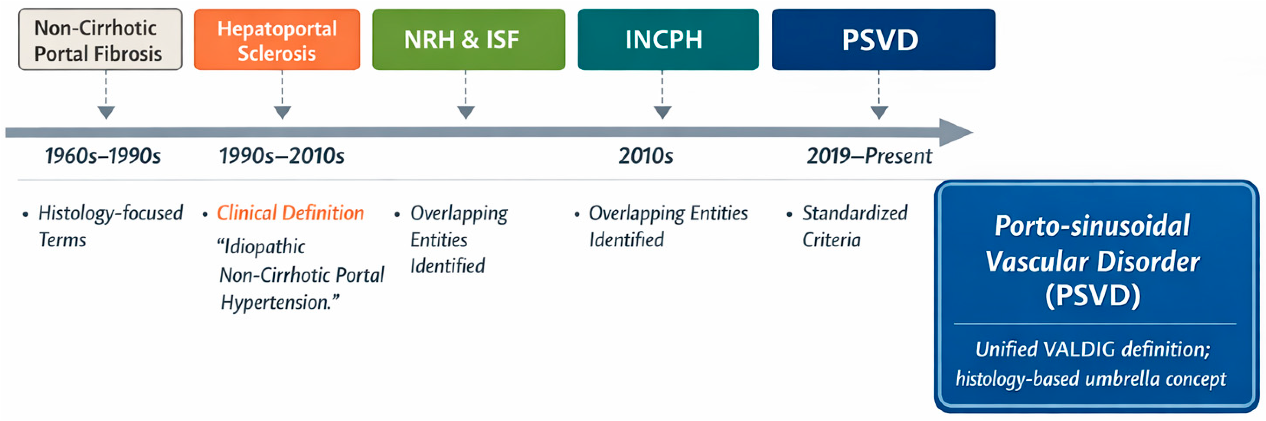
Recognition of the clinical and pathological overlap among these historically distinct entities, along with evidence that they frequently coexist, prompted the need for a comprehensive and unified definition. Responding to this need, the Vascular Liver Disease Interest Group (VALDIG) introduced the term “porto-sinusoidal vascular disease” in 2019, an overarching nosological framework intended to encompass the entire spectrum of vascular liver pathology (
Figure 1) [
4,
8,
9].
The PSVD definition represents a significant advancement by prioritizing specific histopathological lesions as the basis for diagnosis. Unlike earlier definitions such as INCPH, which relied only on clinical signs of portal hypertension [
4], the PSVD approach enables earlier identification by recognizing characteristic liver lesions, even without overt symptoms. This framework also acknowledges pre-clinical or subclinical stages of the disorder [
5,
6,
7,
10]. The new approach aims to support earlier diagnosis, enhance understanding of disease progression, and promote consistency in research.
This review synthesizes current knowledge on PSVD, critically examining its epidemiology, etiology, pathophysiology, and clinical manifestations. It emphasizes diagnostic criteria and methodology, particularly the role of liver biopsy and non-invasive tools. Management strategies, prognosis, and ongoing controversies and knowledge gaps are also discussed [
1].
For this narrative review, we conducted a non-systematic literature search in PubMed/MEDLINE, Embase, and Scopus up to December 2025. The search strategy combined free-text terms and MeSH/Emtree headings related to porto-sinusoidal vascular disorder and its historical nomenclature, including “porto-sinusoidal vascular disorder”, “portosinusoidal vascular disease”, “idiopathic non-cirrhotic portal hypertension”, “INCPH”, “hepatoportal sclerosis”, “nodular regenerative hyperplasia”, and “incomplete septal fibrosis”, together with “portal hypertension”, “non-cirrhotic portal hypertension”, “pathophysiology”, “diagnosis”, and “management”. We screened reference lists of key original articles and reviews for additional relevant studies and prioritized English-language human studies, large cohorts, and recent guideline or consensus documents for inclusion.
2. Epidemiology, Demographics, and Geographic Variation
The epidemiology of PSVD varies greatly by region, likely due to differences in diagnostic practices, clinical awareness, population genetic backgrounds, and environmental exposures [
1]. In Western countries and Singapore, PSVD is a rare cause of portal hypertension, accounting for less than 10% of cases [
1]. This low prevalence in these populations contributes to underrecognition and diagnostic delays.
Epidemiological studies from India report much higher PSVD prevalence, with some cohorts attributing up to 48% of portal hypertension cases to PSVD [
1,
11]. This geographic disparity suggests roles for genetic and environmental factors, though the underlying mechanisms remain unclear.
Recent multicenter studies in Europe and China indicate that PSVD may be more common than previously thought, even in Western populations, with prevalence rates of 15–20% among patients with unexplained liver enzyme elevations or non-cirrhotic portal hypertension [
12,
13]. These findings challenge the traditional view of PSVD as very rare and suggest that the historically low prevalence in Western countries may stem from underrecognition and underdiagnosis rather than true epidemiological differences [
1,
9].
Accurate determination of PSVD incidence trends remains problematic due to multiple methodological constraints. The historical absence of standardized diagnostic criteria, combined with the retrospective nature of the majority of large epidemiological studies, has precluded reliable assessment of true incidence rates [
14]. The disorder is widely recognized as substantially under-recognized within clinical practice and is frequently misdiagnosed as cirrhosis or alternative liver pathologies, particularly in regions where clinical awareness of PSVD remains limited [
15].
Broader adoption of VALDIG diagnostic criteria and increased clinical awareness are expected to raise reported PSVD incidence rates [
5]. This increase should be viewed as improved detection and diagnostic accuracy, not a true rise in disease occurrence. Distinguishing between apparent and actual incidence is essential for interpreting future data.
PSVD affects all age groups. In adults, it is most often diagnosed in middle age, typically at a younger age than cirrhosis-related portal hypertension [
16,
17]. This suggests PSVD may have a distinct natural history and progression.
Studies on sex distribution in PSVD are conflicting, with some indicating a slight male predominance and others a higher prevalence in females [
12]. These discrepancies may reflect differences in the prevalence of associated autoimmune conditions across populations.
PSVD is increasingly recognized in pediatric populations, where it may present with distinct clinical phenotypes and disease manifestations [
16,
17]. Some affected children exhibit overt manifestations of portal hypertension, while others are identified incidentally through discovery of abnormal liver tests in the absence of significant clinical portal hypertension [
16,
17]. The etiology of PSVD in pediatric cases frequently remains unclear despite comprehensive investigation, and the long-term prognosis in this population remains an important area requiring future research and longitudinal follow-up studies.
3. Etiology and Associated Conditions
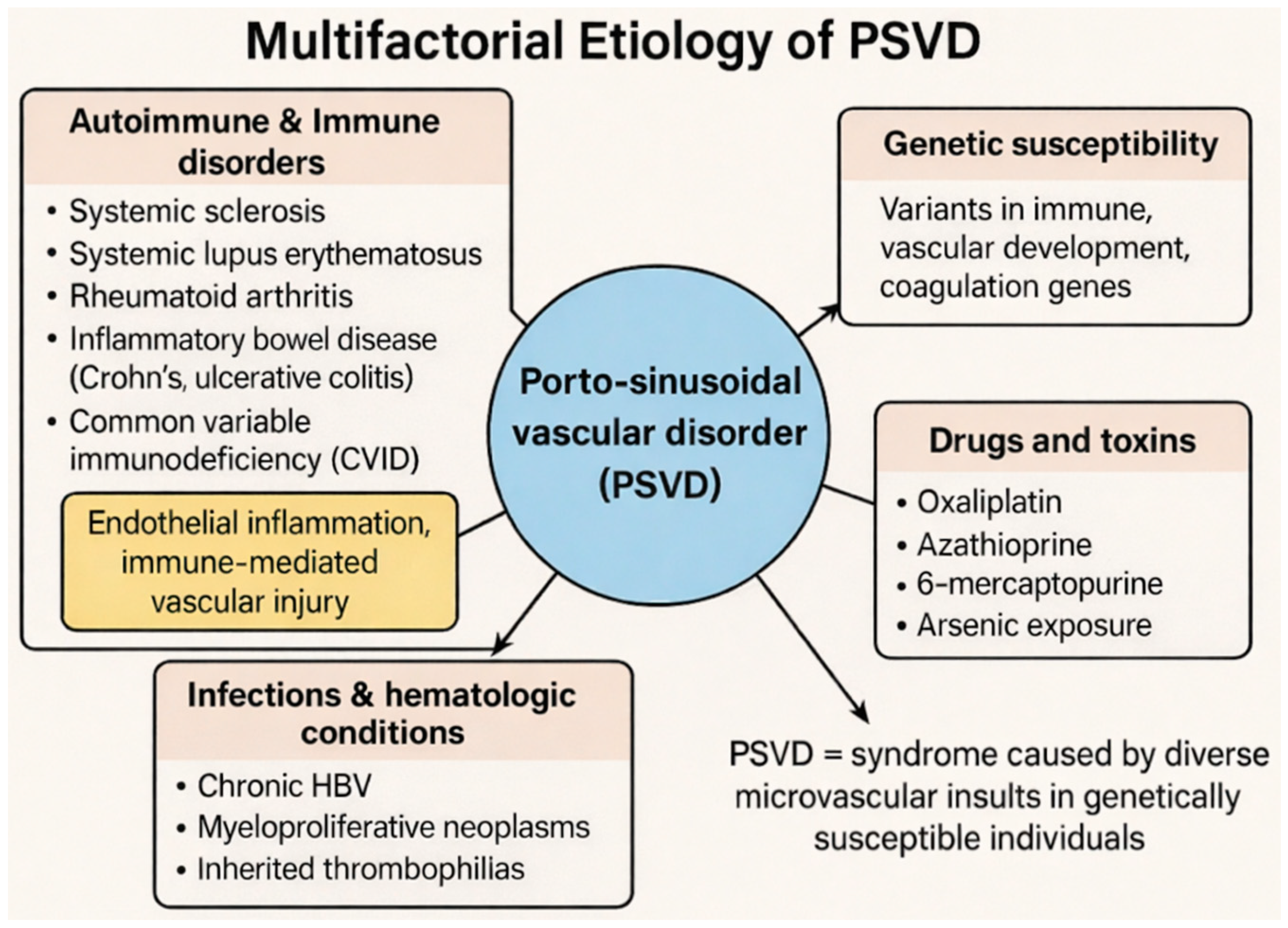
PSVD is best viewed as a syndrome caused by various insults to the hepatic microvasculature instead of one disease with a single cause. Factors contributing to PSVD include genetic susceptibility, immune dysregulation, prothrombotic states, infections, medications, and toxins [
5]. This multifactorial model fits the wide range of clinical presentations, causes, and disease courses observed across patients (
Figure 2).
PSVD is increasingly recognized as being closely associated with systemic autoimmune diseases and may represent a hepatic manifestation of these conditions [
10]. Systemic Sclerosis (Scleroderma) is one of the most frequently reported associations with PSVD, with endothelial dysfunction a shared core pathogenic feature of both conditions [
10]. The pathological mechanisms underlying this association likely involve immune-mediated endothelial injury and vascular remodeling. Systemic Lupus Erythematosus (SLE) has been documented as an associated condition in multiple case series and cohort studies of PSVD patients. Rheumatoid Arthritis has also been identified as an associated condition in patients developing PSVD. Both Crohn’s disease and ulcerative colitis have been associated with PSVD development, suggesting that chronic intestinal inflammation may contribute to hepatic vascular pathology. In addition, a primary immunodeficiency disorder, Common Variable Immunodeficiency (CVID), has been associated with PSVD in multiple case reports and small case series.
Shared mechanisms likely include endothelial inflammation, immune-mediated vascular injury, and immune dysregulation [
10,
12]. PSVD should be considered in patients with autoimmune diseases who develop unexplained liver abnormalities or portal hypertension [
10]. Screening is especially recommended when isolated gamma-glutamyl transferase (GGT) elevation is present, as this may be an early indicator of PSVD.
Recent genetic studies have identified mutations predisposing individuals to PSVD, often involving genes related to immune function, vascular development, and coagulation [
15,
18]. These findings support the view that PSVD arises from environmental or immune triggers in genetically susceptible individuals and may enable future risk stratification and personalized management [
8,
15,
19].
Certain medications and environmental toxins are established causes of the vascular lesions seen in PSVD. Oxaliplatin, a platinum-based chemotherapy drug, is a well-documented and established cause of PSVD, particularly in patients undergoing treatment for colorectal cancer with hepatic metastases [
14,
20]. The mechanism of oxaliplatin-induced vascular injury involves direct endothelial damage and microvascular thrombosis. Azathioprine and 6-mercaptopurine, agents frequently employed in the management of inflammatory bowel disease and in post-transplantation immunosuppression, have been linked to the development of PSVD through immune-mediated and direct toxic mechanisms. Historical environmental and occupational exposure to arsenic has been recognized as a cause of non-cirrhotic portal hypertension, particularly in regions with endemic arsenic contamination of groundwater.
Certain infectious agents and hematological disorders are also recognized as being associated with PSVD development. Chronic hepatitis B virus (HBV) infection may coexist with and contribute to the development of PSVD, thereby complicating diagnostic differentiation [
21]. Additionally, prothrombotic conditions, including myeloproliferative neoplasms and inherited thrombophilias, are established risk factors for PSVD, underscoring the importance of thrombotic mechanisms in its pathogenesis [
22].
4. Pathophysiology and Hemodynamic Mechanisms and a Unified Disease Model
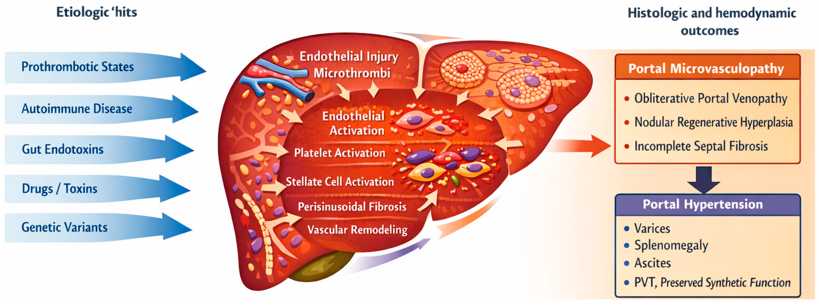
Porto-sinusoidal vascular disorder (PSVD) is now recognized as the final common phenotype resulting from multiple injurious pathways that converge on the endothelial cells of small portal venules and hepatic sinusoids, rather than as a single, uniform disease process (
Figure 3). These convergent insults generate a pro-thrombotic, pro-fibrotic, and pro-inflammatory microenvironment that drives the development of characteristic histological lesions: obliterative portal venopathy (OPV), nodular regenerative hyperplasia (NRH), and incomplete septal fibrosis (ISF).
At the cellular level, diverse insults converge on portal venular and sinusoidal endothelial cells, which serve as primary sensors and amplifiers of injury. Endothelial activation and damage facilitate platelet adhesion, microthrombus formation, and recruitment of inflammatory cells. This process establishes a self-perpetuating pro-thrombotic and pro-inflammatory environment that, in conjunction with stellate-cell activation, drives progressive perisinusoidal and portal-tract fibrogenesis [
2,
4,
15,
23,
24].
Histologically, PSVD is defined by obliterative portal venopathy, nodular regenerative hyperplasia (NRH), and incomplete septal fibrosis, which reflect chronic injury to the intrahepatic portal venous tree and sinusoidal bed [
4,
7]. Thickening and eventual occlusion of small portal venules are attributed to recurrent or persistent microthrombi and platelet aggregates, resulting in progressive loss or “vanishing” of portal veins, redistribution of blood flow, and parenchymal nodular transformation [
4,
23]. Multiple studies report an over-representation of systemic and inherited prothrombotic conditions (such as myeloproliferative neoplasms, protein C deficiency, factor V Leiden, and JAK2 mutations) in PSVD cohorts, supporting a central role for disordered coagulation and endothelial activation [
4,
18,
22]. Molecular studies further implicate altered platelet activation and signaling in the coagulation pathway in vascular remodeling and sinusoidal obliteration [
23]. Clinically, this prothrombotic environment aligns with the high prevalence of portal vein thrombosis in PSVD and distinguishes the disorder from cirrhosis-associated portal hypertension, where architectural collapse rather than primary microvasculopathy predominates [
19,
25].
The term “porto-sinusoidal” highlights that, beyond pre-sinusoidal lesions of small portal venules, the hepatic sinusoids themselves may also be structurally and hemodynamically affected [
2,
4,
7]. Sinusoidal dilatation and perisinusoidal fibrosis can develop through two, not mutually exclusive, mechanisms: (1) as a primary lesion, particularly in cases dominated by nodular regenerative hyperplasia (NRH), where regenerative nodules compress adjacent sinusoids and directly increase sinusoidal [
4,
26,
27], and (2) as a secondary consequence of upstream pre-sinusoidal obstruction from obliterative portal venopathy (OPV), which over time leads to increased sinusoidal pressure, remodeling, and fibrosis [
2,
4,
25,
26]. Hemodynamic studies using the hepatic venous pressure gradient (HVPG) illustrate this duality: many patients exhibit a predominantly pre-sinusoidal pattern, with normal or only mildly elevated HVPG despite clinically significant portal hypertension, while others, typically those with marked NRH, display clearly elevated HVPG, resembling sinusoidal portal hypertension observed in cirrhosis [
2,
4,
25,
27,
28]. The coexistence of presinusoidal and sinusoidal hemodynamic profiles within PSVD underpins the “porto-sinusoidal” nomenclature and accounts for the heterogeneity in clinical presentation and the variable response to non-selective beta-blockers, which are more effective when a sinusoidal component and elevated HVPG are present [
5,
8,
29].
Emerging evidence demonstrates that immune-mediated endothelial damage is a second major driver of PSVD. Studies from VALDIG and other groups report frequent associations with systemic autoimmune diseases (such as systemic lupus erythematosus, systemic sclerosis, and rheumatoid arthritis), primary antibody deficiency syndromes, and inflammatory bowel disease (IBD) [
4,
10,
12]. These conditions share key pathophysiological features with PSVD, including microangiopathy, endothelial dysfunction, and chronic vascular inflammation. Histological analyses of PSVD reveal portal vein endothelialitis and sinusoidal T cell infiltrates, indicating that intrasinusoidal lymphocytes directly contribute to vascular injury and remodeling [
4,
7]. Transcriptomic studies have identified overexpression of genes involved in lymphocyte activation and immune signaling in blood from PSVD patients, further supporting an immune-driven component of portal microvasculopathy [
4]. Autoimmune serological markers (ANA, anti-phospholipid antibodies, and others) are frequently detected and may have diagnostic and prognostic value, although their specificity is limited [
10,
30,
31]. The gut–liver axis has emerged as a critical link between systemic inflammation, procoagulant states, and the vascular lesions of PSVD. Patients with PSVD and portal hypertension exhibit significantly elevated circulating gut-derived endotoxins (such as LPS) compared with both PSVD without portal hypertension and appropriate controls [
24,
32]. These endotoxins activate Toll-like receptor pathways on hepatic endothelial cells and Kupffer cells, promoting endothelial activation, sinusoidal inflammation, and a shift toward a prothrombotic, pro-fibrotic microenvironment [
23,
24]. In IBD-associated PSVD, chronic intestinal inflammation and increased intestinal permeability may amplify this mechanism, providing a continuous source of bacterial products and inflammatory mediators to the portal circulation [
12,
33]. Experimental transcriptomic studies and animal models have begun to reproduce PSVD-like lesions under sustained gut-driven endotoxemia, lending biological plausibility to this axis [
33].
Direct toxic injury to the portal and sinusoidal endothelium is well established in PSVD associated with drug exposure, particularly oxaliplatin and other cytotoxic chemotherapies. Oxaliplatin-related sinusoidal obstruction, peliosis-like changes, and NRH have been described histologically in patients who subsequently meet PSVD criteria, with clinical manifestations ranging from asymptomatic portal hypertension to severe postoperative complications following liver resection for metastases [
14,
26,
34,
35]. The underlying mechanism involves direct endothelial toxicity, sinusoidal wall disruption, and activation of hepatic stellate cells, resulting in sinusoidal dilatation, perisinusoidal fibrosis, and eventual obliteration of small portal venules [
26,
36]. These chemotherapeutic insults often occur in the context of other risk factors, such as thrombophilia or autoimmune disease, thereby integrating into the broader multifactorial model of PSVD pathogenesis [
14,
18]. Genetic studies have begun to identify variants that may predispose individuals to PSVD by altering vascular morphogenesis, immune regulation, or coagulation. Recent research indicates an enrichment of mutations affecting immune cell function and vascular development, including genes involved in angiogenesis, endothelial integrity, and morphogen signaling [
15,
18]. These findings support the concept that PSVD develops when environmental or systemic triggers (such as autoimmune disease, infections, chemotherapeutic drugs, or gut-derived endotoxins) act on a genetically susceptible vascular bed. The presence of familial and congenital vascular anomalies in some patients, and the observation of PSVD-like lesions in models of developmental vascular defects, further emphasize a morphogenetic component to the disorder [
4,
33].
Synthesizing these data, PSVD can be conceptualized as a spectrum of non-cirrhotic intrahepatic vascular liver diseases characterized by the following features.
Portal microvasculopathy, including obliterative portal venopathy, NRH, and incomplete septal fibrosis, serves as the histological hallmark. Prothrombotic states and endothelial dysfunction facilitate the formation and persistence of microthrombi and promote vascular remodeling. Immune-mediated injury and systemic autoimmunity sustain chronic inflammation of the portal and sinusoidal endothelium. Alterations in the gut–liver axis, such as endotoxemia and dysbiosis, amplify both inflammatory and procoagulant pathways. Toxic or chemotherapeutic insults provide additional, often temporally identifiable, injuries to an already vulnerable microvasculature. Genetic susceptibility further modulates individual risk, lesion patterns, and progression toward clinically significant portal hypertension.
This multifactorial, “multiple hit” model accounts for the broad clinical heterogeneity of PSVD, ranging from patients with isolated histological lesions and no portal hypertension to those presenting with severe variceal bleeding or portal vein thrombosis. This perspective supports the classification of PSVD as a syndrome rather than a single disease entity [
5,
6,
31,
37].
Current evidence supports conceptualizing PSVD as the liver’s stereotyped response to diverse upstream insults converging on sinusoidal endothelial cells and hepatic stellate cells, which serve as central integrators of genetic, thrombotic, inflammatory, and toxic factors. Inherited or acquired prothrombotic states promote recurrent microvascular thrombosis in small portal venules and sinusoids, leading to ischemia, parenchymal extinction, and compensatory nodular regeneration that culminate in obliterative portal venopathy and nodular regenerative hyperplasia (OPV/NRH) [
4,
15,
23]. Concurrently, gut-derived endotoxins and systemic immune activation engage pattern-recognition receptors (such as TLR4) on sinusoidal endothelium and Kupffer cells, amplifying endothelial injury, platelet adhesion, and a procoagulant environment, while sustaining low-grade inflammation and perisinusoidal fibrogenesis [
24,
32]. Chemotherapy agents (notably oxaliplatin), thiopurines, and environmental toxins further increase endothelial and microvascular stress, particularly in genetically susceptible individuals with variants in coagulation and morphogenetic pathways [
14,
15]. Activated stellate cells translate these signals into extracellular matrix deposition and aberrant vascular remodeling, favoring incomplete septal fibrosis (ISF) and portal tract hypervascularization [
4,
6]. This framework supports viewing OPV, NRH, and ISF as histological manifestations along a common microvascular injury–repair axis, modulated by the interplay of endothelial damage, thrombotic burden, immune-mediated inflammation, and stellate-cell-driven fibrosis. Collectively, these insults transform the portal–sinusoidal microenvironment into a prothrombotic, profibrotic, and proinflammatory niche. Endothelial activation, platelet aggregation, and stellate-cell-driven matrix deposition progressively remodel the intrahepatic portal tree and sinusoidal bed, resulting in obliterative portal venopathy, nodular regenerative hyperplasia, and incomplete septal fibrosis as distinct histological expressions along a shared injury–repair continuum. This integrative framework advances understanding beyond mere association, providing a final common pathway that links genetic predisposition, thrombosis, and inflammation to the characteristic clinicopathological phenotype of PSVD. The main histologic lesions of PSVD, their pathophysiologic implications, and clinical consequences are summarized in
Table 1.
5. Clinical Presentation and Manifestations
PSVD exhibits an exceptionally broad clinical spectrum, ranging from completely asymptomatic presentations to severe, life-threatening complications of portal hypertension [
7,
8,
37]. Some patients are entirely asymptomatic, with diagnosis made incidentally during liver biopsy performed for investigation of persistently elevated liver enzymes [
10]. Others present with severe, life-threatening complications of clinically significant portal hypertension, including variceal hemorrhage and hepatic encephalopathy.
A key feature of PSVD is that liver synthetic function is generally well preserved, especially in early disease, distinguishing it from decompensated cirrhosis [
37]. This preservation is a hallmark that often points to a non-cirrhotic cause of portal hypertension. Clinicians should see preserved albumin and INR levels with portal hypertension as a ‘red flag for non-cirrhotic portal hypertension.’
When portal hypertension develops in the context of PSVD, the clinical manifestations are indistinguishable from those observed in patients with cirrhosis. Hemorrhage from gastroesophageal varices is a common and serious presenting feature of PSVD, often prompting initial medical evaluation and diagnosis. Enlargement of the spleen is a common physical finding, resulting from portal venous congestion and splenic vascular engorgement. A low platelet count is a frequent laboratory finding, secondary to splenic sequestration of platelets due to splenic enlargement. Fluid accumulation (ascites) in the peritoneal cavity can occur in PSVD, though it is less frequent and less severe than in advanced cirrhosis. Patients with PSVD are at increased risk of thrombosis of the main portal vein (PVT), which can worsen portal hypertension and complicate management.
Laboratory findings in PSVD are often non-specific and may not clearly distinguish it from other liver diseases. Aminotransferase levels may be normal or mildly elevated, reflecting preserved hepatocellular integrity [
12]. Alkaline phosphatase (ALP) and gamma-glutamyl transferase (GGT) may be persistently elevated, often prompting further investigation and serving as a clinical clue to PSVD [
12].
Markers of hepatic synthetic function, including serum albumin and international normalized ratio (INR), typically remain normal until advanced stages of the disease, reflecting the preservation of hepatic function that distinguishes PSVD from cirrhosis. This preservation of synthetic function, despite evidence of portal hypertension, is an important diagnostic clue that should prompt consideration of a non-cirrhotic etiology.
6. Diagnostic Methodology and Criteria
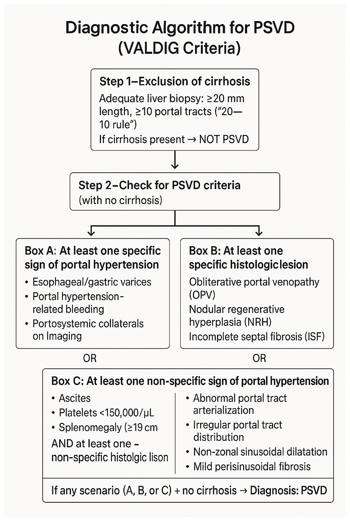
The diagnosis of PSVD is formalized by the VALDIG diagnostic criteria, which rely on a systematic combination of clinical, radiological, and, most importantly, histological findings [
2,
5] (
Figure 4). A key motivation for developing these criteria was to address and substantially reduce the high rates of misclassification of liver diseases, with the ultimate goal of improving diagnostic accuracy and enhancing patient outcomes through earlier and more precise diagnosis [
2,
38].
A definitive diagnosis of PSVD requires the absence of cirrhosis on an adequate liver biopsy specimen plus fulfillment of one of the following diagnostic scenarios [
2,
5]. For instance, patients with unexplained liver enzyme elevation and no cirrhosis on biopsy who are found to have esophageal varices on endoscopy fulfill one of the specific portal hypertension criteria for PSVD diagnosis. The presence of at least one specific sign of portal hypertension (such as esophageal or gastric varices, portal hypertension-related bleeding documented on endoscopy, or portosystemic collateral vessels documented on imaging), OR the presence of at least one specific histological lesion characteristic of PSVD (such as obliterative portal venopathy, nodular regenerative hyperplasia, or incomplete septal fibrosis), OR the presence of at least one non-specific sign of portal hypertension (such as ascites, platelet count less than 150,000/μL, or spleen size greater than or equal to 13 cm), AND at least one non-specific histological lesion characteristic of PSVD. These criteria are intentionally constructed to be highly specific, thereby minimizing misclassification while simultaneously encompassing the full spectrum of the disease, including patients without overt clinical portal hypertension [
2].
Liver biopsy remains the indispensable gold standard for diagnosing PSVD [
6,
38]. Biopsy is essential not only to identify the characteristic histological lesions but also to definitively rule out cirrhosis, which remains the primary differential diagnostic consideration [
6,
38]. For an accurate assessment of the liver parenchyma and portal tracts, the adequacy of a biopsy specimen is crucial. An adequate biopsy specimen, often recalled by the mnemonic ‘20–10 rule’ (at least 20 mm in length containing at least 10 portal tracts), is crucial for accurate assessment [
5,
6].
Specific Histological Lesions include Obliterative Portal Venopathy (OPV), Nodular Regenerative Hyperplasia (NRH), Incomplete Septal Fibrosis (ISF) and Non-specific Histological Lesions. OPV is characterized by the narrowing or complete obliteration of the lumen of small portal venules due to subendothelial fibrosis and endothelial proliferation [
5,
39]. Some portal tracts may show a complete absence or “vanishing” of portal veins, representing end-stage obliteration [
5]. In NRH, the liver parenchyma is transformed into small, regenerative nodules of hepatocytes without the presence of the fibrous septa that characterize cirrhosis [
1]. This gives the liver a subtle nodular architecture on histologic examination. ISF consists of slender fibrous septa that extend from portal tracts into the liver parenchyma but do not connect to form the complete, encircling nodules that define cirrhosis [
29]. Non-specific Histological Lesions include portal tract abnormalities (such as an increased number of arterial profiles relative to bile ducts), irregular distribution of portal tracts, non-zonal sinusoidal dilatation, and mild perisinusoidal fibrosis [
2]. While these lesions are not pathognomonic for PSVD, their presence in the appropriate clinical context supports the diagnosis.
While liver biopsy is the diagnostic gold standard, non-invasive tests and imaging can provide supportive evidence and raise suspicion for PSVD, potentially guiding the decision to biopsy. Cross-sectional imaging studies (CT and MRI) can reveal signs of portal hypertension, such as splenomegaly, ascites, and portosystemic collateral vessels. Certain features on hepatobiliary contrast-enhanced MRI, such as periportal hyperintensity, have been reported to be suggestive of PSVD [
9,
36,
40]. Imaging is also crucial for excluding other causes of portal hypertension, such as Budd–Chiari syndrome, portal vein cavernoma, or hepatic fibrosis patterns suggestive of cirrhosis. Liver Stiffness Measurement (LSM) by transient elastography, such as FibroScan, typically shows lower liver stiffness values in patients with PSVD than in those with cirrhosis presenting with a similar degree of portal hypertension [
25]. This dissociation between relatively low LSM and severe signs of portal hypertension is an important indicator that should prompt consideration of a non-cirrhotic etiology such as PSVD [
1,
27]. This finding is particularly useful in clinical practice for distinguishing PSVD from cirrhosis. The primary differential diagnosis for PSVD is liver cirrhosis, which must be definitively excluded through adequate liver biopsy [
2]. Other conditions to consider in the differential diagnosis include congenital hepatic fibrosis, hepatic sarcoidosis, and sinusoidal obstruction syndrome (SOS), which can also cause microvascular damage but are excluded from the PSVD definition [
2].
7. Management and Therapeutic Strategies
Currently, no therapies specifically target the underlying cause of PSVD or reverse the vascular lesions. Management is adapted from guidelines for portal hypertension in cirrhosis and focuses on preventing and treating complications [
1,
28,
38,
41,
42]. This approach is necessary due to the lack of randomized clinical trials for PSVD, highlighting a major evidence gap. Future research should prioritize developing disease-modifying therapies targeting the underlying mechanisms.
Non-selective Beta-blockers (NSBBs) like propranolol and carvedilol are used for primary and secondary prevention of variceal bleeding. They may be more effective in patients with a sinusoidal component of portal hypertension (such as those with nodular regenerative hyperplasia and elevated HVPG) than in those with mainly pre-sinusoidal obstruction [
5]. This hemodynamic heterogeneity affects the expected efficacy of beta-blockers. Endoscopic Variceal Ligation (EVL) with banding of esophageal varices is the standard of care for preventing and treating variceal hemorrhage. It is preferred over sclerotherapy due to better efficacy and lower complication rates.
The role of anticoagulation in PSVD is complex and debated. Anticoagulation is indicated for patients with portal vein thrombosis to prevent extension and possibly achieve recanalization [
23]. Prophylactic anticoagulation in patients without portal vein thrombosis is controversial and not routinely recommended, though it may be considered in those with a prothrombotic state or strong family history [
23]. Beyond the general principles derived from non-cirrhotic PVT management, PSVD demands a particularly proactive approach to recent, non-cavernomatous portal vein thrombosis. According to current EASL/Baveno-aligned guidance, in cirrhotic candidates for liver transplantation, any recent (<6 months), completely or partially occlusive (>50%) PVT of the main portal trunk—even if asymptomatic—is an urgent indication for full-dose anticoagulation, with the explicit goal of recanalisation to facilitate adequate portal anastomosis and preserve transplantability [
2,
29,
43]. By analogy, and given that PSVD patients are often younger with otherwise favourable post-transplant outcomes, a similar strategy is reasonable in PSVD with recent, non-cavernomatous PVT: prompt initiation of LMWH followed by VKA or DOAC, maintained for at least 6 months, with cross-sectional imaging at 6 months to document recanalisation or progression [
2].
In contrast, the role of primary (prophylactic) anticoagulation in PSVD without established PVT remains uncertain. However, accumulating data on the high prevalence of prothrombotic states in PSVD—including JAK2 V617F-positive myeloproliferative neoplasms, antiphospholipid antibodies, and rare germline variants affecting anticoagulant pathways (e.g., protein C deficiency, SERPINC1/antithrombin and GLA mutations)—support an individualized, lower threshold for prophylactic or long-term anticoagulation in selected high-risk patients, after careful variceal prophylaxis and multidisciplinary assessment [
2,
4,
15,
18]. Prospective studies are needed to define which genetic and acquired thrombophilic profiles in PSVD derive net benefit from primary or extended-duration anticoagulation and to develop evidence-based stopping rules.
Transjugular intrahepatic portosystemic shunt (TIPS) is a salvage therapy for patients with refractory variceal bleeding or ascites unresponsive to medical management [
37]. TIPS effectively controls portal hypertension in PSVD, but its use must be balanced against the risk of hepatic encephalopathy, especially since these patients often have well-preserved liver function [
26,
37]. Although preserved synthetic and hepatocellular function would intuitively be expected to protect against post-TIPS hepatic encephalopathy (HE), the pathophysiology in PSVD is more nuanced. In these patients, basal ammonia detoxification capacity is relatively intact, but creation of a low-resistance portosystemic shunt abruptly diverts portal blood away from functioning hepatocytes, sharply reducing effective hepatic portal perfusion and thereby bypassing this preserved metabolic capacity. The resultant disproportion between portal decompression and hepatocyte exposure to portal flow may explain why a substantial proportion of PSVD/NCPH patients develop post-TIPS HE despite “good” liver function in standard scores (Child–Pugh, MELD) [
1,
5]. This has led to an emphasis on tailoring the degree of shunting: the use of controlled or under-dilated covered stents (e.g., 8-mm PTFE stents rather than full 10-mm dilation) aims to achieve sufficient portal pressure reduction (PPG < 12 mmHg or ≥50% decrease) while preserving an adequate fraction of portal flow through the liver parenchyma [
4,
29]. In selected cases, additional endovascular modulation—such as embolization of large spontaneous portosystemic shunts (e.g., splenorenal shunts) at the time of TIPS—may further help balance effective decompression with controlled encephalopathy risk by redistributing flow through the newly created intrahepatic shunt rather than through unregulated extrahepatic collaterals [
4,
31]. Overall, TIPS in PSVD should be conceived as a “precision” intervention where the target is not maximal, but optimized portal decompression compatible with adequate hepatocyte perfusion and minimal neurocognitive sequelae. The risk of encephalopathy after TIPS may be higher in PSVD due to preserved hepatic synthetic function [
28,
36].
Liver transplantation is the definitive treatment for PSVD patients with end-stage liver failure or complications of portal hypertension unresponsive to other management [
31]. Liver transplantation in PSVD should be considered using broadly similar principles to cirrhosis, but with several important nuances. First, indications and timing are more heterogeneous, because many patients maintain preserved synthetic function for long periods. Transplant referral is generally reserved for those with severe or refractory complications of portal hypertension (recurrent variceal bleeding despite optimal endoscopic/pharmacologic therapy, refractory or recurrent tense ascites, and/or refractory hepatic encephalopathy), and for the small subset who develop progressive liver dysfunction (worsening jaundice, coagulopathy, or parenchymal extinction) or hepatopulmonary syndrome. The presence of chronic, extensive portal vein thrombosis (PVT) may accelerate the need for evaluation, both because it worsens portal hypertension and because complex thrombosis (involving the superior mesenteric/splenic veins or cavernomatous transformation) can limit future transplantability if referral is delayed [
4,
31,
44]. Second, several technical challenges are specific to PSVD: the liver is often normal-sized or only mildly reduced, with frequent hypertrophy of segment I and marked portal hypertension with large portosystemic collaterals and splenomegaly [
1,
4]. Hepatectomy can therefore be more demanding than in a small, shrunken cirrhotic liver, and portal vein anastomosis may require thrombectomy, interposition grafts, or non-standard inflow reconstruction when PVT or cavernoma are present. In addition, large spontaneous shunts (e.g., splenorenal or mesenteric–systemic collaterals) can divert flow away from the graft (“portal steal”), necessitating intraoperative ligation or obliteration and careful modulation of portal inflow to avoid both hypoperfusion and excessive portal hyperperfusion. Third, post-transplant outcomes are overall favorable but not identical to those seen in other indications. In the largest multicenter series to date (79 PSVD recipients), 1-, 2-, and 5-year patient survival rates were approximately 82%, 81%, and 69%, respectively, with similarly good graft survival and histologically proven recurrence in only 3 patients [
44]. However, vascular complications—especially portal vein-related problems—appear more frequent than in standard cirrhotic cohorts, reflecting the pre-existing portal vasculopathy and complex inflow reconstructions [
4,
44]. Overall prognosis after transplantation is strongly influenced by the severity of associated systemic conditions (e.g., autoimmune or hematologic disease) and by baseline renal and hepatic function rather than by PSVD itself [
10,
44]. Long-term outcomes after transplantation are generally favorable, with high patient and graft survival and low risk of recurrence [
31,
41].
9. Controversies, Knowledge Gaps, and Future Research Directions
While the term PSVD has brought greater clinical clarity, some experts have criticized it. They argue that grouping heterogeneous conditions with different causes, mechanisms, and prognoses under one term may create new ambiguities and oversimplify complex processes [
7]. Further refinement into etiological or histological subtypes may be needed as understanding advances [
5].
Histological diagnosis of PSVD is challenging due to the subtle, patchy distribution of lesions and significant variability among pathologists [
6,
39]. Standardizing assessment methods and developing a validated scoring system are essential to improve diagnostic reproducibility and support multicenter research [
6].
Despite recent advances in understanding PSVD, significant knowledge gaps persist. A major knowledge gap concerns the natural history of PSVD patients without portal hypertension at baseline [
29]. Limited evidence suggests progression in this subclinical group may be very slow or absent. Early-stage PSVD could remain indolent for decades, challenging current views of disease progression. Prospective, long-term studies are needed to better define the risk and rate of progression to significant portal hypertension [
6,
37,
38]. This remains a key unanswered question in PSVD research.
Key priorities for future research include [
6,
13,
15,
18,
38,
45,
46].
Prospective Epidemiological Studies: Conducting large, multicenter, population-based studies to establish the true global prevalence and incidence of PSVD, with standardized diagnostic criteria applied across all participating centers.
Natural History Studies: Longitudinally following cohorts of patients, especially those without portal hypertension at baseline, to understand disease progression, identify prognostic biomarkers, and determine the proportion of patients who progress to clinically significant portal hypertension.
Biomarker Development: Identifying and validating non-invasive serum and imaging biomarkers for diagnosis, risk stratification, and monitoring of disease progression, potentially reducing the need for liver biopsy in the future.
Pathophysiological Research: Further elucidating the genetic, molecular, and cellular mechanisms that drive the disease to identify novel therapeutic targets and enable development of disease-modifying therapies [
15,
32,
38].
Interdisciplinary Collaboration: Fostering collaboration between hepatologists, liver pathologists, radiologists, hematologists and rheumatologists, and geneticists to improve recognition of PSVD, enhance diagnostic accuracy, and provide holistic patient care addressing both hepatic and systemic manifestations.
Refined TIPS Risk Stratification: Development and validation of predictive models specifically tailored to PSVD to estimate the risk of post-TIPS hepatic encephalopathy and guide individualized shunt calibration and patient selection.
Surgical Shunt versus TIPS: Comparative studies to clarify whether selective surgical shunts (e.g., distal splenorenal shunt) retain a role in the TIPS era for particular PSVD hemodynamic or clinical phenotypes.
Transplant Registry Studies: Multicenter registry-based analyses focused on PSVD transplantation, assessing technical variations in portal inflow reconstruction and their impact on vascular complications, graft function, and long-term outcomes.