Review

Healing the Liver: Cell and Gene Therapies for Inherited and Acquired Diseases

Department of Health Sciences, University of Piemonte Orientale, 28100 Novara, Italy
*
Author to whom correspondence should be addressed.
Livers 2026, 6(2), 18; https://doi.org/10.3390/livers6020018
Submission received: 13 January 2026 / Revised: 3 March 2026 / Accepted: 5 March 2026 / Published: 10 March 2026

Abstract

The liver plays a fundamental role in maintaining homeostasis thanks to the numerous functions performed by this organ. Non-inherited metabolic liver diseases, inherited metabolic liver diseases, and liver cancers are pathological conditions affecting liver function and that can lead to its failure. To date, for end-stage liver diseases—where the remaining hepatic tissue is no longer capable of regenerating sufficiently rapidly—or for metabolic diseases involving the liver, liver transplantation remains the standard and ideal therapeutic approach. However, this is limited by donor availability, surgical costs, and the tangible risk of autoimmune rejection, which may occur at varying intervals post-surgery. Furthermore, for the duration of their lives, transplant recipients must undergo systemic immunosuppressive treatment to prevent rejection; this is associated with high costs and severe side effects, including infections and secondary malignancies. In this review, we discuss these pathologies and how recent cell-based therapies and/or gene therapy approaches have emerged as promising alternatives that can provide either temporary restoration of hepatic function or long-term benefits, potentially reducing the global burden of liver disorders.

1. Introduction

The liver is a central organ in the human body that is able to maintain systemic homeostasis, holding the highest regenerative capacity and self-healing potential, while playing a pivotal role in host defense against pathogens and participating as a regulator of immune tolerance, thanks also to its anatomical structure and resident cell populations. Parenchymal cells (hepatocytes) and non-parenchymal cells—among them Kupffer cells (KCs), liver sinusoidal endothelial cells (LSECs), biliary epithelial cells (BECs), and other intrahepatic immune cells—coordinate to form a frontline against systemic inflammation and pathogen clearance [1]. Hepatocytes contribute indirectly to immune defense by producing acute-phase proteins and antimicrobial peptides; KCs, liver-resident macrophages, act as phagocytes, efficiently capturing and eliminating circulating bacteria through phagocytosis, cytokine secretion and antigen presentation. LSECs function as immunological gatekeepers, facilitating immune cell trafficking and sensing microbial components through pattern recognition receptors. Cholangiocytes provide the first line of defense within the biliary tract [2]. With these functions, it is easy to notice that the failure of a dedicated cell type leads to different pathological conditions that are now recognized in the extended field of chronic liver diseases, which include inherited metabolic diseases (caused by defects in genes expressed in liver cells); non-inherited metabolic diseases, such as alcohol-associated liver disease (ALD) and the newly named metabolic-dysfunction-associated steatotic liver disease (MASLD); and viral hepatitis and cirrhosis [3].
Inherited metabolic liver diseases (IMLDs) account for up to one third of acute liver failure in childhood. They are a group of more than 400 different genetic disorders specifically involving the liver with mutations reducing or preventing the organ from processing nutrients (fats, proteins, carbohydrates) or causing toxic buildup, and often lead to liver dysfunction, structural abnormalities, cirrhosis, or acute failure, requiring lifelong, specialized care, and in severe cases, a liver transplant (OLT) [4]. This includes the cases of Crigler–Najjar Syndrome (CNS), alpha-1 antitrypsin deficiency (A1ATD), glycogen storage disease type 1 (GSD-1), Wilson’s disease (WD), primary hyperoxaluria (PH) or familial hypercholesterolemia (FH).
CNS is a very rare autosomic recessive genetic disorder caused by complete absence/inactivity (type 1, the more severe form) or partial inactivity (type 2) of the hepatic enzyme UDP-glucuronosyltransferase 1A1 (UGT1A1), resulting in impaired bilirubin conjugation and severe unconjugated hyperbilirubinemia that causes intense, permanent jaundice starting shortly after birth and with a high risk of bilirubin encephalopathy [5].
A1ATD is a genetic disorder caused by mutations in the SERPINA1 gene that is responsible for both the deficiency and presence of abnormal alpha-1 antitrypsin (AAT) protein, the natural inhibitor of neutrophil elastase, that is produced during infections and whose high presence causes damage to lung tissue, thus resulting in emphysema/chronic obstructive pulmonary disease (COPD). The homologous ZZ mutation (Glu342Lys) causes AAT proteins to polymerize (aggregate) inside liver cells (hepatocytes) instead of being released into the bloodstream, triggering an injury cascade and leading to liver injury, fibrosis, cirrhosis, and increased cancer risk [6].
GSD-1 is a rare, autosomal recessive metabolic disorder where the body cannot properly break down glycogen due to a glucose-6-phosphatase (G6Pase-α, encoded by G6PC1 gene; GSD1a, most common) deficiency or to deficiency in the transport protein glucose-6-phosphate translocase (G6PT, encoded by SLC37A4 gene; GSD1b). It causes severe fasting hypoglycemia, lactic acidosis and hyperlipidemia with accumulation of glycogen and fat in the liver and kidneys with consequent hepatomegaly and nephromegaly, resulting in the risk of renal failure and liver tumors (hepatocellular adenomas and hepatocellular carcinoma) in the long term [7].
WD is a rare disorder caused by a mutation in the ATP7B gene, leading to excessive copper accumulation in the liver, brain, eyes, and other tissues, causing irreversible liver damage (cirrhosis, failure), permanent neurological issues (tremors, rigidity), psychiatric disorders (depression, psychosis), and potential death [8].
PH is a rare metabolic disorder where the liver produces excess oxalate, leading to calcium oxalate crystal accumulation, recurrent kidney stones, nephrocalcinosis, and often, end-stage renal disease (ESRD). It is caused by genetic defects in liver-specific enzymes, namely alanine–glyoxylate aminotransferase (AGXT; PH type 1, most common and severe), glyoxylate and hydroxypyruvate reductase (GRHPR; PH type 2), and 4-hydroxy-2-oxoglutarate aldolase 1 (HOGA1; PH type 3) [9,10].
FH is an autosomal dominant disorder caused primarily by inherited mutations in the genes encoding for low-density lipoprotein receptor (LDLR; 85–90% of cases), apolipoprotein B-100 (APOB; 5–10% of cases), proprotein convertase subtilisin/kexin type 9 (PCSK9; <5% of cases) or low-density lipoprotein receptor adaptor protein 1 (LDLRAP1; <1% of cases). These mutations impair the liver’s ability to remove LDL from the blood, and as a consequence, patients suffer from lifelong high LDL levels, and if untreated, significantly raises the risk of premature heart disease [11].
Self-regeneration becomes fundamental when hepatic tissue undergoes damage of various origins that compromises its structure, and consequently, its function—as seen in viral infections (such as hepatitis C), chronic conditions like non-alcoholic fatty liver disease (NAFLD—now recognized as MAFLD, metabolic-dysfunction-associated fatty liver disease), drug abuse and autoimmune responses [12]. Liver disease accounts for approximately 2 million deaths per year worldwide, 1 million due to complications of cirrhosis and 1 million due to viral hepatitis and hepatocellular carcinoma, with differences arising from countries, geographical areas and depending on the availability of the methods of assessment.
The persistence of a pathological condition—whether manifesting as acute injury or evolving into chronic inflammation—combined with abnormal cell regeneration and persistent tissue damage, inevitably leads to fibrosis. If left untreated, fibrosis progresses to cirrhosis, and ultimately, liver cancer [13].
Globally, it is established that 4% of deaths are attributed to liver cirrhosis, hepatic failure, or hepatocellular carcinoma (HCC), all of which originate from initial acute or chronic hepatic injury that becomes irreversible over time, with the progressive loss of metabolic capacity [14].
Acute liver failure (ALF). ALF originates from initial hepatocyte damage caused by viral infections, toxins, or drug-induced injury, which triggers both intrinsic and extrinsic apoptotic pathways [15]. This primary insult compromises KC tolerance, initiating a proinflammatory cascade that recruits systemic monocytes and neutrophils. The interplay between damage-associated molecular patterns (DAMPs) and pathogen-associated molecular patterns (PAMPs) exacerbates this response, leading to secondary immune-mediated necrosis and mitochondrial collapse [16]. As necrotic cells release further DAMPs, a self-sustaining cycle of inflammation is established through the activation of natural killer (NK) and natural killer T (NKT) cells and the secretion of cytokines such as tumor necrosis factor-alpha (TNF-α) and interleukin 1-beta (IL-1β). When this persistent immune activation outweighs the liver’s regenerative capacity and clearance mechanisms, systemic metabolic and hemodynamic instability ensues, resulting in multiorgan failure and terminal liver dysfunction [17].
The causes of ALF vary by region: viral infections (HAV, HBV, and HEV) are the primary drivers in low-income countries, whereas drug-induced liver injury is more prevalent in high-income nations [18,19]. Globally, the WHO estimates that one-third of the population has been infected with HBV or HCV. In highly endemic, low-income regions, HAV exposure is extremely common, affecting over 90% of children by age 10.
Acute-on-chronic liver failure (ACLF). ACLF is a distinct clinical syndrome characterized by an abrupt, life-threatening deterioration in patients with underlying chronic liver disease or cirrhosis. Unlike traditional decompensated cirrhosis—marked by ascites, variceal hemorrhage, or hepatic encephalopathy—ACLF is defined by the presence of concomitant extrahepatic organ failures (OFs) and an exceptionally high short-term mortality rate, mirroring the severity of acute liver failure [20]. In recent decades, diagnostic criteria have evolved significantly but still ongoing discussion exists regarding whether the precipitating insult must be primary hepatic (e.g., viral flare, alcoholic hepatitis) or can include extrahepatic triggers (e.g., sepsis) [21]. The transition from compensated or decompensated cirrhosis to ACLF is driven by an intense systemic inflammatory response. This is sustained by circulating PAMPs and DAMPs. The subsequent multi-organ failure is attributed to a synergistic combination of tissue hypoperfusion, mitochondrial dysfunction, and direct immune-mediated cellular damage [22]. While alcohol and chronic viral hepatitis remain the most prevalent underlying etiologies, nearly 50% of cases lack an identifiable precipitating trigger. Management currently prioritizes supportive care for OF, typically within an intensive care setting.
For those with end-stage manifestations, OLT remains the only definitive treatment, offering favorable long-term survival in selected candidates. Emerging therapies, including bioartificial liver support systems, granulocyte-colony-stimulating factors (G-CSF), and mesenchymal stem cell (MSC) transplantation, are currently under investigation [23,24,25]. However, these modalities remain experimental and are not yet integrated into standard clinical practice. Future research focusing on mechanistic interventions to resolve systemic inflammation is essential to improve patient prognosis.
Liver fibrosis (LF). LF represents a pivotal pathological transition in the progression of various chronic liver diseases (CLDs), serving as the precursor to more severe conditions such as decompensated cirrhosis and HCC [1]. Characterized by a persistent and dysregulated wound-healing response, LF involves the aberrant accumulation of intrahepatic fibrous tissue and a profound imbalance in the synthesis and degradation of the extracellular matrix (ECM) [26].
While initially a dynamic and potentially reversible process, chronic hepatic insult leads to structural and functional architectural distortion of the liver. This progression culminates in end-stage liver disease (ESLD), associated with life-threatening complications including hepatic encephalopathy, gastrointestinal hemorrhage, and hepatorenal syndrome. Recent epidemiological data indicate a rising global prevalence, with advanced fibrosis and cirrhosis affecting approximately 3.3% and 1.3% of the population, respectively—a trend further exacerbated by the increasing incidence of metabolic dysfunction [27]. The etiology of LF exhibits significant geographic and socioeconomic variation: in developing nations, chronic HBV infection remains the primary driver of fibrogenesis, while in Western countries, thanks to the vaccination, MASLD, alcohol misuse and HCV are the predominant triggers that lead to metabolic-dysfunction-associated steatohepatitis (MASH), acknowledging that insulin resistance and systemic inflammation are central to fibrotic progression [28]. LF is still a multi-faceted process driven by three primary interconnected but not overlapping axes, covering hepatic stellate cell (HSC) activation, immune-mediated signaling and oxidative stress/inflammatory cascade. HSCs are the principal effectors of fibrogenesis: upon hepatic injury, quiescent HSCs undergo a phenotypic transformation into activated, myofibroblast-like cells that hold increased contractility, chemotaxis, and augmented secretion of ECM components, particularly Type I and III collagens [29]. The fibrotic microenvironment is regulated by an intricate network of immune cells, including monocyte-derived macrophages, dendritic cells, and NK cells, which secrete a cocktail of potent profibrotic cytokines as tumor growth factor-beta (TGF-β) and platelet-derived growth factor (PDGF), which sustain the activated state of HSCs [30]. In addition, apoptotic hepatocytes release reactive oxygen species (ROSs) and cellular debris, which act as DAMPs [31]. These signals directly activate KCs, the resident hepatic macrophages, which in turn release pro-inflammatory mediators such as TNF-α, IL-1β, and interleukin-6 (IL-6), creating a self-perpetuating cycle of inflammation and matrix deposition [32].
However, if the damage persists, the liver reaches a “breaking point” called cirrhosis. At this stage, the liver becomes so compromised that blood can no longer flow through it properly. This leads to life-threatening complications like internal bleeding, kidney issues, and hepatic encephalopathy, where the brain becomes clouded by toxins that the liver is too damaged to filter out.
Liver cancer. Hepatocellular carcinoma (HCC) and intrahepatic cholangiocarcinoma (iCCA) are the two main primary liver tumors, but they differ significantly in incidence, etiology, and epidemiological trends. HCC is by far the most common form, accounting for approximately 75–85% of primary liver tumors. iCCA is less common, accounting for approximately 10–15% of cases, although studies report an increased incidence of iCCA [33,34]. HCC represents a significant global health burden, currently ranked as the fourth leading cause of cancer-related mortality. Historically, world incidence remained below 1%; however, recent data indicate a rising trajectory. While global variations in HCC prevalence are primarily attributed to chronic viral hepatitis (HBV and HCV), regions with lower viral endemicity are experiencing a demographic shift [35]. Traditionally, ALD has been the dominant risk factor in low-viral regions. However, there is an increasing correlation between HCC and metabolic dysfunction. Current longitudinal data suggest that MAFLD—and its inflammatory progression, MASH—is the fastest-growing etiology of incident liver cancer, with a 39% increase observed between 2015 and 2024. This trend is closely linked to the rising prevalence of metabolic syndrome and Type 2 diabetes mellitus (T2DM) [36]. MAFLD is characterized by hepatic steatosis (lipid accumulation > 5%) in the absence of secondary causes of liver injury. Its pathophysiology is multifactorial, involving insulin-resistance-induced oxidative stress, genetic predisposition, gut microbiome dysbiosis, and lifestyle factors [37]. HBV and HCV are considered the primary drivers of global HCC mortality, accounting for 40% and 29% of deaths, respectively.
Intrahepatic cholangiocarcinoma is primarily caused by chronic inflammation of the bile ducts and liver, often aggravated by cirrhosis, viral hepatitis (B or C), and congenital anomalies such as choledochal cysts and Caroli syndrome. Significant risk factors include exposure to chemical toxins, smoking, obesity, diabetes, metabolic syndrome, and liver parasites [38].
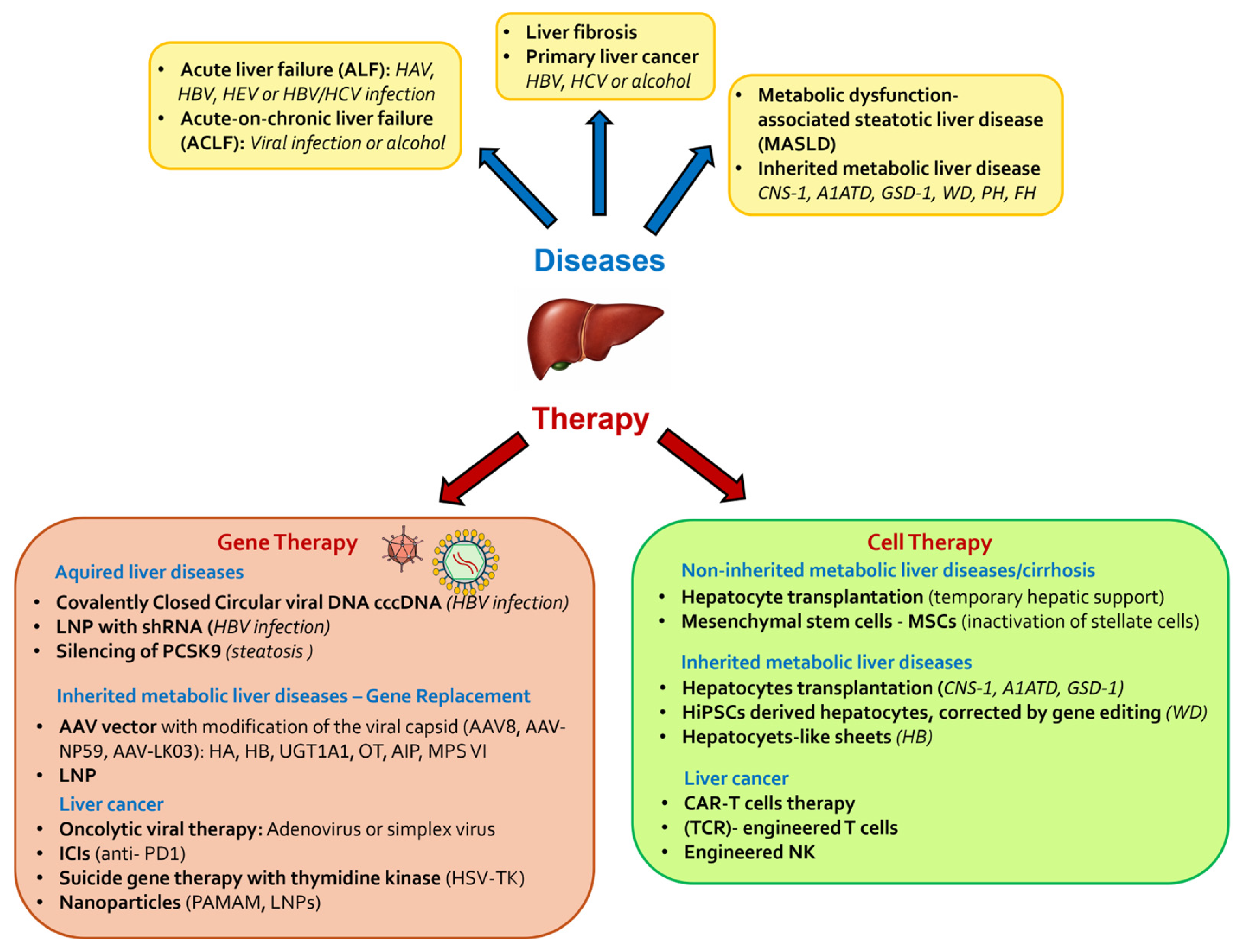
Patients affected by liver diseases often look for less invasive and more accessible treatment options. Although organ transplantation is curative for many, it is limited to a subset of patients due to donor scarcity and challenges in donor–recipient compatibility. In this context, cell-based therapies and/or gene therapy approaches have emerged as promising alternatives that can provide either temporary restoration of hepatic function or long-term benefits, potentially reducing the global burden of liver disorders. In this review we discuss both non-inherited and inherited metabolic liver diseases and how recent cell-based therapies and/or gene therapy approaches have emerged as promising alternatives that can provide either temporary restoration of hepatic function or long-term benefits, potentially reducing the global burden of liver disorders (Figure 1).

2. Cell Therapy

Cell therapy for non-inherited metabolic liver disease. Liver disease can be categorized into three main groups: non-inherited metabolic liver diseases, inherited metabolic liver diseases, and liver cancers. Each category presents unique pathophysiology mechanisms, clinical manifestations, and therapeutic needs. Accordingly, cell-based therapies are being developed and optimized in a disease-specific manner with distinct cellular sources and delivery modalities.
Hepatocyte transplantation (HTx) is based on the infusion of functional hepatocytes to provide temporary hepatic support [39]. In ALF, HTx is designed as a bridge therapy, offering transitory functional hepatic support until the native liver regenerates or a suitable donor organ becomes available for transplantation [40].
Given the liver’s intrinsic regenerative capacity, which can be achieved within weeks, long-term engraftment is not essential in ALF. Nonetheless, the high-inflammatory microenvironment can directly impair the viability of transplanted cells. For this reason, it is important to transplant highly functional hepatocytes capable of rapidly performing key metabolic activities while protecting them from the inflammatory response. One solution has been the encapsulation of hepatocytes within biocompatible matrices. This approach was done by encapsulating human hepatocytes in alginate beads, followed by intraperitoneal administration in eight pediatric ALF patients awaiting liver transplant, without requiring immunosuppression due to alginate beads providing sufficient immunoisolation. In this proof-of-concept study, four patients spontaneously recovered without the need for a liver transplant, while three were stabilized until the availability of the donor organ, underlying the safety and feasibility of this cell-based approach as a temporary metabolic support system [41].
On the other hand, the application of HTx in ACLF remains quite limited and more challenging than in ALF due to the presence of pre-existing cirrhosis, systemic inflammation, and multiorgan failure [42]. Clinical studies evaluated intrasplenic HTx in ACLF patients with a five-year follow-up. Despite transplantation of a large number of allogenic hepatocytes (4.2 × 10¹⁰ to 6 × 10¹⁰ cells), the outcome was variable with three patients recovering from liver failure, one requiring subsequent liver transplantation, and three dying within months post-transplantation, making it very difficult to draw definitive conclusions due to the heterogeneous cell doses and patients characteristics [43]. This pathological setting restricts liver regeneration capacity, resulting in the inefficient use of short-term metabolic support and, therefore, requiring different strategies to promote long-term engraftment while repairing tissue damage. Accordingly, in chronic conditions like ACLF and cirrhosis, alternative cell-based therapies, such as those using mesenchymal stromal cells (MSCs), are being explored [44]. MSCs can be isolated from several tissues, including bone marrow, adipose tissue, and umbilical cord [45,46]. Their low immunogenicity and strong immunomodulatory properties make them attractive candidates for modulating inflammation and promoting tissue repair [47]. Furthermore, the possibility of obtaining autologous MSCs potentially eliminates the need for immunosuppressants and supports their clinical use for liver disorders [48,49,50]. Their therapeutic effect is mediated by their secretome, which modulates the liver microenvironment through paracrine signals [51,52,53]. The MSC secretome comprises a complex mixture of soluble factors (e.g., growth factors, cytokines) and exosomes, which are small extracellular vesicles (EV, 30–150 nanometers in diameter) generated from the cellular endosomal system [54]. These vesicles act as specialized delivery vehicles, transporting microRNAs (miRNAs), messenger RNAs (mRNAs), and other bioactive molecules regulating target liver cells’ fate [55,56].
New evidence indicates that these cell-free products can exhibit immunomodulatory, anti-apoptotic, and pro-regenerative effects of MSCs, thereby shifting the therapeutic focus from living cells to their derived products and underscoring a significant paradigm shift toward acellular clinical strategies. This kind of strategy bypasses the risks associated with live-cell transplantation, such as senescence, microvascular obstruction, or potential tumorigenicity [57].
In preclinical models of acute and chronic liver injury, MSC-conditioned medium and MSC-derived EV reduce hepatocyte apoptosis and oxidative stress, inhibit hepatic stellate cell activation [58] and ECM deposition [59] and promote hepatocyte proliferation and metabolic recovery, thereby improving liver architecture and function [51].
Despite these advantages, clinical studies have highlighted challenges regarding the use of MSCs, including the reduced viability, variability in cell preparations, and incomplete understanding of their mechanisms [60,61]. In addition, uncertainties regarding the optimal MSC source, cell dosage, or route of administration complicate the standardization of treatment protocols for reliable MSC-based therapies [62] (Table 1). The transition to secretome-based products aims to resolve these issues by providing a more consistent and safer alternative to whole-cell therapy.
This approach is being explored for MASLD, which represents the most common chronic liver condition that leads to an increased risk of fibrosis, cirrhosis, and HCC [63]. Recently, pre-clinical studies have shown that MSC-derived EV from the umbilical cord mitigate the inflammatory environment by reducing lipid deposition in hepatocytes by inhibiting the expression of dynamin-related protein 1 (DRP1), whose high expression promote excessive fission, mitochondrial dysfunction, and increased ROS [64].
Cell therapy for inherited metabolic liver disease. Inherited metabolic liver diseases encompass a diverse group of conditions resulting from genetic defects that impair hepatic metabolism [65]. Even though OLT is the main approach to restore the missing metabolic function, the scarcity of donors and the use of lifelong immune suppression limit its application. In this context, the main goal of cell-based therapies is to provide a long-term engraftment and an efficient hepatic repopulation to provide a lasting clinical benefit.
HTx represents a promising and less invasive alternative that aims to restore deficient or absent enzymatic activity. It is estimated that partial repopulation of the liver with functional hepatocytes, corresponding to approximately 5–10% of total liver mass, may be sufficient to achieve meaningful metabolic correction [40]. For instance, HTx has been investigated as a therapeutic option for several inherited metabolic liver diseases, including Crigler–Najjar Syndrome (CNS) type 1, alpha-1 antitrypsin deficiency (A1ATD), glycogen storage disease type 1 (GSD-1), providing transient or partial metabolic correction, improving biochemical parameters, reducing the need for supportive therapies, and delaying liver transplantation [66]. However, the clinical application of HTx faces several challenges. High-quality and viable primary human hepatocytes is often limited by the scarcity of suitable donor organs, which is also limited by the isolation and cryopreservation method that results in batch-to-batch variability and reduced functionality upon thawing, making large-scale expansion very difficult [67,68]. Furthermore, this could also result in low engraftment efficiency [39] (Table 1). To overcome the limited availability of primary human hepatocytes and their rapid loss of activity in vitro [69], researchers are focused on developing alternative strategies to enhance cellular engraftment. To address this challenge, induced pluripotent stem cell (iPSC) technology has emerged as an alternative and innovative tool. Human iPSCs (hiPSCs) generated by reprogramming patient-specific somatic cells using Yamanaka factors (Oct4, Sox2, Klf4, and c-Myc) offer the possibility to differentiate into multiple clinical-grade cell types, enabling personalized therapeutic strategies tailored to each patient’s specific needs [70,71]. Preclinical studies in Wilson’s disease (WD), an autosomal recessive disorder that leads to excess copper deposition in the liver and brain, demonstrated that the use of patient-specific iPSC-derived hepatocytes carrying the correction of the mutated gene using CRISPR/Cas9 technology was able to restore copper metabolism in the WD mouse model, improving hepatic functions [72].
Beyond WD, recent studies in hemophilia B (HB), an X-linked bleeding disorder characterized by impaired clotting factor IX (FIX) activity, have explored the potential of a hiPSC-based strategy for cell therapy. In a 2025 preclinical study, researchers were able to generate FIX-hiPSCs using the Clustered Regularly Interspaced Short Palindromic Repeats (CRISPR)/Cas9 system to differentiate into functional hepatocyte-like cell sheets. When transplanted in NOD/SCID mice, these sheets were capable of engrafting and secreting active FIX, further demonstrating the potential of engineered hiPSC-derived cells for liver-associated disorders [73]. Furthermore, pluripotent stem-cell-derived hepatocyte-like cells have demonstrated successful engraftment and metabolic support in preclinical models of urea cycle defects [74] (Table 1).
In addition to their two-dimensional (2D) differentiation potential, iPSCs have been exploited to generate three-dimensional (3D) liver organoids to recapitulate the complexity of in vivo microenvironment, representing a promising model for strategy for the development of new therapies [75]. This approach is supported by the fact that iPSCs can be directed toward both parenchymal and non-parenchymal liver lineage, which self-organize into organoids maintaining the transcriptomic and metabolic profiles of the native tissue [76]. Moreover, it was shown that iPSCs exhibit pleiotropic hormonal sensitivity. As a matter of fact, recent work has shown that multiple iPSC lines constitutively express the progesterone receptor (PR), while its expression is absent in the corresponding somatic cell, indicating that PR upregulation is acquired during the reprogramming process [77]. This hormonal sensitivity is particularly relevant for modeling liver diseases influenced by the endocrine milieu and further supports the physiological reactivity of iPSC-derived liver organoids.
However, the potential application of hiPSC-derived hepatocyte-like cells remains limited due to their partial phenotypic immaturity and tumorigenic potential [78]. Moreover, current differentiation protocols employ reagents that are not good manufacturing practice (GMP)-compliant, such as serum supplements, extracellular matrices, or growth factors [79], limiting their translation to clinical studies.
Table 1. Comparison of major cellular sources for liver cell therapy.
Cell SourceAdvantagesDisadvantagesDevelopment Stage
Primary hepatocytes
  • Immediate therapeutic functionality after transplantation
  • No need for differentiation protocols
  • Proven ability to engraft and restore metabolic function
  • Limited availability
  • Poor in vitro proliferation
  • Limited long-term engraftment efficiency
  • Requirement of immunosuppression
Phase I/II (e.g., ALF trials) [80]
Mesenchymal stem cells (MSCs)
  • Easily accessible from multiple tissues
  • High proliferative capacity
  • Immunomodulatory/anti-inflammatory properties
  • Low immunogenicity
  • Reduced viability and cell preparation variability
  • Incomplete mechanisms understanding
  • No standardization of protocols
Phase II/III (e.g., cirrhosis) [81]
Induced pluripotent stem cells (iPSCs)
  • Unlimited self-renewal
  • Ability to generate patient-specific cells with precise editing (e.g., CRISPR/Cas9)
  • Generation of different cell types
  • Risk of tumorigenicity
  • Complex and time-consuming differentiation protocols
  • Regulatory and manufactory challenges
Preclinical/early trials
[82]
Cell therapy for liver cancer. Primary liver cancers include a heterogeneous group of malignancies derived from different hepatic cell populations, with HCC representing the most prevalent and lethal form [83]. Despite improvements in the frontline treatments, including tumor resection, liver transplantation, and systemic therapies, liver cancer remains associated with high recurrence rates and poor long-term survival, particularly in advanced stages, due to the high genetic heterogeneity and complexity of the tumor environment [84,85]. In this context, cell-based therapeutic strategies have emerged as a promising approach for personalized therapy, primarily directing the immune system to target tumor cells.
Chimeric antigen receptor (CAR) T-cell therapy showed great potential for hematologic malignancies [86], and it involves the use of genetically engineered T cells to recognize specific tumor cell antigens. However, translating this success to solid tumors, such as HCC, is challenged by distinct biological and microenvironmental characteristics. Nevertheless, several studies have focused on targeting tumor-associated antigens, including glypican-3 (GPC3), a cell-surface protein highly expressed in HCC tissues but minimally or not expressed in healthy liver [87]. Consequently, GPC3 has emerged as one of the most promising targets for CAR-T immunotherapy in HCC, enabling a precision-targeted therapeutic strategy. Preclinical studies of GPC3-targeted CAR-T cells have demonstrated the capability to inhibit tumor growth in xenograft models, while early clinical trials have confirmed the efficacy and safety of this approach in patients with advanced HCC [88,89]. Even though GPC3-target therapeutics are in continuous exploration [90], alternative targets observable in HCC tumors have been investigated, such as alpha-fetoprotein (AFP), which operates as an intracellular and secreted antigen. Thus, it requires the use of the T-cell receptor (TCR)-engineered T cells to recognize the processed peptide antigen presented via the major histocompatibility complex class I (MHC-I), expanding the range of targets beyond surface antigens recognized by CAR-T cells [91].
To overcome HCC heterogeneity, another promising strategy is the development of bi-specific CAR-T capable of targeting two antigens at once, enhancing tumor inhibition. An example of this approach is the bispecific c-Met/programmed death-ligand 1 (PD-L1) CAR-T cells, which have demonstrated strong anti-tumor activity both in vivo and in vitro by simultaneously targeting the tumor antigen c-Met and blocking the PD-L1 immune checkpoint on HCC cells [92].
In addition to T-cell-based strategies, other cellular therapies are being explored for HCC, including engineered NK cells. These cells offer several advantages over CAR-T therapies, including an increased safety profile, a lower frequency of cytokine release syndrome, and the feasibility of using allogeneic cell sources without the risk of graft-versus-host disease [93]. However, the efficacy of NK cells in HCC is hampered by the tumor microenvironment, which promotes NK cell exhaustion due to programmed death-1 (PD-1) upregulation and functional impairment due to downregulation of the NK group 2 member D (NKG2D) receptor on their cell surface [94]. In fact, preclinical studies have engineered CAR-NK cells to express the NKG2D receptor, thereby enhancing NK cytotoxicity by recognizing NKG2D ligands that are frequently upregulated on cancer cells [93].
While in HCC the common target is GPC3, in iCCA-specific targets are being studied for CAR-T-cell therapies, such as the use of EGFR- and CD133-specific CAR-T sequential treatments [95]; the Tn antigen (GalNAcα-Ser/Thr) on mucin-1 (Tn-MUC1), a tumor-specific, truncated O-glycan found in many adenocarcinomas and recognized by the 5E5 antibody; mesotelin, expressed by ovarian cancer, cholangiocarcinoma or mesothelioma (NCT05568680); and integrin αvβ6, which is significantly overexpressed in tissues from CCA patients [96].
In summary, cellular therapies represent a promising approach in the treatment of hepatic pathologies, offering personalized solutions from supporting temporary liver metabolism to permanent genetic correction. Nowadays, the gold standard for liver diseases is mainly organ transplantation, but thanks to technological progress, it is not the only possible solution available. The use of MSCs to modulate the inflammatory environment, the generation and genetic engineering of iPSCs to restore hepatic functions, and the development of CAR-T and NK cell-based technologies are all redefining new therapeutic approaches. Altogether, these approaches address the intrinsic complexity of liver diseases, underlying the need for tailored therapeutic strategies rather than a universal solution.
However, the success of these new strategies relies on overcoming critical aspects such as the standardization of GMP protocols, the optimization of cell maturation and engraftment and better comprehension of the complexity of the tumor environment. Regardless of these challenges, continuous advances in preclinical models are accelerating the clinical translation of cell-based therapies. Current findings confirm that cell therapy is no longer just a hypothetical hope, but it is becoming more of a concrete alternative therapeutic solution able to change the global burden of liver disorders.

3. Gene Therapy

The liver as an ideal platform for gene therapy. The liver stands out anatomically not only as an organ with a central role in various types of metabolism, but also as an exceptionally receptive and functional target for gene therapy strategies, thanks to its anatomical and physiological characteristics [97]. First, its extensive blood supply facilitates systemic delivery of various therapeutic vectors, both viral and non-viral, used for in vivo gene transfer [98]. Furthermore, the liver hosts a large population of cells, including hepatocytes, long-lived cells that, once transduced, ensure both transient and stable and long-term therapeutic transgene expression [99,100,101,102,103]. Finally, and perhaps most crucially, its function as a “factory” for protein processing and secretion makes it an ideal site for correcting enzyme/protein deficiencies that underlie many genetic diseases. Often, genetic correction of even a relatively modest fraction of hepatocytes is sufficient to reach the desired therapeutic threshold [104].
Currently, liver gene therapy is divided into two main strategic pathways. The first, and the most obvious and direct, is gene replacement therapy, primarily used for the treatment of hereditary diseases, which aims to insert a functional copy of a mutated/missing gene [105]. On the other hand, functional gene therapy or therapeutic gene modulation emerged later and is under active study to treat/cure acquired diseases—whether viral, metabolic, or oncological—with the aim of modulating gene expression, for example, by silencing genes whose uncontrolled expression results in disease or by expressing proteins with toxic or immunomodulatory activity [106]. This growing success has been made possible by progressive developments and improvements in viral vector engineering, particularly adeno-associated viral vectors (AAV), which have transformed the liver into a true “pioneer organ” in the field of gene therapy [104].
There are two approaches to gene delivery to the liver: ex vivo and in vivo. Ex vivo gene therapy is performed in combination with autologous cell transplantation, an approach limited by a small and short-term engraftment, and above all by the need to remove tissue (which may present variable degrees of damage depending on the pathology) each time it is necessary to isolate cells for treatment and the viability of these cells following culture and in vitro gene manipulation [104].
Due to these limitations, in vivo gene therapy has become the primary choice for the treatment of genetic liver diseases. The key to any successful gene therapy lies in a vector’s ability to efficiently and safely deliver the therapeutic genetic material (DNA or RNA) into target cells, meaning hepatocytes and/or other hepatic cells in the case of liver-directed gene therapy.
Liver-directed gene therapy for metabolic diseases. For gene transfer, the use of non-viral vectors to deliver naked genetic material into the liver has seen increasing development. However, systems such as hydrodynamic injection, electroporation, and ultrasound are limited to preclinical animal models [98,107,108,109,110], as their clinical use is currently impractical. For this reason, researchers have developed virus-derived vectors, or viral vectors, in parallel for more efficient and long-lasting gene transfer. Various viral vectors are used for gene therapy, and these viral vectors have specific characteristics that make them suitable for different applications.
Adenovirus (Ad)-derived vectors were the first to be successfully used to correct a urea cycle defect in a mouse model deficient in ornithine transcarbamylase (OTC) [111]. However, Ad-induced toxicity caused the death of an 18-year-old boy during a clinical trial for the treatment of OTC in the late 1990s [112]. This fatal event, combined with the growing interest in AAVs, significantly limited the development of adenoviral vectors for liver gene therapy.
AAVs are the most established delivery platform, representing the focus of most clinical trials for genetic liver diseases, especially considering the approval between 2022 and 2023 by the FDA and EMA of Roctavian (Valoctocogene roxaparvovec) and Hemgenix (Etranacogene dezaparvovec), two hepatocyte-directed AAV5-based therapies for the treatment of hemophilia A (Rocatavian) and B (Hemgenix) [113]. These vectors offer significant advantages for in vivo gene therapy since they are nonpathogenic, induce a relatively mild immune response compared to other viruses, and, crucially, can be maintained in episomal form (they do not stably integrate into host DNA), allowing gene expression for years following gene transfer into long-lived/quiescent cells such as hepatocytes, minimizing the risk of insertional mutagenesis [114].
A key advance has been the engineering of existing/known serotypes and the search for/discovery of new ones with lower seroprevalence in the global human population [115,116]. Serotypes such as AAV8 and other engineered capsids, including AAV-NP59 and AAV-LK03, have demonstrated particularly high affinity (tropism) for human hepatocytes following intravenous administration [117,118]. In particular, AAV-LK03 was the first engineered capsid used in clinical studies, leading to therapeutic levels of FVIII in 16 out of 18 treated patients [119].
In addition to hemophilia, approaches involving the use of AAV are under investigation and are being used to treat or cure several other genetic metabolic diseases [114]. The most attractive genetic diseases for gene therapy are those that require novel therapeutic approaches and that allow a therapeutic advantage to be achieved even with a small percentage of transduced cells.
Despite the limited cargo capacity of these viral vectors (~4.7 kb), many inherited liver metabolic diseases are caused by mutations in genes that can be inserted into AAVs [104], such as OTC deficiency, acute intermittent porphyria (AIP), mucopolysaccharidosis type VI (MPS VI), CNS, Gaucher disease, and Fabry disease [101].
OTC deficiency is the most common urea cycle defect caused by the absence of the OTC enzyme and leads to the accumulation of ammonia, which can cause coma, brain damage and death. Despite the tragic death of Jesse Gelsinger following treatment with an adenoviral vector containing the OTC gene [112], gene therapy for this urea cycle disease has developed an approach based on AAV8 containing the OTC gene under the transcriptional control of a liver-specific promoter [120], leading to a phase I/II clinical trial (NTC02991144) with 7 out of 11 patients demonstrating a therapeutic benefit, and for which a phase III study is currently underway (NTC05345171). A similar approach is used by another phase I/II clinical study that is currently recruiting (NTC05092685), which involves the use of the AAV-LK03 capsid, a liver-specific serotype with a lower seroprevalence [121].
CNS can cause irreversible neurologic damage, and eventually death, due to severe unconjugated hyperbilirubinemia caused by deficiency of the enzyme uridine diphosphoglucuronate glucuronosytransferase 1A1 (UGT1A1). In a multicenter phase I/II clinical study, researchers tested a liver-specific gene therapy based on AAV8 containing the UGT1A1 gene, showing the therapy was generally well tolerated, and that patients who received the effective dose were able to completely discontinue daily phototherapy for more than 18 months [122], dramatically improving their quality of life. However, in this approach, complete normalization of bilirubin levels has not yet been achieved, as the values remain stable but above the normal range of healthy subjects [123].
Additionally, in phase I and I/II clinical studies for gene therapy of AIP, MPS VI, Gaucher disease and Fabry disease, AAV gene therapy has demonstrated safety and tolerability, with some significant clinical improvements [101].
However, the use of AAVs is not without challenges. The main one is immunogenicity: a significant number of individuals possess pre-existing neutralizing antibodies against common AAV serotypes (in some cases, up to 70–75%) [114,124], which can render gene therapy treatment ineffective. In this regard, several approaches are under investigation, such as the concomitant administration of nanoparticles containing rapamycin [125], the use of IgG-cleaving endopeptidase imlifidase (IdeS) [126], and the use of plasmapheresis to lower anti-AAV antibodies [127].
Furthermore, the need to administer high doses to achieve sufficient therapeutic transduction can sometimes trigger transaminitis, a T-cell-mediated and innate immunity (via TLR9 receptors)-mediated liver inflammation characterized by elevated plasma levels of liver enzymes (ALT/GPT and AST/GOT), which can compromise the duration of therapy and which is usually managed pharmacologically with corticosteroids [128,129,130].
A further limitation of this therapy is the limited loading capacity of AAVs, which precludes its use for very large genes or genes larger than 4 kb, for which split approaches are under investigation in preclinical studies, as in the case of carbamoyl phosphate synthase (CPS1) deficiency [131], or shortened versions of the therapeutic gene are used, such as the miniATP7B gene used in ongoing clinical trials (NTC04537377 and NTC04884815) for WD.
The main reason why AAVs are chosen for in vivo liver gene therapy (i.e., their non-integrating nature) also represents one of the main limitations for their use, mainly during liver regeneration, but especially in the case of pediatric patients, where hepatocytes proliferation causes dilution, and eventual loss of the therapeutic transgene [114,132].
Alongside AAVs, lentiviral vectors (LVs), derived from HIV, are being studied for their superior loading capacity, which allows them to accommodate expression cassettes up to 9 kb, and their ability to stably integrate into the host cell genome [101]. Although they are most often used for ex vivo therapies, such as CAR-T generation [133], as well as in hematopoietic stem cells in cell and gene therapy approaches for the treatment of immune deficiencies [134] and hemophilia A (HA) [135,136], LVs have proven effective for in vivo gene therapy in several preclinical models, such as mice, dogs and non-human primates (NHPs), especially for the treatment of HA and HB with transgene expression in liver cells, i.e., hepatocytes, LSECs and KCs, by exploiting transcriptional (promoters) and post-transcriptional (miRT) control sequences, thus allowing stable and long-term expression of therapeutic levels of the transgene [137,138,139,140,141,142], and by adding molecules capable of inhibiting phagocytosis (i.e., CD47) on the LV surface, which showed improved in vivo liver gene transfer in NHPs [142].
However, LV-integrating capacity currently limits their use in clinics for in vivo gene therapy since it represents a potential risk of insertional mutagenesis [143].
The emergence of lipid nanoparticles (LNPs), the nonviral technology popularized by mRNA vaccines [144,145], offers a promising alternative. LNPs encapsulate nucleic acid (DNA; mRNA; or small interfering RNA, siRNA) and are efficiently internalized by hepatocytes [146,147]. Their advantages include safety (no viral immunogenicity or minimal/virtually zero risk of integration), the possibility of repeated administration, and increased loading capacity [148,149,150]. They are particularly effective in delivering siRNA for gene silencing, finding application in the treatment not only of genetic disorders, such as hypercholesterolemia and hemophilia [151,152,153,154], but also of liver diseases with different etiologies, such as hepatitis B [155].
The efficacy of LNP technology was confirmed in 2018 by the approval of Patisiran (Onpattro), the first siRNA-based drug formulated in LNPs for the treatment of hereditary transthyretin (hATTR)-mediated amyloidosis. This demonstrated that LNPs can stabilize nucleic acids in the systemic circulation, ensuring effective gene silencing in the liver [156]. At the same time, LNPs have become the platform of choice for mRNA replacement therapy (enzyme replacement therapy) for inherited metabolic diseases and for in vivo genome editing using CRISPR/Cas9 systems [157,158]. Recently, preclinical studies on diseases such as familial hypercholesterolemia have demonstrated that systemic administration of mRNA-encoding genome base editors [159] or programmable editors equipped with transcriptional repressors to achieve permanent epigenetic silencing [160], delivered by LNPs, can induce permanent and therapeutic changes in the liver, overcoming the loading capacity limitations typical of viral vectors.
Additional molecular approaches have been developed and approved in recent years, as in the case of the treatment of PH1, which has evolved over a century from a purely symptomatic approach to targeted genetic therapies. Since its first description in 1925, treatment has evolved from hyperhydration and pyridoxine to combined liver–kidney transplantation, until the 2020 approval of lumasiran (Oxlumo), a drug based on double-stranded RNAi conjugated with N-acetylgalactosamine (GalNAc) for liver targeting that degrades HAO1 mRNA, reducing oxalate production in the liver [161]. Nedosiran (Rivfloza), approved at the end of 2023 for the treatment of primary hyperoxaluria type 1 (PH1) in patients aged 9 years and older, is a small interfering RNA (siRNA) that inhibits the enzyme lactate dehydrogenase (LDH) in the liver to reduce the production of oxalate [162].

4. Gene Therapy for Acquired Diseases: Infections and Steatosis

The scope of gene therapy extends well beyond genetic diseases, offering new hope for acquired chronic liver diseases with different etiologies.
Targeting Chronic Hepatitis B (HBV). Chronic HBV infection poses a significant challenge due to the formation of a stable reservoir of covalently closed circular viral DNA (cccDNA) in the nucleus of hepatocytes. The dominant approach for treating this disease is gene silencing, typically with siRNA or short hairpin RNA (shRNA) delivered, often with LNPs, to degrade viral mRNA and inhibit the replication and production of viral proteins, such as HBsAg [163,164]. Clinical trials using this approach aim to achieve a “functional cure” by reducing HBsAg levels demonstrating that drastically lowering HBsAg is possible and safe, laying the foundation for more powerful combination therapies and highlighting how the reduction of HBsAg levels is the fundamental requirement to allow a combination therapy (e.g., interferon) and/or the patient’s immune system to definitively eliminate the infected cells [165,166].
Steatohepatitis and Liver Injury. Numerous drugs exist for conditions such as MASLD, MASH, hepatitis, fibrosis, and cirrhosis. However, to develop an effective therapy for liver disease, it must be considered that the pathogenesis of any chronic disease is regulated by numerous factors [167,168]. Therefore, a single pharmacological agent is not very effective in these conditions. The development of new gene therapy strategies for the treatment of liver diseases aims to reduce the side effects of the pathogenesis of each disease.
MASLD and MASH are the leading causes of chronic liver disease worldwide [169,170]. Given their multifactorial background, gene therapy focuses on modulating specific metabolic or inflammatory pathways to reduce, and, if possible, reverse liver damage. Examples include hepatic silencing of proprotein convertase subtilisin/kexin type 9 (PCSK9) via siRNA to improve lipid homeostasis and steatosis [171], or to enable targeting key enzymes in lipogenesis [172].
For example, in a recent study, a chimeric AAV8 vector (AAV8.P) carrying a specific shRNA was developed to silence the PCSK9 gene to silence this protein and reduce the progression of atherosclerotic plaques [171]. In mice genetically predisposed to atherosclerosis (ApoE-/-), PCSK9 silencing led to a reduction in plaque area and increased plaque stability. Furthermore, PCSK9 silencing not only affected fat, but also reduced inflammation by reducing infiltration of inflammatory macrophages into vessel walls and decreasing systemic inflammatory markers.
A recent study has presented a new therapeutic strategy to combat MASLD [172]. Researchers found that inhibiting only ATP-citrate lyase (ACLY, a key enzyme in de novo lipogenesis in the liver) in mice fed a high-fat, high-fructose diet (HFHFD) not only failed to improve the disease, but in some cases worsened inflammation and liver damage. In fact, ACLY inhibition leads to an accumulation of polyunsaturated fatty acids (PUFAs), which damages mitochondria and increases the production of ROSs. Oxidative stress drives the liver to produce acetate internally, which activates another enzyme, acetyl-CoA synthetase 2 (ACSS2). ACSS2 thus provides an alternative route to produce acetyl-CoA, once again fueling fat synthesis and creating a vicious cycle that worsens the disease. The study demonstrates that the dual inhibition of ACLY and ACSS2 dramatically reduces fat production, improves mitochondrial function and reduces inflammation, and therefore represents a much more effective synergistic therapeutic strategy to reverse the progression of MASLD/MASH, acting both on fat reduction and on the enhancement of mitochondrial health.
An emerging area of research is the use of gene therapy to selectively express anti-fibrotic factors or silence pro-inflammatory molecules in hepatic stellate cells, which are crucial mediators of LF [173,174]. Similar strategies are being explored for alcohol-related liver injury (ALD), focusing on protection from oxidative stress and reducing inflammation [175,176].
Gene therapy for liver cancer. HCC, often complicated by pre-existing cirrhosis, and iCCA can be treated with gene-therapy-based oncology strategies that offer new approaches to attack the tumor.
Oncolytic virotherapy (OVs) uses genetically modified viruses (typically adenovirus or herpes simplex virus) with the ability to selectively replicate in tumor cells and induce their lysis following proliferation, which occurs via a lytic cycle [177]. For this therapy, OVs are modified to replicate only in the presence of activated tumor pathways common in HCC, ensuring selectivity for tumor cells and avoiding damage/elimination of healthy cells [178,179].
A study addresses one of the major challenges in oncolytic virus therapy: the difficulty of the virus penetrating deep, hypoxic core areas of solid tumors, such as HCC [180]. Hypoxia normally inhibits virus replication and limits its spread from the injection site to the tumor periphery. Furthermore, the dense ECM acts as a physical barrier. The researchers in this study engineered an oncolytic adenovirus with the AFP promoter and hypoxia response elements (HREs), which enhance virus replication precisely when exposed to low oxygen. This virus was able to spread effectively throughout the entire tumor mass, reaching both the hypoxic center and the peripheral regions and in animal models used it showed a superior ability to reduce the tumor mass.
In addition to the direct action of lysis, oncolytic action causes the release of tumor antigens and cytokines, promoting a potent local immune response. These vectors are often administered locally (intratumorally or transarterially) and are in advanced stages of clinical development [181,182].
Another strategy is suicide gene therapy, which involves the delivery of a gene, such as the herpes simplex virus thymidine kinase (HSV-TK), that converts a prodrug administered after gene therapy into a cytotoxic agent within transduced tumor cells only. This approach often uses adenoviral vectors that selectively target tumor cells [183,184].
It is noteworthy that most iCCAs have a non-inflamed TME, with an absence of effector T cells, thus making immune checkpoint inhibitors (ICIs) therapy ineffective. However, these “non-inflamed” iCCAs often exhibit alterations/mutations in genes such as the fibroblast growth factor receptor (FGFR2) and isocitrate dehydrogenases (IDH1/IDH2), making these genes possible candidates as gene therapy targets. For example, a recent study showed that polybromo-1 protein (PBRM1) deficiency is a key factor in iCCA progression. When PBRM1 levels are low, tumor cells are able to avoid “senescence” (a sort of natural cell growth block) and continue to proliferate. In this study, polymeric polyamidoamine (PAMAM) nanoparticles were used to deliver plasmid DNA containing the PBMR1 gene. Increased PBRM1 inhibits the ERK1/2 signaling pathway and reactivates tumor suppressor genes (such as p16 and p53/p21), forcing tumor cells into a state of senescence, thus halting their growth and division. In vivo tests on animal models have shown that this approach can significantly reduce tumor mass and is effective in “reprogramming” tumor cells, reversing the processes that promote malignancy caused by the original lack of PBRM1 [185].
In parallel, gene immunomodulation is being developed. Gene therapy can be used to express agents that block immune checkpoints (such as anti-PD-1) or cytokines that enhance the immune response directly in the tumor microenvironment, transforming immunologically “cold” tumors (capable of evading the immune system) into “hot” targets, recognized and infiltrated by the host immune system [186,187,188]. To this end, mRNA vaccines can be used as an immunotherapy strategy, aiming to “train” the immune system to recognize and attack tumor cells by exploiting tumor-associated antigens. For example, in the case of iCCA, three specific antigens—CD247, FCGR1A, and TRAP—have been identified as novel targets for mRNA vaccines, which are associated with a better prognosis and increased immune cell infiltration. These are considered ideal candidates for the development of future mRNA vaccines against CCA [189].
Cell therapy or gene therapy? While gene therapy is the solution of choice for inherited metabolic disorders, where the repair of a single gene is sufficient to correct the clinical phenotype, cell therapy maintains an irreplaceable role in chronic, non-hereditary disorders. In these, the fibrotic microenvironment and loss of functional mass require exogenous cellular input to promote tissue regeneration, a task that gene manipulation alone cannot accomplish (Table 2).

5. Challenges and Future Prospects

The era of genome editing. Despite its successes, hepatic gene therapy still faces significant challenges. Immunogenicity remains the primary challenge, especially for AAVs. Additionally, transient liver toxicity requires the co-administration of immunosuppressants, and the preexisting presence of neutralizing antibodies precludes this option for many patients. Research is actively working on engineering AAV capsids or new “stealth” serotypes capable of evading immune surveillance, as well as techniques for removing neutralizing antibodies. Moreover, the use of non-integrating systems is limited by conditions where liver cells are actively proliferating, as in liver regeneration, or in an actively growing liver, as in pediatric patients [190,191,192,193].
A further uncertainty concerns the duration of transgene expression, especially in chronically damaged livers, such as liver cirrhosis or MASH, where gene transfer may be less efficient due to cellular transformation and/or chronic inflammation [194,195], and cellular replication may lead to dilution and potential loss of AAV episomes over time.
Looking to the future, the greatest promise lies in genome editing technologies, particularly CRISPR/Cas9 due to its great adaptability [196]. These methods go beyond simply adding a gene: they offer the possibility of permanently correcting the genetic mutation directly in the hepatocyte DNA or specifically inactivating HBV cccDNA, a strategy still in the advanced preclinical phase [197,198,199], and for which a recent phase I clinical trial is under recruitment (NCT06680232).
CRISPR technology offers the possibility of intervening at the level of HCC through non-homologous end-joining (NHEJ) or homology-directed repair (HDR) approaches, to target mutated/involved genes in HCC: NHEJ for the introduction of insertions and deletions (indels) with the aim of eliminating the function of a gene, while HDR for the correction/reinsertion of the function of a gene. Furthermore, the use of catalytically inactive (dead) Cas9 (dCas9) fused to transcriptional activators or repressors allows modulating gene expression in HCC without DNA alteration [184].
Gene editing promises a true “cure-all” for many monogenic diseases, and the crucial challenge of this decade is perfecting the safe and efficient delivery of editing complexes, often using a combination of AAV and LNPs.
iECURE recently presented data from their Phase I/II OTC-HOPE clinical trial (NCT06255782) for the treatment of OTC in newborns. The therapy is based on two AAVs, one carrying the ARCUS nuclease, programmed to cut DNA at the PCSK9 gene locus, and one carrying a functional copy of the OTC gene. Initial data are encouraging, with the first patient showing a complete clinical response, normal ammonia levels, and a reduced need for post-treatment medications, suggesting partial restoration of the OTC enzyme. Although generally well tolerated, temporary liver inflammation (transaminitis) occurred, which resolved with immunosuppression [200].
Using similar approaches, based on genome editing platforms and AAV and/or LNP delivery systems, several genetic liver diseases are actively being researched, such as AIP (https://ir.crisprtx.com/news-releases/news-release-details/crispr-therapeutics-highlights-asgct-oral-presentation-and/; accessed on 2 March 2026), HA and HB [201], WD [202], and MPS VI [203], and these are currently at an advanced preclinical stage.
A recently published article describes a pioneering clinical study on a newborn with a severe form of CPS1 deficiency. Researchers employed base editing (BE) technology, using LNPs as a delivery system and directly correcting the single mutated genetic “letter” in the patient’s DNA without a double-strand break [204]. This study demonstrates the technical feasibility of creating personalized gene editing therapies very quickly (within months). Although long-term follow-up is needed to confirm the safety and durability of the effect, this study represents a milestone for precision medicine and the treatment of rare genetic diseases.
Although NHEJ is useful and efficient for the “knockout” of genes responsible for a pathological condition or that promote tumor (e.g., oncogenes in cholangiocarcinoma), it is also a system prone to errors (indels). A critical challenge for liver-targeted gene editing is the quiescent state of adult hepatocytes, which limits the efficacy of HDR. To overcome this, BE and prime editing (PE) have emerged as high-precision tools that operate without inducing double-strand breaks (DSBs), thereby significantly reducing the risk of large-scale genomic rearrangements or chromothripsis. To ensure clinical safety, rigorous off-target detection via GUIDE-seq or CIRCLE-seq is mandatory to assess genomic stability. For instance, while BE is highly efficient for C-to-T conversions in metabolic defects, PE offers unparalleled versatility for correcting diverse mutations, although its delivery via viral vectors remains a challenge due to the large size of the prime editor machinery [205,206,207,208].
In short, liver gene therapy is rapidly evolving, with the potential to transform the management of a wide range of diseases, from rare metabolic disorders to the most common cancers.
High costs of advanced therapies. The global applicability of these innovations is currently limited by profound systemic asymmetries. While regenerative medicine holds promise for treating chronic liver diseases, access in low- and middle-income countries is hampered by a lack of regulatory infrastructure and adequate economic evaluations [209]. Without reforming reimbursement models and inclusive global governance [210], these therapies risk remaining a privilege for a select few, exacerbating health disparities in regions where viral hepatitis and liver cancer are most endemic. The promise of alleviating the global burden of liver disease will remain unfulfilled if advanced therapies do not reach low-income regions, where the prevalence of viral hepatitis and HCC is highest. Future efforts must focus on point-of-care, decentralized manufacturing—utilizing closed-circuit automated systems—and innovative reimbursement mechanisms to bridge the gap between molecular innovation and global clinical equity.

6. Conclusions

The integration of new cell and gene therapies into the clinic offers patients with liver disease not only symptomatic management, but a real prospect of cure or long-term stabilization. The main advantages of these approaches are i) definitive correction of genetic and metabolic defects, as for patients with rare diseases (e.g., Wilson, Crigler–Najjar), gene therapy allows us to restore the enzyme function at the source, eliminating the need for restrictive diets or chronic replacement therapies [98,101,104,122,211]; ii) regeneration and support in acute liver failure (ALF/ACLF) with cell therapies (MSCs or hepatocytes) act as a life-saving “bridge”, modulating the cytokine storm and stimulating the regeneration of residual tissue, drastically reducing short-term mortality while waiting for a donor organ or promoting spontaneous recovery [212,213]; iii) reversion of fibrosis in MASLD/MASH through the use of engineered macrophages or MSCs, which offers the possibility of reversing the fibrotic process, acting directly on hepatic stellate cells and the inflammatory microenvironment, thus preventing progression to cirrhosis [214,215]; and iv) precision oncology for HCC: cellular immunotherapy (CAR-T) and oncolytic vectors allow us to selectively target tumor cells, overcoming the limitations of chemo-resistance and reducing the systemic toxicity typical of conventional treatments [184,216,217].
Both approaches, cell therapy and gene therapy, aim to reduce the burden on organ transplant waiting lists (OLT) by transforming once-terminal conditions into manageable or curable conditions through minimally invasive procedures.
In conclusion, although challenges remain related to vector safety and standardization of cellular protocols, the results of recent studies and clinical trials indicate that these technologies are no longer a future prospect, but an imminent clinical reality. The liver, thanks to its natural regenerative capacity and vascular accessibility, remains the ideal target organ to lead this revolution in precision medicine.

Author Contributions

All authors (E.B., R.A.C., A.F. and S.M.) participated in the review. All authors have read and agreed to the published version of the manuscript.

Funding

This research received no external funding.

Institutional Review Board Statement

Not applicable.

Informed Consent Statement

Not applicable.

Data Availability Statement

No new data were created or analyzed in this study.

Conflicts of Interest

The authors declare no conflicts of interest.

Abbreviations

KCsKupffer cells
LSECsLiver sinusoidal endothelial cells
BECsBiliary epithelial cells
ALDAlcohol-associated liver disease
MASLDMetabolic-dysfunction-associated steatotic liver disease
NAFLDNon-alcoholic fatty liver disease
MAFLDMetabolic-dysfunction-associated fatty liver disease
HCCHepatocellular carcinoma
ALFAcute liver failure
DAMPsDamage-associated molecular patterns
PAMPsPathogen-associated molecular patterns
TNF-αTumor necrosis factor-alpha
IL-1βInterleukin-1 beta
NKNatural killer cells
NKTNatural killer T cells
HBVHepatitis B virus
HCVHepatitis C virus
HAVHepatitis A virus
ACLFAcute-on-chronic liver failure
OFOrgan failure
OLTOrthotopic liver transplantation
G-CSFGranulocyte-colony-stimulating factor
MSCsMesenchymal stem cells
LFLiver fibrosis
CLDsChronic liver diseases
ECMExtracellular matrix
ESLDEnd-stage liver disease
MASHMetabolic-dysfunction-associated steatohepatitis
HSCsHepatic stellate cells
TGF-βTumor growth factor-beta
PDGFPlatelet-derived growth factor
ROSReactive oxygen species
IL-6Interleukin-6
NASHNon-alcoholic steatohepatitis
T2DMType 2 diabetes mellitus
HTxHepatocyte transplantation
DRP1Dynamin-related protein 1
CNSCrigler–Najjar Syndrome
A1ATDAlpha-1 antitrypsin deficiency
GSD-1Glycogen storage disease type 1
iPSCsInduced pluripotent stem cells
hiPSCsHuman-induced pluripotent stem cells
WDWilson’s disease
HBHemophilia B
FIXFactor IX
GMPGood manufacturing practice
CARChimeric antigen receptor
GPC3Glypican-3
AFPAlpha-fetoprotein
TCRT-cell receptor
MHC-IMajor histocompatibility complex class I
PD-L1Programmed death-ligand 1
PD-1Programmed death-1
NKG2DNK group 2 member D
AAVAdeno-associated viral vector
AdAdenovirus
OTCOrnithine transcarbamylase
AIPAcute intermittent porphyria
MPS VIMucopolysaccharidosis type VI
UGT1A1Diphosphoglucuronate glucuronosytransferase 1A1
IdeSIgG-cleaving endopeptidase imlifidase
CPS1Carbamoyl phosphate synthase
LVsLentiviral vectors
HAHemophilia A
NHPNon-human primate
LNPsLipid nanoparticles
siRNASmall interfering RNA
cccDNAClosed circular viral DNA
shRNAShort hairpin RNA
PCSK9Proprotein convertase subtilisin/kexin type 9
ACLYATP-citrate lyase
HFHFDHigh-fat, high-fructose diet
PUFAsPolyunsaturated fatty acids
ACSS2Acetyl-CoA synthetase 2
OVsOncolytic virotherapy
HREsHypoxia response elements
HSV-TKHerpes simplex virus thymidine kinase
NHEJNon-homologous end-joining
HDRHomology-directed repair
dCas9Dead Cas9

References

  1. Yao, T.; Maccioni, L.; Guan, Y.; Rodrigues, R.M.; Gao, B. Liver as a Key Organ for Systemic Antimicrobial Defense. Hepatol. Commun. 2025, 9, e0814. [Google Scholar] [CrossRef]
  2. Pepe-Mooney, B.J.; Dill, M.T.; Alemany, A.; Ordovas-Montanes, J.; Matsushita, Y.; Rao, A.; Sen, A.; Miyazaki, M.; Anakk, S.; Dawson, P.A.; et al. Single-Cell Analysis of the Liver Epithelium Reveals Dynamic Heterogeneity and an Essential Role for YAP in Homeostasis and Regeneration. Cell Stem Cell 2019, 25, 23–38.e8. [Google Scholar] [CrossRef] [PubMed]
  3. Nicolas, C.T.; Wang, Y.; Nyberg, S.L. Cell Therapy in Chronic Liver Disease. Curr. Opin. Gastroenterol. 2016, 32, 189–194. [Google Scholar] [CrossRef] [PubMed]
  4. Guilder, L.; Pula, S.; Pierre, G. Metabolic Disorders Presenting as Liver Disease. Paediatr. Child Health 2017, 27, 533–539. [Google Scholar] [CrossRef]
  5. Sambati, V.; Laudisio, S.; Motta, M.; Esposito, S. Therapeutic Options for Crigler–Najjar Syndrome: A Scoping Review. Int. J. Mol. Sci. 2024, 25, 11006. [Google Scholar] [CrossRef]
  6. Suri, A.; Patel, D.; Teckman, J.H. Alpha-1 Antitrypsin Deficiency Liver Disease. Clin. Liver Dis. 2022, 26, 391–402. [Google Scholar] [CrossRef]
  7. Zhong, J.; Gou, Y.; Zhao, P.; Dong, X.; Guo, M.; Li, A.; Hao, A.; Luu, H.H.; He, T.; Reid, R.R.; et al. Glycogen Storage Disease Type I: Genetic Etiology, Clinical Manifestations, and Conventional and Gene Therapies. Pediatr. Discov. 2023, 1, e3. [Google Scholar] [CrossRef]
  8. Socha, P.; Czlonkowska, A.; Janczyk, W.; Litwin, T. Wilson’s Disease- Management and Long Term Outcomes. Best Pract. Res. Clin. Gastroenterol. 2022, 56–57, 101768. [Google Scholar] [CrossRef]
  9. Hoppe, B.; Martin-Higueras, C. Improving Treatment Options for Primary Hyperoxaluria. Drugs 2022, 82, 1077–1094. [Google Scholar] [CrossRef]
  10. Martínez-Galindo, R.; Campuzano-Pérez, M.; Konstantouli, A.; Aguilar-Ramírez, M.D.P.; Rodríguez, J.A.M.; Abad-López, P.; Shabaka, A.; Cansino, R. Clinical Approaches and Emerging Therapeutic Horizons in Primary Hyperoxaluria. J. Clin. Med. 2026, 15, 940. [Google Scholar] [CrossRef]
  11. Mikhailova, S.; Ivanoshchuk, D.; Timoshchenko, O.; Shakhtshneider, E. Genes Potentially Associated with Familial Hypercholesterolemia. Biomolecules 2019, 9, 807. [Google Scholar] [CrossRef]
  12. Zheng, W.; Liu, W.-Y.; Lou, W.; Targher, G.; Byrne, C.D.; Hegyi, P.; Eslam, M.; George, J.; Zheng, M.-H. From NAFPD to MAFPD: A Literature Review of Implications of a Nomenclature Change. Hepatobiliary Surg. Nutr. 2025, 14, 97488–97988. [Google Scholar] [CrossRef]
  13. Zanotti, S.; Boot, G.F.; Coto-Llerena, M.; Gallon, J.; Hess, G.F.; Soysal, S.D.; Kollmar, O.; Ng, C.K.Y.; Piscuoglio, S. The Role of Chronic Liver Diseases in the Emergence and Recurrence of Hepatocellular Carcinoma: An Omics Perspective. Front. Med. 2022, 9, 888850. [Google Scholar] [CrossRef] [PubMed]
  14. Gan, C.; Yuan, Y.; Shen, H.; Gao, J.; Kong, X.; Che, Z.; Guo, Y.; Wang, H.; Dong, E.; Xiao, J. Liver Diseases: Epidemiology, Causes, Trends and Predictions. Sig. Transduct. Target Ther. 2025, 10, 33. [Google Scholar] [CrossRef] [PubMed]
  15. Jaeschke, H.; Ramachandran, A. The Multiple Mechanisms and Modes of Cell Death after Acetaminophen Overdose. Explor. Dig. Dis. 2025, 4, 100569. [Google Scholar] [CrossRef] [PubMed]
  16. Stravitz, R.T.; Fontana, R.J.; Karvellas, C.; Durkalski, V.; McGuire, B.; Rule, J.A.; Tujios, S.; Lee, W.M. Acute Liver Failure Study Group. Future Directions in Acute Liver Failure. Hepatology 2023, 78, 1266–1289. [Google Scholar] [CrossRef]
  17. Kostallari, E.; Schwabe, R.F.; Guillot, A. Inflammation and Immunity in Liver Homeostasis and Disease: A Nexus of Hepatocytes, Nonparenchymal Cells and Immune Cells. Cell Mol. Immunol. 2025, 22, 1205–1225. [Google Scholar] [CrossRef]
  18. Castaneda, D.; Gonzalez, A.J.; Alomari, M.; Tandon, K.; Zervos, X.B. From Hepatitis A to E: A Critical Review of Viral Hepatitis. World J. Gastroenterol. 2021, 27, 1691–1715. [Google Scholar] [CrossRef]
  19. Langan, R.C.; Goodbred, A.J. Hepatitis A. Am. Fam. Physician 2021, 104, 368–374. [Google Scholar]
  20. Hasa, E.; Hartmann, P.; Schnabl, B. Liver Cirrhosis and Immune Dysfunction. Int. Immunol. 2022, 34, 455–466. [Google Scholar] [CrossRef]
  21. Maccioni, L.; Fu, Y.; Horsmans, Y.; Leclercq, I.; Stärkel, P.; Kunos, G.; Gao, B. Alcohol-Associated Bowel Disease: New Insights into Pathogenesis. eGastroenterology 2023, 1, e100013. [Google Scholar] [CrossRef] [PubMed]
  22. Krznaric, J.; Papic, N.; Vrsaljko, N.; Gjurasin, B.; Kutlesa, M.; Vince, A. Steatotic Liver Disease and Sepsis Outcomes—A Prospective Cohort Study (SepsisFAT). J. Clin. Med. 2024, 13, 798. [Google Scholar] [CrossRef] [PubMed]
  23. Ali, S.; Haque, N.; Azhar, Z.; Saeinasab, M.; Sefat, F. Regenerative Medicine of Liver: Promises, Advances and Challenges. Biomimetics 2021, 6, 62. [Google Scholar] [CrossRef] [PubMed]
  24. Tsolaki, E. Stem Cell-Based Regenerative Opportunities for the Liver: State of the Art and Beyond. World J. Gastroenterol. 2015, 21, 12334. [Google Scholar] [CrossRef]
  25. Engelmann, C.; Martino, V.D.; Kerbert, A.J.C.; Weil-Verhoeven, D.; Aehling, N.F.; Herber, A.; Thévenot, T.; Berg, T. The Current Status of Granulocyte-Colony Stimulating Factor to Treat Acute-on-Chronic Liver Failure. Semin Liver Dis 2021, 41, 298–307. [Google Scholar] [CrossRef]
  26. Zhang, L.; Liu, T.; Shen, H.; Liu, B.; Mei, Y.; Liu, Y.; Chen, S.; Ma, X.; He, C.; Hu, T.; et al. Advanced Strategies Based on Nanomedicine for Liver Fibrosis Treatment. Int. J. Pharm. 2026, 688, 126437. [Google Scholar] [CrossRef]
  27. Zamani, M.; Alizadeh-Tabari, S.; Ajmera, V.; Singh, S.; Murad, M.H.; Loomba, R. Global Prevalence of Advanced Liver Fibrosis and Cirrhosis in the General Population: A Systematic Review and Meta-Analysis. Clin. Gastroenterol. Hepatol. 2025, 23, 1123–1134. [Google Scholar] [CrossRef]
  28. Li, W.; Alazawi, W.; Loomba, R. Current and Emerging Therapeutic Landscape for Metabolic Dysfunction-Associated Steatohepatitis. Lancet Gastroenterol. Hepatol. 2025, 11, 150–162. [Google Scholar] [CrossRef]
  29. Higashi, T.; Friedman, S.L.; Hoshida, Y. Hepatic Stellate Cells as Key Target in Liver Fibrosis. Adv. Drug Deliv. Rev. 2017, 121, 27–42. [Google Scholar] [CrossRef]
  30. Hu, M.; Wang, Y.; Liu, Z.; Yu, Z.; Guan, K.; Liu, M.; Wang, M.; Tan, J.; Huang, L. Hepatic Macrophages Act as a Central Hub for Relaxin-Mediated Alleviation of Liver Fibrosis. Nat. Nanotechnol. 2021, 16, 466–477. [Google Scholar] [CrossRef]
  31. Heymann, F.; Tacke, F. Immunology in the Liver—From Homeostasis to Disease. Nat. Rev. Gastroenterol. Hepatol. 2016, 13, 88–110. [Google Scholar] [CrossRef] [PubMed]
  32. García-Ruiz, C.; Fernández-Checa, J.C. Mitochondrial Oxidative Stress and Antioxidants Balance in Fatty Liver Disease. Hepatol. Commun. 2018, 2, 1425–1439. [Google Scholar] [CrossRef] [PubMed]
  33. Komuta, M. Histological Heterogeneity of Primary Liver Cancers: Clinical Relevance, Diagnostic Pitfalls and the Pathologist’s Role. Cancers 2021, 13, 2871. [Google Scholar] [CrossRef] [PubMed]
  34. Yao, J.; Liang, X.; Liu, Y.; Li, S.; Zheng, M. Trends in Incidence and Prognostic Factors of Two Subtypes of Primary Liver Cancers: A Surveillance, Epidemiology, and End Results-Based Population Study. Cancer Control 2022, 29, 10732748211051548. [Google Scholar] [CrossRef]
  35. Forouzmand, E.; Gittelman, R.; Selewa, A.; Singer, M.; Hoang, T.; He, Y.; Chudova, D.; Talasaz, A. A Method for Classifying Colorectal Cancer and Gastric/Esophageal Cancer Using Blood-Based Testing. J. Clin. Oncol. 2025, 43, 52. [Google Scholar] [CrossRef]
  36. Sun, J. Navigating the Complex Landscape of Non-alcoholic Fatty Liver Disease: From Genomic Associate to the AMPK/mTOR Pathways. J. Gastro. Hepatol. 2023, 38, 1863–1864. [Google Scholar] [CrossRef]
  37. Berardo, C.; Di Pasqua, L.G.; Cagna, M.; Richelmi, P.; Vairetti, M.; Ferrigno, A. Nonalcoholic Fatty Liver Disease and Non-Alcoholic Steatohepatitis: Current Issues and Future Perspectives in Preclinical and Clinical Research. Int. J. Mol. Sci. 2020, 21, 9646. [Google Scholar] [CrossRef]
  38. Glushko, T.; Costello, J.; Chima, R.; McGettigan, M.; Kim, R.; Jeong, D.; Qayyum, A. Molecular Signatures of Intrahepatic Cholangiocarcinoma: Role in Targeted Therapy Selection. Eur. J. Radiol. 2025, 187, 112056. [Google Scholar] [CrossRef]
  39. Sun, Z.; Yuan, X.; Wu, J.; Wang, C.; Zhang, K.; Zhang, L.; Hui, L. Hepatocyte Transplantation: The Progress and the Challenges. Hepatol. Commun. 2023, 7, e0266. [Google Scholar] [CrossRef]
  40. Nguyen, M.P.; Jain, V.; Iansante, V.; Mitry, R.R.; Filippi, C.; Dhawan, A. Clinical Application of Hepatocyte Transplantation: Current Status, Applicability, Limitations, and Future Outlook. Expert. Rev. Gastroenterol. Hepatol. 2020, 14, 185–196. [Google Scholar] [CrossRef]
  41. Dhawan, A.; Chaijitraruch, N.; Fitzpatrick, E.; Bansal, S.; Filippi, C.; Lehec, S.C.; Heaton, N.D.; Kane, P.; Verma, A.; Hughes, R.D.; et al. Alginate Microencapsulated Human Hepatocytes for the Treatment of Acute Liver Failure in Children. J. Hepatol. 2020, 72, 877–884. [Google Scholar] [CrossRef] [PubMed]
  42. Arroyo, V.; Moreau, R.; Jalan, R. Acute-on-Chronic Liver Failure. N. Engl. J. Med. 2020, 382, 2137–2145. [Google Scholar] [CrossRef]
  43. Wang, F.; Zhou, L.; Ma, X.; Ma, W.; Wang, C.; Lu, Y.; Chen, Y.; An, L.; An, W.; Yang, Y. Monitoring of Intrasplenic Hepatocyte Transplantation for Acute-on-Chronic Liver Failure: A Prospective Five-Year Follow-up Study. Transpl. Proc. 2014, 46, 192–198. [Google Scholar] [CrossRef] [PubMed]
  44. Yadav, P.; Singh, S.K.; Rajput, S.; Allawadhi, P.; Khurana, A.; Weiskirchen, R.; Navik, U. Therapeutic Potential of Stem Cells in Regeneration of Liver in Chronic Liver Diseases: Current Perspectives and Future Challenges. Pharmacol. Ther. 2024, 253, 108563. [Google Scholar] [CrossRef] [PubMed]
  45. Zuk, P.A.; Zhu, M.; Ashjian, P.; De Ugarte, D.A.; Huang, J.I.; Mizuno, H.; Alfonso, Z.C.; Fraser, J.K.; Benhaim, P.; Hedrick, M.H. Human Adipose Tissue Is a Source of Multipotent Stem Cells. Mol. Biol. Cell 2002, 13, 4279–4295. [Google Scholar] [CrossRef]
  46. Wang, H.-S.; Hung, S.-C.; Peng, S.-T.; Huang, C.-C.; Wei, H.-M.; Guo, Y.-J.; Fu, Y.-S.; Lai, M.-C.; Chen, C.-C. Mesenchymal Stem Cells in the Wharton’s Jelly of the Human Umbilical Cord. Stem Cells 2004, 22, 1330–1337. [Google Scholar] [CrossRef]
  47. Liu, P.; Qian, Y.; Liu, X.; Zhu, X.; Zhang, X.; Lv, Y.; Xiang, J. Immunomodulatory Role of Mesenchymal Stem Cell Therapy in Liver Fibrosis. Front. Immunol. 2022, 13, 1096402. [Google Scholar] [CrossRef]
  48. Kharaziha, P.; Hellström, P.M.; Noorinayer, B.; Farzaneh, F.; Aghajani, K.; Jafari, F.; Telkabadi, M.; Atashi, A.; Honardoost, M.; Zali, M.R.; et al. Improvement of Liver Function in Liver Cirrhosis Patients after Autologous Mesenchymal Stem Cell Injection: A Phase I-II Clinical Trial. Eur. J. Gastroenterol. Hepatol. 2009, 21, 1199–1205. [Google Scholar] [CrossRef]
  49. Shi, M.; Zhang, Z.; Xu, R.; Lin, H.; Fu, J.; Zou, Z.; Zhang, A.; Shi, J.; Chen, L.; Lv, S.; et al. Human Mesenchymal Stem Cell Transfusion Is Safe and Improves Liver Function in Acute-on-Chronic Liver Failure Patients. Stem Cells Transl. Med. 2012, 1, 725–731. [Google Scholar] [CrossRef]
  50. Lu, W.; Qu, J.; Yan, L.; Tang, X.; Wang, X.; Ye, A.; Zou, Z.; Li, L.; Ye, J.; Zhou, L. Efficacy and Safety of Mesenchymal Stem Cell Therapy in Liver Cirrhosis: A Systematic Review and Meta-Analysis. Stem Cell Res. Ther. 2023, 14, 301. [Google Scholar] [CrossRef]
  51. Choi, S.; Jeong, J. Mesenchymal Stromal/Stem Cell-Based Therapies for Liver Regeneration: Current Status and Future Directions. Int. J. Mol. Sci. 2026, 27, 619. [Google Scholar] [CrossRef]
  52. Hussain, Z. Mesenchymal Stem Cell and Exosome-Based Therapy for Liver Diseases: Can It Overcome Conventional Therapeutic Inconsistencies? Explor. Dig. Dis. 2025, 4, 100563. [Google Scholar] [CrossRef]
  53. Palamà, M.E.F.; Coco, S.; Shaw, G.M.; Reverberi, D.; Ghelardoni, M.; Ostano, P.; Chiorino, G.; Sercia, L.; Persano, L.; Gagliani, M.C.; et al. Xeno-Free Cultured Mesenchymal Stromal Cells Release Extracellular Vesicles with a “Therapeutic” miRNA Cargo Ameliorating Cartilage Inflammation In Vitro. Theranostics 2023, 13, 1470–1489. [Google Scholar] [CrossRef] [PubMed]
  54. Chattopadhyay, S.; Rajendran, R.L.; Chatterjee, G.; Reyaz, D.; Prakash, K.; Hong, C.M.; Ahn, B.-C.; ArulJothi, K.N.; Gangadaran, P. Mesenchymal Stem Cell-Derived Exosomes: A Paradigm Shift in Clinical Therapeutics. Exp. Cell Res. 2025, 450, 114616. [Google Scholar] [CrossRef] [PubMed]
  55. Kheirkhah, A.H.; Sheykhhasan, M.; Hosseinzadeh, F.; Fath-Bayati, L. Engineered Mesenchymal Stem Cell-Derived Exosomes: A Revolutionary Approach to Unlocking Liver Disease Treatment. Biochem. Biophys. Rep. 2025, 44, 102313. [Google Scholar] [CrossRef]
  56. Hu, C.; Wang, L. Advances in the Treatment of Liver Injury Based on Mesenchymal Stem Cell-Derived Exosomes. Stem Cell Res. Ther. 2024, 15, 474. [Google Scholar] [CrossRef]
  57. Yu, S.; Kong, D.; Lu, B.; Pan, Y.; Zeng, Z.; Fu, Y.; Zhao, Z.; He, K.; Tang, R.; Xia, Q. Mesenchymal Stem Cell-Derived Exosomes as Cell-Free Therapeutics: Mechanistic Insights and Engineering Strategies for Liver Disease Treatment. Stem Cell Res. Ther. 2025, 16, 652. [Google Scholar] [CrossRef]
  58. Lee, C.; Kim, M.; Han, J.; Yoon, M.; Jung, Y. Mesenchymal Stem Cells Influence Activation of Hepatic Stellate Cells, and Constitute a Promising Therapy for Liver Fibrosis. Biomedicines 2021, 9, 1598. [Google Scholar] [CrossRef]
  59. Hazrati, A.; Malekpour, K.; Soudi, S.; Hashemi, S.M. Mesenchymal Stromal/Stem Cells and Their Extracellular Vesicles Application in Acute and Chronic Inflammatory Liver Diseases: Emphasizing on the Anti-Fibrotic and Immunomodulatory Mechanisms. Front. Immunol. 2022, 13, 865888. [Google Scholar] [CrossRef]
  60. Liu, Y.; Dong, Y.; Wu, X.; Xu, X.; Niu, J. The Assessment of Mesenchymal Stem Cells Therapy in Acute on Chronic Liver Failure and Chronic Liver Disease: A Systematic Review and Meta-Analysis of Randomized Controlled Clinical Trials. Stem Cell Res. Ther. 2022, 13, 204. [Google Scholar] [CrossRef]
  61. Lin, B.-L.; Chen, J.-F.; Qiu, W.-H.; Wang, K.-W.; Xie, D.-Y.; Chen, X.-Y.; Liu, Q.-L.; Peng, L.; Li, J.-G.; Mei, Y.-Y.; et al. Allogeneic Bone Marrow-Derived Mesenchymal Stromal Cells for Hepatitis B Virus-Related Acute-on-Chronic Liver Failure: A Randomized Controlled Trial. Hepatology 2017, 66, 209–219. [Google Scholar] [CrossRef]
  62. Wu, M.-C.; Meng, Q.-H. Current Understanding of Mesenchymal Stem Cells in Liver Diseases. World J. Stem Cells 2021, 13, 1349–1359. [Google Scholar] [CrossRef] [PubMed]
  63. Gao, F.-Q.; Zhu, J.-Q.; Feng, X.-D. Innovative Mesenchymal Stem Cell Treatments for Fatty Liver Disease. World J. Stem Cells 2024, 16, 846–853. [Google Scholar] [CrossRef] [PubMed]
  64. Chen, Y.; Yang, F.; Wang, Y.; Shi, Y.; Liu, L.; Luo, W.; Zhou, J.; Yan, Y. Mesenchymal Stem Cell-Derived Small Extracellular Vesicles Reduced Hepatic Lipid Accumulation in MASLD by Suppressing Mitochondrial Fission. Stem Cell Res. Ther. 2025, 16, 116. [Google Scholar] [CrossRef] [PubMed]
  65. Taddei, T.; Mistry, P.; Schilsky, M.L. Inherited Metabolic Disease of the Liver. Curr. Opin. Gastroenterol. 2008, 24, 278–286. [Google Scholar] [CrossRef]
  66. Anand, H.; Nulty, J.; Dhawan, A. Cell Therapy in Congenital Inherited Hepatic Disorders. Best Pract. Res. Clin. Gastroenterol. 2022, 56–57, 101772. [Google Scholar] [CrossRef]
  67. Iansante, V.; Mitry, R.R.; Filippi, C.; Fitzpatrick, E.; Dhawan, A. Human Hepatocyte Transplantation for Liver Disease: Current Status and Future Perspectives. Pediatr. Res. 2018, 83, 232–240. [Google Scholar] [CrossRef]
  68. Papatheodoridi, M.; Mazza, G.; Pinzani, M. Regenerative Hepatology: In the Quest for a Modern Prometheus? Dig. Liver Dis. 2020, 52, 1106–1114. [Google Scholar] [CrossRef]
  69. Lauschke, V.M.; Vorrink, S.U.; Moro, S.M.L.; Rezayee, F.; Nordling, Å.; Hendriks, D.F.G.; Bell, C.C.; Sison-Young, R.; Park, B.K.; Goldring, C.E.; et al. Massive Rearrangements of Cellular MicroRNA Signatures Are Key Drivers of Hepatocyte Dedifferentiation. Hepatology 2016, 64, 1743–1756. [Google Scholar] [CrossRef]
  70. Takahashi, K.; Tanabe, K.; Ohnuki, M.; Narita, M.; Ichisaka, T.; Tomoda, K.; Yamanaka, S. Induction of Pluripotent Stem Cells from Adult Human Fibroblasts by Defined Factors. Cell 2007, 131, 861–872. [Google Scholar] [CrossRef]
  71. Robinton, D.A.; Daley, G.Q. The Promise of Induced Pluripotent Stem Cells in Research and Therapy. Nature 2012, 481, 295–305. [Google Scholar] [CrossRef] [PubMed]
  72. Wei, R.; Yang, J.; Cheng, C.-W.; Ho, W.-I.; Li, N.; Hu, Y.; Hong, X.; Fu, J.; Yang, B.; Liu, Y.; et al. CRISPR-Targeted Genome Editing of Human Induced Pluripotent Stem Cell-Derived Hepatocytes for the Treatment of Wilson’s Disease. JHEP Rep. 2022, 4, 100389. [Google Scholar] [CrossRef]
  73. Bayarsaikhan, D.; Bayarsaikhan, G.; Lee, J.; Okano, T.; Kim, K.; Lee, B. Development of iPSC-Derived FIX-Secreting Hepatocyte Sheet as a Novel Treatment Tool for Hemophilia B Treatment. Stem Cell Res. Ther. 2025, 16, 88. [Google Scholar] [CrossRef] [PubMed]
  74. Laemmle, A.; Poms, M.; Hsu, B.; Borsuk, M.; Rüfenacht, V.; Robinson, J.; Sadowski, M.C.; Nuoffer, J.-M.; Häberle, J.; Willenbring, H. Aquaporin 9 Induction in Human iPSC-Derived Hepatocytes Facilitates Modeling of Ornithine Transcarbamylase Deficiency. Hepatology 2022, 76, 646–659. [Google Scholar] [CrossRef] [PubMed]
  75. Messina, A.; Luce, E.; Hussein, M.; Dubart-Kupperschmitt, A. Pluripotent-Stem-Cell-Derived Hepatic Cells: Hepatocytes and Organoids for Liver Therapy and Regeneration. Cells 2020, 9, 420. [Google Scholar] [CrossRef]
  76. Olgasi, C.; Cucci, A.; Follenzi, A. iPSC-Derived Liver Organoids: A Journey from Drug Screening, to Disease Modeling, Arriving to Regenerative Medicine. Int. J. Mol. Sci. 2020, 21, 6215. [Google Scholar] [CrossRef]
  77. Manganelli, M.; Mazzoldi, E.L.; Ferraro, R.M.; Pinelli, M.; Parigi, M.; Aghel, S.A.M.; Bugatti, M.; Collo, G.; Stocco, G.; Vermi, W.; et al. Progesterone Receptor Is Constitutively Expressed in Induced Pluripotent Stem Cells (iPSCs). Stem Cell Rev. Rep. 2024, 20, 2303–2317. [Google Scholar] [CrossRef]
  78. Yamanaka, S. Pluripotent Stem Cell-Based Cell Therapy-Promise and Challenges. Cell Stem Cell 2020, 27, 523–531. [Google Scholar] [CrossRef]
  79. Novoa, J.J.; Westra, I.M.; Steeneveld, E.; Fonseca Neves, N.; Arendzen, C.H.; Rajaei, B.; Grundeken, E.; Yildiz, M.; van der Valk, W.; Salvador, A.; et al. Good Manufacturing Practice-Compliant Human Induced Pluripotent Stem Cells: From Bench to Putative Clinical Products. Cytotherapy 2024, 26, 556–566. [Google Scholar] [CrossRef]
  80. Nulty, J.; Anand, H.; Dhawan, A. Human Hepatocyte Transplantation: Three Decades of Clinical Experience and Future Perspective. Stem Cells Transl. Med. 2024, 13, 204–218. [Google Scholar] [CrossRef]
  81. Huang, W.-C.; Li, Y.-C.; Chen, P.-X.; Ma, K.S.-K.; Wang, L.-T. Mesenchymal Stem Cell Therapy as a Game-Changer in Liver Diseases: Review of Current Clinical Trials. Stem Cell Res. Ther. 2025, 16, 3. [Google Scholar] [CrossRef]
  82. Ju, R.; Tian, S.; Shang, Y.; Ma, S.; Zhang, M.; Liu, J.; Sun, K.; Cui, L.; Zhou, X.; Han, Y. Hepatocyte-like Cells and Liver Organoids: The Application of iPSCs and Their Derivants for Treating Liver Diseases. Mater. Adv. 2024, 5, 8419–8431. [Google Scholar] [CrossRef]
  83. Siegel, R.L.; Kratzer, T.B.; Giaquinto, A.N.; Sung, H.; Jemal, A. Cancer Statistics, 2025. CA Cancer J. Clin. 2025, 75, 10–45. [Google Scholar] [CrossRef] [PubMed]
  84. Ducreux, M.; Abou-Alfa, G.K.; Bekaii-Saab, T.; Berlin, J.; Cervantes, A.; de Baere, T.; Eng, C.; Galle, P.; Gill, S.; Gruenberger, T.; et al. The Management of Hepatocellular Carcinoma. Current Expert Opinion and Recommendations Derived from the 24th ESMO/World Congress on Gastrointestinal Cancer, Barcelona, 2022. ESMO Open 2023, 8, 101567. [Google Scholar] [CrossRef] [PubMed]
  85. Zheng, J.; Wang, S.; Xia, L.; Sun, Z.; Chan, K.M.; Bernards, R.; Qin, W.; Chen, J.; Xia, Q.; Jin, H. Hepatocellular Carcinoma: Signaling Pathways and Therapeutic Advances. Signal Transduct. Target Ther. 2025, 10, 35. [Google Scholar] [CrossRef]
  86. Zugasti, I.; Espinosa-Aroca, L.; Fidyt, K.; Mulens-Arias, V.; Diaz-Beya, M.; Juan, M.; Urbano-Ispizua, Á.; Esteve, J.; Velasco-Hernandez, T.; Menéndez, P. CAR-T Cell Therapy for Cancer: Current Challenges and Future Directions. Signal Transduct. Target Ther. 2025, 10, 210. [Google Scholar] [CrossRef]
  87. Guo, M.; Zhang, H.; Zheng, J.; Liu, Y. Glypican-3: A New Target for Diagnosis and Treatment of Hepatocellular Carcinoma. J. Cancer 2020, 11, 2008–2021. [Google Scholar] [CrossRef]
  88. Sun, L.; Gao, F.; Gao, Z.; Ao, L.; Li, N.; Ma, S.; Jia, M.; Li, N.; Lu, P.; Sun, B.; et al. Shed Antigen-Induced Blocking Effect on CAR-T Cells Targeting Glypican-3 in Hepatocellular Carcinoma. J. Immunother. Cancer 2021, 9, e001875. [Google Scholar] [CrossRef]
  89. Shi, D.; Shi, Y.; Kaseb, A.O.; Qi, X.; Zhang, Y.; Chi, J.; Lu, Q.; Gao, H.; Jiang, H.; Wang, H.; et al. Chimeric Antigen Receptor-Glypican-3 T-Cell Therapy for Advanced Hepatocellular Carcinoma: Results of Phase I Trials. Clin. Cancer Res. 2020, 26, 3979–3989. [Google Scholar] [CrossRef]
  90. Li, Y.-Q.; Liao, Y.; Zhang, W.; Rao, G.-W.; Zheng, Q. Research Progress of Drugs Targeting GPC3 in Cancer Therapy. Bioorg. Chem. 2025, 169, 109383. [Google Scholar] [CrossRef]
  91. Liu, H.; Xu, Y.; Xiang, J.; Long, L.; Green, S.; Yang, Z.; Zimdahl, B.; Lu, J.; Cheng, N.; Horan, L.H.; et al. Targeting Alpha-Fetoprotein (AFP)-MHC Complex with CAR T-Cell Therapy for Liver Cancer. Clin. Cancer Res. 2017, 23, 478–488. [Google Scholar] [CrossRef]
  92. Jiang, W.; Li, T.; Guo, J.; Wang, J.; Jia, L.; Shi, X.; Yang, T.; Jiao, R.; Wei, X.; Feng, Z.; et al. Bispecific C-Met/PD-L1 CAR-T Cells Have Enhanced Therapeutic Effects on Hepatocellular Carcinoma. Front. Oncol. 2021, 11, 546586. [Google Scholar] [CrossRef] [PubMed]
  93. Tavakoli, S.; Samareh-Salavati, M.; Rahnama, M.A.; Abdolahi, S.; Hassanzadeh, A.; Ghazvinian, Z.; Verdi, J.; Vousooghi, N.; Manoochehrabadi, S.; Chahardouli, B.; et al. Natural Killer Cell Therapy in Hepatocellular Carcinoma: A Comprehensive Review. Discov. Oncol. 2025, 16, 1348. [Google Scholar] [CrossRef] [PubMed]
  94. Polidoro, M.A.; Mikulak, J.; Cazzetta, V.; Lleo, A.; Mavilio, D.; Torzilli, G.; Donadon, M. Tumor Microenvironment in Primary Liver Tumors: A Challenging Role of Natural Killer Cells. World J. Gastroenterol. 2020, 26, 4900–4918. [Google Scholar] [CrossRef] [PubMed]
  95. Feng, K.; Guo, Y.; Liu, Y.; Dai, H.; Wang, Y.; Lv, H.; Huang, J.; Yang, Q.; Han, W. Cocktail Treatment with EGFR-Specific and CD133-Specific Chimeric Antigen Receptor-Modified T Cells in a Patient with Advanced Cholangiocarcinoma. J. Hematol. Oncol. 2017, 10, 4. [Google Scholar] [CrossRef]
  96. Dadgar, N.; Arunachalam, A.K.; Hong, H.; Phoon, Y.P.; Arpi-Palacios, J.E.; Uysal, M.; Wehrle, C.J.; Aucejo, F.; Ma, W.W.; Melenhorst, J.J. Advancing Cholangiocarcinoma Care: Insights and Innovations in T Cell Therapy. Cancers 2024, 16, 3232. [Google Scholar] [CrossRef]
  97. Lorente, S.; Hautefeuille, M.; Sanchez-Cedillo, A. The Liver, a Functionalized Vascular Structure. Sci. Rep. 2020, 10, 16194. [Google Scholar] [CrossRef]
  98. Maestro, S.; Weber, N.D.; Zabaleta, N.; Aldabe, R.; Gonzalez-Aseguinolaza, G. Novel Vectors and Approaches for Gene Therapy in Liver Diseases. JHEP Rep. 2021, 3, 100300. [Google Scholar] [CrossRef]
  99. Smith, A.R.; Rizvi, F.; Everton, E.; Adeagbo, A.; Wu, S.; Tam, Y.; Muramatsu, H.; Pardi, N.; Weissman, D.; Gouon-Evans, V. Transient Growth Factor Expression via mRNA in Lipid Nanoparticles Promotes Hepatocyte Cell Therapy in Mice. Nat. Commun. 2024, 15, 5010. [Google Scholar] [CrossRef]
  100. Khan, S.A.; Benincore-Florez, E.; Nidhi, F.; Álvarez, J.V.; Holder, D.A.; Tomatsu, S. Long-Term Liver-Targeted AAV8 Gene Therapy for Mucopolysaccharidosis IVA. Curr. Issues Mol. Biol. 2025, 47, 900. [Google Scholar] [CrossRef]
  101. Puzzo, F.; Kay, M.A. The deLIVERed Promises of Gene Therapy: Past, Present, and Future of Liver-Directed Gene Therapy. Mol. Ther. 2025, 33, 1966–1987. [Google Scholar] [CrossRef]
  102. Zakas, P.M.; Cunningham, S.C.; Doherty, A.; van Dijk, E.B.; Ibraheim, R.; Yu, S.; Mekonnen, B.D.; Lang, B.; English, E.J.; Sun, G.; et al. Sleeping Beauty mRNA-LNP Enables Stable rAAV Transgene Expression in Mouse and NHP Hepatocytes and Improves Vector Potency. Mol. Ther. 2024, 32, 3356–3371. [Google Scholar] [CrossRef] [PubMed]
  103. Ma, Y.; Wang, S.; Guo, Z.; Ding, T.; Jiang, K.; Tian, K.; Yang, M.; Cui, Z.; Wang, B.; Wang, W.; et al. Hepatocyte-Targeted Lipid Nanoparticle for Full-Length ATP7B mRNA Delivery in Wilson Disease. J. Control. Release 2025, 388, 114305. [Google Scholar] [CrossRef] [PubMed]
  104. Baruteau, J.; Brunetti-Pierri, N.; Gissen, P. Liver-directed Gene Therapy for Inherited Metabolic Diseases. J. Inherit. Metab. Dis. 2024, 47, 9–21. [Google Scholar] [CrossRef] [PubMed]
  105. Yan, Q.; Li, D.; Jia, S.; Yang, J.; Ma, J. Novel Gene-Based Therapeutic Approaches for the Management of Hepatic Complications in Diabetes: Reviewing Recent Advances. J. Diabetes Its Complicat. 2024, 38, 108688. [Google Scholar] [CrossRef]
  106. Butterfield, G.L.; Reisman, S.J.; Iglesias, N.; Gersbach, C.A. Gene Regulation Technologies for Gene and Cell Therapy. Mol. Ther. 2025, 33, 2104–2122. [Google Scholar] [CrossRef]
  107. Sendra, L.; Herrero, M.; Aliño, S. Translational Advances of Hydrofection by Hydrodynamic Injection. Genes 2018, 9, 136. [Google Scholar] [CrossRef]
  108. Yokoo, T.; Kamimura, K.; Abe, H.; Kobayashi, Y.; Kanefuji, T.; Ogawa, K.; Goto, R.; Oda, M.; Suda, T.; Terai, S. Liver-Targeted Hydrodynamic Gene Therapy: Recent Advances in the Technique. World J. Gastroenterol. 2016, 22, 8862. [Google Scholar] [CrossRef]
  109. Endo-Takahashi, Y.; Negishi, Y. Microbubbles and Nanobubbles with Ultrasound for Systemic Gene Delivery. Pharmaceutics 2020, 12, 964. [Google Scholar] [CrossRef]
  110. Noble-Vranish, M.L.; Song, S.; Morrison, K.P.; Tran, D.M.; Sun, R.R.; Loeb, K.R.; Keilman, G.W.; Miao, C.H. Ultrasound-Mediated Gene Therapy in Swine Livers Using Single-Element, Multi-Lensed, High-Intensity Ultrasound Transducers. Mol. Ther. Methods Clin. Dev. 2018, 10, 179–188. [Google Scholar] [CrossRef]
  111. Ye, X.; Robinson, M.B.; Pabin, C.; Quinn, T.; Jawad, A.; Wilson, J.M.; Batshaw, M.L. Adenovirus-Mediated in Vivo Gene Transfer Rapidly Protects Ornithine Transcarbamylase-Deficient Mice from an Ammonium Challenge. Pediatr. Res. 1997, 41, 527–534. [Google Scholar] [CrossRef]
  112. Sibbald, B. Death but One Unintended Consequence of Gene-Therapy Trial. Can. Med. Assoc. J. 2001, 164, 1612. [Google Scholar]
  113. Dougherty, J.A.; Dougherty, K.M. Valoctocogene Roxaparvovec and Etranacogene Dezaparavovec: Novel Gene Therapies for Hemophilia A and B. Ann. Pharmacother. 2024, 58, 834–848. [Google Scholar] [CrossRef] [PubMed]
  114. Wang, J.-H.; Gessler, D.J.; Zhan, W.; Gallagher, T.L.; Gao, G. Adeno-Associated Virus as a Delivery Vector for Gene Therapy of Human Diseases. Sig. Transduct. Target Ther. 2024, 9, 78. [Google Scholar] [CrossRef] [PubMed]
  115. Lopez-Gordo, E.; Chamberlain, K.; Riyad, J.; Kohlbrenner, E.; Weber, T. Natural Adeno-Associated Virus Serotypes and Engineered Adeno-Associated Virus Capsid Variants: Tropism Differences and Mechanistic Insights. Viruses 2024, 16, 442. [Google Scholar] [CrossRef]
  116. Issa, S.S.; Shaimardanova, A.A.; Solovyeva, V.V.; Rizvanov, A.A. Various AAV Serotypes and Their Applications in Gene Therapy: An Overview. Cells 2023, 12, 785. [Google Scholar] [CrossRef]
  117. Pei, X.; Shao, W.; Xing, A.; Askew, C.; Chen, X.; Cui, C.; Abajas, Y.L.; Gerber, D.A.; Merricks, E.P.; Nichols, T.C.; et al. Development of AAV Variants with Human Hepatocyte Tropism and Neutralizing Antibody Escape Capacity. Mol. Ther. Methods Clin. Dev. 2020, 18, 259–268. [Google Scholar] [CrossRef]
  118. Westhaus, A.; Cabanes-Creus, M.; Dilworth, K.L.; Zhu, E.; Salas Gómez, D.; Navarro, R.G.; Amaya, A.K.; Scott, S.; Kwiatek, M.; McCorkindale, A.L.; et al. Assessment of Pre-Clinical Liver Models Based on Their Ability to Predict the Liver-Tropism of Adeno-Associated Virus Vectors. Hum. Gene Ther. 2023, 34, 273–288. [Google Scholar] [CrossRef]
  119. George, L.A.; Monahan, P.E.; Eyster, M.E.; Sullivan, S.K.; Ragni, M.V.; Croteau, S.E.; Rasko, J.E.J.; Recht, M.; Samelson-Jones, B.J.; MacDougall, A.; et al. Multiyear Factor VIII Expression after AAV Gene Transfer for Hemophilia A. N. Engl. J. Med. 2021, 385, 1961–1973. [Google Scholar] [CrossRef]
  120. Wang, L.; Morizono, H.; Lin, J.; Bell, P.; Jones, D.; McMenamin, D.; Yu, H.; Batshaw, M.L.; Wilson, J.M. Preclinical Evaluation of a Clinical Candidate AAV8 Vector for Ornithine Transcarbamylase (OTC) Deficiency Reveals Functional Enzyme from Each Persisting Vector Genome. Mol. Genet. Metab. 2012, 105, 203–211. [Google Scholar] [CrossRef]
  121. Perocheau, D.P.; Cunningham, S.C.; Lee, J.; Antinao Diaz, J.; Waddington, S.N.; Gilmour, K.; Eaglestone, S.; Lisowski, L.; Thrasher, A.J.; Alexander, I.E.; et al. Age-Related Seroprevalence of Antibodies Against AAV-LK03 in a UK Population Cohort. Hum. Gene Ther. 2019, 30, 79–87. [Google Scholar] [CrossRef] [PubMed]
  122. D’Antiga, L.; Beuers, U.; Ronzitti, G.; Brunetti-Pierri, N.; Baumann, U.; Di Giorgio, A.; Aronson, S.; Hubert, A.; Romano, R.; Junge, N.; et al. Gene Therapy in Patients with the Crigler–Najjar Syndrome. N. Engl. J. Med. 2023, 389, 620–631. [Google Scholar] [CrossRef] [PubMed]
  123. Di Dato, F.; D’Uonno, G.; Iorio, R. Crigler-Najjar Syndrome: Looking to the Future Does Not Make Us Forget the Present. Orphanet J. Rare Dis. 2024, 19, 102. [Google Scholar] [CrossRef] [PubMed]
  124. Xiao, W.; Gao, G.; Ling, C.; Herzog, R.W.; Xiao, X.; Samulski, R.J. Impact of Neutralizing Antibodies against AAV Is a Key Consideration in Gene Transfer to Nonhuman Primates. Nat. Med. 2018, 24, 699. [Google Scholar] [CrossRef]
  125. Meliani, A.; Boisgerault, F.; Hardet, R.; Marmier, S.; Collaud, F.; Ronzitti, G.; Leborgne, C.; Costa Verdera, H.; Simon Sola, M.; Charles, S.; et al. Antigen-Selective Modulation of AAV Immunogenicity with Tolerogenic Rapamycin Nanoparticles Enables Successful Vector Re-Administration. Nat. Commun. 2018, 9, 4098. [Google Scholar] [CrossRef]
  126. Leborgne, C.; Barbon, E.; Alexander, J.M.; Hanby, H.; Delignat, S.; Cohen, D.M.; Collaud, F.; Muraleetharan, S.; Lupo, D.; Silverberg, J.; et al. IgG-Cleaving Endopeptidase Enables in Vivo Gene Therapy in the Presence of Anti-AAV Neutralizing Antibodies. Nat. Med. 2020, 26, 1096–1101. [Google Scholar] [CrossRef]
  127. Potter, R.A.; Peterson, E.L.; Griffin, D.; Cooper Olson, G.; Lewis, S.; Cochran, K.; Mendell, J.R.; Rodino-Klapac, L.R. Use of Plasmapheresis to Lower Anti-AAV Antibodies in Nonhuman Primates with Pre-Existing Immunity to AAVrh74. Mol. Ther. Methods Clin. Dev. 2024, 32, 101195. [Google Scholar] [CrossRef]
  128. Ertl, H.C.J. T Cell-Mediated Immune Responses to AAV and AAV Vectors. Front. Immunol. 2021, 12, 666666. [Google Scholar] [CrossRef]
  129. Verdera, H.C.; Kuranda, K.; Mingozzi, F. AAV Vector Immunogenicity in Humans: A Long Journey to Successful Gene Transfer. Mol. Ther. J. Am. Soc. Gene Ther. 2020, 28, 723–746. [Google Scholar] [CrossRef]
  130. Muhuri, M.; Maeda, Y.; Ma, H.; Ram, S.; Fitzgerald, K.A.; Tai, P.W.L.; Gao, G. Overcoming Innate Immune Barriers That Impede AAV Gene Therapy Vectors. J. Clin. Investig. 2021, 131, e143780. [Google Scholar] [CrossRef]
  131. Nitzahn, M.; Allegri, G.; Khoja, S.; Truong, B.; Makris, G.; Häberle, J.; Lipshutz, G.S. Split AAV-Mediated Gene Therapy Restores Ureagenesis in a Murine Model of Carbamoyl Phosphate Synthetase 1 Deficiency. Mol. Ther. 2020, 28, 1717–1730. [Google Scholar] [CrossRef]
  132. Piccolo, P.; Brunetti-Pierri, N. Current and Emerging Issues in Adeno-Associated Virus Vector-Mediated Liver-Directed Gene Therapy. Hum. Gene Ther. 2025, 36, 77–87. [Google Scholar] [CrossRef] [PubMed]
  133. Poorebrahim, M.; Quiros-Fernandez, I.; Fakhr, E.; Cid-Arregui, A. Generation of CAR-T Cells Using Lentiviral Vectors. Methods Cell Biol. 2022, 167, 39–69. [Google Scholar] [CrossRef] [PubMed]
  134. Eshghi, S.; Mousakhan Bakhtiari, M.; Behfar, M.; Izadi, E.; Naji, P.; Jafari, L.; Mohseni, R.; Saltanatpour, Z.; Hamidieh, A.A. Viral-Based Gene Therapy Clinical Trials for Immune Deficiencies and Blood Disorders from 2013 until 2023—An Overview. Regen. Ther. 2025, 28, 262–279. [Google Scholar] [CrossRef] [PubMed]
  135. Srivastava, A.; Abraham, A.; Aboobacker, F.; Singh, G.; Geevar, T.; Kulkarni, U.; Selvarajan, S.; Korula, A.; Dave, R.G.; Shankar, M.; et al. Lentiviral Gene Therapy with CD34+ Hematopoietic Cells for Hemophilia A. N. Engl. J. Med. 2025, 392, 450–457. [Google Scholar] [CrossRef]
  136. Eapen, M.; Malec, L.M.; Armant, M.A.; Johnson, B.D.; Shi, Q.; Xu, H.; Du, L.M.; Jerkins, J.H.; Duffy, L.J.; Bushman, F.D.; et al. Platelet-Targeted Gene Therapy for Hemophilia A with Inhibitor History. N. Engl. J. Med. 2025, 392, 412–414. [Google Scholar] [CrossRef]
  137. Merlin, S.; Cannizzo, E.S.E.S.; Borroni, E.; Bruscaggin, V.; Schinco, P.; Tulalamba, W.; Chuah, M.K.M.K.; Arruda, V.R.V.R.; VandenDriessche, T.; Prat, M.; et al. A Novel Platform for Immune Tolerance Induction in Hemophilia A Mice. Mol. Ther. J. Am. Soc. Gene Ther. 2017, 25, 1815–1830. [Google Scholar] [CrossRef]
  138. Merlin, S.; Famà, R.; Borroni, E.; Zanolini, D.; Bruscaggin, V.; Zucchelli, S.; Follenzi, A. FVIII Expression by Its Native Promoter Sustains Long-Term Correction Avoiding Immune Response in Hemophilic Mice. Blood Adv. 2019, 3, 825–838. [Google Scholar] [CrossRef]
  139. Borroni, E.; Borsotti, C.; Cirsmaru, R.A.; Kalandadze, V.; Famà, R.; Merlin, S.; Brown, B.; Follenzi, A. Immune Tolerance Promotion by LSEC-Specific Lentiviral Vector-Mediated Expression of the Transgene Regulated by the Stabilin-2 Promoter. Mol. Ther. Nucleic Acids 2024, 35, 102116. [Google Scholar] [CrossRef]
  140. Cantore, A.; Ranzani, M.; Bartholomae, C.C.; Volpin, M.; Valle, P.D.; Sanvito, F.; Sergi, L.S.; Gallina, P.; Benedicenti, F.; Bellinger, D.; et al. Liver-Directed Lentiviral Gene Therapy in a Dog Model of Hemophilia B. Sci. Transl. Med. 2015, 7, 277ra28. [Google Scholar] [CrossRef]
  141. Milani, M.; Annoni, A.; Moalli, F.; Liu, T.; Cesana, D.; Calabria, A.; Bartolaccini, S.; Biffi, M.; Russo, F.; Visigalli, I.; et al. Phagocytosis-Shielded Lentiviral Vectors Improve Liver Gene Therapy in Nonhuman Primates. Sci. Transl. Med. 2019, 11, eaav7325. [Google Scholar] [CrossRef] [PubMed]
  142. Milani, M.; Canepari, C.; Liu, T.; Biffi, M.; Russo, F.; Plati, T.; Curto, R.; Patarroyo-White, S.; Drager, D.; Visigalli, I.; et al. Liver-Directed Lentiviral Gene Therapy Corrects Hemophilia A Mice and Achieves Normal-Range Factor VIII Activity in Non-Human Primates. Nat. Commun. 2022, 13, 2454. [Google Scholar] [CrossRef] [PubMed]
  143. Hu, P.; Hao, Y.; Tang, W.; Diering, G.H.; Zou, F.; Kafri, T. Analysis of Hepatic Lentiviral Vector Transduction: Implications for Preclinical Studies and Clinical Gene Therapy Protocols. Viruses 2025, 17, 276. [Google Scholar] [CrossRef] [PubMed]
  144. Hariri, A.; Mirian, M.; Zarepour, A.; Khosravi, A.; Iravani, S.; Zarrabi, A. Lipid Nanoparticles Driving mRNA Vaccine Innovations: From Concept to Clinic. Appl. Mater. Today 2025, 44, 102786. [Google Scholar] [CrossRef]
  145. Hou, X.; Zaks, T.; Langer, R.; Dong, Y. Lipid Nanoparticles for mRNA Delivery. Nat. Rev. Mater 2021, 6, 1078–1094. [Google Scholar] [CrossRef]
  146. Kang, D.D.; Marks, A.; Morla-Folch, J.; Dong, Y.; Brown, B.D.; Teunissen, A.J.P. Targeting and Tracking mRNA Lipid Nanoparticles at the Particle, Transcript and Protein Level. Nat. Biomed. Eng. 2025, 9, 1591–1609. [Google Scholar] [CrossRef]
  147. Hosseini-Kharat, M.; Bremmell, K.E.; Prestidge, C.A. Why Do Lipid Nanoparticles Target the Liver? Understanding of Biodistribution and Liver-Specific Tropism. Mol. Ther. Methods Clin. Dev. 2025, 33, 101436. [Google Scholar] [CrossRef]
  148. Attia, M.S.; Kijanka, G.; Nguyen, N.-T.; Zhang, J.; An, H. Advances and Prospects of RNA Delivery Nanoplatforms for Cancer Therapy. Acta Pharm. Sin. B 2025, 15, 52–96. [Google Scholar] [CrossRef]
  149. Mehta, M.; Bui, T.A.; Yang, X.; Aksoy, Y.; Goldys, E.M.; Deng, W. Lipid-Based Nanoparticles for Drug/Gene Delivery: An Overview of the Production Techniques and Difficulties Encountered in Their Industrial Development. ACS Mater. Au 2023, 3, 600–619. [Google Scholar] [CrossRef]
  150. Wang, J.; Ding, Y.; Chong, K.; Cui, M.; Cao, Z.; Tang, C.; Tian, Z.; Hu, Y.; Zhao, Y.; Jiang, S. Recent Advances in Lipid Nanoparticles and Their Safety Concerns for mRNA Delivery. Vaccines 2024, 12, 1148. [Google Scholar] [CrossRef]
  151. Kenet, G.; Nolan, B.; Zulfikar, B.; Antmen, B.; Kampmann, P.; Matsushita, T.; You, C.-W.; Vilchevska, K.; Bagot, C.N.; Sharif, A.; et al. Fitusiran Prophylaxis in People with Hemophilia A or B Who Switched from Prior BPA/CFC Prophylaxis: The ATLAS-PPX Trial. Blood 2024, 143, 2256–2269. [Google Scholar] [CrossRef]
  152. Young, G.; Kavakli, K.; Klamroth, R.; Matsushita, T.; Peyvandi, F.; Pipe, S.W.; Rangarajan, S.; Shen, M.-C.; Srivastava, A.; Sun, J.; et al. Safety and Efficacy of a Fitusiran Antithrombin-Based Dose Regimen in People with Hemophilia A or B: The ATLAS-OLE Study. Blood 2025, 145, 2966–2977. [Google Scholar] [CrossRef] [PubMed]
  153. Di Fusco, S.A.; Maggioni, A.P.; Bernelli, C.; Perone, F.; De Marzo, V.; Conte, E.; Musella, F.; Uccello, G.; Luca, L.D.; Gabrielli, D.; et al. Inclisiran: A New Pharmacological Approach for Hypercholesterolemia. Rev. Cardiovasc. Med. 2022, 23, 375. [Google Scholar] [CrossRef] [PubMed]
  154. Frampton, J.E. Inclisiran: A Review in Hypercholesterolemia. Am. J. Cardiovasc. Drugs 2023, 23, 219–230. [Google Scholar] [CrossRef] [PubMed]
  155. Okada, H.; Sakamoto, T.; Nio, K.; Li, Y.; Kuroki, K.; Sugimoto, S.; Shimakami, T.; Doi, N.; Honda, M.; Seiki, M.; et al. Lipid Nanoparticle-Encapsulated DOCK11-siRNA Efficiently Reduces Hepatitis B Virus cccDNA Level in Infected Mice. Mol. Ther. Methods Clin. Dev. 2024, 32, 101289. [Google Scholar] [CrossRef]
  156. Adams, D.; Gonzalez-Duarte, A.; O’Riordan, W.D.; Yang, C.-C.; Ueda, M.; Kristen, A.V.; Tournev, I.; Schmidt, H.H.; Coelho, T.; Berk, J.L.; et al. Patisiran, an RNAi Therapeutic, for Hereditary Transthyretin Amyloidosis. N. Engl. J. Med. 2018, 379, 11–21. [Google Scholar] [CrossRef]
  157. Yang, L.; Gong, L.; Wang, P.; Zhao, X.; Zhao, F.; Zhang, Z.; Li, Y.; Huang, W. Recent Advances in Lipid Nanoparticles for Delivery of mRNA. Pharmaceutics 2022, 14, 2682. [Google Scholar] [CrossRef]
  158. Paunovska, K.; Loughrey, D.; Dahlman, J.E. Drug Delivery Systems for RNA Therapeutics. Nat. Rev. Genet. 2022, 23, 265–280. [Google Scholar] [CrossRef]
  159. Musunuru, K.; Chadwick, A.C.; Mizoguchi, T.; Garcia, S.P.; DeNizio, J.E.; Reiss, C.W.; Wang, K.; Iyer, S.; Dutta, C.; Clendaniel, V.; et al. In Vivo CRISPR Base Editing of PCSK9 Durably Lowers Cholesterol in Primates. Nature 2021, 593, 429–434. [Google Scholar] [CrossRef]
  160. Cappelluti, M.A.; Mollica Poeta, V.; Valsoni, S.; Quarato, P.; Merlin, S.; Merelli, I.; Lombardo, A. Durable and Efficient Gene Silencing in Vivo by Hit-and-Run Epigenome Editing. Nature 2024, 627, 416–423. [Google Scholar] [CrossRef]
  161. Kara, G.; Calin, G.A.; Ozpolat, B. RNAi-Based Therapeutics and Tumor Targeted Delivery in Cancer. Adv. Drug Deliv. Rev. 2022, 182, 114113. [Google Scholar] [CrossRef]
  162. Syed, Y.Y. Nedosiran: First Approval. Drugs 2023, 83, 1729–1733. [Google Scholar] [CrossRef] [PubMed]
  163. Zai, W.; Yang, M.; Jiang, K.; Guan, J.; Wang, H.; Hu, K.; Huang, C.; Chen, J.; Fu, W.; Zhan, C.; et al. Optimized RNA Interference Therapeutics Combined with Interleukin-2 mRNA for Treating Hepatitis B Virus Infection. Sig. Transduct. Target Ther. 2024, 9, 150. [Google Scholar] [CrossRef] [PubMed]
  164. Aghamiri, S.; Jafarpour, A.; Gomari, M.M.; Ghorbani, J.; Rajabibazl, M.; Payandeh, Z. siRNA Nanotherapeutics: A Promising Strategy for anti-HBV Therapy. IET Nanobiotechnol. 2019, 13, 457–463. [Google Scholar] [CrossRef]
  165. Yuen, M.-F.; Asselah, T.; Jacobson, I.M.; Brunetto, M.R.; Janssen, H.L.A.; Takehara, T.; Hou, J.L.; Kakuda, T.N.; Lambrecht, T.; Beumont, M.; et al. Efficacy and Safety of the siRNA JNJ-73763989 and the Capsid Assembly Modulator JNJ-56136379 (Bersacapavir) with Nucleos(t)Ide Analogues for the Treatment of Chronic Hepatitis B Virus Infection (REEF-1): A Multicentre, Double-Blind, Active-Controlled, Randomised, Phase 2b Trial. Lancet Gastroenterol. Hepatol. 2023, 8, 790–802. [Google Scholar] [CrossRef]
  166. Yuen, M.-F.; Lim, Y.-S.; Yoon, K.T.; Lim, T.-H.; Heo, J.; Tangkijvanich, P.; Tak, W.Y.; Thanawala, V.; Cloutier, D.; Mao, S.; et al. VIR-2218 (Elebsiran) plus Pegylated Interferon-Alfa-2a in Participants with Chronic Hepatitis B Virus Infection: A Phase 2 Study. Lancet Gastroenterol. Hepatol. 2024, 9, 1121–1132. [Google Scholar] [CrossRef]
  167. Kisseleva, T.; Brenner, D. Molecular and Cellular Mechanisms of Liver Fibrosis and Its Regression. Nat. Rev. Gastroenterol. Hepatol. 2021, 18, 151–166. [Google Scholar] [CrossRef]
  168. Wang, H.; Mehal, W.; Nagy, L.E.; Rotman, Y. Immunological Mechanisms and Therapeutic Targets of Fatty Liver Diseases. Cell Mol. Immunol. 2021, 18, 73–91. [Google Scholar] [CrossRef]
  169. Teng, M.L.; Ng, C.H.; Huang, D.Q.; Chan, K.E.; Tan, D.J.; Lim, W.H.; Yang, J.D.; Tan, E.; Muthiah, M.D. Global Incidence and Prevalence of Nonalcoholic Fatty Liver Disease. Clin. Mol. Hepatol. 2023, 29, S32–S42. [Google Scholar] [CrossRef]
  170. Amini-Salehi, E.; Letafatkar, N.; Norouzi, N.; Joukar, F.; Habibi, A.; Javid, M.; Sattari, N.; Khorasani, M.; Farahmand, A.; Tavakoli, S.; et al. Global Prevalence of Nonalcoholic Fatty Liver Disease: An Updated Review Meta-Analysis Comprising a Population of 78 Million from 38 Countries. Arch. Med. Res. 2024, 55, 103043. [Google Scholar] [CrossRef]
  171. Chen, X.; Sun, M.; Ma, X.; Ma, Y.; Chen, B. Silencing Hepatic PCSK9 via Novel Chimeric AAV8 Mitigates the Progression of Atherosclerosis by Inhibiting Inflammation in ApoE−/− Mice. Mol. Ther. Methods Clin. Dev. 2025, 33, 101390. [Google Scholar] [CrossRef] [PubMed]
  172. Zhang, M.; Ji, J.; Lei, Y.; Qin, F.; Tao, Y.; Li, N.; Bian, J.; Li, Z.; Lai, M.; Qiu, Z. Dual Inhibition of Hepatic ACLY and ACSS2: A Synergistic Approach to Combat NAFLD through Lipogenesis Reduction and Mitochondrial Enhancement. Pharmacol. Res. 2025, 215, 107706. [Google Scholar] [CrossRef] [PubMed]
  173. Huang, J.; Huang, H.; Wang, Y.; Xu, B.; Lin, M.; Han, S.; Yuan, Y.; Wang, Y.; Shuai, X. Retinol-Binding Protein-Hijacking Nanopolyplex Delivering siRNA to Cytoplasm of Hepatic Stellate Cell for Liver Fibrosis Alleviation. Biomaterials 2023, 299, 122134. [Google Scholar] [CrossRef]
  174. Zhao, F.; Niu, X.; Song, G.; Wang, L.; Fu, Y.; Li, S.; Gu, X.; Wang, Q.; Luo, J. Hepatic Stellate Cell-Specific miR-214 Expression Alleviates Liver Fibrosis without Boosting Steatosis and Inflammation. J. Transl. Med. 2025, 23, 810. [Google Scholar] [CrossRef] [PubMed]
  175. Dong, H.; Hao, L.; Zhang, W.; Zhong, W.; Guo, W.; Yue, R.; Sun, X.; Zhou, Z. Activation of AhR-NQO1 Signaling Pathway Protects Against Alcohol-Induced Liver Injury by Improving Redox Balance. Cell. Mol. Gastroenterol. Hepatol. 2021, 12, 793–811. [Google Scholar] [CrossRef]
  176. Liu, Y.; Liu, T.; Zhang, F.; Gao, Y. Unraveling the Complex Interplay between Epigenetics and Immunity in Alcohol-Associated Liver Disease: A Comprehensive Review. Int. J. Biol. Sci. 2023, 19, 4811–4830. [Google Scholar] [CrossRef]
  177. Wong, H.H.; Lemoine, N.R.; Wang, Y. Oncolytic Viruses for Cancer Therapy: Overcoming the Obstacles. Viruses 2010, 2, 78–106. [Google Scholar] [CrossRef]
  178. Gao, Y.; Zhu, Y.; Huang, X.; Ai, K.; Zheng, Q.; Yuan, Z. Gene Therapy Targeting Hepatocellular Carcinoma by a Dual-Regulated Oncolytic Adenovirus Harboring the Focal Adhesion Kinase shRNA. Int. J. Oncol. 2015, 47, 668–678. [Google Scholar] [CrossRef]
  179. Huang, H.; Liu, Y.; Liao, W.; Cao, Y.; Liu, Q.; Guo, Y.; Lu, Y.; Xie, Z. Oncolytic Adenovirus Programmed by Synthetic Gene Circuit for Cancer Immunotherapy. Nat. Commun. 2019, 10, 4801. [Google Scholar] [CrossRef]
  180. Yoon, A.-R.; Hong, J.; Kim, M.; Yun, C.-O. Hepatocellular Carcinoma-Targeting Oncolytic Adenovirus Overcomes Hypoxic Tumor Microenvironment and Effectively Disperses through Both Central and Peripheral Tumor Regions. Sci. Rep. 2018, 8, 2233. [Google Scholar] [CrossRef]
  181. Fretwell, E.C.; Houldsworth, A. Oncolytic Virus Therapy in a New Era of Immunotherapy, Enhanced by Combination with Existing Anticancer Therapies: Turn up the Heat! J. Cancer 2025, 16, 1782–1793. [Google Scholar] [CrossRef] [PubMed]
  182. Abd-Aziz, N.; Poh, C.L. Development of Oncolytic Viruses for Cancer Therapy. Transl. Res. 2021, 237, 98–123. [Google Scholar] [CrossRef] [PubMed]
  183. Kim, Y.-H.; Kim, K.T.; Lee, S.-J.; Hong, S.-H.; Moon, J.Y.; Yoon, E.K.; Kim, S.; Kim, E.O.; Kang, S.H.; Kim, S.K.; et al. Image-Aided Suicide Gene Therapy Utilizing Multifunctional hTERT-Targeting Adenovirus for Clinical Translation in Hepatocellular Carcinoma. Theranostics 2016, 6, 357–368. [Google Scholar] [CrossRef] [PubMed]
  184. Ermi, A.G.; Younis, R.M.; Rodriguez, K.; Sarkar, D. Gene Therapy Strategies for Hepatocellular Carcinoma (HCC): Current Landscape and Future Directions. Cancers 2025, 17, 3608. [Google Scholar] [CrossRef]
  185. Wu, X.; Zhang, Y.; Ding, Y.; Yang, J.; Song, Z.; Lin, S.; Zhang, R.; Wu, J.; Shen, S. Nanosize Non-Viral Gene Therapy Reverses Senescence Reprograming Driven by PBRM1 Deficiency to Suppress iCCA Progression. Adv. Sci. 2025, 12, 2414525. [Google Scholar] [CrossRef]
  186. Lai, I.; Swaminathan, S.; Baylot, V.; Mosley, A.; Dhanasekaran, R.; Gabay, M.; Felsher, D.W. Lipid Nanoparticles That Deliver IL-12 Messenger RNA Suppress Tumorigenesis in MYC Oncogene-Driven Hepatocellular Carcinoma. J. Immunother. Cancer 2018, 6, 125. [Google Scholar] [CrossRef]
  187. Liu, J.-Q.; Zhang, C.; Zhang, X.; Yan, J.; Zeng, C.; Talebian, F.; Lynch, K.; Zhao, W.; Hou, X.; Du, S.; et al. Intratumoral Delivery of IL-12 and IL-27 mRNA Using Lipid Nanoparticles for Cancer Immunotherapy. J. Control. Release 2022, 345, 306–313. [Google Scholar] [CrossRef]
  188. Tanoue, K.; Rosewell Shaw, A.; Watanabe, N.; Porter, C.; Rana, B.; Gottschalk, S.; Brenner, M.; Suzuki, M. Armed Oncolytic Adenovirus–Expressing PD-L1 Mini-Body Enhances Antitumor Effects of Chimeric Antigen Receptor T Cells in Solid Tumors. Cancer Res. 2017, 77, 2040–2051. [Google Scholar] [CrossRef]
  189. Yu, X.; Zhu, L.; Wang, T.; Chen, J. Immune Microenvironment of Cholangiocarcinoma: Biological Concepts and Treatment Strategies. Front. Immunol. 2023, 14, 1037945. [Google Scholar] [CrossRef]
  190. Li, C.; Samulski, R.J. Engineering Adeno-Associated Virus Vectors for Gene Therapy. Nat. Rev. Genet. 2020, 21, 255–272. [Google Scholar] [CrossRef]
  191. Domenger, C.; Grimm, D. Next-Generation AAV Vectors—Do Not Judge a Virus (Only) by Its Cover. Hum. Mol. Genet. 2019, 28, R3–R14. [Google Scholar] [CrossRef]
  192. Wang, Y.; Shao, W. Innate Immune Response to Viral Vectors in Gene Therapy. Viruses 2023, 15, 1801. [Google Scholar] [CrossRef] [PubMed]
  193. Shirley, J.L.; De Jong, Y.P.; Terhorst, C.; Herzog, R.W. Immune Responses to Viral Gene Therapy Vectors. Mol. Ther. 2020, 28, 709–722. [Google Scholar] [CrossRef] [PubMed]
  194. Simoni, C.; Nozi, J.; Starinieri, F.; La Bella, T.; Manta, E.; Negri, C.; Biffi, M.; Norata, R.; Rocchi, M.; Sanvito, F.; et al. Liver Fibrosis Negatively Impacts in Vivo Gene Transfer to Murine Hepatocytes. Nat. Commun. 2025, 16, 2119. [Google Scholar] [CrossRef] [PubMed]
  195. Ferriero, R.; Bruno, G.; Padula, A.; Pisano, S.; Boffa, I.; Gargaro, M.; Imperatore, T.; Battipaglia, M.; Vivenzio, S.; Perna, C.; et al. Impact of Liver Fibrosis on AAV-Mediated Gene Transfer to Mouse Hepatocytes. Nat. Commun. 2025, 16, 2118. [Google Scholar] [CrossRef]
  196. Wang, J.Y.; Doudna, J.A. CRISPR Technology: A Decade of Genome Editing Is Only the Beginning. Science 2023, 379, eadd8643. [Google Scholar] [CrossRef]
  197. Kennedy, E.M.; Kornepati, A.V.R.; Cullen, B.R. Targeting Hepatitis B Virus cccDNA Using CRISPR/Cas9. Antivir. Res. 2015, 123, 188–192. [Google Scholar] [CrossRef]
  198. Kostyushev, D.; Kostyusheva, A.; Ponomareva, N.; Brezgin, S.; Chulanov, V. CRISPR/Cas and Hepatitis B Therapy: Technological Advances and Practical Barriers. Nucleic Acid Ther. 2022, 32, 14–28. [Google Scholar] [CrossRef]
  199. Kostyushev, D.; Kostyusheva, A.; Brezgin, S.; Zarifyan, D.; Utkina, A.; Goptar, I.; Chulanov, V. Suppressing the NHEJ Pathway by DNA-PKcs Inhibitor NU7026 Prevents Degradation of HBV cccDNA Cleaved by CRISPR/Cas9. Sci. Rep. 2019, 9, 1847. [Google Scholar] [CrossRef]
  200. ASGCT 28th Annual Meeting Abstracts. Mol. Ther. 2025, 33, 385–386. [CrossRef]
  201. Harmatz, P.; Prada, C.E.; Burton, B.K.; Lau, H.; Kessler, C.M.; Cao, L.; Falaleeva, M.; Villegas, A.G.; Zeitler, J.; Meyer, K.; et al. First-in-Human in Vivo Genome Editing via AAV-Zinc-Finger Nucleases for Mucopolysaccharidosis I/II and Hemophilia B. Mol. Ther. 2022, 30, 3587–3600. [Google Scholar] [CrossRef] [PubMed]
  202. Padula, A.; Spinelli, M.; Nusco, E.; Bujanda Cundin, X.; Capolongo, F.; Campione, S.; Perna, C.; Bastille, A.; Ericson, M.; Wang, C.-C.; et al. Genome Editing without Nucleases Confers Proliferative Advantage to Edited Hepatocytes and Corrects Wilson Disease. JCI Insight 2023, 8, e171281. [Google Scholar] [CrossRef] [PubMed]
  203. Esposito, F.; Dell’Aquila, F.; Rhiel, M.; Auricchio, S.; Chmielewski, K.O.; Andrieux, G.; Ferla, R.; Horrach, P.S.; Padmanabhan, A.; Di Cunto, R.; et al. Safe and Effective Liver-Directed AAV-Mediated Homology-Independent Targeted Integration in Mouse Models of Inherited Diseases. Cell Rep. Med. 2024, 5, 101619. [Google Scholar] [CrossRef] [PubMed]
  204. Musunuru, K.; Grandinette, S.A.; Wang, X.; Hudson, T.R.; Briseno, K.; Berry, A.M.; Hacker, J.L.; Hsu, A.; Silverstein, R.A.; Hille, L.T.; et al. Patient-Specific In Vivo Gene Editing to Treat a Rare Genetic Disease. N. Engl. J. Med. 2025, 392, 2235–2243. [Google Scholar] [CrossRef]
  205. Simoni, C.; Barbon, E.; Muro, A.F.; Cantore, A. In Vivo Liver Targeted Genome Editing as Therapeutic Approach: Progresses and Challenges. Front. Genome Ed. 2024, 6, 1458037. [Google Scholar] [CrossRef]
  206. VanLith, C.J.; Guthman, R.M.; Nicolas, C.T.; Allen, K.L.; Liu, Y.; Chilton, J.A.; Tritz, Z.P.; Nyberg, S.L.; Kaiser, R.A.; Lillegard, J.B.; et al. Ex Vivo Hepatocyte Reprograming Promotes Homology-Directed DNA Repair to Correct Metabolic Disease in Mice After Transplantation. Hepatol. Commun. 2019, 3, 558–573. [Google Scholar] [CrossRef]
  207. Schindeler, A.; Chu, J.; Au-Yeung, C.; Kao, H.-Y.; Ginn, S.L.; O’Donohue, A.K. In Vivo Precision Base Editing to Rescue Mouse Models of Disease. Mol. Ther. Nucleic Acids 2025, 36, 102622. [Google Scholar] [CrossRef]
  208. Xu, W.; Zhang, S.; Qin, H.; Yao, K. From Bench to Bedside: Cutting-Edge Applications of Base Editing and Prime Editing in Precision Medicine. J. Transl. Med. 2024, 22, 1133. [Google Scholar] [CrossRef]
  209. Kamusheva, M.; Turcu-Stiolica, A.; Gierczyński, J.; Subtirelu, M.-S.; Czech, M.; Petrova, G. Do Advanced Therapies Have a Future in the Low- and Middle-Income Countries—The Case of Bulgaria, Romania, and Poland. Front. Public Health 2021, 9, 729847. [Google Scholar] [CrossRef]
  210. Ogbogu, U.; Albrecht, L. Making Advanced Therapies Affordable and Accessible: Two Strategic Approaches. Dev. World Bioeth. 2025; early view. [Google Scholar] [CrossRef]
  211. Kozlov, D.S.; Rodimova, S.; Filatov, P.; Mozherov, A.; Timashev, P.S.; Zyuzin, M.V.; Kuznetsova, D.S. Genomic Medicine in Hepatology: Mechanisms and Liver Treatment Strategies. Mol. Med. 2025, 31, 302. [Google Scholar] [CrossRef]
  212. Ortuño-Costela, M.C.; Pinzani, M.; Vallier, L. Cell Therapy for Liver Disorders: Past, Present and Future. Nat. Rev. Gastroenterol. Hepatol. 2025, 22, 329–342. [Google Scholar] [CrossRef]
  213. Lin, S.; Gao, H.; Ma, H.; Liao, Z.; Zhang, D.; Pan, J.; Zhu, Y. A Comprehensive Meta-Analysis of Stem Cell Therapy for Liver Failure: Assessing Treatment Efficacy and Modality. Ann. Hepatol. 2025, 30, 101586. [Google Scholar] [CrossRef]
  214. Schwabe, R.F.; Tacke, F.; Sugimoto, A.; Friedman, S.L. Antifibrotic Therapies for Metabolic Dysfunction-Associated Steatotic Liver Disease. JHEP Rep. 2025, 7, 101421. [Google Scholar] [CrossRef]
  215. Steinberg, G.R.; Carpentier, A.C.; Wang, D. MASH: The Nexus of Metabolism, Inflammation, and Fibrosis. J. Clin. Investig. 2025, 135, e186420. [Google Scholar] [CrossRef] [PubMed]
  216. Zhou, Y.; Wei, S.; Xu, M.; Wu, X.; Dou, W.; Li, H.; Zhang, Z.; Zhang, S. CAR-T Cell Therapy for Hepatocellular Carcinoma: Current Trends and Challenges. Front. Immunol. 2024, 15, 1489649. [Google Scholar] [CrossRef]
  217. Hemmati, N. Immunotherapeutic Strategies Based on CAR-T Cells in Hepatocellular Carcinoma. EMJ Oncol. 2025, 13, 140–153. [Google Scholar] [CrossRef]
Figure 1. Cell and gene therapy strategies for the management of liver diseases.
Livers 06 00018 g001
Table 2. Cell therapy and gene therapy: comparison of therapeutic approaches.
FeatureGene TherapyCell Therapy
Ideal PatientMonogenic diseases with a structurally intact liver architecture.Cirrhosis, liver failure, or chronic diseases with diffuse tissue damage.
Mechanism of ActionRestoration of a specific protein or enzymatic function at the molecular level.Replacement of functional cellular mass and paracrine immune modulation.
Main AdvantageHigh precision; potential for a definitive “one-shot” cure.Ability to promote tissue regeneration and provide metabolic support to a compromised organ.
Major ChallengeEfficient delivery to target cells and host immune response to the vector.Scarcity of donor cells (hepatocytes) and low long-term engraftment rates.
Disclaimer/Publisher’s Note: The statements, opinions and data contained in all publications are solely those of the individual author(s) and contributor(s) and not of MDPI and/or the editor(s). MDPI and/or the editor(s) disclaim responsibility for any injury to people or property resulting from any ideas, methods, instructions or products referred to in the content.