Review

TM6SF2 in Hepatic Lipid Metabolism and Chronic Liver Disease

1
Department of Surgery, CCM|CVK, Experimental Surgery, Charité—Universitätsmedizin Berlin, Corporate Member of Freie Universität Berlin and Humboldt–Universität zu Berlin, 13353 Berlin, Germany
2
Clinician Scientist Program, Berlin Institute of Health at Charité—Universitätsmedizin Berlin, 10178 Berlin, Germany
*
Author to whom correspondence should be addressed.
Livers 2026, 6(2), 17; https://doi.org/10.3390/livers6020017
Submission received: 22 November 2025 / Revised: 21 January 2026 / Accepted: 25 February 2026 / Published: 5 March 2026

Abstract

Transmembrane 6 superfamily 2 (TM6SF2) was first described as a key regulator of hepatic lipid metabolism and lipoprotein secretion. Today, TM6SF2 is recognized to influence broader mechanisms in liver physiology and pathology. The protein has been linked to influence protein stability, very-low density lipoprotein (VLDL) assembly and secretion, hepatic lipid accumulation and development of Chronic liver disease (CLD). Furthermore, the TM6SF2 E167K variant has attracted scientific interest as it is associated with an increased risk of MASLD and other progressive liver diseases. This review provides an overview of the current knowledge of TM6SF2 and the E167K variant on hepatic lipid metabolism, VLDL mechanisms, protein interactions, CLD and antitumor immunity.

1. Introduction

Metabolic-associated steatotic liver disease (MASLD) is estimated to have a global prevalence of 25.26% to 30.2% [1,2]. MASLD ranges from simple steatosis to metabolic dysfunction-associated steatohepatitis (MASH) and cirrhosis and can ultimately lead to hepatocellular carcinoma (HCC) [3,4]. Notably, 13–38.2% of all HCC cases are attributable to MASLD [5,6]. MASH is currently the second leading indication for liver transplantation among adults in the United States [7,8].
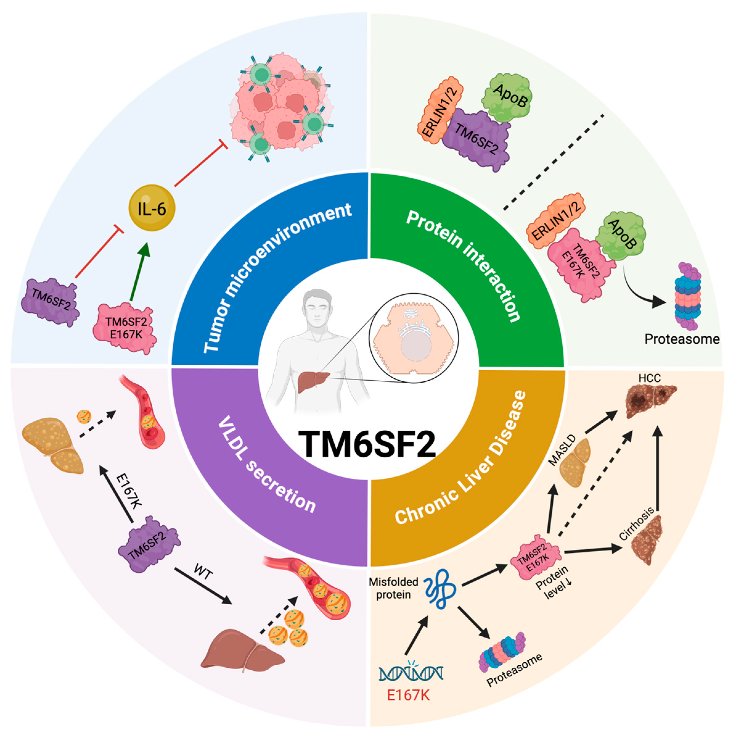
Genome-wide association meta-analyses have identified numerous single-nucleotide polymorphic risk variants associated with MASLD [9], among which the TM6SF2 E167K variant emerged as a clinically relevant genetic determinant for hepatic triglyceride content (HTGC), as well as hepatic fibrosis, cirrhosis and HCC (Figure 1) [9,10,11,12,13,14]. MASLD can be stratified into a cardiometabolic and a liver-specific subtype, characterized by different pathomechanisms [15]. Genetic variants such as TM6SF2 E167K, predominantly contribute to the liver-specific subtype of MASLD, which is associated with intrahepatic lipid retention rather than metabolic dysfunction [15].
The TM6SF2 E167K minor allele is associated with a relative risk ratio of 2.959 [16], indicating a nearly threefold increased risk of MASH in carriers. Notably, the effect allele occurs at a frequency of around 7% in all ancestry cohorts, highlighting its impact on liver health and disease [9].
Although several rare genetic variants of TM6SF2 (L156P, P216L) have been described [17], the TM6SF2 E167K shows the most reproducible effects and has been investigated in several studies. Therefore, this review focuses on the role of TM6SF2 E167K in lipid metabolism and chronic liver diseases. Figure 1 visualizes a summary of TM6SF2 and its functions.

2. TM6SF2: Structure, Expression and Variants

The human TM6SF2 gene is located on chromosome 19 and comprises 10 exons, encoding a 377 amino-acid protein. An alphaFold-generated in-silico model suggests that TM6SF2 has a helix-loop-helix structure with 10 transmembrane segments [17,18]. TM6SF2 is predominantly expressed in the small intestine, liver and kidney, with low expression in other tissues [19]. Within the liver, its expression is essentially in hepatocytes. On a subcellular level, TM6SF2 is localized in the endoplasmic reticulum (ER) and ER-Golgi intermediate compartment (ERGIC) [20], where very-low-density lipoprotein (VLDL) particles undergo lipidation. TM6SF2 has been shown to modulate the lipidation of VLDL and consequently liver and plasma lipid levels [9,10,19,20].
In-silico analyses further predict that the loops facing the luminal side of the ER collectively form the highly conserved EXPanded EBP superfamily (EXPERA) domain, which is defined by a cluster of negatively charged amino acids [18]. The protein contains a predicted catalytic site for 3-beta-hydroxysteroid-8,7-isomerase, an enzyme that converts the double bond from position 8 to position 7 in sterols, necessary for cholesterol synthesis [18].
The rs58542926 variant of the TM6SF2 gene is a nonsynonymous glutamate to lysine substitution at the amino acid residue 167 (E167K) [19]. This single-nucleotide polymorphism (SNP) leads to protein misfolding, resulting in accelerated degradation and lower protein levels [19]. Furthermore, in-silico analyses indicate that the E167K variant may influence the structure of the EXPERA domain due to an exchange of negatively charged glutamate with positively charged lysine, disrupting the electrostatic properties [18,21]. A human hepatocyte cell line with TM6SF2 E167K overexpression showed that RNA levels were comparable to WT, while the protein expression showed a significant reduction, indicating a problem in protein folding [19].
Other clinically relevant variants are rs187429064 (L156P) and rs186811910 (P216L). L156P is associated with an increased risk of steatotic liver disease and steatohepatitis, while P216L is associated with an increased risk of MASH [17]. In addition, L156P may also increase the risk of HCC and fibrosis [17]. L156P substitutes leucine with proline on position 156, which is in the fifth transmembrane α-helix [17]. Consequently, a hydrogen bond is missing, and proline leads to a ~20° kink in the α-helix structure, which has an indirect influence on the E167 residue and thereby on the EXPERA domain [17]. P216L is in the beginning of helix 7 and represents a proline to leucine change [17]. Hypotheses about the effect suggest that the replacement of proline removes a key structural kink, which is critical for physiological protein folding [17].

3. TM6SF2 in Lipid Metabolism

The liver’s hepatocytes have a central role in lipid metabolism by secreting VLDL and taking up fatty acids and lipoproteins. Within the hepatocytes, TG are synthesized and either temporarily stored in lipid droplets or packaged into ApoB-containing particles. Alterations in hepatic lipid mechanisms can shift the balance between storage and secretion. This could lead to an imbalance in the lipid profile of hepatocytes, characterized by the accumulation of cytotoxic lipids. Alteration of the lipid profile is a key mechanism in MALSD pathogenesis. Factors including genetic influences play an important role to study today. We have summarized the key findings of this section in Table 1.
An epidemiological study identified and described the TM6SF2 E167K genetic variant as a determinant associated with increased hepatic steatosis and susceptibility to MASLD [19]. Results showed that plasma triglycerides (TG) and low-density lipoprotein-cholesterol (LDL-c) levels decreased in TM6SF2 E167K carriers, while high-density lipoprotein-cholesterol (HDL-c) levels were not affected [19].
Murine models compared a TM6SF2 knockdown to an overexpression of TM6SF2 and its effects on lipid metabolism. The TM6SF2 knockdown reduced the protein level to 10% and resulted in a decrease in plasma cholesterol and TG levels, while HTGC increased threefold compared to control [19]. Moreover, LDL-c, HDL-c and the TG content of VLDL were reduced in knockdown mice [19]. The decreased VLDL-TG secretion was not associated with reduction in hepatic Apolipoprotein B (ApoB) secretion [19]. This may indicate that TM6SF2 is not required for secreting ApoB-containing lipoproteins, but rather for mobilizing lipids for VLDL assembly in mice [22].
Holmen et al. also used TM6SF2 knockdown mice, which resulted in a nearly 50% reduction of TM6SF2 protein level [21]. Interestingly, only a decrease in total cholesterol (TC) was observed, while TG, HDL-c and LDL-c levels did not differ in knockdown and control [21]. TM6SF2 overexpression in mice resulted in increased TG, LDL-c and TC levels, along with decreased HDL-c levels [21].
Fan et al. investigated the long-term effects of altered TM6SF2 levels on cholesterol metabolism by generating a CRISPR/Cas9-mediated knockout (KO) of TM6SF2 in mice, which were fed a high-fat diet for 10 weeks [23]. Unexpectedly, increased HTGC was observed in TM6SF2 overexpressing mice and plasma TG levels were not affected [23]. In contrary, the TM6SF2 KO mice showed decreased plasma TC and HDL-c, but not LDL-c [23]. However, TG levels were increased and no change in HTGC could be observed in KO mice [23].
The effect of a TM6SF2 KO was also studied in a human hepatoma cell line (Huh-7). Consistent with the results of KO mice [19], knockdown HuH7 cells showed an increase in HTGC and cholesterol esters (CE) concentration [24,25]. Lipid profiles indicate that TM6SF2 knockdown cells were predominantly enriched in TG with saturated and monounsaturated fatty acids with shorter side chain length, while control cells contained relatively more TG with polyunsaturated fatty acids [24,25]. Moreover, glycerol labeling showed higher total levels of TG, phosphatidylcholine (PC), phosphatidylethanolamine and phosphatidylinositol, which later also decreased more rapidly in knockdown cells, indicating an enhanced turnover of glycerolipids [24].
In Huh-7 cells, both TM6SF2 knockdown and TM6SF2 E167K variant overexpression led to comparable increases in hepatic lipid content [25]. The mRNA levels were similar in E167K and WT Huh-7 cells, however the protein level of the E167K variant was reduced by 46% compared to control [19]. This findings again highlight that the E167K variant may results in altered protein folding and protein degradation.
Induced hepatocytes (iHeps) were gene edited, generating isogenic cells with and without the TM6SF2 E167K variant [26]. RNA levels showed no differences between WT and E167K [26]. The TM6SF2 E167K cells showed reduced protein expression compared to control [26]. Furthermore, the intracellular lipid levels increased in the E167K cells compared to WT [26]. The results also showed that ApoB100 protein expression and TC increased intracellularly, while secretion of ApoB100 and VLDL was reduced in E167K cells, again indicating an altered VLDL secretion [26].
Transcriptomic characterization showed that iHeps-TM6SF2 E167K cells modify lipid metabolism by upregulating 153 genes and downregulating 267 genes [26]. Pathways connected to cholesterol-, fatty acid- and glucose metabolism showed increased expressions [26].
Analyses of mitochondria showed a spherical morphology and gene expression analysis revealed that important mitochondrial genes were expressed at lower levels in iHeps-TM6SF2 E167K cells compared with TM6SF2 WT [26]. Besides, altered lipid metabolism in iHep-TM6SF2 E167K cells has been linked to increased ER stress. Analysis of genes indicative of ER and mitochondrial stress showed increased levels in TM6SF2 E167K cells [26].
Pharmacological intervention with 4-Phenylbutyrate has been shown to reduce ER stress [27]. This finding suggests that targeting protein misfolding caused by the TM6SF2 E167K variant may help to reduce ER stress and lipid accumulation [26].
In another study, an isogenic model from induced pluripotent stem cell (iPSC)-derived hepatocyte-like cell (HLC) was developed independently. The HLCs were supplemented with linoleic acid, arachidonic acid and liver X receptor agonist, which has been shown to lead to more physiological phospholipid composition, VLDL lipidation and secretion [28]. Epidemiological research consistently showed that the prevalence of MASLD is higher in men, which may be attributed to differences in sex-specific hormonal patterns [29]. Interestingly, the study investigated how testosterone influences the phenotype of TM6SF2 E167K carriers [28]. Transcriptomic analysis underlined that HLC treatment with dihydrotestosterone (DHT) leads to dose-dependent increase of androgen-responsive genes TSC22D1 and RHOU concentration, reflecting a gene expression pattern characteristic of the male liver [28]. E167K HLCs treated with DHT exhibited increased lipid droplet accumulation and further impairment in arachidonic acid incorporation into PC [28].

3.1. TM6SF2 in VLDL Assembly and Secretion

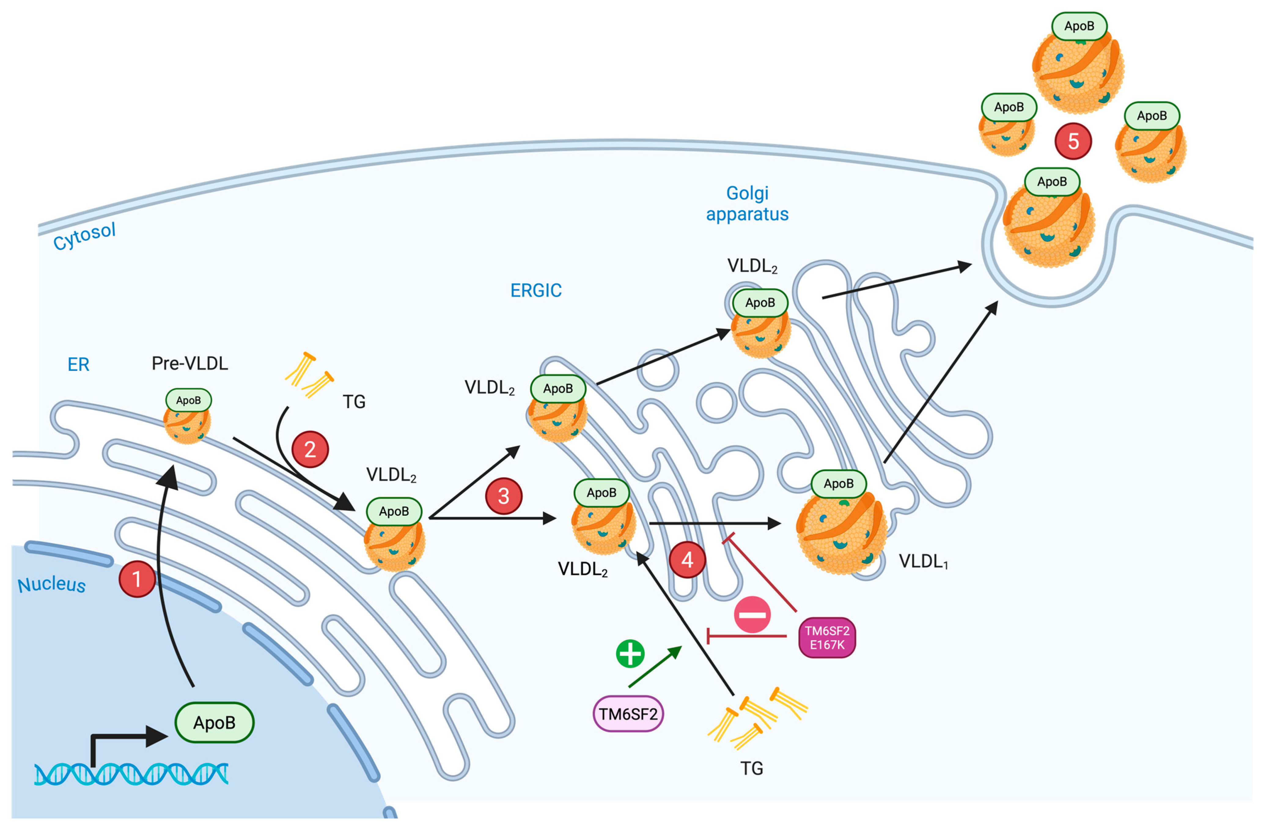
Hepatic cell culture experiments indicate that the assembly of mature VLDL in hepatocytes occurs through multiple (Figure 2) [30]. Initially, pre-VLDL particles are formed in the ER and then enter the secretory pathway, where they undergo gradual lipidation through the addition of TG [30,31]. This stepwise lipidation results in the formation of VLDL2 particles [31]. These particles can either be secreted directly or undergo further lipidation in a smooth membrane compartment, likely the ERGIC, to become large, TG-rich VLDL1 [31].
To investigate the effect of TM6SF2 E167K on VLDL mechanisms, humans homozygous for TM6SF2 E167K were examined in a kinetic study [10]. As expected, carriers of the variant had more than 2-fold higher liver fat content and decreased total plasma ApoB and TG levels [10].
Lipidomic analysis of VLDL1 and VLDL2 showed a decrease in relative abundance of unsaturated fatty acids with increased number of double bonds in TM6SF2 E167K carriers [10]. These findings illustrate that TM6SF2 may influence the assembly and lipid composition of VLDL, particularly polyunsaturated fatty acids.
VLDL1 had a 50% reduced TG content, which represents the main factor for reduced plasma TG levels [10]. Furthermore, VLDL1-ApoB100 was reduced in homozygous TM6SF2 E167K carriers, suggesting a decrease in secretion rate of VLDL1-particles [10]. In contrast, VLDL2 levels were unchanged [10]. VLDL1 and VLDL2 are regulated differently, which suggests that TM6SF2 specifically impacts the VLDL1 pathway [10].
TM6SF2 KO mice studies showed reduced TG levels in the VLDL fraction [19,22]. Surprisingly, mice with TM6SF2 overexpression showed a nearly 34% reduction in TG secretion rate compared to control [32]. Ehrhardt et al. observed reduced TG content in the VLDL and LDL fractions as well as reduced LDL-c, HDL-c and VLDL-c fractions in mice with TM6SF2 overexpression [32].
Despite the reduction in TG secretion from the liver, levels of ApoB100 were similar in KO and WT animals, while ApoB48 secretion was slightly increased [22,33]. In contrast to these results, TM6SF2 transgenic mice showed markedly reduced plasma levels of both ApoB100 and ApoB48, while levels were reduced in liver [32].
The different results in ApoB100 and ApoB48 levels led to the use of an alternative model to study the mechanisms. Homozygous TM6SF2 KO rats showed 6-fold higher HTGC and lower plasma cholesterol levels, with reductions observed in both VLDL-c and HDL-c fractions [34]. Plasma levels of ApoB100 and ApoB48 showed no significant difference between KO and WT rats [34]. Hepatic ApoB100 levels did not differ, while ApoB48 levels were slightly lower in the KO rats [34].
It is important to mention that in humans, ApoB100 is the predominant ApoB isoform produced in the liver, whereas ApoB48 is produced exclusively in the intestine [35]. Murine models differ in this aspect, as ApoB48 also gets produced in the liver [35]. Therefore, it is crucial to highlight that the mentioned ApoB interactions needs to be investigated further. Models with similar ApoB patters as in humans should be studied in the future to validate these findings.

3.2. TM6SF2-ApoB Interaction

TM6SF2 was found to physically interact with ApoB48 in mouse and rat livers [34,36]. Studies showed that TM6SF2 forms a protein complex with ER lipid raft protein (ERLIN) 1, ERLIN2 and ApoB that is essential for ApoB stabilization and VLDL assembly [36]. However, immunoprecipitation of ApoB48 showed that ERLIN1 and ERLIN2 co-immunoprecipitated with TM6SF2, but not directly with ApoB48, suggesting an indirect effect of the two proteins on ApoB48 via TM6SF2 [36]. Subsequently, truncation experiments revealed that the TM6SF2-ApoB48 interaction is mediated through the two luminal loops of TM6SF2 [36].
The stability of the ERLINs-TM6SF2-ApoB48 complex depends on reciprocal stabilization among its components. ERLIN1 and ERLIN2 increased the protein level of TM6SF2 in a dose dependent manner and vice versa [36]. TM6SF2 also stabilizes ApoB48 through its luminal loops, while ERLINs and ApoB48 have little effect on one another [36]. Knockdown of TM6SF2 or ERLINs almost eliminated endogenous ApoB100 and ApoB48 levels without affecting ApoB mRNA, indicating that the protein complex protects ApoB from proteasomal degradation [36].
The TM6SF2 E167K variant was less effective in stabilizing ApoB48. Nevertheless, the E167K variant had no impact in the stabilization of ApoB and ERLINs on TM6SF2 [36]. The impaired TM6SF2-ApoB stabilization likely contributes to defective VLDL lipidation and secretion observed in E167K carriers [36].
Genetic data further support the functional interaction between TM6SF2 and ERLIN1. The ERLIN1 p.Ile291Val SNP is predicted to be a gain of function variant, which reduces the risk for MASLD in homozygous carriers [37]. However, the protective effect was absent in individuals carrying the TM6SF2 E167K variant [37]. This finding underlines the interaction of ERLIN1 and TM6SF2 in lipid metabolism and VLDL secretion.

4. TM6SF2 in Chronic Liver Disease

4.1. Alcohol- and Viral-Associated Liver Disease

Although initially described in MASLD, TM6SF2 E167K also influences ALD and chronic viral hepatitis progression. Autoimmune diseases such as Primary biliary cholangitis (PBC) and primary sclerosing cholangitis (PSC) seem not to be affected by TM6SF2 polymorphisms. Studies suggest that the TM6SF2 E167K variant neither affect the risk for disease progression nor the liver function in PSC patients [38]. We have summarized the key findings of this section in Table 2.
In ALD, the effect of the variant potentially acts synergistically with alcohol consumption. Buch et al. reported that the TM6SF2 E167K variant confers an increased risk for developing ALD-related cirrhosis [39]. The effect was especially strong in individuals with heavy lifetime alcohol intake [39]. Furthermore, carriage of the variant was an additional risk factor for the development of HCC in people with alcohol-related cirrhosis [40,41]. The mechanisms behind the progression of ALD in individuals with the TM6SF2 E167K variant remain unclear and require further investigation.
Viral hepatitis is another etiology of CLD, which represents a major global health concern. It is classified into five main types (A–E), based on the disease-causing virus. Patients with chronic hepatitis B (HBV), C (HCV) and D (HDV) experience a variable susceptibility to develop progressive liver disease. Genetic risk factors such as TM6SF2 E167K contribute to this phenomenon.
Coppola et al. demonstrated that TM6SF2 E167K independently increases the risk of steatosis in chronic HCV patients [42]. Subsequent studies underlined an association of the variant with cirrhosis in chronic HCV patients [43].
Patients who are homozygous for TM6SF2 E167K, had a significantly lower HCV viral load, compared to WT and heterozygous carriers [44]. Histological analyses of chronic HCV patients indicate a modest effect of TM6SF2 E167K on hepatic steatosis [44]. However, after adjusting for HCV genotype, TM6SF2 E167K was associated with non-genotype 3 HCV patients, while genotype 3 influences steatosis independently [44,45]. Additionally, in genotype 1 carriers of HCV, TM6SF2 E167K did not affect the histological severity of liver damage [46]. Meta-analyses confirmed that TM6SF2 E167K contributes to fibrosis progression in chronic HCV patients [47].
Data from chronic HBV patients interestingly show that individuals with the TM6SF2 E167K allele have a modest increase in HBV DNA levels [44]. However, recent studies rather associate TM6SF2 E167K with altered HBsAg-containing subviral particles, since the secretion pathway of HBsAg includes trafficking through the ER-Golgi [48]. Results confirm the hypothesis that TM6SF2 E167K carriers showed a 25% reduction in circulating HBsAg levels compared to WT [48]. Furthermore, the extracellular concentration of HBsAg was almost halved [48]. In agreement with findings from the altered lipid secretion, knockdown of TM6SF2 E167K cells also show intracellular HBsAg retention [48].
TM6SF2 knockdown studies in Huh7.5.1 cells and in primary human hepatocytes showed the molecular impact of the protein on HCV cells and infectious lipoviroparticle production [48]. The results illustrated that TM6SF2 silencing led to a 1.5- and 2-fold increase in intracellular HCV in Huh7.5.1 cells and in primary human hepatocytes [49]. Moreover, viral RNA levels and secretion of infectious viral particles decreased [48,49]. The influence of TM6SF2 on the HCV genome replication, translation and assembly is currently controversial. Tu et al. observed a reduction in the number of positive TM6SF2 KO, HCV infected cells by 77% and significantly reduced intracellular HCV RNA by 82% [48]. They speculate that TM6SF2 knockdown may affect HCV secretion, infection or replication [48].
HDV needs HBsAg to form infectious particles. Thus, it is important to determine whether TM6SF2 knockdown also alters HBsAg secretion, which could explain the changes in HDV particle secretion. Knockdown of TM6SF2 in HDV producing Huh7-END cells showed a significant decrease in HBsAg secretion [48]. The supernatant of TM6SF2 knockdown cells was used to infect Huh7-NTCP cells to determine the effect on HDV secretion. Results showed a reduction in HDV-positive Huh7-NTCP cells, suggesting that secretion of infectious HDV virions was reduced in TM6SF2 knockdown cells [48].

4.2. TM6SF2 in HCC

Analysis of liver tumor tissue in human MASLD-HCC patients showed a downregulated expression of TM6SF2 protein and mRNA levels compared to non-tumor tissue [50]. TM6SF2 KO mice experiments revealed an increased tumor number, maximum tumor diameter and cell proliferation [50]. TM6SF2 overexpression in MASLD-HCC mice with Hepa1-6 tumors showed the opposite phenotype with decreased cell proliferation and reduced tumor weight [50]. In conclusion, these findings demonstrate that TM6SF2 deficiency contributes to MASLD-HCC formation in mice.
Because inflammatory cytokines are responsible for immune-cell recruitment and activation during tumorigenesis, Du et al. used HCC cell line HEPA 1-6 to explore the effect of the TM6SF2 E167K variant on expression of inflammatory cytokines [51]. They found no difference in TNF-α levels among all three TM6SF2 E167K overexpression, WT overexpression and control [51]. TM6SF2- and TM6SF2 E167K overexpression showed higher expression of IL-8 than control, but no significant difference between both overexpression groups [51]. IL-2 and IL-6 increased in TM6SF2 E167K overexpressed cells compared to WT overexpression and control, which had no significant difference [51].
Furthermore, TM6SF2 may impair the tumor immune microenvironment. Especially cytokine-cytokine interaction pathways were enriched in TM6SF2 KO MASLD-HCC mice [50]. TM6SF2 KO in mice livers with Hepa1-6 or RIL-175 tumors showed a decrease in CD8+ T-cells, especially the IFN-γ+ CD8+ cytotoxic T-cells [50]. Overexpression showed the opposite effect, with boosted CD8+ T-cell and cytotoxic CD8+ T-cell activation [50]. Immunofluorescence staining of mice livers with TM6SF2 KO also illustrated that the intratumoral CD8+ T-cells and IFN-γ+ CD8+ T-cells depleted compared to WT [50]. Studies in human primary hepatocytes confirmed the association between CD8+ T-cell infiltration and TM6SF2, implying that TM6SF2 might affect the antitumor response similarly in humans [50]. In summary, these findings underline that TM6SF2 deficiency may impair CD8+ T-cell function to promote tumorigenesis [50]. Hepatic TM6SF2 enhances the cytotoxic CD8+ T-cell mediated antitumor response and thereby inhibits MASLD-HCC progression [50].
IL-6 levels were significantly increased in tumor cells with TM6SF2 KO cells [50]. Elevated IL-6 levels impair CD8+ T-cell proliferation and function, which could be a possible mechanism behind the tumor promoting effect in TM6SF2 KO MASLD-HCC cells [50]. In contrary, TM6SF2 overexpression led to decreased IL-6 levels and thereby enhanced CD8+ T-cell activity [50]. Pharmacological inhibition of IL-6 with Tocilizumab restored CD8+ T-cell function in TM6SF2 KO cells, whereas the addition of IL-6 reversed the stimulatory effect of TM6SF2 overexpression [50]. In conclusion, these findings indicate that TM6SF2 might promote antitumor immunity by suppression of IL-6 secretion, thus preserving cytotoxic CD8+ T-cell responses [50].

5. Discussion

TM6SF2 is a key protein that not only affects metabolic pathways but also stabilizes other proteins which are essential for physiological cellular function. Understanding the role of TM6SF2 is crucial in the context of MASLD to meet the increasing prevalence today and in the future.
Therefore, researchers used diverse methodological approaches to better understand its function in metabolism and disease. However, it is important to highlight the limitations of each model to transparently evaluate the results.
First, lipid metabolism differs between humans and mice, which could influence the value of murine models. While the human TM6SF2 E167K variant is associated with reduced ApoB levels, TM6SF2 KO mice do not show significant changes in ApoB levels. Murine lipid profiles are also characterized by higher HDL and lower LDL levels together with specific differences in VLDL production and hepatic lipid regulation in comparison to humans. Thus, interspecies differences in lipid metabolism highlight the limitations of murine models.
The mouse and human TM6SF2 protein share around 78% sequence identity. Moreover, other SNPs that do not affect TM6SF2 in humans could have functional consequences in mice.
However, the residue affected by the E167K variant is conserved in both mice and humans. Variants in non-conserved regions could affect protein stability, function and regulation, thereby limiting the relevance of murine findings.
Another important aspect to consider is that mice do not develop MASLD under natural conditions. Murine models typically require dietary or genetic interventions which could also influence the function of mouse TM6SF2. In conclusion, murine models represent a valuable model to investigate the mechanisms of TM6SF2.
In-vitro models such as Huh7 and HepG2 cells provide a human background, which avoids interspecies differences. However, as these cell lines are derived from liver tumors, they already exhibit altered metabolic characteristics. Furthermore, studies highlighted that these cell lines are homozygous for the PNPLA3 I148M variant, another major genetic determinant for MALSD development [52]. This means that previous studies on TM6SF2 in these cell lines investigated the variant’s effect in a PNPLA3 I148M minor homozygous background. Due to possible cumulative effects, the results need to be interpreted with caution. In-vitro cultures may also acquire additional mutations due to genetic instability of cancer cells.
Hepatocyte-like cells from iPSCs provide a human and isogenic model in which phenotypic differences can directly be attributed to the genetic background. HLC are similar to hepatocytes but show metabolic differences due to their low expression of key hepatocyte genes. In-vitro findings from HLC will need to be validated. Additionally, the absence of other hepatic cell types such as Stellate cells, Kupffer cells and sinusoidal endothelial cells pose a limitation in modeling hepatic physiology. Besides, in-vitro models lack interaction with small intestines, the organ with the highest TM6SF2 expression, and a central role in lipid metabolism.
Taken together, studies have shown that TM6SF2 impacts hepatic and systemic lipid metabolism, and its TM6SF2 E167K variant significantly influences CLD susceptibility. While both in-vivo and in-vitro models provide valuable insights, each approach comes with limitations that must be considered.
Future studies could explore genotype guided risk stratification and personalized therapeutic strategies for patients carrying TM6SF2 polymorphisms. This approach could help identify high-risk individuals and enable earlier interventions. However, additional research is required to better understand the complex role of TM6SF2 in lipid metabolism and CLD.

Author Contributions

Conceptualization, N.H. and M.A.-N.; resources, N.H.; writing original draft preparation, M.A.-N.; writing review and editing, N.H. and M.A.-N.; visualization, M.A.-N.; supervision, N.H.; project administration, N.H.; funding acquisition, N.H. All authors have read and agreed to the published version of the manuscript.

Funding

This research received no external funding.

Institutional Review Board Statement

Not applicable.

Informed Consent Statement

Not applicable.

Data Availability Statement

The original contributions presented in the study are included in the article, further inquiries can be directed to the corresponding author.

Conflicts of Interest

The authors declare no conflicts of interest.

References

  1. Amini-Salehi, E.; Letafatkar, N.; Norouzi, N.; Joukar, F.; Habibi, A.; Javid, M.; Sattari, N.; Khorasani, M.; Farahmand, A.; Tavakoli, S.; et al. Global Prevalence of Nonalcoholic Fatty Liver Disease: An Updated Review Meta-Analysis comprising a Population of 78 million from 38 Countries. Arch. Med. Res. 2024, 55, 103043. [Google Scholar] [CrossRef] [PubMed]
  2. Younossi, Z.M.; Golabi, P.; Paik, J.M.; Henry, A.; Van Dongen, C.; Henry, L. The global epidemiology of nonalcoholic fatty liver disease (NAFLD) and nonalcoholic steatohepatitis (NASH): A systematic review. Hepatology 2023, 77, 1335–1347. [Google Scholar] [CrossRef]
  3. Bessone, F.; Razori, M.V.; Roma, M.G. Molecular pathways of nonalcoholic fatty liver disease development and progression. Cell. Mol. Life Sci. 2019, 76, 99–128. [Google Scholar] [CrossRef] [PubMed]
  4. Chalasani, N.; Younossi, Z.; Lavine, J.E.; Charlton, M.; Cusi, K.; Rinella, M.; Harrison, S.A.; Brunt, E.M.; Sanyal, A.J. The diagnosis and management of nonalcoholic fatty liver disease: Practice guidance from the American Association for the Study of Liver Diseases. Hepatology 2018, 67, 328–357. [Google Scholar] [CrossRef]
  5. Marrero, J.A.; Fontana, R.J.; Su, G.L.; Conjeevaram, H.S.; Emick, D.M.; Lok, A.S. NAFLD may be a common underlying liver disease in patients with hepatocellular carcinoma in the United States. Hepatology 2002, 36, 1349–1354. [Google Scholar] [CrossRef]
  6. Huang, D.Q.; Mathurin, P.; Cortez-Pinto, H.; Loomba, R. Global epidemiology of alcohol-associated cirrhosis and HCC: Trends, projections and risk factors. Nat. Rev. Gastroenterol. Hepatol. 2023, 20, 37–49. [Google Scholar] [CrossRef]
  7. Cotter, T.G.; Charlton, M. Nonalcoholic Steatohepatitis After Liver Transplantation. Liver Transplant. 2020, 26, 141–159. [Google Scholar] [CrossRef]
  8. Younossi, Z.M.; Henry, L. Epidemiology of non-alcoholic fatty liver disease and hepatocellular carcinoma. JHEP Rep. 2021, 3, 100305. [Google Scholar] [CrossRef]
  9. Chen, Y.; Du, X.; Kuppa, A.; Feitosa, M.F.; Bielak, L.F.; O’Connell, J.R.; Musani, S.K.; Guo, X.; Kahali, B.; Chen, V.L.; et al. Genome-wide association meta-analysis identifies 17 loci associated with nonalcoholic fatty liver disease. Nat. Genet. 2023, 55, 1640–1650. [Google Scholar] [CrossRef]
  10. Borén, J.; Adiels, M.; Björnson, E.; Matikainen, N.; Söderlund, S.; Rämö, J.; Ståhlman, M.; Ripatti, P.; Ripatti, S.; Palotie, A.; et al. Effects of TM6SF2 E167K on hepatic lipid and very low-density lipoprotein metabolism in humans. JCI Insight 2020, 5, e144079. [Google Scholar] [CrossRef]
  11. Sookoian, S.; Castaño, G.O.; Scian, R.; Mallardi, P.; Fernández Gianotti, T.; Burgueño, A.L.; San Martino, J.; Pirola, C.J. Genetic variation in transmembrane 6 superfamily member 2 and the risk of nonalcoholic fatty liver disease and histological disease severity. Hepatology 2015, 61, 515–525. [Google Scholar] [CrossRef]
  12. Liu, Y.-L.; Reeves, H.L.; Burt, A.D.; Tiniakos, D.; McPherson, S.; Leathart, J.B.S.; Allison, M.E.D.; Alexander, G.J.; Piguet, A.-C.; Anty, R.; et al. TM6SF2 rs58542926 influences hepatic fibrosis progression in patients with non-alcoholic fatty liver disease. Nat. Commun. 2014, 5, 4309. [Google Scholar] [CrossRef]
  13. Tang, S.; Zhang, J.; Mei, T.-T.; Guo, H.-Q.; Wei, X.-H.; Zhang, W.-Y.; Liu, Y.-L.; Liang, S.; Fan, Z.-P.; Ma, L.-X.; et al. Association of TM6SF2 rs58542926 T/C gene polymorphism with hepatocellular carcinoma: A meta-analysis. BMC Cancer 2019, 19, 1128. [Google Scholar] [CrossRef]
  14. Yang, J.; Trépo, E.; Nahon, P.; Cao, Q.; Moreno, C.; Letouzé, E.; Imbeaud, S.; Gustot, T.; Deviere, J.; Debette, S.; et al. PNPLA3 and TM6SF2 variants as risk factors of hepatocellular carcinoma across various etiologies and severity of underlying liver diseases. Int. J. Cancer 2019, 144, 533–544. [Google Scholar] [CrossRef] [PubMed]
  15. Luukkonen, P.K. Subtypes of MASLD confer distinct clinical trajectories. J. Hepatol. 2025, 82, 1138–1139. [Google Scholar] [CrossRef] [PubMed]
  16. Kocas-Kilicarslan, Z.N.; Cetin, Z.; Faccioli, L.A.P.; Motomura, T.; Amirneni, S.; Diaz-Aragon, R.; Florentino, R.M.; Sun, Y.; Pla-Palacin, I.; Xia, M.; et al. Polymorphisms Associated With Metabolic Dysfunction-Associated Steatotic Liver Disease Influence the Progression of End-Stage Liver Disease. Gastro Hep Adv. 2024, 3, 67–77. [Google Scholar] [CrossRef] [PubMed]
  17. Huang, H.Y.R.; Vitali, C.; Zhang, D.; Hand, N.J.; Phillips, M.C.; Creasy, K.T.; Scorletti, E.; Park, J.; Regeneron, C.; Schneider, K.M.; et al. Deep metabolic phenotyping of humans with protein-altering variants in TM6SF2 using a genome-first approach. JHEP Rep. 2025, 7, 101243. [Google Scholar] [CrossRef]
  18. Sanchez-Pulido, L.; Ponting, C.P. TM6SF2 and MAC30, new enzyme homologs in sterol metabolism and common metabolic disease. Front. Genet. 2014, 5, 439. [Google Scholar] [CrossRef]
  19. Kozlitina, J.; Smagris, E.; Stender, S.; Nordestgaard, B.G.; Zhou, H.H.; Tybjaerg-Hansen, A.; Vogt, T.F.; Hobbs, H.H.; Cohen, J.C. Exome-wide association study identifies a TM6SF2 variant that confers susceptibility to nonalcoholic fatty liver disease. Nat. Genet. 2014, 46, 352–356. [Google Scholar] [CrossRef]
  20. Mahdessian, H.; Taxiarchis, A.; Popov, S.; Silveira, A.; Franco-Cereceda, A.; Hamsten, A.; Eriksson, P.; van’t Hooft, F. TM6SF2 is a regulator of liver fat metabolism influencing triglyceride secretion and hepatic lipid droplet content. Proc. Natl. Acad. Sci. USA 2014, 111, 8913–8918. [Google Scholar] [CrossRef]
  21. Holmen, O.L.; Zhang, H.; Fan, Y.; Hovelson, D.H.; Schmidt, E.M.; Zhou, W.; Guo, Y.; Zhang, J.; Langhammer, A.; Løchen, M.-L.; et al. Systematic evaluation of coding variation identifies a candidate causal variant in TM6SF2 influencing total cholesterol and myocardial infarction risk. Nat. Genet. 2014, 46, 345–351. [Google Scholar] [CrossRef]
  22. Smagris, E.; Gilyard, S.; BasuRay, S.; Cohen, J.C.; Hobbs, H.H. Inactivation of Tm6sf2, a Gene Defective in Fatty Liver Disease, Impairs Lipidation but Not Secretion of Very Low Density Lipoproteins. J. Biol. Chem. 2016, 291, 10659–10676. [Google Scholar] [CrossRef]
  23. Fan, Y.; Lu, H.; Guo, Y.; Zhu, T.; Garcia-Barrio, M.T.; Jiang, Z.; Willer, C.J.; Zhang, J.; Chen, Y.E. Hepatic Transmembrane 6 Superfamily Member 2 Regulates Cholesterol Metabolism in Mice. Gastroenterology 2016, 150, 1208–1218. [Google Scholar] [CrossRef]
  24. Ruhanen, H.; Nidhina Haridas, P.A.; Eskelinen, E.L.; Eriksson, O.; Olkkonen, V.M.; Kakela, R. Depletion of TM6SF2 disturbs membrane lipid composition and dynamics in HuH7 hepatoma cells. Biochim. Biophys. Acta Mol. Cell Biol. Lipids 2017, 1862, 676–685. [Google Scholar] [CrossRef]
  25. Pant, A.; Chen, Y.; Kuppa, A.; Du, X.; Halligan, B.D.; Speliotes, E.K. Perturbation of TM6SF2 Expression Alters Lipid Metabolism in a Human Liver Cell Line. Int. J. Mol. Sci. 2021, 22, 9758. [Google Scholar] [CrossRef]
  26. Faccioli, L.A.P.; Sun, Y.; Animasahun, O.; Motomura, T.; Liu, Z.; Kurihara, T.; Hu, Z.; Yang, B.; Cetin, Z.; Baratta, A.M.; et al. Human-induced pluripotent stem cell–based hepatic modeling of lipid metabolism–associated TM6SF2-E167K variant. Hepatology 2025, 82, 638–654. [Google Scholar] [CrossRef] [PubMed]
  27. Mai, C.T.; Le, Q.G.; Ishiwata-Kimata, Y.; Takagi, H.; Kohno, K.; Kimata, Y. 4-Phenylbutyrate suppresses the unfolded protein response without restoring protein folding in Saccharomyces cerevisiae. FEMS Yeast Res. 2018, 18, foy016. [Google Scholar] [CrossRef]
  28. Hirai, A.; Yoneyama, Y.; Assi, I.; Osonoi, S.; Sakai, S.; Ohtsu, K.; Takebe, T. Modeling Transmembrane 6 Superfamily Member 2 p.Glu167Lys-associated Steatotic Liver Disease Using Androgen-treated Human Induced Pluripotent Stem Cell-derived Hepatocyte-like Cells. Gastro Hep Adv. 2025, 5, 100774. [Google Scholar] [CrossRef]
  29. Wong, R.J. Epidemiology of metabolic dysfunction-associated steatotic liver disease (MASLD) and alcohol-related liver disease (ALD). Metab. Target. Organ. Damage 2024, 4, 35. [Google Scholar] [CrossRef]
  30. Bostrom, K.; Boren, J.; Wettesten, M.; Sjoberg, A.; Bondjers, G.; Wiklund, O.; Carlsson, P.; Olofsson, S.O. Studies on the assembly of apo B-100-containing lipoproteins in HepG2 cells. J. Biol. Chem. 1988, 263, 4434–4442. [Google Scholar] [CrossRef] [PubMed]
  31. Stillemark-Billton, P.; Beck, C.; Borén, J.; Olofsson, S.-O. Relation of the size and intracellular sorting of apoB to the formation of VLDL 1 and VLDL 2. J. Lipid Res. 2005, 46, 104–114. [Google Scholar] [CrossRef]
  32. Ehrhardt, N.; Doche, M.E.; Chen, S.; Mao, H.Z.; Walsh, M.T.; Bedoya, C.; Guindi, M.; Xiong, W.; Ignatius Irudayam, J.; Iqbal, J.; et al. Hepatic Tm6sf2 overexpression affects cellular ApoB-trafficking, plasma lipid levels, hepatic steatosis and atherosclerosis. Hum. Mol. Genet. 2017, 26, 2719–2731. [Google Scholar] [CrossRef]
  33. Newberry, E.P.; Hall, Z.; Xie, Y.; Molitor, E.A.; Bayguinov, P.O.; Strout, G.W.; Fitzpatrick, J.A.J.; Brunt, E.M.; Griffin, J.L.; Davidson, N.O. Liver-Specific Deletion of Mouse Tm6sf2 Promotes Steatosis, Fibrosis, and Hepatocellular Cancer. Hepatology 2021, 74, 1203–1219. [Google Scholar] [CrossRef]
  34. Luo, F.; Smagris, E.; Martin, S.A.; Vale, G.; McDonald, J.G.; Fletcher, J.A.; Burgess, S.C.; Hobbs, H.H.; Cohen, J.C. Hepatic TM6SF2 Is Required for Lipidation of VLDL in a Pre-Golgi Compartment in Mice and Rats. Cell Mol. Gastroenterol. Hepatol. 2022, 13, 879–899. [Google Scholar] [CrossRef] [PubMed]
  35. Oka, K.; Kobayashi, K.; Sullivan, M.; Martinez, J.; Teng, B.-B.; Ishimura-Oka, K.; Chan, L. Tissue-specific Inhibition of Apolipoprotein B mRNA Editing in the Liver by Adenovirus-mediated Transfer of a Dominant Negative Mutant APOBEC-1 Leads to Increased Low Density Lipoprotein in Mice. J. Biol. Chem. 1997, 272, 1456–1460. [Google Scholar] [CrossRef] [PubMed]
  36. Li, B.-T.; Sun, M.; Li, Y.-F.; Wang, J.-Q.; Zhou, Z.-M.; Song, B.-L.; Luo, J. Disruption of the ERLIN–TM6SF2–APOB complex destabilizes APOB and contributes to non-alcoholic fatty liver disease. PLoS Genet. 2020, 16, e1008955. [Google Scholar] [CrossRef]
  37. Rendel, M.D.; Vitali, C.; Creasy, K.T.; Zhang, D.; Scorletti, E.; Huang, H.; Seeling, K.S.; Park, J.; Hehl, L.; Vell, M.S.; et al. The common p.Ile291Val variant of ERLIN1 enhances TM6SF2 function and is associated with protection against MASLD. Med 2024, 5, 963–980.e5. [Google Scholar] [CrossRef]
  38. Kruk, B.; Liebe, R.; Milkiewicz, M.; Wunsch, E.; Raszeja-Wyszomirska, J.; Lammert, F.; Milkiewicz, P.; Krawczyk, M. PNPLA3 p.I148M and TM6SF2 p.E167K variants do not predispose to liver injury in cholestatic liver diseases: A prospective analysis of 178 patients with PSC. PLoS ONE 2018, 13, e0202942. [Google Scholar] [CrossRef]
  39. Buch, S.; Stickel, F.; Trépo, E.; Way, M.; Herrmann, A.; Nischalke, H.D.; Brosch, M.; Rosendahl, J.; Berg, T.; Ridinger, M.; et al. A genome-wide association study confirms PNPLA3 and identifies TM6SF2 and MBOAT7 as risk loci for alcohol-related cirrhosis. Nat. Genet. 2015, 47, 1443–1448. [Google Scholar] [CrossRef] [PubMed]
  40. Stickel, F.; Buch, S.; Nischalke, H.D.; Weiss, K.H.; Gotthardt, D.; Fischer, J.; Rosendahl, J.; Marot, A.; Elamly, M.; Casper, M.; et al. Genetic variants in PNPLA3 and TM6SF2 predispose to the development of hepatocellular carcinoma in individuals with alcohol-related cirrhosis. Off. J. Am. Coll. Gastroenterol. | ACG 2018, 113, 1475–1483. [Google Scholar] [CrossRef]
  41. Falleti, E.; Cussigh, A.; Cmet, S.; Fabris, C.; Toniutto, P. PNPLA3 rs738409 and TM6SF2 rs58542926 variants increase the risk of hepatocellular carcinoma in alcoholic cirrhosis. Dig. Liver Dis. 2016, 48, 69–75. [Google Scholar] [CrossRef]
  42. Coppola, N.; Rosa, Z.; Cirillo, G.; Stanzione, M.; Macera, M.; Boemio, A.; Grandone, A.; Pisaturo, M.; Marrone, A.; Adinolfi, L.E.; et al. TM6SF2 E167K variant is associated with severe steatosis in chronic hepatitis C, regardless of PNPLA3 polymorphism. Liver Int. 2015, 35, 1959–1963. [Google Scholar] [CrossRef]
  43. Milano, M.; Aghemo, A.; Mancina, R.M.; Fischer, J.; Dongiovanni, P.; De Nicola, S.; Fracanzani, A.L.; D’Ambrosio, R.; Maggioni, M.; De Francesco, R.; et al. Transmembrane 6 superfamily member 2 gene E167K variant impacts on steatosis and liver damage in chronic hepatitis C patients. Hepatology 2015, 62, 111–117. [Google Scholar] [CrossRef] [PubMed]
  44. Eslam, M.; Mangia, A.; Berg, T.; Chan, H.L.; Irving, W.L.; Dore, G.J.; Abate, M.L.; Bugianesi, E.; Adams, L.A.; Najim, M.A.; et al. Diverse impacts of the rs58542926 E167K variant in TM6SF2 on viral and metabolic liver disease phenotypes. Hepatology 2016, 64, 34–46. [Google Scholar] [CrossRef] [PubMed]
  45. Hui, J.M.; Kench, J.; Farrell, G.C.; Lin, R.; Samarasinghe, D.; Liddle, C.; Byth, K.; George, J. Genotype-specific mechanisms for hepatic steatosis in chronic hepatitis C infection. J. Gastroenterol. Hepatol. 2002, 17, 873–881. [Google Scholar] [CrossRef] [PubMed]
  46. Petta, S.; Maida, M.; Grimaudo, S.; Pipitone, R.M.; Macaluso, F.S.; Cabibi, D.; Cammà, C.; Di Marco, V.; Sferrazza, S.; Craxì, A. TM6SF2 rs58542926 is not associated with steatosis and fibrosis in large cohort of patients with genotype 1 chronic hepatitis C. Liver Int. 2016, 36, 198–204. [Google Scholar] [CrossRef]
  47. Liu, Z.; Que, S.; Zhou, L.; Zheng, S.; Romeo, S.; Mardinoglu, A.; Valenti, L. The effect of the TM6SF2 E167K variant on liver steatosis and fibrosis in patients with chronic hepatitis C: A meta-analysis. Sci. Rep. 2017, 7, 9273. [Google Scholar] [CrossRef]
  48. Tu, T.; Ajoyan, H.; Nur Umami, R.; Veeraraghavan, V.; Boldbaatar, D.; Najim, M.A.M.; Khan, A.; Bayoumi, A.; Ho, V.; Eslam, M.; et al. Inhibition of Cellular Factor TM6SF2 Suppresses Secretion Pathways of Hepatitis B, Hepatitis C, and Hepatitis D Viruses. J. Infect. Dis. 2024, 230, 970–981. [Google Scholar] [CrossRef]
  49. Boyer, A.; Park, S.B.; de Boer, Y.S.; Li, Q.; Liang, T.J. TM6SF2 Promotes Lipidation and Secretion of Hepatitis C Virus in Infected Hepatocytes. Gastroenterology 2018, 155, 1923–1935.e8. [Google Scholar] [CrossRef]
  50. Zhang, Y.; Xie, M.; Wen, J.; Liang, C.; Song, Q.; Liu, W.; Liu, Y.; Song, Y.; Lau, H.C.H.; Cheung, A.H.; et al. Hepatic TM6SF2 activates antitumour immunity to suppress metabolic dysfunction-associated steatotic liver disease-related hepatocellular carcinoma and boosts immunotherapy. Gut 2025, 74, 639–651. [Google Scholar] [CrossRef]
  51. Du, S.; Liao, S.; Liu, S.; Xin, Y. TM6SF2 E167K Variant Overexpression Promotes Expression of Inflammatory Cytokines in the HCC Cell Line HEPA 1-6. J. Clin. Transl. Hepatol. 2019, 7, 27–31. [Google Scholar] [CrossRef] [PubMed]
  52. Steigemann, P.; Braeuer, N.; Puetter, V.; Zablowsky, N.; Juenemann, K.; von Nussbaum, F.; Lesche, R.; Dittmar, N.; Schaller, D.; Makowska, Z.; et al. Identification of NUV-244 as a PNPLA3 I148M degrading small molecule. iScience 2025, 28, 112384. [Google Scholar] [CrossRef] [PubMed]
Figure 1. Protein interaction: TM6SF2 forms a protein complex with ERLIN1, ERLIN2 and ApoB, which is essential for ApoB stabilization. TM6SF2 E167K alters this complex and leads to proteasomal degradation of ApoB. Chronic Liver Disease: The TM6SF2 E167K variant leads to misfolding with concomitant protein degradation and thereby lower protein levels. E167K is linked to increased risk of MASLD, liver cirrhosis, and HCC. VLDL secretion: The TM6SF2 WT axis illustrates the physiological secretion of large, TG-rich VLDL particles into the bloodstream. The TM6SF2 E167K variant impairs the secretion and leads to lipid droplet accumulation in the liver. The secreted particles are smaller, and the amount of VLDL particles is reduced. Tumor microenvironment: The TM6SF2 E167K variant shows an increases of IL-6 levels. IL-6 impairs CD8+ T-cell proliferation and function, especially cytotoxic CD8+ T-cells. Consequently, TM6SF2 WT indirectly promotes antitumor immunity by suppression of IL-6. This figure was created with BioRender.com (accessed on 21 November 2025).
Livers 06 00017 g001
Figure 2. VLDL secretion and assembly: (1) ApoB is lipidated co-translationally and forms pre-VLDL particles. (2) In the secretory pathway, pre-VLDL particles undergo further lipidation with triglycerides (TG) to form VLDL2 particles, which are defined as a TG-poor form of VLDL. (3) Next, VLDL2 particles can either be secreted or further lipidated to form VLDL1 particles, the TG-rich form of VLDL. (4) Physiologically, VLDL2 receives bulk lipidation in the endoplasmic reticulum-Golgi intermediate compartment (ERGIC). TM6SF2 E167K impairs the lipidation, causing a reduction of TG content. The variant also leads to a decrease in secretion rate of VLDL1 particles. (5) VLDL1 and VLDL2 exit the hepatocyte and enter the bloodstream. ApoB, Apolipoprotein B; VLDL, very-low density lipoprotein; This figure was created with BioRender.com (accessed on 21 November 2025).
Livers 06 00017 g002
Table 1. Overview of experimental models and key findings on TM6SF2 in Hepatic Lipid Metabolism.
AuthorsExperimental ModelSpecificationMain Findings
Kozlitina et al. [19]In-vivo and in-vitro studiesTM6SF2 knockdown in miceReduced plasma TG and LDL-c levels in TM6SF2 E167K carriers
Reduced plasma cholesterol, TG, LDL-c, HDL-c and TG content of VLDL in mice; HTGC increased threefold in mice
Holmen et al. [21]In-vivo studyTM6SF2 knockdown and overexpression in miceKnockdown: Reduced TC levels
Overexpression: TC, LDL-c and TG increased; HDL-c levels decreased
Smagris et al. [22]In-vivo studyTM6SF2 KO in miceReduced TG in the VLDL fraction
Fan et al. [23]In-vivo studyTM6SF2 KO and overexpression in miceOverexpression: increased TC, LDL-c and HTGC. Plasma TG levels were not affected.
KO: reduced plasma TC and HDL-c levels; LDL-c and HTGC were not affected; TG levels increased
Ruhanen et al. [24]In-vitro studyTM6SF2 KO in Huh-7 cellsIncreased HTGC and CE concentration; Lipid profile shift to TG with saturated and monounsaturated fatty acids with shorter side chain length
Pant et al. [25]In-vitro studyTM6SF2 WT overexpression, knockdown and TM6SF2 E167K overexpression in Huh-7 cellsTM6SF2 knockdown showed increased HTGC, cholesterol esters concentration and enrichment of triglycerides with saturated and monounsaturated fatty acids with short side chains
Faccioli et al. [26]In-vitro studyTM6SF2 WT and TM6SF2 E167K in iHeps cells;ApoB100 expression and TC increased intracellularly in TM6SF2 E167K cells, while secretion of ApoB100 and VLDL was reduced; iHeps-TM6SF2 E167K cells showed 153 upregulated genes and 267 downregulated genes connected to metabolic pathways
Mai et al. [27]In-vitro studySaccharomyces cerevisiae treated with 4-phenylbutyrate4-Phenylbutyrate showed to reduce ER stress
Hirai et al. [28]In-vitro studyHepatocyte-like cells supplemented with linoleic acid, arachidonic acid and liver x receptor agonistTM6SF2 E167K HLCs: Increased HTGC and enlarged lipid droplets; decrease in arachidonic acid-containing polyunsaturated PC species;
DHT treatment led to lipid droplet accumulation and impairment in arachidonic acid incorporation into PC
Wong, R.J. [29]Epidemiological studyEpidemiological study on MASLD and ALD prevalencePrevalence of MASLD is higher in men than in women
Bostrom et al. [30]In-vitro studyApoB100-containing lipoprotein assembly study in HepG2 cellsThe ApoB100-containing lipoprotein assembly undergoes multiple steps;
ApoB100 co-translationally bounds to the ER membrane and gets lipidated sequentially
Stillemark-Billton [31]In-vitro studyVLDL-lipidation observation in McA-RH7777 cellsVLDL1 and VLDL2 undergo separate lipidation pathways; VLDL1 undergoes a two-step process, while VLDL2 is formed through ApoB size-dependent lipidation
Borén et al. [10]Clinical studyExamination of humans homozygous for TM6SF2 E167KDecreased total plasma ApoB and TG levels; Decrease in relative abundance of PUFAs in VLDL1 and VLDL2;
VLDL1 had a 50% reduced TG content
Ehrhardt et al. [32]In-vivo studyTM6SF2 overexpression in mice34% reduction in TG secretion rate; reduced TG content in LDL and VLDL fractions and reduced LDL-c, HDL-c and VLDL-c fractions; reduced ApoB100 and ApoB48 levels
Newberry et al. [33]In-vivo studyTM6SF2 KO in mice with WT and E167K variantKO mice showed reduced VLDL triglyceride secretion with similar ApoB100 levels and slightly higher ApoB48 levels
Luo et al. [34]In-vivo studyTM6SF2 KO in rats6-fold higher HTGC; plasma ApoB levels showed no difference to WT
Oka et al. [35]In-vivo studyLiver-specific inhibition of APOBEC-1In contrast to humans, ApoB48 is also produced in the liver and not just in the intestine
Li et al. [36] In-vivo and in-vitro studiesTAP- mass spectrometry; TM6SF2/ERLIN1/2 Knockdown and overexpression in Huh-7 and HEK293 cells; TM6SF2 Knockdown in miceERLIN 1 and 2, TM6SF2 and ApoB may physically interact.
Rendel et al. [37]Epidemiological studyERLIN1 p.Ile291Val variant interaction with TM6SF2The protective effect of the ERLIN1 variant was absent in TM6SF2 E167K carriers indicating protein interaction
Table 2. Overview of models and key findings on TM6SF2 in Chronic Liver Disease.
AuthorsModelSpecificationMain Findings
Kruk et al. [38]Epidemiological studyCandidate gene association study in PSC patientsTM6SF2 E167K does not appear to influence primary sclerosing cholangitis
Buch et al. [39]Epidemiological studyGWASTM6SF2 E167K is a risk locus for ALD-related cirrhosis
Stickel et al. [40]Epidemiological studyCandidate gene association study in alcohol-related cirrhosisTM6SF2 E167K is an additional risk factor for HCC in patients with alcohol-related cirrhosis
Falleti et al. [41]Epidemiological studyCandidate gene association study in cirrhotic patientsTM6SF2 and PNPLA3 polymorphisms are associated with increased HCC risk in alcohol-related cirrhosis, but not in viral cirrhosis
Coppola et al. [42]Clinical studyHCV positive patients were genotyped for TM6SF2 E167KTM6SF2 E167K is an independent risk factor for steatosis in chronic Hepatitis C Virus (HCV) patients
Milano et al. [43]Clinical studyHCV patients were genotyped for TM6SF2 E167KAssociation of TM6SF2 E67K with cirrhosis in chronic HCV patients
Eslam et al. [44]Clinical studyCandidate gene association study in MASLD and viral hepatitis patientsHomozygous TM6SF2 E167K carriers had a significantly lower HCV viral load than control; the variant is associated with the degree of steatosis in chronic HCV patients;
TM6SF2 E167K is associated with an increase in HBV DNA levels
Hui et al. [45]Clinical studyAnalysis of metabolic risk factors and viral genotype as predictors for steatosis and fibrosisHCV genotype 3 influences steatosis independently of the TM6SF2 variant
Petta et al. [46]Clinical studyCandidate gene association study in genotype 1 HCV patientsTM6SF2 E167K did not affect severity of liver damage in genotype 1 HCV carriers
Liu et al. [47]Meta-analysisMeta-analysis of TM6SF2 E167K in chronic HCV patientsTM6SF2 E167K is associated with fibrosis progression in chronic HCV patients
Tu et al. [48]In-vitro study and human cohort validationTM6SF2 KO and knockdown in Huh-7.1 cells and primary human hepatocytes;
HBV patient cohort correlation
TM6SF2 E167K carriers showed reduction in circulating HBsAg levels and increased intracellular HBsAg retention; increase of HCV levels in TM6SF2 KO cells; reduced secretion of infectious HDV virions in TM6SF2 knockdown cells.
Boyer et al. [49]In-vivo and in-vitro studiesTM6SF2 knockdown and overexpression in Huh7.5.1 cells;
Liver tissue analysis from chronic HCV patients
TM6SF2 knockdown is showed reduced secretion of infectious HCV lipoviroparticles; overexpression led to enhanced lipoviroparticles release
Zhang et al. [50]In-vivo and in-vitro studiesTM6SF2 KO and overexpression in mice; Human MASLD-HCC liver tissueLiver tissue of human HHC patients showed decreased TM6SF2 expression; TM6SF2 KO in mice showed increased tumor diameter and tumor number in mice; TM6SF2 overexpression showed the opposite effect in mice; TM6SF2 might promote antitumor immunity by suppression of IL-6
Du et al. [51]In-vitro studyHEPA 1-6 cells with TM6SF2 WT, overexpression and E167K overexpressionThe two overexpression groups showed higher expression of IL-8 than WT; IL2 and IL-6 increased in TM6SF2 E167K overexpression in comparison to WT and WT overexpression
Disclaimer/Publisher’s Note: The statements, opinions and data contained in all publications are solely those of the individual author(s) and contributor(s) and not of MDPI and/or the editor(s). MDPI and/or the editor(s) disclaim responsibility for any injury to people or property resulting from any ideas, methods, instructions or products referred to in the content.