1. Introduction
Endometriosis is a chronic, estrogen-dependent inflammatory disorder characterized by the presence of functional endometrial tissue outside the uterine cavity. It affects a significant proportion of women of reproductive age and is associated with chronic pelvic pain, infertility, and impaired quality of life. Despite its relatively high prevalence, the condition remains challenging to diagnose due to the lack of specific clinical and diagnostic criteria and the heterogeneity of its presentation.
Endometriotic lesions may occur in various anatomical locations, most commonly involving the ovaries and peritoneum, but may also develop in extrapelvic sites, including the abdominal wall and surgical scars following procedures such as caesarean section, episiotomy, or abdominal surgery, with a reported incidence of 0.03–0.4% [
1,
2]. This clinical heterogeneity is structurally defined by three distinct disease phenotypes: superficial peritoneal lesions, deep infiltrating endometriosis (DIE), and ovarian endometriomas, each exhibiting unique microenvironmental features. Emerging molecular data demonstrate that this phenotypic divergence results from specific somatic and microenvironmental aberrations rather than broad generic pathways: (1) Hormonal and Epigenetic Heterogeneity: Genomic data indicate that deep lesions and endometriomas exhibit severe progesterone resistance driven by the epigenetic hypermethylation of the PR−B (progesterone receptor isoform B) promoter, alongside localized, uninhibited hyperestrogenism mediated by the over-expression of aromatase (CYP19A1) [
3,
4,
5]. (2) Inflammatory and Immunological Heterogeneity: Peritoneal fluid profiling shows that the development of superficial versus deep lesions depends on distinct cytokine gradients; superficial implants are characterized by high levels of localized peritoneal interleukin-6 (IL−6) and tumor necrosis factor-alpha (TNF−α), whereas deep infiltrating lesions exhibit a specific enrichment of transforming growth factor-beta (TGF−β), driving extensive tissue fibrosis and neurogenesis [
6,
7]. (3) Somatic Genetic Heterogeneity: Recent exome sequencing data have confirmed that deep infiltrating endometriosis and ovarian endometriomas frequently harbor distinct epithelial somatic mutations in cancer-associated genes, including ARID1A, PIK3CA, and KRAS, which are completely absent in superficial lesions, providing clear biological evidence that the clinical heterogeneity of the disease is driven by distinct lineage-specific molecular profiles [
3,
8,
9]. While epidemiological investigations have increasingly pointed toward a non-random co-occurrence of endometriosis and cutaneous melanoma, the nature of this association remains conspicuously elusive [
10]. Historically, research has fluctuated between designating this link as a byproduct of shared phenotypic traits—such as sun sensitivity, pigmentary characteristics, and specific melanocytic nevi profiles—and exploring deeper, systemic biological vulnerabilities [
10]. However, a major unresolved gap in the current literature is the lack of a unified framework that synthesizes how these superficial phenotypic overlaps intersect with deeper genetic pleiotropy, shared immune evasion strategies, and endocrine-disrupting pathways. Furthermore, existing studies frequently fail to disentangle true biological syndromology from surveillance bias, a methodological confounding factor that arises because women under active gynecological surveillance for endometriosis are systematically more likely to undergo comprehensive dermatological screenings.
Beyond its gynecological manifestations, endometriosis has increasingly been associated with systemic conditions, including a potential increased risk of certain malignancies. Among these, melanoma has attracted growing attention due to shared biological pathways. Melanoma is a malignant tumor of melanocytes, whose development is influenced by genetic susceptibility, environmental factors—particularly ultraviolet (UV) radiation—and host-related factors such as immune response and hormonal milieu [
11].
Emerging evidence suggests that women with endometriosis may have an increased risk of developing melanoma compared with the general population. In particular, epidemiological studies have reported associations between endometriosis and cutaneous melanoma, as well as links with pigmentary traits and family history of melanoma [
10].
The scientific rationale for exploring a link between endometriosis and melanoma stems from clinical observations of systemic comorbidities in patients exhibiting a background of hyperestrogenism and altered immune surveillance. However, evaluating this intersection is historically hampered by substantial methodological biases, including selection bias within single-center cohorts and surveillance bias driven by the intensive medical tracking inherent to chronic gynecological care. Unpacking this relationship is essential not only to prevent unwarranted clinical anxiety but also to determine whether distinct, high-risk patient subpopulations truly exist [
6]. Regarding disease phenotypes, large-scale epidemiological and genetic linkage data indicate that the risk association with cutaneous melanoma is most pronounced in patients with ovarian endometriosis (endometriomas) and severe, histologically verified pelvic lesions, rather than superficial peritoneal disease alone [
6,
10]. At the molecular level, this phenotypic co-occurrence is driven by specific shared genetic loci and convergent biological pathways: (1) Specific Shared Loci: Genome-wide association studies (GWAS) have identified precise overlapping risk loci on chromosome 9p21.3 (encompassing the CDKN2A and CDKN2B tumor suppressor genes) and chromosome 1q21.3, which represent shared susceptibility regions directly modulating both melanocyte transformation and the hyper-proliferative, apoptosis-resistant behavior of ectopic endometrial cells [
3,
8]. (2) Shared Genetic Pathways: Both pathologies intersect via the dysregulation of the MAPK/ERK (mitogen-activated protein kinase) and PI3K/Akt/mTOR signaling cascades, frequently accelerated by somatic or microenvironmental mutations that drive uninhibited cellular proliferation and tissue invasion [
3,
9]. (3) Shared Hormonal and Immunological Axes: Hyperestrogenemia—specifically via localized estrogen receptor-beta (ERbeta) signaling—promotes both endometrial lesion growth and melanocyte proliferation [
5]. Concurrently, both conditions exploit an identical systemic immune-evasion pathway characterized by natural killer (NK) cell exhaustion and a permissive, interleukin-6 (IL-6)-driven inflammatory microenvironment that facilitates cell survival and angiogenesis [
6,
7]. However, the nature of this association remains unclear. While genetic studies have identified overlapping susceptibility loci between endometriosis and melanoma, the causal pathways linking these conditions have not been fully elucidated [
3]. Moreover, epidemiological data are heterogeneous, and the observed association may reflect shared risk factors rather than a direct causal relationship.
Therefore, the aim of the present study was to systematically evaluate the available evidence on the association between endometriosis and melanoma and to explore the potential genetic, hormonal, and immunological mechanisms underlying this relationship.
To address these critical gaps, the present review offers a timely and comprehensive synthesis of both epidemiological data and emerging molecular evidence. Rather than treating clinical observations and bench science as isolated domains, this study maps the convergent pathways of endometriosis and melanoma across three key dimensions: shared genetic susceptibility loci (such as CDKN2A and PTEN mutations), microenvironmental immune dysregulation (particularly involving natural killer cell impairment), and localized estrogenic hyperactivation. By critically evaluating these mechanisms alongside methodological challenges like detection bias, this work contributes a novel, integrated model that clarifies the oncological risks of endometriosis patients, ultimately providing clinicians with a clearer rationale for personalized dermatological surveillance protocols.
2. Results and Discussion
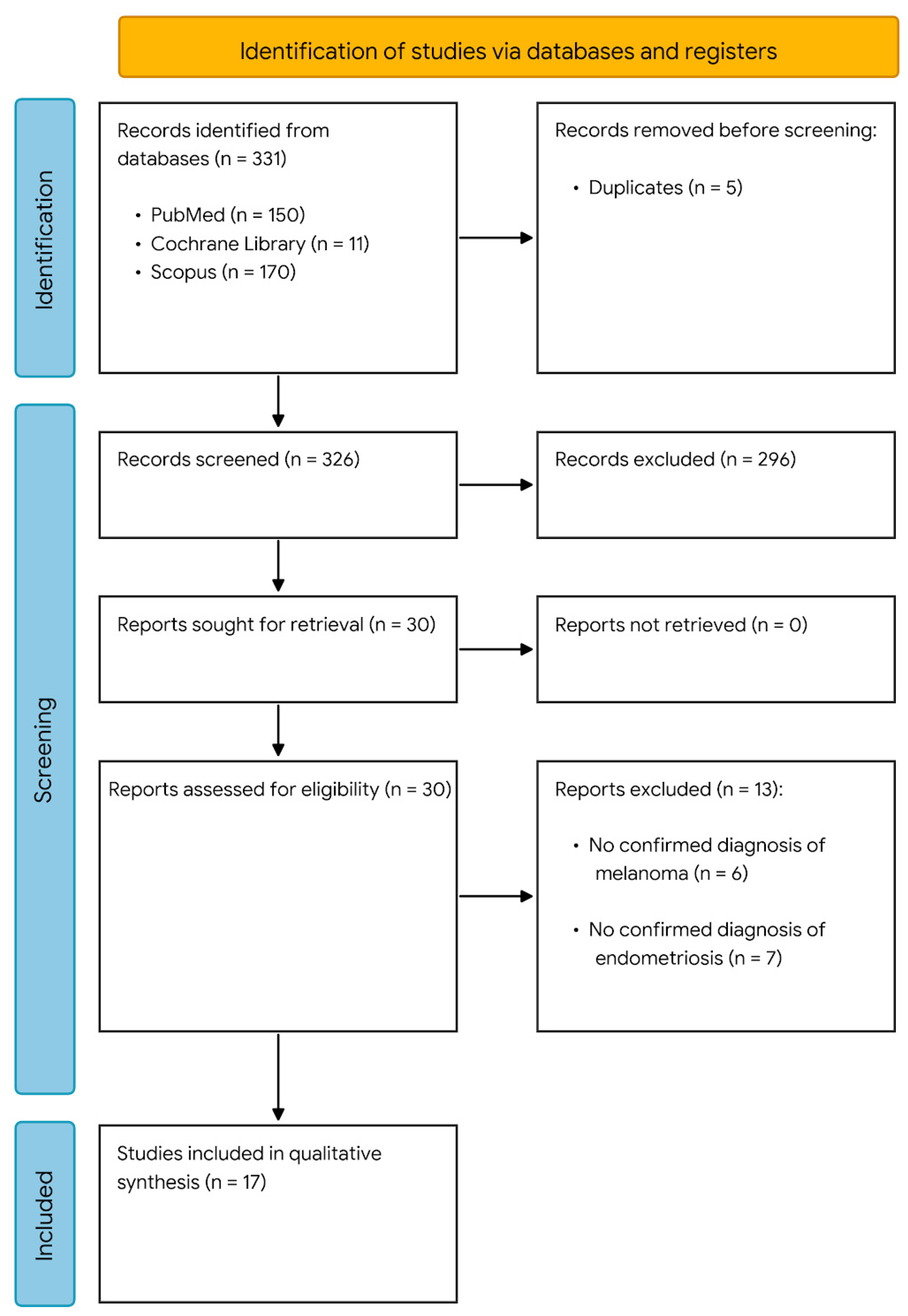
After removal of duplicates, a total of 326 records were identified (
Figure 1).
Following title and abstract screening, 296 articles were excluded. The remaining 30 full-text articles were assessed for eligibility, of which 13 were excluded for not meeting the inclusion criteria. Ultimately, 17 studies were included in the qualitative synthesis. The characteristics of the included studies are summarized in
Table 1. The included studies were categorized into two main groups: (i) genetic and biomolecular studies and (ii) epidemiological investigations.
Genetic and biomolecular studies focused on identifying potential shared mechanisms between endometriosis and melanoma, with particular emphasis on common molecular pathways, as well as hormonal and immunological factors.
Epidemiological studies primarily explored the association between the two conditions from a clinical perspective. Specifically, ten studies evaluated the risk of melanoma occurrence among patients with endometriosis, one study assessed the risk of developing endometriosis in patients with a prior diagnosis of melanoma, and one study investigated the relationship between cutaneous phototype characteristics and the risk of endometriosis.
The epidemiological cohort and case–control studies analyzed in this review demonstrate a persistent, albeit moderate, elevation in melanoma risk among women diagnosed with endometriosis, with hazard ratios consistently hovering between 1.2 and 1.8 [
1,
2]. However, interpreting these findings requires a highly critical approach to confounding variables. A major point of discussion is surveillance (or detection) bias. Women diagnosed with endometriosis often undergo rigorous, long-term medical follow-ups and multi-specialist consultations. This heightened interaction with the healthcare system naturally increases the probability of opportunistic skin examinations, leading to higher detection rates of early-stage, thin melanomas (Breslow thickness less than 1 mm) that might otherwise remain undiagnosed in the general population. This hypothesis is supported by several large-scale database studies where the association between the two conditions weakens significantly when adjusting for the frequency of outpatient clinic visits. Future observational protocols must strictly match cohorts based on healthcare utilization rates to isolate the true biological risk from diagnostic artifacts. Beyond epidemiological correlations, our synthesis highlights a compelling biological convergence that transcends mere statistical association. At the genetic level, genome-wide association studies (GWAS) have identified overlapping susceptibility loci, notably near the CDKN2A/B tumor suppressor locus on chromosome 9p21, a well-established driver of melanoma pathogenesis that also regulates endometrial stromal cell proliferation [
10]. Furthermore, both diseases share alterations in the PI3K/Akt/mTOR and MAPK signaling cascades, often mediated by upstream somatic mutations or epigenetic silencing of PTEN. In endometriosis, these pathway alterations drive benign invasion, migration, and resistance to apoptosis in ectopic endometrial tissues, mirroring the hallmark features of malignant melanocyte transformation. This suggests that while endometriosis is inherently benign, it shares a “pro-invasive” genetic background with cutaneous melanoma, which may explain why certain sub-phenotypes of endometriosis carry a more pronounced risk of subsequent neoplastic development. The local microenvironment plays an equally critical role in promoting both pathologies. Endometriosis is characterized by localized hyperestrogenism and progesterone resistance, which sustain chronic inflammatory states. Interestingly, human melanocytes express functional estrogen receptors (ER-alpha and ER-beta), and estrogen signaling has been shown to modulate melanoma cell proliferation and migration under experimental conditions. This endocrine link is further complicated by shared immune escape mechanisms. In both endometriosis and melanoma, the local tissue microenvironment exhibits marked immune suppression, characterized by decreased cytotoxic activity of natural killer (NK) cells, altered macrophage polarization (M2-like phenotype), and elevated levels of pro-inflammatory cytokines such as IL-6 and TNF-alpha. This systemic and local immune tolerance not only allows ectopic endometrial implants to survive and implant in the peritoneal cavity but may also facilitate the early survival and vascularization of micrometastatic melanoma cells.
2.1. Genetic and Biomolecular Links Between Endometriosis and Melanoma
Genetic correlation analyses suggest that endometriosis shares susceptibility factors with several complex diseases, including melanoma, supporting the involvement of common biological pathways in disease pathogenesis [
3]. However, whether melanoma predisposes to the development of endometriosis remains unclear, as few longitudinal studies have specifically addressed this relationship.
Genome-wide association studies (GWAS) have identified multiple risk loci for both conditions, with approximately 27 loci associated with endometriosis and more than 50 with melanoma [
4,
5,
22]. Genome-wide association studies (GWAS) have successfully mapped numerous risk loci for both conditions. However, a critical distinction must be made between shared genomic loci and shared causal variants. Although identical chromosomal regions harbor susceptibility genes central to both disorders, fine-mapping efforts reveal that the actual association signals are driven by distinct, non-overlapping single-nucleotide polymorphisms (SNPs). This pattern indicates that the genetic intersection stems from a mutual vulnerability within specific regulatory regions or loci, rather than from identical causal nucleotide alterations [
8]. Several genes located within these loci are shared between the two diseases, including CDKN2A, CDKN2B, CDK4, TP53, PTEN, KDR, and FN1, suggesting partially overlapping genetic architectures. Despite this overlap, current evidence indicates that these associations are driven by distinct causal variants rather than a single shared genetic determinant. In particular, loci involving CDKN2A and CDKAL1 have shown the strongest evidence of independent but converging genetic effects, supporting a modest yet significant genetic correlation between melanoma susceptibility and endometriosis [
8].
Crucially, analogous biological paradigms involving the intersection of cell-cycle alterations (TP53, PTEN, CDKN2A) and functional natural killer (NK) cell exhaustion are well-documented in other hormone-driven malignancies, such as endometrial and ovarian carcinomas, as well as in chronic autoimmune inflammatory disorders. In those clinical scenarios, the systematic downregulation of activating NK cell ligands (e.g., MICA/B) synergizes with intrinsic DNA repair deficits to establish a permissive niche for immune evasion. The observation that the endometriosis-melanoma axis shares this precise biomolecular and immunological fingerprint indicates that their co-occurrence is not an isolated anomaly; rather, it reflects a broader, systemic pattern of disrupted immune surveillance and homeostatic failure [
7].
Interestingly, endometriosis does not appear to be genetically associated with pigmentary traits such as skin color, red hair, tanning ability, or nevus count, suggesting that the link between endometriosis and melanoma is unlikely to be mediated by pigmentation-related pathways.
From a pathophysiological perspective, both diseases share key features, including chronic inflammation and hormonal dependence. Endometriosis is characterized by an estrogen-driven inflammatory environment, while the skin itself functions as an endocrine organ capable of local hormone synthesis. Melanocytes express estrogen receptors, and estrogen signaling has been implicated in melanoma development and progression [
23].
Different estrogen receptor subtypes appear to play distinct roles. ERα is predominantly expressed in normal epidermal tissue, whereas ERβ is more highly expressed in melanocytic lesions, including nevi and melanoma. Experimental studies suggest that activation of ERβ inhibit melanoma cell proliferation, potentially through modulation of signaling pathways such as PI3K/Akt [
23]. These findings indicate a complex and context-dependent role of estrogen signaling in both endometriosis and melanoma.
The functional impact of estrogens in melanoma pathogenesis is predominantly dictated by the signaling balance between Erα and Erβ receptor subtypes, which exert opposing effects on tumor progression. While Erβ activation typically mediates oncosuppressive pathways by triggering apoptosis and suppressing epithelial–mesenchymal transition (EMT), alternative signaling via Erα or the G-protein coupled estrogen receptor (GPER) can stimulate melanocyte proliferation and neoangiogenesis under sustained local estradiol concentrations. This dual hormonal axis tightly mirrors the hyperestrogenic microenvironment characteristic of endometriosis, where persistent estrogenic stimulation drives cellular survival and tissue remodeling. This suggests that patient-specific variations in the ERα and ERβ expression ratio might dictate a joint phenotypic susceptibility to both clinical entities [
25,
26]. Immune dysregulation represents an additional shared mechanism. Natural killer (NK) cells, which are central to immune surveillance, exhibit reduced cytotoxic activity in patients with melanoma, and similar alterations have been described in endometriosis [
24]. The compromised cytotoxic efficiency of natural killer (NK) cells provides a compelling immunological link between the two pathologies. Under homeostatic conditions, NK cells clear transformed melanocytes and ectopic endometrial fragments by exocytosing perforin and granzymes to trigger target-cell apoptosis, alongside secreting pro-inflammatory cytokines such as interferon-gamma (IFN-γ). In both melanoma and endometriosis, this surveillance architecture is fundamentally disrupted. The down-regulation of activating receptors (such as NKG2D) paired with an influx of inhibitory signals within the local microenvironment impairs NK cell-mediated clearance, effectively facilitating both the engraftment of endometriotic lesions and the immune evasion of malignant melanocytes [
27]. This impaired immune response contribute to both ectopic endometrial implantation and tumor progression. Overall, current evidence suggests that endometriosis and melanoma share partially overlapping genetic, hormonal, and immunological pathways. However, the observed associations are modest, and the absence of clearly shared causal variants highlights the need for further integrative studies to clarify the underlying mechanisms.
2.2. Phototype Characteristics and the Possible Risk of Endometriosis
A large prospective cohort study conducted within the Nurses’ Health Study II evaluated the association between pigmentary traits and the risk of endometriosis in over 100,000 women. The authors reported that light pigmentation, increased nevus count, and a family history of melanoma were associated with a higher risk of endometriosis, suggesting a potential link between melanocytic characteristics and the disease [
10]. When evaluating these findings, the inherently observational design of the NHS II cohort must be kept in mind. While the substantial sample size lends considerable statistical weight to the epidemiological trends, such data cannot establish direct causality. The noted associations between pigmentary traits and endometriosis risk could still be driven by unmeasured confounding factors or complex gene-environment interactions, meaning these correlations should be viewed as hypothesis-generating rather than definitive proof of a shared etiology [
10].
Interestingly, while natural hair color was not independently associated with endometriosis risk, a differential effect was observed among women with red hair depending on oral contraceptive use. Specifically, the risk of endometriosis was increased in red-haired women who used oral contraceptives, whereas it appeared reduced in those who had never used them. This interaction highlights the role of hormonal modulation, particularly estrogen signaling, in influencing disease expression in genetically predisposed individuals.
From a mechanistic perspective, these findings support the hypothesis that melanocytic and endocrine pathways may intersect in the pathogenesis of endometriosis. Melanocytes express estrogen receptors, and hormonal signaling influence both melanogenesis and ectopic endometrial growth. However, it remains unclear whether pigmentary traits represent causal factors or surrogate markers of underlying genetic or hormonal susceptibility.
Overall, while the association between pigmentary characteristics and endometriosis is intriguing, current evidence remains limited and does not allow definitive conclusions. Further studies are needed to clarify whether these phenotypic traits reflect shared biological mechanisms linking endometriosis and melanoma.
2.3. Risk of Melanoma Occurrence in Patients with Endometriosis
Several epidemiological studies have investigated the potential association between endometriosis and the risk of melanoma, with overall findings suggesting a modest but consistent increase in risk.
Large prospective cohort studies provide the strongest evidence. In a cohort of nearly 100,000 women, Kvaskoff et al. reported a significantly increased risk of cutaneous melanoma among women with a history of endometriosis or uterine fibroids [
12,
28]. This association was further confirmed in an extended follow-up of the same population, with a stronger link observed for cutaneous melanoma compared to non-melanoma skin cancers [
10]. Notably, environmental exposures and treatment modalities for endometriosis did not appear to explain this association, suggesting the involvement of underlying biological mechanisms [
14].
Additional support comes from large population-based studies. Saraswat et al., in a nationwide cohort including over 280,000 women, demonstrated a significantly increased risk of melanoma in patients with surgically confirmed endometriosis, with hazard ratios consistently ranging between 1.5 and 1.8 across different comparison groups [
17]. Similar trends have been reported in other cohort and case–control studies, further reinforcing the hypothesis of an epidemiological association between the two conditions [
16].
Evidence from smaller studies also suggests a potential link between endometriosis and melanoma-related phenotypes. In particular, an increased prevalence of dysplastic nevi and a higher frequency of family history of melanoma have been observed in women with endometriosis, especially in younger populations [
13,
15]. These findings support the hypothesis that shared genetic susceptibility may contribute to the observed association.
However, not all studies have confirmed this relationship. Some case–control studies failed to demonstrate a significant increase in melanoma risk among women with endometriosis [
18,
29]. Similarly, studies relying on self-reported diagnoses have reported null findings, although their reliability is limited by potential misclassification bias [
19].
Finally, emerging evidence suggests that melanoma risk be higher in women with infertility, particularly those with endometriosis-related infertility, and that dysplastic nevi—known precursors of melanoma—may be more prevalent in this population [
20].
Overall, the available evidence suggests a modest but potentially meaningful association between endometriosis and melanoma. However, heterogeneity in study design, population characteristics, and diagnostic criteria limits the ability to draw definitive conclusions. Further large-scale, prospective studies integrating genetic, hormonal, and environmental data are needed to clarify whether this relationship reflects shared risk factors or a true causal link. A pivotal methodological factor that cloud this epidemiological relationship is surveillance bias (or detection bias). Women managed for chronic conditions like endometriosis inherently navigate healthcare systems more frequently and undergo regular specialist evaluations. This heightened medical surveillance logically increases the likelihood that suspicious cutaneous lesions are flagged, examined, and biopsied. Consequently, the modestly elevated risk of melanoma observed in cohort studies might partially reflect a higher diagnosis rate driven by medical proximity rather than a purely biological phenomenon [
30]. A rigorous interpretation of the current literature demands caution, as the reported relative risks and hazard ratios consistently remain below the conservative threshold of 2.0. In observational epidemiology, associations of this magnitude are inherently vulnerable to residual confounding. This interpretative challenge is further compounded by the diagnostic heterogeneity across the tracked cohorts; while certain investigations relied strictly on surgically and histologically verified endometriosis, others utilized radiological criteria or unrefined registry discharge codes, which lack granular detail regarding specific disease phenotypes (e.g., deep endometriosis versus superficial peritoneal lesions) or precise melanoma staging [
21].
2.4. Risk of Endometriosis Occurrence Among Melanoma Patients
Evidence on the risk of developing endometriosis among women with a prior diagnosis of melanoma is limited but suggests an association. In an observational study including 7559 female university students, women with a history of melanoma were significantly more likely to report reproductive-related surgical procedures, including surgery for endometriosis, compared with both women with non-melanoma skin cancers and those without a history of cancer.
Specifically, the odds ratio for prior endometriosis surgery was 3.2 (95% CI 1.0–10.1) when compared with women diagnosed with non-melanoma skin cancer and 3.9 (95% CI 1.2–12.4) when compared with women without cancer, suggesting a three- to fourfold increased likelihood of endometriosis in this population.
Interestingly, no significant difference in endometriosis risk was observed between melanoma patients with and without dysplastic nevi, indicating that the association not be mediated by pigmentary or melanocytic traits.
However, these findings should be interpreted with caution, as the study was based on a relatively small sample and partly relied on self-reported data, introducing potential recall and classification bias. Despite these limitations, the results raise the hypothesis that women with melanoma have an increased predisposition to hormonally mediated gynecological conditions, such as endometriosis.
Overall, the available evidence on the reverse association between melanoma and endometriosis remains scarce, and further well-designed prospective studies are needed to confirm this relationship and to clarify the underlying biological mechanisms [
21].
2.5. Abdominal Wall Endometriosis and Melanoma
Abdominal wall endometriosis (AWE) is a rare form of extrapelvic endometriosis characterized by the presence of functional endometrial tissue within the subcutaneous or muscular layers of the abdominal wall, typically arising in surgical scars following cesarean section or other gynecological procedures. Its estimated incidence ranges from 0.03% to 0.4% among women with prior cesarean deliveries [
31,
32]. Clinically, AWE presents as a painful, cyclic mass near a surgical scar, and diagnosis is often delayed due to nonspecific symptoms and frequent misinterpretation as other soft tissue lesions [
33].
Histologically, AWE is characterized by endometrial glands and stroma within a fibrotic and chronically inflamed microenvironment, reflecting the estrogen-dependent and inflammatory nature of the disease [
33]. Chronic inflammation, oxidative stress, and angiogenesis—hallmarks of endometriosis—have been implicated in processes that contribute to tumorigenesis [
34,
35].
Although the coexistence of AWE and melanoma is extremely rare, this association raises the possibility of shared pathophysiological mechanisms. Both conditions are influenced by hormonal signaling, particularly estrogen pathways, and by immune modulation. In addition, alterations in genes involved in cell-cycle regulation and apoptosis, such as TP53, PTEN, and CDKN2A, have been described in both endometriosis and melanoma [
9,
36,
37].
However, the clinical significance of this association remains uncertain. Current evidence is limited to theoretical considerations and indirect observations, and no direct epidemiological data support a causal relationship between AWE and melanoma. Therefore, while the hypothesis of a shared biological background is intriguing, it should be interpreted with caution.
Further studies are needed to clarify whether extra pelvic forms of endometriosis, such as AWE, represent a distinct biological subset with potential implications for oncological risk, or whether the observed associations reflect broader systemic mechanisms linking endometriosis and malignancy.
3. Methods
This review was conducted in accordance with the Preferred Reporting Items for Systematic Reviews and Meta-Analyses (PRISMA) guidelines to ensure methodological transparency and reproducibility [
38], the checklist of which can be found in the
Supplementary Material. The review protocol was registered on the Open Science Framework (OSF) (DOI:10.17605/OSF.IO/BA5ZD) prior to the conduct of this review. The registration record is publicly available at
https://osf.io/x3vjt (accessed on 20 June 2026).
A comprehensive literature search was performed using PubMed/MEDLINE, Scopus, and the Cochrane Library to identify relevant studies published in English up to 1 February 2025. The search strategy combined Medical Subject Headings (MeSH) and free-text terms, including “melanoma,” “melanocytic carcinoma,” “endometriosis,” “endometrioma,” and “adenomyosis,” using Boolean operators (“AND,” “OR”) to optimize sensitivity and specificity.
It is worth clarifying that the initial retrieval of 326 records did not result from a restrictive search limited to the core terms alone. Instead, a comprehensive search string was deployed, utilizing Medical Subject Headings (MeSH) and free-text expansions—such as ‘adenomyosis’, ‘endometrioma’, and ‘melanocytic carcinoma’—linked via the Boolean operator ‘OR’. This expansive approach was intentionally designed to cast a wide net across the indexed literature, minimizing the risk of omitting relevant downstream analyses prior to manual screening.
To ensure methodological rigor, study eligibility was systematically defined according to the Population, Exposure, Outcome, and Study Design (PECO) framework. Specifically, we included peer-reviewed observational studies—comprising both prospective and retrospective cohort and case–control designs—as well as randomized controlled trials involving adult female populations evaluated within gynecological or oncological settings. Eligible studies were required to investigate patients with a clinically, radiologically, or histopathologically confirmed diagnosis of endometriosis (encompassing superficial peritoneal implants, ovarian endometriomas, or deep infiltrating disease) and report on histologically verified primary cutaneous melanoma using quantitative risk metrics, such as Hazard Ratios, Odds Ratios, or Standardized Incidence Ratios accompanied by 95% Confidence Intervals.
Conversely, we excluded studies that relied solely on self-reported survey data without medical record validation for either condition, as well as those focusing exclusively on non-cutaneous melanoma phenotypes, such as ocular or mucosal variants, or metastatic lesions of unknown primary origin. Non-original research records—including narrative or systematic reviews lacking primary data, meta-analyses, conference proceedings, editorials, letters to the editor, case reports, and small case series with fewer than ten patients—were likewise omitted. In cases involving overlapping patient cohorts where duplicate datasets provided no additional longitudinal follow-up or updated risk estimates, only the investigation with the largest sample size or most comprehensive follow-up was retained. Lastly, non-English publications and studies with completely unavailable full texts despite author contact were excluded. Notably, the identification and removal of duplicate records across databases was managed as a standard PRISMA screening pipeline step prior to eligibility evaluation, rather than serving as an intrinsic study exclusion criterion. Two independent reviewers screened titles and abstracts, followed by full-text evaluation of potentially eligible studies. Disagreements were resolved by consensus.
Data extraction included study design, population characteristics, diagnostic criteria, and main outcomes related to the association between endometriosis and melanoma.
Due to heterogeneity in study design, populations, and outcome measures, a quantitative meta-analysis was not performed, and findings were synthesized qualitatively.
As this study is based on previously published data, ethical approval and informed consent were not required.
4. Conclusions
Current evidence suggests that endometriosis and melanoma may share partially overlapping biological pathways, including genetic susceptibility, hormonal regulation, and immune dysregulation. Genome-wide and biomolecular investigations have highlighted the involvement of identical genes and shared molecular pathways specifically those regulatory axes encompassing TP53, PTEN, and CDKN2A rather than the presence of identical genetic variants or mutations. This evidence points toward a modest genetic correlation. In addition, both diseases appear to be influenced by estrogen signaling and chronic inflammation, which may contribute to dysregulated cellular proliferation and immune responses.
Epidemiological findings indicate a slight but consistent increase in melanoma risk among women with endometriosis, particularly for cutaneous melanoma and in younger populations or those with a family history of dysplastic nevi. Conversely, limited evidence suggests a possible reverse association, with a higher prevalence of endometriosis among women with melanoma. However, heterogeneity across studies, including differences in diagnostic criteria and study design, limits the ability to draw definitive conclusions.
Overall, the association between endometriosis and melanoma appears biologically plausible but remains unproven. In conclusion, the epidemiological and biological association between endometriosis and melanoma remains unconvincing and largely speculative. The current data, while hinting at partially overlapping molecular axes, fail to provide definitive clarity regarding a true causal or functional relationship, primarily due to the methodological constraints of observational designs and pervasive detection biases. Consequently, rather than replicating descriptive epidemiological data, future research must implement longitudinal frameworks and multiomic approaches capable of dissecting confounding factors to determine whether a genuine biological synergy exists.



 ,
,