


Abstract
The application of natural polysaccharides and their sulfated derivatives have already been successfully implemented in the pharmaceutical and food industries, in particular. The present study is concerned with modifying a predominant polysaccharide in the composition of spruce wood, galactoglucomannan (GGM), by sulfation via a urea-sulfamic acid complex in a 1,4-dioxane medium. By varying the sulfation process duration from 30 to 180 min, six novel GGM sulfate samples with different degrees of substitution (DS) of 0.4–1.2 were obtained and studied with a combination of modern physicochemical methods: elemental analysis, Fourier transform infrared (FTIR) spectroscopy, and gel permeation chromatography (GPC). It has been revealed that the sulfation of GGM proceeds without degradation of the main polymer chain, as evidenced by the shift in the main peak toward the high-molecular-weight region in the GPC curves. Moreover, modification of the polysaccharide leads to a significant transformation of the molecular conformation from a dense sphere to a random coil (α from 0.30 to 0.76). Furthermore, it has been determined that sulfate-substituted groups of the GGM tended to decrease the scavenging capacity of the 1,1-diphenyl-2-picrylhydrazyl (DPPH) free radicals. However, the 2,2′-azino-bis(3-ethylbenzothiazoline-6-sulfonic acid) diammonium salt (ABTS) assay showed an increase in the free radical inhibitory capacity of sulfated polysaccharides. This is attributed to the structural and conformational properties of the polysaccharide sulfate derivatives. The maximum anticoagulant activity (ACA) of sulfated GGM (SGGM) is 21.19 ± 2.89 IU/mg and increases with increasing sulfation duration.
1. Introduction
The development and improvement of modern drugs is a vast, constantly progressing field, aimed at increasing the therapeutic efficiency of pharmaceutical agents by simultaneously reducing the active substance dosage and minimizing side effects [1,2]. The advances in drug system development are driven, primarily, by the pursuit of implementing substances with a natural origin. The successful application of this approach allows the use of materials already characterized by the certain inherited biological activity depending on their source [2]. Among the numerous branches, one of the innovative paths is concerned with the application of biopolymers in pharmaceutical research and development studies. In the nearest prospect, biopolymer substances could serve as the basis for both controlled drug carriers and encapsulation materials, as well as for biologically active additives [3,4,5].
Within the framework of the integrated biomass processing strategy, the use of irregular polysaccharides for the production of high-tech, value-added products is of particular importance. Given the predominance of coniferous forests (spruce, pine, and fir) in European ecosystems, the isolation and modification of coniferous polysaccharides—specifically glucomannans and galactoglucomannans—represents an extremely promising and relevant field. Polysaccharides garner great interest due to their unique properties, which facilitate their application in various medical fields [6]. Their inherent biocompatibility minimizes the risk of adverse and immune reactions, a critical factor in the development of active pharmaceutical forms [3,7,8,9,10,11,12]. However, the complex and diverse spatial structure inherent in polysaccharides is closely intertwined with their biological activity and physicochemical properties.
Currently, sulfated polysaccharides have attracted widespread attention due to their unique properties, such as biocompatibility, biodegradability, non-toxicity, renewability, tunability, swelling capacity, and inertness [13]. Despite the diversity of natural sulfated polysaccharides [13,14] found in various living organisms, chemically synthesized derivatives often possess novel or enhanced properties compared to their natural counterparts [10]. Consequently, chemical modification represents a key approach that not only diversifies the types of biopolymers but also expands their range of applications [8,9,15,16]. Numerous studies have already demonstrated the production of modified polysaccharide derivatives with targeted properties [17,18]. For example, some chemically sulfated polysaccharides exhibit superior anticoagulant activity compared to traditional anticoagulants such as heparin [10]. Furthermore, it has been shown that sulfated polysaccharides possess the anticoagulant activity essential for both antithrombotic agents and clot-resistant biomaterials [14,19].
Generally, sulfated polysaccharides exhibit enhanced water solubility and superior biological activity, primarily due to controlled changes in chain conformation [19,20]. However, the relationship between the structure and biological activity of sulfated polysaccharides has not yet been clearly established. Therefore, it is essential to conduct comprehensive research aimed at obtaining novel sulfated polysaccharides and characterizing their biological activity. Such efforts are pivotal for facilitating the development of novel substances that could significantly advance the pharmaceutical and medical fields in the future.
Thus, this study aims to synthesize and characterize novel sulfated derivatives of spruce (Picea abies) galactoglucomannan with a higher density of introduced sulfate groups. Furthermore, to evaluate biomedical potential, including the assessment of the bioactivity of these highly sulfated polysaccharide derivatives, was investigated—in particular, ability to scavenge free radicals using DPPH/ABTS antioxidant assays and effects on blood coagulation in vitro with human blood/plasma clotting tests.
2. Materials and Methods
2.1. Raw Materials
A sawdust fraction (2.0–5.0 mm) of Picea abies, grown in the Krasnoyarsk Territory (Russian Federation) was used as the starting material. The chemical composition of the spruce wood was determined as follows (wt.%): cellulose—44.4, lignin—30.6, hemicelluloses—22.6, extractives—1.8, ash—0.6. The isolation of GGM was performed according to a previously described method [21].
2.2. GGM Sulfation
The obtained GGM was sulfated with sulfamic acid (purity > 99.9%; SCRS, Krasnoyarsk, Russia) in 1,4-dioxane (p.a., ECOS-1, Moscow, Russia) solvent in the presence of urea (p.a., SCRS, Krasnoyarsk, Russia) as organic base under different conditions. In a procedure, 50 mL of 1,4-dioxane, 3.1 g of sulfamic acid, and 1.9 g of urea were placed into a three-necked flask equipped with a thermometer and a mechanical stirrer. The resulting mixture was heated to 50 °C under vigorous stirring, followed by the addition of 1 g of air-dried GGM. The temperature was then raised to 85 °C and stirred at this temperature for 3 h, taking samples every 30 min. Upon completion, the solvent was decanted, and the residue was dissolved in 25 mL of distilled water. Excess sulfamic acid was neutralized with a 25% aqueous ammonia solution (p.a., SCRS, Krasnoyarsk, Russia) until a neutral pH ~ 7.0 was reached. To remove unreacted compounds and low-molecular-weight substances, the products were dialyzed against distilled water for 24 h using a dialysis membrane (MF-5030-46, MFPI, Seguin, TX, USA) with a pore size of 3.5 kDa (water was changed every hour). The resulting SGGM solutions were dried in an oven at 60 °C to obtain film structures, which were subsequently ground into a fine powder for physicochemical and biological studies.
2.3. Physicochemical Study of SGGM Samples
2.3.1. Elemental Analysis
Elemental analysis of the sulfated samples was performed using a Vario EL cube analyzer (Elementar, Langenselbold, Germany) in CHNS mode. The procedure involved sample combustion in an oxygen-rich atmosphere, followed by gas adsorption separation and detection via a thermal conductivity detector. The DS was calculated according to the methodology described in studies [22,23].
2.3.2. Gel-Permeation Chromatograph
The molecular weight characteristics (weight-average molecular weight Mw and polydispersity PDI) of the initial GGM and SGGM samples were determined by GPC using an Agilent 1260 Infinity II Multi-Detector GPC/SEC System chromatograph (Agilent Technologies, Santa Clara, CA, USA) with a refractive detector. The separation was performed using two Agilent PL Aquagel-OH Mixed-M and one Agilent PL Aquagel-OH-30 columns, with a 0.1 M NaNO3 solution combined with NaN3 in deionized water as the eluent. The calibration was performed using Agilent EasiVial PEG/PEO polyethylene glycol standards (Agilent, Santa Clara, CA, USA). The flow rate was 1 mL/min and the sample volume was 100 µL. The analysis was carried out similarly to work [16].
2.3.3. FTIR-Spectroscopy
FTIR spectra of the initial and SGGM samples were recorded in the wavelength range of 4000–400 cm−1 using a Shimadzu IRTracer-100 FTIR spectrometer (Shimadzu, Kyoto, Japan). For analysis, the samples were prepared as KBr pellets by mixing 2 mg of the polymer with 1000 mg of KBr and subsequent pressing. Spectral data were processed using OPUS software version 5.0 (Bruker, Billerica, MA, USA).
2.4. Study of Biological Activity
2.4.1. Antioxidant Activity
The DPPH radical scavenging activity of the SGGM samples was determined according to a previously described method [21]. Briefly, to evaluate the influence of the degree of substitution on antioxidant activity, SGGM-60, 150, and 180 samples were selected. Samples of the GGM and SGGM at different concentrations (0.5; 2, and 5 mg/mL) were mixed with 0.2 mM DPPH solution (purity > 99.9%, Tokyo Chemical Ind. Co. Ltd., Tokyo, Japan) and incubated at 25 °C in the dark for 30 min. Then, the absorbance was measured at 517 nm using an Ecoview UV 6900 spectrophotometer (Shanghai Mapada Instruments Co. Ltd., Shanghai, China). The DPPH radical scavenging activity was calculated using Equation (1):
where AC is the absorption of the DPPH solution without a sample, AS is the absorption of the test sample mixed with the DPPH solution, and AB is the absorption of the sample without the DPPH solution.
The ABTS radical scavenging activity of GGM and SGGM was assessed using a modified method based on similar studies [24,25]. The ABTS radical cation (ABTS•+) was obtained by mixing 7 mM ABTS (purity > 99.9%, Tokyo Chemical Ind. Co. ltd., Tokyo, Japan) and 2.45 mM aqueous potassium persulfate (purity > 99.9%, Chimmed, Moscow, Russia) solution, followed by incubation for 12 h in the dark at 4 °C. After incubation, the solution was diluted with deionized water to an absorbance of 0.700 ± 0.002 at 734 nm [26]. Then, 1 mL of the polysaccharide sample at various concentrations (0.1–2 mg/mL) was mixed with 3 mL of the diluted ABTS solution. The mixture was incubated for 10 min at 20 °C, and the absorbance was measured at 734 nm against distilled water. The control (Ac) contained all reagents except the polysaccharide; As represents the absorbance of the reaction mixture of the polysaccharide with ABTS, and AB is the reaction result of the polysaccharide without ABTS. The ABTS radical scavenging activity was calculated using Equation (2):
2.4.2. Anticoagulant Activity
In the present study the 12 patients’ blood samples were examined (all healthy volunteers signed an “Informed voluntary consent for medical examination and donation of blood and (or) its components” at the National Research Center for Hematology, Moscow, Russia; sample collection period: 5 June 2025–24 June 2025). The blood samples were collected from the cubital vein into a test tube UNIVAC (OOO Eyton, Moscow, Russia; article number 13100.5.4.CN3.2) and mixed up with 3.2 wt.% trisodium citrate solution (9:1 ratio). The platelet-pure human plasma (PPP) was prepared by centrifugation of the stabilized blood using ELMI CM-6M (ELMI, Riga, Latvia) for 20 min at 950 g. The native and sulfated GGM samples were solubilized in an isotonic sodium chloride solution 0.9% NaCl and its impact on blood recalcification time (BRT) and activated partial thromboplastin time (APTT) were tested.
To determine the blood and platelet-poor plasma clotting time the semiautomatic coagulometers (EMKO LLC, Moscow, Russia) equipped with six 2-channel APG2-01 and one 4-channel APG4-03-P, as the reagent of NPO Renam (Moscow, Russia), were used. All of the measurements were performed according to standard operation procedures (SOP No. LPiFG-A-001/01 dated 20 December 2023) of the Laboratory of pathology and pharmacology of Hemostasis of the Federal State Budgetary Institution “National Medical Research Center of Hematology” of the Ministry of Health of the Russian Federation.
The BRT was determined according to the method described in the article [27]. The 0.05 mL sample of blood containing GGM and SGGM with concentration varying in the range 0.0033–3.33 mg/mL was incubated for 1 min at 37 °C. Then, the 0.05 mL of 0.02 M CaCl2 solution was added and the clotting time was recorded. The 2BRT concentrations values were determined for the investigated samples, characterized by doubled blood coagulation time compared to the control sample (blood with 0.9% NaCl; 0 mg/mL).
The APTT test was performed according to the instructions supplied for the PG-7/1 kit (NPO Renam, Moscow, Russia): to the 0.05 mL of plasma sample, containing GGM and SGGM with concentration varied in range 0.0047–0.93 mg/mL, the 0.05 mL of APTT-reagent was added. After the 3 min of incubation at 37 °C the 0.05 mL of 0.025 M CaCl2 was added and plasma clotting time was recorded. The 2APTT concentration values for investigated samples characterized by doubled blood coagulation time compared to the control sample (plasma with 0.9% NaCl; 0 mg/mL). As a result of plotting the curves of plasma coagulation time in the APTT test on the concentration of GGM/SGGM or unfractionated heparin (UFH; Heparin 5000 IU/mL, Belmedpreparaty RUP, Minsk, Belarus), their 2APTT concentrations were determined and ACA (IU/mg) = 2APTTUFH (IU/mL)/2APTTGGM/SGGM (mg/mL) were calculated.
Statistical processing of the results was performed using Primer of Biostatistics 4.03 (The McGraw-Hill Companies, Inc., Columbus, OH, USA) and Statistica 8.0 (StatSoft, Inc., Tulsa, OK, USA). To compare non-normally distributed data (obtained in the BRT test), the non-parametric Mann–Whitney U-test criteria (for unrelated samples) and the Wilcoxon signed-rank test (for related samples) were used. To compare normally distributed data (obtained in the APTT test), the parametric Student t-criteria (for unrelated samples) and Student paired t-test criteria (for related samples) were used.
3. Results
3.1. Physicochemical Characteristics of SGGM Samples
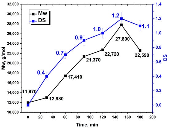
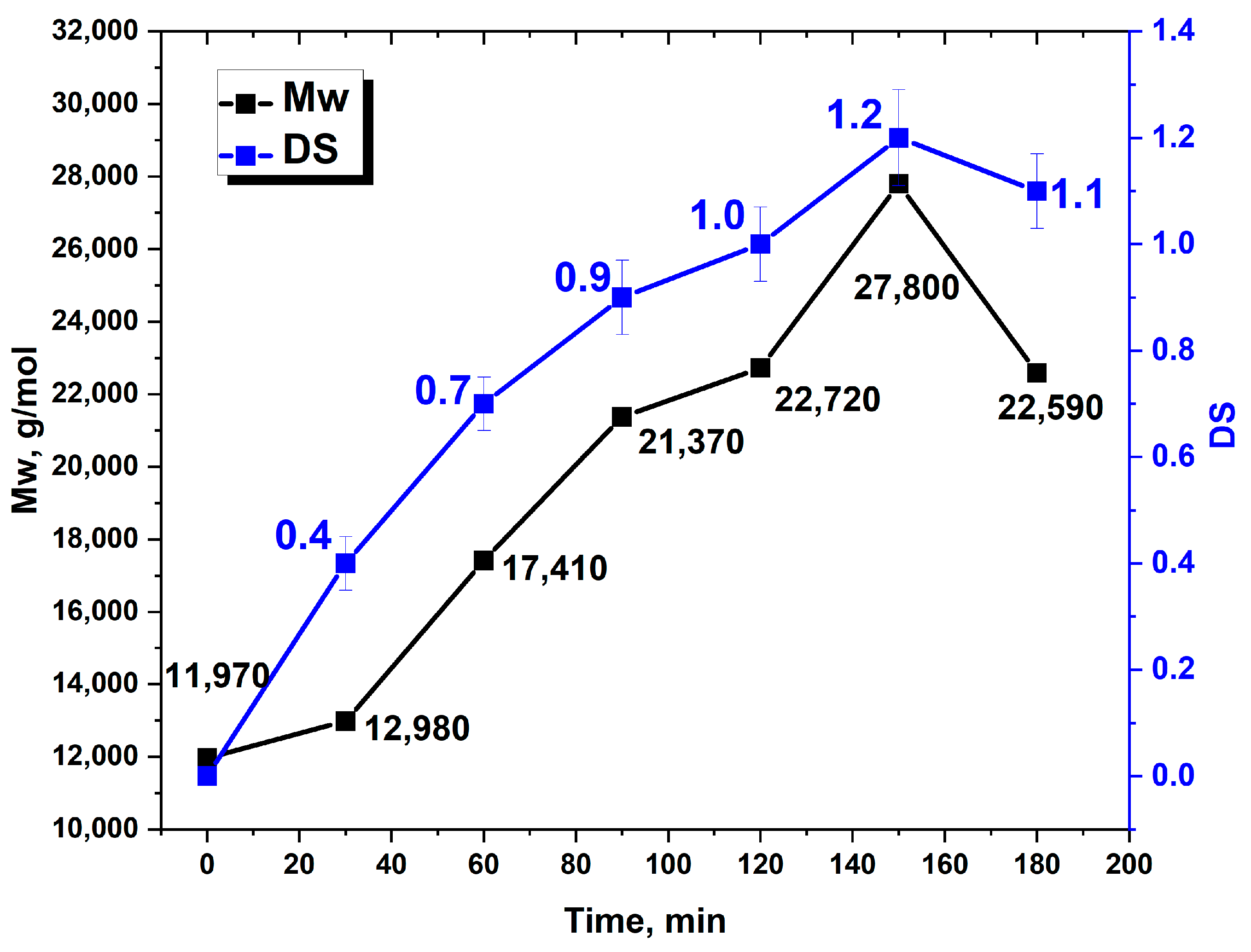
As a result of chemical modification with varying process duration, six samples of SGGM were obtained. The sulfated samples were analyzed by the elemental analysis and gel permeation chromatography, aimed to determine the impact of synthesis conditions on the DS and molecular weight (Mw) depicted in Figure 1.
Figure 1.
The relationship between the duration of the galactoglucomannan sulfation (min), degree of substitution (DS) and weight-average molecular weight (Mw) of the product.
The data demonstrated that the main stage of sulfation occurs within the first 120 min, as evidenced by a proportional increase in the DS to 1.0 and Mw to 22,720 g/mol. Meanwhile, a plateau at 90–120 min range likely caused by delayed formation of the sulfate groups, followed by a slight increase in molecular weight, was observed. As the reaction time increases up to 150 min, the maximum values of the DS to 1.2 and Mw to 27,800 g/mol were achieved. Further increase in the sulfation duration up to 180 min leads to a decrease in the sulfated product with DS to 1.1 and Mw to 22,590 g/mol. Such peculiar pattern could be caused by intensification of the hydrolysis with formation of off-target products [15].
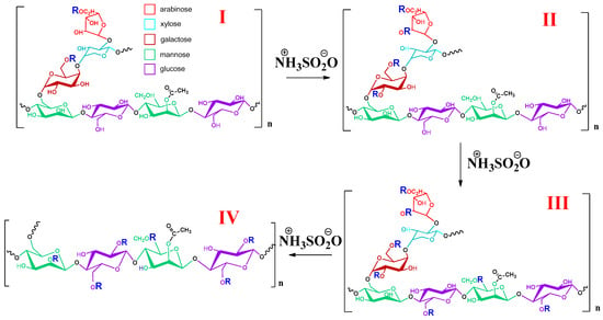
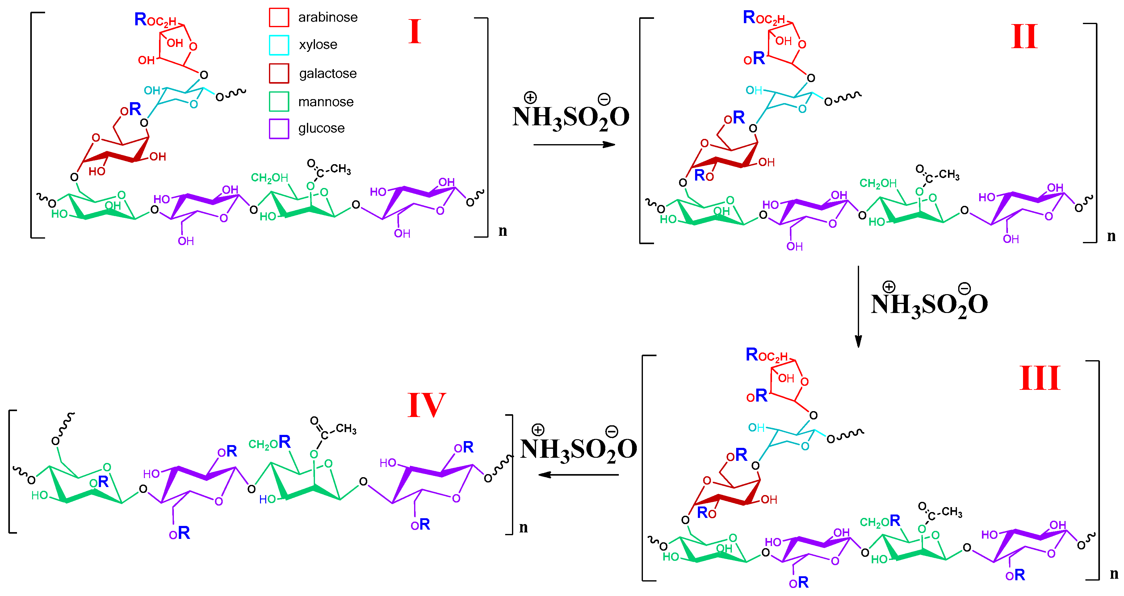
Based on the obtained data, we propose a hypothetical reaction mechanism for the GGM sulfation by sulfamic acid–urea system in 1,4-dioxane medium, which includes four main stages, represented in Figure 2. According to the proposed mechanism, the first stage (I) is initialized by addition of sulfate group at the side links C-5/C-6 position due to the favorable position of hydroxyl groups and absence of the steric hindrance [14]. Further, the second stage (II) is accompanied by substitution of free C-2 and C-4 positions and, accordingly, followed by the macromolecular unfolding due to the electrostatic repulsion of similar charged sulfate groups. In turn, the third stage (III) is characterized by release of the main-chain hydroxyl groups available for substitution. This phenomenon results in obtaining a product with a high DS of 1.2 at 150 min. Nevertheless, a further increase in the sulfation duration to 180 min intensifies the hydrolysis of the side (1 → 6) glycosidic bonds followed by the cleavage of sulfated monosaccharide units (depicted as the fourth stage IV). However, further structural studies are required, which our research team plans to conduct in the near future.
Figure 2.
The hypothetical reaction mechanism of the GGM sulfation by the “sulfamic acid–urea” complex in a 1,4-dioxane medium (R–SO3NH4) medium with a process duration from 0 to 180 min. The structural representations: (I) 5,6-O-side chain sulfated GGM; (II) 2,4,5,6-O-side chain sulfated GGM; (III) 2,4,5,6-O-side chain and 6-O-main chain sulfated GGM; (IV) 3,6-O-main chain sulfated GGM with cleaved (1 → 6) side chain.
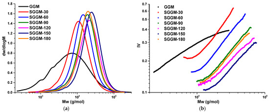
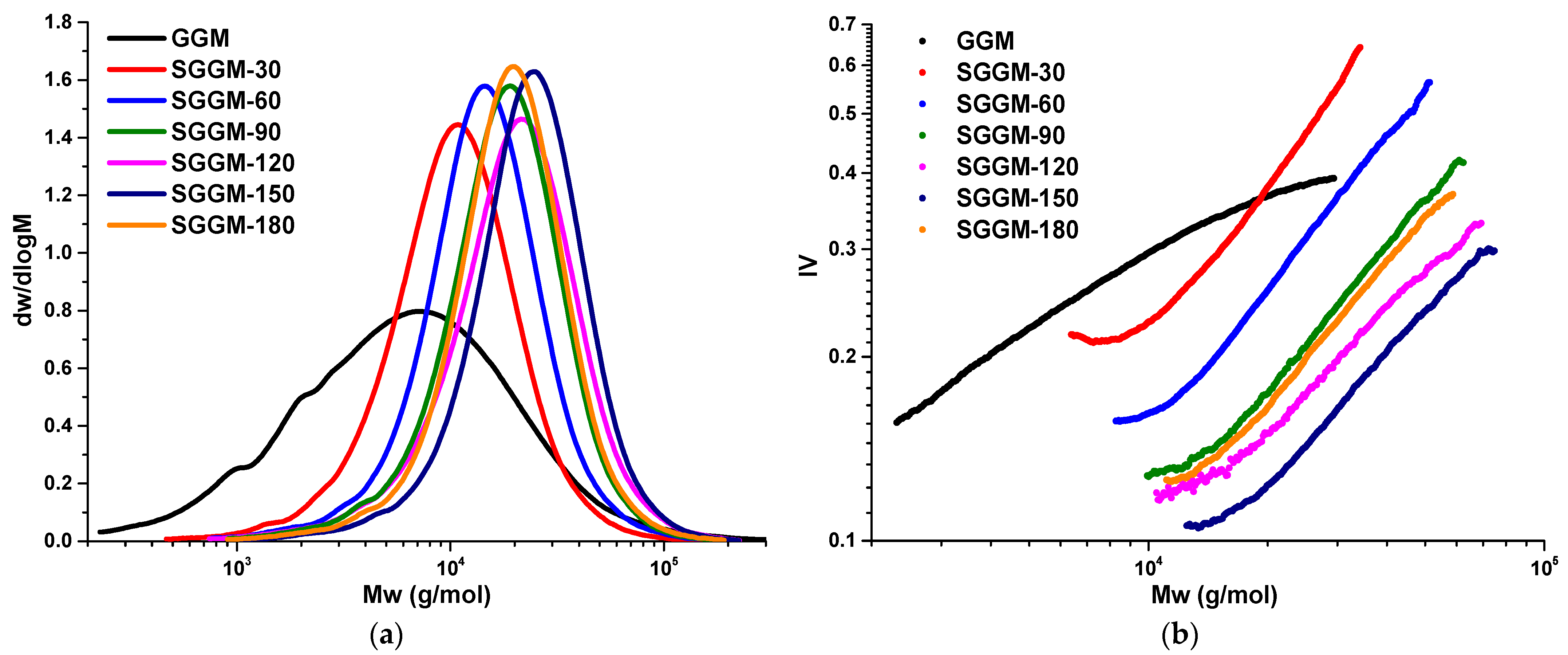
The chain conformation of sulfated polysaccharides is an equally vital factor impacting the derivative’s biological activity [28]. It depends on both native and modified structural characteristics such as the chain length, type and amount of branching, the resulting DS and specific reagents used for sulfation [29,30]. To determine the molecular weight characteristics (Mw and PDI) and K and α coefficients for the Mark–Houwink–Sakurada (MHS) equation characterizing the branching and conformation of polymer molecules, respectively [29,31], the GPC was performed (Figure 3a,b; Table 1).
Figure 3.
The profiles of native GGM and SGGM samples: (a) molecular weight distribution (MWD), (b) curves based on the parameters of the equation of the Mark–Houwink–Sakurada.
Table 1.
Molecular weight characteristics (Mw and PDI) and parameters of the MHS equation (K and α) determined for GGM’s sulfates.
The results allow to announce that the native polysaccharide has a fairly dense spherical shape in an aqueous solution, as is indicated by the α coefficient equal to 0.30 [32]. In addition, the broad, non-uniform molecular weight distribution of the native sample points to the heterogeneity of the GGM molecules (Figure 3a). In turn, the shift in the MWD curves to the high molecular weight region (Figure 3a), caused by the growth of Mw (Table 1), proves that the incorporation of sulfate groups into polysaccharides structure proceeds without the significance of the main chain hydrolysis. It was observed that the profile of the MWD curves narrows due to the removal of low-molecular-weight fragments during dialysis. This results in a significant decrease in the PDI and, accordingly, points to the isolation of polysaccharides with a homogeneous structure.
To conclude, the MHS curve analysis allows to evaluate that, as a result of GGM’s sulfation, a branched-type polysaccharide structure was obtained with conformation of random coil (α value lies in 0.66–0.76 range). Such a type of structure is characterized by better water solubility compared to non-modified samples, which impacts positively on the biological activity [33].
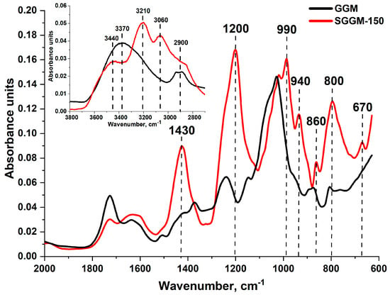
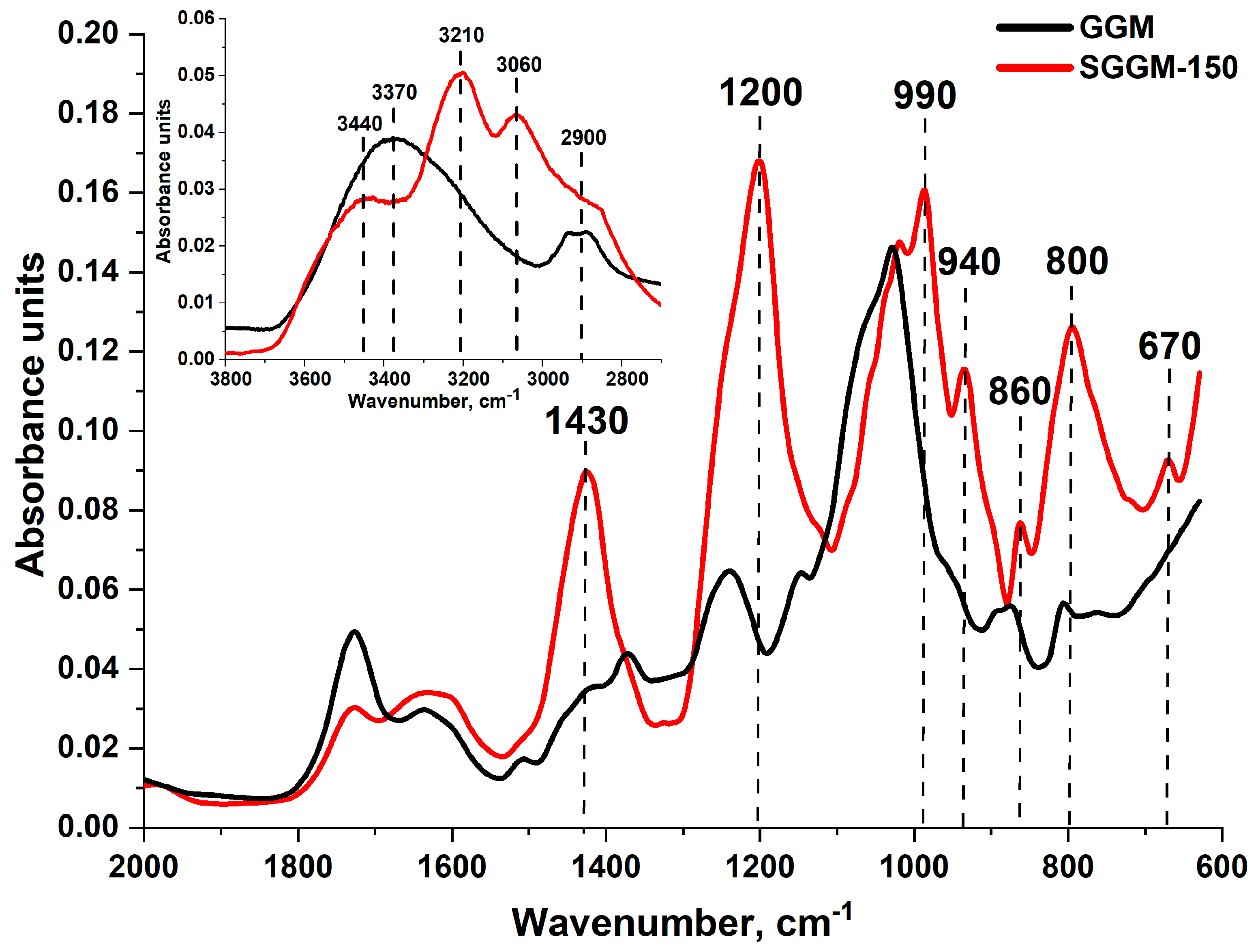
The FTIR spectra of the native GGM and its sulfate with maxima DS are represented in Figure 4 and are characterized by the following specific features. As it was expected, the FTIR spectra of native GGM depicts a broad region of the O–H (~3370 cm−1) and C–H (2900 cm−1) bonds stretching vibrations. This region, as a consequence of GGM’s modification, is altered for sulfate: the distinct peaks of the spectra are overlapped by the ammonium N–H bond stretching vibrations at range of 3440, 3210 and 3060 cm−1 [15]. Additionally, a high-intensity peak observed at 1430 cm−1 indicates the presence of the NH4+ group deformation vibrations [34]. Moreover, a high-intensity band at ~1200 cm−1 and two minor bands at 990 and 940 cm−1 corresponding to S=O were observed. Their mutual presence with bands at 860 and 800 cm−1 also confirms the presence of C-O-S [9,34] sulfate groups, absent in case of native GGM. Finally, an additional band of the sulfate group symmetrical stretching vibrations at about 670 cm−1 also points to the successful sulfation of the GGM [35].
Figure 4.
The FTIR spectra and characteristic absorption bands of native GGM and SGGM with highest DS (SGGM-150).
3.2. Biological Activity of GGM’s Derivatives
The development of synthesis techniques has become a primary focus for studies dedicated to the application of sulfated polysaccharides in medical and food chemistry fields. The extensive research has revealed that the sulfated polysaccharides, as natural functional polymers, exhibit biological activity, including antioxidant and anticoagulant properties. Understanding the radical-triggered mechanism of scavenging is crucial for designing effective drug systems, particularly where oxidative processes are predominant.
3.2.1. The Determination of the GGM Sulfates DS Impact on the Ability to Scavenge Free Radicals
The determination of free radical scavenging capacity is based on the antioxidant’s ability to split off the hydrogen atoms, with subsequent reduction in the free radicals to their non-radical form. It was demonstrated earlier that substances with the -OH, C=O, -COOH, -S and C-O-S functional groups are more effective in scavenging of free radicals [36]. Antioxidant activity (AOA) is commonly expressed by IC50 (mg/mL) values, as the concentration of investigated substance, leading a reduction in radical concentrations up to 50%. The estimated IC50 values for the DPPH and ABTS scavenging by the native and sulfated GGM samples are represented in Table 2.
Table 2.
The IC50 values of the DPPH and ABTS radicals scavenging for native GGM and SGGM samples.
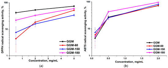
The determined IC50 values for GGM and its derivatives differ significantly from each other and range from 2.28 to 2.44 mg/mL for DPPH and 0.59–0.62 mg/mL for ABTS. Nevertheless, the data depicted in Figure 5 allow a more detailed observation of the dependence of differences and the «structure–properties» pattern.
Figure 5.
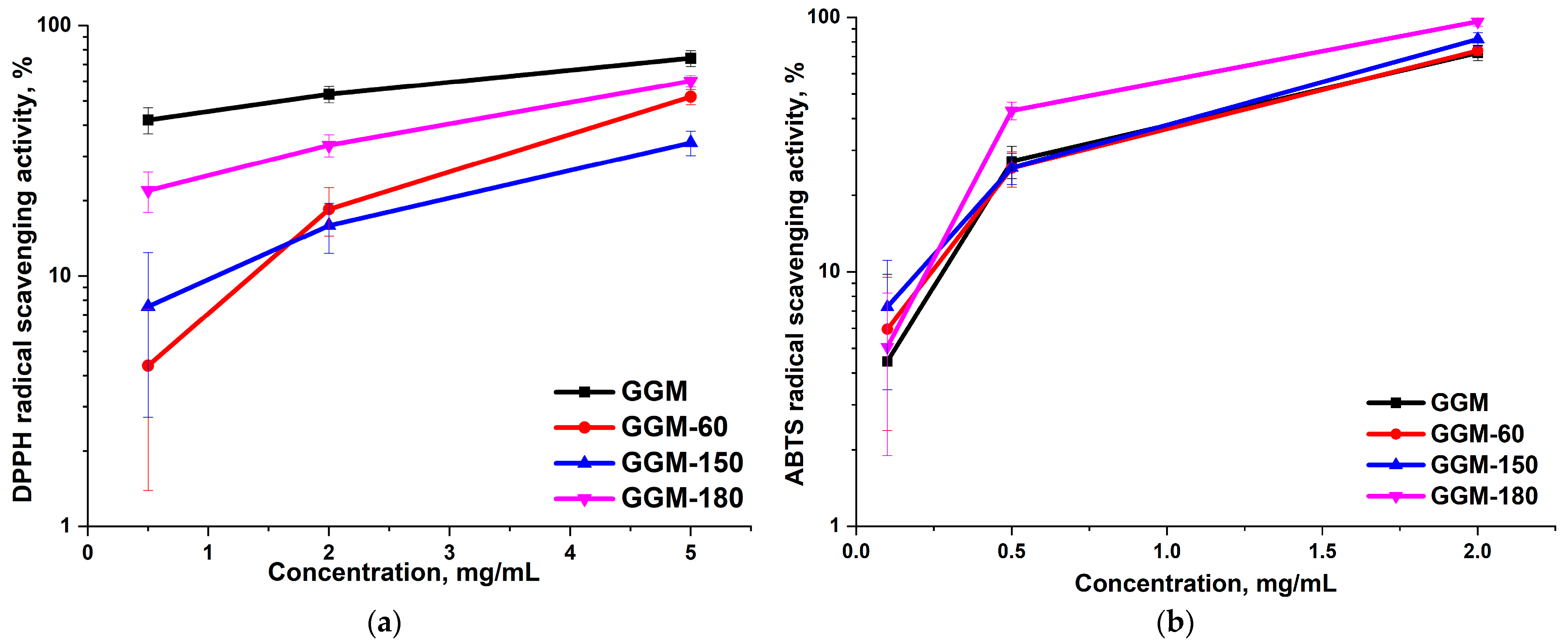
The ability of the native and sulfated GGM to scavenge (a) DPPH and (b) ABTS free radicals (p < 0.05; n = 3).
Thus, the depicted results indicate that the sulfation of GGM leads to a decrease in the ability to inhibit DPPH free radicals, regardless of the derivatives DS (Figure 5a), reaching a maximum of 60% (SGGM-180) at a concentration of 5 mg/mL. At the same time, the native polysaccharide was characterized by the highest AOA (up to 74%) across the entire concentration range among all inspected samples (Figure 5a). This phenomenon is due to the heterogeneous structure of SGGM, which is rich in hydroxyl and carboxyl groups. However, during the modification process, on the one hand, they are replaced by sulfate groups, which lack a mobile proton to inhibit the DPPH radical, and on the other hand, the presence of singly charged functional groups leads to macromolecules unfolding due to the polyelectrolyte effect and an even more branched structure is formed [37], which prevents access to the sterically hindered radical center of DPPH at both low and high concentrations [38]. Thus, this fact indicates that the DPPH scavenging is not the primary antioxidant mechanism inherent in GGM sulfated derivatives, which, by the analogy, has already been observed by the other studies [36,39,40].
Another common spectrophotometric method for assessing the antioxidant activity of substances, including natural polymers, is based on the free radical scavenging ability of ABTS. Although both methods quantify antioxidant capacity, the ABTS method is often considered more effective because it operates in both aqueous and organic solvents and correlates better with other methods. Furthermore, the two methods differ in their transfer mechanisms [41,42]. For this study, relying on the ABTS test results is more appropriate because, firstly, the original GGM and its sulfates are hydrophilic. Secondly, since GGM is modified to yield its salts, the predominant antioxidant mechanism will be based on single or more electron transfer [26]. It was found that the maximum ABTS radical absorption value (96%) was achieved by the SGGM-180 sample at a polysaccharide concentration of 2 mg/mL in solution. This sample combines a high DS (1.1), homogeneity, mobility and moderate branch of the molecules, which together provide such promising results. Obviously, this phenomenon is connected with GGM’s heterogeneous structure, abundant with rich in various functional groups. In summary, the presence of such multiple functional groups empowers the GGM by the perspective AOA properties. In this case, sulfation of the GGM not only provokes the molecule structure and conformation conversion but also results in a significant change in the inhibition of free radicals.
3.2.2. Analysis of the in Vitro Effect of SGGM on Human Blood/Plasma Coagulation
To prevent the occurrence of blood clots (fibrin clots with formed elements of the blood-platelets, erythrocytes, leukocytes) or bleeding, the mammalian body has a protective blood coagulation system (hemostasis system), including the vascular endothelium, platelets, the basis of cellular hemostasis, plasma hemostasis (including coagulation factors; activated coagulation factors-serine proteinases), inhibitors/activators of coagulation factors, extracellular matrix metalloproteinases, and the fibrinolysis system [43]. Activation of the blood coagulation system results in the formation of thrombin (factor IIa), which hydrolyzes plasma-soluble fibrinogen; fibrinogen polymerization results in the formation of insoluble fibrin, the basis of a thrombus. Anticoagulants with different mechanisms of action are also used in clinical practice to prevent and treat thrombosis [44,45]. Anticoagulants are also used in the design of biomaterials with a surface resistant to the formation of blood clots [46,47,48].
It has previously been shown that SGGM (PDI = 1.5; DS = 1.81) in concentrations up to 0.002 mg/mL does not independently provoke human platelet aggregation, does not affect hemolysis of human erythrocytes in vitro [49], inhibits collagen-induced platelet aggregation, and its ACA is associated with the inhibition of the amidolytic activity of factors IIa and Xa, which are independent of antithrombin (plasma inhibitor of serine proteinases of the blood coagulation system) [13].
In this study, we assessed the effect of sample solutions on the time of fibrin clot appearance in the BRT test and on the time of fibrin clot appearance in APTT test.
3.2.3. The Blood Recalcification Time
The BRT test (activation of blood coagulation with the addition of calcium ions) is used in one of the options for setting up thromboelastometry (qualitative and quantitative assessment of the viscosity, strength and elasticity of a whole blood clot, the rate of its formation and subsequent lysis) to monitor hypercoagulation and prescribe optimal doses of AC drugs for the prevention and treatment of thrombosis [50,51].
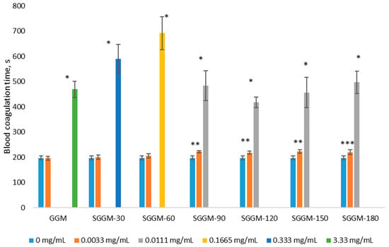
Incubation of blood with the studied samples in the concentration range of 0.011–3.33 mg/mL resulted in a significant increase in the clotting time in the BRT test by 2.1–3.5 times, compared to the control without adding the sample (0 mg/mL—197.38 ± 7.56 s; Figure 6). With increasing sulfation reaction time (30, 60, 90, 120, 150, 180 min), the 2BRT concentrations of SGGM samples reached 0.190 ± 0.039 mg/mL, 0.074 ± 0.014 mg/mL, 0.0097 ± 0.0013 mg/mL, 0.0110 ± 0.0015 mg/mL, 0.0088 ± 0.001 mg/mL, 0.0092 ± 0.0015 mg/mL, respectively, and were significantly lower than 2BRT GGM 2.612 ± 0.31 mg/mL. The lower the 2BRT concentration, the greater the anticoagulant potential of the sample. Based on the 2BRT concentration, the anticoagulant potential of SGGM samples is greater than that of the original GGM.
Figure 6.
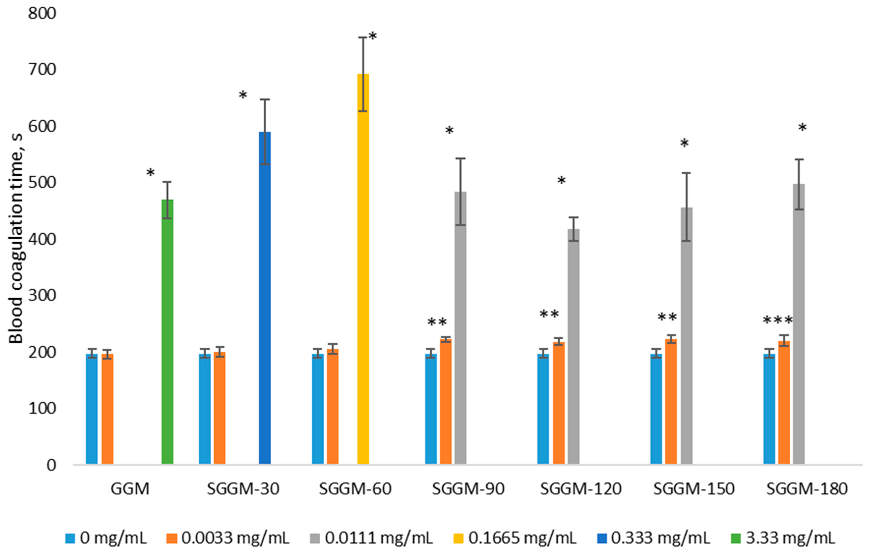
Effect of GGM and SGGM on blood clotting time (s) in the BRT test (* p = 0, ** 0.001 ≤ p ≤ 0.006, *** 0.03 < p < 0.05 difference from readings at 0 mg/mL; n = 6).
The studied samples were shown to prevent fibrin clot formation, and with increasing sulfation reaction time (30, 60, 90, 120, 150, and 180 min), the 2BRT concentrations of SGGM samples were 13.75, 35.3, 269.3, 237.5, 296.8, and 283.9 times lower, respectively, than the 2BRT of GGM.
3.2.4. Activated Partial Thromboplastin Time
In clinical practice, the APTT test is used to monitor therapy with heparin drugs and determine the presence of coagulation inhibitors [52], and in research practice, to determine the presence of ACA in compounds [53]. The APTT test simulates damage to the vascular wall (without thromboplastin) and activation of coagulation (using a contact phase activator and phospholipids).
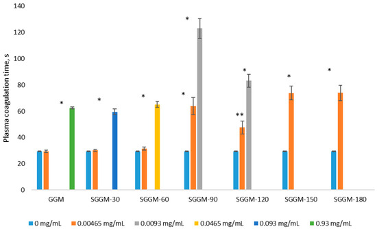
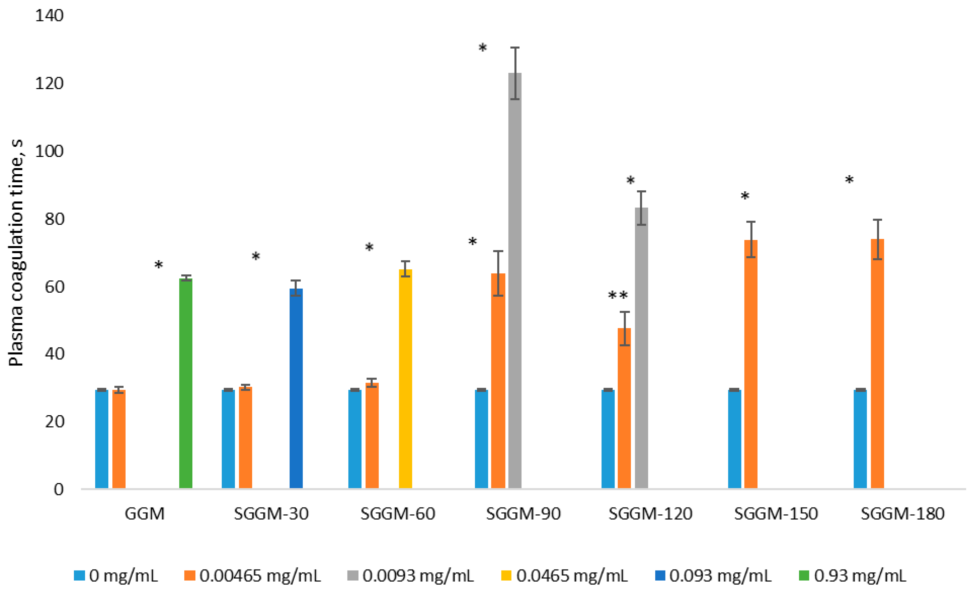
Incubation of human platelet-poor plasma with GGM and SGGM samples in the concentration range of 0.0047–0.93 mg/mL resulted in a significant increase in plasma clotting time in the APTT test, compared to the 0 mg/mL control (Figure 7). For comparison, the addition of the studied SGGM-150 and SGGM-180 (at a concentration of 0.0047 mg/mL), as well as sulfated polysaccharides (at a concentration of 0.0063 mg/mL) isolated from green (Cladophora oligoclada) [54]/brown (Stephanocystis dioica) [53] algae and cuttlefish ink Sepia esculenta [55] to plasma resulted in an increase in plasma clotting time in the APTT test by 2.5, 2.51, 1.5, 4.4, and 1.8 times greater than in the control. It should be noted that the greater increase in plasma clotting time with the addition of sulfated polysaccharide isolated from Stephanocystis dioica may be associated with a higher concentration compared with the concentration of the studied SGGM-150 and SGGM-180.
Figure 7.
Effect of GGM and SGGM on plasma clotting time (s) in the APTT test (* p = 0, ** p = 0.005 difference from readings at 0 mg/mL; n = 6).
In the present study, upon incubation of plasma with 0.2326 IU/mL unfractionated heparin (UFH) (activates plasma inhibitors of serine proteinases of the blood coagulation system–antithrombin and heparin cofactor II [56]) the coagulation time in the APTT test was 159.12 ± 18.99 s (n = 6; 2APTT for UFH 0.0608 ± 0.0036 IU/mL). The concentration of 2APTT for UFH reached 0.0608 ± 0.0036 IU/mL and the concentrations of 2APTT for SGG, SGGM-30, SGGM-60, SGGM-90, SGGM-120, SGGM-150, SGGM-180 were 0.8283 ± 0.0431 mg/mL, 0.0938 ± 0.0064 mg/mL, 0.0389 ± 0.0019 mg/mL, 0.0042 ± 0.0005 mg/mL, 0.0060 ± 0.0005 mg/mL, 0.0033 ± 0.0003 mg/mL, 0.0032 ± 0.0005 mg/mL, respectively. The calculated ACA of GGM was insignificant, 0.0757 ± 0.009 IU/mg, and the ACA of SGGM reached 10.27 ± 0.54 IU/mg–21.19 ± 2.89 IU/mg (Table 3). The highest ACA of 18.52 ± 0.85 IU/mg and 21.19 ± 2.89 IU/mg were shown by samples SGGM-150 and SGGM-180, respectively. The correlation coefficient between the sulfation reaction time (0 min for GGM) and ACA was r = 0.9312 (n = 7; p = 0.002). The obtained results indicate that SGGM samples have ACA that significantly increases with increasing sulfation reaction time. An increase in ACA with an increase in the degree of sulfation was shown using galactan from a South Korean mollusk as an example [20], as well as using aqueous fractions of sulfated galactan (sulfur content 17.4% and 19.3%) from the green alga Codium bernabei, which lengthen the plasma clotting time in the APTT test in the concentration range from 0.01 to 1 mg/mL (by 2.47–3.38 times and 2.6–4.2 times, compared to the control, respectively) [57]. The concentrations of 2APTT and 2BRT of the studied samples were significantly and positively closely related (r = 0.9992; n = 7; p = 0).
Table 3.
Anticoagulant activity of native GGM and SGGM samples.
This may indicate that the ACA of the studied SGGM depends on the plasma components of the blood coagulation system. Indirect confirmation of this conclusion may be the presence of inhibition of the amidolytic activity of factor IIa upon interaction with SGGM (PDI = 1.5; DS = 1.81) [16].
4. Conclusions
Novel sulfated GGM derivatives with degrees of substitution ranging from 0.4 to 1.2 were synthesized from spruce (Picea abies) using a sulfamic acid–urea–1,4-dioxane system. The incorporation of sulfate groups and the resulting structural transformations were confirmed via elemental analysis, GPC, and FTIR spectroscopy. High-intensity bands associated with S=O and C–O–S stretching vibrations in the sulfated GGM were identified in the FTIR spectra. Elemental analysis and GPC data allowed for the determination of the relationship between the DS and sulfation duration, leading to the proposal of a four-stage hypothetical reaction mechanism. It was found that the DPPH scavenging is not the primary antioxidant mechanism inherent in GGM sulfated derivatives. At the same time, the maximum ABTS radical absorption value (96%) was achieved by the SGGM-180 sample at a polysaccharide concentration of 2 mg/mL in solution. Conversely, the SGGM samples effectively prevented the formation of fibrin clots in human blood/plasma in vitro. Increasing the sulfation duration from 90 to 180 min enhanced the anticoagulant activity of the SGGM by an order of magnitude compared to samples obtained between 30 and 60 min. The most promising derivatives, characterized by the highest ACA (18.52 ± 0.85 and 21.19 ± 2.89 IU/mg), represent potential candidates for the design of thromboresistant biomaterials.
Author Contributions
Conceptualization, V.S.B. and Y.N.M.; methodology, Y.N.M. and N.N.D.; validation, V.S.B. and Y.N.M.; formal analysis, V.S.B., N.N.D. and M.V.S.; investigation, V.S.B., N.N.D. and M.V.S.; resources, Y.N.M.; data curation, V.S.B., Y.N.M. and N.N.D.; writing—original draft preparation, V.S.B. and N.N.D.; writing—review and editing, V.S.B., Y.N.M. and N.N.D.; visualization, V.S.B. and M.V.S.; supervision, Y.N.M.; funding acquisition, Y.N.M. All authors have read and agreed to the published version of the manuscript.
Funding
This study was supported by the Russian Science Foundation, project no. 22-73-10212-П, https://rscf.ru/en/project/22-73-10212-П/ (accessed on 6 November 2025).
Institutional Review Board Statement
All procedures performed with human participation comply with the ethical standards of the Russian Bioethics Committee and the Helsinki Declaration of 1964 and its subsequent amendments. An “Informed voluntary consent for medical examination and donation of blood and (or) its components” was obtained from each of the healthy voluntary donors (Appendix No. 2 to Order No. 475 of the National Medical Research Center of Hematology of the Ministry of Health of the Russian Federation dated 2 August 2023). The study was conducted under the agreement No. 1/c-2022 dated 12/19/2022 by the National Medical Research Center of Hematology (Ministry of Health of the Russian Federation, Moscow) and the Institute of Chemistry and Chemical Technology of the Krasnoyarsk Scientific Center, Siberian Branch of the Russian Academy of Sciences (Krasnoyarsk) and in accordance with the protocol of the study in standard operating procedures (SOP No. LPiFG-A-001/01 dated 20 December 2023) of Hemostasis Pathology and Pharmacology Laboratories of the National Medical Research Center of Hematology (Ministry of Health of the Russian Federation, Moscow).
Data Availability Statement
The original contributions presented in this study are included in the article. Further inquiries can be directed to the corresponding author.
Acknowledgments
This study was carried out using the equipment of the Krasnoyarsk Regional Centre for Collective Use, Krasnoyarsk Scientific Center, Siberian Branch of the Russian Academy of Sciences.
Conflicts of Interest
The authors declare no conflicts of interest.
Correction Statement
This article has been republished with a minor correction to the Funding statement. This change does not affect the scientific content of the article.
References
- Mishra, P.; Rawat, B.; Sahu, K.K.; Akram, W. Unveiling the transformative horizon of biopolymers in drug delivery. Polym. Bull. 2025, 82, 8727–8762. [Google Scholar] [CrossRef] [Scilit]
- Pillai, A.; Bhande, D.; Pardhi, V. Controlled Drug Delivery System. In Advanced Drug Delivery: Methods and Applications; Santra, T.S., Shinde, A.U.S., Eds.; Springer Nature: Singapore, 2023; pp. 267–289. [Google Scholar] [CrossRef] [Scilit]
- Ceylan, S.; Akbay, İ.K.; Demir, D. Current Trends in Sustainable Biopolymers for Biomedical Engineering and Pharmaceutical Applications. In Sustainable Biopolymers and Composites for Biomedical Applications; Collins, M.N., Culebras, M., Eds.; Springer Nature: Cham, Switzerland, 2025; pp. 3–26. [Google Scholar] [CrossRef] [Scilit]
- Getahun, M.J.; Kassie, B.B.; Alemu, T.S. Recent advances in biopolymer synthesis, properties, & commercial applications: A review. Process Biochem. 2024, 145, 261–287. [Google Scholar] [CrossRef] [Scilit]
- Li, S.; Xiong, Q.; Lai, X.; Li, X.; Wan, M.; Zhang, J.; Yan, Y.; Cao, M.; Lu, L.; Guan, J.; et al. Molecular Modification of Polysaccharides and Resulting Bioactivities. Compr. Rev. Food Sci. Food Saf. 2016, 15, 237–250. [Google Scholar] [CrossRef] [Scilit] [PubMed]
- Khan, A.; Alamry, K.A.; Asiri, A.M. Multifunctional Biopolymers-Based Composite Materials for Biomedical Applications: A Systematic Review. ChemistrySelect 2021, 6, 154–176. [Google Scholar] [CrossRef] [Scilit]
- Ferreira, S.S.; Passos, C.P.; Madureira, P.; Vilanova, M.; Coimbra, M.A. Structure–function relationships of immunostimulatory polysaccharides: A review. Carbohydr. Polym. 2015, 132, 378–396. [Google Scholar] [CrossRef] [Scilit]
- Xie, J.H.; Jin, M.L.; Morris, G.A.; Zha, X.Q.; Chen, H.Q.; Yi, Y.; Li, J.E.; Wang, Z.J.; Gao, J.; Nie, S.P.; et al. Advances on Bioactive Polysaccharides from Medicinal Plants. Crit. Rev. Food Sci. Nutr. 2016, 56, S60–S84. [Google Scholar] [CrossRef] [Scilit]
- Hao, Z.; Dai, S.; Tan, J.; Gao, Y.; Sang, Y.; Xue, H. Modification, Structural Characterizations, and Biological Activities of Sulfated Polysaccharides: A Review. Starch-Stärke 2024, 76, 2300116. [Google Scholar] [CrossRef] [Scilit]
- Yu, Y.; Shen, M.; Song, Q.; Xie, J. Biological activities and pharmaceutical applications of polysaccharide from natural resources: A review. Carbohydr. Polym. 2018, 183, 91–101. [Google Scholar] [CrossRef] [Scilit]
- Chen, F.; Huang, G. Extraction and antioxidant activities of cushaw polysaccharide. Int. J. Biol. Macromol. 2018, 120, 1646–1649. [Google Scholar] [CrossRef] [Scilit]
- Bourais, I.; Elmarrkechy, S.; Machich, O.; Nassif, A.; Taha, D.; Bouyahya, A. Chapter 26—Biological and pharmaceutical activities of polysaccharides. In Plant Polysaccharides as Pharmaceutical Excipients; Nayak, A.K., Hasnain, M.S., Pal, D., Eds.; Elsevier: Amsterdam, The Netherlands, 2023; pp. 575–607. [Google Scholar] [CrossRef] [Scilit]
- Kalita, P.; Pathak, M.P.; Barbhuiya, P.A.; Nongkhlaw, F.T. Natural sulfated polysaccharides as biomacromolecule in management of metabolic disorders: A comprehensive review. Int. J. Biol. Macromol. 2025, 317, 144856. [Google Scholar] [CrossRef] [Scilit]
- Arlov, Ø.; Rütsche, D.; Asadi Korayem, M.; Öztürk, E.; Zenobi-Wong, M. Engineered Sulfated Polysaccharides for Biomedical Applications. Adv. Funct. Mater. 2021, 31, 2010732. [Google Scholar] [CrossRef] [Scilit]
- Wang, Z.; Xie, J.; Shen, M.; Nie, S.; Xie, M. Sulfated modification of polysaccharides: Synthesis, characterization and bioactivities. Trends Food Sci. Technol. 2018, 74, 147–157. [Google Scholar] [CrossRef] [Scilit]
- Drozd, N.N.; Kuznetsova, S.A.; Malyar, Y.N.; Kazachenko, A.S.; Borovkova, V.S.; Berezhnaya, Y.D. Thrombin and Factor Xa Hydrolysis of Chromogenic Substrates in the Presence of Sulfated Derivatives of Galactomannan and Galactoglucomannan Natural Gels. Pharmaceutics 2022, 14, 2678. [Google Scholar] [CrossRef] [Scilit] [PubMed]
- Liu, T.; Ren, Q.; Wang, S.; Gao, J.; Shen, C.; Zhang, S.; Wang, Y.; Guan, F. Chemical Modification of Polysaccharides: A Review of Synthetic Approaches, Biological Activity and the Structure–Activity Relationship. Molecules 2023, 28, 6073. [Google Scholar] [CrossRef] [Scilit]
- Zhao, T.; Yang, M.; Ma, L.; Liu, X.; Ding, Q.; Chai, G.; Lu, Y.; Wei, H.; Zhang, S.; Ding, C. Structural Modification and Biological Activity of Polysaccharides. Molecules 2023, 28, 5416. [Google Scholar] [CrossRef] [Scilit] [PubMed]
- Yu, Y.; Song, Q.; Huang, L.; Shen, M.; Yu, Q.; Chen, Y.; Xie, J. Immunomodulatory activities of sulfated Cyclocarya paliurus polysaccharides with different degrees of substitution on mouse spleen lymphocytes. J. Funct. Foods 2020, 64, 103706. [Google Scholar] [CrossRef] [Scilit]
- Chen, Q.; Zhang, M.; Liu, Y.; Liu, W.; Peng, C.; Zheng, L. Sulfated Polysaccharides with Anticoagulant Potential: A Review Focusing on Structure-Activity Relationship and Action Mechanism. Chem. Biodivers. 2024, 21, e202400152. [Google Scholar] [CrossRef] [Scilit]
- Borovkova, V.S.; Malyar, Y.N.; Sudakova, I.G.; Chudina, A.I.; Skripnikov, A.M.; Fetisova, O.Y.; Kazachenko, A.S.; Miroshnikova, A.V.; Zimonin, D.V.; Ionin, V.A.; et al. Molecular Characteristics and Antioxidant Activity of Spruce (Picea abies) Hemicelluloses Isolated by Catalytic Oxidative Delignification. Molecules 2022, 27, 266. [Google Scholar] [CrossRef] [Scilit]
- Kazachenko, A.S.; Malyar, Y.N.; Vasilyeva, N.Y.; Borovkova, V.S.; Issaoui, N. Optimization of guar gum galactomannan sulfation process with sulfamic acid. Biomass Convers. Biorefin. 2023, 13, 10041–10050. [Google Scholar] [CrossRef] [Scilit]
- Kazachenko, A.S.; Malyar, Y.N.; Vasilyeva, N.Y.; Bondarenko, G.N.; Korolkova, I.V.; Antonov, A.V.; Karacharov, A.A.; Fetisova, O.Y.; Skvortsova, G.P. «Green» synthesis and characterization of galactomannan sulfates obtained using sulfamic acid. Biomass Convers. Biorefin. 2022, 12, 2705–2714. [Google Scholar] [CrossRef] [Scilit]
- Shang, H.-M.; Zhou, H.-Z.; Yang, J.-Y.; Li, R.; Song, H.; Wu, H.-X. In vitro and in vivo antioxidant activities of inulin. PLoS ONE 2018, 13, e0192273. [Google Scholar] [CrossRef] [Scilit]
- Zhou, H.; Fan, Z.; Li, Y.; Liu, X.; Wang, B.; Xing, J.; He, J.; Zheng, R.; Li, J. Structure–Antioxidant Activity Relationship of Polysaccharides Isolated by Microwave/Ultrasonic-Assisted Extraction from Pleurotus ferulae. Antioxidants 2025, 14, 91. [Google Scholar] [CrossRef] [Scilit]
- Bibi Sadeer, N.; Montesano, D.; Albrizio, S.; Zengin, G.; Mahomoodally, M.F. The Versatility of Antioxidant Assays in Food Science and Safety—Chemistry, Applications, Strengths, and Limitations. Antioxidants 2020, 9, 709. [Google Scholar] [CrossRef] [Scilit]
- Gulliani, G.L.; Hyun, B.H.; Litten, M.B. Blood recalcification time. A simple and reliable test to monitor heparin therapy. Am. J. Clin. Pathol. 1976, 65, 390–396. [Google Scholar] [CrossRef] [Scilit]
- Neupane, S.; Bittkau, K.S.; Alban, S. Size distribution and chain conformation of six different fucoidans using size-exclusion chromatography with multiple detection. J. Chromatogr. A 2020, 1612, 460658. [Google Scholar] [CrossRef] [Scilit]
- Gaborieau, M.; Castignolles, P. Size-exclusion chromatography (SEC) of branched polymers and polysaccharides. Anal. Bioanal. Chem. 2011, 399, 1413–1423. [Google Scholar] [CrossRef] [Scilit]
- Bao, H.; Choi, W.-S.; You, S. Effect of Sulfated Modification on the Molecular Characteristics and Biological Activities of Polysaccharides from Hypsizigus marmoreus. Biosci. Biotechnol. Biochem. 2010, 74, 1408–1414. [Google Scholar] [CrossRef] [Scilit]
- Miranda-Arizmendi, V.; Marquez-Escalante, J.; Rascon-Chu, A.; Martínez-Robinson, K.; Brown-Bojorquez, F.; Carvajal-Millan, E. Chaetoceros muelleri sulfated polysaccharides: Chain conformation, physical characteristics, and morphology. AIMS Biophys. 2024, 11, 455–463. [Google Scholar] [CrossRef] [Scilit]
- Li, S.; Li, J.; Zhi, Z.; Wei, C.; Wang, W.; Ding, T.; Ye, X.; Hu, Y.; Linhardt, R.J.; Chen, S. Macromolecular properties and hypolipidemic effects of four sulfated polysaccharides from sea cucumbers. Carbohydr. Polym. 2017, 173, 330–337. [Google Scholar] [CrossRef] [Scilit] [PubMed]
- Mukherjee, S.; Jana, S.; Khawas, S.; Kicuntod, J.; Marschall, M.; Ray, B.; Ray, S. Synthesis, molecular features and biological activities of modified plant polysaccharides. Carbohydr. Polym. 2022, 289, 119299. [Google Scholar] [CrossRef] [Scilit] [PubMed]
- Tarasevich, B. IR Spectra of the Main Classes of Organic Compounds; Moscow State University: Moscow, Russia, 2012. [Google Scholar]
- Yang, D.; Lin, F.; Huang, Y.; Ye, J.; Xiao, M. Separation, purification, structural analysis and immune-enhancing activity of sulfated polysaccharide isolated from sea cucumber viscera. Int. J. Biol. Macromol. 2020, 155, 1003–1018. [Google Scholar] [CrossRef] [Scilit]
- Ageeli, A.A.; Mohamed, S.F. Structural Characterization, Antioxidant, and Antiviral Activity of Sulfated Polysaccharide (Fucoidan) from Sargassum asperifolium (Turner). J. Agardh. Chem. 2023, 5, 2756–2771. [Google Scholar] [CrossRef] [Scilit]
- Ionin, V.A.; Malyar, Y.N.; Borovkova, V.S.; Zimonin, D.V.; Kazachenko, A.S. TEMPO-Oxidized Spruce Galactoglucomannan–Biopolymer with Enhanced Antioxidant Activity and Selective Heavy-Metal Sorption. Antioxidants 2025, 14, 569. [Google Scholar] [CrossRef] [Scilit]
- Schaich, K.M.; Tian, X.; Xie, J. Hurdles and pitfalls in measuring antioxidant efficacy: A critical evaluation of ABTS, DPPH, and ORAC assays. J. Funct. Foods 2015, 14, 111–125. [Google Scholar] [CrossRef] [Scilit]
- Wang, X.; Wang, J.; Zhang, J.; Zhao, B.; Yao, J.; Wang, Y. Structure–antioxidant relationships of sulfated galactomannan from guar gum. Int. J. Biol. Macromol. 2010, 46, 59–66. [Google Scholar] [CrossRef] [Scilit]
- Shao, P.; Chen, X.; Sun, P. In vitro antioxidant and antitumor activities of different sulfated polysaccharides isolated from three algae. Int. J. Biol. Macromol. 2013, 62, 155–161. [Google Scholar] [CrossRef] [Scilit] [PubMed]
- Platzer, M.; Kiese, S.; Herfellner, T.; Schweiggert-Weisz, U.; Miesbauer, O.; Eisner, P. Common Trends and Differences in Antioxidant Activity Analysis of Phenolic Substances Using Single Electron Transfer Based Assays. Molecules 2021, 26, 1244. [Google Scholar] [CrossRef] [Scilit]
- Rodríguez-Muñoz, M.D.R.; Mora-Garrido, A.B.; Heredia, F.J.; Cejudo-Bastante, M.J.; González-Miret, M.L. Obtention and Characterisation of Antioxidant-Rich Peptides from Defatted Grape Seed Meal Using Different Enzymes. Foods 2025, 14, 1248. [Google Scholar] [CrossRef] [Scilit]
- Grover, S.P.; Mackman, N. Tissue Factor: An Essential Mediator of Hemostasis and Trigger of Thrombosis. Arter. Thromb. Vasc. Biol. 2018, 38, 709–725. [Google Scholar] [CrossRef] [Scilit] [PubMed]
- Valeriani, E.; Di Nisio, M.; Riva, N.; Cohen, O.; Garcia-Pagan, J.C.; Magaz, M.; Porreca, E.; Ageno, W. Anticoagulant therapy for splanchnic vein thrombosis: A systematic review and meta-analysis. Blood 2021, 137, 1233–1240. [Google Scholar] [CrossRef] [Scilit] [PubMed]
- Stuby, J.; Haschke, M.; Tritschler, T.; Aujesky, D. Oral anticoagulant therapy in older adults. Thromb. Res. 2024, 238, 1–10. [Google Scholar] [CrossRef] [Scilit]
- Safi, S.Z.; Fazil, S.; Saeed, L.; Shah, H.; Arshad, M.; Alobaid, H.M.; Rehman, F.; Sharif, F.; Selvaraj, C.; Orakzai, A.H.; et al. Chitosan- and heparin-based advanced hydrogels: Their chemistry, structure and biomedical applications. Chem. Pap. 2024, 78, 9287–9309. [Google Scholar] [CrossRef] [Scilit]
- Bračič, M.; Nagy, B.M.; Plohl, O.; Lackner, F.; Steindorfer, T.; Fischer, R.C.; Heinze, T.; Olschewski, A.; Kleinschek, K.S.; Nagaraj, C.; et al. Antithrombogenic polysaccharide coatings to improve hemocompatibility, protein-repellence, and endothelial cell response. iScience 2024, 27, 110692. [Google Scholar] [CrossRef] [Scilit]
- Pu, X.; Peng, X.; Shi, S.; Feng, S.; Wei, X.; Gao, X.; Yu, X. A New Type of Bioprosthetic Heart Valve: Synergistic Modification with Anticoagulant Polysaccharides and Anti-inflammatory Drugs. ACS Biomater. Sci. Eng. 2025, 11, 634–648. [Google Scholar] [CrossRef] [Scilit] [PubMed]
- Drozd, N.N.; Kuznetsova, S.A.; Malyar, Y.N.; Kazachenko, A.S. Hemocompatibility of Galactomannan and Galactoglucomannan Sulfates in In Vitro Experiments. Bull. Exp. Biol. Med. 2022, 173, 98–104. [Google Scholar] [CrossRef] [Scilit]
- Cui, S.; Jiang, R.; Zhao, J.; Wang, J.; Hu, B.; Li, H.; Chen, S. Thromboelastography-based dynamic evaluation of perioperative coagulation changes and anticoagulant efficacy in lung cancer patients. Thromb. J. 2025, 23, 31. [Google Scholar] [CrossRef] [Scilit]
- Kataria, S.; Juneja, D.; Singh, O. Redefining haemostasis: Role of rotational thromboelastometry in critical care settings. World J. Crit. Care Med. 2025, 14, 102521. [Google Scholar] [CrossRef] [Scilit] [PubMed]
- May, J.E.; Siniard, R.C.; Taylor, L.J.; Marques, M.B.; Gangaraju, R. From Activated Partial Thromboplastin Time to Antifactor Xa and Back Again. Am. J. Clin. Pathol. 2022, 157, 321–327. [Google Scholar] [CrossRef] [Scilit]
- García-Zamora, G.M.; Muñoz-Ochoa, M.; Arvizu-Higuera, D.L.; Rodríguez-Montesinos, Y.E.; López-Vivas, J.M. Anticoagulant activity of sulfated polysaccharides obtained from the brown seaweed Stephanocystis dioica. Hidrobiológica 2025, 35, 49–56. [Google Scholar] [CrossRef] [Scilit]
- He, M.; Yang, Y.; Shao, Z.; Zhang, J.; Feng, C.; Wang, L.; Mao, W. Chemical Structure and Anticoagulant Property of a Novel Sulfated Polysaccharide from the Green Alga Cladophora oligoclada. Mar. Drugs 2021, 19, 554. [Google Scholar] [CrossRef] [Scilit]
- Li, F.; Lin, Z.; Wu, Y.; Luo, P.; Wu, J.; Liu, H. Antioxidant, anticoagulant and thrombolytic properties of SIP-IV, a sulfated polysaccharide from Sepia esculenta ink, and its derivatives. Food Biosci. 2022, 49, 101959. [Google Scholar] [CrossRef] [Scilit]
- Yang, X.; Wang, Q.; Zhang, A.; Shao, X.; Liu, T.; Tang, B.; Fang, G. Strategies for sustained release of heparin: A review. Carbohydr. Polym. 2022, 294, 119793. [Google Scholar] [CrossRef] [Scilit] [PubMed]
- Figueroa, F.A.; Abdala-Díaz, R.T.; Pérez, C.; Casas-Arrojo, V.; Nesic, A.; Tapia, C.; Durán, C.; Valdes, O.; Parra, C.; Bravo-Arrepol, G.; et al. Sulfated Polysaccharide Extracted from the Green Algae Codium bernabei: Physicochemical Characterization and Antioxidant, Anticoagulant and Antitumor Activity. Mar. Drugs 2022, 20, 458. [Google Scholar] [CrossRef] [Scilit] [PubMed]

Disclaimer/Publisher’s Note: The statements, opinions and data contained in all publications are solely those of the individual author(s) and contributor(s) and not of MDPI and/or the editor(s). MDPI and/or the editor(s) disclaim responsibility for any injury to people or property resulting from any ideas, methods, instructions or products referred to in the content. |
© 2026 by the authors. Licensee MDPI, Basel, Switzerland. This article is an open access article distributed under the terms and conditions of the Creative Commons Attribution (CC BY) license.