Article

Cellular Antioxidant Potential and Cytotoxic Activities of Extracellular Polysaccharides Isolated from Lactobacillus graminis Strain KNUAS018

1
Department of Bio-Health Convergence, Kangwon National University, Chuncheon 200-701, Republic of Korea
2
Department of Dental Hygiene, College of Health Science, Kangwon National University, Chungcheong 24341, Republic of Korea
*
Authors to whom correspondence should be addressed.
These authors contributed equally to this work.
Polysaccharides 2025, 6(2), 33; https://doi.org/10.3390/polysaccharides6020033
Submission received: 20 December 2024 / Revised: 6 March 2025 / Accepted: 8 April 2025 / Published: 11 April 2025

Abstract

:
In the present study, exopolysaccharides (EPS-1, EPS-2, and EPS-3) were extracted from Lactobacillus graminis, and their chemical compositions, bioactivities, and cytotoxicity were comprehensively studied. A higher yield was observed for EPS-1 and EPS-2 with 14.38% and 9.24%, respectively. The chemical composition in the samples was studied using FT-IR analysis. The EPS-1 (1 mg/mL) showed higher antioxidant activities with 34.5 ± 6.6% and 93.6 ± 2.3% of DPPH and ABTS radical scavenging, respectively. In the cellular antioxidant assay, the EPS-1 protected oxidative stress-mediated cellular damage in AAPH-treated NIH3T3 cells. In addition, EPS-1 (0.25 mg/mL) treatment augmented the viability of AAPH-stressed RAW264.7 cells (~80%) than AAPH-treated cells (~50%) by reducing the ROS level and associated oxidative damage. Toxicity studies indicated that EPS-1 (1 mg/mL) did not induce notable cytotoxic effects in NIH3T3 cells, RAW264.7 cells, and erythrocytes. Altogether, the findings of this research suggest that L. graminis could be a source of biocompatible polysaccharides with antioxidant properties.

1. Introduction

Inflammation is a complex process in which the immune system is activated and pro-inflammatory mediators are released during the inflammatory response [1]. Inflammatory dysregulation is associated with a wide range of chronic diseases and acute diseases such as cancer, cardiovascular disease, chronic obstructive pulmonary disease, and acute lung injury [2]. When inflammation occurs in the body, oxidative stress often accompanies it. There is an imbalance between the production and accumulation of highly reactive molecules, such as free radicals in the form of reactive oxygen species (ROS) or reactive nitrogen species (RNS), and the ability of the biological system to scavenge them, which leads to tissue damage [3]. Excessive ROS accumulation promotes aging and age-related diseases by interfering with cellular homeostasis in physiological states and the oxidation of biomolecules, including DNA, lipids, and proteins, causing the development of various types of diseases [4]. In addition, the imbalance between ROS and cellular antioxidant activity leads to oxidative stress and, thus, inflammation [5]. Therefore, controlling oxidative stress is essential to prevent further development of inflammation [6]. Currently, the market offers steroidal anti-inflammatory drugs (SAIDs) and non-steroidal anti-inflammatory drugs (NSAIDs) for mitigating inflammation and the associated symptoms of different inflammatory disorders. Prolonged use of SAIDs or NSAIDs may have serious effects on multiple organs, including gastrointestinal bleeding, cardiovascular and renal failure, and other adverse symptoms, so it is imperative to find safer and more effective medications for combating oxidative stress to ameliorate inflammation-induced disease [7].
Extracellular polysaccharides (EPS) have a variety of biological activities, such as antimicrobial anticancer effects and aiding in prevention of insects [8]. In addition, EPS has strong antioxidant activity, which can be used to develop effective and harmless drugs applied to oxidative stress [9]. The EPS are naturally occurring soluble or insoluble polysaccharides that are produced by the microorganisms and then secreted into the extracellular medium [10]. Based on their structures, EPS are classified as homopolysaccharides (HoPS) consisting of one sugar residue monomer and heteropolysaccharides (HePS) consisting of two or more sugar residue monomers or other organic molecules [11]. Recent studies have shown that EPS protects the liver by improving antioxidant status and enhancing anti-inflammatory effects [2]. Moreover, EPS can slow down and treat inflammation by inhibiting T cell activation through activation of TLR4 signaling and up-regulation of IDO expression [12]. Thus, EPS has been revealed to possess substantial antioxidant and anti-inflammatory properties. It has been determined that EPS functions as an antioxidant to avert oxidative stress damage in living cells and as a scavenger of various forms of free radicals in laboratory settings [13].
Lactobacillus (LAB) extracellular polysaccharides have received great attention because of their anticancer, antitumor, anti-inflammatory, immunomodulatory, antithrombotic, hypoglycemic, hypocholesterolemia, antiviral, antidiabetic, and other effects [9]. EPS extraction often faces the problem of difficult preparation and low yield, whereas EPS produced in lactic acid bacteria strains have higher yields. Moreover, Lactobacilli, as Gram-positive catalase-negative rod-shaped bacteria, have been repeatedly shown to be safe in humans [14]. Thus, EPS from LAB is considered one of the most harmless, non-toxic, and cell-free probiotic products available [15]. Based on the above background information, this study aims to investigate the free radical scavenging activity and cytotoxicity (in non-cancerous cells) of the EPS extracted from Lactobacillus graminis strain KNUAS018 and to evaluate their cellular antioxidant potential by modulating oxidative stress.

2. Materials and Methods

2.1. Chemicals and Consumables

The chemicals used in this study are declared with their supplier information as follows. Quercetin dihydrate, Folin–Ciocalteu’s phenol reagent and Triton X-100 (SIGMA, USA), Propidium iodide; PI, Acridine orange; AO, Ethidium bromide; EB, and Rhodamine-123; Rh-123 were obtained from Sigma-Aldrich (St. Louis, MO, USA). Gallic acid, Sulfuric acid, Ascorbic acid; AA, 2,2′-azino-bis(3-ethylbenzothiazoline-6-sulfonic acid) diammonium salt; ABTS, 2,2-diphenyl-1-picrylhydrazyl; DPPH, Ethanol, and hydrogen peroxide were purchased from Daejung (Siheung-si, Republic of Korea). DEAE Sepharose fast flow (GE Healthcare, Uppsala, Sweden), Dulbecco’s modified eagle medium; DMEM, Fetal bovine serum; FBS (HyClone, Logan, UT, USA), De Man Rogosa Sharpe Agar; MRSA, De Man Rogosa Sharpe Broth; MRSB (Oxoid, Basingstoke England), Sheep Blood (Carlina, Seoul, Republic of Korea), 3-Methyl-1-phenyl-5-pyrazolone (TCI, Tokyo, Japan), Phenol and Sodium carbonate (Junsei, Tokyo, Japan), Bradford reagent (Bio-Rad, Hercules, CA, USA), Viability assay kit (Cellomax, Yongin-si, Republic of Korea). Additionally, the murine macrophage RAW264.7 cells and mouse embryonic fibroblast NIH3T3 cells were procured from the Korean Cell Line Bank (KCLB, Seoul, Republic of Korea).

2.2. Culture Condition and Extraction of Crude EPS

In this study, a previously isolated and identified Lactobacillus graminis strain KNUAS018 (L. graminis; GenBank Accession No. OM327574) [16] was used for the production of EPS. The L. graminis were grown on MRSA (de Man Rogosa Sharpe Agar) plates for 24 h and then cultured in 2 L of MRSB (de Man Rogosa Sharpe broth) and incubated in a shaker incubator (200 rpm) at 37 °C for 72 h. After that, bacterial cells were separated from the culture medium via centrifugation at 10,000 rpm for 10 min using a high-speed laboratory centrifuge (Hanil Science Industrial, Gimpo-si Republic of Korea). Subsequently, the resulting supernatant was concentrated using a rotary evaporator at 40 °C. For EPS extraction, a concentrated culture supernatant was mixed with a 95% ethanol ratio of 1:3 (w/v) and allowed to incubate at 4 °C overnight (static conditions). The resulting precipitated EPS was then collected by centrifugation at 10,000 rpm for 20 min. The final crude EPS product was freeze-dried and stored at 4 °C for further analysis.

2.3. Purification and Fractionation of EPS

The crude EPS underwent deproteinization and impurities removal using the Savage solution, which comprises chloroform and n-butyl alcohol in a 4:1 ratio, followed by dialysis against water for 24 h. The crude EPS underwent fractionation through DEAE-Sepharose fast flow column (1.6 × 20 cm) chromatography. The column was initially eluted using distilled water (H2O) followed by various molar concentrations of NaCl (0.1, 0.2, 0.3, 0.4, and 0.5 M). To maintain a consistent flow rate of 1 mL/min, a vacuum pump was employed. Quantification of all fractions was conducted using the phenol–sulfuric acid assay, pooled separately (based on eluent), and labeled.

2.4. Biochemical Assays and Characterization Studies

The protein content in the EPS was quantified using the Bradford method, referencing the bovine serum albumin standard [17]. In the EPS, to determine the total phenol content (TPC), the Folin–Ciocalteu assay was employed, and an aluminum chloride assay was conducted to assess the total flavonoid content (TFC). Furthermore, the protein and nucleic acid levels within the EPS were further analyzed using a UV-vis spectrophotometer (SpectraMax® ABS Plus, Molecular Devices, San Jose, CA, USA) within the 200–400 nm scan range. The functional groups present on EPS were analyzed using a Fourier transform infrared (FT-IR; PerkinElmer Paragon 500, Waltham, MA, USA) spectroscopic study within a scan range of 400–4000 cm−1.

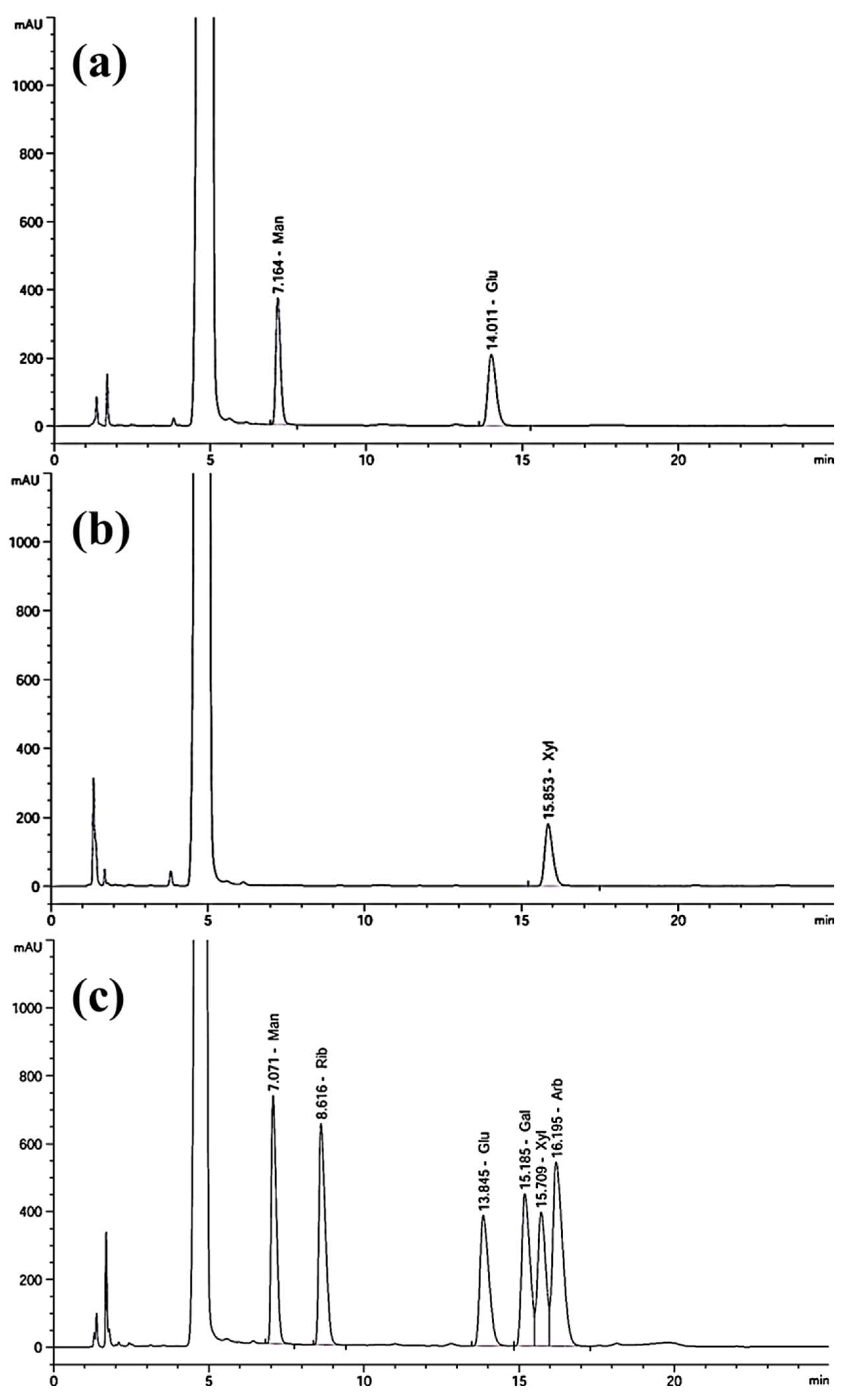
2.5. Monosaccharide Composition Analysis

The monosaccharide composition of EPS was evaluated using HPLC combined with a UV detector (Agilent 1260 Infinity Series, Santa Clara, CA, USA). For the analysis, 10 mg of EPS was dissolved in 10 mL of trifluoroacetic acid (TFA; 4 M), and the mixture was hydrolyzed at 110 °C for 8 h. Then, 200 µL of the hydrolyzed EPS and standard monosaccharide samples (D-(-)-arabinose, D-(-)-glucose, D-(-)-galactose, D-(-)-ribose, D-(-)-mannose, and D-(-)-xylose) were mixed with 240 µL of NaOH (0.3 M) and 240 µL of 3-methyl-1-phenyl-5-pyrazolone (PMP; 0.5 M) prepared in methanol. Afterward, the samples were neutralized using 240 µL of HCl (0.3 M) and vigorously mixed with 1 mL of chloroform. The aqueous layer was then collected and filtered through a 0.22 µm membrane filter for HPLC analysis. The mobile phase consisted of phosphate buffer (0.05 M) and acetonitrile (84:16; v/v), with a flow rate of 1 mL/min. The injection volume was 10 μL and the column temperature was maintained at 40 °C.

2.6. Free Radical Scavenging Assay

The assessment of EPS’s ability to neutralize reactive free radicals was conducted on the ABTS and DPPH free radicals assay. Briefly, the ABTS+ solution was prepared by combining ABTS (7 mM) and potassium persulfate (2.45 mM) at a ratio of 1:0.5 and keeping it in dark conditions for 24 h. The absorbance of the resulting cationic ABTS+ solution was adjusted to 0.7 ± 0.02 with methanol (50%) at 734 nm. For the assay, serially diluted concentrations (15.62 to 1000 μg/mL) of EPS (100 μL) were mixed with ABTS+ (100 μL) and incubated for 10 min at room temperature. Subsequently, the absorbance was measured at 734 nm using a UV-visible spectrophotometer. On the other hand, varying concentrations (ranging from 15.6 to 1000 μg/mL) of EPS (100 μL) were combined with 100 μL of an ethanolic DPPH solution and incubated for 20 min at room temperature. Subsequently, the absorbance of the resulting reaction mixture was measured at 517 nm. The percentage of ABTS and DPPH radicals scavenging was determined using the formula detailed in an earlier investigation [18].

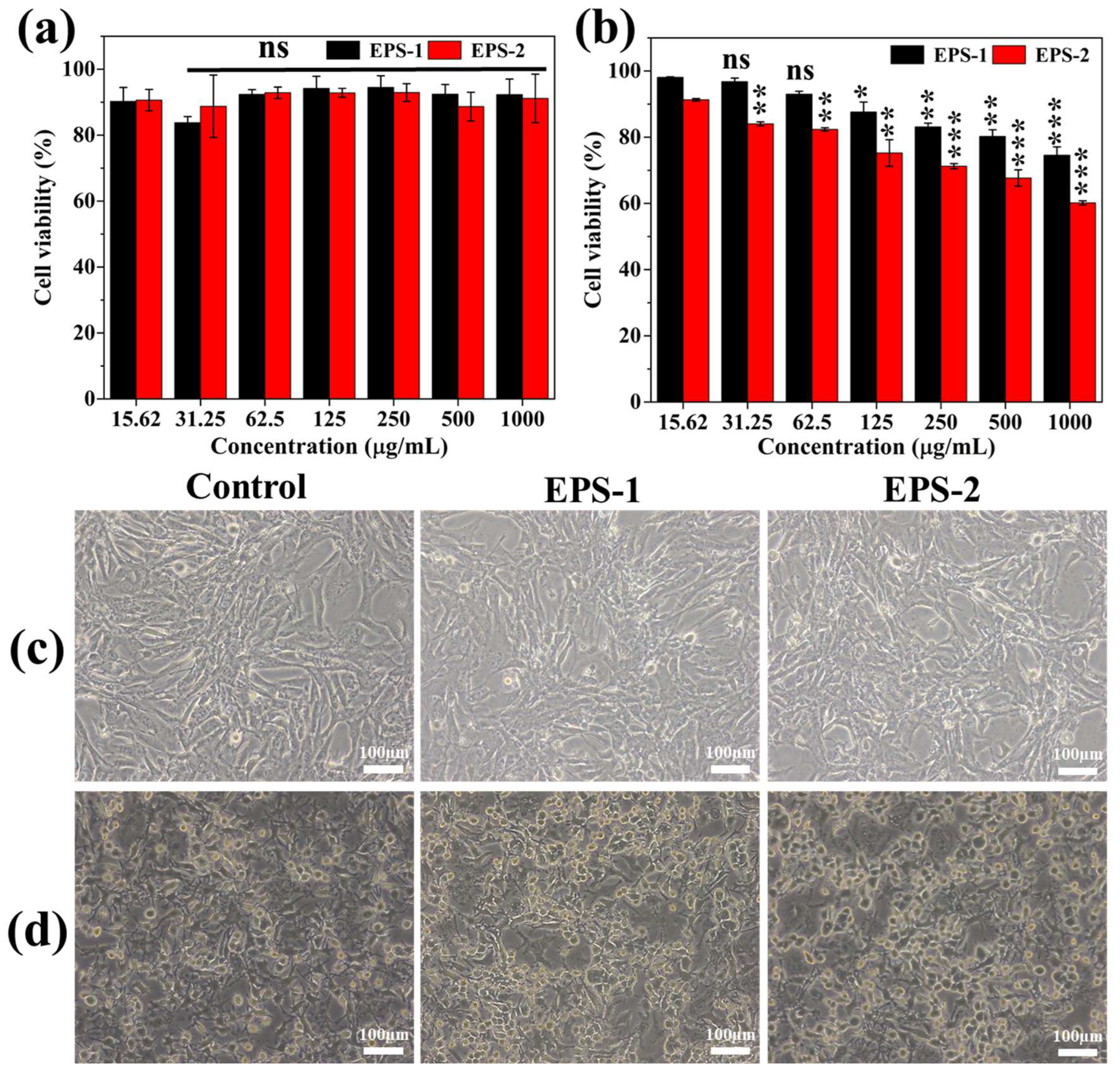
2.7. Cytotoxicity Assay

The potential cytotoxic effects of EPS were evaluated using the WST viability assay kit following the manufacturer’s instructions. Briefly, the NIH3T3 and RAW264.7 cells (1 × 10⁴) were seeded (100 μL; DMEM) into 96-well plates separately and grown overnight in an incubator (5% CO2) at 37 °C. Subsequently, 10 μL of various concentrations (15.62 to 1000 μg/mL) of EPS were added to the respective wells and incubated under the same conditions for another 12 h. Following this incubation period, 10 μL of WST reagent was added to each well and further incubated for 60 min. Then, the optical density was measured at 450 nm using a 96-well plate reader. Finally, the percentage of cytotoxicity was determined using the formula previously described [19]. Furthermore, the results of the WST assays were substantiated by light microscopic examination (in 24 well plates) of samples (EPS-1 and EPS-2)-treated (250 μg/mL) NIH3T3 and RAW264.7 cells.

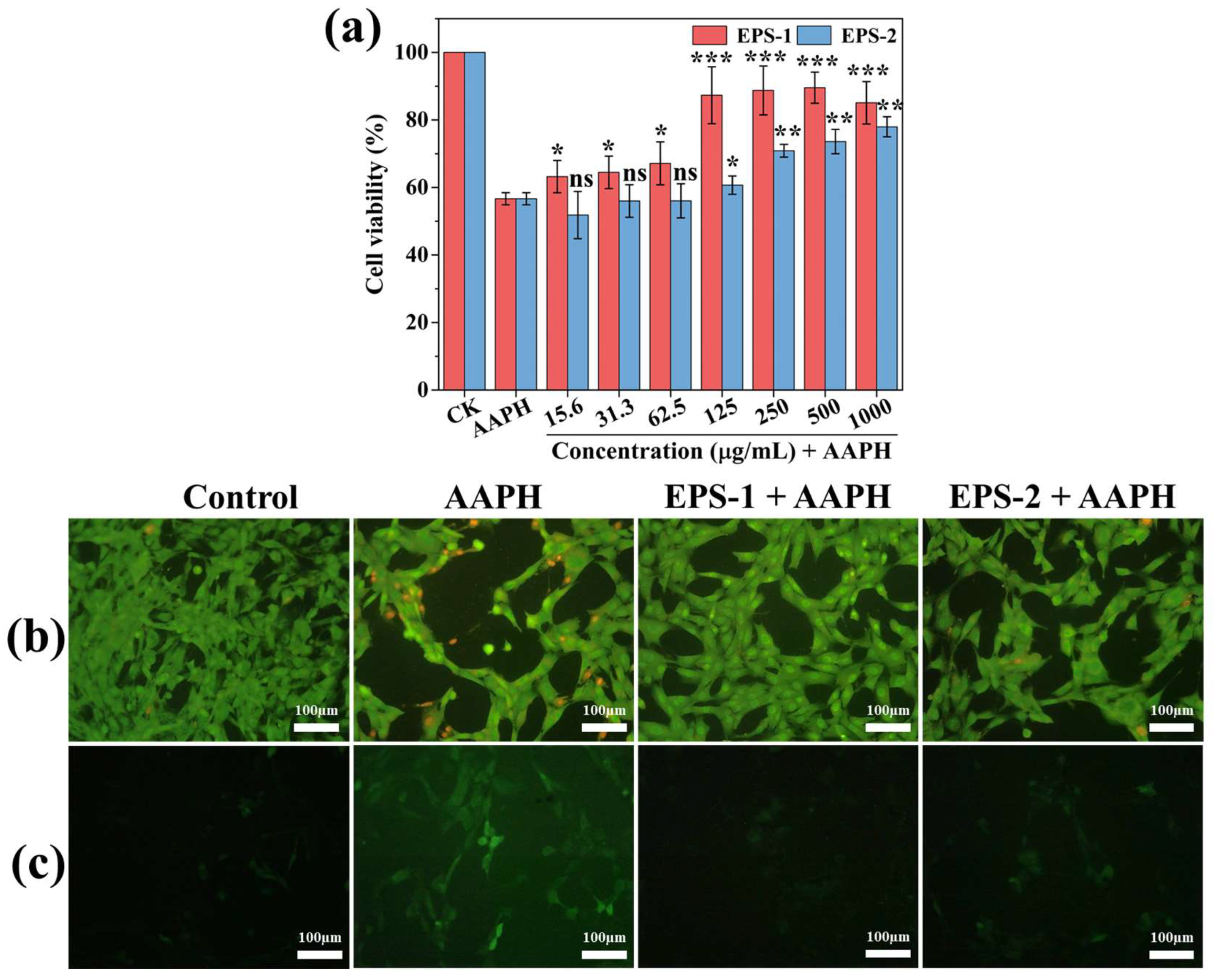
2.8. Cellular Antioxidant Assay

Cellular antioxidant activity of EPS was evaluated in NIH3T3 cells. For the assay, the NIH3T3 cells (1 × 10⁴) were seeded (100 μL; DMEM) into 96-well plates and grown overnight in an incubator (5% CO2) at 37 °C. Then, the EPS was administered at serially diluted concentrations (15.62 to 1000 μg/mL) into the respective wells and incubated further for 4 h at the above-mentioned conditions. Following incubation, the cells were treated with AAPH (500 μg/mL) and incubated for an additional 12 h. Finally, the cytotoxicity was evaluated with the WST kit, and AO/EB dual-staining and DCFH-DA staining was used to investigate the status of reactive oxygen species generation in NIH3T3 cells.

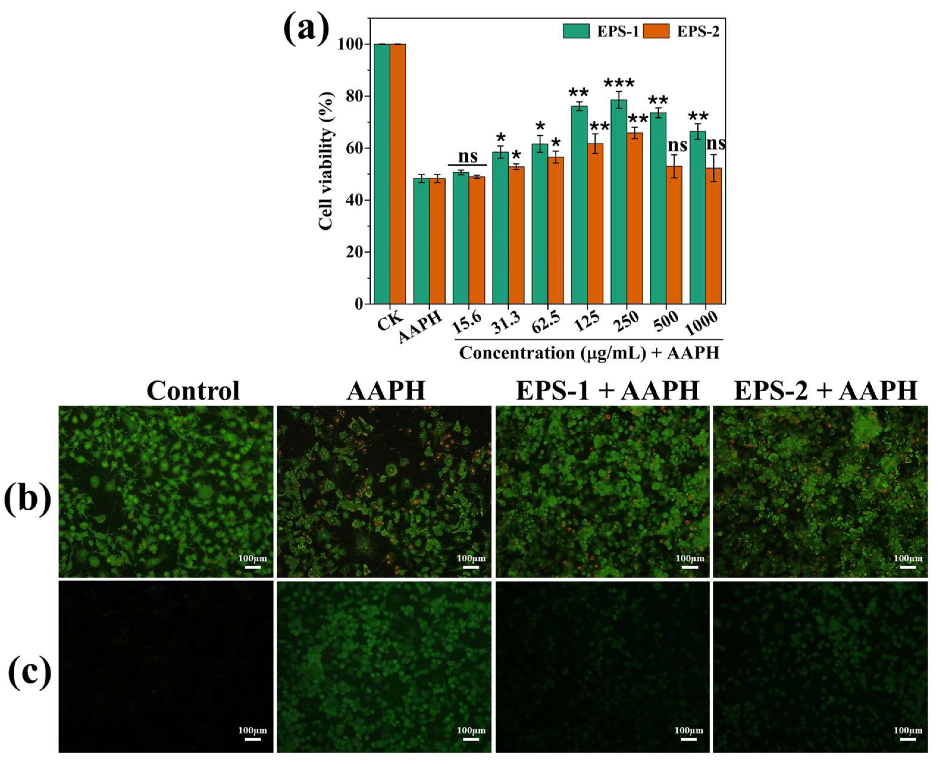
2.9. AAPH-Induced ROS Inhibition Assay

The antioxidant potential of EPS was investigated in AAPH-exposed RAW264.7 cells in terms of their viability by examining reactive oxygen species (ROS) production, and nuclear damage according to the earlier study [20], with some modifications. Specifically, RAW264.7 cells were initially treated with various concentrations (ranging from 15.62 to 1000 μg/mL) of EPS for 4 h and then exposed to AAPH (500 μg/mL), followed by an overnight incubation. On the subsequent day, cell viability was determined using the WST assay. Additionally, the results obtained from the WST assay were confirmed using a fluorescent staining method. The effectiveness of the samples (at a concentration of 250 μg/mL) in mitigating cell death caused by oxidative stress-induced ROS and nucleus damage in AAPH-exposed RAW264.7 cells. The cells were stained with AO/EB dual-staining and DCFH-DA staining solutions for 20 min, observed under a fluorescence microscope, and images were captured at 50 μm scalebar.

2.10. Hemolysis Assay

The hemolytic effect of EPS was assessed on defibrinated sheep blood (Carlina, Republic of Korea) as per a previously established procedure. Initially, the blood was mixed with PBS (pH 7.2) in a 1:10 ratio and the red blood cells (RBCs or erythrocytes) were separated by centrifuging the mix at 2000 rpm for 10 min at 4 °C. The blood plasma (supernatant) was delicately removed, and the RBCs (pellet) were washed with PBS twice. Subsequently, the RBCs were diluted to 4% v/v in PBS, and the experiments were carried out within an ice box. For the assay, the RBCs (200 μL) were added with varying concentrations (ranging from 15.62 to 1000 c) of EPS and incubated for 60 min at 37 °C. Positive and negative controls were established using Triton X-100 and PBS, respectively. Post-incubation, the RBC suspension underwent centrifugation once more at 2000 rpm for 5 min and the supernatant’s absorbance was measured at 540 nm using a multi-well plate reader to calculate the percentage of hemolysis.

2.11. Statistical Analysis

The experimental results are presented as means with standard deviation. Furthermore, a one-way analysis of variance (ANOVA) followed by a post hoc Turkey test (SPSS 26, IBM Inc.) was used to determine the statistical significance of the results. The p-value was lower than 0.05 and was considered statistically significant.

3. Results and Discussion

3.1. EPS Production by L. graminis in MRS Broth

Bacteria generate diverse EPS through various biosynthesis pathways, including the Wzx/Wzy-dependent pathway, synthetase-dependent pathway, ABC transport pathway, and extracellular synthesis pathway. Wzx/Wzy-dependent pathway is the most common biosynthesis mechanism for the production of polysaccharides by Gram-negative bacteria. Specifically, the Wzx/Wzy-dependent pathway begins in the cytoplasm. Firstly, an individual polysaccharide repeated unit (O unit) is translocated to the outer leaflet of the IM by the flippase Wzx, under the regulation of the polysaccharide co-polymerase Wzz, the O unit is constantly polymerized, eventually forming EPS [21,22]. For the ABC transport pathway, free glycosyl units bind to undecaprenyl phosphate on the cell membrane and are continuously catalyzed to form complete polysaccharides. Subsequently, the exocytosis pump complex located on the inner membrane transports them across the inner membrane to the cell surface [23,24]. The synthetase-dependent pathway uses synthetases or membrane subunits to complete polymerization and translocation [25,26]. The extracellular synthesis pathway involves the secretion of sugar precursors and their polymerization at the cell surface, the polymer strand is elongated by the direct addition of monosaccharides obtained by the cleavage of di- or trisaccharides [27]. These pathways necessitate multiple components and involve key stages such as transporting sugars within the cytoplasm, synthesizing sugar-phosphate, creating repetitive units, and polymerization, ultimately resulting in the production of polysaccharides [25]. However, the composition of the culture media affects EPS synthesis. For instance, elevated sugar levels in the culture medium can increase the EPS production in LAB due to an abundant and continual supply of sugar components and increased availability of energy [28]. However, high sugar concentration in the MRS media is the reported reason for the increased production of EPS in Lactobacillus sp. [28,29]. Therefore, in this study, MRS media was used for EPS production from L. graminis strain KNUAS018. The MRS medium is indeed rich in sugars, but exploring waste-derived substrates, such as by-products from the fruit industry, could enhance the circular economy while maintaining high EPS production. For example, agave and bagasse incorporated Brain Heart Infusion (BHI) culture medium increased the EPS production in lactic acid bacteria (LAB), and it had high thermostability. However, the production of EPS was varied among the bacterial strains [30].

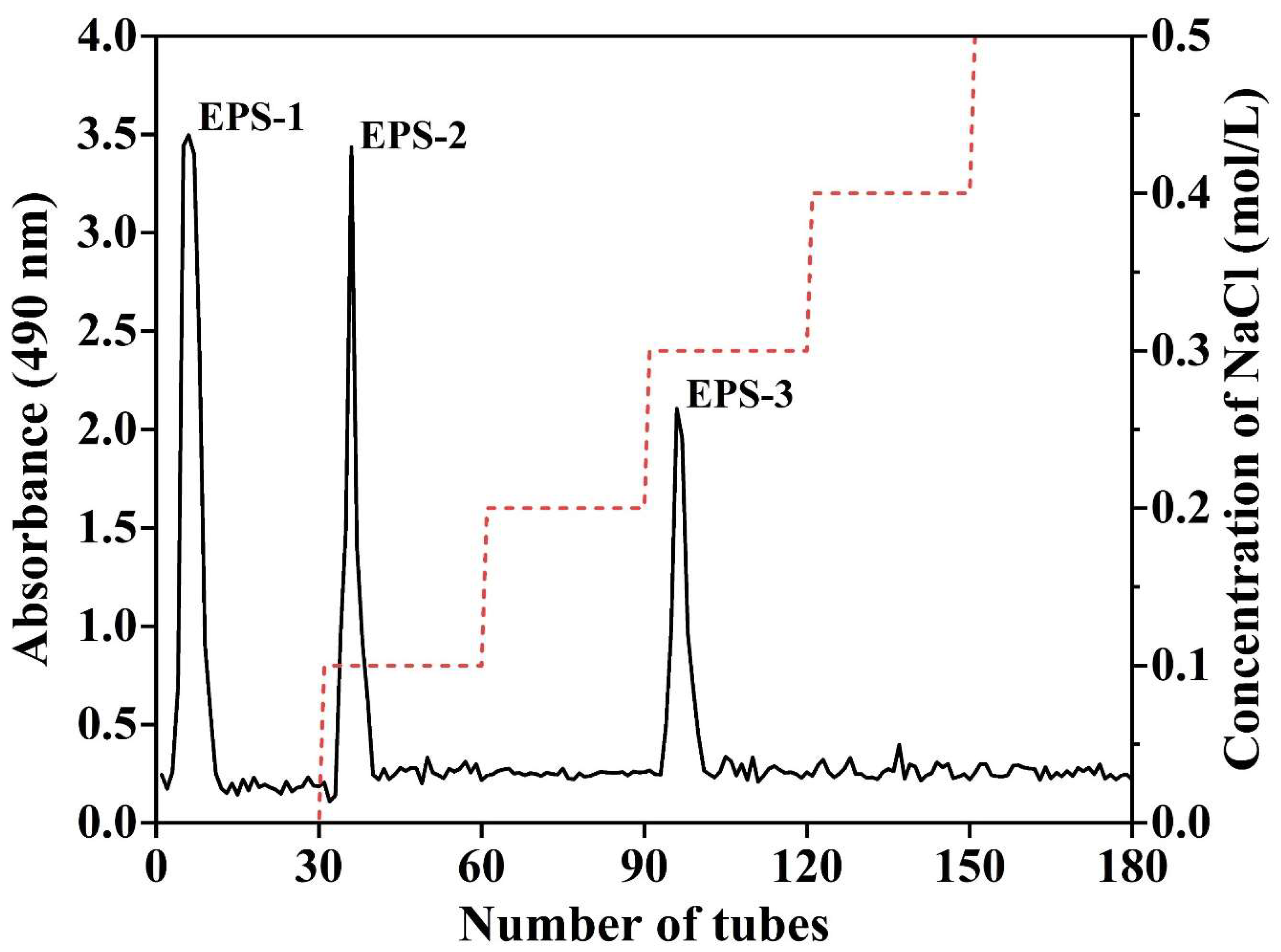
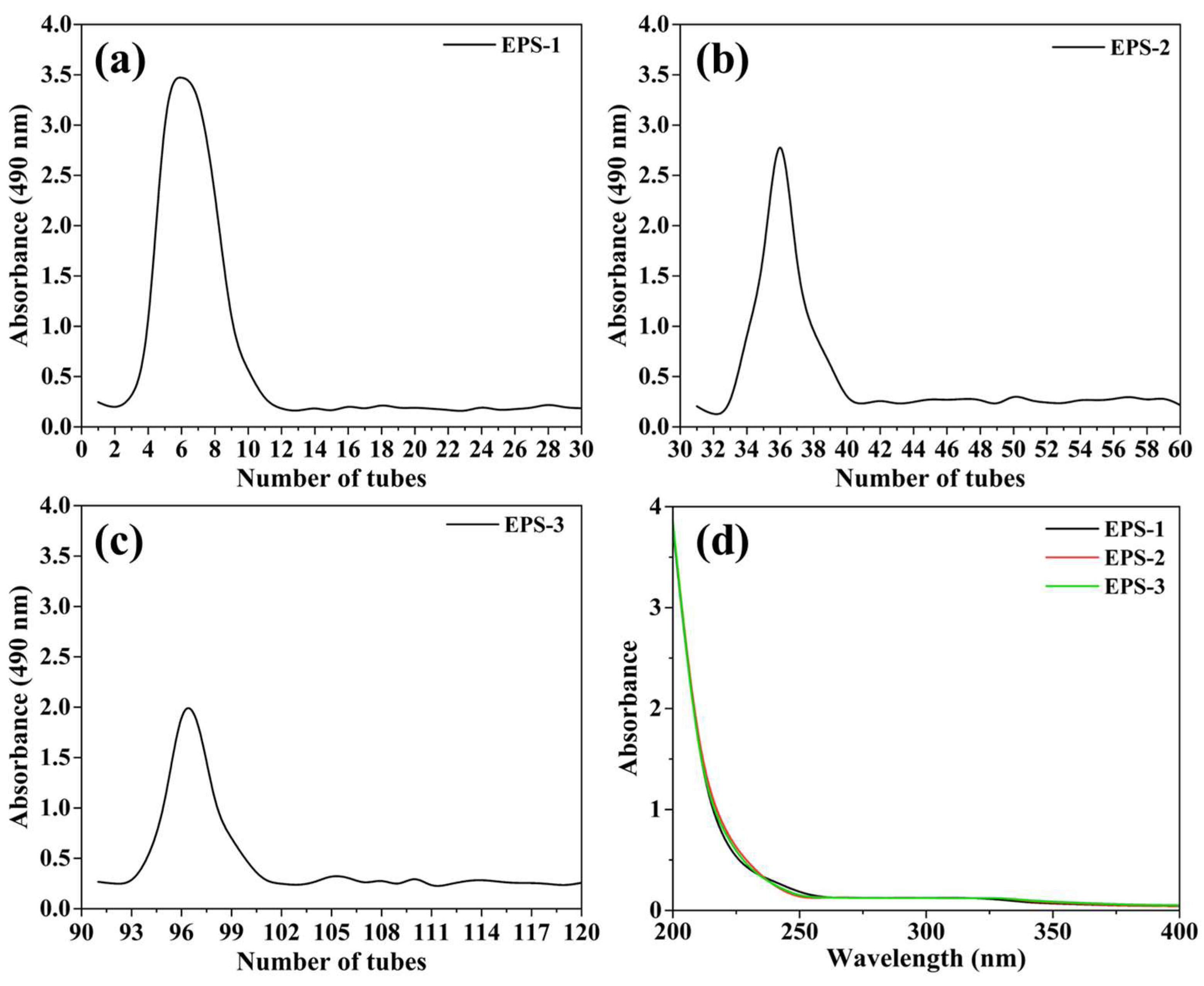
3.2. Isolation, Purification, and Biochemical Composition of EPS
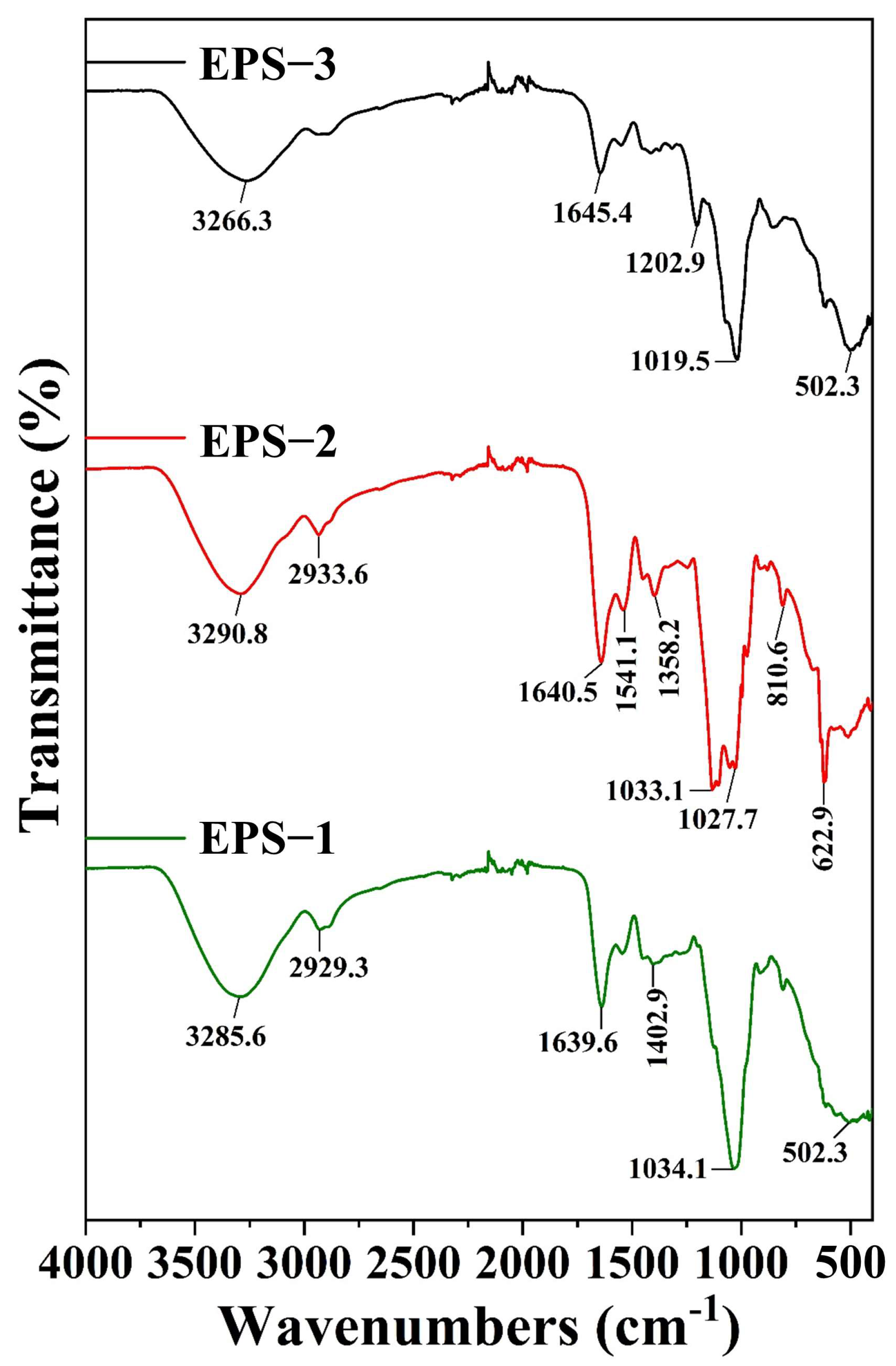
The most commonly used method of crude EPS extraction from LAB is ethanol precipitation [29]. This study utilized cold ethanol (95%) for the precipitation of EPS from the culture supernatant of L. graminis in a 3:1 ratio, which is in line with earlier works [31,32]. The dried weight (DW) of the crude EPS was measured as 1.46 g, as shown in Table 1.
However, crude EPS could contain pigments, proteins, inorganic salts, and non-polar substances. Therefore, the extracted crude EPS underwent deproteinization and decolorization using the Sevage technique, while additional impurities were removed through the dialysis process. Furthermore, the crude EPS underwent fractionation using a DEAE-Sepharose fast flow column, employing distilled water (H2O) and various molar concentrations of NaCl (0.1, 0.2, 0.3, 0.4, and 0.5 M) for elution. Fractions (5 mL each) were collected at 150 min intervals, and total carbohydrate content was determined via the phenol-sulfuric acid assay. A total of 180 tubes were gathered based on different eluents (H2O, 0.1, 0.2, 0.3, 0.4, and 0.5 M NaCl). The elution profile exhibited a total of three fractions (peaks), where there was one pronounced peak in the H2O fraction (EPS-1), one peak in the 0.1 M NaCl fraction (EPS-2), and one peak was observed in the 0.3 M NaCl fraction (EPS-3), suggesting the absence of polysaccharides in other NaCl (0.1, 0.4, and 0.5 M) fractions (Figure 1). However, EPS-1, EPS-2, and EPS-3 indicated a single and symmetrical peak (Figure 2a–c) with a total yield of 14.38%, 9.24%, and 1.78%, respectively (Table 1). Additionally, the chemical composition analysis of samples (EPS-1, EPS-2, and EPS-3) was conducted, and the results are summarized in Table 1. The TPC (mg of GAE/g) of EPS-1, EPS-2, and EPS-3 was found as 0.21 ± 0.02, 0.16 ± 0.01, and 0.09 ± 0.01, respectively. While the TFC (mg of QE/g) was detected as 0.07 ± 0.008 for EPS-1 and 0.04 ± 0.002 for EPS-2, the TFC was not found in EPS-3. An earlier study also reports a trace amount of total phenol (0.38%) and total flavonoids (0.08%) in the polysaccharides isolated from LAB Weissella cibaria [32]. The presence of nucleic acids was not detected in either of the samples (EPS-1, EPS-2, and EPS-3) (Table 1 and Figure 2d).
It is reported that the absence of signals between 260 nm and 280 nm indicated the lack of nucleic acid in the EPS [33]. Similarly, previous research confirms the absence of nucleic acid in the polysaccharides isolated from L. gasseri FR4 using UV–vis spectrophotometry [34]. Moreover, EPS-1 and EPS-2 were not detected for protein presence, while EPS-3 showed a trace (0.03 ± 0.001%) of proteins (Table 1) and corroborated previous findings. A previous study also reports a trace of proteins (1.38%) in the EPS extracted from L. plantarum YW11 [33].

3.3. FT-IR Analysis

The functional groups of the samples (EPS-1, EPS-2, and EPS-3) were analyzed using FT-IR spectroscopy, as shown in Figure 3. The spectrum of EPS-1 indicates peaks at 3285.6 cm−1, 2929.3 cm−1, 1639.6 cm−1, 1402.9 cm−1, 1034.1 cm−1, and 502.3 cm−1. Moreover, EPS-2 showed peaks at 3290.8 cm−1, 2933.6 cm−1, 1640.5 cm−1, 1541.1 cm−1, 1358.2 cm−1, 1033.1 cm−1, 1027.7 cm−1, 810.6 cm−1, and 622.9 cm−1. However, the peaks for EPS-3 were found at 3266.3 cm−1, 1645.4 cm−1, 1202.9 cm−1, 1019.5 cm−1, and 502.3 cm−1. Altogether, the peaks at 3290.8 cm−1, 3285.6 cm−1, and 3266.3 cm−1 correspond to the O-H stretching, and the peaks at 2933.6 cm−1 and 2929.3 cm−1 were associated with C-H stretching [35]. Meanwhile, the peaks at 1645.4 cm−1, 1640.5 cm−1, 1639.6 cm−1, 1541.1 cm−1, 1402.9 cm−1, and 1358.2 cm−1 were related to C=O stretching [36,37]. The characteristic peaks in the FTIR spectrum of a polysaccharide, located between 1320 cm−1 and 1000 cm−1 could be attributed to the C-O stretching of alcohols, carboxylic acids, esters, and ether groups [37]. The peak at 810.6 cm−1 suggests the presence of β-pyranose in carbohydrates [38]. In addition, the peaks at 622.9 cm−1 and 502.3 cm−1 indicate C-H bending and the presence of sulfate groups, respectively [36,39].

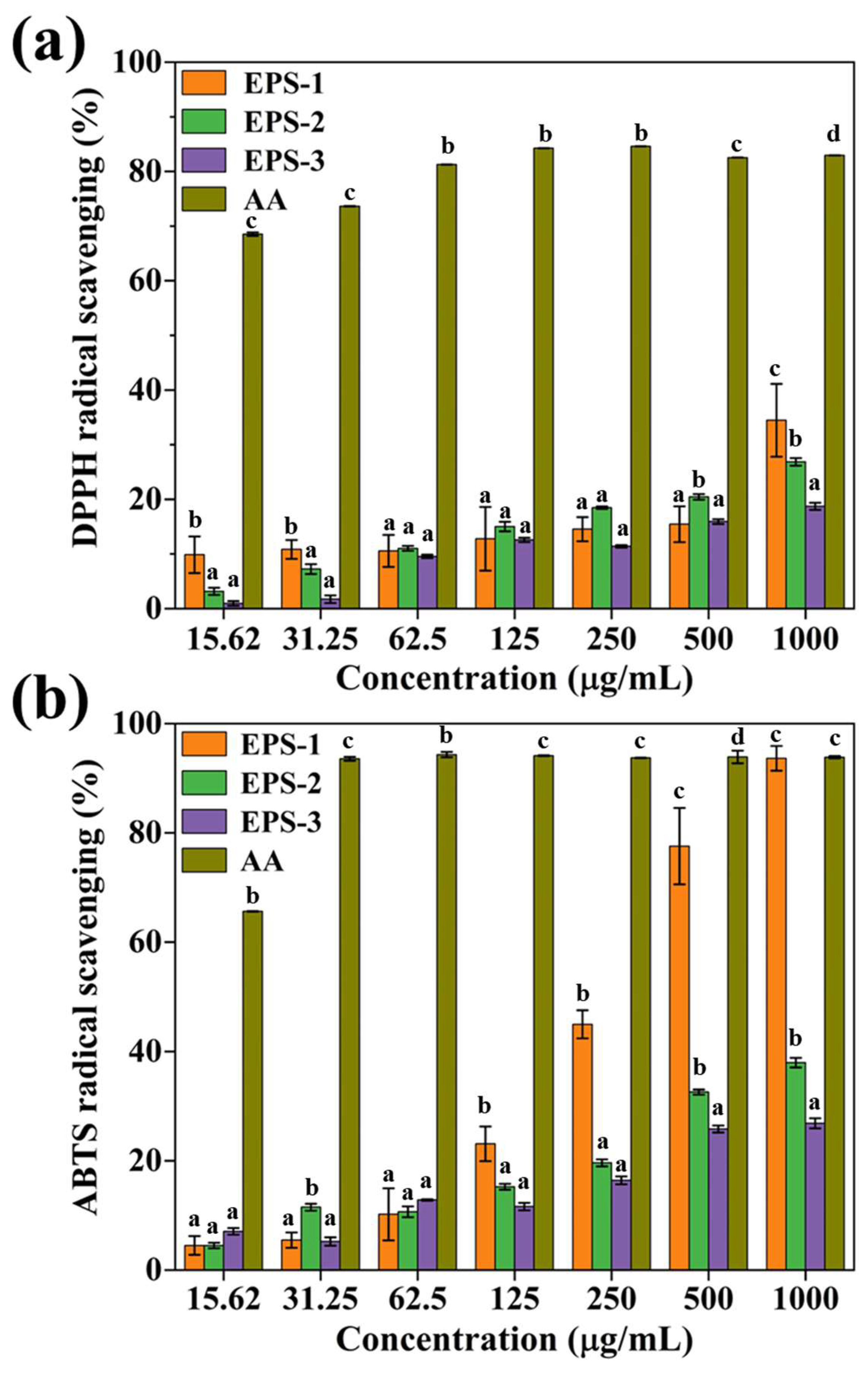
3.4. Free Radical Scavenging Activity

Exposure to elevated levels of free radicals can lead to oxidative stress-induced cell damage, contributing to various diseases. EPS sourced from natural origins is gaining increased attention due to its biocompatibility and antioxidant characteristics [29]. However, the DPPH and ABTS assays are among the widely used methods to evaluate the scavenging abilities of natural compounds against free radicals. In this study, the free radical scavenging abilities of the samples (EPS-1, EPS-2, and EPS-3) were tested against DPPH and ABTS radicals and compared to positive control AA (Figure 4). The DPPH free radical is considered a stable radical that possesses an unpaired valence electron at one atom of nitrogen bridge, but can be significantly reduced when subjected to proto radical scavenger [38]. It has been reported that the EPS derived from various LAB strains display DPPH scavenging capabilities [40]. As per our DPPH assay findings, among the EPSs, the highest tested concentration of (1 mg/mL) EPS-1 exhibited 34.5 ± 6.6% of DPPH scavenging (Figure 4a). However, in previous research, EPS isolated from L. plantarum YW32 displayed 30% of DPPH scavenging activity [41], which was lower compared to the EPS-1 isolated in this study. In the ABTS assay, the transfer of an electron from an antioxidant to ABTS+ demonstrates its capacity to scavenge ABTS+. The results indicate that the ABTS+ scavenging efficacy of EPS-1, EPS-2, and EPS-3 were found to be concentration-dependent. The ABTS+ scavenging of EPS-2 and EPS-3 ranged from 4.5 to 38%. In addition, the EPS-1 indicated 93.6 ± 2.3% of ABTS radical scavenging at 1 mg/mL of tested concentration, which was nearly similar to the same dose of AA (Figure 4b). However, in another study, the ABTS+ scavenging activity of EPS isolated from LAB W. cibaria YB-1 was found to be lower (82.20 ± 1.8%,) compared to EPS-1 [42]. Similarly, even the high dose (5 mg/mL) of EPS derived from L. casei showed ~60% ABTS radicals scavenging activity [43]. The higher scavenging activities of EPS for ABTS+ than for DPPH radicals could be associated with their affinity properties, such as the hydrophilicity of ABTS and hydrophobicity of DPPH [44].

3.5. Monosaccharide Composition of EPS

Even though EPS-3 did not exhibit substantial free radical scavenging activity (DPPH and ABTS radicals) compared to EPS-1 and EPS-2 (Figure 4), EPS-1 and EPS-2 were selected for further analysis. In addition, none of the polysaccharides were isolated from L. graminis not show antibacterial activity. Hence, the monosaccharide composition of EPS-1 and EPS-2 from L. graminis was determined using HPLC (Figure 5). The results reveal that EPS-1 consists of a monosaccharide combination with retention times corresponding to mannose (7.164 min) and glucose (14.011 min) (Figure 5a). The percentage composition of mannose and glucose was 50.67% and 49.32%, respectively. These higher monosaccharide contents are likely produced predominantly during microbial metabolic pathways. Interestingly, EPS-2 exhibited only one monosaccharide peak, corresponding to xylose (15.853 min) (Figure 5b). Additionally, EPS-2 did not show any additional peaks up to a retention time of 25 min, suggesting that it predominantly contains xylose compared to other sugar molecules in the standard. However, the monosaccharide composition in this study was evaluated with a maximum retention time of 25 min, as limited by the availability of standard sugar molecules. The retention times of the detected sugar molecules were appropriately matched with standard monosaccharide compositions (Figure 5c). The previous study evidenced that the polysaccharide isolated from L. casei contains the monosaccharide compositions of glucose (16.03%), mannose (6.94%), arabinose (12.16%), galactose (19.14%), rhamnose (7.93%), fucose (5.30%), ribose (2.95%), Gulcuronic acid (12.37%), Galacturonic acid (6.45%), and xylose (10.72%) [43]. Similarly, L. planetarium JLAU103 contains the monosaccharide composition with the approximate molar ratio arabinose (4.05), rhamnose (6.04), fucose (6.29), xylose (5.22), mannose (1.47), fructose (5.21), galactose (2.24), and glucose (1.83) [45]. However, L. sanfranciscensis possesses only one monosaccharide as glucose due to the pure glucan in the composition [46].

3.6. Cytotoxic Activity

The cytotoxic effect of EPS-1 and EPS-2 was assessed in NIH3T3 and RAW264.7 cell lines using a WST viability assay kit, as presented in Figure 6. The treatment of IPS-1 and IPS-2 did not indicate a noteworthy reduction in the viability of NIH3T3 cells. Even at the highest tested dose (1 mg/mL) of samples (EPS-1 and EPS-2) the cell viability of found >85% (Figure 6a). Similarly, the EPS derived from L. rhamnosus ZY is reported for its nontoxic impact on NIH3T3 cells [47]. However, the EPS-1 and EPS-2 indicated a slight toxic effect in RAW264.7 cells, as shown in Figure 6b. However, the IPS-2 treatments showed slightly higher cytotoxicity compared to IPS-1 treatments, yet the viability of RAW264.7 cells was found to be >75% for the highest tested concentration (1 mg/mL) of IPS-1. In a previous study, polysaccharides from natural sources are reported for their nontoxic impact in RAW264.7 cells [48]. Furthermore, the results of the WST assays were substantiated by light microscopic examination of sample-treated NIH3T3 and RAW264.7 cells. The treatment of IPS-1 and IPS-2 did not indicate any irregular cell morphology of NIH3T3 cells as compared to non-treated (control) cells (Figure 6c). However, the samples (IPS-1 and IPS-2) treatment posed some morphological discrepancies, yet insignificant changes in the cell population of RAW264.7 cells compared to untreated (control) cells (Figure 6d).

3.7. Cellular Antioxidant Activity

It has been reported that the water-soluble azo compound AAPH generates intracellular free radicals, which in turn induces DNA damage and apoptotic cell death [49]. Therefore, we investigated the cellular antioxidant effect of EPS-1 and EPS-2 on NIH3T3 cells exposed to AAPH (Figure 7). The findings indicate that the viability of NIH3T3 cells notably declined (up to 55%) with AAPH treatment, whereas cell viability enhanced in the presence of EPS-1. Although the highest tested dose (1 mg/mL) of EPS-2 also enhanced the cell viability to ~75%, the moderate dose (0.25–0.5 mg/mL) of EPS-2 showed >85% of NIH3T3 cell viability (Figure 7a). Similarly, an earlier study reports that polysaccharides from natural sources decrease the intracellular ROS level and increase the viability of AAPH-stressed Vero cells [50]. Furthermore, in AO/EB dual-staining analysis, the AAPH treatment indicated some red fluorescent (necrotic) cells with fewer populations of NIH3T3 cells as compared to control groups; however, the IPS-1 treatment indicated green fluorescent (live) healthy cells with regular cellular morphology and higher cells population than IPS-2 treated groups (Figure 7b). Moreover, DCFH-DA staining analysis indicated a high ROS level in AAPH-treated cells, while samples (EPS-1 and EPS-2) treatment indicated a reduction in the AAPH-induced ROS production, and the ROS level was found similar to control groups (Figure 7b). Overall, the cellular antioxidant assay suggested that IPS-1 treatment minimizes the AAPH-induced ROS production and associated morphological damage in NIH3T3 cells.

3.8. AAPH-Induced ROS Inhibition Assay

Macrophages are essential components of the host’s defensive mechanism against malignancies and microbial diseases. Their functions include digesting antigens for lymphocytes, phagocytosing cell debris, and releasing cytotoxic chemicals and pro- and anti-inflammatory cytokines to influence the host’s immunological response [51]. The macrophage-like cell line RAW264.7 is one of the most often used cell models in anti-inflammatory studies. Therefore, the anti-inflammatory effects of the samples (EPS-1 and EPS-2) were studied in AAPH-exposed RAW264.7 cells and are presented in Figure 8. It is reported that AAPH-derived free radicals increase the ROS status in the cytosol of RAW264.7 cells and result in oxidative stress associated with inflammation [52]. Compared to control cells, the AAPH treatment indicated a significant reduction (~50%) in the viability of RAW264.7 cells. While samples of (EPA-1 and EPS-2)-treated cells were found for elevated viability, where a moderate dose (125–250 μg/mL) of EPS-1 showed comparatively higher viability (~80%) than EPS-2-treated (~60%) AAPH-stressed RAW264.7 cells (Figure 8a). Moreover, EPS-1 treatment in AAPH-stressed RAW264.7 cells indicated a protective effect against necrotic damage in the AO/EB dual-staining assay (Figure 8b) and reduced ROS level (similar to control cells) in the DCFH-DA staining examination (Figure 8c). These findings indicate that EPS-1 perhaps prevents AAPH stress-induced inflammation in RAW264.7 cells. It has been reported that EPS derived from L. planetarium JLAU103 facilitates anti-inflammatory effects in lipopolysaccharide-stimulated RAW264.7 cells by reducing the release of pro-inflammatory cytokines such as TNF-α and IL-6 [53]. Similarly, another study indicates that L. plantarum LRCC5310-derived EPS treatment decreases the level of pro-inflammatory cytokines (TNF-α and IL-β) compared to LPS-treated RAW264.7 cells [54]. Moreover, the bioactivities of some recently investigated EPS derived from LAB are summarized in Table 2.

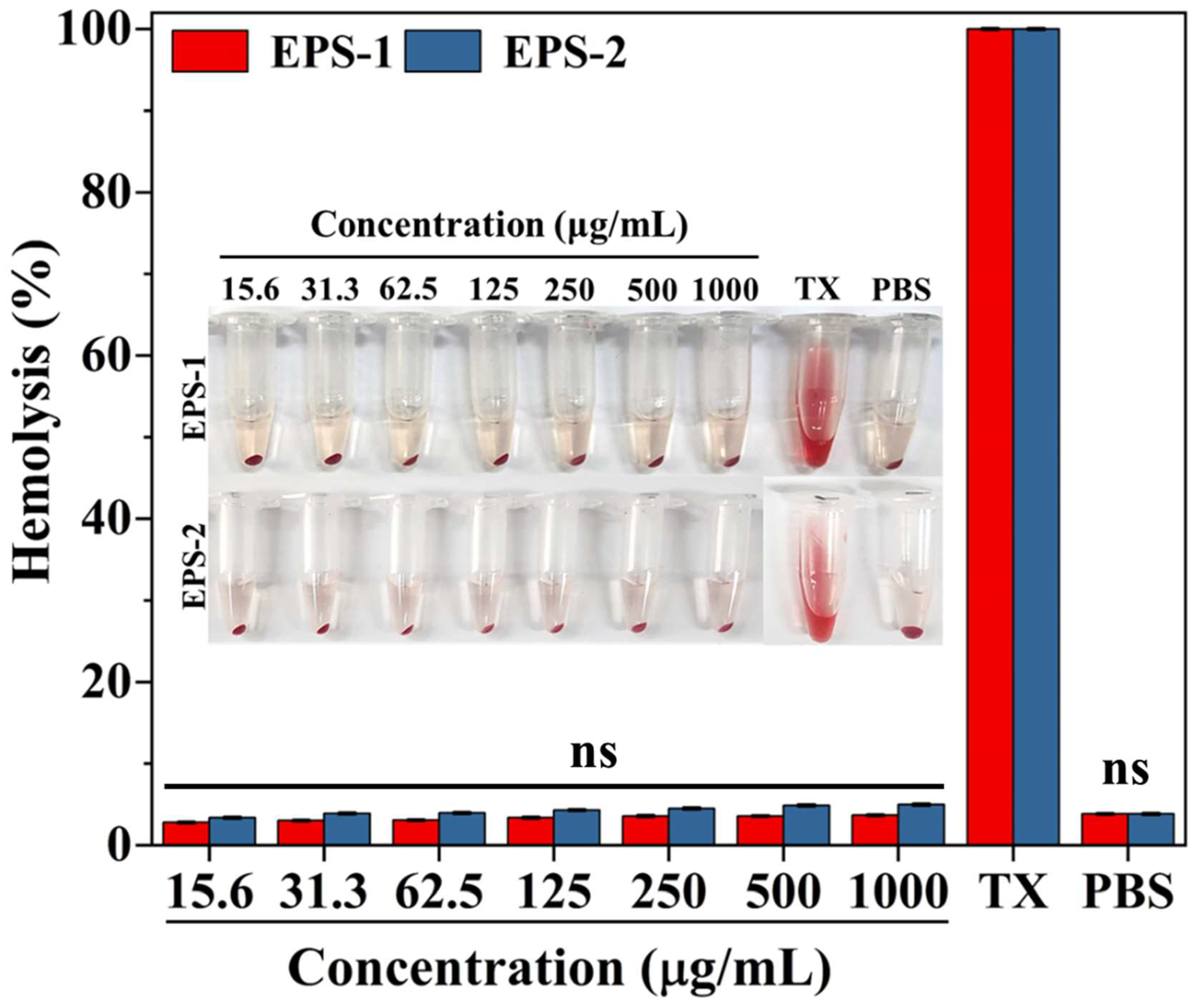
3.9. Hemolytic Activity

The unique characteristics of erythrocytes’ cell membranes enable them to deform, aggregate, or even alter their metabolism in response to changes in their internal environment, making them ideal for studying the direct effects of stimuli [58]. Therefore, the effect of EPS-1 and EPS-2 on the membranes of RBCs was assessed using a hemolysis assay, and the findings are presented in Figure 9. Both the samples (EPS-1 and EPS-2) did not induce significant hemolysis at the tested concentrations (0.0156 to 1 mg/mL), as compared to the positive control (Triton X-100). Even at the highest tested dose (1 mg/mL) of EPS-1 and EPS-2, the hemolysis was found at ~5%, indicating their compatibility with the plasma membrane of RBCs (Figure 9). An earlier investigation also reports that the polysaccharides isolated from Weissella cibaria did not induce hemolysis and so were biocompatible in RBCs [59].

4. Conclusions

EPS isolated from LAB have gained recognition for their potential as a prebiotic in the both food and medical domains and have drawn considerable attention for their industrial utilization. Despite this, there is no documented research in the existing literature regarding the isolation, chemical characterization, antioxidants, and anti-inflammatory activities, as well as cytotoxicity evaluations, of EPS derived from L. graminis strain KNUAS018. In addressing this gap, this study focused on the isolation of three EPS fractions (EPS-1, EPS-2, and EPS-3) from L. graminis, undertaking a comprehensive analysis of their chemical compositions and bioactivities. However, EPS-1 and EPS-2 exhibited high yield and antioxidant activities; particularly, EPS-1 showed superior free radical scavenging activities (ABTS+ and DPPH radicals). In addition, EPS-1 indicated cellular antioxidant effects in AAPH-stressed NIH3T3 cells and antioxidant effects in AAPH-induced RAW264.7 cells via reducing ROS levels. Moreover, no significant cytotoxic impacts were found in EPS-1-treated non-cancer (NIH3T3 and RAW264.7) cells and erythrocytes. Overall, the results of this study imply that EPS derived from L. graminis strain KNUAS018 could function as a lead compound to develop antioxidant agents. However, further research is warranted to elucidate the structural properties of EPS-1 and reveal their exact molecular mechanisms and structure–activity relationships.

Author Contributions

Conceptualization, A.S.; methodology, K.H., K.V.N., and A.S.; software, X.Z.; validation, A.S. and H.-Y.K.; formal analysis, K.H., K.V.N., and X.Z.; investigation, K.H., X.Z., and K.V.N.; resources, H.-Y.K.; data curation, K.H. and X.Z.; writing—original draft preparation, K.V.N.; writing—review and editing, A.S.; supervision, H.-Y.K.; project administration, H.-Y.K.; funding acquisition, A.S. and H.-Y.K. All authors have read and agreed to the published version of the manuscript.

Funding

This work was supported by the 2023 Kangwon National University Development Project and supported by the National Research Foundation of Korea (2021R111A1A01057742; 2022R1A2C2091029).

Institutional Review Board Statement

Not applicable.

Data Availability Statement

All results were presented within the manuscript.

Conflicts of Interest

The authors declare no competing financial interest in this study.

References

  1. Kang, E.; Lee, J.; Seo, S.; Uddin, S.; Lee, S.; Han, S.B.; Cho, S. Regulation of anti-inflammatory and antioxidant responses by methanol extract of Mikania cordata (Burm. f.) BL Rob. leaves via the inactivation of NF-κB and MAPK signaling pathways and activation of Nrf2 in LPS-induced RAW 264.7 macrophages. Biomed. Pharmacother. 2023, 168, 115746. [Google Scholar] [CrossRef] [PubMed]
  2. Song, X.; Liu, Z.; Zhang, J.; Yang, Q.; Ren, Z.; Zhang, C.; Liu, M.; Gao, Z.; Zhao, H.; Jia, L. Anti-inflammatory and hepatoprotective effects of exopolysaccharides isolated from Pleurotus geesteranus on alcohol-induced liver injury. Sci. Rep. 2018, 8, 10493. [Google Scholar] [CrossRef]
  3. Ghavidel, F.; Amiri, H.; Tabrizi, M.H.; Hosseini, H.; Sahebkar, A. The combinational effect of inulin and resveratrol on the oxidative stress and inflammation level in a rat model of diabetic nephropathy. Curr. Dev. Nutr. 2023, 8, 102059. [Google Scholar] [CrossRef] [PubMed]
  4. Huang, Y.-J.; Nan, G.-X. Oxidative stress-induced angiogenesis. J. Clin. Neurosci. 2019, 63, 13–16. [Google Scholar] [CrossRef] [PubMed]
  5. Shoura, S.M.S.; Naghsh, N.; Moslemi, E.; Kavyani, Z.; Moridpour, A.H.; Musazadeh, V.; Dehghan, P. Can resveratrol supplementation affect biomarkers of inflammation and oxidative stress? An umbrella meta-analysis. J. Funct. Foods 2022, 99, 105360. [Google Scholar] [CrossRef]
  6. Gómez, P.I.; Mayorga, J.; Flaig, D.; Castro-Varela, P.; Jaupi, A.; Ulloa, P.A.; Soto-Bartierra, J.; Henríquez, V.; Rojas, V. Looking beyond Arthrospira: Comparison of antioxidant and anti-inflammatory properties of ten cyanobacteria strains. Algal Res. 2023, 74, 103182. [Google Scholar] [CrossRef]
  7. Allu, I.; Kumar Sahi, A.; Kumari, P.; Sakhile, K.; Sionkowska, A.; Gundu, S. A brief review on cerium oxide (CeO2NPs)-based scaffolds: Recent advances in wound healing applications. Micromachines 2023, 14, 865. [Google Scholar] [CrossRef]
  8. Shen, M.; Wang, Y.; Bing, T.; Tang, Y.; Liu, X.; Yu, Y. Alendronate triggered dual-cascade targeting prodrug nanoparticles for enhanced tumor penetration and sting activation of osteosarcoma. Adv. Funct. Mater. 2023, 33, 2307013. [Google Scholar] [CrossRef]
  9. Zhang, L.; Liu, C.; Li, D.; Zhao, Y.; Zhang, X.; Zeng, X.; Yang, Z.; Li, S. Antioxidant activity of an exopolysaccharide isolated from Lactobacillus plantarum C88. Int. J. Biol. Macromol. 2013, 54, 270–275. [Google Scholar] [CrossRef]
  10. Abdel-Wahab, B.A.; F. Abd El-Kareem, H.; Alzamami, A.; A. Fahmy, C.; H. Elesawy, B.; Mostafa Mahmoud, M.; Ghareeb, A.; El Askary, A.; H. Abo Nahas, H.; G. M. Attallah, N.; et al. Novel Exopolysaccharide from Marine Bacillus subtilis with Broad Potential Biological Activities: Insights into Antioxidant, Anti-Inflammatory, Cytotoxicity, and Anti-Alzheimer Activity. Metabolites 2022, 12, 715. [Google Scholar] [CrossRef]
  11. Angelin, J.; Kavitha, M. Exopolysaccharides from probiotic bacteria and their health potential. Int. J. Biol. Macromol. 2020, 162, 853–865. [Google Scholar] [CrossRef]
  12. Zamora-Pineda, J.; Kalinina, O.; Sperling, A.I.; Knight, K.L. Mechanism of TLR4-Mediated Anti-Inflammatory Response Induced by Exopolysaccharide from the Probiotic Bacillus subtilis. J. Immunol. 2023, 211, 1232–1239. [Google Scholar] [CrossRef] [PubMed]
  13. Mahmoud, M.G.; Mohamed, S.S.; Ibrahim, A.Y.; El Awady, M.E.; Youness, E.R. Exopolysaccharide produced by paenibacillus lactes NRC1: Its characterization and anti-inflammatory activity via cyclooxygenases inhibitory activity and modulation of inflammation related cytokines. Der Pharma Chem. 2016, 8, 16–26. [Google Scholar]
  14. Mgomi, F.C.; Yang, Y.-R.; Cheng, G.; Yang, Z.-Q. Lactic acid bacteria biofilms and their antimicrobial potential against pathogenic microorganisms. Biofilm 2023, 5, 100118. [Google Scholar] [CrossRef] [PubMed]
  15. Zhu, Y.; Wang, C.; Jia, S.; Wang, B.; Zhou, K.; Chen, S.; Yang, Y.; Liu, S. Purification, characterization and antioxidant activity of the exopolysaccharide from Weissella cibaria SJ14 isolated from Sichuan paocai. Int. J. Biol. Macromol. 2018, 115, 820–828. [Google Scholar] [CrossRef]
  16. Sathiyaseelan, A.; Saravanakumar, K.; Han, K.; Naveen, K.V.; Wang, M.-H. Antioxidant and Antibacterial Effects of Potential Probiotics Isolated from Korean Fermented Foods. Int. J. Mol. Sci. 2022, 23, 10062. [Google Scholar] [CrossRef]
  17. Bradford, M.M. A rapid and sensitive method for the quantitation of microgram quantities of protein utilizing the principle of protein-dye binding. Anal. Biochem. 1976, 72, 248–254. [Google Scholar] [CrossRef]
  18. Kashyap, P.K.; Chauhan, S.; Negi, Y.S.; Goel, N.K.; Rattan, S. Biocompatible carboxymethyl chitosan-modified glass ionomer cement with enhanced mechanical and anti-bacterial properties. Int. J. Biol. Macromol. 2022, 223, 1506–1520. [Google Scholar] [CrossRef]
  19. Naveen, K.V.; Park, S.; Saravanakumar, K.; Sathiyaseelan, A.; Wang, M.-H. Ultraperformance liquid chromatography-based metabolite profiling and cytotoxic activity of the ethyl acetate extract of two endophytic Penicillium sp. Process Biochem. 2023, 130, 366–378. [Google Scholar] [CrossRef]
  20. Sathiyaseelan, A.; Saravanakumar, K.; Wang, M.-H. Cerium oxide decorated 5-fluorouracil loaded chitosan nanoparticles for treatment of hepatocellular carcinoma. Int. J. Biol. Macromol. 2022, 216, 52–64. [Google Scholar] [CrossRef]
  21. Islam, S.T.; Lam, J.S. Synthesis of bacterial polysaccharides via the Wzx/Wzy-dependent pathway. Can. J. Microbiol. 2014, 60, 697–716. [Google Scholar] [CrossRef] [PubMed]
  22. Becker, A. Challenges and perspectives in combinatorial assembly of novel exopolysaccharide biosynthesis pathways. Front. Microbiol. 2015, 6, 687. [Google Scholar] [CrossRef] [PubMed]
  23. Zhang, L.; Fan, C.; Liu, S.; Zang, Z.; Jiao, L.; Zhang, L. Chemical composition and antitumor activity of polysaccharide from Inonotus obliquus. J. Med. Plants Res. 2011, 5, 1251–1260. [Google Scholar]
  24. Greenfield, L.K.; Whitfield, C. Synthesis of lipopolysaccharide O-antigens by ABC transporter-dependent pathways. Carbohydr. Res. 2012, 356, 12–24. [Google Scholar] [CrossRef]
  25. Schmid, J.; Sieber, V.; Rehm, B. Bacterial exopolysaccharides: Biosynthesis pathways and engineering strategies. Front. Microbiol. 2015, 6, 496. [Google Scholar] [CrossRef]
  26. Liang, S.; Wang, X.; Li, C.; Liu, L. Biological activity of lactic acid bacteria exopolysaccharides and their applications in the food and pharmaceutical industries. Foods 2024, 13, 1621. [Google Scholar] [CrossRef]
  27. Vandana; Das, S. Genetic regulation, biosynthesis and applications of extracellular polysaccharides of the biofilm matrix of bacteria. Carbohydr. Polym. 2022, 291, 119536. [Google Scholar] [CrossRef]
  28. Nguyen, P.-T.; Nguyen, T.-T.; Bui, D.-C.; Hong, P.-T.; Hoang, Q.-K.; Nguyen, H.-T. Exopolysaccharide production by lactic acid bacteria: The manipulation of environmental stresses for industrial applications. AIMS Microbiol. 2020, 6, 451. [Google Scholar] [CrossRef] [PubMed]
  29. Sørensen, H.M.; Rochfort, K.D.; Maye, S.; MacLeod, G.; Brabazon, D.; Loscher, C.; Freeland, B. Exopolysaccharides of lactic acid bacteria: Production, purification and health benefits towards functional food. Nutrients 2022, 14, 2938. [Google Scholar] [CrossRef]
  30. Vázquez-Vargas, C.C.; Cordero-Soto, I.N.; Flores-Maciel, H.A.; Lara-Ceniceros, T.E.; Gallegos-Infante, A.; González-Herrera, S.M.; Ochoa-Martínez, L.A.; Rutiaga-Quiñones, O.M. Bioproduction of exopolysaccharides by lactic acid bacteria using agave by-products. Process Biochem. 2024, 146, 234–240. [Google Scholar] [CrossRef]
  31. Ai, L.; Guo, Q.; Ding, H.; Guo, B.; Chen, W.; Cui, S.W. Structure characterization of exopolysaccharides from Lactobacillus casei LC2W from skim milk. Food Hydrocoll. 2016, 56, 134–143. [Google Scholar] [CrossRef]
  32. Park, S.; Saravanakumar, K.; Sathiyaseelan, A.; Park, S.; Hu, X.; Wang, M.-H. Cellular antioxidant properties of nontoxic exopolysaccharide extracted from Lactobacillales (Weissella cibaria) isolated from Korean kimchi. LWT 2022, 154, 112727. [Google Scholar] [CrossRef]
  33. Wang, J.; Zhao, X.; Yang, Y.; Zhao, A.; Yang, Z. Characterization and bioactivities of an exopolysaccharide produced by Lactobacillus plantarum YW32. Int. J. Biol. Macromol. 2015, 74, 119–126. [Google Scholar] [CrossRef]
  34. Rani, R.P.; Anandharaj, M.; Ravindran, A.D. Characterization of a novel exopolysaccharide produced by Lactobacillus gasseri FR4 and demonstration of its in vitro biological properties. Int. J. Biol. Macromol. 2018, 109, 772–783. [Google Scholar] [CrossRef]
  35. Ibarburu, I.; Puertas, A.I.; Berregi, I.; Rodríguez-Carvajal, M.A.; Prieto, A.; Dueñas, M.T. Production and partial characterization of exopolysaccharides produced by two Lactobacillus suebicus strains isolated from cider. Int. J. Food Microbiol. 2015, 214, 54–62. [Google Scholar] [CrossRef] [PubMed]
  36. Amiri, S.; Mokarram, R.R.; Khiabani, M.S.; Bari, M.R.; Khaledabad, M.A. Exopolysaccharides production by Lactobacillus acidophilus LA5 and Bifidobacterium animalis subsp. lactis BB12: Optimization of fermentation variables and characterization of structure and bioactivities. Int. J. Biol. Macromol. 2019, 123, 752–765. [Google Scholar] [CrossRef] [PubMed]
  37. Imran, M.Y.M.; Reehana, N.; Jayaraj, K.A.; Ahamed, A.A.P.; Dhanasekaran, D.; Thajuddin, N.; Alharbi, N.S.; Muralitharan, G. Statistical optimization of exopolysaccharide production by Lactobacillus plantarum NTMI05 and NTMI20. Int. J. Biol. Macromol. 2016, 93, 731–745. [Google Scholar] [CrossRef]
  38. Li, W.; Ji, J.; Chen, X.; Jiang, M.; Rui, X.; Dong, M. Structural elucidation and antioxidant activities of exopolysaccharides from Lactobacillus helveticus MB2-1. Carbohydr. Polym. 2014, 102, 351–359. [Google Scholar] [CrossRef]
  39. Di, W.; Zhang, L.; Wang, S.; Yi, H.; Han, X.; Fan, R.; Zhang, Y. Physicochemical characterization and antitumour activity of exopolysaccharides produced by Lactobacillus casei SB27 from yak milk. Carbohydr. Polym. 2017, 171, 307–315. [Google Scholar] [CrossRef]
  40. Zhou, Y.; Cui, Y.; Qu, X. Exopolysaccharides of lactic acid bacteria: Structure, bioactivity and associations: A review. Carbohydr. Polym. 2019, 207, 317–332. [Google Scholar] [CrossRef]
  41. Wang, J.; Zhao, X.; Tian, Z.; Yang, Y.; Yang, Z. Characterization of an exopolysaccharide produced by Lactobacillus plantarum YW11 isolated from Tibet Kefir. Carbohydr. Polym. 2015, 125, 16–25. [Google Scholar] [CrossRef] [PubMed]
  42. Ye, G.; Chen, Y.; Wang, C.; Yang, R.; Bin, X. Purification and characterization of exopolysaccharide produced by Weissella cibaria YB-1 from pickle Chinese cabbage. Int. J. Biol. Macromol. 2018, 120, 1315–1321. [Google Scholar] [CrossRef]
  43. Yang, X.; Ren, Y.; Zhang, L.; Wang, Z.; Li, L. Structural characteristics and antioxidant properties of exopolysaccharides isolated from soybean protein gel induced by lactic acid bacteria. LWT 2021, 150, 111811. [Google Scholar] [CrossRef]
  44. Jiang, B.; Wang, L.; Zhu, M.; Wu, S.; Wang, X.; Li, D.; Liu, C.; Feng, Z.; Tian, B. Separation, structural characteristics and biological activity of lactic acid bacteria exopolysaccharides separated by aqueous two-phase system. LWT 2021, 147, 111617. [Google Scholar] [CrossRef]
  45. Min, W.-H.; Fang, X.-B.; Wu, T.; Fang, L.; Liu, C.-L.; Wang, J. Characterization and antioxidant activity of an acidic exopolysaccharide from Lactobacillus plantarum JLAU103. J. Biosci. Bioeng. 2019, 127, 758–766. [Google Scholar] [CrossRef] [PubMed]
  46. Zhang, G.; Zhang, W.; Sun, L.; Sadiq, F.A.; Yang, Y.; Gao, J.; Sang, Y. Preparation screening, production optimization and characterization of exopolysaccharides produced by Lactobacillus sanfranciscensis Ls-1001 isolated from Chinese traditional sourdough. Int. J. Biol. Macromol. 2019, 139, 1295–1303. [Google Scholar] [CrossRef]
  47. Ng, I.S.; Xue, C. Enhanced exopolysaccharide production and biological activity of Lactobacillus rhamnosus ZY with calcium and hydrogen peroxide. Process Biochem. 2017, 52, 295–304. [Google Scholar] [CrossRef]
  48. Wen, Z.-S.; Liu, L.-J.; OuYang, X.-K.; Qu, Y.-L.; Chen, Y.; Ding, G.-F. Protective effect of polysaccharides from Sargassum horneri against oxidative stress in RAW264. 7 cells. Int. J. Biol. Macromol. 2014, 68, 98–106. [Google Scholar] [CrossRef]
  49. Yang, H.-L.; Korivi, M.; Lin, M.-K.; Chang, H.C.-W.; Wu, C.-R.; Lee, M.-S.; Chen, W.T.-L.; Hseu, Y.-C. Antihemolytic and antioxidant properties of pearl powder against 2, 2′-azobis (2-amidinopropane) dihydrochloride-induced hemolysis and oxidative damage to erythrocyte membrane lipids and proteins. J. Food Drug Anal. 2017, 25, 898–907. [Google Scholar] [CrossRef]
  50. Wang, L.; Oh, J.Y.; Hwang, J.; Ko, J.Y.; Jeon, Y.-J.; Ryu, B. In vitro and in vivo antioxidant activities of polysaccharides isolated from celluclast-assisted extract of an edible brown seaweed, Sargassum fulvellum. Antioxidants 2019, 8, 493. [Google Scholar] [CrossRef]
  51. Xu, Y.; Cui, Y.; Yue, F.; Liu, L.; Shan, Y.; Liu, B.; Zhou, Y.; Lü, X. Exopolysaccharides produced by lactic acid bacteria and Bifidobacteria: Structures, physiochemical functions and applications in the food industry. Food Hydrocoll. 2019, 94, 475–499. [Google Scholar] [CrossRef]
  52. Gao, G.; Zhou, J.; Zhou, J.; Wang, H.; Ke, L.; Ding, Y.; Zhang, S.; Ding, W.; Rao, P.; Li, J. Divalent cations of magnesium, iron and copper regulate oxidative responses and inflammatory cytokines in RAW 264.7 macrophages. Food Control 2022, 141, 109212. [Google Scholar] [CrossRef]
  53. Wang, J.; Fang, X.; Wu, T.; Fang, L.; Liu, C.; Min, W. In vitro immunomodulatory effects of acidic exopolysaccharide produced by Lactobacillus planetarium JLAU103 on RAW264.7 macrophages. Int. J. Biol. Macromol. 2020, 156, 1308–1315. [Google Scholar] [CrossRef]
  54. Kim, K.; Lee, G.; Thanh, H.D.; Kim, J.-H.; Konkit, M.; Yoon, S.; Park, M.; Yang, S.; Park, E.; Kim, W. Exopolysaccharide from Lactobacillus plantarum LRCC5310 offers protection against rotavirus-induced diarrhea and regulates inflammatory response. J. Dairy Sci. 2018, 101, 5702–5712. [Google Scholar] [CrossRef] [PubMed]
  55. Adesulu-Dahunsi, A.; Sanni, A.; Jeyaram, K. Production, characterization and in vitro antioxidant activities of exopolysaccharide from Weissella cibaria GA44. LWT 2018, 87, 432–442. [Google Scholar] [CrossRef]
  56. Li, J.-Y.; Jin, M.-M.; Meng, J.; Gao, S.-M.; Lu, R.-R. Exopolysaccharide from Lactobacillus planterum LP6: Antioxidation and the effect on oxidative stress. Carbohydr. Polym. 2013, 98, 1147–1152. [Google Scholar] [CrossRef]
  57. Xu, X.; Qiao, Y.; Peng, Q.; Dia, V.P.; Shi, B. Probiotic activity of ropy Lactiplantibacillus plantarum NA isolated from Chinese northeast sauerkraut and comparative evaluation of its live and heat-killed cells on antioxidant activity and RAW 264.7 macrophage stimulation. Food & Function 2023, 14, 2481–2495. [Google Scholar]
  58. Campos-Sánchez, J.C.; Guardiola, F.A.; Esteban, M.Á. In vitro effects of a natural marine algae polysaccharide (λ-carrageenan) on seabream erythrocytes, tumour cell lines and marine bacterial pathogens. J. Appl. Phycol. 2023, 36, 399–409. [Google Scholar] [CrossRef]
  59. Han, K.; Sathiyaseelan, A.; Saravanakumar, K.; Park, S.-Y.; Shin, S.; Choi, H.B.; Naveen, K.V.; Wang, M.-H. Biomimetic hydroxyapatite-chitosan nanoparticles deliver the erythromycin for improved antibacterial activity. J. Drug Deliv. Sci. Technol. 2022, 72, 103374. [Google Scholar] [CrossRef]
Figure 1. Fractionation profile of extracellular polysaccharides (EPS-1, EPS-2, and EPS-3) extracted from L. graminis by DEAE-Sepharose fast flow column chromatography eluted with different concentrations of NaCl (0, 0.1, 0.2, 0.3, 0.4, and 0.5 M), examined using phenol–sulfuric acid assay. The red dashed line represents the stepwise NaCl concentration gradient (mol/L) used to elute the EPS fractions, reflecting their differential binding affinities to the column.
Polysaccharides 06 00033 g001
Figure 2. The elution curve of extracellular polysaccharides (EPS-1 (a), EPS-2 (b), and EPS-3 (c)) eluted with deionized water, 0.1 mol/L, and 0.3 mol/L of NaCl, respectively, on DEAE-Sepharose column and examined using phenol–sulfuric acid assay and UV spectrum of extracellular polysaccharides (EPS-1, EPS-2, and EPS-3) (d).
Polysaccharides 06 00033 g002
Figure 3. Analysis of the functional groups in extracellular polysaccharides (EPS-1, EPS-2, and EPS-3) extracted from L. graminis using Fourier-transformed infrared (FT-IR) spectroscopy.
Polysaccharides 06 00033 g003
Figure 4. Analysis of the in vitro free radical scavenging activities of extracellular polysaccharides (EPS-1, EPS-2, and EPS-3) extracted from L. graminis compared to standard ascorbic acid (AA) using DPPH radical scavenging (a) and ABTS+ scavenging (b) assay. The results are expressed as the mean; error bars indicate the SD of three independent experiments. The different alphabets on the column indicate the significance among each concentration of the samples (p < 0.05).
Polysaccharides 06 00033 g004
Figure 5. HPLC analysis of monosaccharide composition of bacterial extracellular polysaccharides. Chromatogram of monosaccharides composition of EPS-1 (a) EPS-2 (b) and monosaccharides standards (c).
Polysaccharides 06 00033 g005
Figure 6. Analysis of the cytotoxic effect of extracellular polysaccharides (EPS-1 and EPS-2) extracted from L. graminis in mouse embryonic fibroblast (NIH3T3) cells (a) and murine macrophage (RAW264.7) cells (b) using water-soluble tetrazolium salts (WST) assay, and visualization of cellular morphologies in samples of (EPS-1 and EPS-2)-treated NIH3T3 cells, (c) and RAW264.7 cells (d) using light microscopy. The results are expressed as the mean; error bars indicate the SD of three independent experiments. The samples were compared with the initial concentration (15.6 µg/mL) vs. remaining treated concentrations (31.25–1000 µg/mL). * p < 0.05, ** p < 0.01, *** p < 0.001, and ns—non-significant.
Polysaccharides 06 00033 g006
Figure 7. Analysis of the cellular antioxidant activity of extracellular polysaccharides (EPS-1 and EPS-2) extracted from L. graminis in AAPH-stressed mouse embryonic fibroblast (NIH3T3) cells using water-soluble tetrazolium salts (WST) assay (a), Acridine Orange/Ethidium bromide (AO/EB) dual-staining (b), and 2′,7′-Dichlorodihydrofluorescein diacetate (DCFH-DA) fluorescent staining (c) assay. The samples were compared with the AAPH treatment. * p < 0.05, ** p < 0.01, *** p < 0.001, and ns—non-significant.
Polysaccharides 06 00033 g007
Figure 8. Analysis of the cellular antioxidant activity of extracellular polysaccharides (EPS-1 and EPS-2) extracted from L. graminis in AAPH-exposed RAW264.7 cells using water-soluble tetrazolium salts (WST) assay (a), Acridine Orange/Ethidium bromide (AO/EB) dual-staining (b), and 2′,7′-Dichlorodihydrofluorescein diacetate (DCFH-DA) fluorescent staining (c) assay. The samples were compared with the AAPH treatment. * p < 0.05, ** p < 0.01, *** p < 0.001, and ns—non-significant.
Polysaccharides 06 00033 g008
Figure 9. Analysis of hemolysis activity of extracellular polysaccharides (EPS-1 and EPS-2) extracted from L. graminis in erythrocytes (RBCs) as compared to Triton X-100 (TX: positive control) and phosphate-buffered saline (PBS: negative control). The sample and PBS treatment were non-significant (ns) to TX.
Polysaccharides 06 00033 g009
Table 1. Analysis of the biochemical composition of exopolysaccharide fractions (EPS-1, EPS-2, and EPS-3) derived from L. graminis strain KNUAS018 by spectrometric assays.
SourceEPS-1EPS-2EPS-3
Crude weight (g)1.46
Yield (%)14.389.241.78
Total phenol (mg of GAE/g)0.21 ± 0.020.16 ± 0.010.09 ± 0.01
Total flavonoid (mg of QE/g)0.07 ± 0.0080.04 ± 0.002-
Nucleic acid (%)---
Protein (%)--0.03 ± 0.001
Table 2. Summary of some recent investigations of the exopolysaccharides (EPS) extracted from lactic acid bacteria (LAB) and their targeted antioxidant activities.
Source of EPSMonosaccharide CompositionFree Radical Scavenging (Activity; Concentration Used)Cellular Antioxidant ActivityCytotoxicity Analysis (Viability)Ref.
L. caseiGlucose (16.03%), mannose (6.94%), arabinose (12.16%), galactose (19.14%), rhamnose (7.93%), fucose (5.30%), ribose (2.95%), Gulcuronic acid (12.37%), Galacturonic acid (6.45%), xylose (10.72%)DPPH (IC50: 3.24 mg/mL), hydroxyl radical (IC50: 1.03 mg/mL), and ABTS (IC50: 2.42 mg/mL).--[43]
L. sanfranciscensisGlucoseABTS (93.43%; 1 mg/mL).--[46]
LAB strain GA44Glucose and rhamnoseDPPH (48.9%; 4 mg/mL)
superoxide anion radical (77.1%; 4 mg/mL)
hydroxyl radical (88%; 4 mg/mL).
--[55]
L. plantarum LP6Not evaluatedDPPH (64.85%; 1 mg/mL), Linoleic acid peroxidation (66.5%; 1 mg/mL).-HepG2 (100%; 256–512 μg/mL), Artemia nauplii (95–100%; 1 mg/mL).[56]
L. planetarium JLAU103Approximate molar ratio arabinose (4.05), rhamnose (6.04), fucose (6.29), xylose (5.22), mannose (1.47), fructose (5.21), galactose (2.24), and glucose (1.83) -RAW264.7 (promoted IL-6, TNF-α and NO release), (inhibited COX-2 and iNOS expression), (inhibited NF-κB activation)RAW264.7 (>100%; 0.1 mg/mL)[45,57]
L. graminis strain KNUAS018EPS-1; mannose and glucoseDPPH (34.5 ± 6.6%; 1 mg/mL), ABTS (93.6 ± 2.3%; 1 mg/mL)AAPH-stressed NIH3T3 viability (>85%; 0.25 mg/mL)AAPH-induced RAW264.7 viability (~80%; 0.25 mg/mL) and reduced ROS levelPresent study
Disclaimer/Publisher’s Note: The statements, opinions and data contained in all publications are solely those of the individual author(s) and contributor(s) and not of MDPI and/or the editor(s). MDPI and/or the editor(s) disclaim responsibility for any injury to people or property resulting from any ideas, methods, instructions or products referred to in the content.

