Article

Comparative Toxicity of Four Industrially Important Nanoparticles to the Marine Diatom Phaeodactylum tricornutum: Temporal Dynamics, Hazard Classification, and Phenotypic Response Patterns


by
Marina Alexandrovna Mazur
1,*,
Elena Vladimirovna Zhuravel
2 and
Andrey Alexandrovich Mazur
3,*
1
A.V. Zhirmunsky National Scientific Center of Marine Biology, Far Eastern Branch of the Russian Academy of Sciences (FEB RAS), Palchevskogo 17, 690041 Vladivostok, Russia
2
Institute of the World Ocean, Far Eastern Federal University, Ajax 10, Russky Island, 690922 Vladivostok, Russia
3
V.I. Il’ichev Pacific Oceanological Institute, Far Eastern Branch of the Russian Academy of Sciences (FEB RAS), Baltiyskaya 43, 690041 Vladivostok, Russia
*
Authors to whom correspondence should be addressed.
Ecologies 2026, 7(3), 99; https://doi.org/10.3390/ecologies7030099
Submission received: 17 August 2026 / Revised: 9 September 2026 / Accepted: 9 September 2026 / Published: 11 September 2026

Abstract

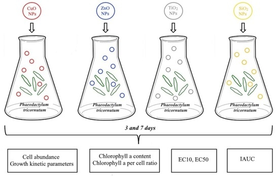
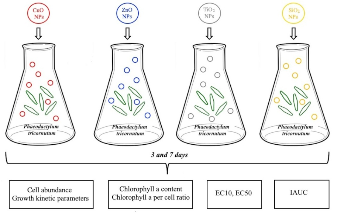
Engineered nanoparticles (ENPs) are increasingly released into marine environments, raising concerns about their potential ecological risks. In this study, we evaluated the toxicity of four widely used ENPs—CuO, ZnO, TiO2, and SiO2—to the marine diatom Phaeodactylum tricornutum over a 7-day exposure. Using a range of endpoints, including cell abundance, growth kinetics, chlorophyll a content, the integral inhibition index (IAUC), and effective concentrations (EC10, EC50), we established a toxicity ranking of CuO > (TiO2 ≈ ZnO) > SiO2 based on IAUC and final cell counts. Each nanoparticle exhibited a distinct phenotypic response pattern: CuO showed cumulative and irreversible toxicity (IAUC = 57.3% at 5 mg/L); TiO2 caused acute toxicity, with cell counts dropping to 29.8% of the control by day 3, followed by partial recovery (59.7% by day 7); ZnO induced delayed hormesis at 0.5 mg/L (125.1% of the control at day 7) but moderate inhibition at higher concentrations; and SiO2 displayed an algistatic, partially reversible effect (IAUC = 37.1%), likely due to physical adhesion and shading. Based on EC50 values, CuO, ZnO, and TiO2 were classified as “Toxic” (GHS Acute Category 2), while SiO2 was borderline. The lowest EC10 values were recorded for CuO (0.21–0.33 mg/L), indicating high chronic hazard. Our findings underscore the importance of integrating acute toxicity data with temporal dynamics and phenotypic response patterns for a reliable environmental risk assessment of ENPs in seawater.

Graphical Abstract

1. Introduction

Nanoparticles (NPs) (1–100 nm) possess unique physicochemical and biological properties due to their high specific surface area. This ensures their bioavailability and wide application in industry, medicine, and science [1,2,3]. By origin, they are classified as natural, incidental, and engineered (ENPs); annually, approximately 1000 Tg of natural NPs and 0.3 Tg of synthetic NPs participate in biogeochemical cycles [4,5].
Global production of engineered nanomaterials (ENMs) has grown substantially over the past decade. In 2023, the global ENM market was valued at USD 12.42 billion, rising to USD 14.34 billion in 2024. This growth corresponds to a compound annual growth rate exceeding 15% [6]. SiO2 NPs are among the most highly produced nanomaterials. Annual global production of specialty silica, including nanoforms, is estimated at over 3 million tons [7,8], with applications ranging from construction materials to pharmaceuticals [9]. Approximately 10,000 tons of TiO2 NPs are used annually in the global production of pigments, photocatalysts, and UV filters [10]. ZnO NPs, with an annual production volume in the range of 550–35,000 tons, are used in plastics, lubricants, batteries, and cosmetics [11]. Although the annual production of CuO NPs is currently modest—estimated at up to 1600 tons—these particles have found widespread applications in batteries, sensors, catalysts, and antimicrobial coatings [12,13].
High production volumes lead to the release of ENPs into coastal marine waters, where they affect the growth, development, and mortality of aquatic organisms. The mechanism of ecotoxicity is based on the excessive generation of reactive oxygen species (ROS). This leads to oxidative stress, immune suppression, and subsequent damage to cellular components, including proteins, cell membranes, and DNA [14,15,16,17,18]. Phytoplankton—the foundation of marine food webs—is a suitable test organism due to its sensitivity to ENPs [19,20,21]. The adverse biological effects of NPs may be associated with adsorption onto cells, internalization, and the release of metal ions [3,22,23].
Phaeodactylum tricornutum Bohlin, 1897 is a widespread pennate diatom that grows in brackish or saline waters. The biology, ecology, and ecotoxicology of P. tricornutum are well understood. Consequently, it is a widely used test organism for assessing seawater quality [22,23,24,25,26]. The sensitivity of microalgae to toxicants depends on cell size, and the smallest species, including P. tricornutum (cell size 5–12 µm), are more susceptible to their effects [27]. The use of P. tricornutum as a test organism is regulated by international standards and Russian guidelines [28,29]. A rapid response to changes in the concentration of toxicants in the test media has been demonstrated for this species in numerous studies [30,31,32,33,34]. Assessing the toxic effects of ENPs on marine diatoms is essential for analyzing their long-term potential impact on marine food webs that are based on phytoplankton production [21,35].
Nevertheless, few studies have compared the toxicity of CuO, ZnO, TiO2, and SiO2 NPs within a single experimental system. Most research addresses only one or two nanoparticle types, hindering direct hazard comparisons. Furthermore, the temporal dynamics (acute against chronic effects) and hormetic responses at low concentrations remain poorly understood.
The aim of this study was to conduct a comparative assessment of the toxicity of four ENPs (CuO, ZnO, TiO2, and SiO2) on the marine diatom P. tricornutum. To achieve this, the following objectives were set: (1) to study cell abundance dynamics and growth kinetic parameters (specific growth rate, doubling time and number of divisions); (2) to analyze chlorophyll a content as a marker of photosynthetic activity; (3) to calculate the integral toxicity index (IAUC); and (4) to determine effective concentrations (EC10, EC50).

2. Materials and Methods

2.1. Object of Investigation

An unialgal culture of the marine diatom Phaeodactylum tricornutum, maintained in the laboratory of the UNESCO International Chair in Marine Ecology at the Institute of the World Ocean, Far Eastern Federal University (Vladivostok, Russia), was used for the experiments. Cells were cultured in F/2 medium (Giard’s medium; composition given in Table 1) prepared with filtered and autoclaved seawater. All culture vessels and media were autoclaved, and aseptic conditions were maintained throughout. Microalgae were grown in a culture incubator KS-200 (JSC “Smolensk SKTB SPU”, Smolensk, Russia) at 3500 lx, a 16:8 h light:dark photoperiod, and 22 ± 1 °C. After reaching the exponential growth phase (9–12 × 104 cells/mL) and acclimating to the growth conditions for 3 days, the stock culture was used for the experiments [28]. Cell density was determined at the start of the experiment and at 24, 48, and 72 h, and on days 4, 5 and 7 using a Goryaev counting chamber (MiniMed, Bryansk, Russia).

2.2. The Characterization of Nanoparticles

Stock suspensions of NPs were prepared using CuO (nominal particle size 50–80 nm; 99.8% purity), ZnO (nominal particle size < 50 nm; 99.5% purity), TiO2 (nominal particle size ≤ 20 nm; 99.5% purity), and SiO2 (nominal particle size ≤ 20 nm; 99.5% purity) from Sigma-Aldrich (Steinheim, Germany). The size of primary particles and their agglomerates was determined by transmission electron microscopy (TEM) according to the procedure previously described by [36]. For TEM analysis, 10 µL of each nanoparticle suspension was deposited onto formvar-coated copper grids and allowed to adsorb for 30 min at room temperature. After removing excess liquid with filter paper and air-drying at room temperature, the grids were examined using a Carl Zeiss Libra 200 transmission electron microscope (Carl Zeiss NTS GmbH, Oberkochen, Germany) operating at 200 kV. At least 100 particles were measured per sample to determine the size distribution. The main properties of the nanoparticles have been discussed in previously published works [37,38,39] and are summarized in Table 2.
Immediately prior to each experiment, stock suspensions of CuO, ZnO, TiO2, and SiO2 NPs were sonicated in a Sapphire ultrasonic bath (A3 Engineering, Moscow, Russia) for 20 min at a power setting of 40 W. Sonication time and power were chosen according to protocols described in earlier studies [37,38,39,40]. Stock suspensions were prepared in double-distilled water. The tested concentrations were 0.2, 0.5, 1, 2 and 5 mg/L for CuO; 0.5, 1, 2, 5, and 10 mg/L for ZnO; and 1, 2, 5, and 10 mg/L for both TiO2 and SiO2. Control cultures were maintained under identical conditions without the addition of NPs.
The concentration ranges for each NP type were selected based on literature data on their toxic effects on diatoms, including P. tricornutum [17,20,24,41,42,43,44]. The selected concentration ranges allowed the determination of threshold concentrations inhibiting growth and photosynthetic activity of P. tricornutum, and enabled calculation of EC10 and EC50 values for this species.

2.3. Phaeodactylum tricornutum Tests

To prepare the experimental solutions, aliquots of stock suspensions of CuO, ZnO, TiO2, or SiO2 were diluted with f/2 culture medium to obtain the desired final concentrations, and a suspension of the microalgae was then added. Each treatment was performed in triplicate. Control cultures were maintained in nanoparticle-free medium. Stock suspensions of nanoparticles were used throughout the entire 7-day exposure period without renewal. This approach (static exposure) was chosen to evaluate the cumulative effect of the particles over time and to simulate a realistic scenario in which particle concentration in the medium may decrease due to aggregation and sedimentation. Such practice is widely used in recent studies [18,20,45].
Cell counts were performed using a Goryaev counting chamber (MiniMed, Bryansk, Russia) under a Zeiss Axio Imager microscope (Carl Zeiss, Oberkochen, Germany) at 24, 48, 72 h and on days 4, 5, and 7. To calculate the effective concentrations causing 10% (EC10(48), EC10(72)) and 50% (EC50(48), EC50(72)) inhibition of growth, cell counts were recorded at 24 h intervals [28].

2.4. Calculation of Growth Kinetic Parameters: Specific Growth Rate (µ), Doubling Time (td), Number of Divisions per Day (M)

From the cell count time series, the following growth parameters were calculated:
Specific growth rate (µ, day−1)—a measure of biomass increase per unit time. It was calculated using natural logarithms according to the formula:
μ = ln N 2 ln N 1 t 2 t 1
where N 1 and N 2 are cell densities (×104 cells/mL) at times t 1 and t 2 (days), and l n denotes the natural logarithm.
Doubling time (td, days)—the period over which the cell count doubles. It was calculated using natural logarithms according to the formula:
t d = ln 2 μ
Number of divisions per day (M, divisions·day−1)—the number of cell cycles per day. It was determined using the formula, where ln denotes the natural logarithm, consistent with the calculation of μ:
M = l o g 2 N 2 l o g 2 N 1 t 2 t 1 = μ ln 2
All parameters were calculated for the time intervals 0–3 days (exponential growth phase) and 0–7 days (the entire exposure period). For each concentration, mean values were calculated from three replicates.

2.5. Calculation of Integral Area Under the Curve (IAUC)

To comprehensively assess the toxic effects over the 7-day exposure period, the area under the growth curve (AUC) and the integral percentage inhibition (IAUC, %) were calculated.
Calculation of AUC. For each sample, growth curves were plotted as cell density (×104 cells mL−1) versus time (days). The area under the curve was calculated using the trapezoidal method by summing the areas for the intervals 0–2, 2–4, and 4–7 days according to the formula:
A U C =   i N i +   N i + 1 2 ×   t i
where N i and N i + 1 are cell densities at the start and end of the interval, respectively, and t i is the interval duration (days).
Calculation of IAUC. IAUC was calculated for each concentration as:
I A U C % =   1 A U C e x p e r i m e n t a l A U C c o n t r o l   × 100 %
where A U C e x p e r i m e n t a l and A U C c o n t r o l are the areas under the curves for the treated and control cultures, respectively. Positive IAUC values indicate growth inhibition, whereas negative values indicate stimulation (hormesis).
The IAUC was selected as the primary endpoint for evaluating growth inhibition, as it integrates all cell densities at different time points, rather than relying solely on a single time point. This approach accounts for the entire growth trajectory, including possible irregularities such as lag phases, nonmonotonic responses, or delayed recovery, which would not be captured by final biomass or growth rate alone. IAUC has been shown to be a sensitive endpoint in algal toxicity testing and is consistent with standard guidelines for growth inhibition tests [46].

2.6. Chlorophyll a Determination

To determine chlorophyll a concentration, 5 mL of algal culture was centrifuged (Cence H-2050R (Hunan XiangYi Laboratory Instrument Development Co., Ltd., Changsha, China)) at 7000 g for 10 min to pellet the cells. The pellet was resuspended in 1 mL of distilled water, and 4 mL of acetone (Ekos-1, Moscow, Russia) was added. To ensure complete pigment extraction, samples were incubated at 4 °C for 24 h in the dark and then centrifuged again for 5 min. Absorbance was measured using a Shimadzu-UV 1800 spectrophotometer (Shimadzu, Kyoto, Japan) at 630, 647, and 664 nm with 80% acetone (Ekos-1, Moscow, Russia) as the blank [47].
Chlorophyll a concentration (μg/L) was calculated using the following formula (without pheophytin correction):
C h l   a = 11.85 × D 664 D 750 1.54 × D 647 D 750 0.08 × ( D 630 D 750 ) × V e V s × l
where
D 664 , D 647 , D 630 —absorbance at the respective wavelengths;
D 750 —baseline absorbance (no pigment absorption);
V e —extract volume (cm3);
V s —sample volume (dm3);
l —cuvette path length (cm).
To minimize potential interference from nanoparticles during chlorophyll a measurement, the samples were centrifuged, and the pigment concentration was calculated using a turbidity correction (subtraction of absorbance at 750 nm, D 750 ). Chlorophyll a content per cell (pg/cell) was calculated by dividing the volumetric chlorophyll a concentration (μg/L) by the cell density (×104 cells/mL). All measurements were performed in triplicate, and mean values were used for further calculations [28].

2.7. Statistical Analysis

The experimental data were analyzed using MS Excel and Statistica v.10 (StatSoft, Inc., Tulsa, OK, USA). Data are presented as arithmetic means ± standard deviation (SD). All experiments were performed in three independent biological replicates (n = 3 flasks), with three technical aliquots taken from each flask. Normality of distribution was assessed using the Shapiro–Wilk test, and homogeneity of variances was verified with Levene’s test. The non-parametric Mann–Whitney U-test was applied for pairwise comparisons between treatments and the control. The Bonferroni correction was applied to control the overall error level. Statistical significance was accepted at p < 0.05.

3. Results

3.1. Effect of CuO NPs on Cell Abundance Dynamics and Chlorophyll a Content in Phaeodactylum tricornutum

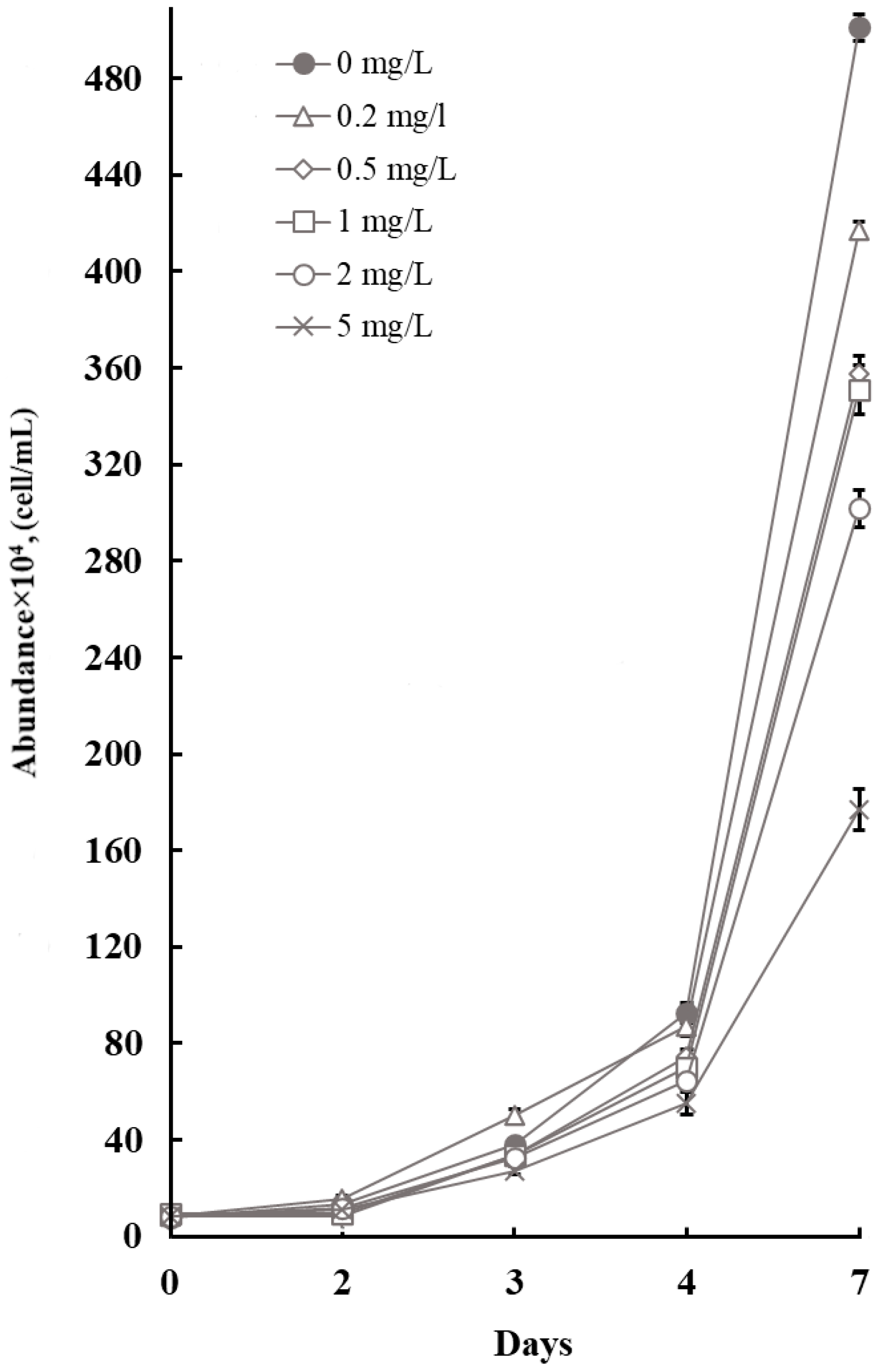
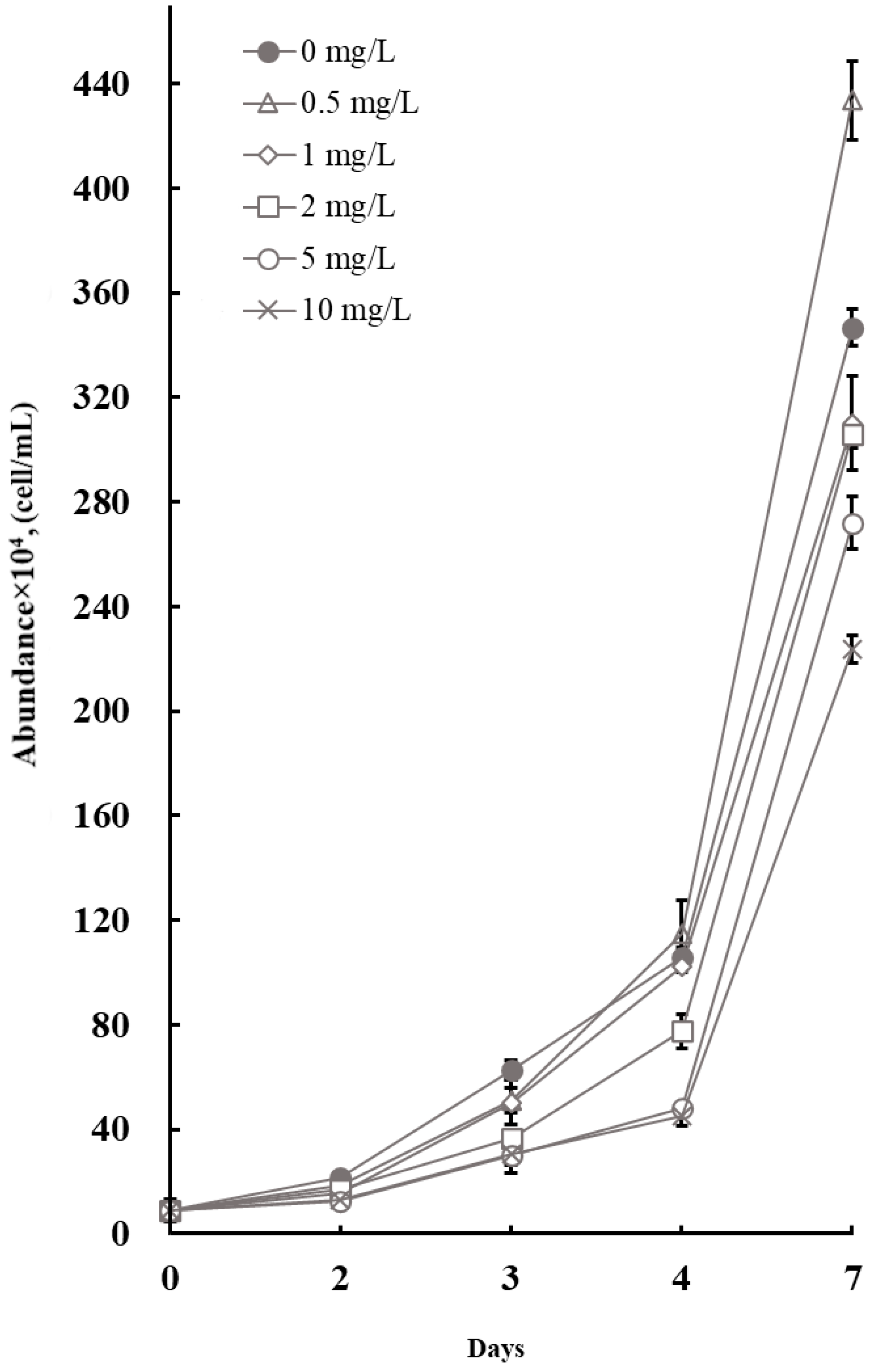
Figure 1 shows the growth dynamics of Phaeodactylum tricornutum during a 7-day exposure to CuO NPs at concentrations ranging from 0.2 to 5 mg/L. On day 3, a statistically significant (p < 0.05) stimulation of cell growth was observed at 0.2 mg/L, whereas significant inhibition (p < 0.05) was observed at the highest concentration tested (5 mg/L). The specific growth rates (μ) (Table S1) at these concentrations were 0.7 ± 0.01 and 0.36 ± 0.05 day−1, respectively, compared to the control value of 0.62 ± 0.06 day−1.
By day 7, a statistically significant (p < 0.05) decrease in cell abundance was observed in all treated cultures. The lowest growth rates were recorded at CuO NPs concentrations of 1, 2, and 5 mg/L (0.44 ± 0.02, 0.42 ± 0.03, and 0.33 ± 0.02 day−1, respectively), which predictably resulted in reduced division rates (M) of 0.64 ± 0.02, 0.61 ± 0.04, and 0.48 ± 0.02, respectively (Table S1). These data indicate a cumulative toxic effect of CuO NPs: initial stimulation at low concentrations is followed by inhibition, and the inhibitory effect of high concentrations intensifies over time.
The lowest calculated effective concentrations causing 10% growth inhibition (EC10(48 h) and EC10(72 h)) were 0.33 and 0.21 mg/L, respectively, whereas the EC50(48h) and EC50(48 h) values were 5.12 and 6.84 mg/L, respectively.
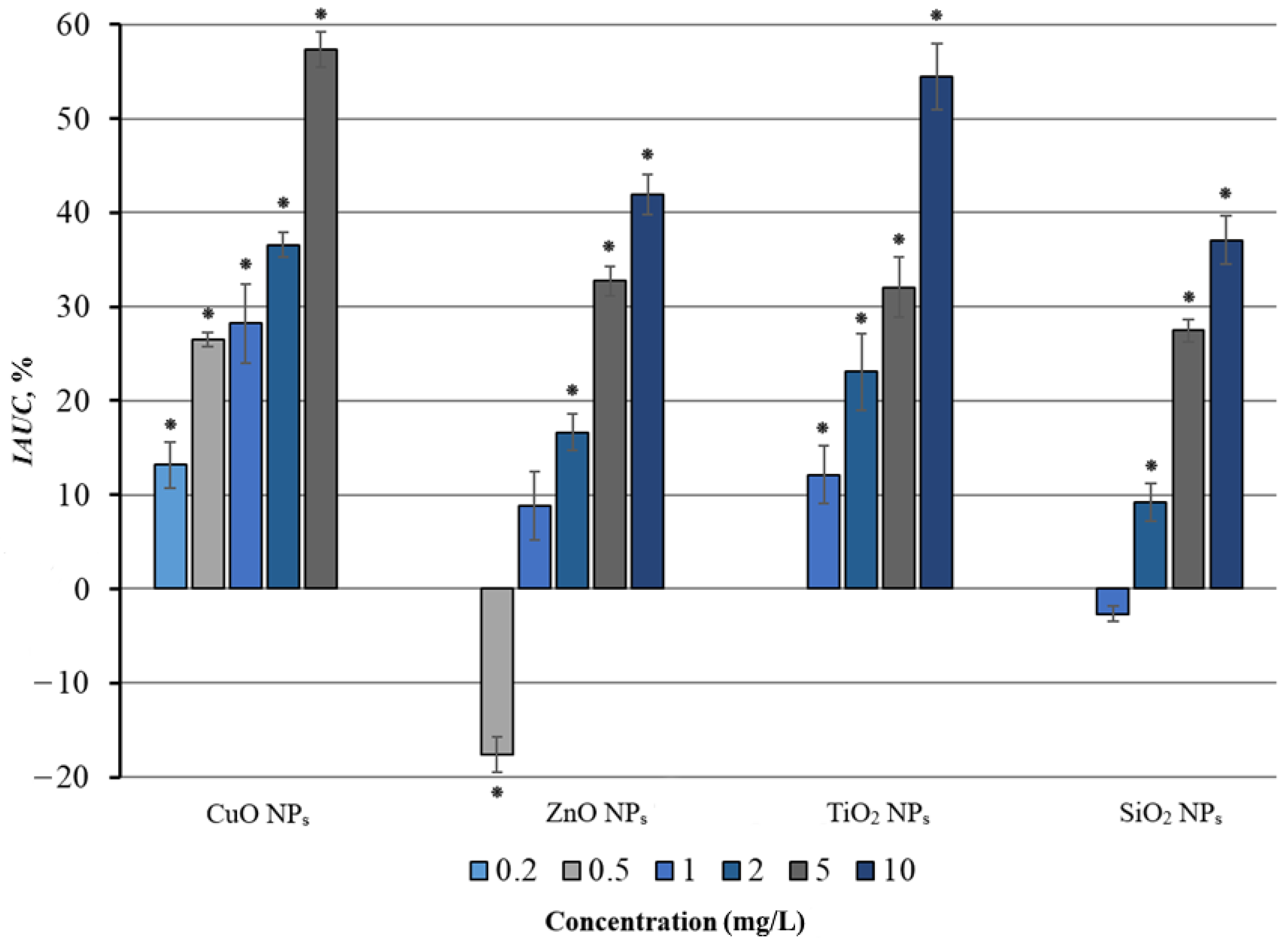
The integrated area under the growth curve and the corresponding percentage inhibition (IAUC, %) relative to the control (Figure 2) revealed statistically significant inhibition (p < 0.05) at the lowest tested concentration (0.2 mg/L), with the maximum effect observed at 5 mg/L. At the highest concentration, the toxic effect resulted in a >50% reduction in productivity (IAUC = 57.33 ± 1.87%).
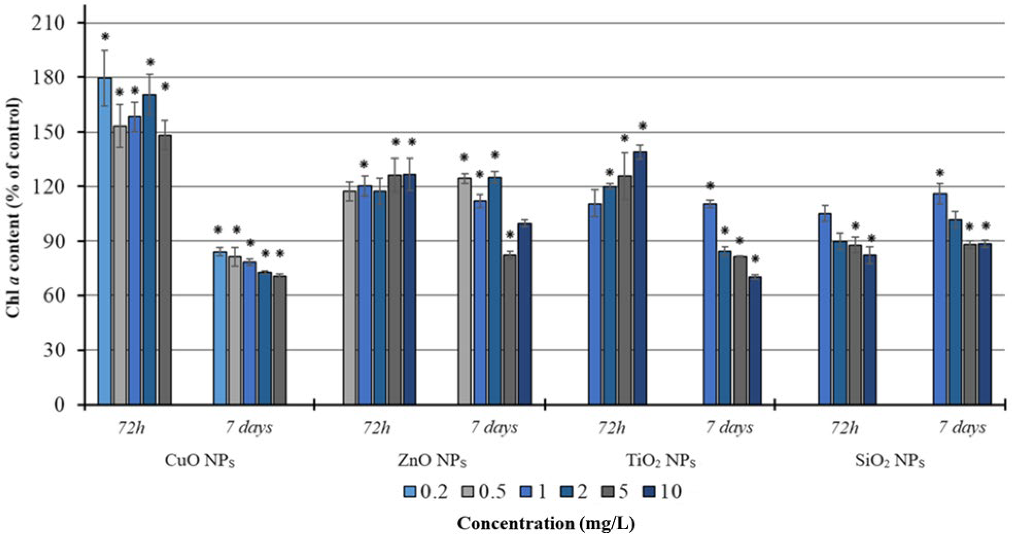
The effect of CuO NPs on the photosynthetic apparatus was also assessed by monitoring chlorophyll a (Chl a) content. On day 3, the highest Chl a/biomass ratios—approximately twice the control values (p < 0.05)—were observed at 2 and 5 mg/L. By day 7, the most pronounced effect was still observed at 5 mg/L, where the Chl a/biomass ratio remained approximately twice that of the control (p < 0.05).

3.2. Effect of ZnO NPs on Cell Abundance and Chlorophyll a Content in Phaeodactylum tricornutum

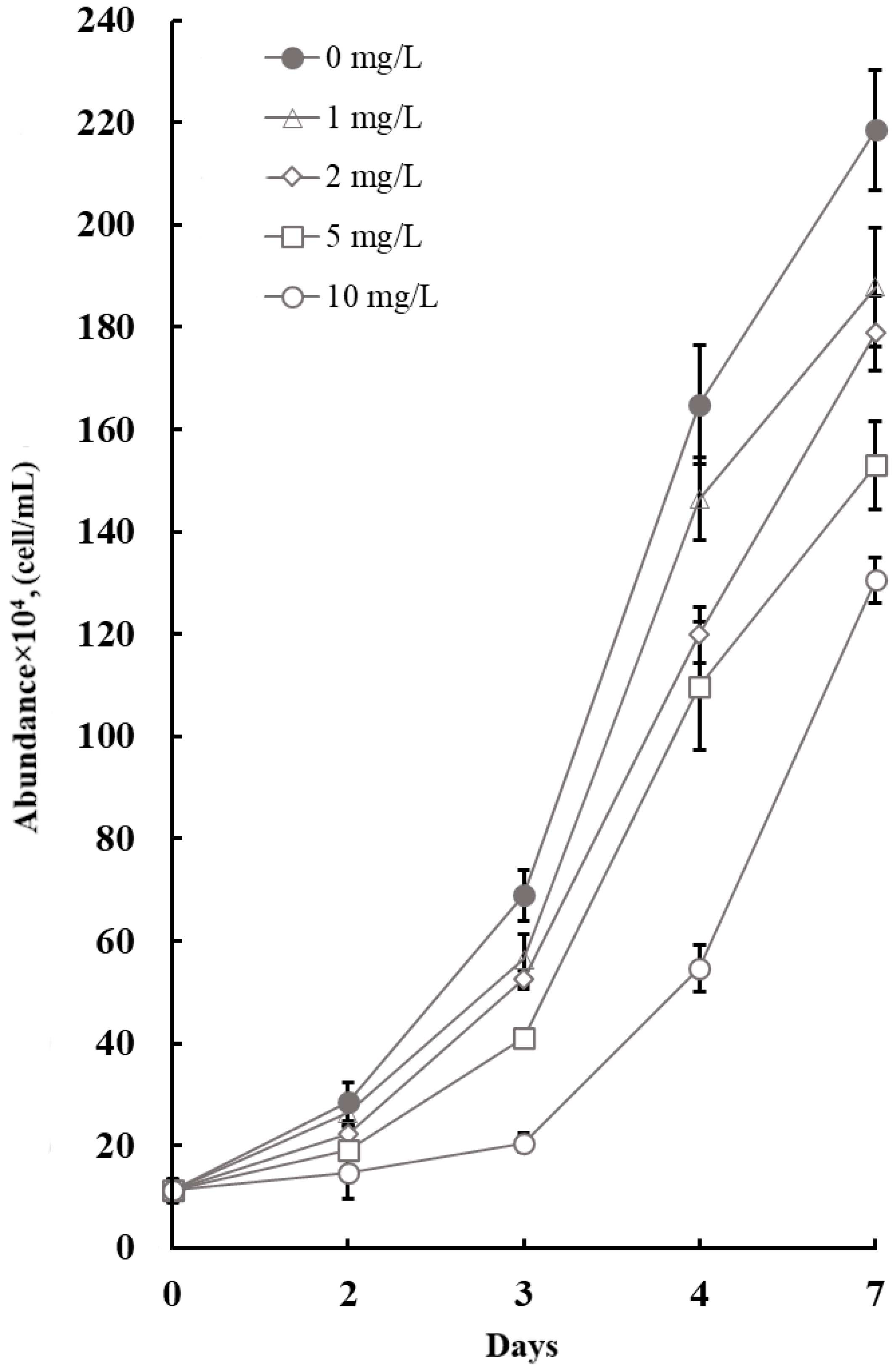
The effect of ZnO NPs at concentrations ranging from 0.5 to 10 mg/L on the growth of P. tricornutum was evaluated over a 7-day period. The results, presented in Figure 3, demonstrate a dose- and time-dependent toxic effect.
At 72 h, a statistically significant inhibition (p < 0.05) of growth was observed at 2–10 mg/L. The greatest inhibitory effect of ZnO NPs was observed during the exponential growth.
By day 7, as the culture entered the stationary phase, a statistically significant stimulation (p < 0.05) was observed at 0.5 mg/L, whereas significant inhibition (p < 0.05) was recorded at 1–10 mg/L. The highest growth rate (μ) was recorded at 0.5 mg/L, exceeding the control by 4%. Notably, in contrast to day 3, the overall toxicity gradient was attenuated by day 7, as reflected in the doubling time (td) values. For example, at 10 mg/L, the doubling time was 22% longer than in the control.
Effective concentrations were calculated from the growth data. The EC10(48 h) and EC10(72 h) values were 0.28 and 0.92 mg/L, while the EC50(48 h) and EC50(72 h) values were 2.15 and 3.18 mg/L, respectively.
To comprehensively assess the effect over the entire exposure period, the integrated area under the growth curve (IAUC) and the corresponding percentage inhibition relative to the control were calculated (Figure 2). The results revealed a 17.6% stimulation of growth at 0.5 mg/L (p < 0.05), and a dose-dependent inhibition at 2, 5, and 10 mg/L, with IAUC values of 16.6, 32.7, and 41.9%, respectively (p < 0.05).
On day 3, the highest Chl a/biomass ratios—exceeding the control by 169.9% and 164.4% (p < 0.05)—were recorded at 5 and 10 mg/L, respectively. On day 7, the highest ratios were observed at 2 and 10 mg/L, exceeding the control by 41.8% and 54.5%, respectively.

3.3. Effect of TiO2 NPs on Cell Abundance Dynamics and Chlorophyll a Content in Phaeodactylum tricornutum

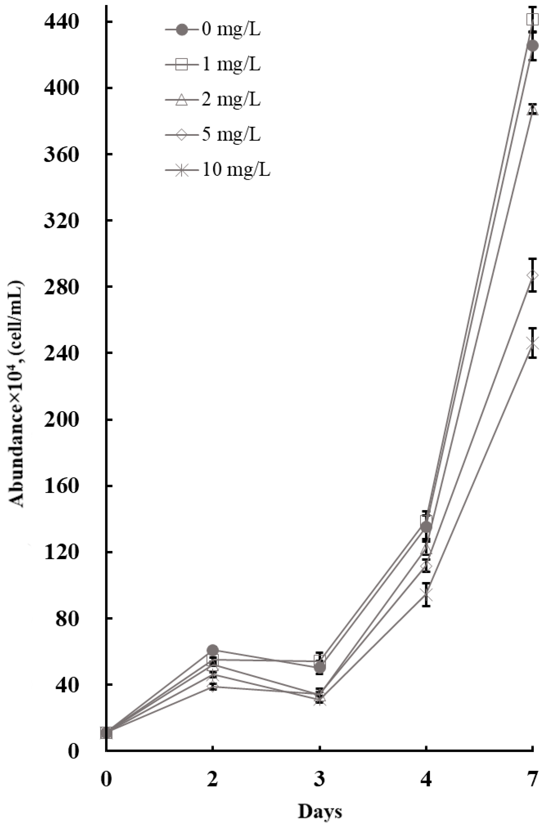
The results of the 7-day exposure to TiO2 NPs are presented in Figure 4. The data revealed a dose- and time-dependent toxic effect. On day 3, a statistically significant inhibition of growth (p < 0.05) was observed at 1–10 mg/L, with the most pronounced effect recorded at 10 mg/L.
At 5 and 10 mg/L (p < 0.05), significant decreases in the specific growth rate (μ), number of divisions per day (M), and doubling time (td) were recorded (Table S1). At 10 mg/L, the minimum values were observed: μ = 0.313 ± 0.03 day−1, M = 0.451 ± 0.04 divisions day−1, and td = 2.23 ± 0.2 days, corresponding to a 56.4% inhibition of growth parameters.
On day 7, a significant inhibition (p < 0.05) of growth was observed at all concentrations tested, with the maximum effect (59.7% inhibition) recorded at 10 mg/L. Nevertheless, the kinetic growth parameters (μ, M, td) suggested that the culture had partially adapted to TiO2 NP exposure. Significant differences from the control (p < 0.05) were observed at ≥2 mg/L. The lowest μ and M values—15.42% below the control—were recorded at 10 mg/L.
The EC10(48 h) and EC10(72 h) values were 0.43 and 0.61 mg/L, while the EC50(48 h) and EC50(72 h) values were 6.47 and 5.28 mg/L, respectively. The IAUC (%) values revealed a clear dose-dependent pattern (Figure 2), with a significant (p < 0.05) 12.1% decrease in productivity already at the lowest tested concentration (1 mg/L). At 2–10 mg/L, IAUC values ranged from 23% to 54.47% inhibition.
On day 3, the highest Chl a/biomass ratios—five times higher than the control—were recorded at 10 mg/L. On day 7, the ratio at 10 mg/L exceeded the control by more than sixfold.

3.4. Effect of SiO2 NPs on Cell Abundance Dynamics and Chlorophyll a Content in Phaeodactylum tricornutum

The results of the 7-day toxicity test with SiO2 NPs are presented in Figure 5. A clear dose-dependent relationship was observed, accompanied by pronounced adaptation of the microalgal culture. On day 3, significant growth inhibition (p < 0.05) was observed at 2, 5, and 10 mg/L. At 1 mg/L, a slight, though statistically non-significant, increase in cell abundance (7.9% above the control, p > 0.05) was observed. The greatest toxic effect was observed at 10 mg/L, where M, μ, and td differed from the control by 41.21% (p < 0.05) (Table S1).
On day 7, significant inhibition (p < 0.05) persisted at 2–10 mg/L, reducing cell abundance by 9.1%, 32.5%, and 42.16% relative to the control, respectively. At 10 mg/L, significant decreases (p < 0.05) in μ and M (13.5%) and a corresponding increase in td (15.6%) were recorded.
The IAUC (%) values, presented in Figure 2, revealed significant inhibition (p < 0.05) at 2, 5, and 10 mg/L, with reductions of 9.15%, 27.5%, and 37% relative to the control, respectively. The EC10(48 h) and EC10(72 h) values were 1.27 and 1.64 mg/L, while the EC50(48 h) and EC50(72 h) values were 10.84 and 5.18 mg/L, respectively.
On day 3, the Chl a/biomass ratio showed significant changes at 2–10 mg/L. By day 7, the ratio increased significantly at 5 and 10 mg/L by 33.8% and 56.8% (p < 0.05), respectively.

4. Discussion

4.1. Comparative Toxicity Ranking and Phenotypic Response Patterns of CuO, ZnO, TiO2, and SiO2 NPs in Phaeodactylum tricornutum

It is worth noting that this study does not include direct measurements of reactive oxygen species (ROS), antioxidant enzyme activities, or dissolved metal ion concentrations. These limitations are acknowledged in Section 4.5. Nevertheless, the observed phenotypic response patterns provide a basis for interpreting the temporal dynamics of toxicity. The discussion presented below focuses on these patterns and relates them to mechanistic hypotheses proposed in the literature.
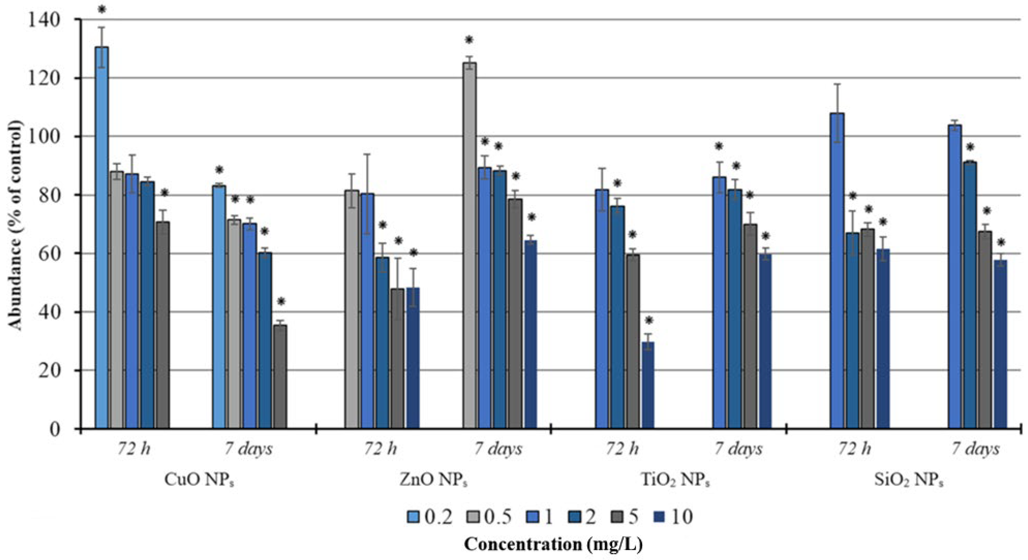
Comparison of IAUC values revealed higher toxicity of TiO2 compared to ZnO NPs. The IAUC for TiO2 NPs was 54.5%, which is 13 percentage points higher than that for ZnO NPs (41.9%), indicating higher integral toxicity of TiO2 NPs at 10 mg/L. However, the differences in final cell counts were not significant (Figure 6).
Our findings are consistent with previously reported data. The observed differences in toxicity are attributable to the distinct phenotypic response pattern of each nanoparticle type. The toxicity mechanism of TiO2 NPs is primarily based on their photocatalytic activity. Exposure to light (e.g., in the culture incubator) induces the generation of reactive oxygen species (ROS) on the surface of TiO2 NPs. The ROS can trigger lipid peroxidation, protein oxidation, and DNA damage [48]. This may contribute to the observed effects in our study. However, since our study did not include dark control experiments or direct ROS measurements, this interpretation remains a hypothesis rather than a confirmed mechanism for our experimental system. Recent studies on microalgae have confirmed these effects: Wang et al. [16] demonstrated that TiO2 NPs inhibit the growth of Microcystis aeruginosa Kützing, 1846 through ROS overproduction and damage to the photosynthetic apparatus. Similarly, Zhu et al. [49] showed that combined exposure to TiO2 NPs and UV-B radiation induces severe oxidative stress in Chlorella pyrenoidosa H.Chick, 1903. Sendra et al. [27] evaluated the toxicity of TiO2 NPs under visible light and ultraviolet (UV) radiation. They used two microalgal species: Chlamydomonas reinhardtii P.A. Dangeard, 1888, and P. tricornutum. Both species showed sensitivity under both conditions, with a more pronounced effect under UV exposure. These findings confirm the high photocatalytic activity of TiO2 NPs. Another possible contributor to the acute toxicity of TiO2 NPs is their high surface energy, which promotes adhesion to cells. Recent studies have confirmed that TiO2 NPs readily adsorb onto algal cell surfaces [49,50]. This process, particularly at high nanoparticle concentrations, may disrupt active transport across the cell membrane and cause localized ROS accumulation [27,51].
Similar to TiO2 NPs, ZnO NPs are known to induce ROS generation and mechanical damage to cell membranes through adhesion [17,52]. The toxicity mechanism of ZnO NPs is based on their partial dissolution in the culture medium, followed by the release of zinc ions (Zn2+), which may explain the observed effects. The released Zn2+ ions subsequently disrupt enzymatic, transport, and metabolic processes in microalgal cells [18,53]. Whereas the photocatalytic generation of ROS by TiO2 NPs is a rapid process, causing membrane damage and metabolic disruption within hours, the release of Zn2+ is more gradual. Moreover, to exert oxidative stress-related toxicity, Zn2+ must first penetrate the cell [40,54,55]. Notably, Baysal et al. [56] demonstrated that ZnO NPs retain their toxicity even when their physicochemical parameters (size, zeta potential, and surface chemistry) change in seawater. This observation supports the interpretation that the less pronounced toxic effects of ZnO NPs in our study are likely due to the gradual release and intracellular accumulation of Zn2+. This contrasts with rapid photocatalytic action of TiO2 NPs, which is mediated by ROS generation.
Both TiO2 and ZnO NPs were less toxic than CuO NPs. The lowest growth rates were observed in cultures exposed to CuO NPs (Table S1). Unlike the other NPs, CuO NPs did not exhibit any recovery of physiological parameters of culture by the end of the experiment. The hormetic effect observed at the lowest concentration on day 3 was replaced by inhibition by day 7. Although this inhibition was weak (IAUC = 13.2 ± 2.5%), it was statistically significant, confirming the transient nature of the stimulation. Even at low concentrations, the accumulation of Cu2+ inside the cells depletes cellular defense systems, leading to decreased M and μ (0.69 ± 0.02 divisions day−1 and 0.48 ± 0.01 day−1, respectively) and increased doubling time (td = 1.44 ± 0.03 days). The most pronounced inhibition was observed at 5 mg/L (IAUC = 57.3 ± 1.9%), where the doubling time exceeded 2 days, and M and μ were 35% below control values (0.48 ± 0.02 divisions day−1 and 0.33 ± 0.02 day−1, respectively). This pattern is consistent with the classical ionic toxicity mechanism, driven by the accumulation of Cu2+ inside algal cells. The gradual dissolution of CuO NPs, accompanied by the release and subsequent accumulation of Cu2+, drives intracellular copper concentration toward a critical threshold. This process leads to oxidative stress and disruption of enzymatic functions, ultimately compromising the cells’ adaptive capacity [55,57]. Our findings are consistent with previous studies, which also reported hormesis at low CuO NPs concentrations and growth inhibition at high concentrations [58,59].
The toxicity of CuO and ZnO NPs observed during the experiment cannot be explained solely by the release of ionic forms of metals into the environment. Recent studies show that a significant fraction of nanoparticles can be internalized by microalgal cells, for example, through endocytosis or passive diffusion. As noted in the review by Wang et al. [60], during cell division, the permeability of the microalgal cell wall increases sharply, which may contribute to more active uptake of NPs. For P. tricornutum, this mechanism may be particularly relevant, as this species is characterized by a thin organic cell wall with a low silicon content, primarily composed of polysaccharides. Furthermore, cytokinesis in P. tricornutum is accompanied by the formation of cleavage furrows and active vesicular transport [61]. Together, these factors may lead to enhanced NPs uptake, even at low concentrations.
After internalization, the nanoparticles undergo intracellular dissolution, releasing metal ions directly into the cytoplasm [24]. This creates a localized, high-dose toxic effect that bypasses membrane transport systems and detoxification mechanisms. This local effect, in turn, can lead to the development of oxidative stress, damage to cell membranes and organelles (including mitochondria and chloroplasts), and, ultimately, to the inhibition of cell division [25]. In contrast, SiO2 NPs were the least toxic among the NPs tested. On day 3, exposure to 10 mg/L SiO2 NPs and 5 mg/L CuO NPs resulted in cell abundances of 61.6% and 70.8% of the control, respectively (p < 0.05). Although both concentrations caused significant inhibition, the effect of SiO2 NPs was less pronounced than that of CuO NPs. The toxicity of SiO2 NPs, although lower overall than that of CuO NPs, may be partly explained by their immediate adsorption onto cell surfaces, leading to physical blocking of light and membrane transport [23,62]. Nevertheless, such surface aggregations do not cause irreversible damage, unlike the direct chemical effects of metal ions (Cu2+, Zn2+).
The Chl a/cell ratio data were consistent with the overall toxicity pattern. The absolute Chl a/cell values were higher under TiO2 NPs treatment (0.67 pg/cell) than under CuO NPs treatment (0.414 pg/cell) (Table S1). However, the relative increase over the control was substantially greater for CuO NPs (201% vs. 115.5% for TiO2). This observation suggests a differential physiological response, although statistical confirmation would be required to validate this difference. This observation may be associated with the accumulation of Cu2+ within microalgal cells. Copper ions are known to replace magnesium ions in the chlorophyll molecule, forming non-functional Cu-chlorophyll [24,62]; however, this substitution occurs primarily at elevated copper concentrations and is associated with photosynthetic impairment. This explains why elevated Chl a content at relatively low cell densities may result from compensatory pigment synthesis in response to stress. It may also result from the formation of non-functional Cu-chlorophyll under copper excess conditions.
The physicochemical properties of the nanoparticles (Table 2) provide a basis for hypothesizing their behavior in aqueous media and the associated toxic effects [63,64]. CuO NPs exhibited the smallest hydrodynamic diameter (100 ± 12 nm) and the most negative zeta potential (–57 ± 4.2 mV), which may indicate higher colloidal stability and reduced aggregation compared to the other nanoparticles. These characteristics likely enhance bioavailability and interaction with algal cells, which is consistent with the observed high toxicity, particularly considering the combined effects of the particles themselves and sustained Cu2+ release. ZnO NPs showed a moderately larger hydrodynamic diameter (200 ± 27 nm) and a lower (in magnitude) zeta potential (–39.4 ± 2.7 mV), which may indicate a greater tendency toward aggregation and partial Zn2+ release, contributing to their moderate toxicity through an ionic mechanism. Despite their small primary size (27 ± 4 nm), TiO2 NPs formed larger agglomerates (190.5 ± 18 nm). They also exhibited a relatively low zeta potential (–13.9 ± 1.1 mV), which may facilitate particle adhesion to cell surfaces and localized photocatalytic ROS generation. SiO2 NPs displayed the largest hydrodynamic diameter (576 ± 104 nm) and the lowest (in magnitude) zeta potential (–11.5 ± 0.7 mV), indicating strong aggregation. The combination of their high specific surface area (613 ± 47 m2/g) with these properties likely leads to shading and particle adhesion to membranes. This suggests a physical rather than direct chemical toxicity.
It is important to note that the nanoparticle characteristics described above were determined directly in stock suspensions prepared using double-distilled water. However, the properties of these particles may vary significantly when they are added to a marine environment. The high ionic strength characteristic of seawater screens the electrostatic repulsion between particles, leading to aggregation and a decrease in colloidal stability [65]. Unlike in deionized water, the hydrodynamic diameter of NPs in seawater increases, while the zeta potential becomes less negative; these factors contribute to reduced electrostatic stabilization. The dissolution of metal oxide NPs in a marine environment proceeds differently. ZnO NPs dissolve significantly faster in this environment compared to other nanoparticles studied [66]. CuO NPs are characterized by extremely slow dissolution in a marine environment [67]. TiO2 NPs are characterized by an extremely low overall dissolution rate in neutral and alkaline environments. However, in the study by Mbanga et al. [68], the dissolution of these particles in seawater occurs faster than in freshwater, indicating some acceleration of the process, although the overall dissolution rate remains extremely low. The dissolution of amorphous silica, including SiO2 NPs, generally occurs faster in a marine environment. However, Spitzmüller et al. [69] reported that high salinity slows the dissolution of silicon nanoparticles, while elevated pH and temperature accelerate this process. Thus, under real-world seawater conditions (with a pH of ~8.0–8.2), the overall dissolution rate of SiO2 NPs will be determined by the balance of these opposing factors. A synthesis of the literature data presented here suggests that aggregation and differences in dissolution kinetics, along with surface properties, may contribute to the observed differences in toxic effects between the types of nanoparticles studied in our experiment.
Based on our findings, the refined toxicity ranking is as follows: CuO > (TiO2 ≈ ZnO) > SiO2. Based on IAUC values, TiO2 NPs exhibited higher overall toxicity compared to ZnO NPs. However, the final cell counts at 5–10 mg/L for TiO2 and ZnO NPs appeared to be relatively close in our experiments, which provided a basis for considering them together in the comparative assessment. The most notable differences in toxicity were observed between this group and CuO NPs, as well as between this group and SiO2 NPs.

4.2. Hormesis: Growth Stimulation Induced by Low Doses of CuO NPs

A notable finding of the study was the stimulation of microalgal growth at the initial stage of the experiment. This occurred upon exposure to a low concentration of CuO NPs (0.2 mg/L). At this concentration of CuO, cell counts were 30.4% higher than control values (p < 0.05), indicating a statistically significant hormetic effect.
This hormetic effect was most pronounced for CuO NPs. At 72 h, an increase in the growth kinetic parameters was observed: the M and μ values exceeded the control by 13%. By the end of the experiment, however, the stimulatory effect had been replaced by inhibition, with a 17% decrease in cell abundance and a >50% reduction in kinetic parameters. The hormetic effect at low CuO NPs concentrations was further confirmed not only by increased cell abundance. It was also supported by an 80% increase in Chl a content (Figure 7) and a 38% increase in the Chl a/cell ratio (Table S1). At low CuO NP concentrations (0.2 mg/L), the observed increase in chlorophyll content per cell may reflect the activation of pigment biosynthesis. This is considered a typical adaptive stress response typically with hormesis.
Our findings are consistent with the literature, which also reports hormesis following exposure to CuO NPs [54]. This effect is attributed to the essential role of copper as a trace element. Copper is required for the synthesis of key proteins (plastocyanin) and enzymes (polyphenol oxidase, cytochrome oxidase, and Cu-Zn-superoxide dismutase) involved in photosynthesis and antioxidant defense [24]. As copper accumulates both in the environment and within intracellular pools, its concentration eventually exceeds the cell’s detoxification capacity. Consequently, the stimulatory effect is replaced by toxicity [70]. Notably, silicon transporters (SITs) in diatoms such as P. tricornutum are not strictly specific. This may facilitate the uptake of certain metals into the cell [71].
The observed hormetic effects are interpreted as potential stress-induced responses. These interpretations are based on literature-supported mechanisms and should be regarded as hypotheses requiring direct experimental confirmation. Overall, the induction of hormesis by NPs of different chemical natures confirms the universality of this phenomenon. However, it also highlights its dependence on both the specific toxicity mechanism and the physiology of the test organism. This section has focused on classical hormesis, characterized by immediate growth stimulation at low concentrations. The delayed hormesis observed with low concentrations of ZnO NPs deserves special attention. It is case, stimulation occurred only after a period of adaptation, and this is discussed in the following section.

4.3. Types of Toxic Effects of the Studied Nanoparticles

Analysis of growth kinetics, Chl a content, and IAUC values allowed us to classify the NPs by the severity of their toxic effects. We also classified them by their temporal dynamics under short- and long-term exposure. The 7-day experiment identified four distinct patterns of temporal toxicity dynamics: acute (TiO2 NPs), cumulative (CuO NPs), adaptive (ZnO NPs), and algostatic (SiO2 NPs).

4.3.1. Acute Toxicity of TiO2 NPs

In our study, the acute toxicity of TiO2 NPs was evidenced by the greatest reduction in cell abundance compared to the control (29.8% of the control at 10 mg/L) as early as 72 h (Figure 6). This was the most pronounced effect among all treatments during the initial phase. This finding is further supported by the kinetic parameters. Specifically, the specific growth rate (μ) and division rate (M) decreased by 56.3%, while the doubling time (td) increased by 129.9%, indicating a critical slowdown of the cell cycle.
An increase in Chl a content by 38.9% (Figure 7) relative to the control was observed. Additionally, the Chl a/cell ratio at 10 mg/L increased 4.7-fold compared to the control, which is another diagnostic indicator of acute toxicity. Taken together, these observations are consistent with a compensatory response of the microalgal culture to acute photocatalytic stress. This response may involve emergency chlorophyll synthesis by surviving cells, possibly related to the photocatalytic activity of TiO2 NPs. A similar pattern was reported by Deng et al. [41], who observed increased Chl a ratio in P. tricornutum cultures exposed to high TiO2 NP concentrations, accompanied by reduced cell abundance. This stimulation of pigment synthesis may reflect an attempt to maintain photosynthetic activity during mass cell death. It may also represent adaptation to increased light exposure after the shading effect of dead cells is removed.
By day 7, however, partial recovery was observed. At 10 mg/L, cell abundance recovered to 59.7% of the control. This recovery was also reflected in the kinetic parameters, while the Chl a/cell ratio differed from the control by only 15.9%. This pattern—initial growth inhibition followed by partial recovery—is characteristic of acute but partially reversible toxicity.

4.3.2. Cumulative Toxicity of CuO NPs

CuO NPs exhibited a distinctly different toxicity pattern. The toxic response was cumulative and irreversible. At 5 mg/L CuO NPs, the IAUC value reached 57.3% (Figure 2). This was the highest value recorded in this study, based on the percentage inhibition relative to the control. Exposure to 5 mg/L resulted in progressively increasing inhibition over time. Cell abundance decreased from 70.7% of the control at 72 h to 35.3% by day 7, accompanied by a 42% reduction in μ and M and a 72.3% increase in td.
Analysis of Chl a accumulation further confirms the irreversible nature of the damage (Figure 7). As cell abundance declined and kinetic parameters deteriorated, the Chl a/cell content increased by 95.2% relative to the control (Table S1). Notably, this hyperpigmentation persisted throughout the experiment, peaking by day 7. This contrasts with TiO2 NPs, where the chlorophyll surge was transient and compensatory. The sustained elevation of Chl a/cell levels, together with the failure of kinetic parameters to recover, may indicate irreversible damage to the photosynthetic apparatus and cell division processes. One possible explanation, supported by Franzitta et al. [24], is the formation of non-functional Cu-chlorophyll. However, this was not directly measured in our study.
The cumulative nature of toxicity observed in the experiment with CuO NPs may be related to the behavior of these particles in the marine environment. A study by Hanna et al. [67] demonstrates an extremely low dissolution rate of CuO NPs in seawater. Complete dissolution of these particles can take several months, whereas ZnO NPs dissolve completely within a few days. From this, it can be concluded that a significant portion of CuO NPs remains in the experimental medium in the solid phase for an extended period. Subsequently, these particles may adhere to cell surfaces and also be internalized.
The dissolution of CuO NPs in the marine environment, followed by the release of Cu2+, is generally considered an important mechanism of acute toxicity in the literature [67], and may contribute to the effects observed in the early stages of our experiment. Internalized particles make a significant contribution to the development of toxic effects resulting from exposure to CuO NPs. Once a particle enters the cell, a slow but sustained release of Cu2+ occurs, leading to an increase in intracellular copper content. This accumulation process ultimately has a negative impact on cellular defense systems [72]. This may account for the cumulative nature of the toxicity observed in our experiment. Our findings of reduced population growth and activated chlorophyll synthesis upon CuO NP exposure are consistent with those of Franzitta et al. [24]. They also observed growth inhibition and accumulation of non-functional Cu-substituted chlorophyll a (Cu-Chl a) in P. tricornutum, indicating damage to the photosynthetic apparatus. Furthermore, similar toxic effects were reported by Shoman et al. [20] for the diatom Thalassiosira weissflogii G. Fryxell & Hasle, 1977. This suggests a general pattern in the mechanism of action.

4.3.3. Adaptive Toxicity of ZnO NPs (Delayed Hormesis)

ZnO NPs exhibited a unique adaptive response characterized as delayed hormesis. Unlike the classic hormesis observed with CuO NPs, the growth-promoting effect of ZnO NPs became apparent only toward the end of the experiment. At 72 h and 0.5 mg/L, a decrease in population size of 18.5% was observed (Figure 6). However, this difference was not statistically significant (p > 0.05). By day 7, however, the pattern had reversed: inhibition was replaced by stimulation, with population size exceeding the control by 25.1% (μ and M were 5.6% higher) (p < 0.05). The IAUC value for this concentration was negative (−17.6%), confirming net stimulation over the entire exposure period. Notably, this was the only treatment in which kinetic parameters exceeded control values by day 7.
At high ZnO NPs concentrations (5 and 10 mg/L), cellular protective reserves were depleted. The IAUC values were 32.8% and 41.9%, respectively, indicating significant integral inhibition (Figure 2). Nevertheless, partial recovery of kinetic parameters was observed. Compared to day 3 values, μ and M had increased by 19.5% by the end of the experiment. The distinguishing ZnO from CuO NPs, where no recovery was observed.
The adaptive nature of the toxicity observed in the experiment with ZnO NPs may be due to the combined effects of both extracellular and intracellular mechanisms. As previously described, ZnO NPs dissolve in seawater fairly quickly (within a few days) [64]. The releasing Zn2+ directly into the environment, which leads to the development of acute stress. However, the uptake of remaining ZnO particles by algal cells, followed by the initiation of intracellular release of ionic forms, cannot be ruled out. The ability of ZnO NPs to induce hormesis in unicellular algae has been confirmed in several studies [73]. These studies described variations in classic hormesis, typically manifesting as rapid growth stimulation at low toxicant concentrations. The delayed hormesis observed at low ZnO NPs concentrations—unique to our study—may reflect the time required for cells to activate defense systems. These include metallothionein synthesis and antioxidant enzymes such as Cu/Zn-superoxide dismutase. Upon cellular uptake, Zn2+ ions are known to induce moderate oxidative stress. This can trigger the synthesis of protective proteins such as metallothioneins and antioxidant enzymes. This mechanism may explain the delayed hormesis observed in our study. Between days 3 and 7, protective proteins accumulate, neutralizing the stress. Notably, zinc is an essential trace element that can compensate for environmental deficiencies and activate metabolic pathways. By day 7, the cells have overcome the stress, demonstrating a transient increase in growth rates relative to the control.
The presumed activation of cellular defense systems contributes to the development of stress resistance in cells, which may manifest as compensatory growth stimulation. Unlike CuO NPs, the rapid dissolution of ZnO NPs [67] does not lead to the accumulation of solid particles, which rules out the possibility of cumulative toxicity and allows cells to adapt to stress. The classic hormesis pattern observed in the CuO NPs experiment at 0.2 mg/L was followed by irreversible inhibition by the end of the experiment. In contrast, for ZnO NPs, hormesis did not become apparent until day 7. This difference in the dynamics of cell numbers may be associated with different kinetics of ionic form release both outside and inside the cell.
This interpretation is proposed as a hypothesis based on literature data and the observed temporal dynamics, and should be considered as a working explanation rather than a direct demonstration of the underlying mechanisms.

4.3.4. Algostatic Effect of SiO2 NPs

SiO2 NPs exhibited a reversible toxicity pattern. At higher concentrations (2–10 mg/L), significant growth inhibition was observed on day 3 (33.2–35.8% below the control) (Figure 6). This was accompanied by a 25.3–28.9% decrease in μ and M and a 32.5–41.2% increase in td—indicating a genuine toxic effect (Table S1). However, by day 7, partial recovery was observed only at 2 mg/L (8.9% below the control). At 5 and 10 mg/L, no such recovery occurred. The IAUC values for 5 and 10 mg/L were 27.4% and 37.1%, respectively. These were the lowest in the study, yet still indicating significant inhibition (Figure 2). Cell counts at 5 and 10 mg/L were 32.5% and 42.1% below the control, respectively. These values were virtually unchanged from day 3. Thus, cell counts stabilized at the achieved level, in contrast to CuO NPs, where inhibition progressively intensified. Notably, partial recovery of kinetic parameters was also observed: μ and M values were only 10.7–15.1% below the control, while td increased by only 10.4–17.1%.
This pattern is characteristic of algostatic toxicity. It involves no increase in cell abundance despite significant improvement in physiological parameters. Notably, cells continue to divide and the division rate is increasing, yet total cell abundance does not rise. This may be explained either by irreversible damage to a subpopulation of cells that failed to recover. Alternatively, it may result from a temporary equilibrium between cell death and division rates among surviving cells.
Analysis of Chl a/cell revealed increased values by the end of the experiment (Table S1). At 5 mg/L, the excess over the control increased from 24.1% on day 3 to 33.3% on day 7. At 10 mg/L, it increased from 31.0% to 55.6%. This may indicate active adaptation of the photosynthetic apparatus to stress. Unlike CuO NPs, where hyperpigmentation may result from inactive Cu-chlorophyll formation, the increased pigment content in the SiO2 NPs experiment may reflect functional compensation by cells. This compensation may be a response to physical stress (adhesion and shading). Silicon is known not to form toxic complexes with chlorophyll. Rather, it is utilized by diatoms for frustule construction [14], which explains the preservation of cell viability and the improvement in kinetic parameters.

4.4. Environmental Implications and Hazard Classification

The NPs were classified according to toxicity criteria established by European regulation REACH (EC) No 1907/2006 [74] (concerning hazard classification for the aquatic environment) and the CLP Regulation (EC No 1272/2008) [75]. In the Russian Federation, there are no direct maximum permissible concentrations (MPCs) for ENPs. However, the criteria for classifying chemical hazards to the aquatic environment are consistent with international approaches and are established in GOST R 54496-2011 [76]. Based on these documents, classification relies on EC50 values according to the following criteria:
  • EC50 ≤ 1 mg/L—“Very Toxic” (GHS Acute Category 1; H400);
  • 1 < EC50 ≤ 10 mg/L—“Toxic” (GHS Acute Category 2; H401);
  • 10 < EC50 ≤ 100 mg/L—“Harmful” (GHS Acute Category 3; H402).
The EC50 values for all NPs are presented in Table 3. Based on the criteria above, most NPs fall into the “Toxic” category. The exception is SiO2 NPs, whose EC50(48 h) value of 10.84 mg/L slightly exceeds the threshold for the “Harmful” category (10 mg/L). These data indicate that, at sufficient concentrations, all tested NPs can acutely inhibit diatom growth.
Based on the EC50 criterion alone, ZnO NPs exhibited the highest acute toxicity (EC50 = 3.18 mg/L). They were followed by TiO2 (5.28 mg/L), CuO (6.84 mg/L), and SiO2 (5.18 mg/L for 72 h, though borderline at 48 h). However, this ranking reflects only acute effects and does not capture the long-term, cumulative toxicity observed for CuO NPs. This acute toxicity ranking is consistent with IAUC and recovery kinetics only in part. Although SiO2 NPs were the least toxic by all criteria, CuO NPs showed the greatest long-term hazard despite their intermediate EC50 value.
The EC10 value serves as an important indicator of chronic toxicity. The European Water Framework Directive [77] recommends using EC10 as an approximation of the NOEC (no observed effect concentration). This is used for establishing safe environmental levels. The calculated EC10 values for NPs were: CuO, 0.21–0.33 mg/L; ZnO, 0.28–0.92 mg/L; TiO2, 0.43–0.61 mg/L; and SiO2, 1.27–1.64 mg/L. The lowest EC10 values were observed for CuO NPs. This may indicate high diatom sensitivity to sublethal concentrations, consistent with the cumulative ionic toxicity mechanism. In contrast, the highest EC10 values were recorded for SiO2 NPs, confirming that it poses the lowest risk under chronic exposure.
Thus, the EC50-based ranking does not fully coincide with the IAUC-derived toxicity order. While ZnO NPs exhibited the highest acute toxicity, CuO NPs demonstrated the most severe long-term hazard due to their cumulative, irreversible effects. These discrepancies stem from the fact that EC50 values reflect acute toxicity during the 48–72 h period following the start of the experiment. In contrast, the IAUC and final cell culture density reflect the integrated effect over the entire 7-day exposure period. It is also important to note that the integrated approach takes into account the dynamics of cell recovery and cumulative damage. The differences in phenotypic response models discussed in Section 4.1 and Section 4.3 explain this fact. Thus, ZnO NPs induce rapid (lowest EC50 value) but partially reversible stress, while CuO NPs act slowly and irreversibly, which explains the high cumulative effect, as measured by IAUC, despite a higher EC50. In the case of TiO2 NPs, acute toxicity was observed in the initial stages, but their subsequent effects were not as irreversible as those of CuO NPs, whereas SiO2 NPs exhibited the weakest toxicity among all the particles studied. This discrepancy highlights that acute criteria alone are insufficient for a complete environmental hazard assessment. Combining both dimensions, the overall hazard profile can be summarized as follows: CuO (high chronic risk, moderate acute toxicity) > ZnO (high acute toxicity, relatively lower chronic risk) ≥ TiO2 (moderate acute and chronic toxicity) > SiO2 (low acute and chronic risk).
In order to assess the environmental risk associated with the EC10 and EC50 values obtained in this study, it is necessary to compare them with the actual concentrations of the respective nanoparticles (NPs) reported in coastal waters. According to the literature, mass concentrations of Cu- and Zn-containing nanoparticles in Black Sea waters range from 0.1 to 1.0 μg/L [78]. In seawater samples from Laizhou Bay (Yellow Sea), the highest average concentrations were observed for Ti-containing NPs (1.78 × 10⁸ particles/L). They were followed by Zn-, Ag-, Cu-, and Au-containing particles [64]. A study of NP concentrations in a Norwegian fjord reported values of up to 2 μg/L for Si-containing NPs and up to 0.127 μg/L for Ti-containing NPs [79]. These findings are consistent with the general observation that detectable nanoparticle concentrations in marine environments typically fall below the microgram-per-liter level.
It is important to note that the EC10 values obtained for all analyzed NPs in this study range from hundreds to thousands of μg/L. These values significantly exceed the levels detected in marine environments. Laboratory toxicity tests necessarily use higher concentrations to reliably detect effects, calculate threshold values, and study mechanisms of action. These concentrations typically exceed environmental levels by several orders of magnitude. It is worth noting that EC10 and EC50 values are essential input parameters for deriving predicted no-effect concentrations (PNEC) within the framework of environmental risk assessment. Standard regulatory guidelines [80,81] prescribe the use of laboratory toxicity data, including EC10 and EC50, for subsequent extrapolation to protective threshold levels for ecosystems.
Despite this, our results show that the potential for cumulative effects of NPs should not be underestimated, especially in areas of localized pollution and in cases of pulse inputs. Furthermore, the need for further development of analytical methods and more comprehensive field studies to refine environmental risk assessments should be emphasized.
The EC10 and EC50 values calculated in this study can be used for preliminary risk assessment. They can also be used for future projects aimed at establishing reference values for NPs in marine waters. However, direct extrapolation of the obtained concentrations to regulatory threshold values as MPCs would require additional supporting data. In particular, further studies assessing chronic toxicity across multiple trophic levels are needed, in accordance with the requirements of Russian standards [76].

4.5. Study Limitations and Future Perspectives

The results of this study should be interpreted with caution when extrapolating to natural ecosystems. This is due to several limitations inherent to the experimental design.
The study employed an in vitro approach using a P. tricornutum monoculture under sterile, controlled conditions. It did not account for interspecies interactions or the influence of microorganisms that could affect nanoparticle aggregation, dissolution, and bioavailability. Furthermore, abiotic parameters were held constant, although their variability (e.g., increased temperature, water acidification, changes in organic matter content) can significantly influence the toxic effects of NPs. In the present study, salinity, temperature, and light conditions remained stable and uniform throughout the experiment.
Although oxidative stress is suggested to play a key role in the observed effects—particularly for CuO and TiO2 NPs—direct measurements of ROS or antioxidant enzyme activity (e.g., superoxide dismutase and catalase) were not performed. Such measurements would provide direct evidence to support the proposed mechanisms. Furthermore, the absence of 50% inhibition at the maximum tested concentration for CuO NPs precluded the calculation of EC50 values at higher concentration ranges.
To further elucidate the toxicity mechanisms of the tested NPs and enhance the environmental relevance of our findings, we propose the following directions for future research:
  • A limitation of this study is the use of nominal nanoparticle concentrations without measuring actual concentrations and/or ion fractions. This precludes the determination of absolute toxicity thresholds. However, it does not affect the comparative ranking of particles under identical experimental conditions. Future studies should include measurements of actual concentrations for more precise quantitative assessment.
  • To directly confirm the contribution of the photocatalytic mechanism to TiO2 toxicity, the experiment should be repeated under complete darkness.
  • Expanding the temperature range (15–25 °C), salinity (25–35‰), and organic matter concentration would bring the experimental conditions closer to real-world environmental scenarios.
  • A limitation of the present study is the lack of post-incubation physicochemical characterization of the nanoparticles in seawater. These parameters are critical for interpreting particle behavior, aggregation state, and bioavailability. Future investigations should incorporate such measurements to better elucidate the relationship between nanoparticle physicochemical properties and their toxicity in marine environments.
  • One limitation of this study is the lack of systematic assessment of nanoparticle interference in spectrophotometric Chl a determination. Nanoparticles may affect the measurements through light scattering or interaction with extracted pigments, potentially leading to artifacts.
  • Concentrations of dissolved metal ions (Cu2+ and Zn2+) were not determined in the test medium. This precluded a quantitative assessment of the relative contributions of ionic and particulate mechanisms to the observed toxicity. Future studies should incorporate measurements of dissolved fractions, e.g., via filtration or ultracentrifugation, to better differentiate between these pathways.
  • Since some NPs—particularly CuO—exhibited cumulative toxicity in this study, their impact on algal growth and viability should be assessed over several division cycles.
  • Transmission electron microscopy (TEM) analysis of P. tricornutum cells after exposure would provide visual confirmation of nanoparticle adhesion, aggregation, and internalization. It would also reveal ultrastructural damage to membranes, chloroplasts, and nuclei.

5. Conclusions

A comparative study of the toxicity of four types of ENPs (CuO, ZnO, TiO2, and SiO2) on the marine diatom Phaeodactylum tricornutum yielded the following main conclusions.
  • Based on the integral toxicity index (IAUC) and final cell counts on day 7, the toxicity ranking was as follows: CuO > (TiO2 ≈ ZnO) > SiO2. CuO NPs proved to be the most toxic (IAUC 57.3%), while SiO2 NPs were the least toxic (IAUC 37.1%). Although TiO2 NPs exhibited higher integral toxicity than ZnO NPs, the final cell counts at 5–10 mg/L for both particle types were comparable in our experiments, which provided a basis for considering them together in the comparative assessment. However, the EC50-based acute toxicity ranking differed (ZnO > TiO2 > CuO > SiO2), reflecting the distinction between short-term and cumulative effects.
  • Classic hormesis was observed for CuO NPs at 0.2 mg/L (130% of the control). Delayed hormesis was characteristic of ZnO NPs at 0.5 mg/L, where inhibition on day 3 was followed by stimulation (125% of the control) on day 7. The observed hormetic effects highlight the complexity of nanoparticle-biota interactions and the importance of considering non-monotonic dose–response relationships in environmental risk assessment.
  • Based on EC50 values, CuO, ZnO, and TiO2 NPs were classified as “Toxic” (Acute Category 2; H401), while SiO2 NPs were classified at the “Toxic/Harmful” boundary (Acute Categories 2/3). The lowest EC10 value was recorded for CuO NPs (0.21–0.33 mg/L), indicating a high chronic hazard.
  • The EC10 and EC50 values obtained in this study can support the development of reference values for NPs in seawater. However, direct application to regulatory thresholds such as MPCs would require additional data (e.g., multi-species chronic tests). The variability of phenotypic response patterns identified must be considered when assessing both short- and long-term risks.
Overall, our findings emphasize that the hazard ranking of ENPs depends not only on their chemical composition but also on the temporal dynamics of their effects. This underscores the need for a multi-parametric approach in nanotoxicity studies, combining integral indices (IAUC), effective concentrations (EC10, EC50), and mechanistic characterization to reliably assess the environmental risks posed by engineered NPs in marine eco-systems.

Supplementary Materials

The following supporting information can be downloaded at: https://www.mdpi.com/article/10.3390/ecologies7030099/s1, Table S1: The effect of CuO, ZnO, TiO2, SiO2 NPs on physiological parameters of P. tricornutum.

Author Contributions

Conceptualization, M.A.M. and E.V.Z.; Methodology, E.V.Z.; Software, A.A.M. and M.A.M.; Formal analysis, M.A.M. and E.V.Z.; Investigation, E.V.Z. and M.A.M.; Resources, A.A.M.; Data curation, A.A.M. and M.A.M.; Writing—original draft, M.A.M.; Writing—review and editing, E.V.Z., A.A.M. All authors have read and agreed to the published version of the manuscript.

Funding

This research was partially supported by the state assignment for research work of V.I. Il’ichev Pacific Oceanological Institute, FEB RAS (No. 124022100077-0).

Data Availability Statement

The original contributions presented in this study are included in the article/Supplementary Material. Further inquiries can be directed to the corresponding authors.

Conflicts of Interest

The authors declare no conflicts of interest.

References

  1. Eker, F.; Duman, H.; Akdaşçi, E.; Bolat, E.; Sarıtaş, S.; Karav, S.; Witkowska, A.M. A Comprehensive Review of Nanoparticles: From Classification to Application and Toxicity. Molecules 2024, 29, 3482. [Google Scholar] [CrossRef] [Scilit] [PubMed]
  2. Wu, D.; Kusada, K.; Yamamoto, T.; Toriyama, T.; Matsumura, S.; Kawaguchi, S.; Kubota, Y.; Kitagawa, H. Platinum-group-metal high-entropy-alloy nanoparticles. J. Am. Chem. Soc. 2020, 142, 13833–13838. [Google Scholar] [CrossRef] [Scilit] [PubMed]
  3. Zhang, N.; Xiong, G.; Liu, Z. Toxicity of metal-based nanoparticles: Challenges in the nano era. Front. Bioeng. Biotechnol. 2022, 10, 1001572. [Google Scholar] [CrossRef] [Scilit] [PubMed]
  4. Hochella, M.F.; Mogk, D.W.; Ranville, J.; Allen, I.C.; Luther, G.W.; Marr, L.C.; McGrail, B.P.; Murayama, M.; Qafoku, N.P.; Rosso, K.M.; et al. Natural, incidental, and engineered nanomaterials and their impacts on the Earth system. Science 2019, 363, 8299. [Google Scholar] [CrossRef] [Scilit] [PubMed]
  5. Raut, S.S.; Singh, R.; Lekhak, U.M. Naturally occurring nanoparticles (NONPs): A review. Next Sustain. 2024, 3, 100037. [Google Scholar] [CrossRef] [Scilit]
  6. Adassooriya, N.M.; Manipura, A.; Husen, A. Industrial Scale-Up Production of Nanoparticles—Challenges and Opportunities. In Industrial Scale Production of Nanoparticles; Adassooriya, N.M., Manipura, A., Husen, A., Eds.; Smart Nanomaterials Technology; Springer: Singapore, 2025; pp. 1–20. [Google Scholar] [CrossRef] [Scilit]
  7. Ale, A.; Gutierrez, M.F.; Rossi, A.S.; Bacchetta, C.; Desimone, M.F.; Cazenave, J. Ecotoxicity of silica nanoparticles in aquatic organisms: An updated review. Environ. Toxicol. Pharmacol. 2021, 87, 103689. [Google Scholar] [CrossRef] [Scilit] [PubMed]
  8. Pilling, R.; Patwardhan, S.V. Recent Advances in Enabling Green Manufacture of Functional Nanomaterials: A Case Study of Bioinspired Silica. ACS Sustain. Chem. Eng. 2022, 10, 12048–12064. [Google Scholar] [CrossRef] [Scilit] [PubMed]
  9. Nayl, A.A.; Abd-Elhamid, A.I.; Aly, A.A.; Bräse, S. Recent progress in the applications of silica-based nanoparticles. RSC Adv. 2022, 12, 13706–13726. [Google Scholar] [CrossRef] [Scilit] [PubMed]
  10. Chandoliya, R.; Sharma, S.; Sharma, V.; Joshi, R.; Sivanesan, I. Titanium Dioxide Nanoparticle: A Comprehensive Review on Synthesis, Applications and Toxicity. Plants 2024, 13, 2964. [Google Scholar] [CrossRef] [Scilit] [PubMed]
  11. Shen, M.; Liu, W.; Zeb, A.; Lian, J.; Wu, J.; Lin, M. Bioaccumulation and phytotoxicity of ZnO nanoparticles in soil-grown Brassica chinensis L. and potential risks. J. Environ. Manag. 2022, 306, 114454. [Google Scholar] [CrossRef] [Scilit] [PubMed]
  12. Labanni, A.; Nasir, M.; Arief, S. Research progress and prospect of copper oxide nanoparticles with controllable nanostructure, morphology, and function via green synthesis. Mater. Today Sustain. 2023, 24, 100526. [Google Scholar] [CrossRef] [Scilit]
  13. Naz, S.; Gul, A.; Zia, M.; Javed, R. Synthesis, biomedical applications, and toxicity of CuO nanoparticles. Appl. Microbiol. Biotechnol. 2023, 107, 1039–1061. [Google Scholar] [CrossRef] [Scilit] [PubMed]
  14. Wang, F.; Zhou, L.; Mu, D.; Zhang, H.; Zhang, G.; Huang, X.; Xiong, P. Current research on ecotoxicity of metal-based nanoparticles: From exposure pathways, ecotoxicological effects to toxicity mechanisms. Front. Public Health 2024, 12, 1390099. [Google Scholar] [CrossRef] [Scilit] [PubMed]
  15. Cai, P.; Li, Q.; Wang, S.; Tan, L.; Wang, J. Cytotoxicity of Single and Binary Mixtures of Copper and Silica Nanoparticles Exposed to Nitzschia closterium f. minutissima. Aquat. Toxicol. 2024, 279, 107211. [Google Scholar] [CrossRef] [Scilit] [PubMed]
  16. Wang, D.; Mao, W.; Zhao, L.; Meng, D.; Wu, T. Effects of Aggregation and Settling of Photoactive TiO2 Nanoparticles on Microcystis aeruginosa and Extracellular Matters Release. Algal Res. 2024, 82, 103626. [Google Scholar] [CrossRef] [Scilit]
  17. Akimov, A.; Solomonova, E.; Shoman, N.; Rylkova, O. Changes in growth rate, fluorescence and cytometric parameters in the microalga Thalassiosira weissflogii and the development of its adaptive potential upon exposure to zinc oxide nanoparticles. J. Appl. Phycol. 2025, 37, 419. [Google Scholar] [CrossRef] [Scilit]
  18. Zakharova, O.V.; Vasyukova, I.A.; Chebotaryova, S.P.; Koiava, E.Y.; Razlivalova, S.S.; Grigoriev, G.V.; Baranchikov, P.A.; Gusev, A.A. Dose-Dependent Effects of ZnO Nanoparticles Towards the Microalgae Lobosphaera: Compensation of Salt Stress at Low Concentration and Toxicity at High Concentrations. Int. J. Mol. Sci. 2025, 26, 9455. [Google Scholar] [CrossRef] [Scilit] [PubMed]
  19. Bameri, L.; Sourinejad, I.; Ghasemi, Z.; Fazelian, N. Toxicological Impacts of TiO2 Nanoparticles on Growth, Photosynthesis Pigments, and Protein and Lipid Content of the Marine Microalga Tetraselmis suecica. Bull. Environ. Contam. Toxicol. 2023, 111, 29. [Google Scholar] [CrossRef] [Scilit] [PubMed]
  20. Shoman, N.; Solomonova, E.; Akimov, A.; Rylkova, O.; Meger, Y. The comprehensive effect of copper oxide nanoparticles on the physiology of the diatom microalga Thalassiosira weissflogii. Funct. Plant Biol. 2023, 50, 612–622. [Google Scholar] [CrossRef] [Scilit] [PubMed]
  21. Solomonova, E.; Shoman, N.; Akimov, A. Effect of zinc oxide and copper oxide nanoparticles on picophytoplankton of the Black sea during the winter season. Aquat. Sci. 2026, 88, 93. [Google Scholar] [CrossRef] [Scilit]
  22. Broccoli, A.; Anselmi, S.; Cavallo, A.; Ferrari, V.; Prevedelli, D.; Pastorino, P.; Renzi, M. Ecotoxicological Effects of New Generation Pollutants (Nanoparticles, Amoxicillin and White Musk) on Freshwater and Marine Phytoplankton Species. Chemosphere 2021, 279, 130623. [Google Scholar] [CrossRef] [Scilit] [PubMed]
  23. Sheng, P.; Xv, L.; Lin, F.; Ding, Y.; Wang, Y.; Sun, B.; Fu, J.; He, Y.; Zhou, D. Toxicity of Engineered Nanomaterials to Microalgae: Mechanisms, Modulating Factors, Combined Effects, and Methodological Advances. Molecules 2026, 31, 2069. [Google Scholar] [CrossRef] [Scilit] [PubMed]
  24. Franzitta, M.; Feijão, E.; Cabrita, M.T.; Gameiro, C.; Matos, A.R.; Marques, J.C.; Goessling, J.W.; Reis-Santos, P.; Fonseca, V.F.; Pretti, C.; et al. Toxicity Going Nano: Ionic Versus Engineered Cu Nanoparticles Impacts on the Physiological Fitness of the Model Diatom Phaeodactylum tricornutum. Front. Mar. Sci. 2020, 7, 539827. [Google Scholar] [CrossRef] [Scilit]
  25. Zhang, Y.; Liu, C.; Li, B.; Chen, L.; Xiang, Z.; Tian, X.; Sun, C.; Gao, J.; Qiao, R.; Hu, S. Mechanisms of ROS-induced mitochondria-dependent apoptosis in Phaeodactylum tricornutum under AgNPs exposure. Front. Mar. Sci. 2024, 11, 1499109. [Google Scholar] [CrossRef] [Scilit]
  26. Nalbantlar, B.; Çakal Arslan, Ö. Which Chemical Poses Greater Ecotoxicological Risk: Bisphenol Z or Bisphenol A Diglycidyl Ether? A Comparative Study on Phaeodactylum tricornutum and Mytilus galloprovincialis in Aquatic Environments. Toxicol. Environ. Chem. 2025, 107, 1186–1207. [Google Scholar] [CrossRef] [Scilit]
  27. Sendra, M.; Moreno-Garrido, I.; Yeste, M.P.; Gatica, J.M.; Blasco, J. Toxicity of TiO2, in nanoparticle or bulk form to freshwater and marine microalgae under visible light and UV-A radiation. Environ. Pollut. 2017, 227, 39–48. [Google Scholar] [CrossRef] [Scilit] [PubMed]
  28. GOST 31960-2012; State standard. Water. Methods for Determining Toxicity by Growth Inhibition of Marine Unicellular Algae Phaeodactylum tricornutum Bohlin and Sceletonema costatum (Greville) Cleve. Order of the Federal Agency for Technical Regulation and Metrology. State Standard (GOST): Moscow, Russian Federation, 2012. Available online: https://files.stroyinf.ru/cat0/3962-3.htm (accessed on 30 June 2026).
  29. ISO 10253:2024; Water Quality—Marine Algal Growth Inhibition Test with Skeletonema sp. and Phaeodactylum tricornutum. International Organization for Standardization. (Replaces ISO 10253:2016); ISO: Geneva, Switzerland, 2024. Available online: https://www.iso.org/standard/85458.html (accessed on 30 June 2026).
  30. Mercado, B.; Valero, N.; Roca-Pérez, L.; Bernabeu-Berni, E.; Andreu-Sánchez, O. Investigation of Metal Toxicity on Microalgae Phaeodactylum tricornutum, Hipersaline Zooplankter Artemia salina, and Jellyfish Aurelia aurita. Toxics 2023, 11, 716. [Google Scholar] [CrossRef] [Scilit] [PubMed]
  31. Rico, M.; Santiago-Díaz, P.; Rivero, A.; Santana-Casiano, J.M. Characterization of polyphenols and carbohydrates exuded by Phaeodactylum tricornutum diatom grown under Cu stress. Sci. Rep. 2024, 14, 9367. [Google Scholar] [CrossRef] [Scilit] [PubMed]
  32. Zhao, D.-S.; Farooq, M.A.; Li, M.; Chen, Y.-T.; Xu, J.-M.; Liu, X.-L.; Zhang, A.; Yan, X.; Zou, H.-X.; Pang, Q. Acute Toxicity of Salicylic Acid and Its Derivatives on the Diatom Phaeodactylum tricornutum: Physico-Biochemical and Transcriptomic Insights. Aquat. Toxicol. 2024, 276, 107116. [Google Scholar] [CrossRef] [Scilit] [PubMed]
  33. Obanya, H.E.; Couceiro, F.; Ford, A.T.; Cabada-Blanco, F. Toxicological assessment of tyre-derived compounds: Effects of 6PPD-quinone, diphenylguanidine, and mercaptobenzothiazole on Phaeodactylum tricornutum. Aquat. Toxicol. 2025, 286, 107457. [Google Scholar] [CrossRef] [Scilit] [PubMed]
  34. Shi, R.; Chen, W.; Hu, Y.; Chen, B.; Yu, X.; Huang, H.; Wang, D.; Hu, S. Responses of Phaeodactylum tricornutum to short-chain chlorinated paraffins: Growth inhibition, photosynthetic damage and oxidative stress. Front. Mar. Sci. 2026, 13, 1826197. [Google Scholar] [CrossRef] [Scilit]
  35. Shi, C.; Yuan, Z.; Zhong, X.; Yang, Q.; Yin, Y.; Hu, L.; Wang, Y.; Liang, Y. Light Intensity Can Significantly Regulate Cadmium Transformation into CdS Nanoparticles in Microalgae (Dunaliella salina and Phaeodactylum tricornutum). Ecotoxicol. Environ. Saf. 2025, 293, 118032. [Google Scholar] [CrossRef] [Scilit] [PubMed]
  36. Kukla, S.; Slobodskova, V.; Mazur, A.; Chelomin, V.; Kamenev, Y. Genotoxic testingof titanium dioxide nanoparticles in far eastern mussels, Mytilus Trossulus. Pollution 2021, 7, 129–140. [Google Scholar] [CrossRef] [Scilit]
  37. Kukla, S.P.; Slobodskova, V.V.; Zhuravel, E.V.; Mazur, A.A.; Chelomin, V.P. Exposure of adult sand dollars (Scaphechinus mirabilis) (Agassiz, 1864) to copper oxide nanoparticles induces gamete DNA damage. Environ. Sci. Pollut. Res. 2022, 29, 39451–39460. [Google Scholar] [CrossRef] [Scilit] [PubMed]
  38. Kukla, S.P.; Chelomin, V.P.; Mazur, A.A.; Slobodskova, V.V.; Mazur, M.A. SiO2 Nanoparticles Suspension Exposures with Marine Invertebrates: Genotoxicity Response. Water 2023, 15, 162. [Google Scholar] [CrossRef] [Scilit]
  39. Kukla, S.; Chelomin, V.; Mazur, A.; Dovzhenko, N.; Slobodskova, V.; Elovskiy, E. Ecotoxicological effects of CuO and TiO2 nanoparticles dietary exposure on the marine gastropod Littorina brevicula. PeerJ 2025, 13, e19838. [Google Scholar] [CrossRef] [Scilit] [PubMed]
  40. Mazur, A.A.; Slobodskova, V.V.; Kukla, S.P.; Chelomin, V.P.; Zhuravel, E.V.; Mazur, M.A. Waterborne exposure of adult sand dollar Scaphechinus mirabilis (Agassiz, 1864), to zinc ions and zinc oxide nanoparticles affects early development of its offspring. Water Air Soil Pollut. 2020, 231, 115. [Google Scholar] [CrossRef] [Scilit]
  41. Deng, X.Y.; Cheng, J.; Hu, X.L.; Wang, L.; Li, D.; Gao, K. Biological effects of TiO2 and CeO2 nanoparticles on the growth, photosynthetic activity, and cellular components of a marine diatom Phaeodactylum tricornutum. Sci. Total Environ. 2017, 575, 87–96. [Google Scholar] [CrossRef] [Scilit] [PubMed]
  42. Li, J.; Schiavo, S.; Rametta, G.; Lucia Miglietta, M.; La Ferrara, V.; Wu, C.; Manzo, S. Comparative toxicity of nano ZnO and bulk ZnO towards marine algae Tetraselmis suecica and Phaeodactylum tricornutum. Environ. Sci. Pollut. Res. 2017, 24, 6543–6553. [Google Scholar] [CrossRef] [Scilit] [PubMed]
  43. Minetto, D.; Libralato, G.; Marcomini, A.; Volpi Ghirardini, A. Potential effects of TiO2 nanoparticles and TiCl4 in saltwater to Phaeodactylum tricornutum and Artemia franciscana. Sci. Total Environ. 2017, 579, 1379–1386. [Google Scholar] [CrossRef] [Scilit] [PubMed]
  44. Wang, J.; Tan, L.; Li, Q.; Wang, J. Toxic effects of nSiO2 and mPS on diatoms Nitzschia closterium f. minutissima. Mar. Environ. Res. 2024, 193, 106298. [Google Scholar] [CrossRef] [Scilit] [PubMed]
  45. Tzanakis, N.; Aravantinou, A.F.; Manariotis, I.D. Short-Term Toxicity of ZnO Nanoparticles on Microalgae at Different Initial Nutrient Concentrations. Sustainability 2023, 15, 7853. [Google Scholar] [CrossRef] [Scilit]
  46. Sabine, D.; Stephan, B.; Linda, H.; Marco, K.; Magali, S.; Joern, W.; Silvia, P. A critical examination of the protection level for primary producers in the first tier of the aquatic risk assessment for plant protection products. Environ. Sci. Eur. 2023, 35, 70. [Google Scholar] [CrossRef] [Scilit]
  47. Jeffrey, S.W.; Mantoura, R.F.C.; Wright, S.W. Phytoplankton Pigments in Oceanography: Guidelines to Modern Methods; UNESCO Publishing: Paris, France, 2002. [Google Scholar]
  48. Baltar, B.J.; Vieira, J.T.; Meire, R.O.; Suguihiro, N.M.; Rodrigues, S.P. The currently knowledge on toxicity of TiO2 nanoparticles in microalgae: A systematic review. Aquat. Toxicol. 2025, 287, 107530. [Google Scholar] [CrossRef] [Scilit] [PubMed]
  49. Zhu, L.; Feng, S.; Li, Y.; Sun, X.; Sui, Q.; Chen, B.; Qu, K.; Xia, B. Physiological and Transcriptomic Analysis Reveals the Toxic and Protective Mechanisms of Marine Microalga Chlorella pyrenoidosa in Response to TiO2 Nanoparticles and UV-B Radiation. Sci. Total Environ. 2024, 912, 169174. [Google Scholar] [CrossRef] [Scilit] [PubMed]
  50. Bakir, M.; Abad-Alvaro, I.; Laborda, F.; Slaveykova, V.I. Comparative assessment of uptake and effects of TiO2 and CeO2 nanoparticles in Algae using advanced single-entity analytical techniques. Aquat. Toxicol. 2025, 286, 107430. [Google Scholar] [CrossRef] [Scilit] [PubMed]
  51. Sivakumar, M.; Dhinakarasamy, I.; Chakraborty, S.; Clements, C.; Thirumurugan, N.K.; Chandrasekar, A.; Vinayagam, J.; Kumar, C.; Thirugnanasambandam, R.; Kumar, V.R.; et al. Effects of titanium oxide nanoparticles on growth, biochemical composition, and photosystem mechanism of marine microalgae Isochrysis galbana COR-A3. Nanotoxicology 2025, 19, 156–179. [Google Scholar] [CrossRef] [Scilit] [PubMed]
  52. Shoman, N.; Solomonova, E.; Akimov, A.; Rylkova, O.; Mansurova, I. Activation of stress reactions in the dinophyte microalga Prorocentrum cordatum as a consequence of the toxic effect of ZnO nanoparticles and zinc sulfate. Aquat. Toxicol. 2024, 272, 106964. [Google Scholar] [CrossRef] [Scilit] [PubMed]
  53. Tan, S.; Wen, F.; Liu, D.; Lu, H.; Li, L.; Zhu, L. Physiological Responses and Lipid Accumulation of Freshwater Microalgae Chlorella sorokiniana under Short-Term Zinc Stress in Water Solution. Algal Res. 2024, 80, 103528. [Google Scholar] [CrossRef] [Scilit]
  54. Liu, J.; Feng, X.; Wei, L.; Chen, L.; Song, B.; Shao, L. The toxicology of ion-shedding zinc oxide nanoparticles. Crit. Rev. Toxicol. 2016, 46, 348–384. [Google Scholar] [CrossRef] [Scilit] [PubMed]
  55. Hazeem, L. Single and Combined Toxicity Effects of Zinc Oxide Nanoparticles: Uptake and Accumulation in Marine Microalgae, Toxicity Mechanisms, and Their Fate in the Marine Environment. Water 2022, 14, 2669. [Google Scholar] [CrossRef] [Scilit]
  56. Baysal, A.; Saygin, H.; Ustabasi, G.S. Physicochemical Transformation of ZnO and TiO2 Nanoparticles in Sea Water and Its Impact on Bacterial Toxicity. Environ. Health Eng. Manag. 2019, 6, 73–80. [Google Scholar] [CrossRef] [Scilit]
  57. Bamary, S.; Einali, A.; Azizian-Shermeh, O. Oxidative stress tolerance induced by copper oxide nanoparticles in the green microalga Dunaliella bardawil is independent of β-carotene accumulation and osmoprotectant metabolites. Aquaculture 2025, 605, 742426. [Google Scholar] [CrossRef] [Scilit]
  58. Rudi, L.; Cepoi, L.; Chiriac, T.; Djur, S. Interactions Between Potentially Toxic Nanoparticles (Cu, CuO, ZnO, and TiO2) and the Cyanobacterium Arthrospira platensis: Biological Adaptations to Xenobiotics. Nanomaterials 2025, 15, 46. [Google Scholar] [CrossRef] [Scilit] [PubMed]
  59. Shoman, N.; Solomonova, E.; Akimov, A.; Rylkova, O. Interaction of copper oxide nanoparticles with the cyanobacterium Synechococcus sp.: Toxic effects and oxidative stress development. J. Appl. Phycol. 2025, 37, 2497–2508. [Google Scholar] [CrossRef] [Scilit]
  60. Wang, F.; Guan, W.; Xu, L.; Ding, Z.; Ma, H.; Ma, A.; Terry, N. Effects of Nanoparticles on Algae: Adsorption, Distribution, Ecotoxicity and Fate. Appl. Sci. 2019, 9, 1534. [Google Scholar] [CrossRef] [Scilit]
  61. Tanaka, A.; De Martino, A.; Amato, A.; Huysman, M.J.J.; Mathieu, B.; Rostaing, P.; Tirichine, L.; Bowler, C. Ultrastructure and Membrane Traffic During Cell Division in the Marine Pennate Diatom Phaeodactylum tricornutum. Protist 2015, 166, 506–521. [Google Scholar] [CrossRef] [Scilit] [PubMed]
  62. Rocha, G.S.; Lombardi, A.T.; Espíndola, E.L.G. Effects of copper and cadmium mixtures on photosynthetic parameters of the freshwater microalga Ankistrodesmus densus (Chlorophyceae). J. Appl. Phycol. 2025, 37, 3771–3783. [Google Scholar] [CrossRef] [Scilit]
  63. Castanha, R.F.; Pereira, A.D.E.S.; Villarreal, G.P.U.; Vallim, J.H.; Pertrini, F.S.; Jonsson, C.M.; Fraceto, L.F.; Castro, V.L.S.S.D. Ecotoxicity Studies of Two Atrazine Nanoformulations: From the Evaluation of Stability in Media to the Effects on Aquatic Organisms. Environ. Pollut. 2023, 335, 122235. [Google Scholar] [CrossRef] [Scilit] [PubMed]
  64. Li, G.; Liu, X.; Wang, H.; Liang, S.; Xia, B.; Sun, K.; Li, X.; Dai, Y.; Yue, T.; Zhao, J.; et al. Detection, distribution and environmental risk of metal-based nanoparticles in a coastal bay. Water Res. 2023, 242, 120242. [Google Scholar] [CrossRef] [Scilit] [PubMed]
  65. Wang, H.; Perron, M.M.; Cantwell, M.G.; Wu, F.; Burgess, R.M.; Portis, L.M.; Ho, K.T. Stability and aggregation of silver and titanium dioxide nanoparticles in seawater: Role of salinity and dissolved organic carbon. Environ. Toxicol. Chem. 2014, 33, 1023–1029. [Google Scholar] [CrossRef] [Scilit] [PubMed]
  66. Gelabert, A.; Sivry, Y.; Ferrari, R.; Akrout, A.; Cordier, L.; Nowak, S.; Menguy, N.; Benedetti, M.F. Uncoated and coated ZnO nanoparticle life cycle in synthetic seawater. Environ. Toxicol. Chem. 2014, 33, 341–349. [Google Scholar] [CrossRef] [Scilit] [PubMed]
  67. Hanna, S.K.; Miller, R.J.; Zhou, D.; Keller, A.A.; Lenihan, H.S. Accumulation and toxicity of metal oxide nanoparticles in a soft-sediment estuarine amphipod. Aquat. Toxicol. 2013, 142–143, 441–446. [Google Scholar] [CrossRef] [Scilit] [PubMed]
  68. Mbanga, O.; Cukrowska, E.; Gulumian, M. Dissolution of titanium dioxide nanoparticles in synthetic biological and environmental media to predict their biodurability and persistence. Toxicol. Vitr. 2022, 84, 105457. [Google Scholar] [CrossRef] [Scilit] [PubMed]
  69. Spitzmüller, L.; Nitschke, F.; Rudolph, B.; Berson, J.; Schimmel, T.; Kohl, T. Dissolution control and stability improvement of silica nanoparticles in aqueous media. J. Nanoparticle Res. 2023, 25, 40. [Google Scholar] [CrossRef] [Scilit]
  70. Zhu, Y.; Xu, J.; Lu, T.; Zhang, M.; Ke, M.; Fu, Z.; Pan, X.; Qian, H. A comparison of the effects of copper nanoparticles and copper sulfate on Phaeodactylum tricornutum physiology and transcription. Environ. Toxicol. Pharmacol. 2017, 56, 43–49. [Google Scholar] [CrossRef] [Scilit] [PubMed]
  71. Zhou, B.; Ma, J.; Chen, F.; Zou, Y.; Wei, Y.; Zhong, H.; Pan, K. Mechanisms underlying silicon-dependent metal tolerance in the marine diatom Phaeodactylum tricornutum. Environ. Pollut. 2020, 262, 114331. [Google Scholar] [CrossRef] [Scilit] [PubMed]
  72. Wan, J.K.; Chu, W.L.; Kok, Y.Y.; Cheong, K.W. Assessing the toxicity of copper oxide nanoparticles and copper sulfate in a tropical Chlorella. J. Appl. Phycol. 2018, 30, 3153–3165. [Google Scholar] [CrossRef] [Scilit]
  73. Agathokleous, E.; Feng, Z.Z.; Iavicoli, I.; Calabrese, E.J. The two faces of nanomaterials: A quantification of hormesis in algae and plants. Environ. Int. 2019, 131, 105044. [Google Scholar] [CrossRef] [Scilit] [PubMed]
  74. European Parliament; Council of the European Union. Regulation (EC) No 1907/2006 of the European Parliament and of the Council of 18 December 2006 concerning the Registration, Evaluation, Authorisation and Restriction of Chemicals (REACH). Off. J. Eur. Union 2006, L396, 1–849. Available online: https://eur-lex.europa.eu/legal-content/EN/TXT/?uri=CELEX:32006R1907 (accessed on 30 June 2026).
  75. European Parliament; Council of the European Union. Regulation (EC) No 1272/2008 of the European Parliament and of the Council of 16 December 2008 on classification, labelling and packaging of substances and mixtures, amending and repealing Directives 67/548/EEC and 1999/45/EC, and amending Regulation (EC) No 1907/2006. Off. J. Eur. Union. 2008, L353, 1–1355. Available online: https://eur-lex.europa.eu/legal-content/EN/TXT/?uri=CELEX:32008R1272 (accessed on 30 June 2026).
  76. GOST 32424-2013; State standard. Classification of Hazard of Chemical Products for Environmental Impact. General Principles. Order of the Federal Agency for Technical Regulation and Metrology No. 834-st; State Standard (GOST): Moscow, Russian Federation, 2013. Available online: https://base.garant.ru/71109674/ (accessed on 30 June 2026).
  77. European Parliament; Council of the European Union. Directive 2000/60/EC of the European Parliament and of the Council of 23 October 2000 establishing a framework for Community action in the field of water policy. Off. J. Eur. Union. 2000, L327, 1–73. Available online: https://eur-lex.europa.eu/legal-content/EN/TXT/?uri=CELEX:32000L0060 (accessed on 30 June 2026).
  78. Kuznetsova, O.V.; Keppler, B.K.; Timerbaev, A.R. Analysis of Engineered Nanoparticles in Seawater Using ICP-MS-Based Technology: From Negative to Positive Samples. Molecules 2023, 28, 994. [Google Scholar] [CrossRef] [Scilit] [PubMed]
  79. Bruvold, A.S.; Bienfait, A.M.; Ervik, T.K.; Loeschner, K.; Valdersnes, S. Vertical distribution of inorganic nanoparticles in a Norwegian fjord. Mar. Environ. Res. 2023, 188, 105975. [Google Scholar] [CrossRef] [Scilit] [PubMed]
  80. ECHA. Guidance on Information Requirements and Chemical Safety Assessment. Chapter R.10: Characterisation of Dose [Concentration]-Response for Environment; European Chemicals Agency: Helsinki, Finland, 2008; Available online: https://echa.europa.eu/guidance-documents/guidance-on-information-requirements-and-chemical-safety-assessment (accessed on 3 September 2026).
  81. ECHA. Appendix R10-2: Recommendations for Nanomaterials Applicable to Chapter R.10; European Chemicals Agency: Helsinki, Finland, 2012. [Google Scholar]
Figure 1. Growth curves of P. tricornutum at different concentrations of CuO NPs.
Ecologies 07 00099 g001
Figure 2. Percentage inhibition (IAUC) for P. tricornutum. *—significantly different from the control at p < 0.05.
Ecologies 07 00099 g002
Figure 3. Growth curves of P. tricornutum at different concentrations of ZnO NPs.
Ecologies 07 00099 g003
Figure 4. Growth curves of P. tricornutum at different concentrations of TiO2 NPs.
Ecologies 07 00099 g004
Figure 5. Growth curves of P. tricornutum at different concentrations of SiO2 NPs.
Ecologies 07 00099 g005
Figure 6. Cell abundance of P. tricornutum cultures (as percentage of control) upon exposure to the tested nanoparticles. *—significant difference from the control at p < 0.05.
Ecologies 07 00099 g006
Figure 7. Chlorophyll a content in P. tricornutum cells (as percentage of control) upon exposure to the tested nanoparticles. *—significant difference from the control at p < 0.05.
Ecologies 07 00099 g007
Table 1. Microalgae cultivation conditions.
ParametersCultivation Conditions
Temperature22 ± 1 °C
pH8.0 ± 0.2
Salinity32.48 ± 0.12‰
Light intensity3500 lx, cool white fluorescent
Light cycle16:8 h light:dark
Nutrient mediumf/2 medium (without vitamins)
Component concentrations (g/L):NaNO375
NaH2PO4·2H2O5
Na2SiO3 · 9H2O30
CuSO4 · 5H2O0.01
ZnSO4∙7H2O0.022
CoCl2∙6H2O0.01
MnCl2∙4H2O0.18
Na2MoO4 · 2H2O0.0063
Na2EDTA·2H2O4.36
FeCl3 · 6H2O3.15
Controlf/2 medium (without ENP addition)
Table 2. Physical characteristics of the nanoparticles used.
NPsSize, nmHydrodynamic Size, nmPurity, %Total Surface Area, m2/gZeta Potential, mV
CuO50 ± 8100 ± 1299.829 ± 6−57 ± 4.2
ZnO45 ± 5200 ± 2799.558 ± 9−39.4 ± 2.7
TiO227 ± 4190.5 ± 1899.550 ± 15−13.9 ± 1.1
SiO220 ± 5576 ± 10499.5613 ± 47−11.5 ± 0.7
Table 3. Acute toxicity classification (48/72 h) of nanoparticles according to European criteria.
NPsEC50(48/72), mg/LEU CategoryGHS Category
CuO5.12/6.84ToxicAcute 2 (H401)
ZnO2.15/3.18ToxicAcute 2 (H401)
TiO26.47/5.28ToxicAcute 2 (H401)
SiO210.84/5.18Harmful/ToxicAcute 2/Acute 3
Disclaimer/Publisher’s Note: The statements, opinions and data contained in all publications are solely those of the individual author(s) and contributor(s) and not of MDPI and/or the editor(s). MDPI and/or the editor(s) disclaim responsibility for any injury to people or property resulting from any ideas, methods, instructions or products referred to in the content.