Review

Engineering Applications of Biomechanics in Medical Sciences: Insights from Musculoskeletal and Cardiovascular Systems—A Narrative Review of the 2020–2026 Literature


1
College of Engineering and Technology, American University of the Middle East, Egaila 54200, Kuwait
2
Department of Mechanical Engineering, Faculty of Engineering, Karamanoglu Mehmetbey University, Karaman 70100, Türkiye
3
Department of Mechanical Engineering, Faculty of Engineering and Architecture, Recep Tayyip Erdogan University, Rize 53100, Türkiye
*
Author to whom correspondence should be addressed.
Eng 2026, 7(5), 235; https://doi.org/10.3390/eng7050235
Submission received: 22 January 2026 / Revised: 29 April 2026 / Accepted: 11 May 2026 / Published: 13 May 2026
(This article belongs to the Special Issue Interdisciplinary Insights in Engineering Research 2026)

Abstract

Biomechanics sits at the interface of engineering and medical sciences, offering essential insight into how tissues, organs, and biological systems respond to mechanical loading. This review brings together recent advances in musculoskeletal and cardiovascular biomechanics, illustrating how experimental techniques, computational modeling, and multiscale analysis are used to characterize load transfer, tissue deformation, fatigue, and injury mechanisms. In musculoskeletal applications, predictive simulations, wearable sensing technologies, and neuromechanical assessment tools support improved injury prevention, rehabilitation planning, and assistive device development. In the cardiovascular domain, patient-specific modeling, fluid–structure interaction analyses, and advanced imaging approaches clarify how hemodynamics, vessel wall mechanics, and device–tissue interactions influence disease progression, implant performance, and therapeutic outcomes. Emerging technologies including artificial intelligence, machine learning, digital twin frameworks, biofabrication, soft robotics, and self-powered sensing are enabling data-driven, real-time, and personalized interventions that connect mechanistic understanding with clinical practice. Despite these advances, challenges remain in accounting for individual variability, integrating multiscale data, and translating computational predictions into clinically validated solutions. By emphasizing interdisciplinary strategies that unite biomechanics, computational analytics, and innovative device engineering, this review outlines a pathway toward predictive, patient-centered healthcare and next-generation therapeutic and rehabilitation solutions.

1. Introduction

Biomechanics can be more broadly described as a quantitative discipline grounded in the application of fundamental mechanics spanning continuum, solid, and fluid mechanics to biological systems, rather than being limited solely to engineering mechanics. It draws not only on applied engineering approaches but also on foundational theoretical frameworks that govern the behavior of both hard and soft biological tissues. In particular, developments in the rheology of blood and other complex biofluids including suspensions of cells and microorganisms have contributed to the emergence of new theoretical areas, such as active matter and swimmer dynamics, which extend beyond traditional engineering formulations.
In this way, biomechanics provides a structured and rigorous framework for understanding how living tissues and organs respond to forces, deform, and interact under physiological conditions [1]. Within the medical sciences, this inherently interdisciplinary field acts as a bridge between engineering analysis and clinical practice, supporting the development of diagnostic tools, therapeutic devices, implants, and surgical strategies grounded in a mechanistic understanding of biological function. For example, biomechanical principles underpin diagnostic approaches such as strain-based cardiac assessment, hemodynamic indices derived from medical imaging, and motion analysis for injury risk evaluation. These examples are discussed in the subsequent sections of the manuscript.
From an engineering standpoint, biological structures can be viewed as complex load-bearing systems subjected to stresses, strains, and deformations under both physiological and pathological conditions. Classical concepts used to describe engineered materials such as elasticity, viscoelasticity, fatigue, and fracture mechanics are directly applicable to biological tissues, albeit with added complexity arising from material heterogeneity, anisotropy, active remodeling, and neuromechanical control. In addition, phenomena such as poroelasticity, which captures fluid–solid interactions within tissues, as well as growth and remodeling processes that describe time-dependent structural adaptation, are fundamental to accurately representing the behavior of many biological systems. As a result, biomechanics extends beyond explaining motion or structural function, offering predictive capabilities for injury mechanisms, disease progression, and treatment outcomes.
Recent experimental and computational advances have significantly expanded the scope and impact of biomechanics in medical research. High-resolution imaging, motion capture technologies, and numerical simulations including finite element analysis (FEA) [2] and computational fluid dynamics (CFD) now allow detailed replication and interrogation of biological processes. In addition, emerging techniques such as Digital Volume Correlation (DVC), which enables full-field internal strain measurement, and 4D flow MRI, which captures time-resolved blood flow dynamics, are increasingly important for linking structure, deformation, and flow in vivo. Fox et al. [3] exemplify this approach through studies on load carriage, where engineering-based gait analysis reveals kinematic, spatiotemporal, and kinetic adaptations to increased mechanical demand. By identifying thresholds beyond which compensatory strategies elevate injury risk, such work informs ergonomic design, assistive technologies, and evidence-based injury-prevention guidelines.
In a related context, Babangida et al. [4] emphasize the growing role of wearable sensing technologies in occupational biomechanics. Their work demonstrates how inertial measurement units (IMUs), electromyography (EMG), pressure sensors, and emerging soft and optoelectronic devices can overcome many of the limitations associated with traditional observational methods. Continuous, real-time monitoring enables integration with smart personal protective equipment (PPE), sensor fusion, and feedback-driven systems, supporting data-informed interventions aimed at reducing musculoskeletal injury risk and improving workplace safety. However, these technologies are not without limitations, including challenges related to sensor calibration, measurement accuracy under real-world conditions, and variability across users and environments. In addition, concerns surrounding data privacy, security, and ethical use remain important considerations for large-scale deployment. Together, these studies highlight the increasing translation of biomechanical insight into practical preventive and assistive solutions.
Computational modeling has likewise become central to modern biomechanical applications. Alaneme et al. [5] review the use of FEA to predict the mechanical performance and biodegradation behavior of magnesium-based biomaterials for orthopedic and cardiovascular implants. By systematically varying geometry, material properties, constitutive models, and boundary conditions, such simulations reduce experimental cost and complexity while achieving strong agreement with experimental observations. In this context, boundary conditions in biological systems are typically derived from experimental measurements, medical imaging, or physiologically based assumptions, although their accurate definition remains a key source of uncertainty. Validation strategies for these models, including comparison with experimental data and in vivo observations, are discussed in later sections of the manuscript. These findings underscore how engineering analysis can guide implant design and support the clinical translation of biodegradable biomaterials.
Beyond physical loading alone, cognition has emerged as an important nontraditional factor influencing biomechanical outcomes. Avedesian et al. [6] systematically review evidence suggesting that increased cognitive load reflected in factors such as reaction time, dual-tasking demands, and working memory capacity can alter lower-limb biomechanics and may contribute to elevated musculoskeletal injury risk in athletic populations. This relationship is supported by neuromechanical studies showing that cognitive stress can modify motor control strategies, joint loading patterns, and movement variability under dynamic conditions. Their findings highlight the importance of integrating cognitive and neuromechanical considerations into biomechanical modeling and injury-prevention strategies. A more detailed discussion of neural control mechanisms and neuromechanical modeling frameworks is provided in later sections of the manuscript.
Cardiovascular biomechanics represents another domain in which engineering analysis directly connects organ-level modeling with clinical relevance. Rodero et al. [7] review state-of-the-art cardiac mechanics models, ranging from ventricular and atrial simulations to fully coupled four-chamber representations that integrate tissue mechanics with circulatory dynamics. While these models effectively link cellular-scale mechanics to whole-organ function, their clinical adoption remains limited by high computational demands, often requiring significant computational resources and long simulation times, lack of standardized software platforms, regulatory barriers, and insufficient validation. To address these challenges, reduced-order modeling and surrogate modeling approaches are increasingly being explored as efficient alternatives, offering substantial reductions in computational cost while preserving essential physiological fidelity. The review emphasizes the need for data integration, model verification, and appropriately scaled model complexity to enable reliable clinical application.
Musculoskeletal injury mechanisms are further explored in the context of overuse and fatigue. Bustos et al. [8] provide a comprehensive review supporting the development of biomechanical risk-assessment toolkits for overuse-related injuries. By combining functional biomechanics with biological markers, their work illustrates the multidimensional nature of injury processes encompassing mechanical load, cumulative fatigue, and individual susceptibility and underscores the need for validated, sensor-based protocols to support early detection, rehabilitation, and prevention. In this context, fatigue modeling approaches are often adapted from classical engineering fatigue theories, such as stress–life and strain–life frameworks; however, these are extended to account for biological complexities. Unlike traditional engineering materials, biological tissues exhibit active repair and remodeling processes, meaning that damage accumulation is partially offset by time-dependent healing and adaptation mechanisms, which are increasingly being incorporated through growth–remodeling and recovery-based modeling frameworks.
At the tissue and cellular scales, Mukund et al. [9] conceptualize skeletal muscle as an integrated biomechanical system in which force generation arises from coordinated multiscale processes involving myogenesis, sarcomeric organization, neuromuscular signaling, extracellular matrix interactions, and vascular support. In addition to this framework, other muscle modeling approaches are also widely used in biomechanics, including Hill-type muscle models for macroscopic force prediction, continuum-based active muscle models for tissue-level deformation, and motor unit-based neuromuscular control models that capture activation dynamics and recruitment strategies. In these neuromuscular formulations, motor control is typically represented through activation signals governed by neural drive, often incorporating feedback mechanisms that link sensory input to muscle response. This systems-level perspective links molecular dysfunction to altered mechanical performance, offering mechanistic insight relevant to disease modeling, rehabilitation engineering, and pharmacologically guided interventions.
Emerging technologies continue to broaden the scope of biomechanics within medical sciences. Haleem et al. [10] highlight the role of nanotechnology and nanomedicine in enabling mechanically informed interventions, including targeted drug delivery, implantable devices, tissue engineering strategies, diagnostics, and wearable physiological monitoring systems. At the nanoscale, mechanical mechanisms such as ligand–receptor interactions, nanoparticle deformation, surface adhesion, and local stress–strain effects influence how materials interact with cells and biological interfaces. These nanoscale mechanical behaviors are then translated to tissue- and organ-level responses through collective cellular signaling, extracellular matrix remodeling, and changes in local mechanical properties, ultimately affecting macroscopic biomechanical function. By combining nanoscale control with artificial intelligence and predictive analytics, these approaches enhance therapeutic precision, reduce adverse effects, and support personalized and preventive healthcare.
Spinal biomechanics is also undergoing active development. Alizadeh et al. [11] review cervical spine models, noting that most existing efforts focus on crash scenarios rather than occupational or daily-life activities. This historical focus is largely due to the availability of high-quality experimental crash data, established regulatory interest in automotive safety, and the relative difficulty of capturing in vivo loading conditions during everyday tasks. Common modeling approaches rely on multibody dynamics, simplified muscle representations, Hill-type muscle models, and inverse dynamics for force estimation. While injury thresholds have been proposed in the context of high-impact events such as whiplash and automotive trauma, comparable validated thresholds for low-load occupational or daily-life activities remain limited. However, limited task-specific validation remains a key limitation, highlighting the need for EMG-driven, person- or activity-specific models that more accurately capture muscle geometry and force generation to improve ergonomic assessment, injury prevention, and rehabilitation outcomes.
Collectively, these studies demonstrate the breadth of engineering applications within biomechanics, spanning musculoskeletal, cardiovascular, and cellular systems; predictive modeling; sensor-based monitoring; and emerging nanomedicine-driven interventions. Persistent gaps in the literature include limited integration of cognitive and neuromechanical factors, insufficient task- and patient-specific model validation, high computational costs, software standardization challenges, and barriers to translating multiscale mechanistic insights into routine clinical practice. Addressing these challenges through interdisciplinary, data-driven approaches is essential to enhance the predictive capability of biomechanical models, optimize medical device and implant design, and support individualized diagnostics, rehabilitation, and preventive care.
This review is based on a targeted survey of the biomechanics literature identified through Web of Science, Scopus, and Google Scholar, with particular emphasis on publications from 2020 to 2026, complemented by selected seminal studies to provide historical and conceptual context. A targeted survey approach was adopted rather than a fully systematic review due to the interdisciplinary and rapidly expanding nature of biomechanics, where relevant studies span multiple subfields and are distributed across engineering, medical, and computational journals. This approach allowed for a more conceptually integrative synthesis while still maintaining methodological transparency. Approximately 438 studies were initially screened, of which 238 were ultimately included based on the eligibility criteria. Search queries combined biomechanics-related terms with mechanics- and application-focused keywords, including musculoskeletal and cardiovascular biomechanics, tissue mechanics, injury mechanisms, fatigue, multiscale modeling, wearable sensors, computational biomechanics, fluid–structure interaction, and machine learning. Boolean operators (AND, OR) were applied to refine and combine keywords across databases to ensure comprehensive coverage of relevant literature. Studies were included if they presented experimental, computational, or data-driven investigations directly addressing the mechanical behavior, functional performance, or clinical relevance of biological tissues, organs, or biomechanically informed devices. Conference papers were included only when they provided substantial methodological or computational contributions not yet available in journal form, while review articles were primarily used for background context and framework development rather than as primary sources of evidence. Priority was given to peer-reviewed articles demonstrating methodological rigor and clear translational relevance. Studies primarily focused on non-mechanical biological processes, purely descriptive clinical observations, or lacking sufficient engineering and quantitative analysis were excluded to maintain a mechanics-centered perspective. Screening and selection were performed through a structured review process; however, study selection was not fully blinded or duplicated, which may introduce potential bias despite careful cross-checking by the authors.

2. Fundamentals of Biomechanics

Biomechanics sits at the crossroads of engineering mechanics and the biological sciences, offering a systematic way to understand how the human body produces, transmits, and withstands mechanical forces. Rather than being limited to an engineering perspective, it also reflects deeper theoretical foundations in continuum and fluid mechanics, which are essential for describing the behavior of both solid tissues and complex biological fluids under physiological conditions. From an engineering viewpoint, the body can be represented as a complex mechanical system composed of rigid and deformable components (bones, muscles, tendons, ligaments, soft tissues, and fluid-filled structures) that together provide stability, enable movement, and support physiological function. These components operate under multiaxial loading, continuously adapt to mechanical stimuli, and combine passive material behavior with active biological control.
The primary aim of biomechanics is to quantify these interactions using tools drawn from statics, dynamics, continuum mechanics, and constitutive theory. This quantitative framework underpins the analysis of injury mechanisms, clinical diagnosis, implant and device design, and the development of personalized treatment strategies. While the governing principles originate from classical mechanics, living tissues exhibit nonlinear, time-dependent, and adaptive behavior that fundamentally distinguishes them from engineered materials. As a result, biomechanics extends traditional mechanical concepts into a domain where structure and function are inseparably linked, forming the foundation of modern musculoskeletal and cardiovascular engineering.

2.1. Core Mechanical Principles

Biomechanics is grounded in Newtonian mechanics, including force and moment balance, conservation of momentum, and continuum descriptions of deformation. Biological tissues subjected to external loads must satisfy equilibrium while maintaining kinematic compatibility. These requirements are captured by the equations of motion, which describe how internal stresses balance applied, inertial, and body forces under both static and dynamic conditions.
In quasi-static activities such as standing or slow walking, inertial effects are minimal. Under these conditions, joint reaction forces, ligament loads, and muscle forces can be estimated using rigid-body assumptions and free-body diagrams, forming the basis of classical gait and posture analysis. In contrast, dynamic movements (running, jumping, or rapid directional changes) require explicit consideration of inertia. Ground reaction forces during running, for example, may reach several times body weight, leading to high transient stresses in joints and soft tissues. Impact scenarios, such as falls or collisions, further demand strain-rate-sensitive formulations, as biological materials stiffen and fail differently under rapid loading.
Many tissues are also exposed to repetitive loading, making fatigue and vibration [12] concepts central to biomechanical analysis. Bones experience millions of loading cycles over a lifetime [13], and orthopedic implants must be designed to resist fatigue-related failure. Unlike conventional materials, however, biological tissues can repair and remodel, complicating direct application of classical fatigue theories. Muscle–tendon units exhibit frequency-dependent stiffness, and the spine shows resonance behavior relevant to whole-body vibration exposure. Capturing these responses requires models that incorporate viscoelasticity, damping, and adaptive mechanisms.
Fundamental mechanical measures (stress, strain, and elastic modulus) remain essential descriptors of tissue behavior, but biological materials rarely follow linear elastic laws. Instead, they are typically nonlinear, anisotropic, and time dependent. Arterial walls stiffen at large strains due to collagen fiber recruitment, skin exhibits hysteresis during cyclic loading, and cartilage displays fluid–solid interactions that require poroelastic descriptions. Despite significant advances, important limitations persist. Many material properties are obtained from ex vivo experiments that cannot fully replicate in vivo conditions such as pre-stress, hydration, or active muscle forces. Combined with uncertain boundary conditions and inter-individual variability, these factors mean that biomechanical models must be interpreted cautiously and refined continuously.

2.2. Constitutive Behavior of Biological Materials

Biological tissues display mechanical responses that are markedly more complex than those of conventional engineering materials. Their behavior is shaped by heterogeneity, anisotropy, viscoelasticity, and an inherent capacity for growth, remodeling, and self-repair. Unlike metals or polymers, which are typically characterized by relatively well-defined elastic or plastic regimes, biological tissues exhibit strongly nonlinear mechanical behavior, including nonlinear elasticity, strain stiffening, and region-dependent stiffness evolution. Tendons, for example, remain compliant at low load levels to permit efficient movement but progressively stiffen under higher loads to protect against injury. Similarly, arterial walls must accommodate cyclic pressure fluctuations while maintaining structural integrity, leading to pronounced nonlinear stress–strain responses combined with time-dependent behavior. These functional requirements result in mechanical responses that cannot be captured by simple constitutive descriptions.
The elastic behavior of soft tissues is therefore commonly described using hyperelastic or structurally motivated constitutive models. In tendons and ligaments, the characteristic initial “toe region” reflects the gradual uncrimping of collagen fibers, followed by a stiffer response once fibers become aligned with the loading direction. Constitutive formulations such as Mooney–Rivlin, Ogden, Fung-type exponential, and Holzapfel–Gasser–Ogden (HGO) models have been widely employed, each with distinct advantages and limitations. Mooney–Rivlin models are computationally efficient but limited in capturing strong nonlinear stiffening; Ogden models provide greater flexibility for large deformations; Fung-type exponential models effectively describe soft tissue stiffening but lack explicit structural interpretation; and HGO models are particularly suited for fiber-reinforced tissues such as arteries because they explicitly account for fiber orientation and dispersion. The relevance of hyperelastic modeling was further demonstrated by Sugerman et al. [14], who investigated thrombus mimics subjected to large-deformation shear. Their experiments revealed strain stiffening, hysteresis, and a negative Poynting effect, with the Ogden model providing the closest agreement with the observed mechanical response.
Beyond elasticity, viscoelastic effects are a defining characteristic of many biological materials, appearing as creep, stress relaxation, and sensitivity to loading rate. Typical biological tissues exhibit relaxation time scales ranging from seconds (e.g., soft connective tissues) to several minutes or even hours (e.g., ligaments and cartilage under sustained loading), depending on water content and microstructural composition. Lin et al. [15] examined metal-coordination hydrogels that exhibit pronounced rate-dependent viscoelasticity, strain softening, and self-healing behavior, and proposed constitutive descriptions that incorporate cross-link kinetics and inelastic conformational changes. At the cellular scale, Fielding et al. [16] studied confluent cell monolayers and captured nonlinear rheological phenomena, including strain stiffening, stress overshoot, and a finite yield stress. Their findings linked macroscopic mechanical responses to underlying changes in cell shape and collective rearrangements. Together, these studies highlight the limitations of classical viscoelastic formulations such as Kelvin–Voigt or quasi-linear viscoelastic models which assume linear stress–strain relations and time-invariant material parameters, making them inadequate for tissues with evolving microstructure. The summary of the constitutive models is given in Table 1.
Anisotropy and microstructural organization further play a central role in governing tissue mechanics. Motiwale et al. [17] emphasized how fiber–matrix and fiber–fiber interactions control anisotropic and nonlinear responses in biological materials. Clear examples include tendons and ligaments, where collagen fibers are strongly aligned along the primary loading direction, resulting in direction-dependent stiffness; arterial walls, where circumferential fiber orientation leads to higher stiffness in the hoop direction than in the axial direction; and skeletal muscle, which exhibits anisotropy due to highly ordered sarcomere alignment. In a bio-inspired context, Greco et al. [18] demonstrated that platelet geometry and volume fraction dictate bending stiffness [19] and penetration resistance in nacre-like composites. Their introduction of a “protecto-flexibility” metric illustrates how carefully designed microarchitectures can reconcile competing requirements of stiffness and compliance. Despite these advances, many existing constitutive models still struggle to represent dynamic fiber recruitment, spatial heterogeneity, and microstructural evolution under complex loading conditions.
Recent work increasingly combines computational mechanics with data-driven approaches to address these limitations. Liu et al. [20] proposed a physics-informed neural network framework that distinguishes population-level trends from individual-specific mechanical responses while enforcing physical constraints such as convexity. Applied to aneurysmal aortic tissue, this approach outperformed traditional HGO models in capturing patient-specific behavior. Similarly, Moreno Mateus et al. [21] integrated magneto-active substrates with multiscale simulations to investigate tissue and cellular responses under spatially heterogeneous loading, offering insights into deformation modes that are difficult to access using conventional experimental techniques alone. Modern computational approaches increasingly incorporate fiber recruitment mechanics and spatial heterogeneity using structure-based constitutive laws, multiscale finite element frameworks [22], and data-driven surrogate models that approximate evolving internal tissue architecture.
Additional complexity arises from plasticity, damage accumulation, and active mechanical behavior. Bone [23] and fibrous tissues progressively accumulate microdamage under cyclic loading [24], while skeletal muscle actively generates force and adapts its mechanical properties in response to use, disuse, or pathology. Such phenomena are rarely incorporated into traditional constitutive frameworks, which typically focus on passive material responses under idealized conditions. This represents a key limitation in current modeling approaches, particularly for applications involving long-term adaptation or disease progression.
In summary, advances in constitutive modeling of biological tissues increasingly depend on the close integration of experimental observation with computational and data-driven methods. Studies spanning hydrogels, thrombus mimics, bio-inspired composites [25], cellular assemblies, and patient-specific tissues illustrate the diversity of available modeling strategies. Nevertheless, significant challenges remain in developing unified, predictive frameworks capable of capturing multiscale heterogeneity, active remodeling, complex loading histories, and long-term adaptation. Addressing these challenges will be critical for the continued advancement of biomechanics-based medical engineering and clinically relevant modeling tools.

2.3. Multiscale and Hierarchical Organization

A defining characteristic of biological systems is their hierarchical organization across multiple spatial and temporal scales. Mechanical behavior observed at the organ level does not arise in isolation, but instead emerges from coordinated interactions spanning molecular, cellular, and tissue scales. At the molecular level, proteins such as collagen and elastin determine fundamental material properties; at the cellular level, cells sense and respond to mechanical cues through mechanotransduction; at the tissue level, organized structures such as fiber networks govern load distribution; and at the organ level, these combined effects enable integrated physiological function. This multilevel coupling fundamentally distinguishes biomechanics from most classical engineering disciplines and explains why seemingly minor changes at the microscale can drive functional adaptation, degeneration, or disease. For clarity, this hierarchy can be conceptually viewed as a bottom-up system in which molecular interactions influence cellular behavior, which in turn shapes tissue structure and ultimately determines organ-level mechanics. Evidence from tissue engineering and mechanobiology increasingly shows that replicating hierarchical organization rather than matching bulk material properties alone is essential for restoring or maintaining physiological function. A schematic representation of this multiscale hierarchy is recommended (e.g., molecular → cellular → tissue → organ) to visually support this concept.
At the molecular and nanoscale, structural proteins such as collagen, elastin, and proteoglycans establish the mechanical backbone of many tissues. Collagen, for instance, exhibits a high tensile modulus on the order of ~1–2 GPa at the fibril level, providing strength and load-bearing capacity, whereas elastin is significantly more compliant (typically in the range of ~0.1–1 MPa), enabling reversible deformation and elasticity. Proteoglycans, although not primary load-bearing components, contribute to compressive resistance and viscoelastic behavior through their ability to retain water and generate osmotic swelling pressures. Salvatore et al. [26] present type I collagen as a canonical example of hierarchical design, extending from amino acid sequences to triple helices, fibrils, and macroscopic fibers. Mechanical responses at higher scales are strongly influenced by nanoscale features such as fibril diameter, cross-link density, and supramolecular arrangement. Importantly, processing steps including extraction, crosslinking, fabrication, and sterilization can significantly alter collagen architecture across these scales, leading to measurable changes in stiffness, strength, and cellular response. Although advanced techniques such as SAXS, WAXS, AFM, and FTIR have improved nanoscale characterization, predictive frameworks that reliably translate molecular-scale alterations into tissue-level mechanical behavior remain underdeveloped.
At the cellular scale, hierarchy becomes an active regulator of biological function rather than a passive structural feature. Yang et al. [27] demonstrate that nano-to-micro hierarchical surface topography alone without biochemical cues can modulate stem cell morphology, focal adhesion formation, cytoskeletal tension, and mechanotransduction pathways such as YAP/TAZ. By reproducing collagen-like anisotropy, they show that physical structure can directly guide osteogenic differentiation. While these findings complement molecular-scale collagen studies, they also reveal a persistent limitation: many mechanotransduction investigations rely on simplified, two-dimensional or static systems, restricting their relevance to dynamic, three-dimensional tissues encountered in vivo.
At the mesoscale, where cellular behavior interacts with extracellular matrix organization, hierarchical design becomes critical for functional tissue regeneration. Addressing tendon-to-bone repair, Ma et al. [28] combine bottom-up growth of calcium silicate nanowires with top-down 3D printing of alginate hydrogels, producing architectures that span nano- to microscale features. This multiscale reinforcement improves mechanical performance while simultaneously providing aligned biochemical and topographical cues that promote stem cell differentiation and fibrocartilage formation. In contrast to collagen-based approaches, this strategy illustrates how synthetic and inorganic components can bridge steep mechanical gradients between soft and hard tissues. However, challenges related to long-term remodeling, vascularization, and patient-specific variability remain largely unresolved.
At the tissue scale, emergent mechanical behavior arises from coordinated microstructure, porosity, and anisotropy. Sprio et al. [29] show that biomorphic hydroxyapatite scaffolds derived from natural wood exhibit unconventional ceramic behavior, including pronounced anisotropy, enhanced ductility, and tensile strength exceeding compressive strength. These properties originate from the replication of aligned macrochannels interconnected by microscale tubules, a hierarchical architecture that improves damage tolerance while supporting osteogenic and angiogenic responses. Despite these advantages, translating such biomorphic designs into scalable and patient-specific manufacturing strategies remains a significant challenge.
At the organ and system level, hierarchy manifests through integrated networks and coupled physical processes. Connor et al. [30] describe vascular engineering as inherently multiscale, spanning cellular capillaries to large conduit vessels. Although bioprinting and microfluidic techniques have enabled the creation of perfusable microvasculature, reproducing native branching hierarchies, adaptive remodeling, and biomechanical–biological coupling remains difficult. These limitations constrain the viability of large engineered tissues and highlight the need to integrate vascular hierarchies with load-bearing scaffolds such as those proposed by Ma et al. [28] and Sprio et al. [29].
From a broader engineering standpoint, Koushik et al. [31] synthesize these concepts in the context of bone tissue engineering, emphasizing that effective scaffolds must simultaneously provide mechanical support and biological functionality through hierarchical porosity, composition, and spatial organization. Advances in multi-material additive manufacturing, combined with medical imaging and finite element analysis, now allow increasingly patient-specific designs that better reflect native bone hierarchy. Nevertheless, critical gaps persist, including limited understanding of healing processes across scales, challenges in multi-material fabrication, and the need for adaptive, stimuli-responsive scaffolds.
Finally, Wei et al. [32] frame these developments within a broader bioinspired paradigm, arguing that natural materials achieve multifunctionality through bottom-up self-assembly across scales. While additive manufacturing technologies offer unprecedented control over hierarchical architecture, cross-scale integration remains fragmented. Most engineered systems are still optimized for individual scales rather than for fully coupled hierarchical performance.
Taken together, these studies make clear that hierarchy is not merely a structural attribute but a governing principle linking mechanics, biology, and function. Despite substantial progress in characterizing and reproducing hierarchical features at individual scales, major challenges remain in integrating molecular, cellular, tissue, and organ-level mechanisms into unified experimental and computational frameworks. Addressing these gaps is essential for advancing predictive multiscale biomechanics and for the development of next-generation bioinspired and biomedical materials.

2.4. Computational Modeling

The intricate geometry, nonlinear material behavior, and multiphysics coupling characteristic of biological systems rarely admit closed-form analytical solutions, making computational modeling a cornerstone of modern biomechanics. Finite element analysis (FEA) and computational fluid dynamics (CFD) remain the most widely used tools, enabling prediction of stresses, strains, deformations, and flow fields in tissues, organs, and biomedical devices. These approaches have been successfully applied in clinical biomechanics, for example in patient-specific stent design to optimize deployment and reduce restenosis risk, in joint replacement analysis to predict implant longevity and stress shielding, and in CFD-based assessment of blood flow patterns to evaluate aneurysm rupture risk or atherosclerotic plaque progression. Fluid–structure interaction (FSI) models further extend these capabilities by capturing the bidirectional coupling between deformable solids and fluids, as exemplified by pulsatile blood flow interacting with compliant vascular walls. Despite their maturity, these approaches remain sensitive to assumptions regarding material properties, boundary conditions, mesh design, and experimental or in vivo validation. For instance, assuming rigid arterial walls instead of compliant ones can significantly alter predicted wall shear stress distributions, while the use of simplified linear elastic material models in soft tissues may underestimate localized deformation and stress concentrations, ultimately affecting clinical interpretation.
Recent efforts increasingly focus on improving reproducibility and transparency in computational biomechanics. Malik-Sheriff et al. [33] describe BioModels, a curated repository containing thousands of computational models designed to promote reuse, semantic consistency, and benchmarking across platforms. While many biomechanical simulations remain highly customized and patient-specific, initiatives such as BioModels highlight the importance of shared standards for building confidence in computational predictions. However, a clear gap persists between the availability of standardized repositories and their widespread adoption in biomechanics, where bespoke modeling workflows remain the norm.
At smaller scales, continuum-based methods are often insufficient to capture emergent biological behavior. Sivakumar et al. [34] employ agent-based models to study multicellular spheroids, demonstrating how cell–cell adhesion and signaling give rise to tissue-level organization. In this context, agent-based models complement continuum approaches by explicitly representing individual cells and their interactions, enabling the capture of discrete, stochastic, and heterogeneous behaviors that are difficult to incorporate into continuum formulations, which typically assume spatial averaging and homogenization. Such approaches address a key limitation of classical continuum mechanics: the difficulty of linking molecular or cellular parameters directly to collective phenomena. A complementary hybrid strategy is presented by Hynes et al. [35], who integrate computational flow modeling with bioprinted vascular constructs to study circulating tumor cell dynamics. Their work illustrates how tightly coupled computational–experimental pipelines can isolate the role of hydrodynamics in complex biological processes, bridging multiple length scales more effectively than simulation or experimentation alone.
Comparable modeling challenges arise in engineered soft systems, which often mirror biomechanical complexity. Nguyen and Zhang [36] model fabric-based soft pneumatic actuators using finite element methods, capturing large deformations, anisotropy, and nonlinear material behavior. Although focused on wearable robotics, this work underscores issues common to biomechanics, including parameter sensitivity, multi-material coupling, and the need for rigorous experimental validation. Such parallels reinforce the transferability of modeling strategies across biological and bioinspired systems.
As computational predictions increasingly inform clinical decisions and device certification, credibility assessment has become a central concern. Viceconti et al. [37] propose a structured framework emphasizing context of use, verification, validation, and uncertainty quantification (V&V&UQ), directly addressing a longstanding gap in biomechanical modeling practice. Complementing this perspective, Sevink et al. [38] highlight persistent challenges in data management, interoperability, and reproducibility across heterogeneous simulation platforms, issues that become particularly acute in multiscale and multiphysics applications.
Overall, while FEA, CFD, and FSI remain indispensable for tissue- and organ-level analysis, their predictive value is often constrained by limited validation, inconsistent standards, and incomplete uncertainty quantification. Emerging repositories, discrete modeling approaches, and hybrid experimental–computational pipelines offer promising extensions, but fully reliable, transferable, and clinically actionable models will require systematic credibility assessment and improved data integration across scales.

2.5. Experimental Methods and Validation

Experimental biomechanics provides the empirical foundation upon which computational models are built and evaluated. Mechanical testing remains central to characterizing the constitutive behavior of biological tissues. Uniaxial and biaxial tensile tests define basic stress–strain relationships, shear tests probe anisotropy and viscoelasticity, and indentation methods [39] capture localized stiffness in heterogeneous tissues such as cartilage. While ex vivo experiments offer controlled conditions, they often fail to replicate in vivo states, including residual stress, hydration, active contraction, and biological remodeling. For example, arterial tissues in vivo are pre-stressed and subjected to pulsatile pressure, which significantly influences their stiffness and deformation behavior; similarly, cartilage exhibits load-dependent fluid pressurization in vivo that is difficult to reproduce ex vivo, leading to differences in measured stiffness and time-dependent response. In skeletal muscle, active contraction and neural activation further modify mechanical properties, which are absent in passive ex vivo testing. These factors limit the physiological relevance of ex vivo measurements and highlight the importance of integrating experimental data with in vivo observations and computational modeling.
These experimental principles extend naturally to emerging bio-composites and biofabricated materials. Attias et al. [40] systematically investigate mycelium-based materials, demonstrating how fungal species, substrate composition, and mold geometry influence density, water uptake, and compressive strength. Their work establishes reproducible fabrication protocols that support sustainable material design, while also identifying open challenges related to aesthetics, scalability, and underexplored applications. Such studies highlight the growing need for standardized testing frameworks tailored to unconventional biomaterials.
Advanced imaging and structural characterization techniques play a complementary role in validation across length scales. Motion capture combined with force platforms enables detailed analysis of musculoskeletal kinematics and kinetics, while particle image velocimetry, laser Doppler velocimetry, and 4D flow MRI resolve complex cardiovascular flow patterns. Lombardo et al. [41] review scattering techniques including small-angle X-ray, neutron, and light scattering as non-invasive tools for probing biomaterial structure and dynamics from nanometer to micrometer scales. When coupled with appropriate modeling, these ensemble-averaged methods reveal interactions and conformational changes that remain inaccessible to conventional imaging, though limitations in spatial resolution and model dependence persist.
Biocompatibility testing represents another essential experimental pillar, particularly for materials intended for clinical use. Hosseinpour et al. [42] outline structured evaluation strategies for endodontic materials, spanning cytotoxicity assays, local tissue response, preclinical testing, and clinical studies. They emphasize that assay selection strongly influences outcomes and that adherence to international standards is critical for reproducibility. Importantly, biocompatibility is closely linked to biomechanical performance, as the mechanical properties of a material such as stiffness, strength, and surface characteristics directly influence its interaction with surrounding tissues. For example, excessive stiffness mismatch between an implant and native tissue can lead to stress shielding, while inadequate mechanical integrity may result in premature failure or poor functional integration. In addition, surface mechanics and microstructure can affect cell adhesion, proliferation, and tissue regeneration, further coupling biological response with mechanical behavior. The increasing complexity of nanomaterials and tissue-engineered constructs further complicates validation, reinforcing the need for rigorous and harmonized testing protocols.
Experimental data also underpin computational credibility assessment. Viceconti et al. [37] stress that meaningful validation requires systematic verification, validation, and uncertainty quantification aligned with regulatory frameworks. In this context, innovative experimental methods can play an enabling role. Acín Peres et al. [43] introduce a respirometry technique that preserves mitochondrial function in frozen samples, allowing standardized, high-sensitivity measurements across laboratories and retrospective studies. Such approaches help overcome practical constraints while improving data consistency.
Finally, rigorous experimental design and transparent reporting remain prerequisites for meaningful validation. Heinrich et al. [44] provide best-practice guidelines for pharmacological evaluation of bioactive preparations, emphasizing appropriate controls, verification of material identity, and alignment between experimental models and research questions. Persistent methodological weaknesses underscore the importance of critical appraisal and reproducibility in studies of complex biological systems.
Taken together, these studies reaffirm that experimental biomechanics is indispensable for quantifying material behavior, validating computational predictions, and enabling translation. Major gaps remain in standardized protocols for novel materials, spatiotemporal resolution of deformation and flow measurements, and integration of heterogeneous in vitro, ex vivo, and in vivo datasets. Hybrid strategies that combine mechanical testing, advanced imaging, scattering methods, and standardized validation frameworks with computational modeling offer the most promising path forward.

2.6. Coupled and Dynamic Biomechanical Systems

Biological systems operate as tightly coupled, dynamic networks in which mechanical, neural, and biological processes interact continuously. Nowhere is this complexity more evident than in the musculoskeletal and cardiovascular systems, where tissue mechanics, muscle activation, structural dynamics, and neural control are inseparably linked. Yu et al. [45] illustrate this principle in neuromotor systems, showing that central pattern generators coupled with sensory feedback produce more robust and adaptable motor behaviors than isolated oscillators. While feedback improves adaptability, it also increases sensitivity to internal noise, underscoring the necessity of closed-loop system analysis.
Human–machine interaction provides a further illustration of coupling effects. Yan et al. [46] develop a four-degree-of-freedom analytical model of human–exoskeleton interaction (see Figure 1) that captures elastic and viscous coupling between the user and device. Experimental validation demonstrates that factors such as mass ratio, joint misalignment, coupling location, and soft-tissue properties significantly influence tracking accuracy and interface forces. Although gravity compensation and active user input improve kinematic tracking, they do not necessarily reduce interaction loads, which is biomechanically significant because elevated interface forces can increase localized tissue stress, discomfort, and the risk of soft-tissue injury, even when movement appears mechanically accurate. This highlights that accurate motion tracking alone is not sufficient for safe or effective device performance, as minimizing harmful load transfer between the device and the human body is equally critical. These findings closely parallel Yu et al.’s [45] conclusion that realistic modeling must integrate biological and mechanical subsystems.
Coupled modeling is equally critical in craniofacial biomechanics. Guo et al. [48] integrate subject-specific imaging, jaw kinematics, EMG-driven muscle activation, and flexible multibody dynamics to predict temporomandibular joint loading and muscle co-contraction. Validation against healthy subjects demonstrates the importance of patient-specific modeling for capturing neuromuscular–skeletal interactions. Similarly, Cop et al. [49] propose a unified framework for estimating time-varying joint stiffness by combining system identification with high-density EMG-informed musculoskeletal models. This approach reconciles data-driven and model-based estimates, enabling more generalizable assessments across individuals and tasks.
Roupa et al. [50] place these developments in historical context, tracing the evolution of musculoskeletal modeling from simplified two-dimensional representations to advanced three-dimensional neuromusculoskeletal frameworks. While early two-dimensional models provided valuable initial insights, they are inherently limited in their ability to capture out-of-plane motions, complex joint kinematics, and realistic muscle coordination patterns, which are essential for representing true physiological movement in three-dimensional space. As a result, such simplified approaches may lead to inaccuracies in estimating joint loads, muscle forces, and movement dynamics, particularly in activities involving multi-planar motion. They highlight trade-offs between computational cost, anatomical fidelity, and predictive capability, noting that integration of experimental data, optimization strategies, and control-based tuning substantially enhances model performance. Across these studies, recurring themes emerge: muscle redundancy, multibody dynamics, and neural control are central to understanding movement.
Analogous challenges appear at smaller scales. Scott et al. [51] demonstrate that single-particle tracking in cellular biomechanics reveals dynamic force interactions and transport mechanisms, while also exposing the need for standardized protocols and harmonized analysis. These issues mirror validation and reproducibility challenges encountered at macroscopic scales, reinforcing the universality of coupling and uncertainty across biomechanics.
Despite substantial progress, major gaps remain. Multiscale coupling from molecular mechanotransduction to organ-level behavior is constrained by computational cost and incomplete biological understanding. Subject-specific variability, tissue heterogeneity, and uncertainty in neural control further limit predictive accuracy. Moreover, integration of experimental data with simulation frameworks remains fragmented across laboratories, hindering reproducibility and generalization.
In summary, understanding coupled and dynamic biomechanical systems requires unified experimental–computational approaches that integrate neural control, tissue mechanics, and multibody dynamics. Such integration can be achieved through emerging methodological frameworks, including hybrid experimental–computational pipelines that combine in vivo measurements with subject-specific simulations, physics-informed machine learning models that embed governing mechanical laws into data-driven predictions, and digital twin platforms that continuously update patient-specific models using real-time sensor and imaging data. In addition, closed-loop control frameworks linking wearable sensing, neuromuscular modeling, and adaptive feedback systems offer a practical pathway for integrating biomechanics with real-world applications in rehabilitation and assistive technologies. Continued development of closed-loop, multiscale, and patient-specific frameworks, supported by rigorous validation and data-driven methods, will be essential for advancing predictive modeling in clinical biomechanics, rehabilitation engineering, and assistive technologies.

3. Musculoskeletal Biomechanics and Engineering Applications

The musculoskeletal system constitutes the main load-bearing and motion-enabling framework of the human body, supporting movement, mechanical stability, and organ protection. From an engineering standpoint, it functions as a complex multibody assembly of rigid and deformable elements (bones, muscles, tendons, ligaments, and joints) each characterized by distinct material behaviors and mechanical roles. In everyday activities, musculoskeletal tissues are subjected to substantial mechanical loads; for example, walking can generate joint reaction forces of approximately 2–3 times body weight at the hip and knee, while running or jumping may increase these loads to 5–10 times body weight. Similarly, tendons such as the Achilles tendon can experience stresses exceeding 50–100 MPa during high-intensity activities, reflecting their critical load-bearing function. A comprehensive understanding of both individual tissue mechanics and their coupled interactions under healthy and pathological conditions is fundamental for the development of orthopedic implants, prostheses, rehabilitation technologies, and bioinspired mechanical systems.

3.1. Structural and Mechanical Characteristics of Musculoskeletal Components
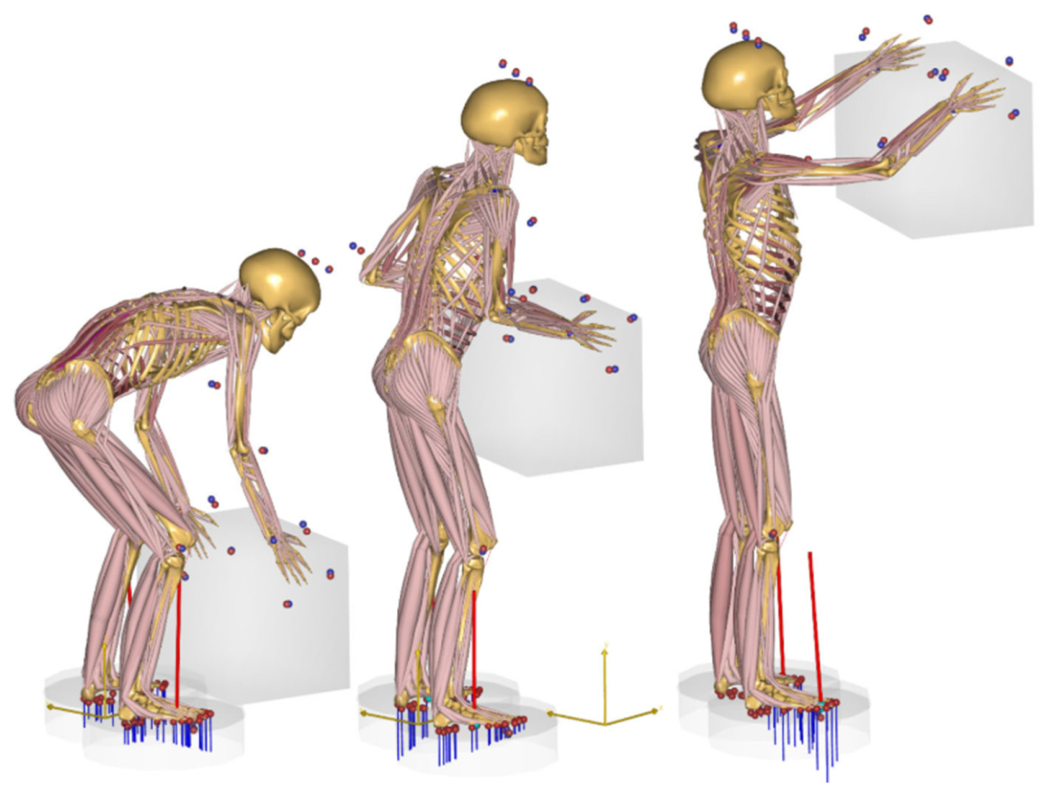
Bone is a hierarchically organized composite material optimized for high stiffness, strength, and low mass. Its cortical and trabecular architectures continuously adapt to mechanical loading through remodeling mechanisms consistent with Wolff’s law. As a result, multiscale experimental and numerical approaches are essential for accurately characterizing bone mechanics (Figure 2). Galbusera et al. [52] emphasize finite element modeling (FEM) as a central tool in musculoskeletal analysis, highlighting the importance of medical imaging for geometry reconstruction, mesh generation, and patient-specific material assignment. While personalized digital twins offer enhanced predictive capability, uncertainties in individual material properties and realistic in vivo loading remain limiting factors. Complementary work by Al-Barghouthi et al. [53] demonstrates that macroscopic bone elasticity and permeability arise directly from microstructural features such as trabecular thickness, connectivity, and anisotropy. Lin et al. [54] further contribute practical methodologies for extracting compact bone mechanical parameters from bilinear stress–strain responses, supporting both scaffold design and comparative biomechanical studies. Extending these concepts to synthetic analogs, Li et al. [55] review approaches for improving the load-bearing performance of calcium phosphate ceramics while maintaining bioactivity for bone repair.
At the nanoscale, recent studies on mineralized collagen fibrils, the fundamental building blocks of bone, have provided deeper mechanistic insight into energy dissipation and viscoelasticity. Milazzo and co-workers [56,57] demonstrate that mineralization level, hydration state, and loading rate strongly influence the dynamic mechanical response, with increased mineral content enhancing stiffness while intrafibrillar water significantly increases viscous damping and reduces relaxation times. Furthermore, their work on transient loading reveals that mineralized fibrils exhibit distinct mechanical responses in gap and overlap regions, with mineral accumulation enhancing stiffness locally while maintaining strong damping capacity under high-rate loading. These findings establish that nanoscale mechanisms govern macroscopic bone toughness and highlight the critical role of water-mediated viscoelasticity in mitigating damage under dynamic conditions.
Muscle tissue operates as an active mechanical actuator, transforming biochemical energy into force and motion, while its passive response is governed by hierarchical fiber organization. A similar concept of hierarchical organization is well established in other musculoskeletal tissues such as bone, where structural features span multiple scales, from nanoscale collagen–mineral composites to microscale osteons in cortical bone and trabecular networks in cancellous bone, each contributing to overall mechanical strength and energy dissipation. Gotti et al. [58] show that electrospun, muscle-inspired nanofibrous constructs can replicate nonlinear, multiscale stress–strain behavior through controlled fiber alignment. Similarly, Guo et al. [59] relate tissue viscoelasticity to fractal microarchitectures using hyper-cell models, demonstrating that hierarchical organization governs fractional-order relaxation behavior across scales. These findings reinforce the notion that muscle mechanics emerge from coupled nano- and microscale structures, while also highlighting persistent challenges in linking hierarchy to functional performance in vivo. Translating these principles into engineering design, Guan et al. [60] introduce bioinspired soft–rigid lattice architectures with adjustable stiffness and anisotropy, illustrating how hierarchical structuring can guide load transfer and mechanical adaptability in robotic systems [1].
Tendons and ligaments transmit muscular forces to bone and stabilize joints, exhibiting collagen-driven anisotropic and nonlinear responses typically described using hyperelastic or fiber-reinforced models. At the microscale, collagen fibrils, organized in a crimped and highly aligned architecture, govern the characteristic nonlinear stress–strain response of tendons. Recent microstructural modeling work by Gregory et al. [61] introduces a physically grounded framework that separates elastic and plastic fibril behavior while incorporating statistical distributions of yield and rupture stretches. This approach successfully reproduces experimentally observed features such as post-yield stiffening and step-like failure, which are not captured by traditional phenomenological models, thereby strengthening the link between fibril-level mechanisms and macroscopic tendon response.
Guo et al. [62] reveal scale-dependent mechanical behavior in articular cartilage, showing that stiffness at macro- and microscales correlates with proteoglycan content and load-bearing capacity, whereas nanoscale collagen fibrils remain mechanically stable. This finding underscores that emergent tissue mechanics depend more on structural organization than on intrinsic constituent stiffness. Consistent with this, nanoscale experimental studies by Svensson et al. [63] show that individual human tendon collagen fibrils exhibit only modest changes in stiffness and strength with aging, despite pronounced functional decline at the whole-tissue level, suggesting that higher-level structural organization, not intrinsic fibril properties, dominates age-related mechanical degradation.
Belluzzi et al. [64] and Jerban et al. [65] further demonstrate that cartilage and meniscus mechanics are strongly influenced by aging, pathology, and loading history, with MRI under mechanical load revealing early degenerative changes undetectable in conventional imaging. These studies expose limitations in current experimental techniques for capturing multiscale behavior under physiologically relevant conditions. At larger length scales, Zellers et al. [66] demonstrate that tendon tensile mechanics correlate strongly with collagen organization at the fascicle level, whereas advanced glycation end-products (AGEs) show weak or negligible correlation. This highlights that structural disorganization across hierarchical levels plays a more dominant role than biochemical alterations in governing tendon mechanical performance, particularly in pathological conditions such as diabetes.
From a clinical perspective, Borjali et al. [67] show that tendon-to-bone fixation strength depends critically on both structural alignment and surgical technique, with direct implications for repair strategies.
Fascia and connective tissues add further complexity to musculoskeletal mechanics. Kodama et al. [68] demonstrate that fascial stiffness, tension, and viscoelasticity regulate tissue sliding, neural mobility, and fibrosis risk, with mechanical loading driving fibroblast-to-myofibroblast differentiation. These responses are further modulated by age, hormonal status, and obesity, highlighting the need for quantitative, patient-specific assessment tools.
At the multicellular scale, Efremov et al. [69] show that the collective mechanics of cell aggregates depend on extracellular matrix composition, intercellular junctions, and contractility, exhibiting solid- or fluid-like behavior depending on timescale. Building on this foundation, more recent studies have significantly advanced our understanding by treating cell aggregates as active, multiphase materials rather than passive viscoelastic bodies. For instance, Dolega et al. [70] reveal that multicellular spheroids behave as active poroelastic systems, where cells, extracellular matrix, and interstitial fluid interact to govern overall mechanical response. A key highlight of this work is the demonstration that the bulk modulus and hydraulic permeability are not intrinsic constants, but instead emerge from internal active stresses and fluid transport mechanisms. Their findings further show that cells can be effectively modeled as incompressible inclusions embedded within a permeable matrix, providing a physically grounded explanation for stress distribution and time-dependent deformation in aggregates.
Complementing these insights, Ayan et al. [71] approach spheroids from a mechanics-of-manipulation perspective, demonstrating that their behavior during bioprinting is governed by a balance between spheroid surface tension and the yield stress of the surrounding medium. A key highlight is the identification of force-controlled transfer and positioning mechanisms, which enable precise, reproducible assembly of three-dimensional tissue constructs. This work not only advances biofabrication but also provides direct experimental evidence of how spheroid-scale mechanics influence structural organization and stability in engineered tissues.
From a measurement standpoint, Boot et al. [72] introduce a significant methodological advance by developing a high-throughput microfluidic platform for spheroid mechanophenotyping. The key highlight here is the ability to quantify viscoelastic properties across multiple spheroids simultaneously, overcoming the limitations of traditional single-sample techniques such as micropipette aspiration. Their results demonstrate consistent mechanical characterization across different cell types, reinforcing the link between single-cell properties, cell–cell interactions, and emergent tissue-level mechanics.
In addition to passive mechanical behavior, Nakano et al. [73] provide compelling evidence that cell aggregates exhibit active mechanical communication. Studying cardiomyocyte spheroids, they show that mechanical compression enhances both beating frequency and energy output, and can even induce synchronized activity between adjacent aggregates. A key highlight of this work is the identification of mechanically mediated coupling as a governing mechanism, demonstrating that collective function in tissues is not solely regulated by biochemical or electrical signaling, but also by mechanical interactions.
Reinforcing the importance of advanced measurement techniques, Dallara et al. [74] demonstrate that Digital Volume Correlation (DVC) enables three-dimensional strain mapping in heterogeneous musculoskeletal tissues, effectively linking microstructural features to functional mechanics. Together, these studies highlight a critical shift in the field: collective mechanics of cell aggregates are now understood as the result of tightly coupled phenomena involving active stresses, fluid transport, interfacial mechanics, and dynamic cell–cell communication. This emerging perspective underscores the need for integrated experimental and theoretical frameworks capable of capturing the full complexity of living, adaptive materials across scales.
Figure 2. A musculoskeletal model illustrating box lifting from the floor to a shelf. The blue lines beneath the feet indicate simulated pressure distributions, which collectively generate the ground reaction forces shown by the red vectors. Blue and red markers denote the alignment of marker coordinates between the experimental motion data and the model. Reprinted from Ref. [75] with permission from MDPI, 2025.
Eng 07 00235 g002
Across these studies, several consistent themes emerge. Musculoskeletal mechanics are inherently hierarchical, with nanoscale and microscale organization governing macroscopic function [43,53,54,58,59,60,66]. Recent advances further confirm that nanoscale mechanisms such as collagen fibril viscoelasticity, mineralization, and hydration play a decisive role in energy dissipation and damage resistance [56,57], while microscale organization (e.g., fibril alignment and fascicle structure) governs nonlinear and failure behavior [52,53,66]. Multiscale experimental approaches ranging from μCT and DVC to AFM and load-bearing MRI are essential for capturing emergent behavior [52,64,65,74], yet integrating these datasets into predictive, patient-specific models remains challenging. Hierarchical design principles consistently enhance both native tissue function and engineered constructs, from scaffolds and tendon–bone interfaces to bioinspired robotic systems [55,58,60]. Ultimately, the strong coupling between structure, loading, and pathology underscores the need for dynamic, individualized assessment frameworks capable of capturing nonlinear, anisotropic, and time-dependent responses [53,64,66,68].

3.2. Joint Mechanics and Load Transfer
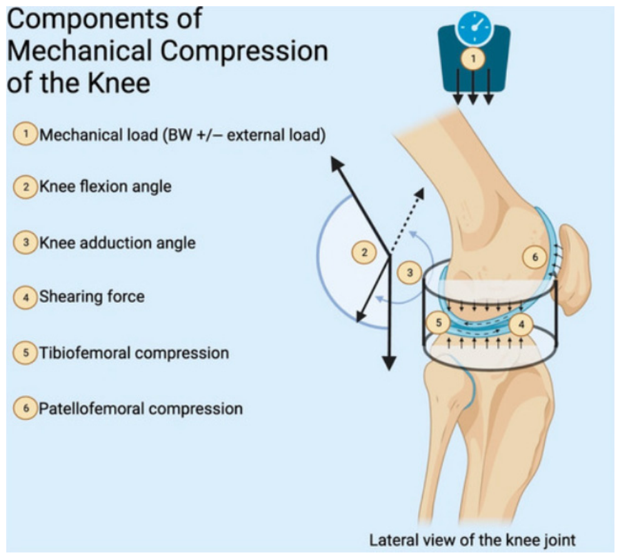
Human joints such as the hip, knee, and shoulder are sophisticated mechanical systems that enable controlled motion while efficiently distributing loads (see Figure 3). Their behavior is governed by contact mechanics, lubrication, cartilage viscoelasticity, and coordinated neuromuscular control. Healthy articular cartilage facilitates low-friction motion and shock absorption through its biphasic collagen–proteoglycan matrix, whereas degenerative conditions such as osteoarthritis disrupt load sharing and elevate contact stresses (Prathap et al. [76]; Layton et al. [77]; Logerstadt et al. [78]; Zdero et al. [79]). Finite element and multibody dynamic models are therefore essential for quantifying joint stresses, deformation, kinematics, and muscle forces, supporting prosthesis design, arthroplasty optimization, and functional assessment under physiological loading.
Recent studies further demonstrate that joint load transfer is highly sensitive not only to material properties but also to individual joint geometry. Deng et al. [80] show that even subtle variations in knee shape can lead to significant and sometimes unexpected changes in contact and fluid pressures following meniscectomy. A key highlight of this work is that joints deviating from the “average” geometry may experience elevated stresses, offering a mechanistic explanation for the wide variability in clinical outcomes. This finding reinforces the importance of subject-specific modeling in predicting joint mechanics and guiding surgical decision-making.
Advances in sensing and assistive technologies have broadened the scope of joint mechanics analysis. The study in [81] analyzes AI and machine learning integration in self-powered IoT sensors across major application domains, highlighting healthcare as the most developed area and reporting very high model accuracies up to 99.92% in some applications. Chen et al. [82] report reductions of 20–28% in lower-back and shoulder moments during patient-handling tasks with exoskeletal assistance, while Christens et al. [83] describe a modular full-body exoskeleton designed to support walking, lifting, and carrying in older adults. Despite these benefits, challenges related to device mass, joint alignment, control strategies, and upper-limb dexterity remain.
Beyond external assistance, implant design has emerged as a critical factor in restoring physiological load transfer. Saeidi et al. [84] demonstrate through cadaveric experiments that load-sharing knee implants can reduce medial joint contact pressures by approximately 18% without altering native joint geometry, highlighting their potential to mitigate osteoarthritis progression. Similarly, Munford et al. [85] show that conventional solid implants significantly disrupt natural stress distribution, leading to underloading of bone and potential resorption, whereas titanium lattice implants are capable of restoring near-native load transfer. A key highlight here is the recognition that implant stiffness and architecture must be carefully tailored to preserve physiological mechanical environments. Extending this further, Stoddart et al. [86] reveal that total knee arthroplasty introduces substantial strain shielding (up to ~30%), while partial and multi-compartmental procedures better maintain natural load transfer patterns. These findings collectively emphasize that preserving physiological load pathways is essential for long-term joint health and implant success.
Joint loading is highly dependent on task execution and technique. Hua et al. [87] show that different curling delivery styles impose distinct demands on lower-limb joints, with toe contact increasing hip and knee moments and outward-toed full-foot contact emphasizing ankle and subtalar control. These findings highlight the need for movement-specific injury prevention strategies. Similarly, Huang et al. [88] demonstrate that suspended-load backpacks reduce lower-limb muscle activity and joint work, improving metabolic efficiency while increasing shoulder loading, revealing trade-offs in load redistribution. Zaman et al. [89] extend this work by developing subject-specific predictions of asymmetric lifting motions, showing that optimizing dynamic joint torque constraints across multiple joints can reduce injury risk. Huysamen et al. [90] further identify hip and shoulder torque–power requirements as key targets for exoskeleton-assisted lifting design.
Figure 3. Mechanical loading applied to the knee. Reprinted from Ref. [91] with permission from MDPI, 2024.
Eng 07 00235 g003
Quantitative benchmarks of joint loading during daily activities further emphasize substantial inter-individual variability. Layton et al. [77] report peak joint forces ranging from 0.5 to 6.4 times body weight, with variability approaching ~20–30% depending on the activity type, modeling assumptions, and subject-specific biomechanical parameters such as gait pattern, body mass, and joint alignment, challenging the applicability of standardized reference loads. Barrutia et al. [92] address this issue using a mechanical knee phantom incorporating soft-tissue deformation, revealing reduced effective exoskeleton assistance due to energy absorption at the human–device interface. Miskovic et al. [93] demonstrate that hybrid systems combining soft hip exosuits with rigid knee exoskeletons can reduce metabolic cost (~16%) and muscle activity while preserving natural kinematics, offering a balanced multi-joint load transfer strategy.
Beyond external devices, intrinsic load transfer along the kinetic chain plays a crucial role in joint mechanics. Almansoof et al. [94] emphasize coordinated activation of muscles, fascia, and neural tissues to efficiently transmit energy from the lumbopelvic–hip complex to the limbs, reducing distal joint overload. At the tissue level, Rajesh et al. [95] show that engineered bioreactors applying cyclic stretch, hydrostatic pressure, and dynamic compression can reproduce physiological loading environments to stimulate tendon, ligament, and cartilage regeneration, with outcomes strongly dependent on loading magnitude, frequency, and duration. Zdero et al. [79] further stress the importance of accurately quantifying joint contact forces, noting limitations in both in vitro pressure films and in vivo motion-based estimations.
Overall, these studies highlight the inherently multiscale nature of joint mechanics and load transfer, spanning cartilage mechanotransduction, whole-body kinematics, wearable sensing, and exoskeletal augmentation. Experimental and computational investigations consistently show that cartilage degeneration and altered contact mechanics significantly affect load distribution and joint stresses [76,77,78,79], while subject-specific geometric variations further modulate pressure distributions and fluid mechanics within the joint [80]. Importantly, recent advances underline that joint mechanics is not governed by a single factor but by the interplay between geometry, material behavior, and intervention strategies such as implants or assistive devices. For instance, implant-related studies demonstrate that restoring physiological load transfer depends critically on stiffness matching and structural design [84,85,86], whereas assistive technologies such as exoskeletons and wearable systems actively redistribute joint loads and influence muscle coordination and metabolic cost [82,83,93]. The growing body of evidence points toward a paradigm shift from generic models to fully subject-specific, geometry-aware, and function-driven approaches. This shift is further supported by advances in AI-enabled sensing and real-time monitoring, which provide subject-specific in vivo data for predictive modeling and rehabilitation [81], as well as by studies highlighting the importance of task-specific loading conditions and movement strategies in determining joint response [87,88,89]. Despite significant progress, major challenges remain in integrating subject-specific in vivo data with computational models, accounting for soft-tissue contributions and human–device interaction effects [92], and optimizing assistive technologies to balance load reduction, metabolic efficiency, and kinematic fidelity. Future advances will rely on hybrid experimental–computational frameworks, AI-driven monitoring, and multi-joint augmentation strategies that better replicate physiological load transfer while minimizing injury risk.

3.3. Orthopedic Implant Design and Evaluation

Biomechanical principles form the foundation of orthopedic implant design, optimization, and evaluation, encompassing joint replacements, fixation systems, and load-bearing scaffolds. Engineering analysis informs material selection, implant geometry, and surface engineering to promote physiological load transfer while limiting stress shielding, an adverse condition in which excessive implant stiffness reduces mechanical stimulation of surrounding bone, potentially triggering bone resorption and implant loosening.
Recent evidence further emphasizes that stress shielding is not governed by material stiffness alone, but rather by a complex interaction between implant material and geometry. Park et al. [96] demonstrate that changes in material from titanium to cobalt–chromium can either increase or decrease bone resorption depending on implant design and anatomical location. A key highlight of this study is that identical materials may produce different remodeling responses when paired with different geometries, reinforcing the need to evaluate design–material coupling rather than treating them as independent variables.
Advances in implant materials continue to address these challenges. Hussein et al. [97] introduce a near-β Ti–30Nb–3Ag alloy exhibiting a 24–43% reduction in elastic modulus compared with Ti–6Al–4V and commercially pure titanium, alongside improved corrosion resistance and antibacterial behavior. Similarly, Baltatu et al. [98] report enhanced mechanical performance and osseointegration in Ti–Mo–Zr–Ta–Si alloys through controlled silicon additions. Kelly et al. [99] demonstrate that laser powder bed–fused porous titanium scaffolds enable stiffness tailoring and improved bone ingrowth, though mechanical strength and interfacial shear resistance remain strongly dependent on porosity. Beyond metallic systems, Bagherifard et al. [100] show that bio-nanocomposites [101] such as TCP–WS–Zr combine high strength, regulated biodegradation, and improved bone regeneration, underscoring the potential of tissue-engineered implant materials.
Structural design strategies play a critical role in mitigating stress shielding. Safavi et al. [102] review porous implant architectures including uniform, graded, and patient-specific configurations demonstrating superior biomechanical performance for personalized designs. Boudjemaa et al. [103] show that honeycomb-inspired interfacial geometries reduce peak stresses by over 50% while maintaining acceptable displacements, highlighting the influence of geometry on load absorption. Polymer-based alternatives [104], such as PEEK, PEKK, and PAEK/HA composites, reviewed by McMullan et al. [105] and Ma et al. [106], offer bone-like stiffness, radiolucency, and reduced stress shielding. When combined with additively manufactured porous structures, these polymers show promise, although achieving sufficient bioactivity and long-term mechanical stability remains challenging.
Beyond generic design strategies, recent studies highlight the importance of precise implant positioning and alignment in controlling load transfer. Dong et al. [107] demonstrate that small variations in tibial prosthesis alignment particularly varus angle can significantly alter contact pressures on the polyethylene liner, with optimized configurations reducing peak stresses to approximately 16 MPa. A key highlight of this work is the integration of finite element analysis with orthogonal experimental design, enabling systematic and efficient optimization of implantation parameters. This approach provides a practical pathway toward patient-specific surgical planning and improved implant longevity.
Topology optimization (TO) and additive manufacturing (AM) have fundamentally transformed implant design workflows. Kladovasilakis et al. [108] and Wu et al. [109] demonstrate that bioinspired lattice architectures and hierarchical TO can achieve substantial mass reductions (~38%), enhanced porosity, and improved stress distribution in hip implants while maintaining acceptable safety margins. Schwarz Diamond and Gyroid lattices show particularly favorable mechanical performance. Extending these methods to craniofacial applications, van Kootwijk et al. [110] report that fully porous, TO-designed Ti–6Al–4V ELI mandibular implants achieve comparable quasi-static and fatigue behavior with reduced mass, supporting clinical feasibility. Across these studies, finite element analysis remains the primary preclinical evaluation tool, complemented by experimental validation through mechanical testing, digital image correlation, and fatigue assessment.
Translating these design advances into clinical practice, Tang et al. [111] provide evidence that porous metal pillar augmentation can effectively manage tibial bone defects in total knee arthroplasty. A key highlight is the demonstration of improved clinical outcomes such as increased range of motion and functional scores alongside stable fixation with minimal radiolucent lines over a two-year follow-up period. Importantly, no cases of implant loosening or revision were reported, suggesting that porous, load-sharing structures can enhance both mechanical stability and biological integration in vivo.
Biological integration is equally essential for long-term implant success. Grzeskowiak et al. [112] emphasize that both initial mechanical stability and long-term fixation depend on tissue response, vascularization, and healing dynamics. Surface modification, porous architectures, and bioactive coatings enhance osseointegration and interface durability. Cyphert et al. [113] focus on antimicrobial implant strategies with controlled drug release, validated through in vivo and subcutaneous models. In contrast, Sun et al. [114] highlight the lack of standardization in evaluating degradable magnesium implants across animal models and testing protocols, complicating cross-study comparison and clinical translation.
Despite significant progress, several challenges persist. Biological validation often trails mechanical assessment, with limited long-term in vivo and clinical data. Most current experimental and preclinical studies typically span short to mid-term durations (ranging from a few weeks to several months, and in some cases up to 1–2 years for animal models), which is often insufficient to capture long-term biological adaptation, material degradation, or chronic tissue remodeling processes. As a result, this temporal limitation restricts the ability to reliably predict long-term clinical performance, implant survival, and patient-specific outcomes. This limitation is reflected across multiple studies, where promising short-term outcomes in porous and load-sharing implants [111] and bioactive or degradable systems [100,114] are not yet supported by consistent long-term validation frameworks, particularly for degradation behavior and biological remodeling. Furthermore, the study in [112] emphasizes that long-term implant success depends on dynamic biological processes such as vascularization and tissue integration, which are rarely captured in short-duration studies.
Many finite element models assume homogeneous bone properties and linear material behavior, neglecting frictional interfaces, cyclic loading, and multi-material constructs. However, more advanced computational approaches demonstrate the importance of incorporating these complexities: the studies [108,109] show that topology-optimized and lattice-based designs significantly alter stress distribution and load transfer, while Dong et al. [107] highlight that implant alignment and positioning can strongly influence local contact mechanics and stress fields. In addition, Park et al. [96] demonstrate that stress shielding is governed by the coupled interaction between material and geometry, rather than material stiffness alone, further exposing the limitations of simplified modeling assumptions. Moreover, standardized metrics for assessing stress shielding, porosity-dependent performance, osseointegration, and degradation kinetics remain underdeveloped. This gap is reinforced by findings from [102,103], who show that porous and bioinspired architectures significantly influence mechanical performance but lack unified evaluation criteria, while Kelly et al. [99] demonstrate that porosity-driven improvements in bone ingrowth are highly dependent on structural parameters that are not consistently standardized across studies. Addressing these limitations will require multi-objective hierarchical TO, functionally graded lattices, patient-specific AM solutions, and improved preclinical standardization.
Importantly, emerging evidence suggests that future implant design must move toward fully integrated frameworks that simultaneously consider material selection, structural geometry, implantation strategy, and patient-specific anatomy. Rather than optimizing these factors independently, their coupled effects on load transfer, stress distribution, and biological response must be addressed holistically. This paradigm shift is strongly supported by multiple studies: Park et al. [96] demonstrate material–geometry coupling effects, Dong et al. [107] highlight the critical role of implantation strategy and alignment, and the studies [99,102] show how structural design and porosity influence both mechanical and biological outcomes. Together, these works establish that implant performance emerges from the interaction of multiple design variables rather than isolated optimization.
In summary, the convergence of biomechanics, advanced materials, surface engineering, and additive manufacturing enables rational orthopedic implant design that balances load transfer, stress shielding mitigation, and biological integration. Integrated computational–experimental frameworks are essential for preclinical assessment, while future advances will depend on standardized validation protocols, multi-material strategies, and systematic long-term performance evaluation to bridge the gap between laboratory innovation and clinical application. Recent developments in advanced alloys [97,98], bio-nanocomposites [100], and additively manufactured porous scaffolds [99] further illustrate the shift toward multifunctional implant systems that integrate mechanical performance, bioactivity, and controlled degradation. However, as consistently noted across these studies, achieving reliable long-term clinical validation remains a critical unresolved challenge.

3.4. Injury Mechanics and Rehabilitation Engineering

Biomechanical analysis plays a central role in elucidating injury mechanisms and guiding rehabilitation engineering, linking injury prevention, therapeutic device development, and functional recovery. By combining kinematic and kinetic analysis with motion capture, ground reaction force measurements, and electromyography, high-risk movement patterns can be identified and addressed through personalized rehabilitation programs. Zhao [115] shows that these tools support neuromuscular training and equipment optimization in sports, with increasing integration of machine learning approaches such as artificial neural networks (ANNs), support vector machines (SVMs), random forest classifiers, and recurrent neural networks (RNNs), particularly long short-term memory (LSTM) models for time-series biomechanical data to improve injury prediction and reduce reinjury risk by identifying movement patterns, fatigue states, and abnormal joint loading trajectories.
Recent advances further demonstrate that biomechanical analysis is no longer confined to laboratory environments. Slade et al. [116] introduce a wearable system capable of estimating metabolic energy expenditure in real time with significantly lower error (~13%) compared to conventional smartwatches (>40%). A key highlight of this work is the use of lower-limb inertial sensors (shank and thigh), which more accurately capture dynamic activities such as walking, running, and stair climbing. This represents a major step toward continuous, real-world biomechanical monitoring, enabling more precise assessment of functional recovery and rehabilitation progress outside controlled laboratory settings.
Complementary sport-specific studies by Lucarno et al. [117] and Trasolini et al. [118] reveal that injuries such as anterior cruciate ligament rupture in female soccer players and throwing-related shoulder and elbow injuries commonly arise from noncontact, multiplanar loading and kinetic-chain dysfunctions, emphasizing the need for task-specific neuromuscular rehabilitation rather than isolated joint strengthening. From a biomechanical perspective, ACL rupture is strongly associated with high knee abduction moments, excessive anterior tibial shear forces, increased knee valgus angles, and poor neuromuscular control during landing and cutting maneuvers. Similarly, throwing-related injuries are linked to extreme joint loading conditions, including high shoulder external rotation torques, elevated elbow valgus stress, and rapid angular velocity transfer along the proximal-to-distal kinetic chain, which places repetitive tensile and shear loads [119] on soft tissues.
Bridging laboratory biomechanics with practical clinical assessment, Peebles et al. [120] demonstrate that portable and low-cost systems such as force-sensing insoles and tablet-based motion tracking can reliably quantify landing biomechanics in non-laboratory environments. A key highlight is the identification of asymmetries in plantar force impulse and knee flexion range of motion in ACL-reconstructed patients, providing objective metrics for return-to-sport decision-making. This work highlights the growing feasibility of translating injury risk assessment into routine clinical and field settings.
Rehabilitation engineering increasingly translates biomechanical insight into therapeutic interventions through robotic and mechatronic systems. Reviews by Gull et al. [121] and Gandolfi et al. [122] highlight the potential of upper-limb exoskeletons to restore functional movement following neurological or neuromuscular injury, emphasizing kinematic compatibility, compliant control, and safe human–robot interaction. However, they also identify persistent challenges, including the lack of standardized design criteria, inconsistent reporting of mechanical and control parameters, and limited regulatory approval. Dong et al. [107] extend these observations to parallel ankle rehabilitation robots, showing that while advanced actuators and multimodal training promote central nervous system reorganization, robust clinical validation remains limited. Vaida et al. [123] provide a design-driven example in which experimental kinematic data from post-stroke patients directly informed workspace definition, singularity avoidance, and numerical simulation of a lower-limb parallel robot, illustrating how injury mechanics can directly guide rehabilitation device design.
Wearable technologies complement robotic systems by enabling continuous biomechanical monitoring for injury prevention and recovery optimization. Rebelo et al. [124] and Seckin et al. [125] demonstrate that wearable sensors and force platforms can quantify mechanical loading, fatigue, and workload metrics, supporting individualized training modulation and rehabilitation tracking in environments where laboratory-based motion capture is impractical. Ortiz-Padilla et al. [126] further show that video-based motion analysis can estimate forces and identify stress concentrations associated with fatigue fractures or ligament injury.
Extending these capabilities to population-scale insights, Khurshid et al. [127] demonstrate through large-scale wearable accelerometry (UK Biobank, ~96,000 participants) that objectively measured physical activity is strongly associated with reduced incidence across more than half of evaluated diseases. A key highlight is that wearable-derived metrics show significantly stronger associations with disease risk reduction than self-reported activity, emphasizing the importance of objective biomechanical monitoring. This finding not only reinforces the role of physical activity in injury prevention and recovery but also underscores the potential of wearable technologies to inform long-term rehabilitation strategies and public health interventions.
Despite these advances, the real-time integration of force estimation with predictive injury and recovery models remains underdeveloped.
Biomechanical modeling provides a critical foundation for injury analysis and rehabilitation planning. Roupa et al. [50] review multibody dynamics, musculoskeletal (MSK), and neuromusculoskeletal (NMSK) models, highlighting trade-offs between computational efficiency and physiological realism. While NMSK models capture neural activation and muscle coordination in detail, their computational cost limits routine clinical use. In contrast, MSK models offer practical tools for estimating joint loading and informing rehabilitation protocols and assistive device optimization.
Neurorehabilitation strategies increasingly integrate neural interfaces and advanced control schemes. Bai et al. [128] demonstrate that brain–computer interfaces combined with functional electrical stimulation can improve post-stroke upper-limb function, although long-term efficacy and the added value of adjunctive neuromodulation remain uncertain. Akbari et al. [129] extend these concepts to home-based, IoT-enabled rehabilitation platforms that integrate sensors, mechatronic devices, and tele-supervision, enabling data-driven early intervention and improving accessibility, an approach accelerated by pandemic-related constraints.
Across sports biomechanics, robotic rehabilitation, wearable sensing, and modeling, several overarching limitations remain. Standardization of mechanical design, control strategies, and evaluation metrics is still limited, hindering comparison across studies and slowing clinical translation [50,107,121,122]. Many systems remain at the prototype stage, with insufficient large-scale clinical validation or regulatory certification [127,128,129]. Moreover, while biomechanical analysis can identify high-risk movement patterns, real-time integration of force estimation, fatigue prediction, and adaptive load modulation into rehabilitation systems is still lacking [115,116,124,125].
Importantly, emerging evidence suggests a paradigm shift toward continuous, real-world, and data-driven rehabilitation ecosystems, where wearable sensing, portable biomechanics, and AI-based analytics operate synergistically [116,120,126,127]. Rather than relying solely on episodic laboratory assessments, future systems are expected to provide continuous feedback, enabling adaptive rehabilitation that responds dynamically to patient-specific biomechanical states [116,123,127].
In conclusion, injury mechanics and rehabilitation engineering represent a rapidly evolving intersection of biomechanics, robotics, sensing technologies, and neuroengineering. Although substantial progress has been made in translating mechanical insight into rehabilitation devices and personalized treatment strategies, future research must prioritize standardized validation, real-time biomechanical feedback, AI-driven prediction, and user-specific adaptive control [50,115,121,122,127]. Such integrated approaches are essential for delivering safe, effective, and scalable rehabilitation solutions that meaningfully connect engineering innovation with clinical outcomes.

4. Cardiovascular and Hemodynamic Biomechanics

The cardiovascular system operates as a continuously active, tightly integrated transport network responsible for delivering blood, oxygen, and nutrients throughout the body. From an engineering viewpoint, it represents a highly efficient fluid–structure interaction (FSI) system, in which pulsatile blood flow dynamically couples with compliant vessel walls subjected to cyclic mechanical loading. In physiological conditions, this system is characterized by well-defined hemodynamic ranges, including arterial blood pressures of approximately 80–120 mmHg (systolic/diastolic), blood flow velocities ranging from ~0.1–0.3 m/s in microcirculation to ~0.5–1.5 m/s in large arteries such as the aorta, and wall shear stresses typically on the order of 1–2 Pa in healthy vessels. Deviations from these ranges are strongly associated with vascular remodeling and disease progression. For example, regions of low or oscillatory wall shear stress are known to promote endothelial dysfunction, increased permeability, and inflammatory signaling, which contribute to the initiation and progression of atherosclerotic plaques. Conversely, abnormally high or highly fluctuating hemodynamic loads can weaken the vessel wall structure over time, promoting extracellular matrix degradation and elastin fragmentation, which are key factors in aneurysm initiation and expansion. Quantitative biomechanical analysis of this interaction is essential for understanding normal hemodynamics, pathological progression, and the rational design of cardiovascular devices, including stents, grafts, embolic agents, and prosthetic valves.

4.1. Mechanical Behavior of Blood and Vessel Walls

The mechanical response of blood vessels emerges from a multiscale, strongly coupled system involving flowing blood, vascular cells, and extracellular matrix (ECM) components interacting under time-dependent loading. Blood vessels exhibit pronounced nonlinearity, anisotropy, and viscoelasticity, and actively remodel their material properties in response to changes in pressure, flow, and biochemical signaling. Blood itself is not merely a passive loading medium; it generates shear forces at the endothelial interface and, under pathological conditions, forms mechanically active structures such as thrombi. Mechanical compatibility among blood, vessel wall, and implanted or engineered materials therefore governs stress transmission, energy dissipation, and long-term structural integrity under both physiological and diseased states.
To mathematically capture these complex behaviors, arterial wall mechanics are commonly represented using constitutive models such as hyperelastic formulations (e.g., Neo-Hookean, Mooney–Rivlin, and Ogden models), which describe nonlinear elastic responses, as well as structurally informed models like the Holzapfel–Gasser–Ogden framework, which explicitly accounts for collagen fiber orientation and dispersion. In addition, viscoelastic extensions of these models are often employed to represent time-dependent behavior such as stress relaxation and creep under cyclic loading conditions.
The summary of the studies investigating mechanical behavior of blood and vessel walls in the literature are summarized in Table 2.
To facilitate cross-scale comparison of vascular mechanics, representative studies are commonly organized according to shared mechanical themes such as nonlinearity, anisotropy, viscoelasticity, compliance matching, and flow–structure coupling rather than by biological application alone. This perspective reveals recurring methodological approaches, convergent mechanical behaviors, and persistent limitations in constitutive modeling, multiaxial testing, and long-term validation across native tissues, engineered constructs, and device-based interventions.
At the cellular level, vascular mechanics arise from coordinated interactions between endothelial cells (ECs) and vascular smooth muscle cells (VSMCs). Mendez-Barbero et al. [130] emphasize that EC–VSMC coupling regulates circumferential stress, wall stiffness, permeability, and vascular tone through mechanosensitive signaling pathways. Shear stress from blood flow and cyclic stretch from pulsatile pressure are transduced into biochemical responses that modulate cytoskeletal tension, contractility, and ECM synthesis. When this coupling is disrupted, through altered junctional mechanics, impaired paracrine signaling, or abnormal ECM turnover, vessels undergo maladaptive remodeling, manifested mechanically as increased stiffness, wall thickening, and reduced elastic recoil. Liu et al. [131] further elucidate these mechanotransduction pathways, describing how ion channels, integrins, cytoskeletal networks, and nuclear deformation convert mechanical stimuli into transcriptional responses. Despite substantial mechanistic insight, quantitative links between specific signaling disruptions and changes in constitutive parameters (e.g., compliance, tangent modulus, or relaxation times) remain poorly established, limiting predictive modeling.
The ECM dominates load bearing in the vessel wall and largely defines its nonlinear stress–strain response. Yanagisawa et al. [132] demonstrate that elastin fibers, organized into elastin–contractile units with smooth muscle cells, are essential for maintaining arterial compliance and the Windkessel effect by storing elastic energy during systole and releasing it during diastole. Progressive elastin degradation shifts load sharing to stiffer collagen fibers, increasing wall stiffness and altering stress distributions. Cai et al. [133] show that hypertension-driven ECM remodeling changes the collagen–elastin balance, reinforcing a feedback loop between altered mechanics and pathological adaptation. Yamashiro et al. [134] provide a mechanistic bridge between ECM mechanics and cellular signaling by identifying thrombospondin-1 as a mediator of strain-dependent integrin engagement and YAP activation. Together, these studies demonstrate that vessel wall mechanics cannot be captured by single-modulus descriptions; instead, evolving fiber recruitment, anisotropy, and microstructural heterogeneity must be incorporated into constitutive frameworks. Nevertheless, many existing models still assume spatial homogeneity and neglect region-specific ECM architecture.
Table 2. Comparative summary of representative biomechanical studies on blood, vessel walls, and vascular constructs.
ReferenceSystem/ScalePrimary Mechanical FocusMethods/ModelsKey Mechanical FindingsLimitations/Gaps
Mendez-Barbero et al. [130]Native vessel wall (cellular–tissue)EC–VSMC coupling, wall stiffness, complianceCell–cell interaction analysis, in vitro modelsDisrupted EC–VSMC communication leads to maladaptive stiffening and loss of elasticityLacks direct quantification of constitutive parameters
Liu et al. [131]Native vessel wall (cellular)Mechanotransduction under shear and stretchMolecular and cellular mechanobiologyIdentifies pathways converting mechanical stress to biochemical signalingMostly qualitative, limited linkage to macroscopic mechanics
Yanagisawa et al. [132]Aorta (tissue)Elastic recoil, Windkessel mechanicsStructural–mechanical analysis, genetic modelsElastin–contractile units govern cyclic energy storage and complianceLimited patient-specific mechanical data
Cai et al. [133]Arteries (tissue)ECM remodeling, stiffness in hypertensionECM analysis, mechanical inferenceECM degradation increases arterial stiffness and dysfunctionSimplified material assumptions
Yamashiro et al. [134]Vessel wall (molecular–tissue)ECM-mediated mechanotransductionGenetic mouse models, signaling analysisThbs1–integrin–YAP axis links strain to remodelingContext-dependent effects complicate translation
Blinkouskaya et al. [135]Aging tissues (organ-level analogy)Stiffening due to microstructural degenerationContinuum mechanics, imagingMultiscale degeneration drives macroscopic stiffnessIndirect application to vessels
Sanchez-Molina et al. [136]Cerebral veins (tissue)Nonlinearity, viscoelasticity, strain-rate effectsMicrotensile testing, constitutive modelingViscoelasticity significant at high strain ratesLimited to specific vessel type
Tutwiler et al. [137]Blood clots (material)Fracture toughness, rupture mechanicsMechanical testing, FE simulationsClot failure governed by strain-driven fiber ruptureDoes not include vessel wall interaction
Zheng et al. [138]Blood–thrombus–wall systemFlow–structure interaction, embolizationPhase-field + particle-based modelingPredicts clot deformation and detachment under flowRequires high computational cost
Brusokas et al. [139]Veins with thrombus (system)Wall compliance effects3D numerical simulationsWall elasticity strongly alters clot deformationIdealized geometries
Jeong et al. [140]Vascular grafts (tissue–device)Compliance mismatch, IHReview, mechanical testingMismatch alters WSS and promotes intimal hyperplasiaNo standardized compliance metrics
Moore et al. [141]Small-diameter graftsElasticity, complianceMaterial comparison reviewNatural materials better match vessel mechanicsLimited long-term data
Wang2 et al. [142]Engineered SDBVsNonlinear stress–strain, burst pressureReview of fabrication & testingHybrid materials offer tunable mechanicsVariability across studies
Kuang et al. [143]Biodegradable graftsLoad-bearing & remodelingNanofiber core–shell design, animal modelsMechanical support transitions to regenerated tissueScaling to humans uncertain
Jia et al. [144]Tri-layer vascular graftCompliance, burst strengthMechanical testing, in vivo studiesLayered design mimics native mechanicsManufacturing complexity
Zhou et al. [145]Bioprinted vesselsTunable complianceCoaxial 3D bioprintingBalanced mechanical support and biological functionShort-term evaluation
Wang et al. [146]Bioprinted conduitsEnergy dissipation, elasticityDouble-network hydrogelsVenous vs. arterial mechanics replicatedFatigue behavior not assessed
Daniel et al. [147]Decellularized HUVBiphasic stress–strainMechanical testing, cell seedingPreserves native vessel mechanicsDonor variability
Grossbacher et al. [148]Biofabricated vesselsAnisotropy, reinforcementVolumetric bioprinting + MEWFiber reinforcement enables vessel-like mechanicsComplex fabrication
Distler et al. [149]Soft tissues/hydrogelsNonlinearity, asymmetry, viscoelasticityMulti-modal mechanical testingSimple blends replicate complex tissue mechanicsNot vascular-specific
Choi et al. [150]Soft tissue (oral mucosa)ECM architecture, anisotropyTensile testing, SEMFiber orientation dictates stiffness and failureStatic loading only
Song et al. [151]Bone–implant system (analogy)Compliance matchingCompression testing, FE analysisPorosity reduces stiffness mismatchDifferent loading environment
Collins et al. [152]Engineered scaffoldsMicrostructure–mechanics linkCAD, imaging, FE modelingArchitecture dictates mechanical responseLimited biological validation
Hu et al. [153]Embolic materialsInjectability vs. mechanical stabilityIn vivo porcine modelsECM hydrogel balances occlusion and wall safetyLong-term remodeling unknown
Xin et al. [154]Metamaterial scaffoldsAuxetic behavior, J-shaped responseMechanical testing, design modelingProgrammable vessel-like mechanicsBiological integration not tested
Lin et al. [155]Vascular stentsRadial/axial mechanicsFE analysis, in vitro testsNPR stents reduce wall injuryMaterial optimization needed
Salama et al. [156]Biodegradable metalsStiffness–degradation balanceReview of AM iron implantsPorosity tunes mechanical compatibilityCorrosion control challenges
Dastagir et al. [157]TEVGsPulsatile conditioningFlow bioreactor, mechanical testingPhysiological loading improves strengthLimited long-term patency
Devillard et al. [158]Engineered vesselsMechanical maturationBioprinting, bioreactorsConditioning essential for durabilityScale-up challenges
Campinho et al. [159]EndotheliumFlow-induced mechanicsLive imaging, mechanobiologyShear stress governs morphogenesisMostly developmental context
Insights from other soft tissues reinforce the central role of ECM organization in governing mechanical behavior. Choi et al. [150] show that collagen fiber orientation alone produces substantial regional variation in stiffness, strength, and failure strain in human oral mucosa: aligned fiber networks yield higher stiffness and strength, whereas multidirectional arrangements promote compliance and earlier failure. These findings closely parallel vascular behavior, where collagen orientation dictates anisotropy and rupture risk. Distler et al. [149] further demonstrate that replicating native tissue mechanics requires capturing nonlinear, asymmetric, and time-dependent responses across multiple loading modes. Their alginate–gelatin hydrogels reproduce compression–tension asymmetry, conditioning effects, and viscoelastic relaxation observed in brain tissue, underscoring the limitations of vascular graft studies that report only uniaxial tensile properties while neglecting torsion, compression [160], and cyclic loading experienced in vivo.
Similarly, multiscale cardiac studies reinforce that microstructural organization particularly collagen fiber architecture and myofiber orientation plays a decisive role in organ-level mechanical function and disease progression. Neekalanten et al. [161] demonstrate that in pulmonary hypertension, right ventricular remodeling is strongly linked to collagen fiber tautness and myofiber reorientation, which reduce tissue anisotropy and contribute to maladaptive stiffening and ventricular–arterial uncoupling. Their multiscale analysis shows that fiber-level remodeling mechanisms, rather than hypertrophy alone, govern the transition from adaptive to maladaptive mechanical behavior, highlighting the importance of incorporating microstructural remodeling into biomechanical models.
Aging introduces additional complexity through progressive, multiscale degeneration. Although focused on neural tissue, Blinkouskaya et al. [135] provide a useful mechanical analogy linking microstructural degradation to macroscopic stiffening and shape change. In blood vessels, endothelial dysfunction, smooth muscle atrophy, elastin fragmentation, and collagen accumulation collectively reduce compliance and alter nonlinear mechanical response. Sanchez-Molina et al. [136] contribute rare high-resolution mechanical data on cerebral bridging veins, revealing pronounced nonlinear elasticity, viscoelastic stress relaxation, and strain-rate sensitivity. Their results show that viscoelastic effects dominate at high strain rates, such as during traumatic loading, whereas quasi-static behavior is governed primarily by nonlinear elasticity. A persistent limitation in vascular biomechanics remains the scarcity of age-dependent human mechanical data, which restricts validation of constitutive models for clinical prediction.
Multiscale experimental investigations of myocardial tissue further illustrate how mechanical behavior emerges from interactions across length scales. Neelakantan et al. [162] present a multiscale experimental framework linking sarcomere-level contraction to organ-level cardiac function, showing that active stress generation, relaxation behavior, and anisotropic contraction vary significantly between fiber bundles and ventricular wall tissue. Their findings demonstrate that myocardial contractility is strongly direction-dependent and varies through the ventricular wall thickness, emphasizing that organ-level cardiac performance cannot be accurately predicted without incorporating fiber orientation, transmural structure, and time-dependent active behavior into biomechanical models.
Blood itself plays an active mechanical role, particularly during clot formation, where it transitions from fluid-like to solid-like behavior. Tutwiler et al. [137] quantify the fracture toughness of fibrin clots, demonstrating that failure is governed by strain-driven fiber rupture rather than stress-based criteria, with direct implications for embolization risk (see Figure 4) under flow-induced loading. Zheng et al. [138] integrate blood flow, platelet aggregation, and thrombus mechanics within a unified computational framework, capturing how shear forces deform clots and redistribute stresses to the vessel wall. Brusokas et al. [139] further show that vessel wall compliance strongly modulates clot deformation and local hemodynamics, with compliant walls amplifying clot strain compared to rigid-wall assumptions. These findings expose a major limitation of many thrombosis models: neglecting wall deformability leads to inaccurate predictions of clot stability and embolic potential.
Figure 4. Schematic representation of the stasis and stenosis models: (a) normal blood flow; (b) stenosis model, where a thrombus forms due to partial obstruction of blood flow; and (c) stasis model, where a thrombus develops due to complete obstruction of blood flow. Reprinted from Ref. [163] with permission from MDPI, 2023.
Eng 07 00235 g004
In addition to thrombosis mechanics, recent work by Mukherjee et al. [164] highlights the importance of advanced imaging-derived strain metrics for detecting early myocardial dysfunction. Their study on radiation-induced cardiotoxicity shows that principal strain measurements derived from cardiac MRI can detect subclinical deterioration in cardiac contractility earlier than conventional strain metrics, particularly by capturing transmural heterogeneity in myocardial deformation. This work demonstrates that regional and layer-specific mechanical analysis provides more sensitive indicators of cardiac dysfunction than global mechanical metrics, reinforcing the importance of spatially resolved biomechanical assessment in cardiovascular modeling and diagnosis.
Mechanical mismatch between native vessels and implanted materials remains a central challenge in vascular device design. Jeong et al. [140] demonstrate that compliance mismatch between grafts and host arteries disrupts wall shear stress distributions and promotes intimal hyperplasia. Moore et al. [141] and Wang et al. [146] similarly show that excessive stiffness in synthetic grafts triggers maladaptive biological responses, reinforcing the importance of compliance-matched designs. Analogous principles are well established in orthopedic biomechanics, where Song et al. [151] show that stiff, solid implants induce stress shielding, while porous TPMS architectures reduce modulus mismatch and improve load transfer. In contrast, Hu et al. [153] develop an ECM-based embolic hydrogel that combines shear-thinning injectability with solid-state mechanical stability and controlled degradation, representing a rare example in which delivery, load-bearing function, and biological remodeling are addressed simultaneously.
Fabrication strategy has emerged as a key determinant of mechanical performance. Collins et al. [152] articulate a general biomechanical principle: microstructural architecture governs macroscopic mechanical behavior. Although discussed in the context of bone scaffolds, the implications for vascular mechanics are direct controlled porosity, anisotropy, and hierarchical organization dictate stiffness, strength, and failure. Advanced imaging, CAD-based design, and finite element analysis now enable prediction of deformation and rupture risk, as demonstrated in aneurysm biomechanics. Distler et al.’s [149] work further shows that even relatively simple material systems can reproduce complex tissue mechanics when composition and processing are guided by systematic mechanical characterization. However, translation to clinically validated vascular constructs remains constrained by the lack of long-term cyclic fatigue data.
Biofabrication strategies increasingly seek to replicate native vessel mechanics through hierarchical design. Kuang et al. [143], Jia et al. [144], Zhou et al. [145], and Wang et al. [146] demonstrate that multilayered, core–shell, or double-network architectures distribute mechanical functions across layers, combining early load-bearing with gradual biological replacement. Daniel et al. [147] preserve native biphasic mechanics using decellularized human umbilical vein scaffolds, while Grossbacher et al. [148] integrate volumetric bioprinting with melt electrowriting to achieve anisotropic reinforcement. Despite promising results, challenges remain in fatigue resistance, scalability, and long-term remodeling under physiological loading.
Emerging material systems further expand the vascular biomechanical design space. Xin et al. [154] introduce auxetic metamaterials with programmable, J-shaped stress–strain responses resembling arterial mechanics, while Lin et al. [155] develop shape-memory stents incorporating negative Poisson’s ratio geometries to reduce vessel wall injury during deployment. Salama et al. [156] review biodegradable iron-based implants that balance initial mechanical support with gradual degradation, although controlling degradation kinetics while preserving mechanical integrity under cyclic loading remains unresolved.
Mechanical conditioning is consistently shown to be essential for functional maturation. Dastagir demonstrates that pulsatile flow enhances the strength and performance of spider silk–based vascular grafts, aligning with Devillard et al.’s [158] emphasis on bioreactor-based dynamic conditioning. Campinho et al. [159] further show that endothelial cells actively interpret mechanical cues to guide morphogenesis, reinforcing the limitation of purely static mechanical testing.
In summary, the biomechanics of blood and vessel walls is governed by the coupled effects of microstructure, material behavior, mechanotransduction, and dynamic loading [130,131,132]. Evidence spanning vascular biology, thrombosis, tissue engineering, and comparative soft-tissue mechanics consistently highlights anisotropy, nonlinearity, viscoelasticity, and compliance matching as fundamental principles [133,136,140,141,149]. Despite substantial progress, critical gaps remain in standardized mechanical characterization, integration of blood–wall–device interactions, and validation of multiscale models using human data [137,138,139]. Addressing these challenges will require rigorously integrated frameworks combining advanced fabrication, multimodal mechanical testing, and patient-specific computational modeling to enable durable, predictive, and personalized vascular therapies [143,148,152,158].

4.2. Hemodynamic Parameters and Flow Patterns
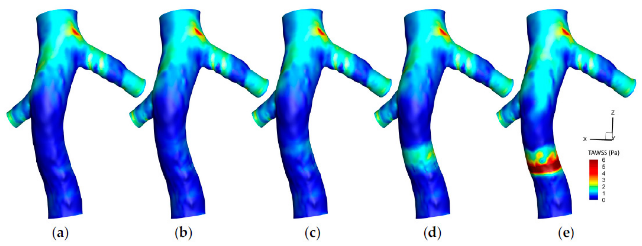
Hemodynamic forces are fundamental to cardiovascular function, governing endothelial behavior, vascular remodeling, and the onset and progression of disease. Parameters such as blood pressure, flow velocity, wall shear stress (WSS), oscillatory shear index (OSI), time-averaged WSS (TAWSS), and relative residence time (RRT) collectively define the mechanical environment within arteries and veins. Extensive experimental and computational evidence links regions of low or oscillatory WSS to atherosclerotic plaque formation, while elevated tensile stresses are closely associated with aneurysm initiation and rupture. Although blood flow in healthy arteries is predominantly laminar, disturbances frequently arise near bifurcations, stenoses, curved segments, and implanted devices. These disturbed flow patterns characterized by separation, recirculation, and oscillatory shear play a decisive role in localized endothelial dysfunction and spatially heterogeneous disease development.
Patient-specific computational studies have provided critical insight into how altered hemodynamics contribute to vascular pathology. Ferdows et al. [165] developed a coronary artery disease framework based on CCTA-derived three-dimensional reconstructions, demonstrating strong correlations between WSS-related indices and stenosis geometry and severity. Their analysis included velocity, pressure, WSS, TAWSS, OSI, RRT, and computational fractional flow reserve (cFFR), revealing that sequential or proximally located stenoses markedly intensify flow disturbances. These configurations produced elevated velocities, localized WSS peaks, and pronounced oscillatory flow in constricted and bifurcating regions. While the approach enables non-invasive assessment of endothelial risk (analogous in concept to particle image velocimetry (PIV)) simplifying assumptions such as rigid vessel walls and Newtonian blood behavior limit its physiological completeness.
The importance of vascular compliance and blood rheology is highlighted in fluid–structure interaction (FSI) studies. Kumar et al. [166] applied patient-specific three-dimensional FSI models to normal and stenosed carotid arteries and showed that hypertension leads to reduced WSS and increased OSI in regions prone to recirculation, particularly within the carotid sinus and downstream of stenoses. Incorporating shear-thinning behavior through Carreau–Yasuda rheology proved essential for accurate WSS prediction, underscoring the sensitivity of disturbed flow metrics to endothelial dysfunction and plaque localization. Complementary findings by Buradi et al. [167] demonstrated that regions of low TAWSS often coincide with elevated OSI and RRT, creating slow-moving, oscillatory flow environments that favor particle residence and endothelial injury. Together, these studies emphasize the complementary and interdependent roles of WSS, OSI, and RRT in characterizing shear-driven vascular risk.
Anatomical complexity further amplifies hemodynamic disturbances. Investigations of abdominal aortic bifurcations by Soares et al. [168] revealed that the abdominal wall region exhibits particularly high atherogenic potential, while bifurcation zones are susceptible to thrombosis due to persistent recirculation. Using 4D-flow MRI, Bai et al. [169] showed that the curved middle cerebral artery (MCA) experiences low minimum TAWSS and elevated OSI along its inner curvature, identifying preferential sites for plaque formation. Similarly, Yang et al. [170] demonstrated that carotid artery stenosis elevates WSS at constrictions while inducing complex OSI and RRT distributions both upstream and at bifurcations, depending on stenosis severity and location. Rahmati et al. [171] extended these observations using FSI simulations of progressive common iliac artery stenosis, showing that wall displacement, TAWSS, OSI, and endothelial cell activation potential (ECAP) vary systematically with disease severity, with downstream regions exposed to low shear and increased thrombosis risk. Supporting these findings, Ziegler et al. [172] reported a strong correlation between WSS and near-wall turbulent kinetic energy (nwTKE) in asymptomatic carotid plaques using 4D-flow MRI, suggesting that local flow dynamics, rather than geometry alone, govern shear-related stress distributions.
Hemodynamics also play a critical role in plaque evolution and destabilization. Oyejide et al. [173] combined transient two-way FSI with CFD to examine carotid plaques, showing that high stresses concentrate at plaque attachment edges and exposed surfaces, with localized WSS peaks influenced by plaque size, orientation, and mechanical properties. Severe lumen narrowing generated accelerated jets capable of destabilizing plaques, reinforcing the importance of patient-specific modeling for stroke risk assessment. Clinical relevance is further supported by studies of saphenous vein grafts, where Khan et al. [174] observed significantly lower normalized WSS (WSS*) in pre-diseased segments compared to healthy regions. Ahmadpour et al. [175] showed that severe stenoses generate sharp WSS spikes surrounded by near-zero shear zones, with disturbances propagating into adjacent branches and promoting multi-site plaque development. Rostam et al. [176] demonstrated that atherosclerotic occlusions induce disturbed flow and elevated wall stress, conditions that favor intracranial aneurysm formation. Incorporating multiphase effects, Zare Jouneghani et al. [177] developed a two-phase FSI model of the left circumflex coronary artery that explicitly accounted for red blood cell dynamics, capturing reductions in WSS and increases in OSI associated with the Fahraeus–Lindqvist effect and improving overall predictive accuracy.
Beyond adult vascular disease, hemodynamic forces are equally influential in developmental and congenital contexts. Salman et al. [178] reviewed embryonic cardiac development and showed that disturbed flow modulates endocardial cell behavior and gene expression, shaping cardiac morphology and contributing to congenital heart defects (CHDs). While zebrafish and chicken embryos provide effective experimental models, comparable human fetal data remain limited. In congenital pathology, Rafieri et al. [179] investigated coarctation of the aorta (CoA) using FSI and demonstrated that local WSS distributions, when combined with pressure gradients, offer a more accurate assessment of disease severity than conventional clinical metrics. Device-related studies further highlight the importance of realistic modeling: Moradicheghamahi et al. [180] showed that incorporating hyperelastic vessel walls in simulations of inferior vena cava (IVC) filters significantly alters predictions of TAWSS, OSI, and RRT, particularly in the presence of large clots (Figure 5).
Hemodynamic alterations are also central to aneurysmal and dilated vessels. Sheikh et al. [181] linked abnormal WSS, OSI, and RRT to cerebral aneurysm initiation and rupture risk, while Takehera et al. [182] observed non-laminar flow with low WSS and high OSI in dilated infrarenal aortas using 4D-flow MRI, highlighting flow-driven atherogenic potential. In pulmonary arterial hypertension, Shi et al. [183] reconstructed patient-specific arterial trees and reported low WSS, elevated OSI and RRT, and pronounced helical flow in proximal vessels. Urschel et al. [184] further emphasized the role of the endothelial glycocalyx in mediating WSS-dependent plaque localization. Ramses et al. [185] synthesized these insights in abdominal aortic aneurysm (AAA) research, identifying WSS, TAWSS, OSI, RRT, and ECAP as promising biomechanical biomarkers for risk stratification and treatment planning.
Figure 5. Time-Averaged Wall Shear Stress (TAWSS) contours on the Inferior vena cava (IVC) wall for different cases: (a) IVC without a filter; (b) IVC with an unoccluded filter; (c) IVC with a filter and a small, captured clot; (d) IVC with a filter and a medium captured clot; (e) IVC with a filter and a large, captured. Reprinted from Ref. [180] with permission from MDPI, 2025.
Eng 07 00235 g005
Finally, studies integrating advanced imaging and numerical simulations reveal pronounced spatial heterogeneity in cerebral and coronary arteries. Bai et al. [169] reported marked variations in WSS and OSI along the curved MCA, with inner curvatures experiencing the highest oscillatory shear. Oyejide et al. [173] showed that carotid plaques are subjected to localized high WSS, whereas healthy arteries exhibit peak shear near bifurcations. Clinical observations by Khan et al. [174] confirmed that saphenous vein graft stenosis is associated with localized WSS reduction, while Ahmadpour et al. [175] demonstrated that severe stenosis can propagate hemodynamic disturbances across arterial branches. Taken together, these findings underscore the necessity of patient-specific modeling approaches that integrate vascular geometry, flow dynamics, vessel compliance, and multiphase blood behavior to fully capture the complex interplay of hemodynamic forces underlying vascular pathology.
In summary, the accumulated evidence demonstrates that cardiovascular hemodynamics is governed by a tightly coupled interplay between vascular geometry, flow disturbances, and vessel wall mechanics, where parameters such as WSS, OSI, TAWSS, and RRT consistently emerge as key indicators of vascular health and disease progression. Across a wide range of patient-specific studies, disturbed flow patterns at bifurcations, stenoses, and curved vessels have been strongly associated with atherogenesis, plaque progression, and thrombotic risk, while elevated and oscillatory shear environments continue to explain spatially heterogeneous endothelial dysfunction [165,166,167]. Advanced imaging and FSI-based simulations further reinforce that anatomical complexity and wall compliance significantly amplify these effects, with consistent findings across cerebral, carotid, coronary, and peripheral arteries [169,170,171]. Moreover, plaque destabilization, aneurysm development, and graft failure are increasingly understood as emergent outcomes of localized hemodynamic extremes rather than purely geometric narrowing, highlighting the predictive value of multiphysics modeling and patient-specific reconstruction [173,174,181,185]. Collectively, these studies emphasize that robust prediction of vascular pathology requires fully integrated frameworks that combine realistic blood rheology, wall deformation, and high-resolution flow characterization to capture the true complexity of in vivo hemodynamic environments.

4.3. Fluid–Structure Interaction (FSI) Modeling
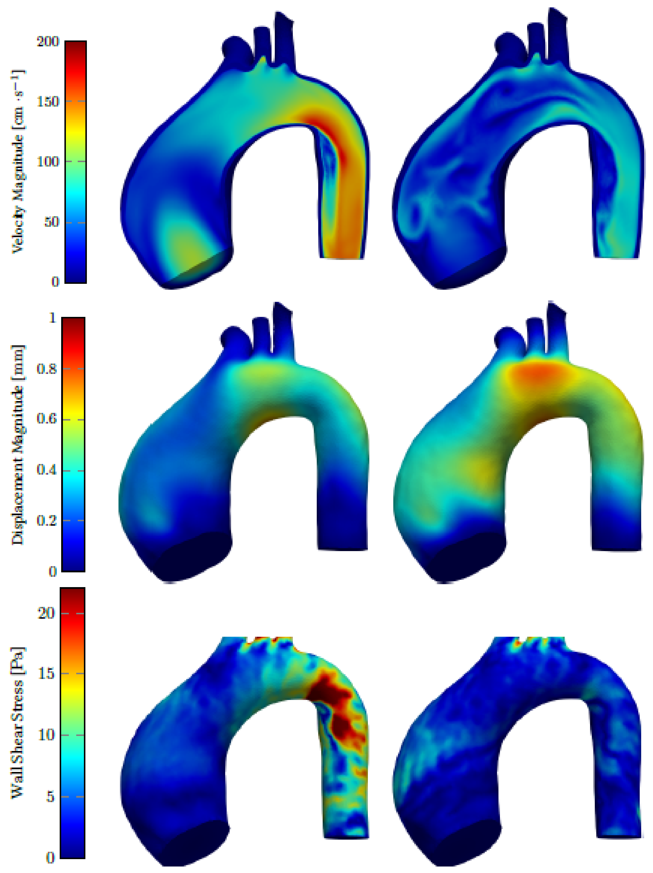
Fluid–structure interaction (FSI) modeling has become a cornerstone of cardiovascular biomechanics because blood flow and vessel deformation are fundamentally interdependent. While conventional CFD captures flow patterns without accounting for vessel compliance, and standalone structural models ignore fluid-driven forces, FSI frameworks couple both domains to resolve pressure, velocity, and wall deformation simultaneously. This coupled representation significantly improves the prediction of cardiovascular device performance by more accurately capturing real physiological loading conditions, including pulsatile pressure transmission, wall deformation-induced flow changes, and localized stress distributions. As a result, FSI models enable better assessment of device–tissue interactions, such as stent expansion behavior, graft compliance mismatch, and prosthetic valve leaflet dynamics, which are critical for predicting risks like restenosis, thrombosis, or device fatigue. This integrated approach has proven essential for patient-specific cardiovascular analysis (Figure 6), medical device evaluation, atherosclerotic disease modeling, biological transport, and microscale elastofluidic systems. In line with this perspective, Mukherjee et al. [186] emphasize that integrating computational modeling with emerging technologies such as 3D bioprinting enables the creation of more realistic, patient-specific cardiovascular platforms, where hemodynamics and structural behavior must be accurately captured to predict tissue performance and disease progression. Similarly, Neelakantan et al. [187] demonstrate the importance of subject-specific FSI modeling in pulmonary hypertension, showing that increased distal vascular resistance primarily drives the rise in pulmonary artery pressure, while decreased arterial compliance significantly increases characteristic impedance and alters pressure pulsatility. Their subject-specific one-dimensional FSI model, informed by mechanical testing, imaging, and hemodynamic data, highlights how different vascular remodeling mechanisms contribute differently to right ventricular afterload, providing a clearer biomechanical understanding of disease progression and RV failure. At the same time, it exposes methodological and computational challenges that continue to limit widespread clinical adoption.
Figure 6. Velocity magnitude, displacement magnitude and wall shear stress distribution analysis at 150 (left) and 300 ms (right). Reprinted from Ref. [188] with permission from MDPI, 2022.
Eng 07 00235 g006
Several studies highlight the strengths of classical FSI formulations in capturing arterial compliance, wall stress, and pressure-wave propagation. Syed et al. [189] demonstrate that FSI enables realistic assessment of aneurysm growth, stenosis progression, and vessel remodeling. Similarly, work by Dake et al. [190], Athani et al. [191], Kumar et al. [192], and Rostam et al. [176] shows that abnormal distributions of WSS, OSI, and von Mises stress often missed by rigid-wall models can serve as biomechanical indicators of aneurysm instability, plaque development, and vascular degeneration. These capabilities also support virtual intervention planning, stent deployment analysis, and evaluation of biomechanical loading under patient-specific conditions. This aligns with the broader vision presented by Niederer et al. [193], who highlight that physics-based computational models provide a unifying framework for integrating patient-specific imaging and diagnostic data, enabling prediction of treatment outcomes that cannot be inferred from isolated measurements alone. However, high computational cost, numerical stability issues, and the absence of standardized validation protocols continue to hinder routine clinical implementation.
Extending beyond large-vessel mechanics, Musharaf et al. [194] contrast traditional vascular FSI with microscale elastofluidic applications, reviewing numerical strategies ranging from finite and boundary element methods to lattice Boltzmann, immersed boundary, molecular dynamics, and hybrid solvers. While LBM is particularly effective for multiphase microscale flows and MD captures atomistic interactions beyond continuum assumptions, no single method is universally optimal. The authors advocate AI-assisted hybrid approaches and real-time simulation capabilities, aligning with emerging digital twin concepts discussed by Brown et al. [195], which aim to make FSI more efficient and clinically accessible. Such developments resonate with the outlook of Trayanova et al. [196], where the integration of mechanistic modeling with machine learning and patient-specific data is identified as a key pathway toward precision cardiology and improved prediction of complex cardiovascular phenomena. At the microscale, Ames et al. [197] further demonstrate the growing importance of high-performance computing in FSI by performing large-scale GPU-accelerated simulations resolving the deformation of millions of red blood cells in high-hematocrit blood flow. Their work shows that while immersed boundary FSI methods enable detailed cell-resolved hemodynamics, the main computational bottleneck in large multiscale simulations is data movement between CPU and GPU systems rather than the fluid or structural solvers themselves. This highlights that future progress in multiscale cardiovascular FSI will depend not only on improved physical models but also on advances in parallel computing and data communication strategies.
Accurate representation of blood rheology remains a persistent challenge. Garg et al. [198] emphasize that many cardiovascular FSI studies rely on Newtonian or overly simplified viscoelastic models, neglecting shear-thinning behavior, viscoplastic effects, wall slip, and particle-scale interactions. These simplifications can significantly distort predictions of WSS, pressure, and wall deformation. Kumar et al. [192] reinforce this point by demonstrating that Newtonian and Carreau–Yasuda formulations yield markedly different WSS and OSI distributions, underscoring the importance of rheological fidelity for reliable hemodynamic assessment. In particular, non-Newtonian rheological assumptions become critically important in physiological regimes characterized by low shear rates (e.g., venous circulation, recirculation zones, aneurysms), high hematocrit conditions, microcirculation, and regions with disturbed or oscillatory flow. In such cases, shear-thinning and viscoelastic effects significantly alter predicted velocity profiles, wall shear stress, and thrombus-related transport phenomena, whereas in large arteries under high shear rates, Newtonian approximations may still remain acceptable.
Across a wide range of studies including those by Dake et al. [190], Athani et al. [191], Kumar et al. [192], Rostam et al. [176], Oyejide et al. [173], Moradicheghamahi et al. [180], and Laha et al. [199], FSI consistently reveals biomechanical mechanisms that fluid-only or rigid-wall models cannot capture. Two-way coupling allows realistic simulation of aneurysmal wall motion, valve leaflet dynamics, and stent–artery interaction, leading to more accurate distributions of pressure, shear stress, and oscillatory flow. In contrast, rigid-wall CFD models neglect wall deformation and fluid–structure interaction, which often leads to underestimation or overestimation of wall shear stress, inaccurate oscillatory shear index predictions, and unrealistic flow patterns, particularly in compliant vessels or in the presence of plaques, thrombi, or medical devices. For example, Kumar et al. show that hypertension reduces WSS while increasing OSI, conditions associated with plaque growth, whereas Rostam et al. [176] link elevated wall stresses from atherosclerotic occlusions to intracranial aneurysm initiation. Oyejide et al. [173] further demonstrate that plaque rupture risk depends strongly on wall compliance, while Moradicheghamahi et al. [180] show that rigid-wall assumptions significantly distort WSS and OSI predictions in IVC filter simulations, particularly in the presence of large thrombi.
Mesh-free FSI approaches offer additional advantages when large deformations or complex moving boundaries are involved. Studies by Laha et al. [199], Mohammed et al. [200], Shi et al. [201], and Toma et al. [202] employ smoothed particle hydrodynamics (SPH) or SPH–FEM hybrids to avoid mesh entanglement and capture highly transient dynamics. Valente et al. [188] modeled ascending thoracic aortic aneurysms using SimVascular, while Laha et al. [199] demonstrated that SPH–FSI accurately reproduces the transient behavior of bileaflet mechanical heart valves, validated against 4D MRI with errors below 5%. Mohammed et al. [200] introduced a discrete multiphysics SPH framework capable of predicting stent deformation and von Mises stress directly from flow velocity and material properties (Figure 7). Continued improvements in SPH discretization by Shi et al. [201] and Toma et al. [202] have further enhanced accuracy, making mesh-free FSI a competitive alternative to traditional methods.
Figure 7. The dimensionless von Mises stress for two different stents for different stent property and fluid average velocity values. Reprinted from Ref. [200] with permission from MDPI, 2021.
Eng 07 00235 g007
The relevance of FSI extends beyond cardiovascular mechanics. Chen et al. [203] apply FSI to embolism transport, platelet aggregation, tubular flows, and kidney stone formation, illustrating the limitations of CFD-only approaches in biological systems. Salman et al. [178] highlight the critical role of FSI in embryonic heart development, where wall shear stress regulates gene expression and morphogenesis, reinforcing the importance of fluid–solid coupling in congenital heart disease.
Despite these advances, broader challenges remain. Brown et al. [195] emphasize the lack of standardized frameworks for data assimilation, uncertainty quantification, sensitivity analysis, and reproducibility. Even with GPU acceleration and machine-learning-based surrogates, computational demands remain a barrier to clinical translation. Moreover, fully coupled models that integrate FSI with electrophysiology, myocardial contraction, and whole-body circulation are still computationally intensive and difficult to validate -a challenge also emphasized by Trayanova et al. [196], who note the complexity of multiscale integration from cellular to organ-level phenomena. From a computational standpoint, classical CFD models are relatively less expensive and easier to implement but lack fidelity in deformable systems, whereas fully coupled FSI simulations introduce substantial computational burdens due to mesh deformation, coupling iterations, and time-step constraints. High-fidelity approaches such as SPH, immersed boundary methods, or cell-resolved simulations further increase cost, often requiring GPU acceleration and parallel computing. In terms of validation, CFD models are relatively mature with well-established benchmarks, while FSI and multiscale models still face challenges in experimental validation, patient-specific calibration, and reproducibility. Consequently, while CFD is widely used in clinical and industrial workflows, FSI and advanced multiscale frameworks remain in a transitional stage, with growing but still limited clinical readiness due to their complexity, computational expense, and lack of standardized validation protocols.
Taken together, these studies those by [173,176,180,189,190,191,199,200,202] portray FSI modeling as a rapidly evolving but still maturing field. By capturing interactions among arteries, valves, plaques, clots, stents, and microfluidic structures, FSI provides insights that uncoupled simulations cannot achieve. Persistent limitations including simplified material descriptions, rheological assumptions, high computational cost, and inconsistent validation must be addressed, as also emphasized by [195,196,198]. Emerging mesh-free methods, hybrid solvers, and digital twin frameworks point toward a transformative future, as highlighted by [194,195,197]. A structured comparison of available frameworks suggests that while CFD offers robustness, low cost, and high clinical maturity, FSI provides superior physical realism at increased computational expense, and multiscale or particle-based methods deliver the highest fidelity at the cost of significant complexity and limited validation maturity. Bridging these trade-offs through hybrid modeling, standardized validation pipelines, and reduced-order or AI-assisted approaches will be essential to advance clinical translation. In this context, the convergence of FSI, patient-specific modeling, and data-driven approaches as highlighted by Mukherjee et al. [186], Trayanova et al. [196], and Niederer et al. [193], positions computational biomechanics as a central pillar in advancing precision cardiovascular medicine.

4.4. Cardiovascular Device Engineering
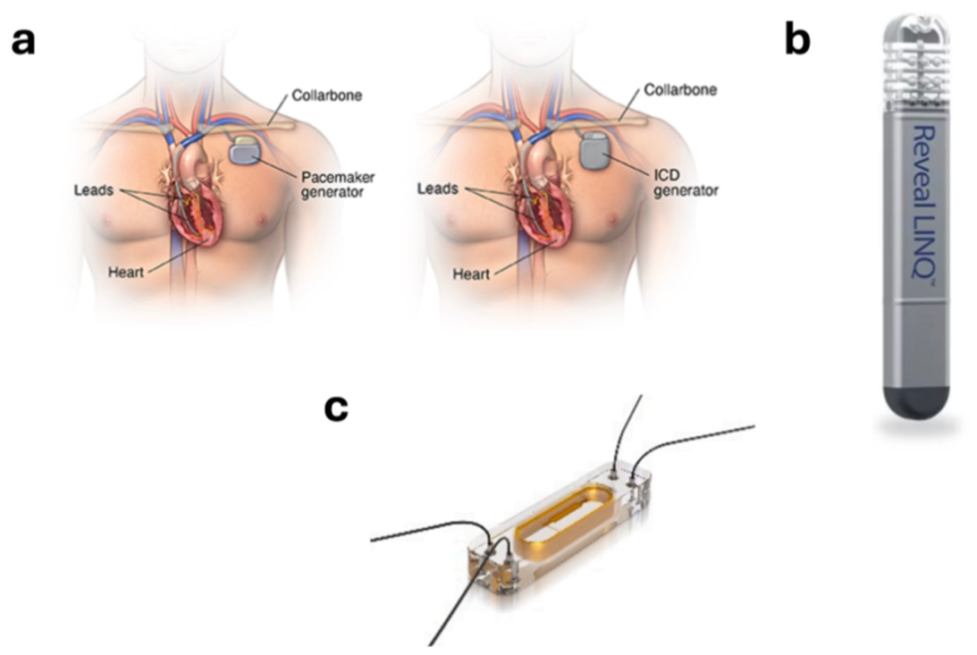
Cardiovascular biomechanics plays a pivotal role in designing, optimizing, and validating devices (see Figure 8) that interact with blood flow and vascular tissue. For example, coronary stents are biomechanically optimized to balance radial strength, flexibility, and fatigue resistance, with finite element simulations used to evaluate deployment behavior, vessel wall stress distribution, and long-term cyclic durability under pulsatile loading conditions. Similarly, transcatheter heart valves are designed using detailed biomechanical analyses of leaflet kinematics, contact stresses, and flow-induced shear forces to minimize regurgitation and improve durability. Vascular grafts are another key example, where compliance matching with native arteries is critical to reduce flow disturbances and intimal hyperplasia, while endovascular aneurysm repair (EVAR) devices are optimized to ensure secure fixation and minimize endoleak risk under physiological pressure conditions. Similarly, both native and prosthetic heart valves require detailed analysis of leaflet motion, flow-induced stresses, and long-term performance. Emerging materials and manufacturing strategies such as shape-memory alloys, biodegradable polymers, and 3D-printed scaffolds are enabling devices that adapt more naturally to physiological conditions. Biomechanical modeling helps quantify compliance, flow uniformity, and thrombogenic risk, while pulsatile flow loops and mock circulatory systems remain essential for validating predictions prior to clinical deployment.
With cardiovascular disease remaining the leading global cause of mortality, the demand for innovative devices that restore, monitor, or augment cardiac function is high. The field brings together biomaterials, biofabrication, regulatory science, and advanced sensing, with a focus on biocompatibility, mechanical robustness, physiological functionality, and long-term host integration. Recent research has explored surface engineering, regulatory pathways, implantable devices, biofabrication strategies, self-powered systems, and tissue-engineered constructs, revealing both impressive advances and ongoing challenges.
Figure 8. (a) Pacemaker and implantable cardioverter-defibrillator (ICD). (b) Medtronic Reveal LINQ insertable cardiac monitor. (c) CardioMEMS device for monitoring pulmonary artery pressure. Reprinted from Ref. [204] with permission from MDPI, 2024.
Eng 07 00235 g008
Surface engineering has emerged as a key strategy for improving device performance. Zhao et al. [205] describe approaches ranging from physical, chemical, and biological modifications to gene-based strategies. Micro- and nanoscale topographies can guide endothelial alignment and reduce thrombogenicity, while biochemical agents including peptides, growth factors, antibodies, and nitric oxide donors enhance endothelial adhesion. Material selection remains critical: metallic stents offer high strength but higher thrombosis risk, natural biomaterials are biocompatible but mechanically limited, and synthetic polymers can be tuned for specific properties but often require surface functionalization for hemocompatibility. Gene engineering adds another layer, enabling controlled expression of angiogenic or antiproliferative factors. However, few studies integrate these strategies systematically in clinically validated devices, underscoring the need for comprehensive design frameworks that address both short- and long-term performance.
Regulatory frameworks are essential for translating innovations safely into clinical practice. Aycock et al. [206] describe the FDA OSEL Cardiovascular Research Program, which standardizes preclinical testing across in vitro, animal, and computational platforms for valves, pacemakers, ventricular assist devices, ablation catheters, and mapping systems. While such cross-disciplinary approaches facilitate patient access and reduce uncertainty, gaps remain in predicting long-term device–host interactions and aligning regulatory protocols with rapidly evolving biofabrication methods.
Recent innovations also focus on minimally invasive implants and energy solutions. Wang et al. [207] discuss devices such as stents, transcatheter valves, occluders, synthetic grafts, and injectable hydrogels, highlighting challenges in biocompatibility, mechanical integrity, and integration. Zhang et al. [208] emphasize long-term energy solutions for implants, including biomechanical and biochemical harvesters and mid-field electromagnetic wireless power transfer (WPT), which allow continuous, noninvasive energy delivery to high-demand devices such as smart stents.
Self-powered and wearable technologies complement traditional implants. Wang et al. [209] report triboelectric nanogenerators (TENGs) from biocompatible PVA composites that harvest low-level biomechanical energy from human motion. Optimized designs allow TENGs to detect subtle skin deformations corresponding to pulse signals. Hughes et al. [210] review consumer wearables (smartwatches and activity trackers) for arrhythmia detection and remote monitoring, though challenges remain in standardization, clinical integration, and measurement accuracy. Together, these approaches highlight the promise of patient-centric, self-powered systems, albeit with ongoing validation needs.
Cardiac tissue engineering offers complementary solutions. Veldhuizen et al. [211] present a microfluidic platform for 3D cardiac tissue formation, co-culturing hPSC-derived cardiomyocytes with fibroblasts in collagen hydrogels and staggered micropost topographies to encourage anisotropic organization, functional maturation, and synchronous contractions. Wang et al. [212] and Poerio et al. [213] extend these approaches through 3D bioprinting, fabricating anisotropic cardiac patches with controlled cell organization and scaffold geometry. Yet limitations persist, including incomplete maturation, insufficient vascularization, and difficulties scaling for clinical applications.
Surface modifications continue to enhance vascular device performance. Yang et al. [214] report endothelium-mimicking coatings for metallic stents using metal–catechol–amine chemistry to sustain nitric oxide release and enable heparin conjugation. Han et al. [215] describe multilayer soft electronic arrays integrated into endocardial balloon catheters for high-resolution mapping, stimulation, ablation, and electroporation. Combining bioactive coatings with soft electronics improves both functionality and safety.
Regenerative strategies leverage biodegradable polymers and hydrogels. Nasr et al. [216] review natural, synthetic, and nanostructured polymers, highlighting trade-offs between mechanical properties, biocompatibility, and degradation profiles. Self-healing hydrogels (Maeso et al. [217]) restore structural integrity after myocardial injury, support cell proliferation, and modulate inflammation. Naumova et al. [218] explore biological pacemakers using cell- or gene-based approaches as alternatives to electronic devices, though challenges remain regarding safety, stability, and integration.
Despite these advances, critical hurdles persist. Long-term integration with host tissue while minimizing immune response, thrombosis, or biofilm formation remains unresolved; Mostafavi et al. [219] highlight implant-associated biofilm infections. Tissue-engineered constructs face challenges in scaling, maturation, and vascularization. Regulatory and standardization gaps are particularly pronounced for self-powered devices and biofabricated tissues. Addressing these challenges requires interdisciplinary collaboration, combining materials science, mechanics, biology, and clinical expertise to produce devices that are robust, biocompatible, and functionally sophisticated.
The field of cardiovascular device engineering is increasingly converging across surface and material engineering, self-powered sensing, tissue engineering, and biofabrication [205,214,215]. Surface coatings, gene-based strategies, and biomimetic hydrogels enhance biocompatibility [205,216,217], while wearable sensors and wireless energy systems reduce reliance on batteries [208,209,210]. Engineered cardiac tissues and 3D bioprinting platforms provide physiologically relevant in vitro models for drug testing and disease modeling [211,213]. Biodegradable polymers, self-healing hydrogels, and biological pacemakers represent next-generation alternatives to conventional implants [216,217,218]. Mechanically informed modeling enables analysis of plaque formation, aneurysm expansion, and arterial dissection [173,181,185], while computational hemodynamics supports virtual surgeries and predictive interventions [165,166,171]. Integration of imaging data with machine learning further enhances identification of mechanical biomarkers, paving the way toward truly personalized cardiovascular care [127,169,172].

4.5. Cross-Domain Biomechanical Synthesis

While musculoskeletal and cardiovascular biomechanics are often presented as separate fields, a closer examination reveals that they are governed by many of the same underlying mechanical principles. Making these connections explicit helps provide a more unified perspective and strengthens the broader engineering framework that this review aims to highlight.
One clear commonality is the role of cyclic loading and fatigue. In the musculoskeletal system, repeated loading over time contributes to microdamage, remodeling, and overuse injuries. Similarly, in the cardiovascular system, the continuous pulsatile nature of blood flow subjects vessel walls and implanted devices to cyclic stresses that influence fatigue, disease progression, and long-term durability. In both cases, tissue response depends on the balance between accumulated mechanical damage and biological repair processes.
A related parallel can be observed in load transfer and stiffness mismatch. In orthopedic applications, excessively stiff implants can lead to stress shielding, reducing the mechanical stimulus required for healthy bone maintenance. In vascular applications, a similar issue arises when there is a compliance mismatch between native vessels and implanted grafts or stents, which can disrupt local hemodynamics and contribute to complications such as intimal hyperplasia. Although the contexts differ, both scenarios emphasize the importance of mechanical compatibility between biological tissues and engineered systems.
Both systems also rely on hierarchical organization to manage load transfer across scales. In musculoskeletal tissues, structure ranges from collagen fibrils to whole joints, enabling efficient force transmission and adaptability. In vascular tissues, layered architectures and fiber-reinforced structures play a comparable role in controlling deformation and stress distribution. When this hierarchical organization is disrupted due to injury, degeneration, or disease, mechanical function is compromised in both systems.
Another shared concept is mechanotransduction, whereby cells respond to mechanical stimuli. In bone and muscle, mechanical loading influences growth, remodeling, and repair through cellular signaling pathways. In the vascular system, endothelial and smooth muscle cells respond to shear stress and cyclic strain, regulating processes such as vascular adaptation and disease development. Despite differences in biological function, the underlying principle of mechanically driven cellular response remains consistent.
Finally, both domains face similar challenges in modeling and validation. Patient-specific variability, uncertainties in material properties, and difficulties in replicating physiological conditions limit the predictive accuracy of both musculoskeletal and cardiovascular models. Simplifying assumptions, while often necessary, can reduce clinical reliability, highlighting the need for improved validation strategies and more robust, data-driven approaches across both fields.
Overall, these shared principles reinforce the idea that musculoskeletal and cardiovascular biomechanics are not isolated areas but rather interconnected applications of common mechanical concepts. Recognizing and leveraging these connections can support more integrated modeling approaches, improved device design, and ultimately more effective patient-specific solutions.

5. Conclusions, Open Challenges and Future Directions

5.1. Summary and Concluding Remarks

This review demonstrates how biomechanics provides a unifying engineering framework for understanding the mechanical behavior of biological tissues, organs, and integrated physiological systems. A central outcome of the reviewed literature is the consistent identification of biomechanics as a multiscale, strongly coupled system in which nanoscale structure, microscale organization, and macroscale loading collectively govern functional performance and failure. Across musculoskeletal, cardiovascular, and cellular scales, the combined use of experimental measurements and advanced computational modeling enables detailed assessment of load transmission, tissue deformation, fatigue accumulation, and injury mechanisms. Key quantitative findings across studies reveal that physiological loading is highly nonlinear and subject-specific—for example, joint forces reaching up to 5–10× body weight, vascular wall shear stresses on the order of 1–2 Pa in healthy conditions, and significant variability (~20–30%) in biomechanical responses due to individual geometry and material heterogeneity. In vascular systems, hemodynamic forces and fluid–structure interactions are shown to play a central role in governing normal function, disease initiation and progression, and the performance of therapeutic devices. At the tissue and cellular levels, force generation, adaptation, and repair emerge from tightly coupled molecular, structural, and neuromechanical processes acting across multiple scales.
A major synthesized result emerging from this review is that structural organization—rather than intrinsic material properties alone—dominates mechanical behavior in both musculoskeletal and cardiovascular systems. Evidence consistently shows that hierarchical architecture (e.g., collagen alignment, trabecular connectivity, vascular fiber orientation) governs stiffness, anisotropy, energy dissipation, and failure mechanisms. This leads to a key paradigm shift: accurate biomechanical prediction requires structure-informed and geometry-aware models rather than homogeneous material assumptions.
Despite major advances in predictive modeling, sensor-based monitoring, and numerical simulation, significant challenges remain. A critical limitation identified across domains is the gap between high-fidelity models and clinically applicable tools, primarily due to uncertainties in in vivo material properties, loading conditions, and long-term biological adaptation. Capturing patient-specific variability, translating multiscale biomechanical insights into routine clinical practice, and incorporating cognitive and neuromechanical influences continue to limit widespread adoption. Furthermore, most current validation studies remain short- to mid-term, limiting confidence in long-term predictions of implant performance, tissue remodeling, and disease progression. These observations highlight the need for interdisciplinary, data-driven approaches that connect fundamental biomechanics with applied goals in injury prevention, rehabilitation engineering, and medical device development, while also laying the groundwork for emerging paradigms such as AI-enabled modeling and digital twin systems.
Within the cardiovascular domain, patient-specific simulations have provided critical insight into how complex flow structures, vessel wall mechanics, and device–tissue interactions influence disease evolution, injury risk, and implant durability. A key result across hemodynamic studies is the strong correlation between disturbed flow metrics—such as low time-averaged wall shear stress (TAWSS), high oscillatory shear index (OSI), and increased relative residence time (RRT)—and the spatial localization of atherosclerosis, thrombosis, and aneurysm development. Accurate representation of material behavior, non-Newtonian blood rheology, wall compliance, and multiscale coupling is essential for capturing physiological and pathological responses. Notably, fluid–structure interaction (FSI) modeling consistently outperforms rigid-wall or fluid-only approaches by capturing wall deformation, stress redistribution, and coupled flow dynamics, although at a significantly higher computational cost. In parallel, progress in surface engineering, biodegradable and self-healing materials, tissue-engineered constructs, and self-powered sensing technologies demonstrates the growing impact of combining computational predictions with targeted experimental validation.
Several emerging trends can be clearly identified from the reviewed body of work. First, there is a transition from generic, population-based models toward fully patient-specific, geometry-aware, and data-driven frameworks. Second, hybrid modeling approaches combining physics-based simulations with machine learning are gaining momentum to overcome computational limitations and improve predictive capability. Third, there is a growing shift from static, laboratory-based assessment toward continuous, real-world monitoring using wearable sensors and IoT-enabled systems. Fourth, biofabrication and additive manufacturing [220] are enabling functionally graded, hierarchical, and compliance-matched designs that better replicate native tissue mechanics. Finally, integrated systems combining sensing, actuation, and energy harvesting are paving the way for adaptive, self-powered biomedical devices.
Taken together, cardiovascular biomechanics, hemodynamics, and device engineering form a comprehensive framework for understanding vascular function and guiding therapeutic strategies. Advanced imaging, fluid–structure interaction modeling, and simulation-based analyses reveal how localized flow patterns, wall shear stress, and compliance regulate endothelial behavior, plaque development, aneurysm risk, and post-intervention outcomes. At the same time, the design and optimization of cardiovascular devices—including stents, valves, grafts, tissue-engineered implants, and wearable monitoring systems—benefit from rigorous computational analysis integrated with material and surface engineering innovations. Emerging developments in biofabrication, soft electronics, energy-harvesting systems, and personalized implantable devices further expand opportunities for minimally invasive, patient-specific therapies. Overall, the convergence of multiscale biomechanics, advanced computation, and smart biomedical systems represents a clear new direction in the field, moving toward predictive, adaptive, and fully personalized healthcare solutions. Collectively, these advances underscore the importance of coupling mechanistic understanding with predictive modeling and device innovation to improve clinical outcomes and guide the next generation of biomechanically informed interventions.

5.2. Challenges and Limitations

Despite substantial progress, several enduring challenges continue to restrict the clinical translation of biomechanical research. A key overarching limitation identified across the reviewed literature is the mismatch between high-fidelity biomechanical models and their practical clinical applicability. One of the most persistent challenges is the difficulty of capturing patient-specific variability across biological scales, from molecular and cellular processes to tissue, organ, and system-level mechanics. Biological heterogeneity, complex anatomical geometries, and inter-individual differences in material properties and physiological loading conditions constrain the generalizability of both experimental observations and computational predictions. Importantly, studies consistently report variability on the order of ~20–30% in biomechanical responses due to subject-specific geometry and loading conditions, highlighting the limitations of population-averaged models.
In addition to these technical limitations, regulatory translation of cardiovascular biomechanical devices remains a major challenge. Regulatory approval processes require rigorous evidence of safety, effectiveness, and long-term reliability, typically through a combination of preclinical testing, standardized bench validation, animal studies, and controlled clinical trials. However, biomechanical models often lack standardized validation frameworks, and variability in simulation protocols, material assumptions, and patient-specific inputs makes it difficult to meet regulatory expectations consistently. A critical gap identified in the literature is the absence of universally accepted protocols for in silico validation, uncertainty quantification, and model credibility assessment, which limits the integration of computational biomechanics into regulatory pathways. Furthermore, the absence of universally accepted guidelines for in silico testing, uncertainty quantification, and model credibility assessment continues to slow the adoption of computational biomechanics in regulatory decision-making for device approval.
In musculoskeletal biomechanics, task-specific validation remains limited, particularly for dynamic daily activities and occupational settings. Although wearable sensors and real-time monitoring technologies generate large volumes of data, integrating these measurements into predictive frameworks remains challenging, especially when accounting for cognitive load, neuromechanical control, and fatigue-related effects on injury risk. A key limitation is that current models often decouple mechanical loading from neuromuscular control and fatigue evolution, reducing their ability to predict real-world injury mechanisms. Multiscale modeling approaches that attempt to bridge molecular, tissue, and organ-level phenomena often face high computational costs and a lack of standardized software pipelines, impeding routine clinical use. Additionally, most multiscale frameworks remain computationally intensive and are not yet compatible with real-time or near-real-time clinical decision-making.
Cardiovascular biomechanics presents additional complexities. Common modeling assumptions such as rigid vessel walls, Newtonian blood behavior, or simplified constitutive laws can lead to substantial deviations from physiological reality. Evidence across multiple studies shows that neglecting wall compliance or non-Newtonian rheology can significantly alter predicted wall shear stress (WSS), oscillatory shear index (OSI), and pressure distributions, particularly in low-shear or disturbed flow regions. Incorporating non-Newtonian flow effects, wall compliance, and multiscale interactions significantly increases computational demands, while experimental validation of patient-specific simulations remains limited. Fluid–structure interaction modeling, in particular, continues to face challenges related to numerical stability, convergence, and verification in complex, multiphase, or multiscale flow environments. Moreover, high-fidelity FSI and particle-based simulations often require GPU acceleration and advanced parallel computing, creating a barrier to widespread clinical implementation.
Device-related challenges further complicate translation. Long-term functional integration of cardiovascular implants with host tissue remains difficult, with unresolved issues including thrombosis, immune response, biofilm formation, incomplete vascularization, and insufficient maturation of engineered tissues. A consistent finding across studies is that mechanical mismatch—particularly compliance mismatch between devices and native tissue—remains a primary driver of adverse outcomes such as intimal hyperplasia and device failure.
To address thrombosis risk, current strategies include surface modification techniques such as heparin coating, nitric oxide-releasing surfaces, and endothelialization approaches that promote rapid formation of a non-thrombogenic endothelial layer over the implant. In addition, optimizing device geometry to minimize flow disturbances and regions of low wall shear stress can significantly reduce platelet activation and clot formation. To mitigate immune responses, approaches such as the use of biocompatible and immunomodulatory materials, controlled release of anti-inflammatory agents, and bioinspired surface engineering that mimics native extracellular matrix structures have shown promise in reducing foreign body reactions and improving long-term tissue acceptance. However, a key limitation is that most of these strategies are evaluated in short-term or simplified experimental conditions, with limited long-term in vivo validation under realistic physiological loading.
Regulatory and standardization barriers also slow clinical adoption, particularly for emerging technologies such as self-powered sensors, wearable systems, and biofabricated constructs. In particular, these technologies must navigate stringent regulatory approval pathways, such as the U.S. Food and Drug Administration (FDA) clearance process and the European CE marking framework, both of which require comprehensive evidence of safety, performance, biocompatibility, and long-term reliability. The complexity of these approval routes is further increased for computationally informed or personalized biomechanical devices, where demonstrating clinical equivalence and validation across diverse patient populations remains challenging. This highlights a broader trend: regulatory frameworks are currently not fully adapted to accommodate AI-driven, patient-specific, and continuously evolving biomechanical systems.
Looking forward, several emerging directions are required to overcome these challenges. These include the development of standardized validation protocols for computational models, integration of real-time wearable data into predictive frameworks, adoption of hybrid physics-based and AI-driven modeling approaches, and advancement of reduced-order models to enable clinically feasible simulations. In addition, improved long-term in vivo studies, uncertainty-aware modeling, and robust data assimilation techniques will be essential to bridge the gap between experimental research and clinical translation.
Addressing these limitations will require coordinated, interdisciplinary strategies that combine experimental validation, advanced simulations, multiscale modeling, and real-world clinical data. Ultimately, overcoming these barriers is essential not only for improving model accuracy, but also for enabling scalable, reliable, and patient-specific biomechanical solutions in clinical practice.

5.3. Artificial Intelligence and Data-Driven Biomechanics

Artificial intelligence and machine learning are increasingly reshaping biomechanics by enabling data-driven analysis of movement, injury risk, and rehabilitation outcomes. Importantly, these AI-based approaches do not replace physics-based biomechanical models but rather complement them. Physics-based models provide mechanistic understanding grounded in continuum mechanics, tissue constitutive laws, and fluid–structure interactions, while AI methods enhance these frameworks by improving parameter estimation, reducing computational cost, and identifying complex nonlinear patterns that are difficult to capture analytically. In hybrid modeling strategies, machine learning can also be used to calibrate or accelerate finite element and musculoskeletal simulations, or to infer boundary conditions and material properties from experimental or clinical data. In sports and human performance contexts, AI-based approaches have enabled accurate motion tracking, technique assessment, and injury prediction, supporting real-time feedback and personalized training strategies (Soufavi et al. [221]; Molavian et al. [222]). While these systems demonstrate strong agreement with expert evaluation and promising predictive capability, challenges related to data quality, interpretability, ethical use, and real-world deployment remain unresolved.
In cardiovascular biomechanics, machine learning has emerged as a complementary tool to physics-based modeling, offering accelerated computation, improved handling of high-dimensional data, and enhanced detection of complex spatiotemporal patterns (Arzani et al. [223]). For instance, Mehdi et al. [224] introduce a fully non-invasive framework that uses cardiac strain data and a multi-fidelity machine learning approach (combining simulation data with limited patient data) to accurately identify myocardial infarction regions without relying on contrast agents. This highlights how AI can bridge gaps where clinical data are scarce while reducing patient risk.
Similarly, Mukherjee et al. [225] address a critical limitation in cardiac biomechanics by developing an in silico heart phantom to benchmark strain quantification methods. Their work provides a much-needed validation framework for improving the reliability and clinical applicability of strain-based biomarkers, particularly in complex 4D cardiac motion environments.
Beyond cardiovascular applications, AI-driven models are also being applied across human and animal biomechanics to predict tissue responses, locomotor behavior, and musculoskeletal dynamics (Moulooadi et al. [226]; Smirnov et al. [227]). However, widespread adoption will depend on improved integration with established biomechanical frameworks, standardized datasets, and rigorous validation protocols.
Overall, AI-driven approaches offer substantial potential to enhance predictive accuracy, support individualized assessment, and enable real-time decision-making. Realizing this potential will require close collaboration between engineers, clinicians, and data scientists to ensure transparency, robustness, and clinical relevance.

5.4. Digital Twin Technology in Healthcare

Digital twin technology represents a natural progression of multiscale biomechanics, enabling the creation of patient-specific virtual models that evolve dynamically with incoming clinical data. By integrating imaging, sensor measurements, computational modeling, and AI, digital twins provide a pathway toward more personalized diagnosis, simulation, and intervention strategies.
At the joint and movement level, digital twins support individualized rehabilitation planning and neuromusculoskeletal assessment by combining detailed anatomical models with real-time functional data (Liu et al. [228]; Simonetti et al. [229]).
At a broader system level, they are increasingly viewed as platforms for predictive and personalized care, integrating biomechanics with imaging and data-driven analytics (Diniz et al. [230]; Sun et al. [231]).
Recent large-scale work by Qian et al. [232] further demonstrates the power of cardiac digital twins, where thousands of patient-specific models were constructed to uncover mechanistic insights into cardiac electrophysiology. Their findings show how variations in cardiac function such as conduction velocity and electrical activity can be directly linked to anatomy, lifestyle, and disease, highlighting the ability of digital twins to bridge population-level data with individualized physiological understanding.
From a broader clinical perspective, Coorey et al. [233] emphasize that digital twins are central to advancing precision medicine in cardiovascular disease. Their review highlights how real-time, data-integrated virtual patients could improve diagnosis and treatment selection, while also noting key barriers such as ethical considerations, data integration challenges, and regulatory limitations that currently restrict full clinical implementation.
Despite their promise, digital twin approaches still face several challenges, including multimodal data fusion, computational efficiency, modeling of aging and damage, privacy concerns, and the need for regulatory approval. Additionally, many current implementations remain closer to advanced simulation frameworks rather than fully realized real-time digital twins.
Beyond patient-specific care, digital twins are also being explored for medical device testing and regulatory evaluation through integrated robotic–biomechanical systems (Quinn et al. [234]).
Overall, digital twin technology holds transformative potential for biomechanics and healthcare. However, achieving widespread clinical adoption will require standardized architectures, rigorous validation strategies, and seamless integration with AI and clinical workflows to ensure safe, effective, and scalable deployment.

5.5. Future Perspectives and Outlook

The convergence of biomechanics, artificial intelligence, and digital twin technologies signals a shift toward predictive, personalized, and proactive healthcare. Future efforts are expected to focus on fully integrated, multiscale platforms that combine computational modeling, continuous sensor monitoring, real-time feedback, and adaptive predictive analytics. Such systems have the potential to dynamically guide rehabilitation, optimize device performance, and mitigate injury risk by responding to evolving physiological and environmental conditions.
Achieving this vision will require sustained interdisciplinary collaboration across biomechanics, materials science, robotics, bioengineering, and clinical medicine, alongside careful attention to validation, scalability, and regulatory requirements. By tightly coupling mechanistic understanding with technological innovation and patient-centered care, these advances promise to move healthcare beyond reactive treatment toward a more anticipatory and individualized paradigm.

Author Contributions

Conceptualization, M.D.; Methodology, M.D.; Formal analysis, M.D.; Investigation, M.D., A.M. and U.K.; Writing—original draft, M.D.; Writing—review and editing, M.D., A.M. and U.K. All authors have read and agreed to the published version of the manuscript.

Funding

This research received no external funding.

Institutional Review Board Statement

Not applicable.

Informed Consent Statement

Not applicable.

Data Availability Statement

No new data were created or analyzed in this study. Data sharing is not applicable to this article.

Conflicts of Interest

The authors declare no conflicts of interest.

References

  1. Knudson, D. Fundamentals of Biomechanics; Springer: Boston, MA, USA, 2007. [Google Scholar]
  2. Demiral, M.; Abbassi, F.; Mamedov, A. Deformation characteristics in micro-cutting of single crystal copper: Effect of cutting tool geometry and friction. J. Mech. Sci. Technol. 2022, 36, 1903–1912. [Google Scholar] [CrossRef]
  3. Fox, B.D.; Judge, L.W.; Dickin, D.C.; Wang, H. Biomechanics of Military Load Carriage and Resulting Musculoskeletal Injury: A Review. J. Orthop. Orthop. Surg. 2020, 1, 6–11. [Google Scholar] [CrossRef]
  4. Babangida, A.A.; Caraballo-Arias, Y.; Decataldo, F.; Violante, F.S. Advancing Occupational Medicine through Wearable Technology: A Review of Sensor Systems for Biomechanical Risk Assessment and Work-Related Musculoskeletal Disorder Prevention. ACS Sens. 2025, 10, 5410–5432. [Google Scholar] [CrossRef]
  5. Alaneme, K.K.; Kareem, S.A.; Olajide, J.L.; Sadiku, R.E.; Bodunrin, M.O. Computational biomechanical and biodegradation integrity assessment of Mg-based biomedical devices for cardiovascular and orthopedic applications: A review. Int. J. Lightweight Mater. Manuf. 2022, 5, 251–266. [Google Scholar] [CrossRef]
  6. Avedesian, J.M.; Forbes, W.; Covassin, T.; Dufek, J.S. Influence of Cognitive Performance on Musculoskeletal Injury Risk: A Systematic Review. Am. J. Sports Med. 2022, 50, 554–562. [Google Scholar] [CrossRef] [PubMed]
  7. Rodero, C.; Baptiste, T.M.; Barrows, R.K.; Lewalle, A.; Niederer, S.A.; Strocchi, M. Advancing clinical translation of cardiac biomechanics models: A comprehensive review, applications and future pathways. Front. Phys. 2023, 11, 1306210. [Google Scholar] [CrossRef]
  8. Bustos, A.O.; Belluscio, V.; Camomilla, V.; Lucangeli, L.; Rizzo, F.; Sciarra, T.; Martelli, F.; Giacomozzi, C. Overuse-related injuries of the musculoskeletal system: Systematic review and quantitative synthesis of injuries, locations, risk factors and assessment techniques. Sensors 2021, 21, 2438. [Google Scholar] [CrossRef] [PubMed]
  9. Mukund, K.; Subramaniam, S. Skeletal muscle: A review of molecular structure and function, in health and disease. Wiley Interdiscip. Rev. Syst. Biol. Med. 2020, 12, e1462. [Google Scholar] [CrossRef]
  10. Haleem, A.; Javaid, M.; Singh, R.P.; Rab, S.; Suman, R. Applications of nanotechnology in medical field: A brief review. Glob. Health J. 2023, 7, 70–77. [Google Scholar] [CrossRef]
  11. Alizadeh, M.; Knapik, G.G.; Mageswaran, P.; Mendel, E.; Bourekas, E.; Marras, W.S. Biomechanical musculoskeletal models of the cervical spine: A systematic literature review. Clin. Biomech. 2020, 71, 115–124. [Google Scholar] [CrossRef]
  12. Muhammad, R.; Demiral, M.; Roy, A.; Silberschmidt, V.V. Modelling the dynamic behaviour of hard-to-cut alloys under conditions of vibro-impact cutting. J. Phys. Conf. Ser. 2013, 451, 012030. [Google Scholar] [CrossRef]
  13. Demiral, M.; Duran, E.T. Failure analysis of resistance spot-welded structure using XFEM: Lifetime assessment. Appl. Sci. 2023, 13, 10923. [Google Scholar] [CrossRef]
  14. Sugerman, G.P.; Kakaletsis, S.; Thakkar, P.; Chokshi, A.; Parekh, S.H.; Rausch, M.K. A whole blood thrombus mimic: Constitutive behavior under simple shear. J. Mech. Behav. Biomed. Mater. 2021, 115, 104216. [Google Scholar] [CrossRef] [PubMed]
  15. Lin, J.; Zheng, S.Y.; Xiao, R.; Yin, J.; Wu, Z.L.; Zheng, Q.; Qian, J. Constitutive behaviors of tough physical hydrogels with dynamic metal-coordinated bonds. J. Mech. Phys. Solids 2020, 139, 103935. [Google Scholar] [CrossRef]
  16. Fielding, S.M.; Cochran, J.O.; Huang, J.; Bi, D.; Marchetti, M.C. Constitutive model for the rheology of biological tissue. Phys. Rev. E 2023, 108, L042602. [Google Scholar] [CrossRef]
  17. Motiwale, S.; Sacks, M.S.; Austin, A. Structural Constitutive Models for Soft Biological Tissues and Biomaterials: The Role of Mechanical Interactions. Mech. Soft Mater. 2025, 7, 1. [Google Scholar] [CrossRef]
  18. Greco, F.; Leonetti, L.; Pranno, A.; Rudykh, S. Mechanical behavior of bio-inspired nacre-like composites: A hybrid multiscale modeling approach. Compos. Struct. 2020, 233, 111625. [Google Scholar] [CrossRef]
  19. Kadioglu, F.; Demiral, M. Failure behaviour of the single lap joints of angle-plied composites under three point bending tests. J. Adhes. Sci. Technol. 2020, 34, 531–548. [Google Scholar] [CrossRef]
  20. Liu, M.; Liang, L.; Sun, W. A generic physics-informed neural network-based constitutive model for soft biological tissues. Comput. Methods Appl. Mech. Eng. 2020, 372, 113402. [Google Scholar] [CrossRef]
  21. Moreno-Mateos, M.A.; Gonzalez-Rico, J.; Nunez-Sardinha, E.; Gomez-Cruz, C.; Lopez-Donaire, M.L.; Lucarini, S.; Arias, A.; Muñoz-Barrutia, A.; Velasco, D.; Garcia-Gonzalez, D. Magneto-mechanical system to reproduce and quantify complex strain patterns in biological materials. Appl. Mater. Today 2022, 27, 101437. [Google Scholar] [CrossRef]
  22. Oliaei, S.N.B.; Demiral, M. Finite element modeling of mechanical micromachining. In Advanced Machining Processes: Innovative Modeling Techniques; CRC Press: Boca Raton, FL, USA, 2017; pp. 245–280. [Google Scholar]
  23. Demiral, M.; Abdel-Wahab, A.; Silberschmidt, V. A numerical study on indentation properties of cortical bone tissue: Influence of anisotropy. Acta Bioeng. Biomech. 2015, 17, 3–14. [Google Scholar]
  24. Demiral, M.; Abbassi, F.; Zahedi, A.; Akpinar, S. Critical assessment of the bonded single lap joint exposed to cyclic tensile loading. Aerospace 2023, 10, 454. [Google Scholar] [CrossRef]
  25. Demiral, M.; Saracyakupoglu, T.; Sahin, B.; Köklü, U. Minimizing delamination in CFRP laminates: Experimental and numerical insights into drilling and punching effects. Polymers 2025, 17, 3056. [Google Scholar] [CrossRef]
  26. Salvatore, L.; Gallo, N.; Natali, M.L.; Terzi, A.; Sannino, A.; Madaghiele, M. Mimicking the Hierarchical Organization of Natural Collagen: Toward the Development of Ideal Scaffolding Material for Tissue Regeneration. Front. Bioeng. Biotechnol. 2021, 9, 644595. [Google Scholar] [CrossRef]
  27. Yang, L.; Ge, L.; Zhou, Q.; Mokabber, T.; Pei, Y.; Bron, R.; van Rijn, P. Biomimetic Multiscale Hierarchical Topography Enhances Osteogenic Differentiation of Human Mesenchymal Stem Cells. Adv. Mater. Interfaces 2020, 7, 2000385. [Google Scholar] [CrossRef]
  28. Ma, H.; Yang, C.; Ma, Z.; Wei, X.; Younis, M.R.; Wang, H.; Li, W.; Wang, Z.; Wang, W.; Luo, Y.; et al. Multiscale Hierarchical Architecture-Based Bioactive Scaffolds for Versatile Tissue Engineering. Adv. Healthc. Mater. 2022, 11, e2102837. [Google Scholar] [CrossRef] [PubMed]
  29. Sprio, S.; Panseri, S.; Montesi, M.; Dapporto, M.; Ruffini, A.; Dozio, S.M.; Cavuoto, R.; Misseroni, D.; Paggi, M.; Bigoni, D.; et al. Hierarchical porosity inherited by natural sources affects the mechanical and biological behaviour of bone scaffolds. J. Eur. Ceram. Soc. 2020, 40, 1717–1727. [Google Scholar] [CrossRef]
  30. O’Connor, C.; Brady, E.; Zheng, Y.; Moore, E.; Stevens, K.R. Engineering the multiscale complexity of vascular networks. Nat. Rev. Mater. 2022, 7, 702–716. [Google Scholar] [CrossRef]
  31. Koushik, T.M.; Miller, C.M.; Antunes, E. Bone Tissue Engineering Scaffolds: Function of Multi-Material Hierarchically Structured Scaffolds. Adv. Healthc. Mater. 2023, 12, e2202766. [Google Scholar] [CrossRef]
  32. Wei, J.; Pan, F.; Ping, H.; Yang, K.; Wang, Y.; Wang, Q.; Fu, Z. Bioinspired Additive Manufacturing of Hierarchical Materials: From Biostructures to Functions. Research 2023, 6, 164. [Google Scholar] [CrossRef]
  33. Malik-Sheriff, R.S.; Glont, M.; Nguyen, T.V.; Tiwari, K.; Roberts, M.G.; Xavier, A.; Vu, M.T.; Men, J.; Maire, M.; Kananathan, S.; et al. BioModels-15 years of sharing computational models in life science. Nucleic Acids Res. 2020, 48, D407–D415. [Google Scholar] [CrossRef] [PubMed]
  34. Sivakumar, N.; Warner, H.V.; Peirce, S.M.; Lazzara, M.J. A computational modeling approach for predicting multicell spheroid patterns based on signaling-induced differential adhesion. PLoS Comput. Biol. 2022, 18, e1010701. [Google Scholar] [CrossRef] [PubMed]
  35. Hynes, W.F.; Pepona, M.; Robertson, C.; Alvarado, J.; Dubbin, K.; Triplett, M.; Adorno, J.J.; Randles, A.; Moya, M.L. Examining metastatic behavior within 3D bioprinted vasculature for the validation of a 3D computational flow model. Sci. Adv. 2020, 6, eabb3308. [Google Scholar] [CrossRef]
  36. Nguyen, P.H.; Zhang, W. Design and Computational Modeling of Fabric Soft Pneumatic Actuators for Wearable Assistive Devices. Sci. Rep. 2020, 10, 9638. [Google Scholar] [CrossRef]
  37. Viceconti, M.; Pappalardo, F.; Rodriguez, B.; Horner, M.; Bischoff, J.; Musuamba Tshinanu, F. In silico trials: Verification, validation and uncertainty quantification of predictive models used in the regulatory evaluation of biomedical products. Methods 2021, 185, 120–127. [Google Scholar] [CrossRef] [PubMed]
  38. Sevink, G.J.; Liwo, J.A.; Asinari, P.; MacKernan, D.; Milano, G.; Pagonabarraga, I. Unfolding the prospects of computational (bio)materials modeling. J. Chem. Phys. 2020, 153, 100901. [Google Scholar] [CrossRef]
  39. Demiral, M.; Roy, A.; El Sayed, T.; Silberschmidt, V.V. Influence of strain gradients on lattice rotation in nano-indentation experiments: A numerical study. Mater. Sci. Eng. A 2014, 608, 73–81. [Google Scholar] [CrossRef]
  40. Attias, N.; Danai, O.; Abitbol, T.; Tarazi, E.; Ezov, N.; Pereman, I.; Grobman, Y.J. Mycelium bio-composites in industrial design and architecture: Comparative review and experimental analysis. J. Clean. Prod. 2020, 246, 119037. [Google Scholar] [CrossRef]
  41. Lombardo, D.; Calandra, P.; Kiselev, M.A. Structural characterization of biomaterials by means of small angle x-rays and neutron scattering (Saxs and sans), and light scattering experiments. Molecules 2020, 25, 5624. [Google Scholar] [CrossRef]
  42. Hosseinpour, S.; Gaudin, A.; Peters, O.A. A critical analysis of research methods and experimental models to study biocompatibility of endodontic materials. Int. Endod. J. 2022, 55, 346–369. [Google Scholar] [CrossRef]
  43. Acin-Perez, R.; Benador, I.Y.; Petcherski, A.; Veliova, M.; Benavides, G.A.; Lagarrigue, S.; Caudal, A.; Vergnes, L.; Murphy, A.N.; Karamanlidis, G.; et al. A novel approach to measure mitochondrial respiration in frozen biological samples. EMBO J. 2020, 39, e104073. [Google Scholar] [CrossRef]
  44. Heinrich, M.; Appendino, G.; Efferth, T.; Fürst, R.; Izzo, A.; Kayser, O.; Pezzuto, J.; Viljoen, A. Best practice in research-overcoming common challenges in phytopharmacological research. J. Ethnopharmacol. 2020, 246, 112230. [Google Scholar] [CrossRef]
  45. Yu, Z.; Thomas, P.J. Dynamical consequences of sensory feedback in a half-center oscillator coupled to a simple motor system. Biol. Cybern. 2021, 115, 135–160. [Google Scholar] [CrossRef]
  46. Yan, Y.; Chen, Z.; Huang, C.; Guo, Q. Modelling and analysis of coupling dynamics of swinging a lower limb exoskeleton. Nonlinear Dyn. 2023, 111, 1213–1234. [Google Scholar] [CrossRef]
  47. Li, Q.; Haraguchi, N.; Yoshimura, B.; Wang, S.; Yoshida, M.; Hase, K. Biomechanical Effects of a Passive Lower-Limb Exoskeleton Designed for Half-Sitting Work Support on Walking. Sensors 2025, 25, 4999. [Google Scholar] [CrossRef] [PubMed]
  48. Guo, J.; Chen, J.; Wang, J.; Ren, G.; Tian, Q.; Guo, C. EMG-assisted forward dynamics simulation of subject-specific mandible musculoskeletal system. J. Biomech. 2022, 139, 111143. [Google Scholar] [CrossRef] [PubMed]
  49. Cop, C.P.; Cavallo, G.; Veld, R.C.V.; Koopman, B.F.; Lataire, J.; Schouten, A.C.; Sartori, M. Unifying system identification and biomechanical formulations for the estimation of muscle, tendon and joint stiffness during human movement. Prog. Biomed. Eng. 2021, 3, 033002. [Google Scholar] [CrossRef]
  50. Roupa, I.; da Silva, M.R.; Marques, F.; Gonçalves, S.B.; Flores, P.; da Silva, M.T. On the Modeling of Biomechanical Systems for Human Movement Analysis: A Narrative Review. Arch. Comput. Methods Eng. 2022, 29, 4915–4958. [Google Scholar] [CrossRef]
  51. Scott, S.; Weiss, M.; Selhuber-Unkel, C.; Barooji, Y.F.; Sabri, A.; Erler, J.T.; Metzler, R.; Oddershede, L.B. Extracting, quantifying, and comparing dynamical and biomechanical properties of living matter through single particle tracking. Phys. Chem. Chem. Phys. 2022, 25, 1513–1537. [Google Scholar] [CrossRef]
  52. Galbusera, F.; Cina, A.; Panico, M.; Albano, D.; Messina, C. Image-based biomechanical models of the musculoskeletal system. Eur. Radiol. Exp. 2020, 4, 49. [Google Scholar] [CrossRef]
  53. Al-Barghouthi, A.; Lee, S.; Solitro, G.F.; Latta, L.; Travascio, F. Relationships Among Bone Morphological Parameters and Mechanical Properties of Cadaveric Human Vertebral Cancellous Bone. JBMR Plus 2020, 4, 49. [Google Scholar] [CrossRef]
  54. Lin, C.Y.; Kang, J.H. Mechanical properties of compact bone defined by the stress-strain curve measured using uniaxial tensile test: A concise review and practical guide. Materials 2021, 14, 4224. [Google Scholar] [CrossRef]
  55. Li, Q.; Feng, C.; Cao, Q.; Wang, W.; Ma, Z.; Wu, Y.; He, T.; Jing, Y.; Tan, W.; Liao, T.; et al. Strategies of strengthening mechanical properties in the osteoinductive calcium phosphate bioceramics. Regen. Biomater. 2023, 10, rbad013. [Google Scholar] [CrossRef]
  56. Milazzo, M.; David, A.; Jung, G.S.; Danti, S.; Buehler, M.J. Molecular origin of viscoelasticity in mineralized collagen fibrils. Biomater. Sci. 2021, 9, 3390–3400. [Google Scholar] [CrossRef] [PubMed]
  57. Milazzo, M.; Jung, G.S.; Danti, S.; Buehler, M.J. Mechanics of mineralized collagen fibrils upon transient loads. ACS Nano 2020, 14, 8307–8316. [Google Scholar] [CrossRef] [PubMed]
  58. Gotti, C.; Sensini, A.; Fornaia, G.; Gualandi, C.; Zucchelli, A.; Focarete, M.L. Biomimetic Hierarchically Arranged Nanofibrous Structures Resembling the Architecture and the Passive Mechanical Properties of Skeletal Muscles: A Step Forward Toward Artificial Muscle. Front. Bioeng. Biotechnol. 2020, 8, 767. [Google Scholar] [CrossRef] [PubMed]
  59. Guo, J.B.; Liang, T.; Che, Y.J.; Yang, H.L.; Luo, Z.P. Structure and mechanical properties of high-weight-bearing and low-weight-bearing areas of hip cartilage at the micro- and nano-levels. BMC Musculoskelet. Disord. 2020, 21, 425. [Google Scholar] [CrossRef]
  60. Guan, Q.; Dai, B.; Cheng, H.H.; Hughes, J. Lattice structure musculoskeletal robots: Harnessing programmable geometric topology and anisotropy. Sci. Adv. 2025, 11, eadu9856. [Google Scholar] [CrossRef]
  61. Gregory, J.; Hazel, A.L.; Shearer, T. A microstructural model of tendon failure. J. Mech. Behav. Biomed. Mater. 2021, 122, 104665. [Google Scholar] [CrossRef]
  62. Guo, J.; Yin, Y.; Peng, G. Fractional-order viscoelastic model of musculoskeletal tissues: Correlation with fractals. Proc. R. Soc. A Math. Phys. Eng. Sci. 2021, 477, 20200990. [Google Scholar] [CrossRef]
  63. Svensson, R.B.; Eriksen, C.S.; Tran, P.H.; Kjaer, M.; Magnusson, S.P. Mechanical properties of human patellar tendon collagen fibrils. An exploratory study of aging and sex. J. Mech. Behav. Biomed. Mater. 2021, 124, 104864. [Google Scholar] [CrossRef]
  64. Belluzzi, E.; Todros, S.; Pozzuoli, A.; Ruggieri, P.; Carniel, E.L.; Berardo, A. Human Cartilage Biomechanics: Experimental and Theoretical Approaches towards the Identification of Mechanical Properties in Healthy and Osteoarthritic Conditions. Processes 2023, 11, 1014. [Google Scholar] [CrossRef]
  65. Jerban, S.; Chang, E.Y.; Du, J. Magnetic resonance imaging (MRI) studies of knee joint under mechanical loading: Review. Magn. Reson. Imaging 2020, 65, 27–36. [Google Scholar] [CrossRef] [PubMed]
  66. Zellers, J.A.; Eekhoff, J.D.; Walk, R.E.; Hastings, M.K.; Tang, S.Y.; Lake, S.P. Human Achilles tendon mechanical behavior is more strongly related to collagen disorganization than advanced glycation end-products content. Sci. Rep. 2021, 11, 24147. [Google Scholar] [CrossRef]
  67. Borjali, A.; Nourani, A.; Moeinnia, H.; Mohseni, M.; Korani, H.; Ghias, N.; Chizari, M. Comparison of mechanical properties in interference screw fixation technique and organic anterior cruciate ligament reconstruction method: A biomechanical study. BMC Musculoskelet. Disord. 2021, 22, 1047. [Google Scholar] [CrossRef]
  68. Kodama, Y.; Masuda, S.; Ohmori, T.; Kanamaru, A.; Tanaka, M.; Sakaguchi, T.; Nakagawa, M. Response to Mechanical Properties and Physiological Challenges of Fascia: Diagnosis and Rehabilitative Therapeutic Intervention for Myofascial System Disorders. Bioengineering 2023, 10, 474. [Google Scholar] [CrossRef] [PubMed]
  69. Efremov, Y.M.; Zurina, I.M.; Presniakova, V.S.; Kosheleva, N.V.; Butnaru, D.V.; Svistunov, A.A.; Rochev, Y.A.; Timashev, P.S. Mechanical properties of cell sheets and spheroids: The link between single cells and complex tissues. Biophys. Rev. 2021, 13, 541–561. [Google Scholar] [CrossRef]
  70. Dolega, M.; Zurlo, G.; Le Goff, M.; Greda, M.; Verdier, C.; Joanny, J.F.; Cappello, G.; Recho, P. Mechanical behavior of multi-cellular spheroids under osmotic compression. J. Mech. Phys. Solids 2021, 147, 104205. [Google Scholar] [CrossRef]
  71. Ayan, B.; Celik, N.; Zhang, Z.; Zhou, K.; Kim, M.H.; Banerjee, D.; Wu, Y.; Costanzo, F.; Ozbolat, I.T. Aspiration-assisted freeform bioprinting of pre-fabricated tissue spheroids in a yield-stress gel. Commun. Phys. 2020, 3, 183. [Google Scholar] [CrossRef] [PubMed]
  72. Boot, R.C.; Roscani, A.; van Buren, L.; Maity, S.; Koenderink, G.H.; Boukany, P.E. High-throughput mechanophenotyping of multicellular spheroids using a microfluidic micropipette aspiration chip. Lab Chip 2023, 23, 1768–1778. [Google Scholar] [CrossRef]
  73. Nakano, K.; Nanri, N.; Tsukamoto, Y.; Akashi, M. Mechanical activities of self-beating cardiomyocyte aggregates under mechanical compression. Sci. Rep. 2021, 11, 15159. [Google Scholar] [CrossRef]
  74. Dall’Ara, E.; Tozzi, G. Digital volume correlation for the characterization of musculoskeletal tissues: Current challenges and future developments. Front. Bioeng. Biotechnol. 2022, 10, 1010056. [Google Scholar] [CrossRef] [PubMed]
  75. Rasmussen, J. From Knowledge to Leverage: How to Use Musculoskeletal Simulation to Design Exoskeleton Concepts. Appl. Sci. 2025, 15, 5903. [Google Scholar] [CrossRef]
  76. Kumar, J.P.; Kumar, M.A.; Venkatesh, D. Healthy gait: Review of anatomy and physiology of knee joint. Int. J. Curr. Res. Rev. 2020, 12, 5903. [Google Scholar] [CrossRef]
  77. Layton, R.; Messenger, N.; Stewart, T. Characteristics of hip joint reaction forces during a range of activities. Med. Eng. Phys. 2022, 108, 103894. [Google Scholar] [CrossRef]
  78. Logerstedt, D.S.; Ebert, J.R.; MacLeod, T.D.; Heiderscheit, B.C.; Gabbett, T.J.; Eckenrode, B.J. Effects of and Response to Mechanical Loading on the Knee. Sports Med. 2022, 52, 201–235. [Google Scholar] [CrossRef]
  79. Zdero, R.; Brzozowski, P.; Schemitsch, E.H. Experimental Methods for Studying the Contact Mechanics of Joints. BioMed Res. Int. 2023, 2023, 4914082. [Google Scholar] [CrossRef]
  80. Deng, R.; Uzuner, S.; Li, L.P. Impact of knee geometry on joint contact mechanics after meniscectomy. Sci. Rep. 2024, 14, 28595. [Google Scholar] [CrossRef] [PubMed]
  81. Rosca, C.M.; Stancu, A. Integration of AI in self-powered IoT sensor systems. Appl. Sci. 2025, 15, 7008. [Google Scholar] [CrossRef]
  82. Chen, Y.; Yin, W.; Zheng, L.; Mehta, R.; Zhang, X. Biodynamic Modeling and Analysis of Human-Exoskeleton Interactions in Simulated Patient Handling Tasks. Hum. Factors 2025, 67, 641–655. [Google Scholar] [CrossRef]
  83. Christensen, S.; Rafique, S.; Bai, S. Design of a powered full-body exoskeleton for physical assistance of elderly people. Int. J. Adv. Robot. Syst. 2021, 18, 1–15. [Google Scholar] [CrossRef]
  84. Saeidi, M.; Kelly, P.A.; Netzel, C.; Scadeng, M.; Kumar, P.; Prendergast, D.; Neitzert, T.; Ramezani, M. Preliminary biomechanical cadaver study investigating a new load-sharing knee implant. J. Exp. Orthop. 2021, 8, 61. [Google Scholar] [CrossRef] [PubMed]
  85. Munford, M.J.; Stoddart, J.C.; Liddle, A.D.; Cobb, J.P.; Jeffers, J.R. Total and partial knee arthroplasty implants that maintain native load transfer in the tibia. Bone Jt. Res. 2022, 11, 91–101. [Google Scholar] [CrossRef]
  86. Stoddart, J.C.; Garner, A.; Tuncer, M.; Amis, A.A.; Cobb, J.; van Arkel, R.J. Load transfer in bone after partial, multi-compartmental, and total knee arthroplasty. Front. Bioeng. Biotechnol. 2024, 12, 1274496. [Google Scholar] [CrossRef]
  87. Hua, L.; Zhu, Y.; Bi, G.; Zhu, M.; Diao, Y. Effects of sliding techniques on lower limb biomechanics and muscle synergy during curling delivery: A focus on joint kinetics and muscle coordination. Front. Hum. Neurosci. 2025, 19, 1587118. [Google Scholar] [CrossRef]
  88. Huang, L.; Yang, Z.; Wang, R.; Xie, L. Physiological and biomechanical effects on the human musculoskeletal system while carrying a suspended-load backpack. J. Biomech. 2020, 108, 109894. [Google Scholar] [CrossRef] [PubMed]
  89. Zaman, R.; Xiang, Y.; Cruz, J.; Yang, J. Three-dimensional asymmetric maximum weight lifting prediction considering dynamic joint strength. Proc. Inst. Mech. Eng. Part H J. Eng. Med. 2021, 235, 437–446. [Google Scholar] [CrossRef]
  90. Huysamen, K.; Power, V.; O’Sullivan, L. Kinematic and kinetic functional requirements for industrial exoskeletons for lifting tasks and overhead lifting. Ergonomics 2020, 63, 818–830. [Google Scholar] [CrossRef] [PubMed]
  91. Jahn, J.; Ehlen, Q.T.; Huang, C.Y. Finding the Goldilocks zone of mechanical loading: A comprehensive review of mechanical loading in the prevention and treatment of knee osteoarthritis. Bioengineering 2024, 11, 110. [Google Scholar] [CrossRef]
  92. Barrutia, W.S.; Bratt, J.; Ferris, D.P. A Human Lower Limb Mechanical Phantom for the Testing of Knee Exoskeletons. IEEE Trans. Neural Syst. Rehabil. Eng. 2023, 31, 2497–2506. [Google Scholar] [CrossRef]
  93. Mišković, L.; Tricomi, E.; Zhang, X.; Missiroli, F.; Krstanović, K.; Petrič, T.; Masia, L. Hybrid Rigid-Soft and Pneumatic-Electromechanical Exoskeleton for Multi-Joint Lower Limb Assistance. IEEE Trans. Med. Robot. Bionics 2024, 6, 1180–1189. [Google Scholar] [CrossRef]
  94. Almansoof, H.S.; Nuhmani, S.; Muaidi, Q. Role of kinetic chain in sports performance and injury risk: A narrative review. J. Med. Life 2023, 16, 1591. [Google Scholar] [CrossRef]
  95. Lp, M.R.L.; Agrawal, D.K. Biomechanical Forces in the Tissue Engineering and Regeneration of Shoulder, Hip, Knee, and Ankle Joints. J. Biotechnol. Biomed. 2023, 6, 491–500. [Google Scholar] [CrossRef]
  96. Park, H.J.; Bae, T.S.; Kang, S.B.; Baek, H.H.; Chang, M.J.; Chang, C.B. A three-dimensional finite element analysis on the effects of implant materials and designs on periprosthetic tibial bone resorption. PLoS ONE 2021, 16, e0246866. [Google Scholar] [CrossRef]
  97. Hussein, M.A.; Azeem, M.A.; Kumar, A.M.; Saravanan, S.; Ankah, N.; Sorour, A.A. Design and processing of near-β Ti–Nb–Ag alloy with low elastic modulus and enhanced corrosion resistance for orthopedic implants. J. Mater. Res. Technol. 2023, 24, 259–273. [Google Scholar] [CrossRef]
  98. Baltatu, M.S.; Spataru, M.C.; Verestiuc, L.; Balan, V.; Solcan, C.; Sandu, A.V.; Geanta, V.; Voiculescu, I.; Vizureanu, P. Design, synthesis, and preliminary evaluation for ti-mo-zr-ta-si alloys for potential implant applications. Materials 2021, 14, 6806. [Google Scholar] [CrossRef]
  99. Kelly, C.N.; Wang, T.; Crowley, J.; Wills, D.; Pelletier, M.H.; Westrick, E.R.; Adams, S.B.; Gall, K.; Walsh, W.R. High-strength, porous additively manufactured implants with optimized mechanical osseointegration. Biomaterials 2021, 279, 121206. [Google Scholar] [CrossRef]
  100. Bagherifard, A.; Yekta, H.J.; Aghdam, H.A.; Motififard, M.; Sanatizadeh, E.; Nejad, M.G.; Esmaeili, S.; Saber-Samandari, S.; Sheikhbahaei, E.; Khandan, A.; et al. Improvement in osseointegration of tricalcium phosphate-zircon for orthopedic applications: An in vitro and in vivo evaluation. Med. Biol. Eng. Comput. 2020, 58, 1681–1693. [Google Scholar] [CrossRef]
  101. Chen, C.; Araby, S.; Demiral, M.; Cai, R.; Yang, X.; Wang, W.; Meng, Q. Fatigue behavior and tribological properties of laser additive manufactured aluminum alloy/boron nitride nanosheet nanocomposites. J. Mater. Res. Technol. 2022, 20, 3930–3948. [Google Scholar] [CrossRef]
  102. Safavi, S.; Yu, Y.; Robinson, D.L.; Gray, H.A.; Ackland, D.C.; Lee, P.V. Additively manufactured controlled porous orthopedic joint replacement designs to reduce bone stress shielding: A systematic review. J. Orthop. Surg. Res. 2023, 18, 42. [Google Scholar] [CrossRef] [PubMed]
  103. Boudjemaa, I.; Khatir, O.; Hamada, A.; Benkhettou, A.; Sahli, A.; Abdoune, Y.; Drici, G. Enhanced Orthopedic Implant Design for Transfemoral Amputation Incorporating a Honeycomb Structure Technology. Mech. Adv. Mater. Struct. 2024, 32, 3664–3670. [Google Scholar] [CrossRef]
  104. Demiral, M. Strength in adhesion: A multi-mechanics review covering tensile, shear, fracture, fatigue, creep, and impact behavior of polymer bonding in composites. Polymers 2025, 17, 2600. [Google Scholar] [CrossRef] [PubMed]
  105. McMullan, R.; Golbang, A.; Salma-Ancane, K.; Ward, J.; Rodzen, K.; Boyd, A.R. Review of 3D Printing of Polyaryletherketone/Apatite Composites for Lattice Structures for Orthopedic Implants. Appl. Sci. 2025, 15, 1804. [Google Scholar] [CrossRef]
  106. Ma, H.; Suonan, A.; Zhou, J.; Yuan, Q.; Liu, L.; Zhao, X.; Lou, X.; Yang, C.; Li, D.; Zhang, Y.G. PEEK (Polyether-ether-ketone) and its composite materials in orthopedic implantation. Arab. J. Chem. 2021, 14, 102977. [Google Scholar] [CrossRef]
  107. Dong, M.; Zhou, Y.; Li, J.; Rong, X.; Fan, W.; Zhou, X.; Kong, Y. State of the art in parallel ankle rehabilitation robot: A systematic review. J. Neuroeng. Rehabil. 2021, 18, 52. [Google Scholar] [CrossRef]
  108. Kladovasilakis, N.; Tsongas, K.; Tzetzis, D. Finite element analysis of orthopedic hip implant with functionally graded bioinspired lattice structures. Biomimetics 2020, 5, 44. [Google Scholar] [CrossRef]
  109. Wu, N.; Li, S.; Zhang, B.; Wang, C.; Chen, B.; Han, Q.; Wang, J. The advances of topology optimization techniques in orthopedic implants: A review. Med. Biol. Eng. Comput. 2021, 59, 1673–1689. [Google Scholar] [CrossRef] [PubMed]
  110. van Kootwijk, A.; Moosabeiki, V.; Saldivar, M.C.; Pahlavani, H.; Leeflang, M.A.; Niar, S.K.; Pellikaan, P.; Jonker, B.P.; Ahmadi, S.M.; Wolvius, E.B.; et al. Semi-automated digital workflow to design and evaluate patient-specific mandibular reconstruction implants. J. Mech. Behav. Biomed. Mater. 2022, 132, 105291. [Google Scholar] [CrossRef] [PubMed]
  111. Tang, Q.; Guo, S.; Deng, W.; Zhou, Y. Using novel porous metal pillars for tibial bone defects in primary total knee arthroplasty. BMC Musculoskelet. Disord. 2023, 24, 829. [Google Scholar] [CrossRef]
  112. Grzeskowiak, R.M.; Schumacher, J.; Dhar, M.S.; Harper, D.P.; Mulon, P.Y.; Anderson, D.E. Bone and Cartilage Interfaces With Orthopedic Implants: A Literature Review. Front. Surg. 2020, 7, 601244. [Google Scholar] [CrossRef]
  113. Cyphert, E.L.; Zhang, N.; Learn, G.D.; Hernandez, C.J.; Von Recum, H.A. Recent Advances in the Evaluation of Antimicrobial Materials for Resolution of Orthopedic Implant-Associated Infections in Vivo. ACS Infect. Dis. 2021, 7, 3125–3160. [Google Scholar] [CrossRef]
  114. Sun, Y.; Helmholz, H.; Willumeit-Römer, R. Preclinical in vivo research of magnesium-based implants for fracture treatment: A systematic review of animal model selection and study design. J. Magnes. Alloys 2021, 9, 351–361. [Google Scholar] [CrossRef]
  115. Zhao, F. The Application of Sports Biomechanics in Sports Injury Prevention and Rehabilitation. Front. Sport Res. 2024, 3, 060320. [Google Scholar] [CrossRef]
  116. Slade, P.; Kochenderfer, M.J.; Delp, S.L.; Collins, S.H. Sensing leg movement enhances wearable monitoring of energy expenditure. Nat. Commun. 2021, 12, 4312. [Google Scholar] [CrossRef]
  117. Lucarno, S.; Zago, M.; Buckthorpe, M.; Grassi, A.; Tosarelli, F.; Smith, R.; Della Villa, F. Systematic Video Analysis of Anterior Cruciate Ligament Injuries in Professional Female Soccer Players. Am. J. Sports Med. 2021, 49, 1794–1802. [Google Scholar] [CrossRef]
  118. Trasolini, N.A.; Nicholson, K.F.; Mylott, J.; Bullock, G.S.; Hulburt, T.C.; Waterman, B.R. Biomechanical Analysis of the Throwing Athlete and Its Impact on Return to Sport. Arthrosc. Sports Med. Rehabil. 2022, 4, e83–e91. [Google Scholar] [CrossRef]
  119. Gomah, M.; Demiral, M. An experimental and numerical investigation of an improved shearing process with different punch characteristics. Stroj. Vestn. J. Mech. Eng. 2020, 66, 375–384. [Google Scholar] [CrossRef]
  120. Peebles, A.T.; Miller, T.K.; Queen, R.M. Landing biomechanics deficits in anterior cruciate ligament reconstruction patients can be assessed in a non-laboratory setting. J. Orthop. Res. 2022, 40, 150–158. [Google Scholar] [CrossRef] [PubMed]
  121. Gull, M.A.; Bai, S.; Bak, T. A review on design of upper limb exoskeletons. Robotics 2020, 9, 16. [Google Scholar] [CrossRef]
  122. Gandolfi, M.; Valè, N.; Posteraro, F.; Morone, G.; Dell’orco, A.; Botticelli, A.; Dimitrova, E.; Gervasoni, E.; Goffredo, M.; Zenzeri, J.; et al. State of the art and challenges for the classification of studies on electromechanical and robotic devices in neurorehabilitation: A scoping review. Eur. J. Phys. Rehabil. Med. 2021, 57, 831–840. [Google Scholar] [CrossRef] [PubMed]
  123. Vaida, C.; Birlescu, I.; Pisla, A.; Ulinici, I.M.; Tarnita, D.; Carbone, G.; Pisla, D. Systematic Design of a Parallel Robotic System for Lower Limb Rehabilitation. IEEE Access 2020, 8, 34522–34537. [Google Scholar] [CrossRef]
  124. Rebelo, A.; Martinho, D.V.; Valente-dos-Santos, J.; Coelho-e-Silva, M.J.; Teixeira, D.S. From data to action: A scoping review of wearable technologies and biomechanical assessments informing injury prevention strategies in sport. BMC Sports Sci. Med. Rehabil. 2023, 15, 169. [Google Scholar] [CrossRef]
  125. Seçkin, A.Ç.; Ateş, B.; Seçkin, M. Review on Wearable Technology in Sports: Concepts, Challenges and Opportunities. Appl. Sci. 2023, 13, 10399. [Google Scholar] [CrossRef]
  126. Ortiz-Padilla, V.E.; Ramírez-Moreno, M.A.; Presbítero-Espinosa, G.; Ramírez-Mendoza, R.A.; Lozoya-Santos, J.d.J. Survey on Video-Based Biomechanics and Biometry Tools for Fracture and Injury Assessment in Sports. Appl. Sci. 2022, 12, 3981. [Google Scholar] [CrossRef]
  127. Khurshid, S.; Weng, L.C.; Nauffal, V.; Pirruccello, J.P.; Venn, R.A.; Al-Alusi, M.A.; Benjamin, E.J.; Ellinor, P.T.; Lubitz, S.A. Wearable accelerometer-derived physical activity and incident disease. npj Digit. Med. 2022, 5, 131. [Google Scholar] [CrossRef]
  128. Bai, Z.; Fong, K.N.; Zhang, J.J.; Chan, J.; Ting, K.H. Immediate and long-term effects of BCI-based rehabilitation of the upper extremity after stroke: A systematic review and meta-analysis. J. Neuroeng. Rehabil. 2020, 17, 57. [Google Scholar] [CrossRef]
  129. Akbari, A.; Haghverd, F.; Behbahani, S. Robotic Home-Based Rehabilitation Systems Design: From a Literature Review to a Conceptual Framework for Community-Based Remote Therapy During COVID-19 Pandemic. Front. Robot. AI 2021, 8, 612331. [Google Scholar] [CrossRef]
  130. Méndez-barbero, N.; Gutiérrez-muñoz, C.; Blanco-Colio, L.M. Cellular crosstalk between endothelial and smooth muscle cells in vascular wall remodeling. Int. J. Mol. Sci. 2021, 22, 7284. [Google Scholar] [CrossRef] [PubMed]
  131. Liu, S.; Lin, Z. Vascular Smooth Muscle Cells Mechanosensitive Regulators and Vascular Remodeling. J. Vasc. Res. 2022, 59, 90–113. [Google Scholar] [CrossRef] [PubMed]
  132. Yanagisawa, H.; Wagenseil, J. Elastic fibers and biomechanics of the aorta: Insights from mouse studies. Matrix Biol. 2020, 85–86, 160–172. [Google Scholar] [CrossRef]
  133. Cai, Z.; Gong, Z.; Li, Z.; Li, L.; Kong, W. Vascular Extracellular Matrix Remodeling and Hypertension. Antioxid. Redox Signal. 2021, 34, 765–783. [Google Scholar] [CrossRef]
  134. Yamashiro, Y.; Thang, B.Q.; Ramirez, K.; Shin, J.; Kohata, T.; Ohata, S.; Nguyen, A.V.; Ohtsuki, S.; Nagayama, K.; Yanagisawa, H. Matrix mechanotransduction mediated by thrombospondin-1/integrin/YAP in the vascular remodeling. Proc. Natl. Acad. Sci. USA 2020, 117, 9896–9905. [Google Scholar] [CrossRef] [PubMed]
  135. Blinkouskaya, Y.; Caçoilo, A.; Gollamudi, T.; Jalalian, S.; Weickenmeier, J. Brain aging mechanisms with mechanical manifestations. Mech. Ageing Dev. 2021, 200, 111575. [Google Scholar] [CrossRef]
  136. Sánchez-Molina, D.; García-Vilana, S.; Llumà, J.; Galtés, I.; Velázquez-Ameijide, J.; Rebollo-Soria, M.C.; Arregui-Dalmases, C. Mechanical behavior of blood vessels: Elastic and viscoelastic contributions. Biology 2021, 10, 831. [Google Scholar] [CrossRef]
  137. Tutwiler, V.; Singh, J.; Litvinov, R.I.; Bassani, J.L.; Purohit, P.K.; Weisel, J.W. Rupture of blood clots: Mechanics and pathophysiology. Sci. Adv. 2020, 6, eabc0496. [Google Scholar] [CrossRef]
  138. Zheng, X.; Yazdani, A.; Li, H.; Humphrey, J.D.; Karniadakis, G.E. A three-dimensional phase-field model for multiscale modeling of thrombus biomechanics in blood vessels. PLoS Comput. Biol. 2020, 16, e1007709. [Google Scholar] [CrossRef]
  139. Brusokas, M.; Borodinas, S.; Jasevičius, R. Three-Dimensional Modeling of the Behavior of a Blood Clot Using Different Mechanical Properties of a Blood Vessel. Mathematics 2025, 13, 285. [Google Scholar] [CrossRef]
  140. Jeong, Y.; Yao, Y.; Yim, E.K. Current understanding of intimal hyperplasia and effect of compliance in synthetic small diameter vascular grafts. Biomater. Sci. 2020, 8, 4383–4395. [Google Scholar] [CrossRef] [PubMed]
  141. Moore, M.J.; Tan, R.P.; Yang, N.; Rnjak-Kovacina, J.; Wise, S.G. Bioengineering artificial blood vessels from natural materials. Trends Biotechnol. 2022, 40, 693–707. [Google Scholar] [CrossRef] [PubMed]
  142. Wang, D.; Xu, Y.; Li, Q.; Turng, L.S. Artificial small-diameter blood vessels: Materials, fabrication, surface modification, mechanical properties, and bioactive functionalities. J. Mater. Chem. B 2020, 8, 1801–1822. [Google Scholar] [CrossRef]
  143. Kuang, H.; Wang, Y.; Shi, Y.; Yao, W.; He, X.; Liu, X.; Mo, X.; Lu, S.; Zhang, P. Construction and performance evaluation of Hep/silk-PLCL composite nanofiber small-caliber artificial blood vessel graft. Biomaterials 2020, 259, 120288. [Google Scholar] [CrossRef]
  144. Jia, W.; Li, M.; Weng, H.; Gu, G.; Chen, Z. Design and comprehensive assessment of a biomimetic tri-layer tubular scaffold via biodegradable polymers for vascular tissue engineering applications. Mater. Sci. Eng. C 2020, 110, 110717. [Google Scholar] [CrossRef]
  145. Zhou, X.; Nowicki, M.; Sun, H.; Hann, S.Y.; Cui, H.; Esworthy, T.; Lee, J.D.; Plesniak, M.; Zhang, L.G. 3D Bioprinting-Tunable Small-Diameter Blood Vessels with Biomimetic Biphasic Cell Layers. ACS Appl. Mater. Interfaces 2020, 12, 45904–45915. [Google Scholar] [CrossRef]
  146. Wang, D.; Maharjan, S.; Kuang, X.; Wang, Z.; Mille, L.S.; Tao, M.; Yu, P.; Cao, X.; Lian, L.; Lv, L.; et al. Microfluidic bioprinting of tough hydrogel-based vascular conduits for functional blood vessels. Sci. Adv. 2022, 8, eabq6900. [Google Scholar] [CrossRef]
  147. Daniel, J.; Abe, K.; McFetridge, P.S. Development of the human umbilical vein scaffold for cardiovascular tissue engineering applications. ASAIO J. 2005, 51, 252–261. [Google Scholar] [CrossRef] [PubMed]
  148. Größbacher, G.; Bartolf-Kopp, M.; Gergely, C.; Bernal, P.N.; Florczak, S.; de Ruijter, M.; Rodriguez, N.G.; Groll, J.; Malda, J.; Jungst, T.; et al. Volumetric Printing Across Melt Electrowritten Scaffolds Fabricates Multi-Material Living Constructs with Tunable Architecture and Mechanics. Adv. Mater. 2023, 35, e2300756. [Google Scholar] [CrossRef] [PubMed]
  149. Distler, T.; Schaller, E.; Steinmann, P.; Boccaccini, A.R.; Budday, S. Alginate-based hydrogels show the same complex mechanical behavior as brain tissue. J. Mech. Behav. Biomed. Mater. 2020, 111, 103979. [Google Scholar] [CrossRef] [PubMed]
  150. Choi, J.J.E.; Zwirner, J.; Ramani, R.S.; Ma, S.; Hussaini, H.M.; Waddell, J.N.; Hammer, N. Mechanical properties of human oral mucosa tissues are site dependent: A combined biomechanical, histological and ultrastructural approach. Clin. Exp. Dent. Res. 2020, 6, 602–611. [Google Scholar] [CrossRef]
  151. Song, K.; Wang, Z.; Lan, J.; Ma, S. Porous structure design and mechanical behavior analysis based on TPMS for customized root analogue implant. J. Mech. Behav. Biomed. Mater. 2021, 115, 104222. [Google Scholar] [CrossRef]
  152. Collins, M.N.; Ren, G.; Young, K.; Pina, S.; Reis, R.L.; Oliveira, J.M. Scaffold Fabrication Technologies and Structure/Function Properties in Bone Tissue Engineering. Adv. Funct. Mater. 2021, 31, 2010609. [Google Scholar] [CrossRef]
  153. Hu, J.; Altun, I.; Zhang, Z.; Albadawi, H.; Salomao, M.A.; Mayer, J.L.; Hemachandra, L.P.P.; Rehman, S.; Oklu, R. Bioactive-Tissue-Derived Nanocomposite Hydrogel for Permanent Arterial Embolization and Enhanced Vascular Healing. Adv. Mater. 2020, 32, e2002611. [Google Scholar] [CrossRef]
  154. Xin, X.; Liu, L.; Liu, Y.; Leng, J. 4D Printing Auxetic Metamaterials with Tunable, Programmable, and Reconfigurable Mechanical Properties. Adv. Funct. Mater. 2020, 30, 2004226. [Google Scholar] [CrossRef]
  155. Lin, C.; Zhang, L.J.; Liu, Y.J.; Liu, L.W.; Leng, J.S. 4D printing of personalized shape memory polymer vascular stents with negative Poisson’s ratio structure: A preliminary study. Sci. China Technol. Sci. 2020, 63, 578–588. [Google Scholar] [CrossRef]
  156. Salama, M.; Vaz, M.F.; Colaço, R.; Santos, C.; Carmezim, M. Biodegradable Iron and Porous Iron: Mechanical Properties, Degradation Behaviour, Manufacturing Routes and Biomedical Applications. J. Funct. Biomater. 2022, 13, 72. [Google Scholar] [CrossRef]
  157. Dastagir, K.; Dastagir, N.; Limbourg, A.; Reimers, K.; Strauß, S.; Vogt, P.M. In vitro construction of artificial blood vessels using spider silk as a supporting matrix. J. Mech. Behav. Biomed. Mater. 2020, 101, 103436. [Google Scholar] [CrossRef] [PubMed]
  158. Devillard, C.D.; Marquette, C.A. Vascular Tissue Engineering: Challenges and Requirements for an Ideal Large Scale Blood Vessel. Front. Bioeng. Biotechnol. 2021, 9, 721843. [Google Scholar] [CrossRef]
  159. Campinho, P.; Vilfan, A.; Vermot, J. Blood Flow Forces in Shaping the Vascular System: A Focus on Endothelial Cell Behavior. Front. Physiol. 2020, 11, 552. [Google Scholar] [CrossRef] [PubMed]
  160. Kadioglu, F.; Demiral, M. Design of adhesively-bonded single lap joints for automotive applications under compressive loading. Results Eng. 2025, 25, 104176. [Google Scholar] [CrossRef]
  161. Neelakantan, S.; Vang, A.; Mehdi, R.R.; Phelan, H.; Nicely, P.; Imran, T.; Zhang, P.; Choudhary, G.; Avazmohammadi, R. Right ventricular stiffening and anisotropy alterations in pulmonary hypertension: Mechanisms and relations to right heart failure. J. Am. Heart Assoc. 2025, 14, e037126. [Google Scholar] [CrossRef] [PubMed]
  162. Neelakantan, S.; Kumar, M.; Mendiola, E.A.; Phelan, H.; Serpooshan, V.; Sadayappan, S.; Avazmohammadi, R. Multiscale characterization of left ventricle active behavior in the mouse. Acta Biomater. 2023, 162, 240–253. [Google Scholar] [CrossRef]
  163. Ayyoub, S.; Orriols, R.; Oliver, E.; Ceide, O.T. Thrombosis models: An overview of common in vivo and in vitro models of thrombosis. Int. J. Mol. Sci. 2023, 24, 2569. [Google Scholar] [CrossRef]
  164. Mukherjee, T.; Elliott, S.; Manikandan, N.; Higgins, T.-J.; Zhong, Y.; Montalvo, S.K.; Saha, D.; Wansapura, J.; Avazmohammadi, R.; Alluri, P.G. Principal strain analysis for early detection of radiation-induced cardiotoxicity in a mouse model. Int. J. Radiat. Oncol. 2025, 123, 306–315. [Google Scholar] [CrossRef]
  165. Ferdows, M.; Hoque, K.E.; Bangalee, M.Z.; Xenos, M.A. Wall shear stress indicators influence the regular hemodynamic conditions in coronary main arterial diseases: Cardiovascular abnormalities. Comput. Methods Biomech. Biomed. Eng. 2023, 26, 235–248. [Google Scholar] [CrossRef]
  166. Kumar, N.; Pai, R.; Khader, S.M.A.; Khan, S.H.; Kyriacou, P.A. Influence of blood pressure and rheology on oscillatory shear index and wall shear stress in the carotid artery. J. Braz. Soc. Mech. Sci. Eng. 2022, 44, 510. [Google Scholar] [CrossRef]
  167. Buradi, A.; Mahalingam, A. Numerical Analysis of Wall Shear Stress Parameters of Newtonian Pulsatile Blood Flow Through Coronary Artery and Correlation to Atherosclerosis. In Advances in Mechanical Engineering; Lecture Notes in Mechanical Engineering; Springer: Berlin/Heidelberg, Germany, 2020; pp. 107–118. [Google Scholar]
  168. Soares, A.A.; Carvalho, F.A.; Leite, A. Wall Shear Stress-Based Hemodynamic Descriptors in the Abdominal Aorta Bifurcation: Analysis of a Case Study. J. Appl. Fluid Mech. 2021, 14, 1657–1668. [Google Scholar] [CrossRef]
  169. Bai, X.; Fu, M.; Li, Z.; Gao, P.; Zhao, H.; Li, R.; Sui, B. Distribution and regional variation of wall shear stress in the curved middle cerebral artery using four-dimensional flow magnetic resonance imaging. Quant. Imaging Med. Surg. 2022, 12, 5462–5473. [Google Scholar] [CrossRef] [PubMed]
  170. Yang, J.; Zhang, Y.; Xue, J.; Guo, Y.; Liu, S.; Yao, Y.; Zhong, H.; Quan, A.; Yang, J. Hemodynamic effects of stenosis with varying severity in different segments of the carotid artery using computational fluid dynamics. Sci. Rep. 2025, 15, 4896. [Google Scholar] [CrossRef] [PubMed]
  171. Rahmati, N.; Pouraliakbar, H.; Eskandari, A.; Javari, K.; Jabbarinick, A.; Sadeghipour, P.; Soltani, M.; Alimohammadi, M. The Impact of Stenosis Severity on Hemodynamic Parameters in the Iliac Artery: A Fluid–Structure Interaction Study. Bioengineering 2025, 12, 1042. [Google Scholar] [CrossRef]
  172. Ziegler, M.; Alfraeus, J.; Good, E.; Engvall, J.; De Muinck, E.; Dyverfeldt, P. Exploring the relationships between hemodynamic stresses in the carotid arteries. Front. Cardiovasc. Med. 2021, 7, 617755. [Google Scholar] [CrossRef]
  173. Oyejide, A.J.; Awonusi, A.A.; Ige, O.E. Fluid-structure interaction study of hemodynamics and its biomechanical influence on carotid artery atherosclerotic plaque deposits. Med. Eng. Phys. 2023, 117, 103998. [Google Scholar] [CrossRef]
  174. Khan, M.O.; Tran, J.S.; Zhu, H.; Boyd, J.; Packard, R.R.; Karlsberg, R.P.; Kahn, A.M.; Marsden, A.L. Low Wall Shear Stress Is Associated with Saphenous Vein Graft Stenosis in Patients with Coronary Artery Bypass Grafting. J. Cardiovasc. Transl. Res. 2021, 14, 770–781. [Google Scholar] [CrossRef]
  175. Ahmadpour-B, M.; Nooraeen, A.; Tafazzoli-Shadpour, M.; Taghizadeh, H. Contribution of atherosclerotic plaque location and severity to the near-wall hemodynamics of the carotid bifurcation: An experimental study and FSI modeling. Biomech. Model. Mechanobiol. 2021, 20, 1069–1085. [Google Scholar] [CrossRef]
  176. Rostam-Alilou, A.A.; Jarrah, H.R.; Zolfagharian, A.; Bodaghi, M. Fluid–structure interaction (FSI) simulation for studying the impact of atherosclerosis on hemodynamics, arterial tissue remodeling, and initiation risk of intracranial aneurysms. Biomech. Model. Mechanobiol. 2022, 21, 1393–1406. [Google Scholar] [CrossRef]
  177. Jouneghani, F.Z.; Ghomashchi, R.; Amabili, M.; Ghayesh, M.H. Biomechanical stress profiling in coronary arteries via two-phase blood FSI. Biomech. Model. Mechanobiol. 2025, 24, 2171–2193. [Google Scholar] [CrossRef]
  178. Salman, H.E.; Yalcin, H.C. Computational modeling of blood flow hemodynamics for biomechanical investigation of cardiac development and disease. J. Cardiovasc. Dev. Dis. 2021, 8, 14. [Google Scholar] [CrossRef]
  179. Rafieianzab, D.; Abazari, M.A.; Soltani, M.; Alimohammadi, M. The effect of coarctation degrees on wall shear stress indices. Sci. Rep. 2021, 11, 12757. [Google Scholar] [CrossRef]
  180. Moradicheghamahi, J.; Goswami, D. Impact of Vein Wall Hyperelasticity and Blood Flow Turbulence on Hemodynamic Parameters in the Inferior Vena Cava with a Filter. Micromachines 2025, 16, 51. [Google Scholar] [CrossRef]
  181. Sheikh, M.A.A.; Shuib, A.S.; Mohyi, M.H.H. A review of hemodynamic parameters in cerebral aneurysm. Interdiscip. Neurosurg. Adv. Tech. Case Manag. 2020, 22, 100716. [Google Scholar] [CrossRef]
  182. Takehara, Y.; Isoda, H.; Takahashi, M.; Unno, N.; Shiiya, N.; Ushio, T.; Goshima, S.; Naganawa, S.; Alley, M.; Wakayama, T.; et al. Abnormal flow dynamics result in low wall shear stress and high oscillatory shear index in abdominal aortic dilatation: Initial in vivo assessment with 4D-flow MRI. Magn. Reson. Med. Sci. 2020, 19, 235–246. [Google Scholar] [CrossRef] [PubMed]
  183. Shi, J.; Liang, J.; Wang, J.; Wang, H.; Wang, Z.; Zhang, X.; Wu, G.; Tian, S.; Wei, W. Assessment of biomechanical properties in pulmonary arterial hypertension: A computational fluid dynamics study of the extensive pulmonary arterial tree. BMC Pulm. Med. 2025, 25, 175. [Google Scholar] [CrossRef] [PubMed]
  184. Urschel, K.; Tauchi, M.; Achenbach, S.; Dietel, B. Investigation of wall shear stress in cardiovascular research and in clinical practice—From bench to bedside. Int. J. Mol. Sci. 2021, 22, 5635. [Google Scholar] [CrossRef] [PubMed]
  185. Ramses, R.; Agu, O. From Heart to Abdominal Aorta: Integrating Multi-Modal Cardiac Imaging Derived Haemodynamic Biomarkers for Abdominal Aortic Aneurysm Risk Stratification, Surveillance, Pre-Operative Assessment and Therapeutic Decision-Making. Diagnostics 2025, 15, 2497. [Google Scholar] [CrossRef]
  186. Mukherjee, T.; Amoli, M.S.; Rezapourdamanab, S.; El Shammas, L.R.; Tomov, M.L.; Mendiola, E.A.; Serpooshan, V.; Avazmohammadi, R. Computational Modeling Meets 3D Bioprinting: Emerging Synergies in Cardiovascular Disease Modeling. Adv. Healthc. Mater. 2026, e03034. [Google Scholar] [CrossRef] [PubMed]
  187. Neelakantan, S.; Mendiola, E.A.; Zambrano, B.; Vang, A.; Myers, K.J.; Zhang, P.; Choudhary, G.; Avazmohammadi, R. Dissecting contributions of pulmonary arterial remodeling to right ventricular afterload in pulmonary hypertension. Bioeng. Transl. Med. 2025, 10, e70035. [Google Scholar] [CrossRef] [PubMed]
  188. Valente, R.; Mourato, A.; Brito, M.; Xavier, J.; Tomás, A.; Avril, S. Fluid–Structure Interaction Modeling of Ascending Thoracic Aortic Aneurysms in SimVascular. Biomechanics 2022, 2, 189–204. [Google Scholar] [CrossRef]
  189. Syed, F.; Khan, S.; Toma, M. Modeling dynamics of the cardiovascular system using fluid-structure interaction methods. Biology 2023, 12, 1026. [Google Scholar] [CrossRef]
  190. Dake, P.G.; Mukherjee, J.; Sahu, K.C.; Pandit, A.B. Computational fluid dynamics in cardiovascular engineering: A comprehensive review. Trans. Indian Natl. Acad. Eng. 2024, 9, 335–362. [Google Scholar] [CrossRef]
  191. Athani, A.; Ghazali, N.N.N.; Badruddin, I.A.; Usmani, A.Y.; Amir, M.; Singh, D.; Khan, S.H. Image-Based Hemodynamic and Rheological Study of Patient’s Diseased Arterial Vasculatures Using Computational Fluid Dynamics (CFD) and Fluid–Structure Interactions (FSI) Analysis: A review. Arch. Comput. Methods Eng. 2025, 32, 1427–1457. [Google Scholar] [CrossRef]
  192. Kumar, N.; H, G.; A, G.; Kumar, S. Fluid-structure interaction analysis of stenosed carotid arteries: Evaluating hemodynamics, plaque vulnerability, and endothelial stimulation potential. Results Eng. 2025, 27, 105943. [Google Scholar] [CrossRef]
  193. Niederer, S.A.; Lumens, J.; Trayanova, N.A. Computational models in cardiology. Nat. Rev. Cardiol. 2019, 16, 100–111. [Google Scholar] [CrossRef]
  194. Musharaf, H.M.; Roshan, U.; Mudugamuwa, A.; Trinh, Q.T.; Zhang, J.; Nguyen, N.T. Computational Fluid–Structure Interaction in Microfluidics. Micromachines 2024, 15, 897. [Google Scholar] [CrossRef]
  195. Brown, A.L.; Liu, J.; Ennis, D.B.; Marsden, A.L. Cardiac mechanics modeling: Recent developments and current challenges. J. Elast. 2025, 158, 28. [Google Scholar] [CrossRef]
  196. Trayanova, N.A.; Lyon, A.; Shade, J.; Heijman, J. Computational modeling of cardiac electrophysiology and arrhythmogenesis: Toward clinical translation. Physiol. Rev. 2024, 104, 1265–1333. [Google Scholar] [CrossRef]
  197. Ames, J.P.; Puleri, D.F.; Balogh, P.; Gounley, J.; Draeger, E.W.; Randles, A. Multi-GPU immersed boundary method hemodynamics simulations. J. Comput. Sci. 2020, 44, 101153. [Google Scholar] [CrossRef]
  198. Garg, A.; Akkinepally, B.; Sarkar, J.; Pattanayek, S.K. Emerging Perspectives in Non-Newtonian Fluid Dynamics: Research Gaps, Evolving Methods, and Conceptual Limitations. Phys. Fluids 2025, 37, 071401. [Google Scholar] [CrossRef]
  199. Laha, S.; Fourtakas, G.; Das, P.K.; Keshmiri, A. Smoothed particle hydrodynamics based FSI simulation of the native and mechanical heart valves in a patient-specific aortic model. Sci. Rep. 2024, 14, 6762. [Google Scholar] [CrossRef] [PubMed]
  200. Mohammed, A.M.; Ariane, M.; Alexiadis, A. Fluid-structure interaction in coronary stents: A discrete multiphysics approach. ChemEngineering 2021, 5, 60. [Google Scholar] [CrossRef]
  201. Shi, W.K.; Chen, C.; Peng, Y.X.; Shen, Y.M.; Chen, J.Q.; Liu, M.B. A 3D multi-resolution SPH-FEM coupling model with boundary penetration prevention technique for violent fluid-structure interactions. Comput. Methods Appl. Mech. Eng. 2026, 449, 118546. [Google Scholar] [CrossRef]
  202. Toma, M.; Chan-Akeley, R.; Arias, J.; Kurgansky, G.D.; Mao, W. Fluid–structure interaction analyses of biological systems using smoothed-particle hydrodynamics. Biology 2021, 10, 185. [Google Scholar] [CrossRef]
  203. Chen, Z.; Zhu, L.T.; Luo, Z.H. Characterizing Flow and Transport in Biological Vascular Systems: A Review from Physiological and Chemical Engineering Perspectives. Ind. Eng. Chem. Res. 2024, 63, 4–36. [Google Scholar] [CrossRef]
  204. Iqbal, S.M.A.; Leavitt, M.A.; Mahgoub, I.; Asghar, W. Advances in cardiovascular wearable devices. Biosensors 2024, 14, 525. [Google Scholar] [CrossRef]
  205. Zhao, J.; Feng, Y. Surface Engineering of Cardiovascular Devices for Improved Hemocompatibility and Rapid Endothelialization. Adv. Healthc. Mater. 2020, 9, e2000920. [Google Scholar] [CrossRef]
  206. Aycock, K.; Blinova, K.; Casciola, M.; Craven, B.; Di Prima, M.; Souza, G.D.; Duraiswamy, N.; Farahmand, M.; Hariharan, P.; Herbertson, L.; et al. Cardiovascular Medical Devices: Regulatory Science Research Overview in the Office of Science and Engineering Laboratories (OSEL) at the Food and Drug Administration (FDA). In Proceedings of the 2021 Design of Medical Devices Conference, Minneapolis, MN, USA, 12–15 April 2021. [Google Scholar]
  207. Wang, Y.; Li, G.; Yang, L.; Luo, R.; Guo, G. Development of Innovative Biomaterials and Devices for the Treatment of Cardiovascular Diseases. Adv. Mater. 2022, 34, e2201971. [Google Scholar] [CrossRef] [PubMed]
  208. Zhang, J.; Das, R.; Zhao, J.; Mirzai, N.; Mercer, J.; Heidari, H. Battery-Free and Wireless Technologies for Cardiovascular Implantable Medical Devices. Adv. Mater. Technol. 2022, 7, 2101086. [Google Scholar] [CrossRef]
  209. Wang, R.; Mu, L.; Bao, Y.; Lin, H.; Ji, T.; Shi, Y.; Zhu, J.; Wu, W. Holistically Engineered Polymer–Polymer and Polymer–Ion Interactions in Biocompatible Polyvinyl Alcohol Blends for High-Performance Triboelectric Devices in Self-Powered Wearable Cardiovascular Monitorings. Adv. Mater. 2020, 32, 2002878. [Google Scholar] [CrossRef]
  210. Hughes, A.; Shandhi, M.M.H.; Master, H.; Dunn, J.; Brittain, E. Wearable Devices in Cardiovascular Medicine. Circ. Res. 2023, 132, 652–670. [Google Scholar] [CrossRef] [PubMed]
  211. Veldhuizen, J.; Cutts, J.; Brafman, D.A.; Migrino, R.Q.; Nikkhah, M. Engineering anisotropic human stem cell-derived three-dimensional cardiac tissue on-a-chip. Biomaterials 2020, 256, 120195. [Google Scholar] [CrossRef]
  212. Wang, Z.; Wang, L.; Li, T.; Liu, S.; Guo, B.; Huang, W.; Wu, Y. 3D bioprinting in cardiac tissue engineering. Theranostics 2021, 11, 7948–7969. [Google Scholar] [CrossRef]
  213. Poerio, A.; Guibert, B.; Leroux, M.M.; Mano, J.F.; Cleymand, F.; Jehl, J.P. Mechanical characterization of 3D-printed patterned membranes for cardiac tissue engineering: An experimental and numerical study. Biomedicines 2023, 11, 963. [Google Scholar] [CrossRef]
  214. Yang, Y.; Gao, P.; Wang, J.; Tu, Q.; Bai, L.; Xiong, K.; Qiu, H.; Zhao, X.; Maitz, M.F.; Wang, H.; et al. Endothelium-mimicking multifunctional coating modified cardiovascular stents via a stepwise metal-catechol-(amine) surface engineering strategy. Research 2020, 2020, 9203906. [Google Scholar] [CrossRef] [PubMed]
  215. Han, M.; Chen, L.; Aras, K.; Liang, C.; Chen, X.; Zhao, H.; Li, K.; Faye, N.R.; Sun, B.; Kim, J.H.; et al. Catheter-integrated soft multilayer electronic arrays for multiplexed sensing and actuation during cardiac surgery. Nat. Biomed. Eng. 2020, 4, 997–1009. [Google Scholar] [CrossRef]
  216. Nasr, S.M.; Rabiee, N.; Hajebi, S.; Ahmadi, S.; Fatahi, Y.; Hosseini, M.; Bagherzadeh, M.; Ghadiri, A.M.; Rabiee, M.; Jajarmi, V.; et al. Biodegradable nanopolymers in cardiac tissue engineering: From concept towards nanomedicine. Int. J. Nanomed. 2020, 15, 4205–4224. [Google Scholar] [CrossRef]
  217. Maeso, L.; Eufrásio-da-Silva, T.; Deveci, E.; Dolatshahi-Pirouz, A.; Orive, G. Latest progress of self-healing hydrogels in cardiac tissue engineering. Biomed. Microdevices 2024, 26, 4205–4224. [Google Scholar] [CrossRef] [PubMed]
  218. Naumova, N.; Iop, L. Bioengineering the cardiac conduction system: Advances in cellular, gene, and tissue engineering for heart rhythm regeneration. Front. Bioeng. Biotechnol. 2021, 9, 673477. [Google Scholar] [CrossRef]
  219. Mostafavi, E.; Dubey, A.K.; Walkowiak, B.; Kaushik, A.; Ramakrishna, S.; Teodori, L. Antimicrobial surfaces for implantable cardiovascular devices. Curr. Opin. Biomed. Eng. 2022, 23, 100406. [Google Scholar] [CrossRef]
  220. Demiral, M.; Otkur, M. From Mechanics to Machine Learning in Additive Manufacturing: A Review of Deformation, Fatigue, and Fracture. Technologies 2026, 14, 218. [Google Scholar] [CrossRef]
  221. Souaifi, M.; Dhahbi, W.; Jebabli, N.; Ceylan, H.İ.; Boujabli, M.; Muntean, R.I.; Dergaa, I. Artificial Intelligence in Sports Biomechanics: A Scoping Review on Wearable Technology, Motion Analysis, and Injury Prevention. Bioengineering 2025, 12, 887. [Google Scholar] [CrossRef] [PubMed]
  222. Molavian, R.; Fatahi, A.; Abbasi, H.; Khezri, D. Artificial Intelligence Approach in Biomechanics of Gait and Sport: A Systematic Literature Review. J. Biomed. Phys. Eng. 2023, 13, 383–402. [Google Scholar] [CrossRef] [PubMed]
  223. Arzani, A.; Wang, J.X.; Sacks, M.S.; Shadden, S.C. Machine Learning for Cardiovascular Biomechanics Modeling: Challenges and Beyond. Ann. Biomed. Eng. 2023, 50, 615–627. [Google Scholar] [CrossRef]
  224. Mehdi, R.R.; Kadivar, N.; Mukherjee, T.; Mendiola, E.A.; Bersali, A.; Shah, D.J.; Karniadakis, G.; Avazmohammadi, R. Non-Invasive Diagnosis of Chronic Myocardial Infarction via Composite In-Silico-Human Data Learning. Adv. Sci. 2025, 12, e06933. [Google Scholar] [CrossRef]
  225. Mukherjee, T.; Usman, M.; Mehdi, R.R.; Mendiola, E.; Ohayon, J.; Lindquist, D.; Shah, D.; Sadayappan, S.; Pettigrew, R.; Avazmohammadi, R. In-silico heart model phantom to validate cardiac strain imaging. Comput. Biol. Med. 2024, 181, 109065. [Google Scholar] [CrossRef]
  226. Mouloodi, S.; Rahmanpanah, H.; Gohery, S.; Burvill, C.; Tse, K.M.; Davies, H.M. What can artificial intelligence and machine learning tell us? A review of applications to equine biomechanical research. J. Mech. Behav. Biomed. Mater. 2021, 123, 104728. [Google Scholar] [CrossRef] [PubMed]
  227. Smirnov, Y.; Smirnov, D.; Popov, A.; Yakovenko, S. Solving musculoskeletal biomechanics with machine learning. PeerJ Comput. Sci. 2021, 7, e663. [Google Scholar] [CrossRef]
  228. Liu, T.; Sun, L.; Sun, C.; Chen, Z.; Li, J.; Su, P. A Digital Twin System for the Sitting-to-Standing Motion of the Knee Joint. Electronics 2025, 14, 2867. [Google Scholar] [CrossRef]
  229. Simonetti, D.; Hendriks, M.; Koopman, B.; Keijsers, N.; Sartori, M. A wearable gait lab powered by sensor-driven digital twins for quantitative biomechanical analysis post-stroke. Wearable Technol. 2024, 5, e13. [Google Scholar] [CrossRef]
  230. Diniz, P.; Grimm, B.; Garcia, F.; Fayad, J.; Ley, C.; Mouton, C.; Oeding, J.F.; Hirschmann, M.T.; Samuelsson, K.; Seil, R. Digital twin systems for musculoskeletal applications: A current concepts review. Knee Surg. Sports Traumatol. Arthrosc. 2025, 33, 1892–1910. [Google Scholar] [CrossRef]
  231. Sun, T.; Wang, J.; Suo, M.; Liu, X.; Huang, H.; Zhang, J.; Zhang, W.; Li, Z. The Digital Twin: A Potential Solution for the Personalized Diagnosis and Treatment of Musculoskeletal System Diseases. Bioengineering 2023, 10, 627. [Google Scholar] [CrossRef]
  232. Qian, S.; Ugurlu, D.; Fairweather, E.; Toso, L.D.; Deng, Y.; Strocchi, M.; Cicci, L.; Jones, R.E.; Zaidi, H.; Prasad, S.; et al. Developing cardiac digital twin populations powered by machine learning provides electrophysiological insights in conduction and repolarization. Nat. Cardiovasc. Res. 2025, 4, 624–636. [Google Scholar] [CrossRef]
  233. Coorey, G.; Figtree, G.A.; Fletcher, D.F.; Snelson, V.J.; Vernon, S.T.; Winlaw, D.; Grieve, S.M.; McEwan, A.; Yang, J.Y.H.; Qian, P.; et al. The health digital twin to tackle cardiovascular disease—A review of an emerging interdisciplinary field. npj Digit. Med. 2022, 5, 126. [Google Scholar] [CrossRef]
  234. Quinn, A.R.; Saxby, D.J.; Yang, F.; de Sousa, A.C.; Pizzolato, C. A digital twin framework for robust control of robotic-biological systems. J. Biomech. 2023, 152, 111557. [Google Scholar] [CrossRef] [PubMed]
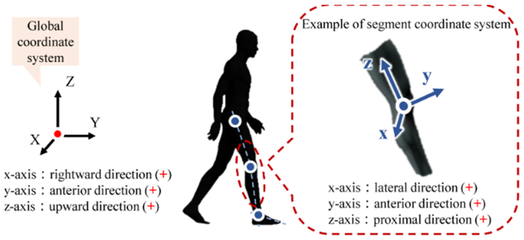
Figure 1. A rigid-body, segmental model of the human body. Reprinted from Ref. [47] with permission from MDPI, 2025.
Eng 07 00235 g001
Table 1. Comparison of commonly used hyperelastic and viscoelastic constitutive models for soft biological tissues, including their main features, advantages, limitations, and typical applications.
Mooney–RivlinHyperelastic (phenomenological)Polynomial strain energy functionSimple, easy to implement, good for moderate strainsCannot capture strong strain stiffening or anisotropyRubber-like materials, soft tissues under moderate deformation
OgdenHyperelastic (phenomenological)Power-law strain energy functionVery good for large deformation and nonlinear stiffeningParameters lack direct physical meaningThrombus, skin, brain tissue, large strain problems
Fung ExponentialHyperelastic (phenomenological)Exponential stress–strain relationCaptures exponential stiffening behavior of biological tissuesDoes not explicitly include fiber orientationSoft tissues, arteries, cartilage
Holzapfel–Gasser–Ogden (HGO)Hyperelastic (structurally motivated)Includes fiber orientation and dispersionPhysically meaningful, captures anisotropyMore complex, more parameters requiredArteries, ligaments, fiber-reinforced tissues
Kelvin–VoigtLinear viscoelasticSpring and dashpot in parallelSimple creep behaviorCannot model stress relaxation wellBasic viscoelastic approximation
MaxwellLinear viscoelasticSpring and dashpot in seriesModels stress relaxationCannot model creep accuratelyPolymers, simple viscoelastic materials
Quasi-Linear Viscoelastic (QLV)Nonlinear viscoelasticSeparates time and strain effectsWidely used for biological tissuesAssumes separable behavior (not always realistic)Tendons, ligaments
Microstructural/Network ModelsPhysically basedIncludes fiber network, cross-links, kineticsCaptures remodeling, healing, rate effectsComputationally complexHydrogels, cell tissues, biological networks
Disclaimer/Publisher’s Note: The statements, opinions and data contained in all publications are solely those of the individual author(s) and contributor(s) and not of MDPI and/or the editor(s). MDPI and/or the editor(s) disclaim responsibility for any injury to people or property resulting from any ideas, methods, instructions or products referred to in the content.