Technical Note

Low-Extrusion-Force Injectable Chitosan Gel Microparticles for Effective Wound Dressing in Endoscopic Sinus Surgery


1
Department of Chemical Engineering, Graduate School of Science and Engineering, Kagoshima University, 1-21-40 Korimoto, Kagoshima 890-0065, Japan
2
Research and Development Department, iCUREX Inc., 1-21-40 Korimoto, Kagoshima 890-0065, Japan
3
Department of Applied Chemistry, Faculty of Engineering, University of Miyazaki, 1-1 Gakuen Kibanadai Nishi, Miyazaki 889-2192, Japan
4
Nose and Smell Clinic Ikebukuro, 1-27-10 Minami-Ikebukuro, Toshima-ku, Tokyo 171-0022, Japan
5
Department of Otolaryngology, Musashi Kosugi Hospital, Nippon Medical School, 1-383 Kosugi-cho, Nakahara-ku, Kawasaki 211-8533, Kanagawa, Japan
6
Department of Otolaryngology-Head and Neck Surgery, Graduate School of Medical and Dental Sciences, Kagoshima University, 8-35-1 Sakuragaoka, Kagoshima 890-8520, Japan
7
Department of Otolaryngology, Omori Red Cross Hospital, 4-30-1 Chuo, Ota-ku, Tokyo 143-8527, Japan
*
Author to whom correspondence should be addressed.
Submission received: 28 December 2025 / Revised: 11 January 2026 / Accepted: 15 January 2026 / Published: 20 January 2026
(This article belongs to the Section Materials Engineering)

Abstract

Chitosan hydrogels are effective wound dressings that promote healing through the synergy of chitosan’s inherent biological properties and the moist environment they maintain. We previously developed hydrogel microparticles using a highly biocompatible chitosan derivative with superior therapeutic effects. This study aimed to enhance their clinical translation for Endoscopic Sinus Surgery (ESS) by optimizing preparation conditions to achieve an extrusion force of <20 N, facilitating ergonomic, single-handed administration by surgeons. While reducing the particle size alone was insufficient to lower the extrusion force significantly, the introduction of a mechanical “kneading” process to de-agglomerate microparticle aggregates resulted in a substantial reduction in the required force from 213 ± 80 N to approximately 47 N. By further optimizing the polymer concentration to 5.0% (w/v), we successfully reduced the maximum extrusion force to below 20 N (17 ± 1 N). These results demonstrate that the optimized injectable chitosan gel microparticles achieve the practical usability required for precise surgical maneuvers during ESS.

1. Introduction

Wound treatment generally follows two primary approaches: (i) dry healing, which involves desiccating the wound area to form a scab, and (ii) moist healing, which maintains a hydrated environment. Moist healing is recognized for promoting faster and superior tissue regeneration compared to dry healing [1]. Medical devices designed to cover wound surfaces and maintain this moisture are referred to as wound dressings. Hydrogels, characterized by their high water content, are widely utilized as wound dressings due to their capacity to sustain a hydrated interface. In this study, we focused on chitosan as the polymeric framework for the three-dimensional network structure of these hydrogels.
Chitosan is a polysaccharide derived from the deacetylation of chitin via alkaline treatment. It is a highly promising material for wound dressings owing to its high biocompatibility, biodegradability, antimicrobial properties, hemostatic activity, and accelerated wound-healing effects [2]. However, chitosan is typically soluble only in acidic solvents, resulting in acidic hydrogels that limit medical applications [3]. Furthermore, gelation often requires cytotoxic additives, such as chemical cross-linkers or polymerization initiators [4,5,6]. To address these issues, we successfully developed a chitosan gel with high biosafety [7,8,9,10]. Specifically, we demonstrated that gluconic acid-modified chitosan (CG) is soluble in neutral water, and the resulting neutral solution can be gelled via autoclave (AC) treatment without toxic additives.
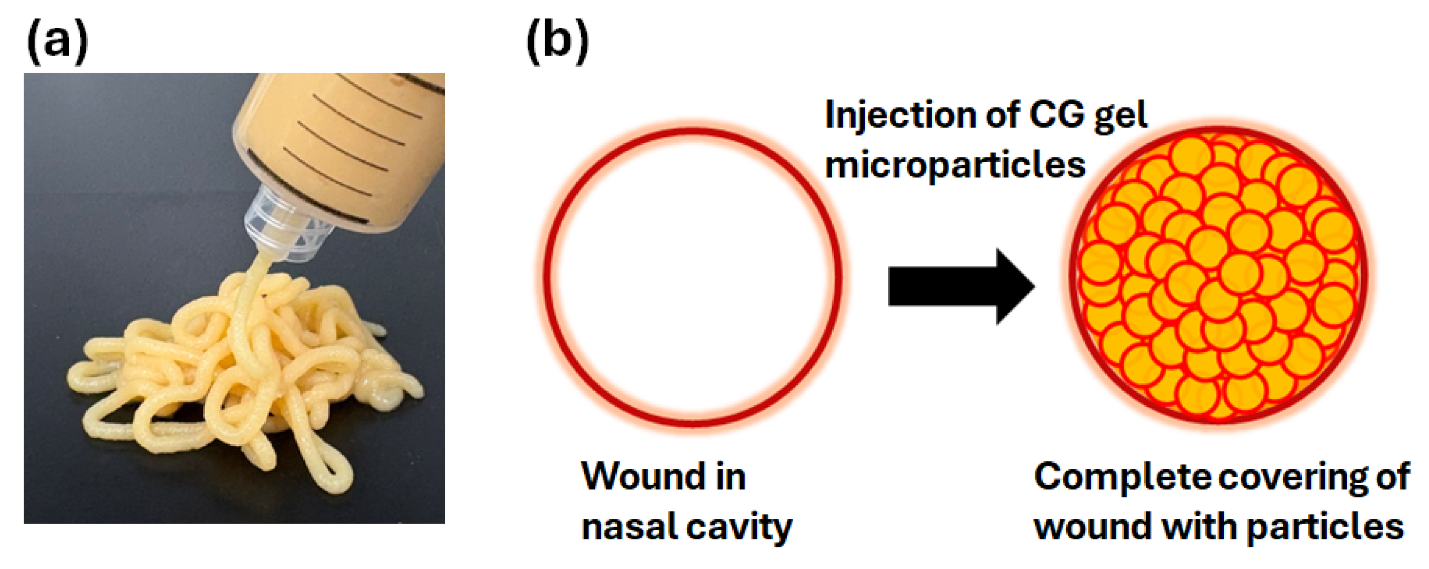
In the current study, we focused on applying this highly biocompatible CG gel to Endoscopic Sinus Surgery (ESS), the standard surgical treatment for chronic sinusitis resistant to drug therapy [11]. During ESS, the resection of mucosa and cartilage from the nasal sinuses generates significant wounding and prolonged postoperative bleeding. Conventionally, sheet- or sponge-type wound dressings have been used for hemostasis [12,13]. However, in the narrow sinonasal cavities, these dressings are time-consuming to insert and difficult to adapt to irregular wound surfaces without leaving gaps. To overcome these limitations, we comminuted pre-formed CG gel using high-speed agitation to form gel microparticles. This allows for the injection of the gel into the nasal sinuses via a syringe (Figure 1a). These injectable microparticles can fill the nasal cavity at a high density, ensuring complete and easy coverage of the wound surface (Figure 1b). We have previously demonstrated that these microparticles exhibit superior healing and hemostatic effects compared to conventional clinical dressings in animal and clinical studies [14]. However, the clinical usability of these injectable microparticles from the surgeon’s perspective remains insufficiently explored. ESS requires precise manipulation within a confined space; surgeons typically hold an endoscope in one hand while operating instruments with the other hand. Therefore, optimal usability depends on the surgeon’s ability to easily and single-handedly inject the CG gel microparticles from the syringe. Given that the maximum pinch force for women is reported to be approximately 64 N [15], an extrusion force of <20 N is considered ideal for fine surgical maneuvers. Thus, the present study aimed to investigate the preparation conditions necessary to ensure that CG gel microparticles can be extruded from a syringe with a force of <20 N.

2. Materials and Methods

2.1. Materials

N-hydroxysuccinimide, 2-morpholinoethanesulfonic acid, sodium gluconate, and sodium chloride were purchased from FUJIFILM Wako Pure Chemical Corp. (Osaka, Japan). 1-Ethyl-3-(3-dimethylaminopropyl)carbodiimide hydrochloride was purchased from Peptide Institute, Inc. (Osaka, Japan). These reagents were used to prepare CG. An autoclavable 20 mL syringe (CHITOSLA syringe) and a 20 mL Terumo syringe (SS-20LZ) were obtained from iCUREX Inc. (Kagoshima City, Japan) and Terumo Corp. (Tokyo, Japan), respectively. Both syringes have a 20 mm inner diameter barrel and a tapered tip terminating in a 2 mm diameter, 10 mm long orifice.

2.2. Preparation of CG Gel Microparticles

CG was prepared according to our previously reported method [7], with a gluconic acid content of 8–12% (relative to the free amino groups of unmodified chitosan). CG was dissolved in a dilute hydrochloric acid aqueous solution (pH 4), followed by the dropwise addition of a NaOH aqueous solution under vigorous stirring to obtain a neutral 2% (w/v) CG aqueous solution. Sodium chloride was added to the solution to ensure isotonicity. This CG solution was subjected to AC treatment (121 °C, 20 min) to induce partial gelation. The resulting mixture was comminuted using a high-speed blender to obtain a CG gel microparticle suspension. The particle size distribution was measured using a laser diffraction analyzer (LA-920, Horiba, Ltd., Kyoto, Japan). The suspension was then centrifuged to remove free water, adjusting the final CG concentration to 5.0% or 6.5% (w/v) relative to the final suspension volume. The resulting suspension was loaded into an autoclavable 20 mL syringe and subjected to a final AC treatment (121 °C, 20 min) for sterilization. The extrusion force was subsequently measured using a tabletop universal tester (MCT-2150W, A&D Co., Ltd., Tokyo, Japan) at a constant plunger speed (60 mm/min).
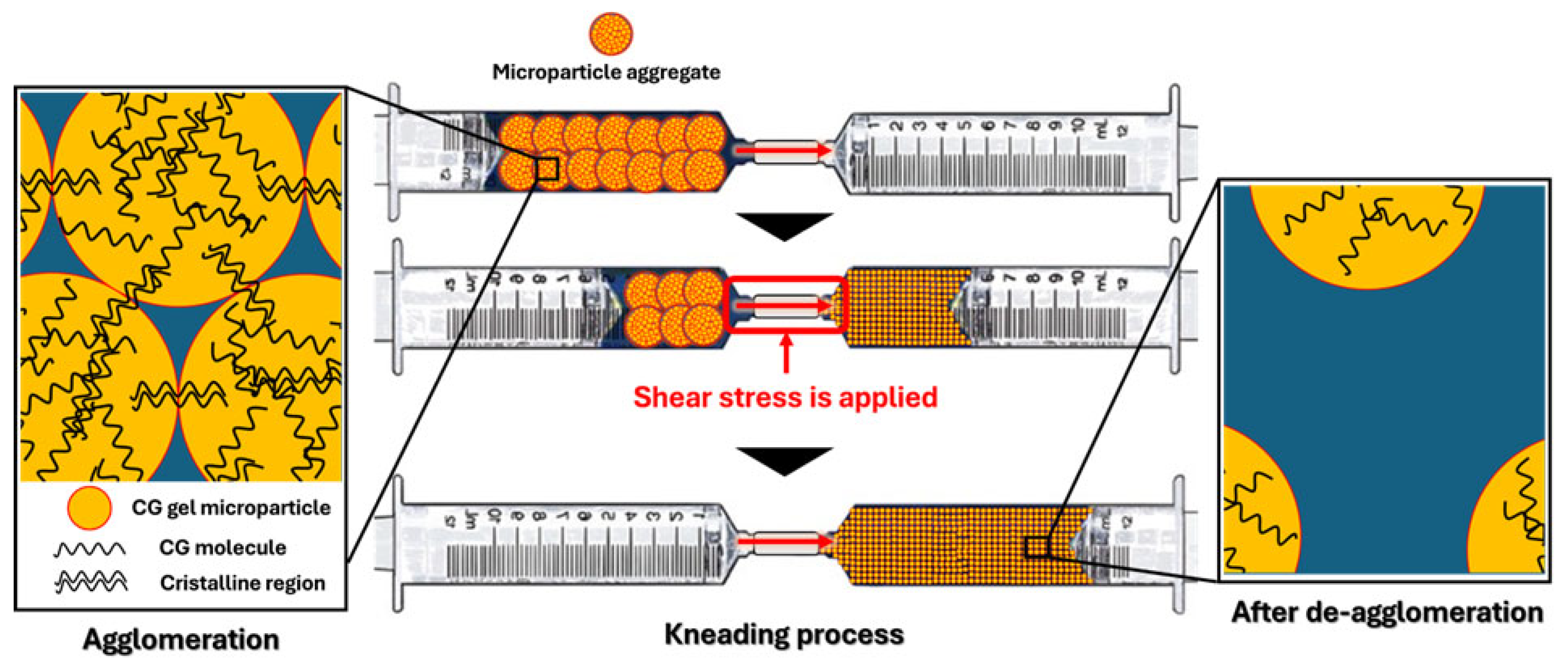
For samples requiring “kneading” within the syringe, the following procedure was performed: the outlet of the gel microparticle-filled syringe was connected to the outlet of a 20 mL Terumo syringe using a plastic connector (inner diameter: 4 mm). By pressing the plunger, the gel microparticles were transferred to the other syringe through the connector (defined as 1 kneading cycle) (Figure 2). Returning the gel to the original syringe constituted two kneading cycles. Multiple kneading cycles were performed to investigate the relationship between the number of kneading cycles and the maximum extrusion force. Experimental data are presented as means ± standard deviations (n = 3).

3. Results and Discussion

In this study, we aimed to identify the optimal preparation conditions for CG gel microparticles that can be extruded from a syringe with a force of <20 N to enable precise surgical maneuvers during ESS.
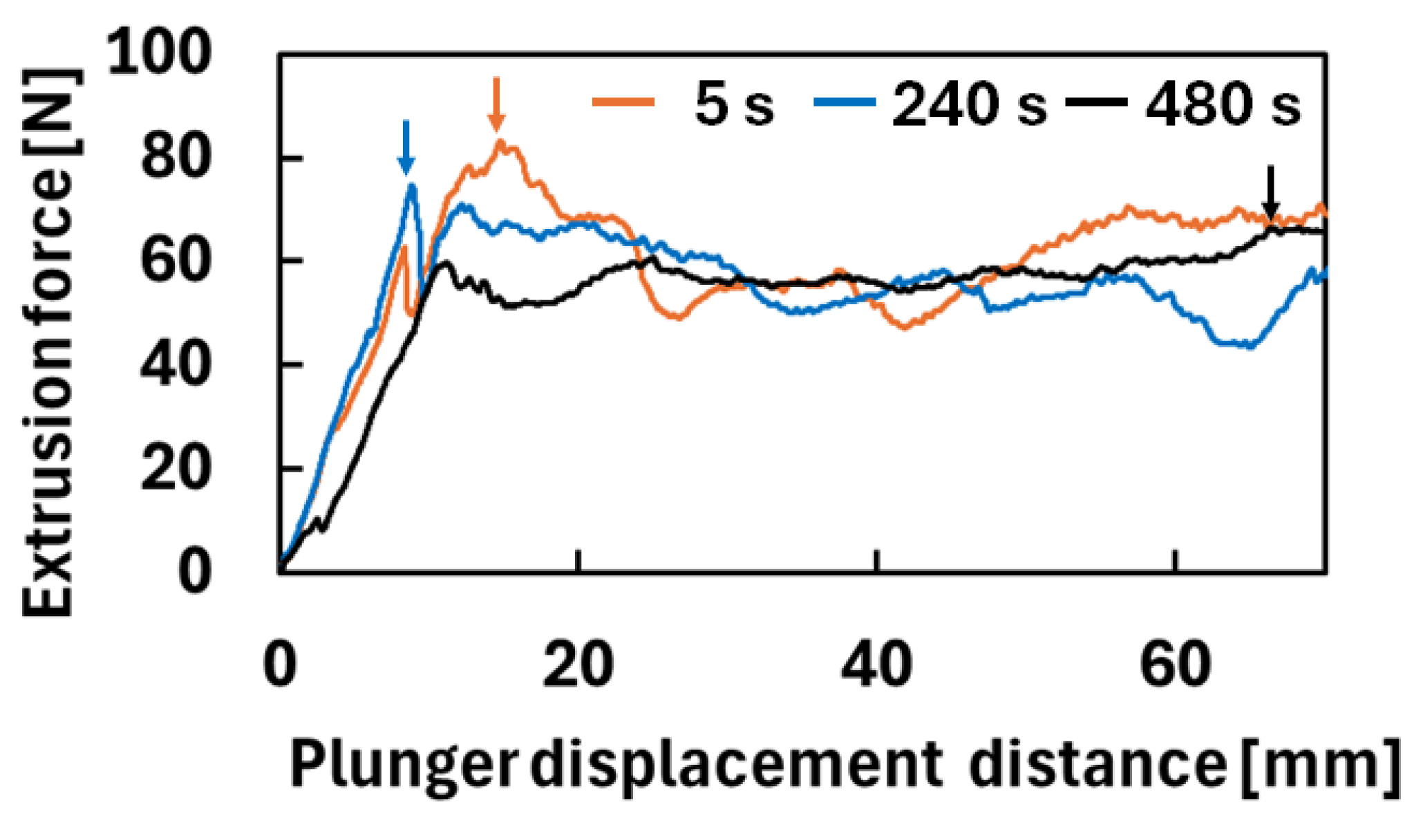
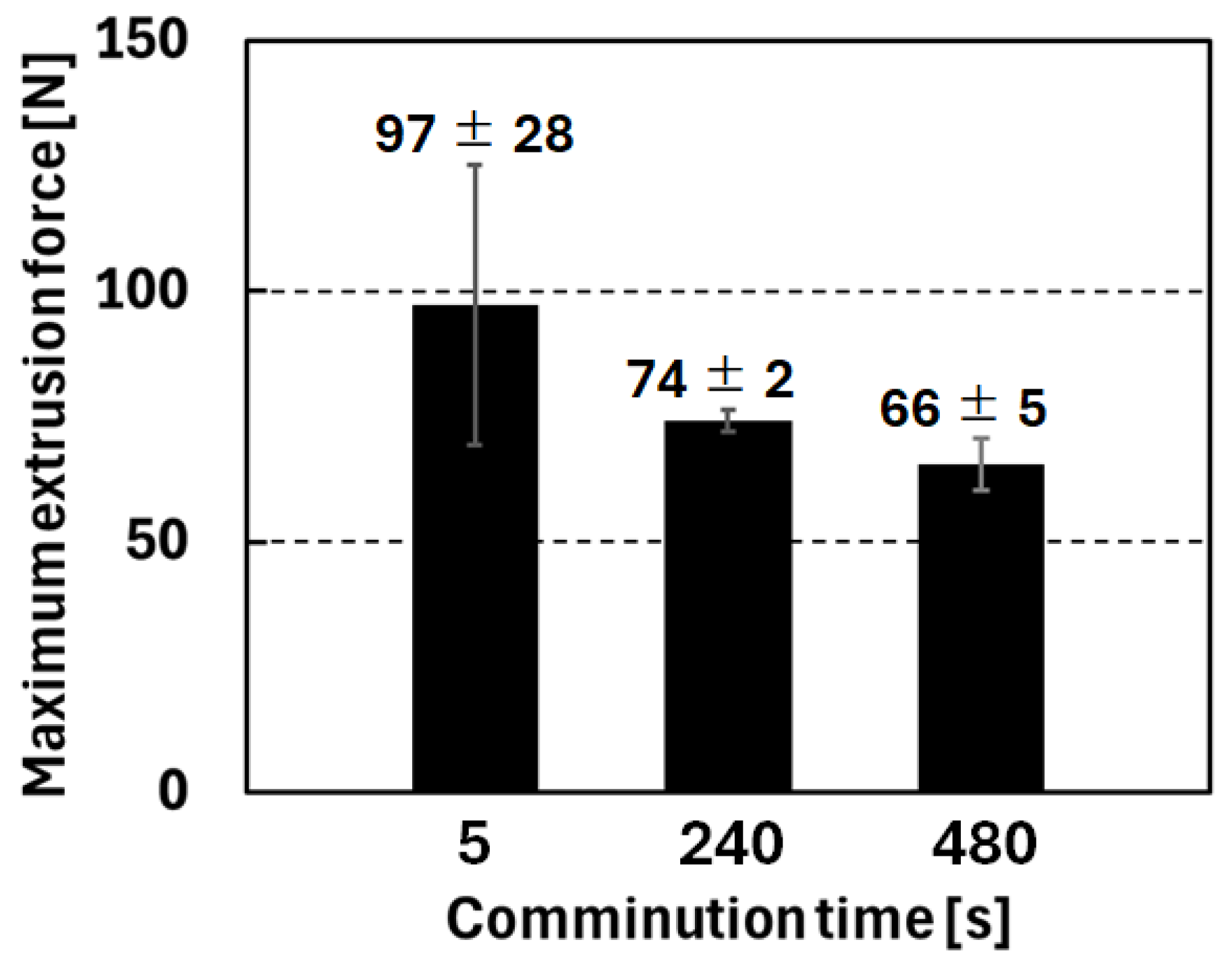
When the CG gel was not comminuted (at 1 kneading cycle and 6.5% (w/v) CG concentration), the extrusion force exceeded 100 N, precluding single-handed injection (the reason for performing 1 kneading cycle here is described later). We hypothesized that reducing the particle size would enhance fluidity and decrease the required force. First, we investigated the relationship between comminution time and particle size (Table 1). Without comminution (at 0 s of comminution), numerous particles with diameters of >1 mm were observed. In contrast, the particle size decreased as the comminution time increased from D10/D50/D90 = 23/82/261 μm at 5 s of comminution to D10/D50/D90 = 4/9/70 μm at 480 s of comminution. Figure 3 shows the relationships between the extrusion forces and the plunger displacement distance for various comminution times (at 6.5% (w/v) CG concentration and 1 kneading cycle). The maximum displacement distance of the plunger (=the length of the syringe barrel) was 70 mm. In Figure 3, arrows define maximum extrusion forces observed during extrusion of gel microparticles from the syringe, which we aimed to reduce to <20 N in this study. Figure 4 summarizes the maximum extrusion forces. The maximum extrusion forces at 0 s and 5 s of comminution were >100 N and 97 ± 28 N, respectively, which was reduced to 66 ± 5 N by increasing the comminution time to 480 s. These results sufficiently demonstrate the clear correlation between comminution time and extrusion force. Furthermore, the data points at 240 s and 480 s demonstrate that while increasing comminution time continues to reduce the force, the rate of reduction begins to stabilize, suggesting that 480 s is an optimized point for practical application.
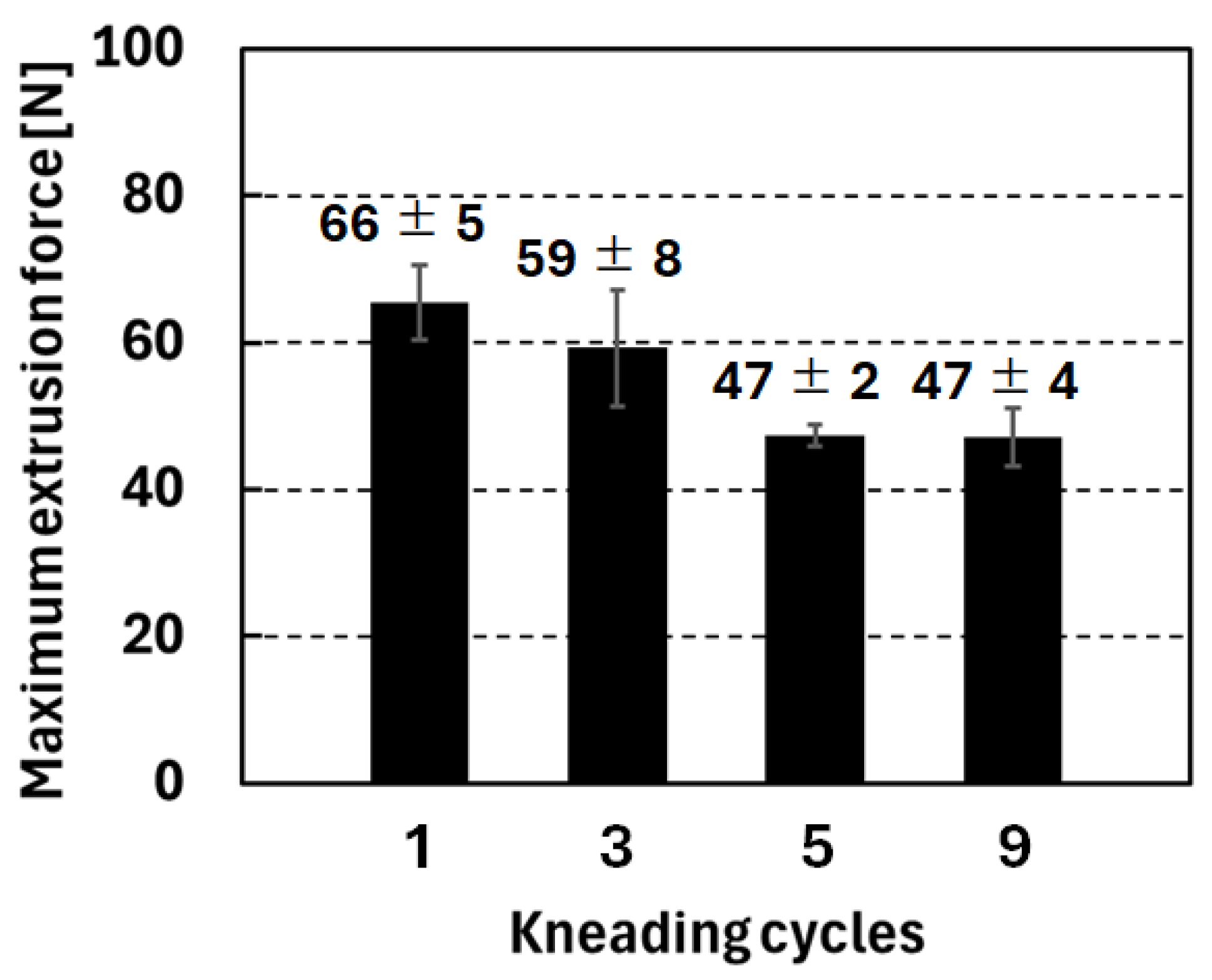
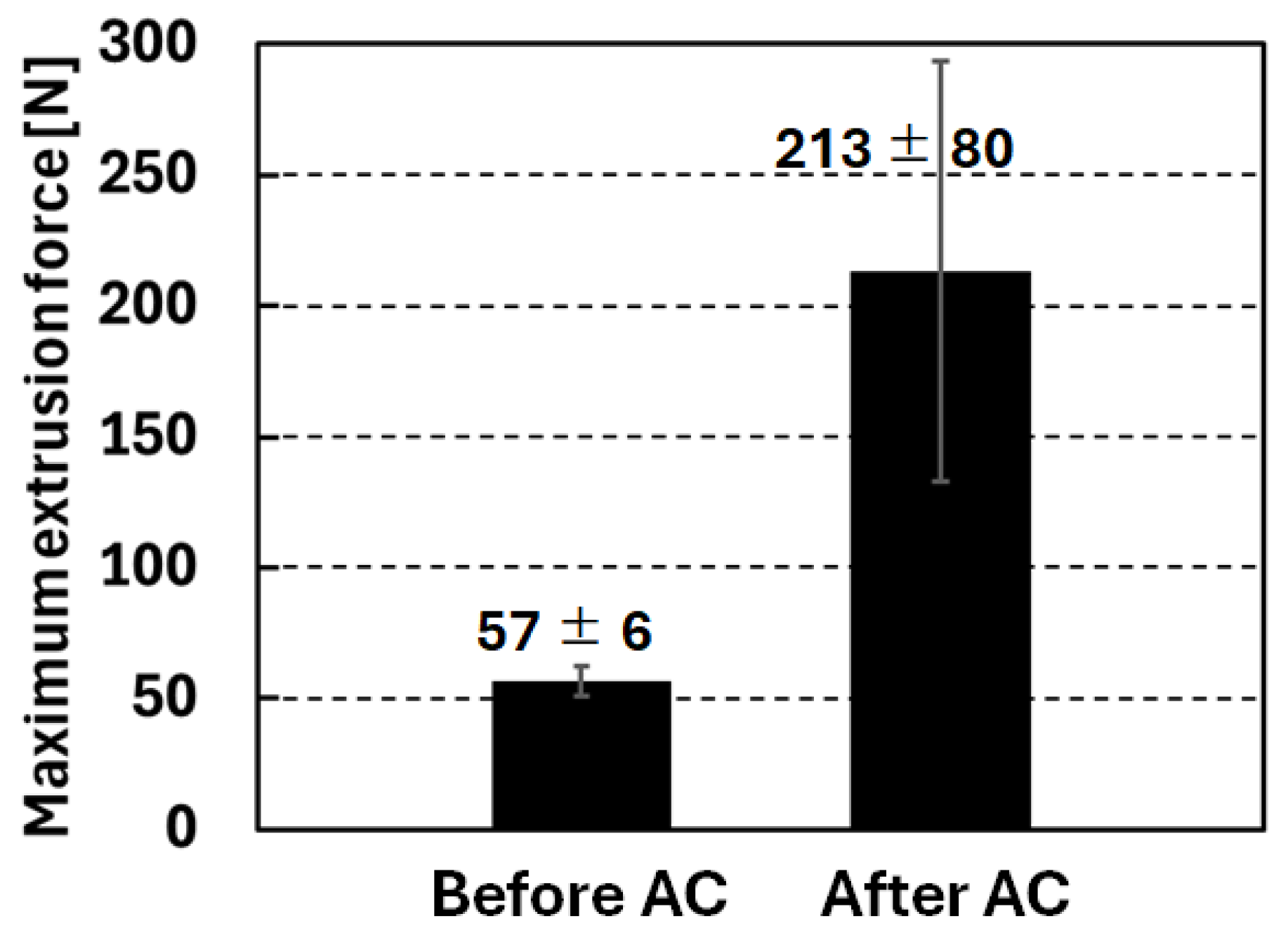
The impact of the sterilization process was then evaluated. Figure 4 shows the maximum extrusion forces at 1 kneading cycle. Without kneading (at 0 kneading cycle), the extrusion force was 213 ± 80 N after the final AC treatment; however, the extrusion force before the final AC treatment was 57 ± 6 N (at 480 s of comminution and 6.5% (w/v) CG concentration) (Figure 5). The significant increase in extrusion force caused by the final AC treatment is likely attributed to the following mechanism. Chitosan is inherently a highly crystalline substance [17]. To disrupt its crystalline structure and dissolve it in water, intramolecular electrostatic repulsion via the protonation of amino groups in the glucosamine units of chitosan is required; based on this principle, chitosan dissolves in acidic aqueous solutions. When such a chitosan aqueous solution is neutralized, the intermolecular electrostatic repulsion is lost, leading to precipitation due to recrystallization. In contrast, our CG is synthesized by chemically modifying the amino groups of chitosan with gluconic acid. Even when neutralized after dissolution in an acidic aqueous solution, the steric hindrance provided by the gluconic acid moieties suppresses molecular recrystallization, allowing the CG to maintain a dissolved state. When this neutral CG aqueous solution is subjected to AC treatment, the thermal motion of water molecules becomes vigorous due to heating. We believe this causes the dehydration of the CG molecules, promoting crystallization, which then acts as physical cross-linking points of CG molecules to induce gelation [9]. By a similar mechanism, physical cross-links likely formed between the CG gel microparticles within the syringe during the final AC treatment (Figure 2). This presumably led to the aggregation of the microparticles, resulting in the increased extrusion force after the final AC treatment (Figure 5). Consequently, we investigated whether these aggregates could be de-agglomerated by applying shear stress to the aggregates after the final AC sterilization. Specifically, the outlet of the CG gel microparticle-filled syringe was connected to the outlet of the other syringe using a plastic connector (inner diameter: 4 mm), and the gel microparticles were transferred to the other syringe through the connector (kneading operation, Figure 2). We anticipated that the shear stress would de-agglomerate the aggregates. For this test, the CG concentration was set to 6.5% (w/v). As expected, the kneading operation successfully reduced the extrusion force from 213 ± 80 N (without kneading, Figure 5) to approximately 47 N after 5 and 9 kneading cycles (Figure 6). We confirmed that the reduction in the extrusion force by kneading operation was observed for another CG concentration.
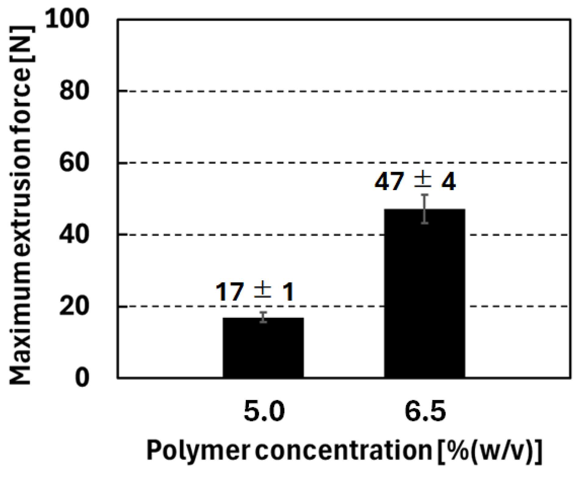
Finally, to further reduce the extrusion force, the CG concentration was decreased from 6.5% to 5.0% (w/v) at 9 kneading cycles. Under these optimized conditions, we successfully achieved the target extrusion force of <20 N (17 ± 1 N) (Figure 7).
The kneading operation is intended to be performed by the users (surgeon or assistant) immediately before clinical use. This ensures that sterile gel microparticles are retained in the closed syringes until the point of care.

4. Conclusions

We demonstrated that reducing the particle size and polymer concentration of CG gel microparticles, combined with the application of the kneading operation, enables the successful reduction in the extrusion force of CG gel microparticles from 213 ± 80 N to <20 N. This reduction in extrusion force facilitates the precise surgical maneuvers required for ESS.

Author Contributions

Conceptualization, T.Y. and T.T.; methodology, Y.N., Y.Y., T.Y., Y.O. and T.T.; validation, Y.N., T.Y. and T.T.; formal analysis, Y.N.; investigation, Y.N., Y.Y., K.H., M.K., M.Y. (Masaru Yamashita) and S.M.; resources, T.Y. and T.T.; data curation, Y.N. and T.T.; writing—original draft preparation, Y.N.; writing—review and editing, T.T.; supervision, M.Y. (Masahiro Yoshida), K.H., M.K., M.Y. (Masaru Yamashita), S.M. and T.T.; project administration, T.T.; funding acquisition, T.Y. and T.T. All authors have read and agreed to the published version of the manuscript.

Funding

This research was partially supported by AMED (Japan Agency for Medical Research and Development) under Grant Number JP24ym0126142, by JSPS (Japan Society for the Promotion of Science) KAKENHI under Grant Number 24K21704 and 24K01238, and by joint research funds from iCUREX Inc.

Institutional Review Board Statement

Not applicable.

Informed Consent Statement

Not applicable.

Data Availability Statement

The original contributions presented in this study are included in the article. Further inquiries can be directed to the corresponding author.

Acknowledgments

The authors would like to thank Saki Kobaru (Kagoshima University) for her valuable technical advice and support.

Conflicts of Interest

T. Takei and Y. Nagase are affiliated with Kagoshima University and hold dual appointments at iCUREX Inc. T. Yoshinaga is affiliated with iCUREX Inc. T. Takei and T. Yoshinaga are shareholders of iCUREX Inc. M. Yamashita (Kagoshima University) serves as a consultant for iCUREX Inc. K. Hosoya is affiliated with Musashi Kosugi Hospital of Nippon Medical School and holds a position at Nose and Smell Clinic Ikebukuro, which is a private clinic. This work was conducted as a joint research project between Kagoshima University and iCUREX Inc., and T. Takei has received research funds from iCUREX Inc. Y. Yamashita, M. Yoshida, and T. Takei are inventors of a registered patent related to this work (Japanese Patent No. 7596001, titled “Hydrogel for Treating Body Cavity Injuries and Body Cavity Wound Treatment Kit”). The remaining authors declare that the research was conducted in the absence of any commercial or financial relationships that could be construed as a potential conflict of interest.

References

  1. Winter, D.G.; Scales, J.T. Effect of air drying and dressings on the surface of a wound. Nature 1963, 197, 91–92. [Google Scholar] [CrossRef] [Scilit] [PubMed]
  2. Zhao, J.; Oiu, P.; Wang, Y.; Wang, Y.; Zhou, J.; Zhang, B.; Zhang, L.; Gou, D. Chitosan-based hydrogel wound dressing: From mechanism to applications, a review. Int. J. Biol. Macromol. 2023, 244, 125250. [Google Scholar] [CrossRef] [Scilit] [PubMed]
  3. Thomas, N.; Paneo, M.A.; Mo’o, F.R.; Puluhulawa, L.E.; Lati, M.S. Formulation of natural hydrogel from bromelain enzym and alginate-chitosan and the effectiveness test by in vivo in healing burns. J. Med. Pharm. Chem. Res. 2025, 7, 944–959. [Google Scholar]
  4. Hamedi, H.; Moradi, S.; Hudson, S.M.; Tonelli, A.E. Chitosan based hydrogels and their applications for drug delivery in wound dressings: A review. Carbohydr. Polym. 2018, 199, 445–460. [Google Scholar] [CrossRef] [Scilit] [PubMed]
  5. Martínez-Mejía, G.; Vázquez-Torres, N.A.; Castell-Rodríguez, A.; del Río, J.M.; Corea, M.; Jiménez-Juárez, R. Synthesis of new chitosan-glutaraldehyde scaffolds for tissue engineering using Schiff reactions. Colloids Surf. A Physicochem. Eng. Asp. 2019, 579, 123658. [Google Scholar] [CrossRef] [Scilit]
  6. Than-ardna, B.; Tamura, H.; Furuike, T. Preparation, characterization, and properties of chitosan-based semi-interpenetrating polymer networks and poly(2-hydroxyethyl methacrylate) structure. Macromol. Chem. Phys. 2022, 223, 2200282. [Google Scholar] [CrossRef] [Scilit]
  7. Takei, T.; Nakahara, H.; Ijima, H.; Kawakami, K. Synthesis of a chitosan derivative soluble at neutral pH and gellable by freeze–thawing, and its application in wound care. Acta Biomater. 2012, 8, 686–693. [Google Scholar] [CrossRef] [Scilit] [PubMed]
  8. Takei, T.; Danjo, S.; Sakoguchi, S.; Tanaka, S.; Yoshinaga, T.; Nishimata, H.; Yoshida, M. Autoclavable physically-crosslinked chitosan cryogel as a wound dressing. J. Biosci. Bioeng. 2018, 125, 490–495. [Google Scholar] [CrossRef] [Scilit] [PubMed]
  9. Yamashita, Y.; Ohzuno, Y.; Saito, Y.; Fujiwara, Y.; Yoshida, M.; Takei, T. Autoclaving-triggered hydrogelation of chitosan-gluconic acid conjugate aqueous solution for wound healing. Gels 2023, 9, 280. [Google Scholar] [CrossRef] [Scilit] [PubMed]
  10. Yamashita, Y.; Ohzuno, Y.; Yoshida, M.; Takei, T. Autoclaving achieves pH-neutralization, hydrogelation, and sterilization of chitosan hydrogels in one step. Macromol 2024, 4, 376–386. [Google Scholar] [CrossRef] [Scilit]
  11. Smith, K.A.; Smith, T.L.; Mace, J.C.; Rudmik, L. Endoscopic sinus surgery compared to continued medical therapy for patients with refractory chronic rhinosinusitis. Int. Forum Allergy Rhinol. 2014, 4, 823–827. [Google Scholar] [CrossRef] [Scilit] [PubMed]
  12. Okushi, T.; Yoshikawa, M.; Otori, N.; Matsuwaki, Y.; Asaka, D.; Nakayama, T.; Morimoto, T.; Moriyama, H. Evaluation of symptoms and QOL with calcium alginate versus chitin-coated gauze for middle meatus packing after endoscopic sinus surgery. Auris Nasus Larynx 2012, 39, 31–37. [Google Scholar] [CrossRef] [Scilit] [PubMed]
  13. Hosoya, K.; Maeda, Y.; Komachi, T.; Sato, K.; Okubo, K. AQUACEL® Ag Advantage reduces the stress of postoperative packing removal after endoscopic sinus surgery. Braz. J. Otorhinolaryngol. 2023, 89, 101292. [Google Scholar] [CrossRef] [Scilit] [PubMed]
  14. Yamashita, Y.; Hosoya, K.; Fujiwara, Y.; Saito, Y.; Yoshida, M.; Matsune, S.; Okubo, K.; Takei, T. Injectable chitosan hydrogel particles as nasal packing materials after endoscopic sinus surgery for treatment of chronic sinusitis. Gels 2025, 11, 60. [Google Scholar] [CrossRef] [Scilit] [PubMed]
  15. Astin, A.D. Finger force capability: Measurement and prediction using anthropometric and myoelectric measures. Master’s Thesis, Virginia Polytechnic Institute and State University, Blacksburg, VA, USA, 16 December 1999. [Google Scholar]
  16. Yamanami, T.; Tsuchida, S.; Ieda, K. Chitosan Microparticles. Japanese Patent JP 4976662 B2, 18 July 2012. [Google Scholar]
  17. Rinaudo, M. Chitin and chitosan: Properties and applications. Prog. Polym. Sci. 2006, 31, 603–632. [Google Scholar] [CrossRef] [Scilit]
Figure 1. (a) Injectable CG gel microparticles. (b) The microparticles allow the nasal cavity to be filled with a high density of the gel microparticles.
Eng 07 00053 g001
Figure 2. Kneading process to de-agglomerate CG gel microparticle aggregates.
Eng 07 00053 g002
Figure 3. Typical relationships between extrusion forces and plunger displacement distance for various comminution times. Arrows show maximum extrusion forces, which we aimed to reduce to <20 N in this study.
Eng 07 00053 g003
Figure 4. Maximum extrusion forces of CG gel microparticle from syringes for various comminution times at 1 kneading cycle (n = 3).
Eng 07 00053 g004
Figure 5. Maximum extrusion forces of CG gel microparticle from syringes before and after final AC treatment at 0 kneading cycle (n = 3).
Eng 07 00053 g005
Figure 6. Maximum extrusion forces of CG gel microparticle from syringes for various kneading cycles (n = 3). The maximum forces were successfully reduced from 213 ± 80 N (without kneading, Figure 5) to approximately 47 N after 5 and 9 kneading cycles.
Eng 07 00053 g006
Figure 7. Maximum extrusion forces of CG gel microparticle from syringes for each CG concentration (n = 3).
Eng 07 00053 g007
Table 1. Relationship between comminution time and particle size distribution of CG gel microparticles.
Comminution Time [s]D10 [μm]D50 [Median, μm]D90 [μm]
52382261
240819122
4804970
Measurement settings and conditions: Dispersion medium: Distilled water, Relative refractive index: 1.5 [16], Sonication: None, Particle state: Swollen.
Disclaimer/Publisher’s Note: The statements, opinions and data contained in all publications are solely those of the individual author(s) and contributor(s) and not of MDPI and/or the editor(s). MDPI and/or the editor(s) disclaim responsibility for any injury to people or property resulting from any ideas, methods, instructions or products referred to in the content.