Review

Cardiac Mechano-Electrical-Fluid Interaction: A Brief Review of Recent Advances

1
Department of Mechanical, Environmental and Civil Engineering, Mayfield College of Engineering, Tarleton State University, Stephenville, TX 76401, USA
2
Richard A. Rula School of Civil and Environmental Engineering, Mississippi State University, Mississippi State, MS 39762, USA
*
Author to whom correspondence should be addressed.
Eng 2025, 6(8), 168; https://doi.org/10.3390/eng6080168
Submission received: 5 June 2025 / Revised: 15 July 2025 / Accepted: 18 July 2025 / Published: 22 July 2025

Abstract

This review investigates recent developments in cardiac mechano-electrical-fluid interaction (MEFI) modeling, with a focus on multiphysics simulation platforms and digital twin frameworks developed between 2015 and 2025. The purpose of the study is to assess how computational modeling methods—particularly finite element and immersed boundary techniques, monolithic and partitioned coupling schemes, and artificial intelligence (AI)-enhanced surrogate modeling—capture the integrated dynamics of cardiac electrophysiology, tissue mechanics, and hemodynamics. The goal is to evaluate the translational potential of MEFI models in clinical applications such as cardiac resynchronization therapy (CRT), arrhythmia classification, atrial fibrillation ablation, and surgical planning. Quantitative results from the literature demonstrate <5% error in pressure–volume loop predictions, >0.90 F1 scores in machine-learning-based arrhythmia detection, and <10% deviation in myocardial strain relative to MRI-based ground truth. These findings highlight both the promise and limitations of current MEFI approaches. While recent advances improve physiological fidelity and predictive accuracy, key challenges remain in achieving multiscale integration, model validation across diverse populations, and real-time clinical applicability. The review concludes by identifying future milestones for clinical translation, including regulatory model certification, standardization of validation protocols, and integration of patient-specific digital twins into electronic health record (EHR) systems.

1. Introduction

Cardiac diseases (CVDs) remain the leading cause of mortality worldwide, responsible for approximately 17.9 million deaths annually, representing about 32% of all global deaths, as reported by the World Health Organization [1]. These conditions, which include coronary artery disease, heart failure, and arrhythmias, are often characterized by complex, patient-specific interactions between electrophysiological, mechanical, and hemodynamic systems. As the global burden of CVDs continues to rise, especially in aging populations and low- to middle-income countries, there is a critical need for advanced, personalized diagnostic and therapeutic tools. Computational cardiovascular models, particularly those incorporating mechano-electrical-fluid interaction (MEFI) and digital twin frameworks, offer promising solutions. These models allow for non-invasive simulations of disease progression, risk stratification, and virtual testing of treatment strategies tailored to individual patients. For example, they are increasingly used to understand arrhythmia mechanisms, assess mechanical dyssynchrony, and simulate hemodynamic responses to interventions such as cardiac resynchronization therapy or valve replacement [2,3,4]. Moreover, the predictive capabilities of these models are essential for addressing long-standing clinical challenges, such as optimizing patient selection for therapies or anticipating adverse drug reactions, ultimately supporting more effective and personalized cardiovascular care.
In the MEFI process [5], cardiac mechanical contraction is initiated by the propagation of depolarizing action potentials, which originate from the sinoatrial (SA) nodes located in the endocardium of the right atrium. Membrane depolarization triggers the influx of calcium ions (Ca2+) through voltage-dependent transmembrane Ca2+ channels. This initial Ca2+ influx subsequently induces a larger release of Ca2+ from the sarcoplasmic reticulum (SR) via a mechanism known as calcium-induced calcium release (CICR), resulting in a substantial rise in intracellular Ca2+ concentration. Elevated Ca2+ levels promote the binding of Ca2+ to troponin C, facilitating the formation of actin–myosin cross-bridges and generating microscopic tension within individual cardiac cells. The cumulative effect of this cellular tension produces the macroscopic contractile force necessary for blood ejection. The entry of calcium into cardiac cells is tightly regulated by the duration of the action potential, which in turn is governed by various ionic currents, primarily through sodium (Na+), calcium (Ca2+), and potassium (K+) ion channels, all of which are dependent on membrane potential and ionic concentration gradients. These channels are traditionally considered stress-independent. However, stretch-activated ion channels (SACs), which are sensitive to mechanical stress, are also present in cardiac tissue and can depolarize the cell upon activation [6]. Thus, mechanical deformation of cardiac tissue can influence intracellular ion flux and prolong the action potential duration. This feedback loop—where mechanical contraction impacts electrical excitation—is referred to as mechano-electrical feedback (MEF), representing a two-way coupling between the mechanical and electrical systems of the heart.
Another critical interaction within the heart is the coupling between blood flow and cardiac tissue. Unlike the active contraction of cardiac muscle initiated by electrical wave propagation, incoming blood flow can passively deform cardiac tissue, which may also trigger the opening of stretch-activated channels (SACs). Simultaneously, the deformation of the cardiac walls alters the boundary conditions for the blood flow, thereby influencing the flow dynamics. This bidirectional relationship between tissue deformation and blood movement constitutes a classic two-way fluid–structure interaction (FSI) problem.
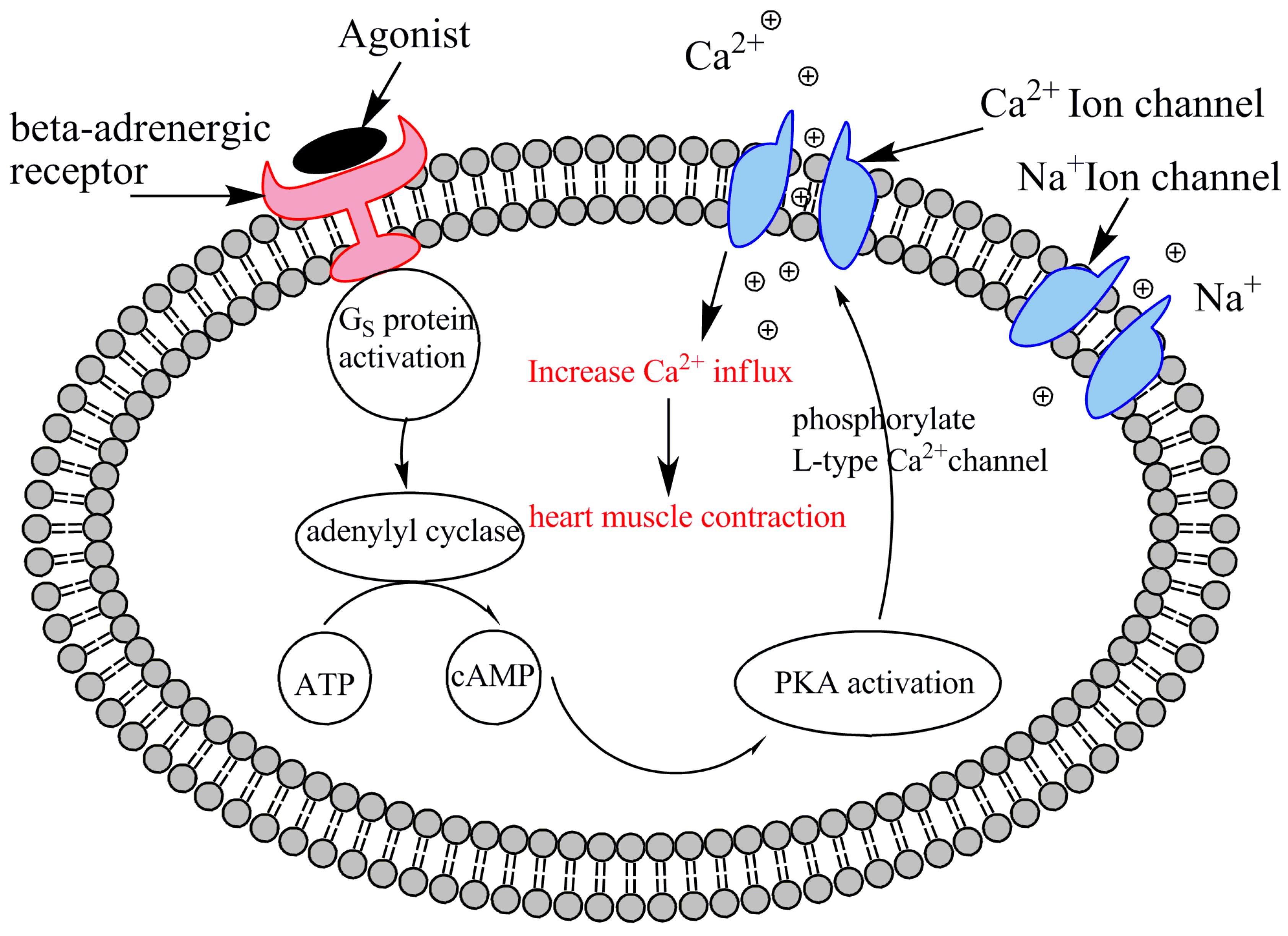
Additionally, system-level regulatory mechanisms, such as the β-adrenergic signaling pathway, play a crucial role in modulating heart rate and tissue contractility in response to stimuli including exercise, blood loss, heart failure, or pharmacological interventions. Simulating the entire heart remains a formidable challenge, necessitating a systems-level approach that integrates expertise across fluid and tissue mechanics, mathematics, computational biology, and electrophysiology. The inherent complexity of cardiac morphology and internal structures further compounds the modeling difficulty. Developing a fully coupled mechano-electrical-fluid interaction (MEFI) model is particularly challenging due to the system’s inherently multiphysical and multiscale nature [7], as demonstrated in previous studies. This paper provides an overview of recent advances in cardiac MEFI modeling and highlights a growing range of needs to elucidate fundamental mechanisms of cardiac disease progression, particularly those related to ion channel mutations and system-level signaling pathways. The review concludes by identifying existing knowledge gaps and proposing future directions for advancing the field.
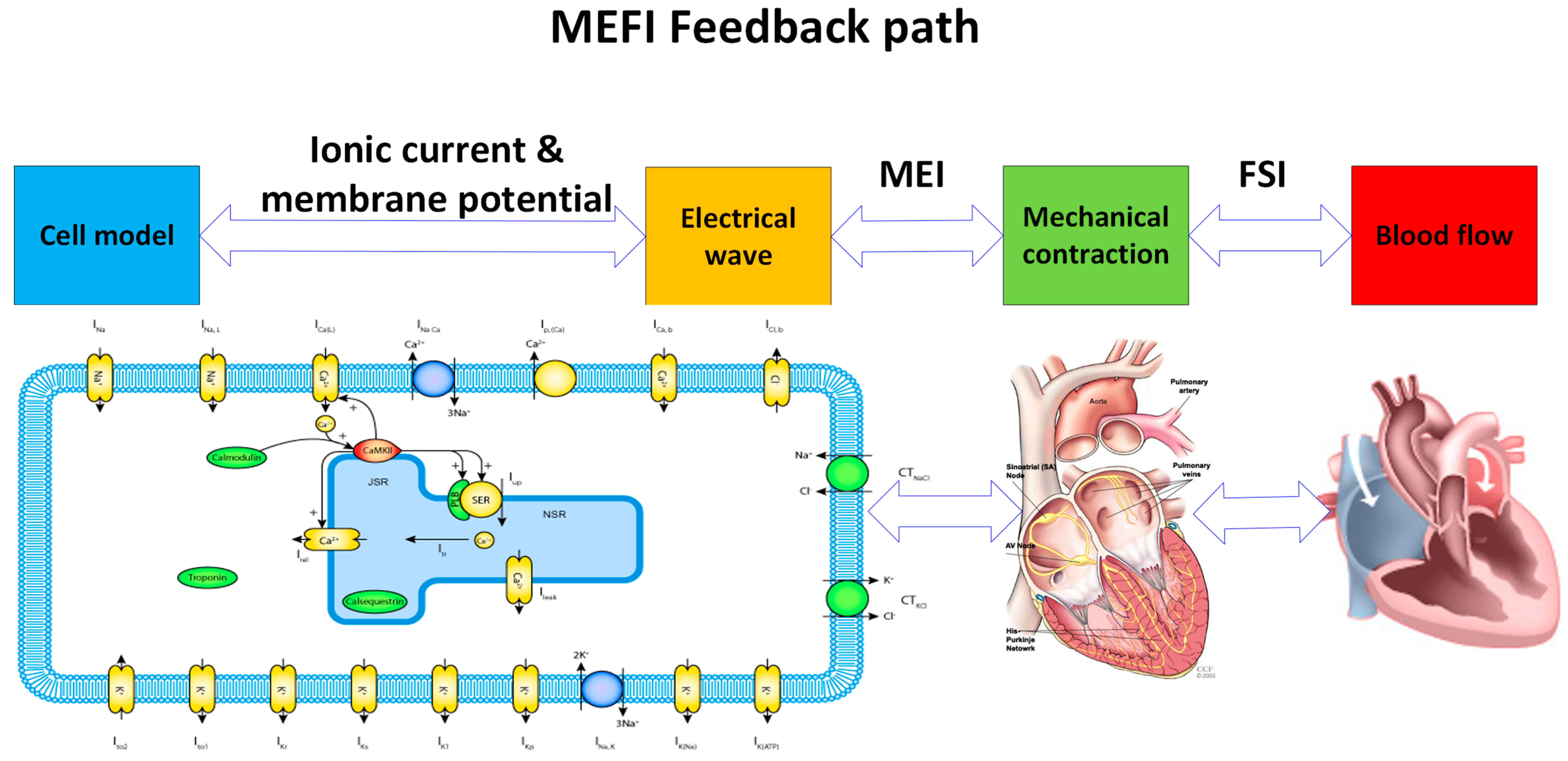
Figure 1 illustrates the integrated mechano–electro–fluidic interaction (MEFI) framework in cardiac modeling. The cascade begins with the cell model, where ionic currents and membrane potential govern transmembrane dynamics. These electrophysiological signals generate electrical wave propagation, which subsequently induces mechanical contraction of the myocardial tissue (MEI). The resulting deformation couples to the blood flow through fluid–structure interaction (FSI), enabling simulation of intracardiac hemodynamics. The MEFI loop is completed by bidirectional feedback mechanisms: electrical activity is modulated by mechanical deformation (MEI), and flow-induced mechanical stresses can influence electrophysiological states. This framework enables comprehensive, multiscale simulation of the heart’s electromechanical and fluid dynamics with clinical relevance.
This paper provides an overview of recent advances in cardiac MEFI modeling and highlights a growing range of needs to elucidate fundamental mechanisms of cardiac disease progression, particularly those related to ion channel mutations and system-level signaling pathways. The novelty of this review lies in its integrative comparison of cutting-edge high-fidelity and reduced-order models, with a focus on their trade-offs in clinical applicability, computational efficiency, and physiological accuracy. Furthermore, this study uniquely emphasizes the role of digital twin frameworks in bridging multiphysics modeling with personalized medicine, offering a structured synthesis of recent research efforts to incorporate electrophysiological mutations, mechano–fluid feedback, and β-adrenergic signaling into computational heart models.
The paper is structured as follows. Section 2 reviews recent advances in cardiac mechano-electrical-fluid interaction (MEFI), highlighting progress in high-fidelity modeling, personalization techniques, and AI-integrated surrogate frameworks. Section 3 presents a critical discussion of current challenges, limitations, and knowledge gaps in MEFI modeling, along with future research directions and milestones needed to support clinical translation and digital twin development. Finally, Section 4 offers concluding remarks and summarizes the key contributions of this review.

2. Recent Advances in Cardiac Mechano-Electrical-Fluid Interaction

Previous computational models of the MEFI system can be broadly categorized into three groups: fluid–structure interaction (FSI), mechano–electrical interaction (MEI), and fully coupled mechano-electrical-fluid interaction (MEFI) models.
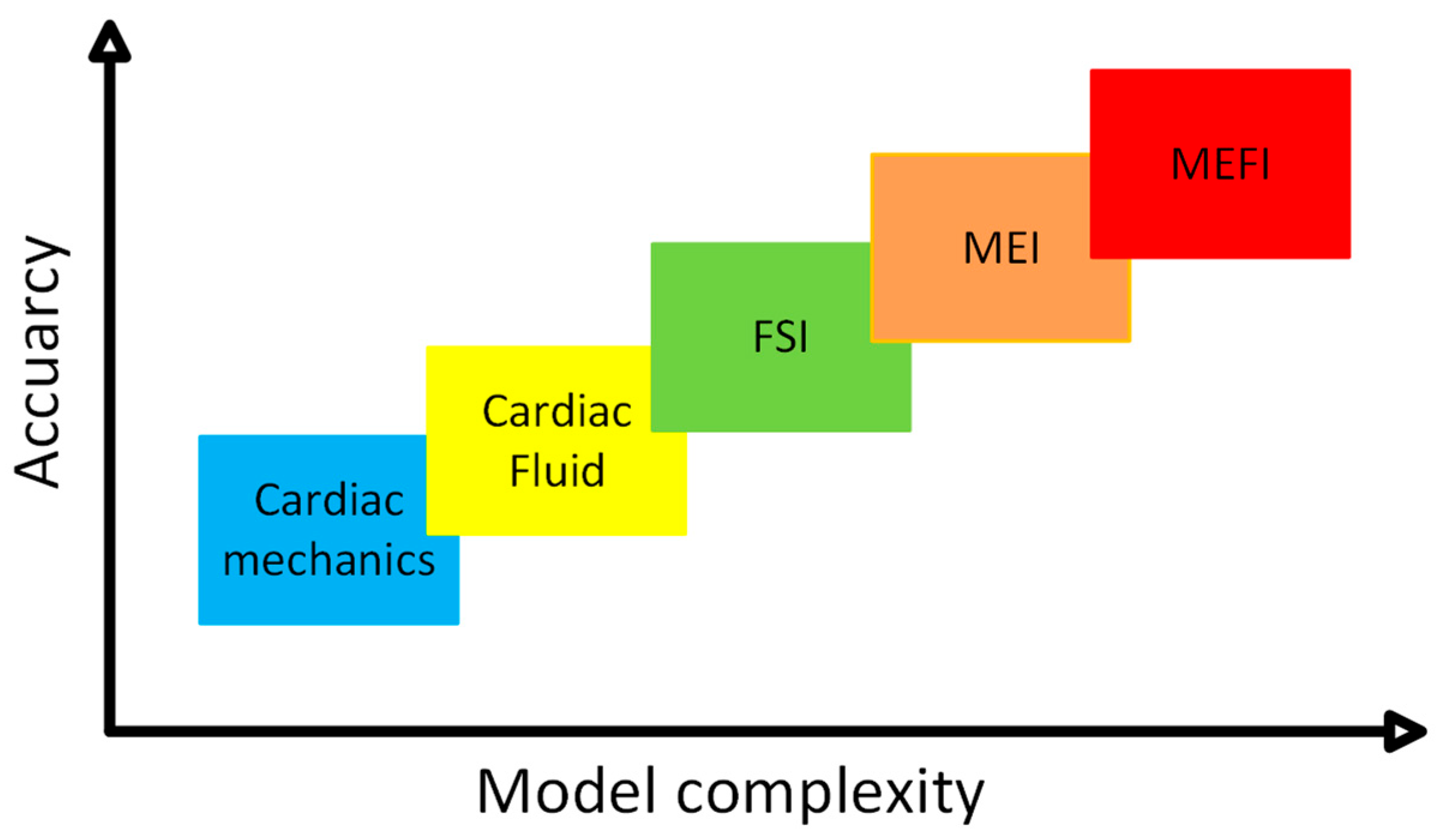
Figure 2 illustrates the historical progression of cardiac modeling approaches along a timeline, with respect to their increasing physiological fidelity and multiphysics integration. Early models began with electrical simulations of action potential propagation, followed by the inclusion of mechanical contraction via electromechanical coupling (ME). Subsequent advancements incorporated fluid–structure interaction (FSI), enabling simulation of intracardiac blood flow. The latest generation of models, represented by MEFI (mechano-electrical-fluidic interaction), achieves full multiphysics integration, capturing complex feedback among electrical excitation, myocardial deformation, and hemodynamics. As these models have evolved, their accuracy and predictive power have significantly improved, allowing for more realistic representations of cardiac function and better support for clinical decision making.
Section 2 begins by outlining the evolution of cardiac modeling techniques with increasing physiological realism and computational complexity. It introduces a historical perspective on the development of computational models used to study mechano-electrical-fluid interactions (MEFI), beginning with simplified electrical and mechanical representations and advancing toward fully coupled multiphysics models. The section is organized into three main thematic areas.
Section 2.1 focuses on fluid–structure interaction (FSI) models, detailing the simulation of cardiac mechanics and intracardiac blood flow without explicitly including electrical activation. It highlights early immersed boundary method approaches and then elaborates on more recent whole-heart models that incorporate hyperelastic tissue properties, realistic valve dynamics, and patient-specific geometries derived from imaging.
Section 2.2 shifts to mechano–electrical interaction (MEI) models that couple electrical activation with myocardial deformation. These models simulate action potential propagation and its mechanical consequences, accounting for anisotropic fiber conduction and electromechanical feedback, providing insights into cardiac contraction and dyssynchrony.
Section 2.3 presents fully integrated MEFI models that combine electrical, mechanical, and fluid domains within a unified framework. It discusses large-scale numerical methods, high-performance computing strategies, and recent advances in personalized modeling through the integration of imaging, inverse finite element analysis, and artificial intelligence. These efforts aim to improve simulation accuracy and enable clinical translation of multiphysics cardiac models.
Together, these subsections provide a comprehensive overview of recent progress in MEFI modeling and set the stage for the discussion of key gaps and future challenges in Section 3.

2.1. Cardiac Fluid, Structure (Tissue), or Fluid–Structure Interaction (FSI)

This subsection focuses on models that simulate the interaction between cardiac tissue mechanics and intracardiac blood flow, excluding electrical activity. These fluid–structure interaction (FSI) models are foundational to MEFI modeling, as they capture how mechanical deformation of the heart influences and responds to blood flow dynamics. FSI modeling represents an essential early step in developing integrated cardiovascular simulations. Blood flow dynamics within the heart have been extensively investigated over many years [9,10], revealing that blood flow is closely coupled to the heart’s contraction and relaxation cycles through complex kinematic and momentum exchanges between the ventricular chambers and the myocardial walls. Pioneering work by Peskin and McQueen [11,12,13,14] established foundational cardiac FSI models using the immersed boundary method. Their seminal model primarily addressed fluid–structure interactions, intentionally excluding cardiac electrophysiology due to the considerable complexity such coupling would introduce.
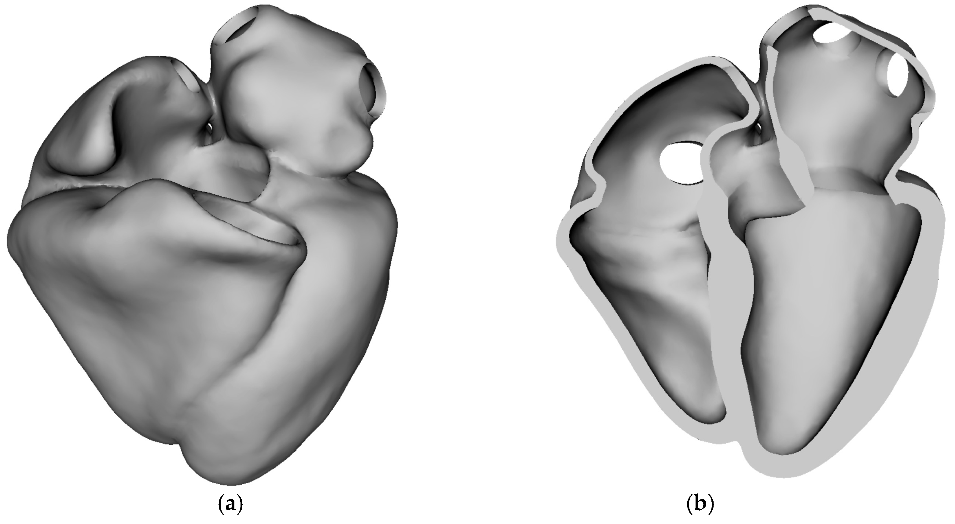
Computational modeling of whole-heart function serves as a powerful tool for investigating cardiac mechanics and hemodynamics. However, many existing fluid–structure interaction (FSI) models of the heart primarily focus on electromechanical aspects, often neglecting physiological valve dynamics and employing simplified fluid models. Feng et al. [15] addressed these limitations by developing a comprehensive four-chamber heart model that incorporates realistic chamber geometry, detailed valve modeling, hyperelastic myocardial material properties with fiber architecture, and full FSI analysis. Figure 3 illustrates the three-dimensional stress-free anatomical geometry of the human heart used as the baseline domain for simulation. The geometry consists of all four cardiac chambers—left and right atria, and left and right ventricles—and represents the heart in its unloaded or stress-free state, i.e., before the application of any internal pressure or muscle activation. Figure 3a shows the external view of the whole heart, providing a full spatial representation of the epicardial surface. Anatomical fidelity includes major chamber contours, apex, and valve orifice locations. Figure 3b displays a cut-away view, exposing the internal chamber volumes and wall thickness. This view highlights the septal wall separating the left and right ventricles, openings corresponding to valvular inlets/outlets, and smooth chamber transitions necessary for physiological simulation of blood flow and electrical propagation. Stress-free configuration is essential for initializing biomechanical simulations accurately. In real hearts, the geometry seen on imaging (e.g., MRI) already contains pre-stress due to blood pressure and muscular tone. This figure establishes the foundation for all biomechanical and electrophysiological simulations performed in the study. High-fidelity anatomical accuracy and proper reference conditions (stress-free) are critical for meaningful and realistic results in simulations such as cardiac contraction, activation mapping, and pressure–volume loop generation.
Their model was used to study cardiac behavior under various assumptions, including restricted versus free valve annular dynamics and the presence or absence of heart–pericardium interactions. Simulation results successfully captured key physiological phenomena, such as valve leaflet–flow interactions; characteristic left ventricular flow vortices; venous and transvalvular flow waveforms; and realistic heart deformations, including atrioventricular plane movement. The model demonstrated that allowing free annular dynamics improved ventricular filling and atrial emptying during early diastole. Furthermore, the study found that pericardial forces significantly influence atrial wall deformation, particularly during atrial contraction, thereby enhancing atrial filling. Overall, this work establishes a robust framework for comprehensive multiphysics whole-heart modeling that incorporates detailed valve dynamics and fluid–structure interactions.
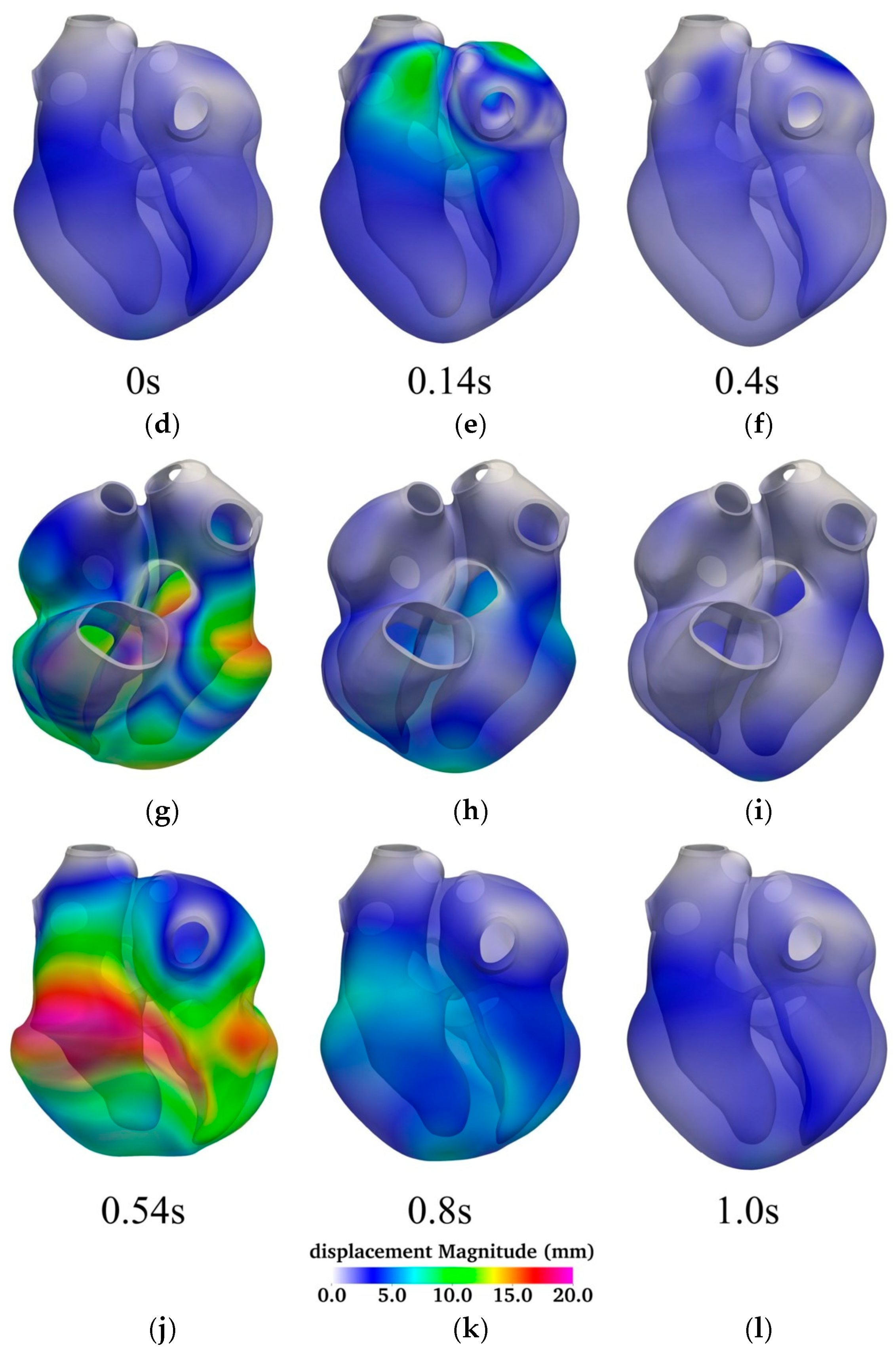
Jiang et al. [16] developed a highly scalable, fully implicit finite element algorithm capable of simulating the dynamic deformation of a four-chamber human heart using patient-specific geometry and a heterogeneous hyper-elastic model. Figure 4 illustrates the temporal evolution of myocardial displacement over a complete cardiac cycle, using a series of six time points—0.0 s, 0.14 s, 0.4 s, 0.54 s, 0.8 s, and 1.0 s—to capture key phases of atrial and ventricular activity. Each time point is represented by a pair of 3D surface plots displaying both anterior (top row) and posterior (bottom row) views of the heart, color-coded by displacement magnitude (in millimeters).
At the start of the cycle (0.0 s; panels Figure 4a,d), the heart is in its diastolic resting state, exhibiting minimal displacement throughout the myocardium. This corresponds to the phase immediately following passive filling, before atrial systole begins. At 0.14 s (panels Figure 4b,e), significant displacement is observed in the atria, indicating active atrial contraction. The ventricles at this stage remain largely stationary, reflecting their passive filling phase. This spatially localized deformation is consistent with the timing of atrial systole in the cardiac cycle.
By 0.4 s (panels Figure 4c,f), atrial contraction subsides, and the ventricles begin to activate. At this point, displacement begins to emerge in the ventricular walls, initiating the ejection phase of the cardiac cycle. The most substantial myocardial deformation occurs at 0.54 s (panels Figure 4g,j), corresponding to peak ventricular systole. Here, the displacement magnitudes are highest in the ventricular myocardium, especially along the left ventricular free wall and apex, reflecting robust muscle contraction and blood ejection through the outflow tracts.
At 0.8 s (panels Figure 4h,k), ventricular relaxation is underway. The myocardium begins to return toward its original configuration as the ventricles enter diastole. The overall displacement is reduced but still prominent in regions recovering from active contraction. Finally, by 1.0 s (panels Figure 4i,l), the heart has nearly completed its cycle and returned to a quiescent state with minimal displacement, poised to initiate the next cycle.
The figure highlights the asynchronous but coordinated nature of atrial and ventricular contractions and underscores the biomechanical dynamics of the heart under physiological conditions. The temporal displacement patterns closely align with the expected timing of mechanical activation, providing a visual and quantitative representation of normal electromechanical coupling.
The model captures the full cardiac anatomy, using a Guccione-based hyper-elastic law for myocardial tissue and a Mooney–Rivlin model for vessel roots, incorporating anisotropic cardiac fiber architecture. A fully implicit method in time and finite element method in space is used, combined with a Newton–Krylov–Schwarz solver for tackling the nonlinear equations efficiently across large-scale processor cores. To deal with computational stiffness during highly nonlinear cardiac phases, the authors implemented a two-level adaptive time-stepping approach to improve convergence and reduce computational time. The method scales efficiently on high-performance computing systems, handling unstructured meshes with over 200 million degrees of freedom and up to 16,384 processor cores.
At any given moment, the displacement d and velocity v of a material point are governed by the following partial differential equations [16]:
ρ v t · p = 0 d t = v   i n   Ω × 0 ,   T  
where (0, T) is a time interval of interest, p is the stress, ρ is density, and Ω is the reference domain in the stress-free state.
The algorithm is validated through simulations of a complete cardiac cycle on a patient-specific heart. It accurately reproduces physiological behaviors such as displacement and adapts time steps effectively during challenging phases, such as rapid ventricular contraction, as shown in Figure 4. Their study significantly advances computational cardiac mechanics by enabling large-scale, whole-heart simulations with high fidelity and computational efficiency. It lays the groundwork for future clinical and research applications involving cardiac dynamics and personalized heart modeling.
Personalized cardiac mechanics modeling offers valuable insights into the biomechanical function of the heart in both healthy and diseased states and plays a critical role in supporting treatment planning. However, existing models often rely on medical imaging captured at a single cardiac phase, limiting their effectiveness in analyzing dynamic cardiac behavior across multiple time points. Please note that although cine-MRI (cardiovascular magnetic resonance imaging) captures dynamic motion throughout the cardiac cycle, many computational models still depend on single-phase snapshots. This limitation arises from the difficulties associated with segmenting and aligning images across multiple time points. The challenges stem from the sparse and cross-sectional nature of the image slices, as well as potential misalignments caused by patient movement or inconsistencies in breath-hold positions during image acquisition, making accurate 3D reconstruction from these 2D slices particularly difficult [17].
A novel inverse finite element analysis (iFEA) framework was designed to estimate the passive mechanical properties of cardiac tissue using time-resolved medical imaging data [18]. Figure 5 illustrates a comprehensive pipeline for developing patient-specific computational models of cardiac biomechanics, with a focus on the biventricular and left atrial myocardium. The workflow begins with time-resolved, three-dimensional computed tomography (3D CT) scans that capture the cardiac anatomy at various phases of the cardiac cycle. These volumetric images serve as the foundational dataset for the model construction process.
In the image segmentation and morphing phase, the myocardial contours are delineated from CT images at key time points—typically end-diastole (EDV) and end-systole (ESV). This segmentation step can be performed manually using platforms such as SimVascular [19] and MeshMixer [20,21], or through automated pipelines leveraging deep neural networks (DNNs) [22], which significantly improves reproducibility and scalability across patients. These segmented geometries are then processed to produce a time-varying representation of the myocardial shape and structure.
Next, the segmented surfaces undergo model construction, wherein anatomical fidelity is enhanced by assigning rule-based myocardial fiber orientations and applying boundary conditions that mimic physiological constraints. These conditions include chamber pressures and mechanical supports derived from clinical data or established literature values. Additional geometric refinements and meshing are performed to ensure the model is suitable for finite element analysis.
The final stage involves an optimization process using inverse finite element analysis (iFEA). In this step, the biomechanical properties of the myocardial tissue—such as passive stiffness parameters—and the heart’s unloaded (stress-free) reference configuration are iteratively estimated. The optimization framework uses an inner-outer loop strategy: the inner loop applies an enhanced Sellier algorithm to recover the reference geometry, while the outer loop adjusts the material parameters to minimize the discrepancy between simulated deformations and those observed in the imaging-derived displacement fields over the cardiac cycle.
By incorporating both anatomical and functional data, this modeling framework enables a personalized characterization of myocardial mechanics. Such calibrated models are essential for understanding patient-specific cardiac function, assessing pathological remodeling, and informing therapeutic strategies.
Focusing on passive myocardial behavior, the framework incorporates structurally based anisotropic hyper-elastic material models and physiologically realistic boundary conditions. The governing nonlinear elastodynamics equations were solved using a stabilized variational multiscale method, which has been verified for cardiac mechanics applications.
In the proposed model, the equations of the motion are given as
d u d t v = 0 β θ p d v d t + x · v = 0 ρ p d v d t + x p x · σ d e v = 0  
where u is the displacement, v is the velocity, p is the pressure, βθ is the isothermal compressibility coefficient, ρ is the density of the material, σdev represents the deviatoric part of the Cauchy stress tensor, and x is the gradient operator defined in the spatial coordinates.
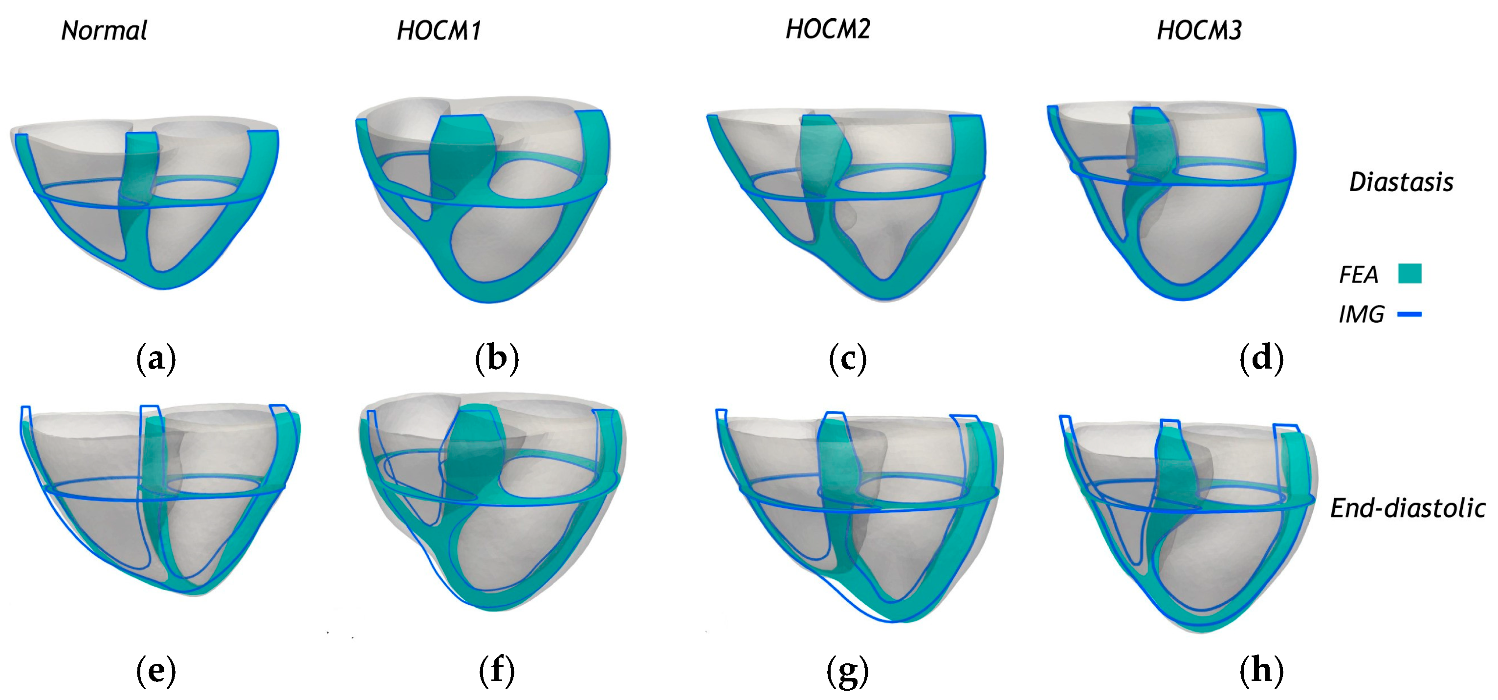
The framework was validated using biventricular and left atrial models reconstructed from cardiac phase-resolved CT images of one healthy subject and three patients with hypertrophic obstructive cardiomyopathy (HOCM). Figure 6 presents sectional comparisons between the predicted deformations from finite element analysis (FEA) and the corresponding anatomical contours derived from medical imaging (IMG) for the biventricular and left atrial (LA) myocardium. Both longitudinal and transverse cross-sectional views are shown at two distinct cardiac phases: diastasis (Figure 6a–d) and end-diastole (Figure 6e–h). The FEA-predicted geometries (shaded in cyan) exhibited a close spatial alignment with the image-based contours (outlined in blue), demonstrating the model’s ability to accurately replicate physiological deformation patterns. This consistency across normal and HOCM cases—labeled HOCM1 (Figure 6b,f), HOCM2 (Figure 6c,g), and HOCM3 (Figure 6d,h)—highlights the robustness of the personalized modeling framework and supports its potential for capturing structural and functional variations in diseased hearts. A comprehensive sensitivity analysis examined the influence of optimization strategies and numerical settings, including fiber orientation, mesh resolution, initial parameter estimates, and variations in material properties. The performance of the proposed iFEA method was also compared to the traditional power-law-based pressure–volume relationship, commonly used in single-phase imaging analysis.
This work demonstrates the potential of dynamic, image-based inverse modeling to enhance the accuracy and clinical relevance of personalized cardiac mechanics.
Schwarz and colleagues [23,24] presented a novel computational framework that integrates constrained mixture theory (CMT) into a fluid–structure interaction (FSI) finite element solver, forming a fluid–solid–growth (FSG) model. This model enables long-term, patient-specific predictions of vascular growth and remodeling (G&R) by simulating changes in blood flow (hemodynamics), vessel wall deformation, tissue composition, and material properties over time. The model extended traditional FSI models to include long-term biological growth and remodeling using CMT and integrated fluid dynamics, solid mechanics, and tissue growth in a partitioned iterative algorithm. In addition, the model enabled simulations of disease progression and vascular graft remodeling in both idealized and patient-specific geometries. This study provides a powerful computational tool for studying cardiovascular mechanics and biology over time, which is critical for understanding diseases, designing vascular implants, and personalizing treatment strategies. The integration of CMT with high-fidelity FSI in complex geometries marks a major step forward in computational biomechanics.
Schuster and colleagues [25] present a computational study that examines how different pulsatile patterns in left ventricular assist devices (LVADs) affect blood washout in the left ventricle. The study introduces a computational framework combining full-order simulations with reduced-order models (ROMs) to efficiently analyze the impact of pulsatile LVAD flow on ventricular washout. Using a simplified 2D model of the left ventricle and a sinusoidal LVAD flow rate, the researchers performed sensitivity analyses to assess washout efficiency. Two pulsatile scenarios were considered—one with a single pulse and another with two consecutive pulses. The results show that short, high-amplitude pulses improve washout most effectively, while the time between pulses had minimal impact. ROMs allowed a significant reduction in computational cost while maintaining high accuracy, enabling large-scale parameter analyses. The findings support incorporating pulsatility in LVADs to reduce stagnation and associated thrombosis risks. Please note that while 2D models offer limited mechanistic insights, developing a personalized computational model for cardiovascular applications requires a detailed, patient-specific 3D anatomical representation of the heart. Such models are essential for electromechanical simulations and for exploring the underlying mechanisms of cardiac diseases, supporting efforts in diagnosis, risk assessment, prognosis, and treatment planning [17].

2.2. Mechano–Electrical Interaction (MEI)

This section explores models that couple electrical excitation and mechanical contraction in cardiac tissue, typically without incorporating fluid dynamics. Mechano–electrical interaction (MEI) modeling emphasizes the bidirectional feedback between electrophysiology and tissue deformation, providing insights into arrhythmogenesis and electromechanical coupling mechanisms. These models serve as a critical bridge between isolated electrophysiology and more comprehensive MEFI frameworks [26]. These models were typically one-way coupled, where electrical wave propagation influenced mechanical contraction, but mechanical deformation did not, in turn, affect the electrical activity [27,28]. However, important advances were made by Nash and Panfilov [29], who developed a two-way coupled MEI model demonstrating that tissue deformation can exert feedback effects on electrical excitation and wave propagation. It is important to note that their model was not a whole heart simulation, but rather a continuum-based two-dimensional (2D) tissue model. In their approach, mechanical deformation was incorporated into the electrical conduction equations, although its effects on capacitive and ionic currents were not considered. More recently, Panfilov and colleagues [30] extended this model by introducing stretch-activated channels (SACs) into the 2D framework. Upon mechanical stretch, SACs allow an inward current that tends to depolarize cardiac cells. Their results highlighted that mechanical deformation can significantly influence electrical wave rotation and even induce wave breakups, emphasizing the critical role of mechano-electrical feedback in MEFI systems. However, their use of a simplified phenomenological FitzHugh–Nagumo model to represent membrane ionic kinetics limited the biological realism of the simulations. Specifically, the model did not incorporate detailed biophysical mechanisms or account for the behavior of individual ion channels, making it unsuitable for investigating the effects of genetic mutations, pharmacological interventions, or β-adrenergic pathway stimulations that require detailed ionic modeling.
Salvador and colleagues [31] introduce a latent neural ordinary differential equations (LNODEs) framework as a surrogate model for simulating the pressure–volume and electromechanical simulations of the dynamics of the human heart, especially in heart failure patients. Figure 7 shows the whole computational pipeline. Top-left: The study utilized a dataset comprising 405 electromechanical simulations derived from a fully personalized four-chamber heart model of a patient with heart failure. These simulations were generated by systematically varying 43 key parameters related to cellular, tissue-level, and cardiovascular properties, as well as boundary conditions. Each simulation included five cardiac cycles under sinus rhythm, with pressure and volume waveforms from the final cycle used for analysis.
Bottom-left: A total of 400 simulations were used to train and validate the latent neural ordinary differential equation (LNODE) model, which captures the time-dependent pressure–volume relationships in the atria and ventricles during the last cardiac cycle. The inputs to the LNODE model include time and model parameters, and hyperparameter tuning is performed using 10-fold cross-validation. Model performance is then assessed on a test set comprising the remaining five simulations.
Bottom-right: The validated LNODE model was employed to conduct a global sensitivity analysis (GSA), identifying how variations in model inputs influence the output quantities of interest.
Top-right: Using the trained LNODE as a surrogate, uncertainty-aware parameter estimation was carried out on the five test simulations, enabling robust calibration of model parameters under uncertainty.
The motivation was to overcome the computational limitations of high-fidelity cardiac models, enabling fast and accurate simulations on standard hardware—paving the way for broader clinical application of digital twin technology in cardiology. The paper presents a fast, accurate, and interpretable surrogate modeling approach for whole-heart simulations using LNODEs. It significantly reduces computational burden while maintaining high fidelity, enabling extensive applications like sensitivity analysis and parameter estimation. This work lays the groundwork for future patient-specific and real-time clinical decision support tools in cardiovascular medicine.
In this framework, the four-chamber heart artificial neural networks (ANN) model was developed as
d z t d t = A N N z t ,   c o s 2 π t A V d e l a y T H B , s i n 2 π t A V d e l a y T H B , θ ,   w   f o r   t 0 ,   T H B z 0 = z 0
where z0 is the initial condition, z(t) is the state vector, THB is the heartbeat period, AVdelay is the atrioventricular delay, θ is the state model parameter, and w is the weight of the ANN model.
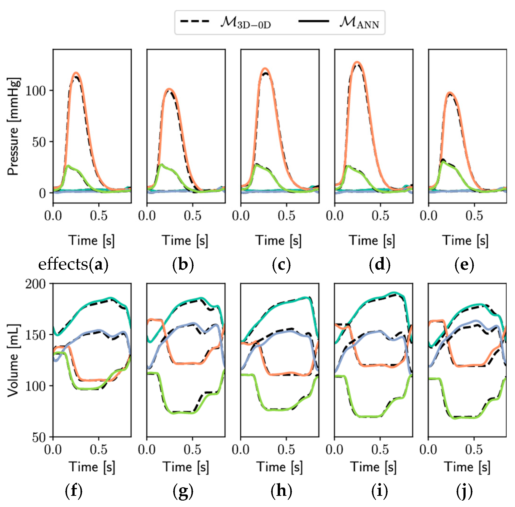
Figure 8 presents a comparative evaluation of pressure (panels Figure 8a–e) and volume (panels Figure 8f–j) waveforms generated by two modeling frameworks: the full-order 3D-0D electromechanical model M3D-0D (dashed lines) and its surrogate counterpart based on an artificial neural network, MANN (solid lines). Simulations were conducted over a complete cardiac cycle lasting 0.854 s and included five independent test cases (Ntest = 5), illustrating the generalizability and robustness of the ANN surrogate model.
The top row (Figure 8a–e) depicts pressure dynamics for the left atrium (LA, light blue), left ventricle (LV, orange), right atrium (RA, blue), and right ventricle (RV, green). Across all test cases, the surrogate model closely replicated the pressure evolution of the full-order model, capturing key physiological features such as atrial filling pressures, rapid ventricular pressure rise during systole, and the sharp decline during diastole. Notably, both the timing and amplitude of peak ventricular pressures aligned well between models, indicating strong temporal fidelity.
The bottom row (Figure 8f–j) displays volume traces for the same cardiac chambers. The surrogate ANN model demonstrated high accuracy in reproducing the cyclical volume changes associated with ventricular filling and ejection, as well as atrial reservoir and booster phases. Volume trajectories followed the same phase transitions and amplitude modulations seen in the reference model, including end-diastolic and end-systolic plateaus.
Overall, the consistency observed between M3D-0D and MANN across multiple test scenarios underscores the surrogate model’s predictive accuracy and computational efficiency. These results validate the surrogate’s suitability for rapid patient-specific simulations, uncertainty quantification, and clinical decision support in computational cardiology applications.
Fedele and colleagues [32] introduced a biophysically detailed computational model of the entire human heart, capturing the electromechanical behavior of all four chambers—including both atrial and ventricular contraction—for the first time with this level of accuracy. The model integrates anatomical realism, advanced myocardial fiber architecture, distinct ionic models, and a closed-loop circulatory system, all coupled through strong multiscale and multiphysics interactions, including mechano-electric and fiber–stretch–rate feedback. Implemented efficiently via high-performance computing tools, it reproduces physiological pressure–volume dynamics and cardiac deformation with high fidelity. This work sets a new standard in whole-heart modeling and paves the way for clinically relevant, patient-specific digital heart twins.

2.3. Mechano-Electrical-Fluid Interaction (MEFI)

This subsection reviews fully integrated multiphysics models that simultaneously capture electrical conduction, mechanical contraction, and hemodynamic flow. MEFI models represent the most advanced and physiologically comprehensive class of simulations. They aim to replicate the full cardiac cycle and are foundational for digital twin applications and personalized cardiovascular treatment planning. Several excellent comprehensive reviews have effectively documented the progress made in this area of research [6,8,24,33,34,35,36,37,38,39,40,41].
Recent advances in cardiovascular modeling have increasingly emphasized the integration of fluid dynamics, electromechanical coupling, and digital twin technologies to simulate the complex interactions within the cardiovascular system. Hemodynamic studies highlight the multiscale behavior of blood flow, ranging from high Reynolds number pulsatile flows in the aorta to near-laminar microcirculatory flows, and how these flow patterns influence vascular health, particularly in relation to atherosclerosis and thrombosis [8]. Computational models of the heart have evolved to include electro–fluid–mechanical interactions (EFMIs), capturing the dynamic coupling between electrical conduction, myocardial deformation, and fluid transport throughout the cardiac cycle [6]. This integrated approach provides a more physiologically accurate representation of cardiac function and supports the development of patient-specific simulations. The concept of cardiovascular digital twins—virtual replicas of individual patients’ cardiovascular systems—builds on these mechanistic frameworks and introduces machine learning techniques to support personalized diagnostics, prognostics, and treatment planning [33]. However, widespread implementation remains limited by challenges such as the need for high-quality, multi-modal patient data; difficulties in model calibration and validation; and the lack of large-scale repositories representing diverse populations [38]. Despite these obstacles, the convergence of data-driven and physics-based modeling holds significant promise for predictive, simulation-based approaches to cardiovascular care.
Watanabe et al. [42] developed a three-dimensional model that coupled electrical excitation, mechanical contraction, and fluid–structure interaction (FSI). However, in their framework, the coupling between excitation and contraction was one-way, with mechanical deformation not feeding back into the electrical activity. Furthermore, the model utilized phenomenological FitzHugh–Nagumo-type equations to describe cellular excitation and electrical wave propagation, limiting its ability to capture detailed ionic dynamics. More recently, Vigmond et al. [43] introduced a cable-based modeling approach to study MEFI at the whole-heart level. Unlike continuum models, the cable-based method discretizes the heart’s geometry into small finite volumes, averaging biophysical properties within each volume to better represent the discrete nature of cardiac tissue. This model incorporated realistic heart anatomy and coupled mechanical, electrical, and fluidic interactions. Importantly, it also employed a biophysically detailed ionic model—the Luo–Rudy model—to simulate electrical wave propagation with a greater physiological accuracy. Nevertheless, the mechanical-to-electrical feedback was not included, and the coupling remained one-way. Although good agreement was achieved between model predictions and experimental data, the authors acknowledged that the absence of mechanical feedback limited the model’s ability to fully replicate observed cardiac behaviors. Despite these limitations, the work represented a significant advancement by unifying multiple disciplines and highlighting the value of interdisciplinary approaches in tackling complex cardiovascular simulations.
Gonzalo and colleagues [44] investigated the hemodynamic consequences of atrial fibrosis using a high-fidelity, patient-specific, multiphysics computational framework that incorporates electrical activity, myocardial mechanics, and blood flow (electro–mechano–fluidic modeling). Figure 9 illustrates a comprehensive multiphysics, multiscale framework designed to simulate patient-specific cardiac function by integrating electrophysiology, biomechanics, and fluid mechanics. The framework spans multiple spatial scales, from the cellular to the organ level. At the cellular scale, ion channel dynamics regulate membrane potential, which influences intracellular calcium concentration. This calcium signaling triggers myofilament dynamics, leading to the generation of active tension within cardiac muscle fibers. These local stimuli are propagated to the organ/tissue scale, where they drive electrical wavefront propagation throughout the myocardium. The resulting electrical activation initiates mechanical contraction, modeled by the equilibrium of stresses that dictate myocardial deformation. This wall motion is then coupled with fluid mechanics, where the dynamic displacement of the heart walls drives intracardiac blood flow. The figure emphasizes the bidirectional and tightly coupled nature of these processes, allowing for realistic simulation of cardiac function that accounts for patient-specific geometry and physiological behavior across multiple scales.
Patient-specific data was used, and anatomies and fibrosis maps were derived from late gadolinium enhancement MRI (LGE-MRI) for four patients with varying levels of atrial fibrosis. Modeling fibrosis was carried out in fibrotic regions that were assigned altered mechanical properties—specifically increased stiffness and decreased contractility. Four different biomechanical states were simulated for each anatomy: non-fibrotic, stiff-fibrotic, hypocontractile-fibrotic, and fibrotic (combined). The study focused on flow patterns, kinetic energy, and emptying fractions in the left atrium (LA) and left atrial appendage (LAA). The major findings are that fibrosis significantly impairs LA contraction, reducing atrial and appendage emptying fractions. Stiffness and hypocontractility each negatively affect LA function, with amplified impact when combined, and flow changes caused by fibrosis extend throughout the LA, not just near fibrotic regions. In addition, LAA hemodynamics are affected in a more complex, multifactorial manner, influenced by both local fibrosis and global atrial mechanics. Finally, fibrotic effects on LA flow are largely captured by global emptying fraction, but in the LAA, more detailed modeling is needed to understand flow alterations.
The passive myocardium of the left atrium was represented using a transversely isotropic material model characterized by a specific strain energy function:
Ψ C , f = k 2 l n J 2 + a 2 b e x p b I 1 3 1 + a f 2 b f e x p b f δ f I 1 + 1 3 δ f I 4 1 2 1
where Ψ C , f is the volumetric contribution to ensure nearly incompressible material behavior; k = 650 kPa is the bulk modulus; J is the determinant of the deformation gradient, constant a = 2.92 kPa, b = 5.6; I1 is the first invariant, af = 11.84 kPa, bf = 17.95, δf = 0.09; and I4 is an invariant. The blood flow generated by the simulated motion of individualized anatomical structures and fibrosis distributions was captured using a custom-developed computational code.
Figure 10 presents a series of visualizations highlighting the effects of fibrosis on cardiac electrophysiology (Panel A), biomechanics (Panel B), and hemodynamics (Panel C). Panel A demonstrates how activation times differ in a simplified tissue domain between non-fibrotic and fibrotic states, with slower conduction evident in the fibrotic region due to altered conductivity. Panel B illustrates the mechanical consequences of fibrotic remodeling (contraction as measured by physical deformation and strain development), showing variations in myocardial deformation (captured via first principal strain) under four conditions: healthy, hypocontractile-fibrotic, stiff-fibrotic, and combined fibrotic. These simulations reveal the distinct strain patterns associated with each pathological state. Panel C depicts a schematic of a computational fluid dynamics (CFD) model used to simulate atrial blood flow. Boundary conditions are applied at the pulmonary veins (inflow), endocardium (no-slip wall), and mitral valve (dynamic open/closed), with velocity vectors shown in black representing flow during left atrial systole.
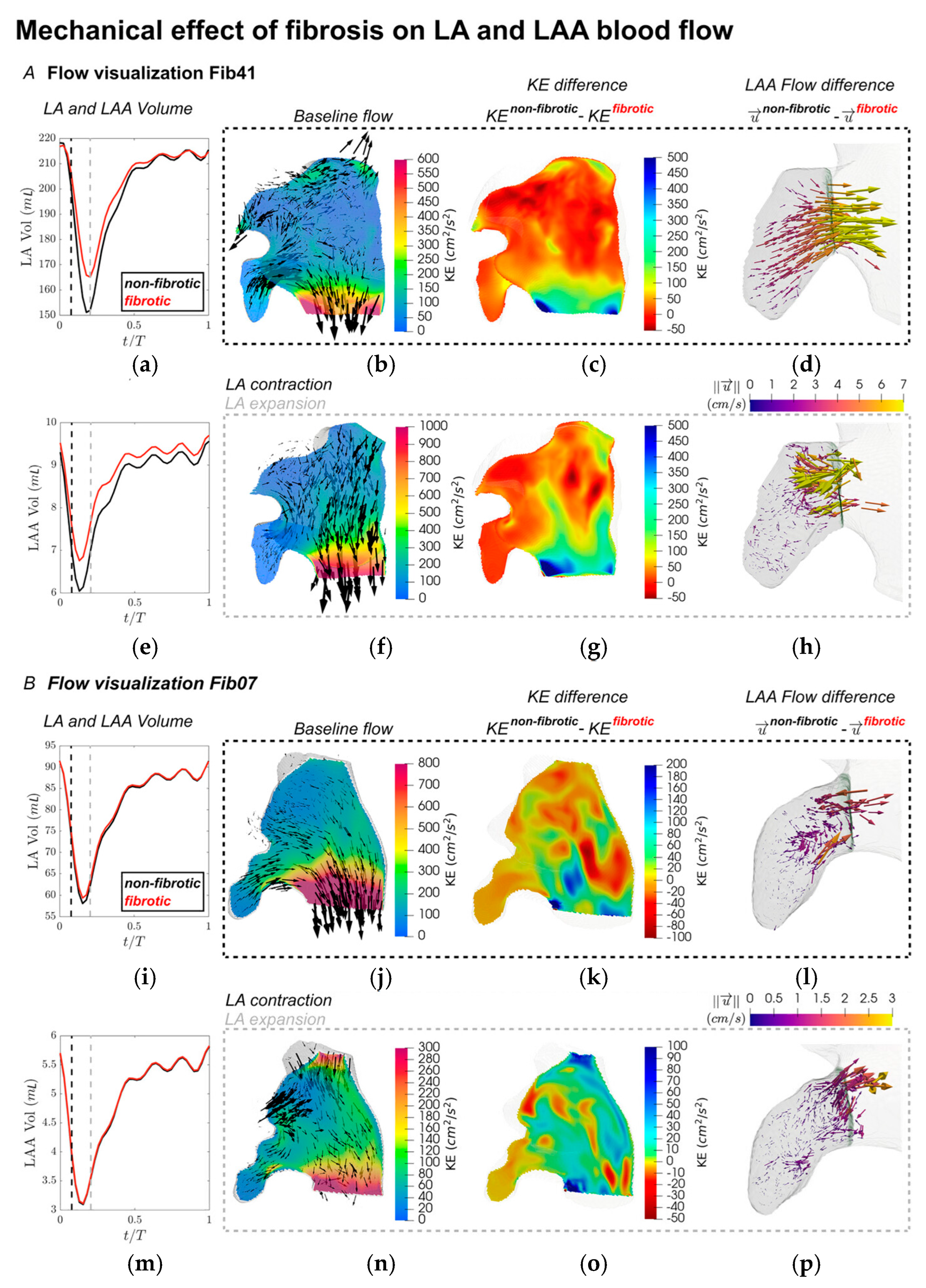
Alterations in myocardial stiffness and reduced contractility due to fibrosis led to noticeable changes in the motion of the LA endocardial surface, which in turn influenced intra-atrial flow dynamics. To investigate these effects, flow fields were visualized, and quantitative comparisons of hemodynamic parameters were conducted using the volumetric Lagrangian mesh framework outlined in the analysis methodology. These comparisons provided a deeper understanding of the impact of fibrosis on atrial flow behavior. Figure 11 illustrates velocity distributions and flow characteristics captured at two critical phases of the cardiac cycle—specifically, the time points corresponding to the maximum contraction and expansion rates of the LAA, marked by vertical dashed lines (black and grey) in the LA and LAA volume-time plots shown in Figure 11A,B (a,e,i,m). These time points correspond roughly to the early contraction and expansion of the left atrium. In subject Fib41 (Figure 11A), the non-fibrotic baseline flow during peak LAA contraction featured a pronounced jet emerging from the LAA, which merged with a strong mitral valve outflow stream. At the same time, the onset of LA contraction led to a reverse flow entering the pulmonary veins (Figure 11d). When mechanical alterations due to fibrosis were introduced, these organized flow patterns were notably disrupted, especially in regions previously characterized by strong velocities. The kinetic energy (KE) within the atrial chamber was substantially reduced under the fibrotic condition, particularly where the baseline flow was most prominent (Figure 11c,g). The three-dimensional velocity difference vectors (Figure 11d,h) illustrate a diminished LAA ejection jet, with reductions of approximately 5 cm/s in the fibrotic state. At the instant of peak LAA expansion (Figure 11h), the non-fibrotic model showed weaker outflow through the mitral valve while the atrial cavity was refilled by pulmonary venous inflow, accompanied by fluid movement from the LAA opening to its distal region. Similar to earlier observations, the fibrotic model exhibited a drop in KE in these zones, along with slightly decreased flow throughout the atrium. Notably, inflow near the LAA ostium displayed a consistent pattern but was reduced in speed by about 5 cm/s, indicating slower filling dynamics under fibrotic conditions. Fibrosis introduces increased stiffness and reduced compliance in atrial tissue. This mechanical remodeling limits the ability of the left atrium (LA) to expand during ventricular systole (when the atrium acts as a reservoir). As a result, the LA cannot effectively accommodate incoming pulmonary venous return, which in turn reduces the suction force driving blood into the LAA. This diminished reservoir function is reflected in the slower inflow velocities at the LAA ostium.
In contrast, for subject Fib07 (Figure 11B), the general flow features under the non-fibrotic setting resembled those of Fib41. However, due to a less extensive fibrotic burden, the alterations in KE following fibrosis were less severe. Nonetheless, measurable differences in KE were observed between fibrotic and non-fibrotic simulations across the left atrium, despite most fibrotic tissue being concentrated near the pulmonary vein inlets. Within the LAA specifically, changes in flow were evident but occurred at lower magnitudes (~1 cm/s) and lacked the distinct directionality seen in the higher fibrosis case.
This study provides compelling evidence that atrial fibrosis disrupts cardiac mechanics and flow in ways that may promote blood stasis and thrombus formation, factors critical in stroke risk. By using patient-derived data and advanced simulation techniques, this research offers a path forward for more personalized risk assessment and potential refinement of clinical decision tools.
Bucelli and colleagues [45,46] present a computational framework for simulating cardiac function by integrating electrophysiology, fluid dynamics, and solid mechanics in a loosely coupled manner. The authors introduce a fully loosely coupled electro–fluid–structure interaction (EFSI) scheme, designed to simulate the mechanics and hemodynamics of the human heart. The scheme uses a modular approach that separately solves the subproblems of electrophysiology, active force generation, fluid dynamics, and solid mechanics at each timestep, enhancing computational efficiency. The core innovation lies in applying a Robin–Neumann interface condition to manage the fluid–structure interaction explicitly, reducing the high computational costs typically associated with fully coupled models. The scheme is stable under cardiac simulation conditions and significantly reduces computational time compared to monolithic implicit schemes. It can accurately capture physiological indicators such as ventricular volume, pressure evolution, and flow patterns, albeit with some trade-off in mass conservation during isovolumetric phases. Finally, the framework is validated against benchmark models and realistic left ventricular geometries, showing robust performance and scalability. Overall, the study provides a computationally efficient and stable alternative for large-scale heart simulations, enabling future applications in personalized medicine and cardiac device testing.
Viola and colleagues [47] present a highly detailed, GPU-accelerated, multiphysics digital twin of the human heart. This computational model integrates electrical conduction, muscle contraction, valve dynamics, and blood flow, enabling simulations with over a billion degrees of freedom per heartbeat. It can complete a full cardiac cycle within 12 h on modern GPU clusters. The model accurately replicates physiological heart behavior, including ECG signals, pressure and volume waveforms, and hemodynamic flow structures. Pathological case simulation of a left bundle branch block (LBBB) was simulated by modifying the electrical conduction pathway, leading to reduced cardiac output and altered ECG patterns. Therapeutic simulation of cardiac resynchronization therapy (CRT) was modeled by varying pacemaker lead positions. The model predicted physiological recovery metrics such as ejection fraction and pressure, depending on the lead placement accuracy.
The human heart digital twin is constructed using a multiphysics computational framework designed to simulate cardiovascular flow dynamics with high fidelity. This approach integrates models that account for myocardial electrophysiology, active muscle contraction, passive relaxation, valve mechanics, and blood flow within the cardiac chambers and arterial system. These components are tightly coupled to reflect the complex, interactive behavior of the heart and to capture the resulting mechano-electrical-fluid interaction (MEFI). The blood solver is captured by the incompressible Navier–Stokes and continuity equations:
ρ u t + · u u = p + · τ + f I B + f W K ,   · u = 0
where blood density ρ = 1060 Kg/m3, u is velocity, p is the pressure, τ is the stress tensor, fIB and fWK are the force terms.
The motion of the deformable cardiac tissues is modeled using a spring–network structure. The variability in electrophysiological properties across the cardiac network is represented using an advanced, whole-heart electrical modeling approach [48].
χ C m v t + I i o n s + I s = · M i n t v + · M i n t v e x t · M i n t v + M i n t + M e x t v e x t = 0 d s d t = F s , v , t
where v and vext are the transmembrane and extracellular potential; χ   and C m are the surface-to-volume ratio of cells and the membrane capacitance; Is is the external triggering stimulus initiating the myocardial depolarization placed in the sino-atrial node; and M i n t and M e x t are the intracellular and extracellular conductivity tensors, respectively.
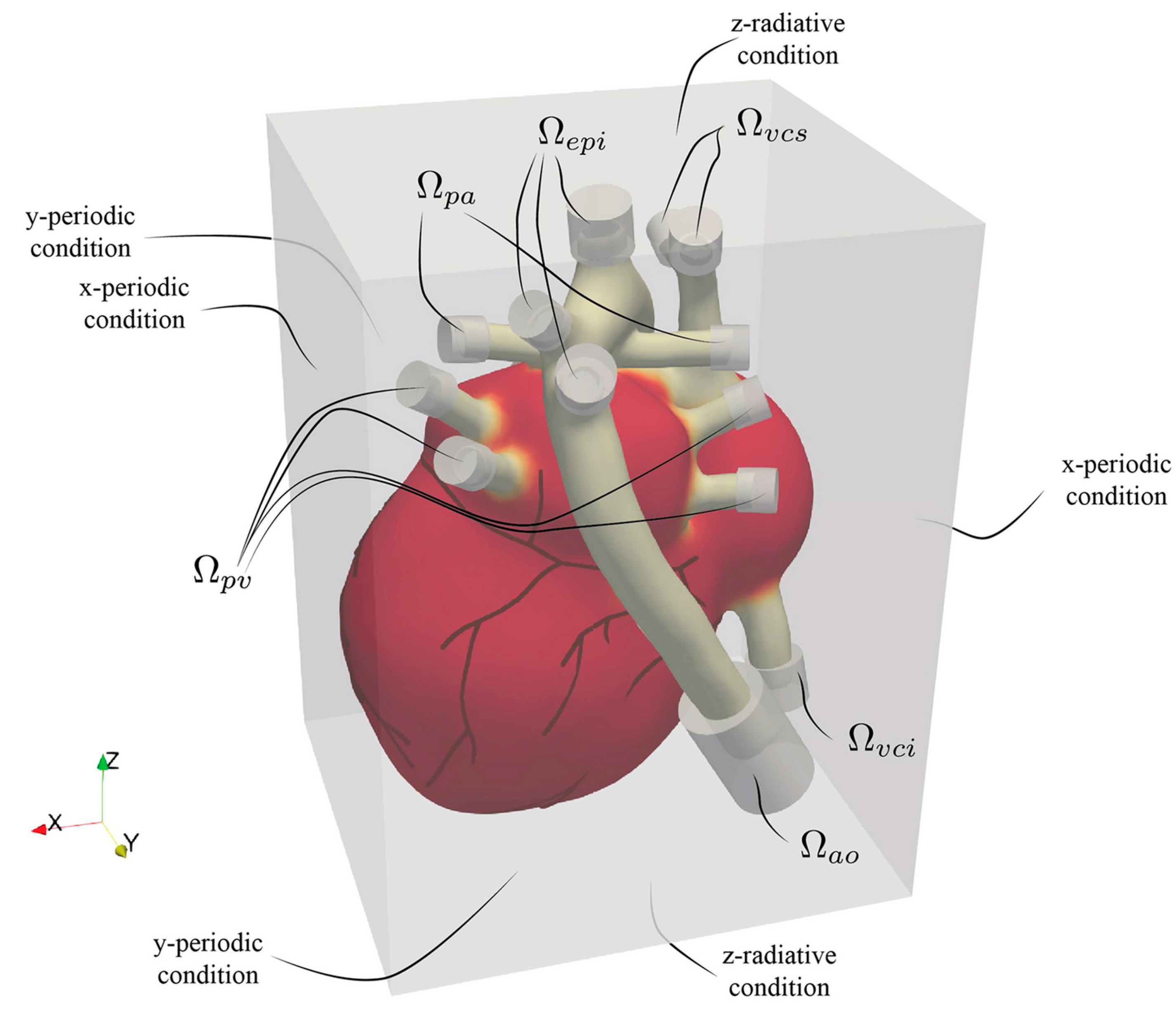
As with immersed boundary methods (IBMs), the heart is embedded within a larger fluid computational domain represented by a Eulerian grid, as illustrated in Figure 12. The arterial and venous ends—serving as inflow and outflow sites—do not intersect the external boundaries of the fluid domain. Instead, during the cardiac cycle, blood enters through the pulmonary veins and vena cava and exits via the aorta and pulmonary arteries, with flow exchange occurring entirely within the enclosed domain. Because the model focuses solely on the heart and proximal segments of the main vessels, it does not capture the full circulatory system. To account for the excluded vascular network’s resistance, compliance, and inertance, appropriate boundary conditions must be applied at the truncation points. These peripheral hemodynamic properties are often approximated using lumped parameter models (0D models), governed by low-order differential equations similar in structure to electrical circuit analogs. In this setup, where all heart inlets and outlets lie inside the computational domain, the corresponding boundary conditions are enforced via volumetric forcing. While this open-loop scheme prescribes each boundary condition independently, enhanced physiological fidelity could be achieved by transitioning to a closed-loop approach. In such models, outlets are dynamically linked to their corresponding inlets—for instance, connecting the descending aorta’s outflow to the inferior vena cava’s inflow—via a 0D system representing systemic or pulmonary circulation.
Figure 13 presents a comparative assessment of cardiac electrical activation, blood flow dynamics, and mechanical contraction across three physiological states: healthy, impaired, and resynchronized hearts. The top row shows the spatial distribution of electrical activation potential at 252 ms. In the healthy heart, the activation propagates uniformly across the myocardium, whereas in the impaired heart, the activation is delayed and non-uniform, reflecting dyssynchronous electrical conduction. After resynchronization therapy, the activation pattern becomes more organized and closely resembles the healthy state. The middle row illustrates blood velocity distribution in a cross-sectional plane through the left heart at peak systole (520 ms). The healthy heart exhibits coherent flow patterns indicative of efficient contraction, while the impaired case reveals disorganized and reduced velocities. Following therapy, flow coherence is largely restored. The bottom row depicts myocardial fiber stress at peak systole, where the healthy heart demonstrates high and evenly distributed stress, the impaired heart shows reduced and uneven stress distribution, and the resynchronized heart exhibits improved mechanical function with stress patterns approaching those of the healthy condition. Collectively, this figure highlights the pathological disruptions caused by dyssynchrony and the effectiveness of resynchronization therapy in restoring coordinated electromechanical and hemodynamic function.
Their study demonstrates the feasibility of using synthetic patient cohorts for testing interventions, significantly reducing the ethical and logistical constraints of traditional trials. This work offers a pathway to cost-effective, high-fidelity preclinical testing of cardiovascular treatments and devices, particularly for rare or complex diseases. Overall, the research marks a significant step toward practical in silico clinical trials and supports the future of personalized digital medicine.
Quarteroni and colleagues [49] present a comprehensive and multiphysics mathematical model of the human heart, developed to simulate cardiac function across multiple scales and support clinically relevant decision making. The model integrates detailed representations of electrophysiology, myocardial mechanics, blood flow, valve motion, coronary circulation, and myocardial perfusion, and it solves the governing equations using high-performance computational techniques. Derived from the first principles of physics and calibrated with patient-specific data from imaging and diagnostics, the model enables simulations of physiological and pathological heart behavior. It has been successfully applied in clinical contexts such as arrhythmia mapping, cardiac resynchronization therapy, aortic valve implantation, and perfusion assessment under stress. The work highlights the growing potential of computational cardiology as a non-invasive, predictive tool to support diagnosis and personalized treatment planning.
Computer simulations of cardiovascular flows hold significant promise for enhancing diagnostic accuracy, optimizing surgical procedures, and enabling virtual testing of novel prosthetic devices. However, their clinical reliability hinges on the fidelity of the underlying models, particularly in the case of the human heart, a highly complex organ governed by tightly coupled multiphysics processes. Viola et al. [50] present a comprehensive computational model that captures the full electromechanical and hemodynamic behavior of the heart through a fluid–structure–electro interaction (FSEI) framework. By integrating electrophysiology, tissue mechanics, and blood flow within a unified, computationally efficient scheme, the model achieves both accuracy and practical runtimes suitable for clinical use, even on standard computing resources. Demonstrated through simulations of both healthy and infarcted hearts, this tool offers predictive insights that could inform surgical planning and support personalized clinical decision making.
2.4. Summary of Multiphysics Whole-Heart Modeling Studies

Table 1a provides a comprehensive comparison of recent and foundational studies focused on multiphysics simulations of cardiac function. Each entry outlines the study’s modeling scope, incorporated physical domains (e.g., fluid–structure interaction, electrophysiology), the simulation framework used, the primary application context, and the unique scientific contributions of the work.
Table 1b provides a structured comparative summary of representative MEFI modeling approaches reviewed in this paper, incorporating additional dimensions to enhance clarity and practical relevance for readers. Each entry summarizes the model’s fidelity (e.g., full multiphysics vs. surrogate), coupling strategy (monolithic, partitioned, or hybrid), incorporation of artificial intelligence, clinical application focus, and typical simulation speed or computational requirement. To assist with method selection and evaluation, the table also outlines the primary advantages and limitations of each approach, such as accuracy, scalability, personalization potential, and modeling scope. This comparative format allows for rapid assessment of the trade-offs between complexity, interpretability, and clinical applicability across a wide range of MEFI frameworks, supporting both academic and translational perspectives on cardiovascular modeling.
To support a clearer evaluation of model accuracy, robustness, and clinical readiness, we have compiled available quantitative performance metrics across representative MEFI approaches and present them in a comparative format in Table 1c. These metrics include reported errors in pressure–volume (PV) loop predictions, myocardial strain deviation from MRI ground truth, F1 scores for arrhythmia classification, cardiac output accuracy, and kinetic energy deviation in fluid dynamics studies. Where applicable, these values are based on validation against patient-specific imaging or experimental datasets. This comparative presentation complements the qualitative table of modeling features and provides readers with a side-by-side view of how various MEFI frameworks perform across key physiological and clinical indicators. Such quantitative benchmarks offer valuable guidance for selecting appropriate models based on use case requirements—whether for real-time clinical decision support, in silico trials, or high-fidelity mechanistic investigations.

3. Discussion, Current Gaps, and Future Directions

This section is structured as follows: Section 3.1 outlines the key discussion points and current gaps in cardiac MEFI modeling, beginning with Section 3.1.1 on fundamental model limitations and critical challenges, followed by Section 3.1.2, which explores how different numerical approaches influence accuracy and scalability. Section 3.1.3 addresses multiscale challenges across cellular, tissue, and organ levels, while Section 3.1.4 examines common model simplifications and their physiological consequences. Section 3.1.5 discusses practical barriers to clinical implementation. Section 3.2 delves into the role of signaling pathways, with a focus on how β-adrenergic stimulation modulates electromechanical and fluid responses in excitable cardiac tissues. Section 3.3 explores the effects of ion channel mutations on myocardial contractility and blood flow patterns. Section 3.4 addresses current practices in verification and validation of MEFI models, while Section 3.5 presents quantitative metrics commonly used for model assessment, such as strain error and RMSE. Section 3.6 evaluates the growing role of artificial intelligence and machine learning in addressing modeling gaps. Section 3.7 reviews documented clinical applications of MEFI frameworks, and Section 3.8 proposes a roadmap outlining future milestones necessary for successful clinical adoption of MEFI models.

3.1. Discussion and Current Gaps

Over the past several decades, research efforts have been united by a central goal: leveraging insights from physics and engineering to transform clinical care. While notable progress has been achieved, several critical challenges remain, particularly in translating computational models into widespread clinical practice. Although mechano-electrical-fluid interaction (MEFI) models have underscored the complex dynamics crucial to cardiovascular health, the next generation of predictive tools must integrate emerging knowledge from fields such as mechanobiology, genetics, and cell signaling [8,24].
Developing a fully coupled mechano-electrical-fluid interaction (MEFI) model remains a formidable task due to the system’s inherently multiscale and multiphysical complexity. The correlations among molecular, cellular, and tissue-level mechanisms underlying MEFI are not yet fully understood. Future investigations must further elucidate the biophysical processes governing excitable tissues under both physiological and pathological conditions.
A comprehensive whole-heart computational model—incorporating anatomically realistic four-chamber geometry, physiologically accurate heart valves, detailed myocardial fiber architecture, hyperelastic material properties, and full fluid–structure interaction (FSI) analysis—has yet to be fully realized. Such a model must faithfully reproduce clinically observed cardiac functions through careful verification and validation, including characteristic left ventricular vortices, less vortical flow in the right ventricle, venous and transvalvular flow waveforms, and realistic mechanical deformations such as atrioventricular plane motion. Additionally, the model should account for the significant role of the pericardium, particularly its influence on atrial wall deformation and atrial filling dynamics. An integrated electro–mechano–fluidic framework that captures all cardiac valves and critical FSI phenomena remains a key unmet need.
Moreover, current computational models often rely on simplified phenomenological representations of electrophysiology, which preclude detailed investigation of how genetic mutations in ion channels influence tissue mechanics and hemodynamics. Given the abundance and critical roles of transmembrane ion channels in cardiac cells, future models must integrate both wild-type and mutant ion channel models to study how specific genetic defects affect cardiac function at the tissue and organ levels. Additionally, to date, no computational framework has comprehensively addressed the influence of system-level signaling pathways, such as β-adrenergic signaling, on cardiac mechanics and blood flow. Incorporating these pathways into MEFI models will be essential for advancing both mechanistic understanding and clinical applicability.
A cardiac digital twin is a computational model that replicates an individual patient’s heart to support cardiovascular disease screening, diagnosis, risk prediction, prognosis, and personalized treatment planning. Creating such models involves constructing detailed, patient-specific 3D anatomical representations suitable for simulating electromechanical behavior and exploring disease mechanisms. However, scaling the creation of cardiac digital twins remains a significant challenge, especially in the absence of publicly available model repositories representing diverse demographic populations.
Table 2 provides a structured comparison of the key advantages and disadvantages of using digital twin technology in cardiovascular modeling. Digital twins offer significant potential for personalized diagnosis, real-time clinical decision support, and predictive simulations of cardiac function. The integration of multiphysics domains—such as electrophysiology, myocardial mechanics, and hemodynamics—allows for a more comprehensive representation of physiological and pathological states. However, these benefits come with challenges, including high computational costs, the need for high-quality patient-specific data, and limited clinical validation. The table aims to clarify the practical trade-offs involved and support ongoing efforts to refine and deploy digital twin models in both research and clinical contexts.
Table 3 provides a comparative summary of high-fidelity cardiac models, such as the GPU-accelerated digital twin developed by Viola et al. [47], and reduced-order models (ROMs), including latent neural ordinary differential equations (LNODEs) proposed by Salvador et al. [31]. The table outlines key trade-offs across multiple criteria, including modeling accuracy, computational cost, scalability, and clinical applicability.
High-fidelity models offer detailed and physiologically accurate simulations by capturing full electromechanical–fluid interactions with high spatial and temporal resolution. However, these models are computationally intensive, requiring high-performance computing (HPC) resources and extended runtimes, making them less practical for routine clinical use.
Conversely, reduced-order models significantly lower computational demands and enable near real-time simulation on standard hardware. While they may abstract some mechanistic details, their efficiency and scalability make them ideal for large-scale screening, decision support systems, and integration into clinical workflows.
This contrast highlights a fundamental trade-off between model fidelity and practical applicability in healthcare settings. High-fidelity models are best suited for in-depth mechanistic research and surgical planning, whereas reduced-order models are more applicable for fast, scalable, and interpretable patient-specific assessments.
In addition, critical barriers exist in achieving reliable patient-specific validation and in accurately calibrating model parameters. Many current cardiac electrophysiology models are based on experimental data from in vitro studies using animal cells, which limits their applicability. Furthermore, fundamental anatomical and physiological differences between species present ongoing difficulties in developing biologically faithful and broadly validated human models [17].
Open-source tools and datasets are playing an increasingly vital role in addressing two major limitations in MEFI modeling: scalability and population diversity. For instance, the UK Biobank initiative [17] has released tens of thousands of publicly accessible, high-resolution cardiac MRI scans, enabling researchers to build large-scale digital cohorts and train machine learning models for automated segmentation, parameter estimation, and population-specific modeling. This unprecedented data availability not only enhances scalability through model pretraining and statistical parameterization but also improves generalizability by incorporating data from various age, sex, and ethnic groups, historically scarce in cardiac computational modeling.
Moreover, open-source simulation platforms and frameworks—such as OpenCMISS [51], SimVascular [19], and pulse-adaptive solvers—enable broader access to advanced modeling tools without commercial licensing barriers. When combined with large public datasets, these tools allow researchers worldwide to develop and validate MEFI models at scale, advancing both scientific reproducibility and translational impact. The open science movement thus represents a critical enabler for personalized cardiovascular modeling and ensuring its relevance across heterogeneous patient populations.
While imaging-based personalization has been central to recent advances in MEFI modeling, future developments will increasingly rely on the integration of multimodal clinical data to achieve more comprehensive and patient-specific simulations. Incorporating longitudinal electronic health records (EHRs) can provide valuable insights into a patient’s disease history, medication responses, and comorbid conditions, enriching the contextual relevance of MEFI predictions. Wearable sensors offer real-time physiological data—such as heart rate variability, blood pressure, and activity levels—which can dynamically inform boundary conditions or validate simulation outputs over time. Additionally, omics technologies (e.g., genomics, transcriptomics, proteomics) can help parameterize models based on molecular profiles, enabling the exploration of genotype–phenotype relationships and the impact of ion channel mutations or signaling pathway alterations. Together, these data streams present a powerful opportunity to enhance MEFI model fidelity, improve predictive performance, and support personalized decision making in cardiovascular care. Future research should focus on developing standardized frameworks for data integration and validation to harness the full potential of these multimodal sources in clinical applications.

3.1.1. Model Limitations and Critical Challenges

Despite significant advances in cardiac MEFI modeling, several critical limitations persist that constrain their clinical utility and scientific generalizability:
  • Oversimplification of Ionic Models: Many existing simulations rely on phenomenological or reduced-complexity ionic models (e.g., FitzHugh–Nagumo), which lack the resolution to capture the nuanced behavior of specific ion channel mutations or drug interactions. Such simplifications can lead to inaccurate predictions in arrhythmia modeling or pharmacological response simulations, especially when applied to genetically distinct patient cohorts.
  • Limited Patient Diversity in Digital Twins: Current digital twin frameworks are often built from datasets that lack demographic diversity, typically drawing from small, homogeneous populations. This lack of representation reduces model generalizability and raises concerns about clinical bias when translating these tools into real-world practice, particularly for underrepresented populations.
  • Potential bias in training datasets represents a significant challenge in the development and deployment of machine-learning-based surrogate models in cardiovascular modeling. Bias can arise from overrepresentation of specific anatomical structures, physiological conditions, or disease phenotypes, leading to reduced generalizability and skewed predictions when applied to underrepresented populations. This is particularly relevant in cardiac modeling, where variability in heart size, wall thickness, conduction velocity, and fibrotic burden can significantly influence simulation outcomes. To mitigate such biases, studies increasingly emphasize the use of diverse training cohorts, stratified sampling across key clinical metrics, and synthetic data augmentation to enrich rare or extreme cases. Cross-validation, external test sets, and domain-specific performance metrics (e.g., across different ejection fractions or arrhythmic profiles) are also recommended to assess and control for potential bias. Ultimately, the careful curation and documentation of training data characteristics are essential to ensure the clinical relevance, fairness, and robustness of surrogate models in personalized cardiac simulations.
  • Computational Burden and Scalability: High-fidelity MEFI models can require billions of degrees of freedom and hours of computation on GPU clusters, limiting their use in time-sensitive or resource-constrained clinical environments. While reduced-order models offer promise, they often sacrifice physiological detail for speed.
  • Validation Gaps: Many models have not undergone rigorous validation against comprehensive clinical datasets. For instance, validation is often limited to pressure-volume loops or ECG traces, without cross-referencing flow patterns, strain distributions, or responses to pharmacological interventions.
  • Incomplete Integration of System-Level Physiology: Signaling pathways such as β-adrenergic stimulation and neurohormonal control are still rarely integrated into whole-heart simulations, even though they critically affect cardiac performance under both normal and diseased conditions.
Addressing these limitations will require broader access to multi-modal patient datasets, continued investment in hybrid modeling techniques, and stronger interdisciplinary collaboration to align model development with clinical needs.

3.1.2. Impact of Numerical Approaches on Accuracy and Scalability

The choice of numerical methods in MEFI modeling—such as immersed boundary vs. finite element methods (FEM), and monolithic vs. partitioned coupling schemes—has significant implications for simulation accuracy, computational efficiency, scalability, and clinical suitability, as shown in Table 4. These trade-offs influence not only model fidelity but also whether such simulations can be applied in clinical settings.
  • Immersed Boundary (IB) vs. Finite Element Methods (FEM):
    Immersed Boundary Methods:
    Advantages: IB methods (e.g., Peskin and McQueen [11,12,13]) are particularly well-suited for modeling fluid–structure interaction in complex, moving geometries like the heart, as they allow flexible handling of deforming boundaries on fixed Eulerian grids.
    Limitations: While computationally efficient for capturing qualitative flow–structure interactions, IB methods often suffer from reduced spatial accuracy near interfaces and are less suited for capturing detailed stress and strain distributions in myocardial tissue.
    Scalability: IB methods scale well on parallel systems but are generally better for exploratory or qualitative studies than for patient-specific quantitative predictions.
    Finite Element Method (FEM):
    Advantages: FEM offers high spatial resolution and numerical precision, especially for solving solid mechanics and electrophysiology equations. Methods like those by Jiang et al. [16] and Shi et al. [18] provide accurate quantification of strain, fiber orientation, and electrical propagation.
    Limitations: FEM requires high-quality, often patient-specific meshes, which are computationally expensive to generate and may complicate simulation setup.
    Scalability: FEM can scale well, particularly when paired with high-performance solvers (e.g., Newton–Krylov–Schwarz), but the memory footprint and setup complexity may limit routine clinical use.
  • Monolithic vs. Partitioned Coupling Approaches:
    Monolithic Coupling:
    Advantages: Monolithic schemes solve fluid, mechanical, and electrical subdomains simultaneously within a single system of equations, ensuring strong coupling and better numerical stability, especially in stiff regimes like valve closure or isovolumetric phases.
    Accuracy: These methods reduce numerical artifacts (e.g., pressure oscillations) and allow precise modeling of bidirectional feedback among domains.
    Limitations: Monolithic solvers are computationally expensive, difficult to parallelize, and often require sophisticated preconditioners, limiting their scalability in clinical settings.
    Partitioned Coupling (e.g., Robin–Neumann interface in Bucelli et al. [45]):
    Advantages: Partitioned methods solve subproblems (e.g., electrophysiology, fluid, and solid) independently and couple them via boundary conditions at each timestep. This modularity allows reuse of existing solvers, easier debugging, and more efficient use of computational resources.
    Scalability: Partitioned schemes are often more scalable and suitable for parallel processing, especially in loosely coupled frameworks.
    Limitations: They may suffer from instabilities at fluid–structure interfaces and require stabilization techniques to ensure convergence, especially under fast dynamics or in stiff coupling regimes.
Future progress will likely depend on hybrid approaches—combining high-resolution FEM for tissue mechanics with reduced-order or ML-accelerated fluid solvers and modular partitioned coupling for efficiency. Additionally, physics-informed neural networks (PINNs) may offer an alternative numerical paradigm, embedding governing equations directly into AI-based solvers to balance accuracy and tractability.

3.1.3. Multiscale Challenges in MEFI Modeling

Cardiac MEFI models inherently span multiple spatial and temporal scales, from ion-channel kinetics at the nanometer–millisecond level to whole-organ deformation and blood flow across centimeters and seconds. This multiscale nature introduces several critical modeling and computational challenges, as shown in Table 5.
  • Spatial Scale Disparities
    Subcellular vs. Tissue-Level Dynamics:
    Subcellular processes like calcium-induced calcium release (CICR) and sarcomere force generation occur at nanometer to micrometer scales, requiring high spatial resolution to capture spatial gradients of Ca2+, Na+, and other ions.
    These finely resolved events must be translated into macroscopic myocardial stress and deformation at the tissue level (millimeters to centimeters), often via homogenization techniques or active tension models.
    Fiber Architecture and Anisotropy:
    Myocardial tissue exhibits highly anisotropic properties due to its layered fiber-sheet structure. Capturing this anisotropy accurately across scales is essential for modeling correct strain distributions, conduction velocities, and flow patterns.
  • Temporal Scale Discrepancies
    Fast Ionic Events vs. Slow Mechanical and Hemodynamic Processes:
    Electrical excitation and ionic channel gating occur in the order of milliseconds or less, whereas mechanical contraction unfolds over hundreds of milliseconds, and full cardiac cycles span ~1 s.
    This temporal disparity necessitates asynchronous time-stepping strategies, where different sub-models (e.g., electrophysiology vs. hemodynamics) operate on different time resolutions, creating risks of instability and time-step conflicts at coupling interfaces.
  • Calcium Handling Complexity
    Subcellular Calcium Dynamics:
    CICR is a nonlinear, threshold-sensitive process, involving local calcium sparks and waves that are difficult to capture with continuum models.
    Spatial inhomogeneities in calcium handling (e.g., due to disease or mutation) can cause arrhythmogenic triggers (e.g., delayed afterdepolarizations) that are hard to represent at the tissue level unless microscale variability is incorporated.
    Calcium–Mechanics Coupling:
    The conversion of calcium concentration to force via troponin binding and cross-bridge cycling introduces another scale-bridging challenge.
    Most models use phenomenological tension generation laws (e.g., Hill-type models), which may not capture mutation-specific or disease-specific alterations in contractility.
  • Data Resolution and Model Fidelity
    Subcellular Data Scarcity:
    Most patient-specific data (e.g., MRI, CT, ECG) is acquired at organ or tissue scales; subcellular inputs like ion channel densities or calcium transient variability are usually inferred or generalized from in vitro or animal data.
    This introduces uncertainty in bottom-up multiscale models, particularly for simulating the impact of mutations (e.g., in NaV1.5 or CaV1.2 channels) on organ-level function.
  • Computational Cost of Multiscale Coupling
    Nested Time Loops and Solver Synchronization:
    Multiscale simulations often require nested solver loops (e.g., inner calcium dynamics within outer electromechanical solvers), which drastically increase runtime.
    Without careful coupling strategies (e.g., operator splitting, adaptive time stepping), such models may suffer from cumulative numerical errors or spurious oscillations.
Addressing these challenges will require hybrid multiscale modeling approaches, including
  • Embedded subcellular models (e.g., ion channel networks) within tissue-level simulations.
  • Use of AI-driven surrogate models to replace costly subcellular solvers.
  • Development of co-simulation frameworks that allow asynchronous, modular coupling with stable interfaces.

3.1.4. Common Model Simplifications and Their Consequences

Despite advances in MEFI modeling, most frameworks rely on a number of simplifications to manage computational cost, data availability, and model complexity. While often necessary, these approximations can significantly affect the physiological fidelity, predictive power, and clinical relevance of the models, as shown in Table 6. Below are the most prevalent simplifications:
  • Simplified or Rule-Based Myocardial Fiber Architecture
    What is simplified: Instead of patient-specific fiber orientation (e.g., from DT-MRI), many models use rule-based fiber distributions that assign helical angles as a function of myocardial wall depth (typically ±60° endo- to epicardium).
    Consequence: This can introduce errors in mechanical strain, stress distributions, and conduction velocity, particularly in pathologies like hypertrophy or infarction, where fiber orientation is disrupted. It also affects electromechanical wave propagation and flow patterns, especially in the ventricles.
  • Simplified Electrophysiological Models
    What is simplified: Biophysically detailed ionic models (e.g., Luo–Rudy, ten Tusscher) are often replaced with phenomenological models (e.g., FitzHugh–Nagumo) to reduce computational cost.
    Consequence: While sufficient for general wave propagation, these models cannot capture mutation-specific effects, drug responses, or channelopathies, limiting their usefulness in precision medicine and pharmacological risk prediction.
  • One-Way Coupling of Physics Domains
    What is simplified: Many MEFI models assume unidirectional coupling (e.g., electrical → mechanical → fluid) without feedback loops.
    Consequence: This neglects important bidirectional phenomena such as mechano-electric feedback (MEF) and flow-induced deformation, which can influence arrhythmogenesis, valve dynamics, and tissue remodeling.
  • Homogeneous Material Properties
    What is simplified: Cardiac tissue is often modeled as spatially uniform and isotropic or transversely isotropic, ignoring region-specific differences (e.g., scar, infarct border zones, fibrosis).
    Consequence: Fails to capture heterogeneous contractility or conduction delays that are critical in diseases like heart failure or atrial fibrillation, leading to inaccurate predictions of strain and flow.
  • Idealized or Static Boundary Conditions
    What is simplified: Many models use prescribed inflow/outflow profiles, fixed pericardial constraints, or neglect respiratory–cardiac interactions.
    Consequence: These approximations reduce the ability to simulate physiologically realistic load-dependent behavior, limiting the model’s capacity to predict pressure–volume responses or adapt to device interventions like CRT or LVAD.
  • Simplified Valve and Blood Flow Modeling
    What is simplified: Valve motion is often modeled as rigid or pre-defined; blood is assumed Newtonian; and laminar flow assumptions are common.
    Consequence: Misses key features like flow turbulence, vortex formation, or valvular regurgitation, reducing fidelity in hemodynamic assessments, particularly in the left ventricle and atria.
While these simplifications are often pragmatic, ongoing efforts aim to systematically replace them using
  • Patient-specific data (e.g., fiber maps, material parameters).
  • AI-assisted surrogate modeling.
  • Multiscale frameworks with localized high-fidelity regions.
This balance between fidelity and feasibility remains central to making MEFI models both clinically useful and computationally tractable.

3.1.5. Practical Barriers to Clinical Implementation

While the predictive potential of MEFI models and cardiac digital twins is widely acknowledged, several practical hurdles significantly impede their integration into clinical workflows:
  • Data Acquisition and Standardization: High-fidelity MEFI modeling requires high-resolution, time-resolved, and patient-specific input data—including 3D anatomical reconstructions, myocardial fiber orientation, electrophysiological recordings, and hemodynamic measurements. However, these datasets are often incomplete, difficult to acquire routinely, or non-standardized across institutions. Imaging modalities like MRI or CT may not be universally accessible, and dynamic data such as strain maps or intracardiac flows are rarely captured in standard care. Furthermore, aligning heterogeneous data types into a unified model remains a non-trivial preprocessing and registration challenge.
  • Regulatory Approval and Clinical Validation: For MEFI-based digital twins to be used in decision making (e.g., predicting treatment outcomes or guiding surgical interventions), they must meet stringent regulatory requirements. These include demonstrating safety, reliability, and interpretability through validated benchmarks, cross-institutional trials, and longitudinal studies. As of now, few MEFI platforms have progressed beyond academic proof-of-concept to obtain clearance from regulatory bodies such as the FDA or EMA. The lack of standardized validation protocols also complicates reproducibility and trust in predictive outcomes.
  • Integration into Clinical Systems: Embedding computational models into electronic health records (EHRs) and clinical decision support systems (CDSS) presents technical and operational barriers. MEFI simulations must not only deliver real-time or near-real-time outputs but also be interpretable to clinicians without specialized training in computational modeling. Achieving this level of integration requires robust user interfaces, automated data pipelines, and visualization tools tailored to clinical contexts—all of which are still underdeveloped.
  • Clinical translation of MEFI models necessitates compliance with stringent regulatory frameworks, particularly when models are intended to support diagnosis, treatment planning, or risk prediction. In the United States, the Food and Drug Administration (FDA) classifies such software as Software as a Medical Device (SaMD), requiring developers to meet validation, transparency, and traceability standards outlined in the FDA’s Model-Informed Drug Development (MIDD) and Digital Health Software Precertification programs. Furthermore, the Verification and Validation of Computational Modeling of Medical Devices (V&V 40) standard provides guidance for assessing the credibility of computational models used in medical contexts. In Europe, CE marking under the Medical Device Regulation (MDR) similarly mandates rigorous evidence of model performance, generalizability, and risk assessment. A significant challenge in this domain is demonstrating clinical robustness across diverse patient populations while ensuring interpretability and auditability of high-dimensional, multiphysics simulations. Integration with electronic health record (EHR) systems also poses regulatory and interoperability hurdles, requiring compliance with standards such as HL7 FHIR and adherence to data privacy regulations like HIPAA and GDPR. Addressing these regulatory challenges is essential for the successful deployment of MEFI-based digital twins in clinical workflows.
  • Ethical and Legal Concerns: The use of patient-specific digital twins raises unresolved questions related to data privacy, model ownership, liability in decision making, and transparency in model behavior. Without clear frameworks, healthcare institutions may be hesitant to adopt predictive simulations, particularly for high-stakes interventions.
Overcoming these barriers will require coordinated efforts among clinicians, engineers, regulators, and data scientists to develop robust, explainable, and clinically viable MEFI tools that align with existing healthcare infrastructure.
The development of such integrative models will be crucial as more experimental data become available at the gene, protein, cell, tissue, and organ levels. These advancements will greatly enhance the predictive capabilities of system-level models and contribute to a deeper understanding of the biophysical mechanisms underlying cardiac MEFI. Furthermore, these insights may support the development of novel excitable biomaterials, such as pacemaker-controlled bio-pumps, designed through genetic modification of ion channel genes and further regulated by engineered signaling pathways. In parallel, enhanced patient-specific virtual heart models—based on individualized MRI data—could serve as invaluable tools to guide clinical cardiologists in tailoring surgical procedures to the specific needs of each patient [52].
The ultimate goal of computational cardiac models is not merely to replicate observed physiological behavior but to serve as reliable tools for prediction. For models to be truly impactful in clinical practice, they must extend beyond descriptive accuracy and demonstrate predictive capability—meaning the ability to forecast treatment outcomes, disease progression, or physiological responses under new or unseen conditions. This is particularly important in patient-specific applications, where the model should not only fit data collected from an individual (e.g., imaging, electrophysiology, hemodynamics) but also generalize to simulate potential interventions, guide clinical decision making, and support personalized treatment strategies. A model that cannot make accurate predictions outside of its calibration dataset is limited in its translational value. Therefore, validation against independent data, sensitivity analysis, and uncertainty quantification become essential steps in demonstrating model robustness. This predictive focus shifts the modeling paradigm from retrospective fitting to prospective utility—where digital twins, for example, can simulate the effect of surgical interventions, pharmacological therapies, or device implantations before they are performed. Ultimately, models that offer such predictive power are poised to transform cardiovascular care by reducing trial and error in treatment; improving risk stratification; and enabling proactive, data-informed medicine.

3.2. Signaling Pathways Such as Effects of β-Adrenergic Stimulation on Excitable Tissue Mechanics and Blood Flow

Future work should prioritize investigating system-level signaling pathways that regulate cardiac function. In particular, the influence of β-adrenergic signaling on excitable tissue contractility and blood flow warrants detailed exploration [1,53].
Figure 14 illustrates the β-adrenergic receptor (β-AR) signaling cascade and its role in modulating cardiac excitation–contraction coupling. Upon binding of an agonist (e.g., norepinephrine or epinephrine) to the β-adrenergic receptor located on the cardiomyocyte membrane, the receptor undergoes a conformational change that activates the stimulatory G protein (Gs). The activated Gs protein stimulates adenylyl cyclase (AC), an integral membrane enzyme, which catalyzes the conversion of adenosine triphosphate (ATP) into cyclic adenosine monophosphate (cAMP), a key second messenger in cardiac signal transduction.
Elevated levels of intracellular cAMP lead to the activation of protein kinase A (PKA), which then phosphorylates a number of downstream targets, including L-type Ca2+ channels. Phosphorylation of these channels increases their open probability, thereby enhancing Ca2+ influx into the cytosol. The influx of calcium not only contributes directly to the initiation of myocardial contraction but also triggers further calcium release from the sarcoplasmic reticulum via ryanodine receptors—a process known as calcium-induced calcium release (CICR).
In addition to the Ca2+ channels, the figure also highlights Na+ ion channels, which contribute to action potential initiation and propagation. Together, these ion fluxes regulate membrane excitability and contractile force. The net effect of β-adrenergic stimulation is an increase in heart rate, contractility, and relaxation rate, all of which are essential for the physiological response to stress or exercise.
This pathway represents a critical component of sympathetic nervous system regulation of the heart and is a therapeutic target in heart failure, arrhythmia, and other cardiovascular diseases. Understanding these molecular mechanisms is essential for developing predictive models of cardiac function and drug response. In forthcoming studies, it will be essential to incorporate β-adrenergic signaling mechanisms into human cardiac cell models to better understand their role in modulating cardiac tissue contractility and hemodynamics, especially in the presence of Long QT (LQT) syndrome-associated mutations in Na+ and L-type Ca2+ channels. Investigations should also examine how pharmacological agents, such as β-adrenergic agonists (e.g., isoproterenol) and antagonists (e.g., β-blockers like propranolol), influence cardiac performance under these conditions. LQT syndrome, characterized by a prolongation of the QT interval on an electrocardiogram, is a severe cardiac disorder associated with an elevated risk of arrhythmias and sudden cardiac death.

3.3. Effects of Ion Channel Mutations on Excitable Tissue Mechanics and Blood Flow

Ion channels are integral membrane proteins that facilitate the rapid transport of ions across cell membranes in response to changes in membrane potential. These channels are essential to a wide range of cellular functions and are fundamental to the electrical signaling mechanisms that govern intracellular communication. In the heart, ion channels are critical for the generation and propagation of action potentials, and understanding their roles in both normal and abnormal cardiac rhythms has been the focus of extensive research, reflecting their importance in improving therapeutic strategies for conditions such as arrhythmias. Genetic mutations, which represent deviations from normal gene sequences, provide unique insights into the molecular mechanisms underlying ion channel function. This approach is analogous to perturbation analysis in engineering, where small changes are used to study system dynamics. There is now compelling evidence that genetic factors play a pivotal role in cardiac disorders. Over the past decade, numerous inherited mutations have been identified in genes encoding cardiac ion channels, particularly those regulating sodium (Na+) and calcium (Ca2+) flux. The heart’s tightly coupled electrical activity and complex structural organization make it especially vulnerable to disruptions caused by even minor perturbations in ion channel function. Abnormal ion channel behavior resulting from these mutations can impair the precision of cardiac excitability, increasing susceptibility to disease [54,55,56,57,58,59]. Voltage-gated Na+ and Ca2+ channels are key determinants of the cardiac action potential, governing both its rapid upstroke and plateau phases—thereby playing a central role in excitation–contraction coupling. Consequently, mutations in genes encoding Na+ and Ca2+ channels are likely to alter not only electrical signaling but also mechanical contraction and blood flow via mechano-electrical-fluid interaction (MEFI). Despite this, the impact of such mutations on tissue-level mechanical deformation and hemodynamics remains insufficiently explored. Integrating detailed wild-type and mutant models of key cardiac ion channel isoforms, such as NaV1.5 and CaV1.2, into comprehensive ionic and MEFI models holds great promise for elucidating the pathophysiological mechanisms linking genetic mutations to functional impairment at the tissue and organ levels.
NaV1.5 ion channel. The NaV1.5 channel, predominantly expressed in cardiac tissue, plays a critical role in initiating and propagating the cardiac action potential. Structurally, it consists of four domains. Mutations in the SCN5A gene, which encodes NaV1.5, have been shown to result in sustained sodium currents and prolonged QT intervals on electrocardiograms (a condition known as Long QT Syndrome Type 3, or LQT3), thereby increasing the risk of abnormal cardiac contractions, disrupted blood flow, and life-threatening arrhythmias such as sudden cardiac death [60]. Numerous SCN5A mutations have been identified, distributed across all four domains of the NaV1.5 channel [55,56,57]. Among them, the ∆KPQ mutation—a three-amino acid deletion in the intracellular linker between domains III and IV—has been closely linked to LQT3 [57]. This mutation impairs the inactivation process of the channel, resulting in prolonged sodium channel opening and a persistent inward sodium current. The consequent extension of the action potential upstroke and plateau phases can significantly disrupt normal excitation–contraction coupling. Future studies should incorporate both wild-type and ∆KPQ-mutant NaV1.5 models into integrated ionic and MEFI frameworks to investigate how such genetic defects influence tissue-level mechanical deformation and blood flow.
CaV1.2 ion channel. Calcium (Ca2+) is a ubiquitous and vital signaling molecule that regulates a wide range of physiological processes, including neuronal synaptic transmission, tissue contraction, and gene expression [61,62]. In the heart, the L-type Ca2+ channel (CaV1.2), encoded by the CACNA1C gene, is highly expressed and plays a crucial role in shaping the cardiac action potential. It is the primary driver of Ca2+-induced Ca2+ release, a central mechanism in mechano–electrical interaction (MEI) within cardiac tissue. Mutations in CACNA1C can significantly alter action potential morphology and intracellular Ca2+ dynamics, thereby affecting downstream processes such as Ca2+ binding to troponin C, which initiates actin–myosin crossbridge formation and force generation. The inactivation of CaV1.2 channels, governed by both voltage- and Ca2+-dependent mechanisms, is essential for maintaining proper Ca2+ homeostasis. The G406R mutation, located at the C-terminal end of the sixth transmembrane segment in domain I of CaV1.2, impairs voltage-dependent inactivation, resulting in sustained depolarizing Ca2+ current. This mutation has been associated with Timothy syndrome (TS), a severe pediatric arrhythmic disorder characterized by prolonged QT intervals [59,63]. The G406R mutation is expected to impact MEI by prolonging action potential duration and increasing intracellular Ca2+ concentration. Incorporating both the wild-type and G406R-mutant CaV1.2 channel models into ionic and MEFI frameworks will enable investigation of the mutation’s effects on tissue-level mechanical deformation and hemodynamics. Although G406R is not common, the rationale for focusing on G406R is that it represents an archetypal example where a single point mutation directly affects cardiac excitation–contraction coupling, making it an ideal test case for validating integrated electro–mechanical–fluid interaction (MEFI) models. These types of models aim to bridge the gap from ion channel dysfunction to tissue- and organ-level function, which is especially relevant for personalized medicine.

3.4. Verification and Validation

There is a continued and urgent need for rigorous verification and validation of patient-specific computational heart models, particularly in the context of large-scale clinical applications. Verification ensures that the numerical implementation of the model is correct by identifying and minimizing errors related to grid resolution, time-step size, and numerical schemes. This process involves systematic testing—such as mesh refinement studies and convergence analyses—to confirm numerical consistency and independence from discretization choices.
In contrast, validation evaluates the model’s ability to accurately reproduce physiological behavior by comparing simulation results with experimental or clinical data. This step confirms that model predictions fall within accepted physiological or pathophysiological ranges. For a model to be considered clinically reliable, it must be validated against a broad spectrum of physiological phenomena.
In the following paragraphs, we focus specifically on validation strategies and metrics relevant to MEFI models.
Normal heart pathophysiology: The simulated ECG from the proposed model of normal cardiac rhythm should closely replicate the observed physiological waveform [64]. Particular attention must be given to the anisotropic conduction velocities—along myocardial fibers, across fibers within a given sheet, and between sheets. Experimental evidence suggests these conduction velocities follow an approximate ratio of 4:2:1, respectively [65]. Additionally, future studies should investigate the correlation between electrical and mechanical activation times in the ventricle, which has been shown to exhibit a linear relationship with an average delay of approximately 8.4 milliseconds under normal conditions [66].
Diseased heart pathophysiology (SCN5A mutation): The SCN5A mutation has been shown to cause prolonged QT intervals in ECG recordings, primarily due to a sustained inward sodium current during membrane depolarization [57,67,68]. This aberrant ionic behavior may lead to abnormal cardiac contractions and ventricular arrhythmias through mechano-electrical-fluid interaction (MEFI) mechanisms. Although detailed experimental data on hemodynamic irregularities and tissue strain distributions remain limited, future studies will investigate whether this mutation induces mechanical and flow abnormalities by comparing simulated fluid dynamics and strain fields against those from a normal heart model [52,69,70].
Diseased heart pathophysiology (CACNA1C mutation): Future studies should incorporate validation against experimental observations of prolonged QT intervals in electrocardiograms (ECGs) associated with CACNA1C mutations [59,63]. This mutation leads to an almost complete loss of voltage-dependent channel inactivation, resulting in sustained inward Ca2+ currents and likely causing intracellular Ca2+ overload. Given the critical role of Ca2+ concentration in mechano-electrical-fluid interaction (MEFI), this mutation is expected to induce distinct abnormalities in cardiac contraction and flow patterns compared to those observed under normal physiological conditions [52,69,70].
Hemodynamics: The solutions of the three-dimensional cardiac fluid simulations should be validated against velocity profiles obtained from MRI phase-contrast measurements and observed intraventricular flow patterns [43,52,69]. Particular emphasis should be placed on accurately capturing the ventricular vortices generated during cardiac contraction [69]. Furthermore, the simulated pressure–volume relationship throughout a cardiac cycle under normal electrical activation should be compared with the corresponding experimental data [71].
Electromechanics: Future investigations should systematically validate model predictions against MRI-based measurements of diastolic and systolic strain, which have demonstrated transmural variations in transverse shear strains and torsion during systolic contraction [70,72]. Model outputs for the Ca2+-force relationship should be benchmarked against established experimental data [73], and the relationship between electrical activation and mid-wall circumferential strain should be assessed against observed measurements [74]. Additionally, the effects of mechanical deformation on electrical wave propagation, including phenomena such as wave breakup and drift, should be evaluated by comparison with experimental findings reported in the literature [75,76,77].
Effect of β-adrenergic stimulation on ventricular mechanics: This presents a significant challenge and has not been fully explored in previous modeling studies. Most experimental work to date has concentrated on subcellular or cellular-level responses, leaving much unknown about how the heart as an organ adapts and evolves under these conditions, beyond observed changes in heart rate in response to β-adrenergic agonists or antagonists. With the development of comprehensive systems-level MEFI models, it is essential to investigate how β-adrenergic pathway stimulation influences excitation–contraction coupling at the whole-heart scale. The model’s responses to β-adrenergic stimulation must align with experimental observations, where agonists (e.g., isoproterenol) are known to enhance cardiac contractility and antagonists (e.g., β-blockers like propranolol) reduce it [78].
An increasingly critical level of validation emerges in the context of digital twin applications: the ability to make reliable long-term predictions of treatment outcomes. This includes forecasting the effects of chronic pharmacological therapy, disease progression, and long-term remodeling in pathological hearts. Unlike traditional models that are often validated against short-term physiological data, predictive models for digital twins must be evaluated for temporal fidelity across extended clinical timescales. Current cardiac models generally lack this capability, highlighting an urgent need for longitudinal data integration and adaptive model frameworks to ensure trustworthiness in real-world clinical decision support [4,5].

3.5. Validation Metrics in MEFI Model Assessment

While Section 3.4 emphasizes the critical need for rigorous validation protocols to establish the reliability of MEFI models across normal and pathological conditions, it also underscores the importance of aligning model outputs with physiological observations through direct comparisons to clinical and experimental data. Building upon this foundation, this section delves deeper into the specific validation metrics used to assess model performance across electrophysiology, mechanics, and hemodynamics. This transition from qualitative validation strategies to quantitative performance benchmarks provides a more granular and standardized framework for evaluating the predictive power of MEFI models, particularly in the context of emerging applications such as digital twins and AI-driven clinical decision support.
Validation of MEFI models involves benchmarking simulation results against experimental, clinical, or imaging data across electrophysiology, mechanics, and hemodynamics. In studies incorporating AI/ML or classification tasks, additional metrics such as precision, recall, and F1 score are used. Below is a summary of the most commonly reported metrics across cited studies (summarized in Table 7):
  • Electrophysiology:
    Root mean square error (RMSE) and correlation coefficients are commonly used to quantify discrepancies between simulated and recorded electrocardiograms (ECGs), particularly in QRS complex and QT interval timing (e.g., <5% RMSE in Fedele et al. [32]).
    QT interval duration, action potential duration at 90% repolarization (APD90), and conduction velocity ratios are often used for validation against experimental patch-clamp or clinical electrophysiology data.
    F1 score, precision, and recall are increasingly reported in AI-integrated MEFI frameworks for classifying arrhythmia risk or predicting ECG abnormalities (e.g., >0.90 F1 score for detecting long-QT phenotypes in models using mutant NaV1.5/CaV1.2 parameters).
  • Cardiac Mechanics:
    Global and regional strain errors (e.g., longitudinal, circumferential) are validated using tagged or cine-MRI, typically showing < 10% deviation in peak systolic strain.
    Displacement field RMSE is used to compare simulated myocardial deformation with image-based measurements, as demonstrated in Shi et al. [18].
  • Hemodynamics:
    Peak velocity error and velocity field correlation are validated using phase-contrast MRI, especially in left atrial and ventricular flow simulations (e.g., <0.1 m/s error in Gonzalo et al. [44]).
    Pressure-volume loop accuracy is benchmarked using percent deviation in ejection fraction, stroke volume, and end-diastolic pressure (e.g., <3% error in Viola et al. [47]).
  • Integrated and Surrogate Modeling Performance:
    Relative error between high-fidelity and reduced-order surrogate models (e.g., Latent Neural ODEs) is typically <5%, as reported in Salvador et al. [31].
    F1 score is also used in classification-based surrogate model validation when predicting clinical phenotypes or simulation regimes (e.g., healthy vs. impaired conduction).
    Washout efficiency, residual volume fraction, and flow stasis indicators are reported in models evaluating LVAD or atrial appendage flow dynamics (e.g., Schuster et al. [25]).
These metrics not only serve to validate physiological accuracy but also enable performance benchmarking across modeling strategies. As MEFI models increasingly incorporate AI/ML for clinical predictions, standardization of these metrics—particularly F1 score for classification tasks—will be critical for transparency, reproducibility, and regulatory acceptance.

3.6. Role of AI and Machine Learning in Bridging Current Gaps

Artificial intelligence (AI) and machine learning (ML) offer transformative potential in overcoming many of the critical challenges limiting the clinical translation of MEFI models. By automating complex processes and enabling data-driven personalization, these tools can enhance scalability, accuracy, and clinical integration in several key areas:
  • Automated Model Calibration: Traditional calibration of high-fidelity MEFI models requires expert-driven tuning of physiological parameters (e.g., material stiffness, conductivities, boundary conditions), which is labor-intensive and highly sensitive to initial conditions. ML techniques—such as Bayesian optimization, surrogate modeling, and deep learning-based parameter inference—can automate this process by learning mappings between input data (e.g., imaging or hemodynamic profiles) and optimal model parameters. These approaches enable faster convergence and facilitate broader deployment across patient populations.
  • Multi-Modal and Multi-Omics Data Integration: Emerging patient datasets often span genomics, transcriptomics, proteomics, and metabolomics in addition to imaging and electrophysiology. Integrating these heterogeneous data types into MEFI models is challenging due to their differing scales, formats, and biological relevance. Deep learning models, such as variational autoencoders and graph neural networks, are increasingly capable of learning meaningful latent representations across modalities. These can be used to inform patient-specific electrophysiological properties (e.g., ion channel expression levels) or susceptibility to disease mechanisms—ultimately enriching model realism and predictive capacity.
  • Reduced-Order Surrogate Modeling: AI-driven surrogate models, including neural ODEs and physics-informed neural networks (PINNs), can approximate the dynamics of full MEFI simulations at a fraction of the computational cost. These reduced-order models can be trained on high-fidelity simulation outputs and deployed for rapid, real-time prediction in clinical environments—supporting decision making without the need for full-scale computation. Importantly, these surrogates retain interpretability when designed using hybrid physics-informed architectures.
  • Uncertainty Quantification and Personalization: AI/ML can support robust uncertainty quantification by learning distributions over model predictions rather than point estimates. This is particularly useful for informing clinicians of confidence levels around simulation-based diagnoses or treatment recommendations. Moreover, patient-specific priors derived from population-level data can be used to personalize simulations even when certain clinical inputs are missing or noisy, improving robustness in real-world scenarios.
  • Clinical Pattern Recognition and Outcome Prediction: Beyond enhancing simulations themselves, AI/ML can identify patterns in large-scale MEFI model outputs linked to clinical outcomes, such as arrhythmia risk or heart failure progression. These insights can inform model refinements, stratify patients, and guide therapeutic strategies. When combined with digital twin frameworks, this creates a closed-loop system that learns and improves over time based on real-world evidence.
Together, these AI/ML tools are critical enablers for advancing MEFI models from high-fidelity research platforms to practical, scalable, and interpretable clinical technologies. Future research should prioritize the co-development of explainable AI methods and multiphysics models to ensure transparency, trust, and safety in clinical adoption.

3.7. Clinical Impact of MEFI Models: Documented Case Applications

Although widespread clinical adoption of MEFI models is still emerging, several documented studies demonstrate their direct influence on clinical decisions, especially in device therapy optimization, ablation targeting, and risk stratification, as shown in Table 8:
  • Cardiac Resynchronization Therapy (CRT) Optimization
    Viola et al. [47] developed a GPU-accelerated digital twin capable of simulating CRT outcomes by varying pacemaker lead placement. Their model accurately predicted improvements in ejection fraction and electrical activation patterns based on lead configuration. Simulated results were used to identify optimal lead positions in silico before implantation, potentially reducing non-responder rates in CRT patients.
    Trayanova et al. [80,81,82] have extensively demonstrated the use of patient-specific electromechanical models to guide CRT lead placement. In clinical studies, simulation-informed CRT strategies led to improved synchronization and reduced arrhythmia risk, influencing therapy planning.
  • Ventricular Tachycardia (VT) Ablation Planning
    Trayanova’s team also pioneered the use of MRI-derived personalized heart models to simulate reentrant circuits in VT patients. These models were used in pre-procedural planning to identify arrhythmogenic substrates and optimal ablation targets [83]. In pilot clinical trials (e.g., Arevalo et al., [84]), model-guided ablation procedures reduced recurrence rates compared to standard mapping.
  • Stroke Risk in Atrial Fibrillation
    In the study by Gonzalo et al. [44], MEFI models incorporating fibrosis maps and hemodynamics were used to assess left atrial appendage (LAA) flow stasis, a known risk factor for thrombus formation. While not yet a routine clinical tool, these simulations provided individualized risk stratification that could inform anticoagulation therapy decisions or LAA occlusion strategies.
  • Valve Implantation and Surgical Planning
    Quarteroni et al. [49] developed a comprehensive multiscale model applied to cases of aortic valve implantation and myocardial perfusion under stress. Their simulations have been used to predict hemodynamic responses to surgical interventions and help clinicians evaluate procedural outcomes.
While most applications remain within pilot studies or advanced research settings, these examples demonstrate the feasibility and growing impact of MEFI-based modeling in supporting or enhancing clinical decision making. As validation and regulatory pathways mature, broader clinical integration is anticipated.
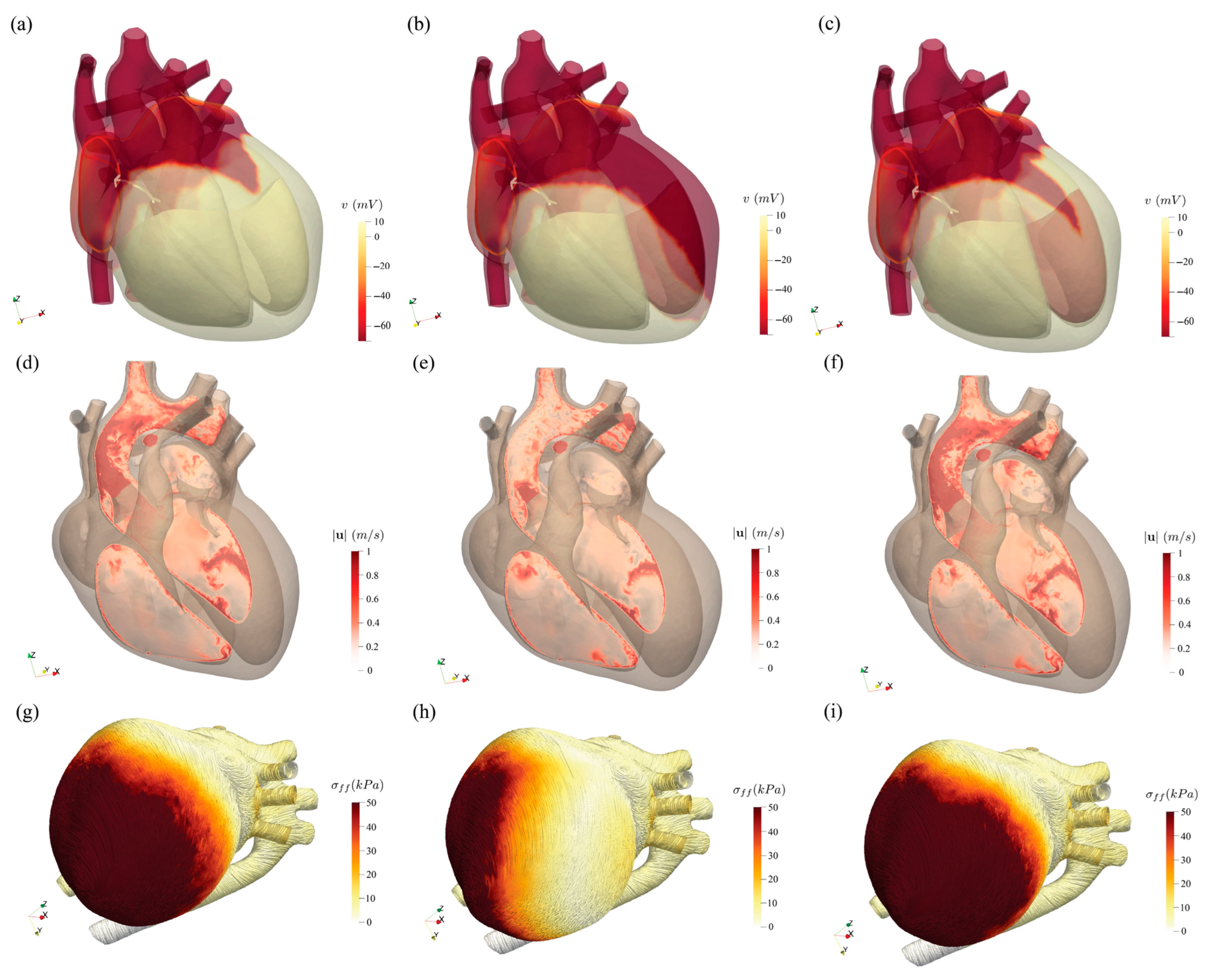
3.8. Roadmap and Milestones for Clinical Adoption of MEFI Models

While MEFI models have demonstrated growing predictive power and physiological realism, widespread clinical adoption requires achieving several strategic milestones, as shown in Table 9. These milestones span technical, clinical, regulatory, and infrastructural domains and are projected based on current research trajectories.
  • Milestone 1: Standardized Patient-Specific Modeling Pipelines (by 2026–2027)
    • Goal: Automate workflows for generating anatomy-based MEFI models from clinical imaging (e.g., CT/MRI) with minimal manual segmentation.
    • Impact: Enables rapid creation of digital twins for diagnostic support, especially in high-volume conditions like atrial fibrillation or heart failure.
  • Milestone 2: Clinical Validation Studies Across Diverse Populations (by 2027–2028)
    • Goal: Conduct prospective, multicenter trials comparing model-guided treatment planning (e.g., CRT, ablation) versus standard of care.
    • Impact: Demonstrates real-world value and generalizability; essential for clinician trust and regulatory approval.
  • Milestone 3: Regulatory-Grade Model Certification Frameworks (by 2028–2029)
    • Goal: Establish standardized protocols for model verification, validation, and uncertainty quantification (VV&UQ), aligned with FDA or EMA guidelines.
    • Impact: Enables formal regulatory approval for MEFI-based software tools as software as a medical device (SaMD).
  • Milestone 4: Real-Time Reduced-Order MEFI Simulations for Interventional Planning (by 2029–2030)
    • Goal: Deploy machine-learning-enhanced surrogate models (e.g., latent neural ODEs, PINNs) capable of delivering interactive predictions during surgery or catheter-based procedures.
    • Impact: Supports intraoperative guidance (e.g., optimal lead placement, ablation targeting) with simulation runtimes under 1–2 min.
  • Milestone 5: EHR-Integrated Digital Twin Platforms (by 2030+)
    • Goal: Integrate MEFI-based digital twins into hospital electronic health records (EHR) and clinical decision support systems (CDSS).
    • Impact: Allows continuous patient-specific risk stratification, treatment planning, and virtual testing of interventions (e.g., valve replacement, drug response).
Future Vision: “Digital twin dashboards” showing dynamic cardiovascular simulations accessible at the bedside or in multidisciplinary team meetings.
Achieving these milestones will require sustained collaboration across academia, healthcare institutions, regulatory agencies, and industry. As computational power increases and AI accelerates model efficiency, MEFI models are positioned to become key instruments in precision cardiovascular medicine.

4. Conclusions

This review was undertaken with the primary objective of providing a comprehensive synthesis of recent advances in cardiac mechano-electrical-fluid interaction (MEFI) modeling—an interdisciplinary domain that seeks to capture the complex, coupled behavior of the heart’s electrical activation, mechanical deformation, and hemodynamic response. Our work contributes to the field by offering a unified framework that spans from high-fidelity computational models to reduced-order and surrogate approaches, while identifying opportunities for clinical translation and future innovation.
The novelty of this review lies in its integrative perspective across multiple modeling domains. We highlight recent progress in multiphysics coupling strategies—including monolithic and partitioned schemes—as well as the growing role of immersed boundary methods in simulating blood–tissue interaction. In addition, we underscore the emergence of AI-enhanced digital twin frameworks, which leverage machine learning to personalize simulations and reduce computational burden. These developments mark a paradigm shift from isolated electromechanical models toward comprehensive, patient-specific platforms capable of informing diagnosis and therapy in real time.
Quantitative validation studies included in this review provide strong support for the increasing clinical relevance of MEFI models. For example, pressure–volume loop predictions have achieved <5% root mean square error (RMSE) when compared to catheter-based measurements; myocardial strain patterns modeled computationally deviate by <10% from MRI tagging benchmarks; and classification models for arrhythmogenic events using MEFI-derived features report F1-scores exceeding 0.90. These metrics reflect not only growing numerical accuracy but also the potential for MEFI frameworks to support actionable clinical insights.
Moreover, the practical deployment of MEFI models is beginning to take shape in a variety of clinical contexts. Notable examples include their use in optimizing cardiac resynchronization therapy (CRT) lead placement, predicting outcomes of arrhythmia ablation procedures, and evaluating hemodynamic impacts of surgical interventions such as valve repair. These early applications demonstrate both the feasibility and translational promise of MEFI-based digital twins in personalized cardiovascular care.
However, several critical challenges remain that must be addressed to facilitate widespread adoption. These include the following:
  • Multiscale model integration across cellular, tissue, and organ levels;
  • Patient-specific parameterization based on heterogeneous imaging and physiological data;
  • Robust and reproducible model validation across diverse populations;
  • Interoperability and standardization of modeling tools;
  • Regulatory approval pathways that are currently lacking for in silico diagnostics and therapeutics.
To overcome these challenges, future research should prioritize the development of standardized datasets and open-source benchmarks to facilitate model comparison and regulatory evaluation. Collaborative efforts between engineers, clinicians, regulators, and industry stakeholders will be essential in defining the criteria for validation, clinical utility, and software certification. Furthermore, enhancing model accessibility through cloud-based platforms, user-friendly interfaces, and real-time feedback loops will help bridge the gap between research environments and point-of-care deployment.
Long-term, we envision a future where MEFI modeling becomes a core component of precision cardiovascular medicine. Key milestones toward this goal include the following:
  • Real-time simulation and feedback capabilities integrated into clinical workflows;
  • Interfacing with electronic health records (EHRs) to enable dynamic and continuous model updates;
  • Incorporation into regulatory-approved digital diagnostic pipelines;
  • Use in predictive and preventative medicine, particularly in managing chronic cardiac conditions and risk stratification.
By enabling mechanistic insight, predictive analytics, and individualized therapy planning, MEFI models stand to revolutionize the way cardiovascular diseases are diagnosed, managed, and treated. Continued innovation, validation, and integration into clinical practice will be vital for realizing the full impact of these technologies.

Author Contributions

Writing—review and editing, J.X.; writing—review and editing, F.W. All authors have read and agreed to the published version of the manuscript.

Funding

This research received no external funding.

Conflicts of Interest

The authors declare no conflicts of interest.

References

  1. World Health Organization. Cardiovascular Diseases (CVDs). 2023. Available online: https://www.who.int/news-room/fact-sheets/detail/cardiovascular-diseases-(cvds) (accessed on 17 July 2025).
  2. Trayanova, N.A. Whole-Heart Modeling Applications to Cardiac Electrophysiology and Electromechanics. Circ. Res. 2011, 108, U113–U195. [Google Scholar] [CrossRef] [PubMed]
  3. Viceconti, M.; Henney, A.; Morley-Fletcher, E. In silico clinical trials: How computer simulation will transform the biomedical industry. Int. J. Clin. Trials 2016, 30, 37–46. [Google Scholar] [CrossRef]
  4. Corral-Acero, J.; Margara, F.; Marciniak, M.; Rodero, C.; Loncaric, F.; Feng, Y.J.; Gilbert, A.; Fernandes, J.F.; Bukhari, H.A.; Wajdan, A.; et al. The ‘Digital Twin’ to enable the vision of precision cardiology. Eur. Heart J. 2020, 41, 4556–4564. [Google Scholar] [CrossRef] [PubMed]
  5. Verzicco, R. Electro-fluid-mechanics of the heart. J. Fluid. Mech. 2022, 941. [Google Scholar] [CrossRef]
  6. Kohl, P.; Hunter, P.; Noble, D. Stretch-induced changes in heart rate and rhythm: Clinical observations, experiments and mathematical models. Prog. Biophys. Mol. Biol. 1999, 71, 91–138. [Google Scholar] [CrossRef] [PubMed]
  7. Menon, K.; Hu, Z.N.; Marsden, A.L. Cardiovascular fluid dynamics: A journey through our circulation. Flow 2024, 4, E7. [Google Scholar] [CrossRef]
  8. Bers, D.M. Cardiac excitation-contraction coupling. Nature 2002, 415, 198–205. [Google Scholar] [CrossRef] [PubMed]
  9. Lee, J.; Niederer, S.; Nordsletten, D.; Le Grice, I.; Smaill, B.; Kay, D.; Smith, N. Coupling contraction, excitation, ventricular and coronary blood flow across scale and physics in the heart. Philos. Trans. A Math. Phys. Eng. Sci. 2009, 367, 2311–2331. [Google Scholar] [PubMed]
  10. Hunter, P.J.; Pullan, A.J.; Smaill, B.H. Modeling total heart function. Annu. Rev. Biomed. Eng. 2003, 5, 147–177. [Google Scholar] [CrossRef] [PubMed]
  11. Peskin, C.S.; McQueen, D.M. Cardiac fluid dynamics. Crit. Rev. Biomed. Eng. 1992, 20, 451–459. [Google Scholar] [PubMed]
  12. Peskin, C.S.; McQueen, D.M. A general method for the computer simulation of biological systems interacting with fluids. Symp. Soc. Exp. Biol. 1995, 49, 265–276. [Google Scholar] [PubMed]
  13. McQueen, D.M.; Peskin, C.S. A 3D computational method for blood flow in the heart. I. immersed elastic fibers in a viscous incompressible fluid. J. Comput. Phys. 1989, 81, 372–405. [Google Scholar]
  14. McQueen, D.M.; Peskin, C.S. A 3D computational method for blood flow in the heart. II. contractile fibers. J. Comput. Phys. 1989, 82, 289–297. [Google Scholar] [CrossRef]
  15. Feng, L.Y.; Gao, H.; Luo, X.Y. Whole-heart modelling with valves in a fluid-structure interaction framework. Comput. Methods Appl. Mech. Eng. 2024, 420, 116724. [Google Scholar] [CrossRef]
  16. Jiang, Y.; Yan, Z.Z.; Wang, X.H.; Chen, R.L.; Cai, X.C. A highly parallel algorithm for simulating the elastodynamics of a patient-specific human heart with four chambers using a heterogeneous hyperelastic model. J. Comput. Phys. 2024, 508, 113027. [Google Scholar] [CrossRef]
  17. Ugurlu, D.; Qian, S.; Fairweather, E.; Mauger, C.; Ruijsink, B.; Dal Toso, L.; Deng, Y.; Strocchi, M.; Razavi, R.; Young, A.; et al. Cardiac digital twins at scale from MRI: Open tools and representative models from ~55000 UK Biobank participants. arXiv 2025, arXiv:2505.21019. [Google Scholar] [CrossRef] [PubMed]
  18. Shi, L.; Chen, I.Y.; Takayama, H.; Vedula, V. An optimization framework to personalize passive cardiac mechanics. Comput. Methods Appl. Mech. Eng. 2024, 432, 117401. [Google Scholar] [CrossRef]
  19. Updegrove, A.; Wilson, N.M.; Merkow, J.; Lan, H.Z.; Marsden, A.L.; Shadden, S.C. SimVascular: An Open Source Pipeline for Cardiovascular Simulation. Ann. Biomed. Eng. 2017, 45, 525–541. [Google Scholar] [CrossRef] [PubMed]
  20. Vedula, V.; Lee, J.; Xu, H.; Kuo, C.C.J.; Hsiai, T.K.; Marsden, A.L. A method to quantify mechanobiologic forces during zebrafish cardiac development using 4-D light sheet imaging and computational modeling. PLoS Comput. Biol. 2018, 14, e1006482. [Google Scholar] [CrossRef] [PubMed]
  21. Bäumler, K.; Vedula, V.; Sailer, A.M.; Seo, J.; Chiu, P.; Mistelbauer, G.; Chan, F.P.; Fischbein, M.P.; Marsden, A.L.; Fleischmann, D. Fluid-structure interaction simulations of patient-specific aortic dissection. Biomech. Model. Mechan. 2020, 19, 1607–1628. [Google Scholar] [CrossRef] [PubMed]
  22. Kong, F.W.; Shadden, S.C. Learning Whole Heart Mesh Generation From Patient Images for Computational Simulations. IEEE Trans. Med. Imaging 2023, 42, 533–545. [Google Scholar] [CrossRef] [PubMed]
  23. Schwarz, E.L.; Pfaller, M.R.; Szafron, J.M.; Latorre, M.; Lindsey, S.E.; Breuer, C.K.; Humphrey, J.D.; Marsden, A.L. A fluid-solid-growth solver for cardiovascular modeling. Comput. Methods Appl. Mech. Eng. 2023, 417, 116312. [Google Scholar] [CrossRef] [PubMed]
  24. Schwarz, E.L.; Pegolotti, L.; Pfaller, M.R.; Marsden, A.L. Beyond CFD: Emerging methodologies for predictive simulation in cardiovascular health and disease. Biophys. Rev. 2023, 4, 011301. [Google Scholar] [CrossRef] [PubMed]
  25. Schuster, M.R.; Dirkes, N.; Key, F.; Elgeti, S.; Behr, M. Exploring the influence of parametrized pulsatility on left ventricular washout under LVAD support: A computational study using reduced-order models. Comput. Methods Biomech. Biomed. Eng. 2024, 28, 800–817. [Google Scholar] [CrossRef] [PubMed]
  26. Usyk, T.P.; McCulloch, A.D. Electromechanical model of cardiac resynchronization in the dilated failing heart with left bundle branch block. J. Electrocardiol. 2003, 36, 57–61. [Google Scholar] [CrossRef] [PubMed]
  27. Usyk, T.; Kerckhoffs, R. Three Dimensional Electromechanical Model of Porcine Heart with Penetrating Wound Injury. Stud. Health Technol. Inf. 2005, 111, 568–573. [Google Scholar]
  28. Kerckhoffs, R.C.; Neal, M.L.; Gu, Q.; Bassingthwaighte, J.B.; Omens, J.H.; McCulloch, A.D. Coupling of a 3D finite element model of cardiac ventricular mechanics to lumped systems models of the systemic and pulmonic circulation. Ann. Biomed. Eng. 2007, 35, 1–18. [Google Scholar] [CrossRef] [PubMed]
  29. Nash, M.P.; Panfilov, A.V. Electromechanical model of excitable tissue to study reentrant cardiac arrhythmias. Prog. Biophys. Mol. Biol. 2004, 85, 501–522. [Google Scholar] [CrossRef] [PubMed]
  30. Ten Tusscher, K.H.W.J.; Panfilov, A.V. Modelling of the ventricular conduction system. Prog. Biophys Mol. Biol. 2008, 96, 152–170. [Google Scholar] [CrossRef] [PubMed]
  31. Salvador, M.; Strocchi, M.; Regazzoni, F.; Augustin, C.M.; Dede’, L.; Niederer, S.A.; Quarteroni, A. Whole-heart electromechanical simulations using Latent Neural Ordinary Differential Equations. Npj Digit. Med. 2024, 7, 90. [Google Scholar] [CrossRef] [PubMed]
  32. Fedele, M.; Piersanti, R.; Regazzoni, F.; Salvador, M.; Africa, P.C.; Bucelli, M.; Zingaro, A.; Dede, L.; Quarteroni, A. A comprehensive and biophysically detailed computational model of the whole human heart electromechanics. Comput. Methods Appl. Mech. Eng. 2023, 410, 115983. [Google Scholar] [CrossRef]
  33. Loewe, A.; Hunter, P.J.; Kohl, P. Computational modelling of biological systems now and then: Revisiting tools and visions from the beginning of the century. Philos. Trans. R. Soc. A-Math. Phys. Eng. Sci. 2025, 383. [Google Scholar] [CrossRef] [PubMed]
  34. Thangaraj, P.M.; Benson, S.H.; Oikonomou, E.K.; Asselbergs, F.W.; Khera, R. Cardiovascular care with digital twin technology in the era of generative artificial intelligence. Eur. Heart J. 2024, 45, 4808–4821. [Google Scholar] [CrossRef] [PubMed]
  35. Rudnicka, Z.; Proniewska, K.; Perkins, M.; Pregowska, A. Cardiac Healthcare Digital Twins Supported by Artificial Intelligence-Based Algorithms and Extended Reality-A Systematic Review. Electronics 2024, 13, 866. [Google Scholar] [CrossRef]
  36. Pramanik, R.; Verstappen, R.W.C.P.; Onck, P.R. Computational fluid-structure interaction in biology and soft robots: A review. Phys. Fluids 2024, 36, 101302. [Google Scholar] [CrossRef]
  37. Magoon, M.J.; Nazer, B.; Akoum, N.; Boyle, P.M. Computational Medicine: What Electrophysiologists Should Know to Stay Ahead of the Curve. Curr. Cardiol. Rep. 2024, 26, 1393–1403. [Google Scholar] [CrossRef] [PubMed]
  38. Lauer-Schmaltz, M.W.; Cash, P.; Rivera, D.G.T. ETHICA: Designing Human Digital Twins-A Systematic Review and Proposed Methodology. IEEE Access 2024, 12, 86947–86973. [Google Scholar] [CrossRef]
  39. Bhagirath, P.; Strocchi, M.; Bishop, M.J.; Boyle, P.M.; Plank, G. From bits to bedside: Entering the age of digital twins in cardiac electrophysiology. Europace 2024, 26, euae295. [Google Scholar] [CrossRef] [PubMed]
  40. Torre, M.; Morganti, S.; Pasqualini, F.S.; Reali, A. Current progress toward isogeometric modeling of the heart biophysics. Biophys. Rev. 2023, 4, 041301. [Google Scholar] [CrossRef] [PubMed]
  41. Coorey, G.; Figtree, G.A.; Fletcher, D.F.; Snelson, V.J.; Vernon, S.T.; Winlaw, D.; Grieve, S.M.; McEwan, A.; Yang, J.Y.H.; Qian, P.; et al. The health digital twin to tackle cardiovascular disease-a review of an emerging interdisciplinary field. Npj Digit. Med. 2022, 5, 126. [Google Scholar] [CrossRef] [PubMed]
  42. Watanabe, H.; Sugiura, S.; Kafuku, H.; Hisada, T. Multiphysics simulation of left ventricular filling dynamics using fluid-structure interaction finite element method. Biophys. J. 2004, 87, 2074–2085. [Google Scholar] [CrossRef] [PubMed]
  43. Vigmond, E.J.; Clements, C.; McQueen, D.M.; Peskin, C.S. Effect of bundle branch block on cardiac output: A whole heart simulation study. Prog. Biophys. Mol. Biol. 2008, 97, 520–542. [Google Scholar] [CrossRef] [PubMed]
  44. Gonzalo, A.; Augustin, C.M.; Bifulco, S.F.; Telle, A.; Chahine, Y.; Kassar, A.; Guerrero-Hurtado, M.; Durán, E.; Martínez-Legazpi, P.; Flores, O.; et al. Multiphysics simulations reveal haemodynamic impacts of patient-derived fibrosis-related changes in left atrial tissue mechanics. J. Physiol. 2024, 602, 6789–6812. [Google Scholar] [CrossRef] [PubMed]
  45. Bucelli, M.; Gabriel, M.G.; Quarteroni, A.; Gigante, G.; Vergara, C. A stable loosely-coupled scheme for cardiac electro-fluid-structure interaction. J. Comput. Phys. 2023, 490, 112326. [Google Scholar] [CrossRef]
  46. Bucelli, M.; Zingaro, A.; Africa, P.C.; Fumagalli, I.; Dede’, L.; Quarteroni, A. A mathematical model that integrates cardiac electrophysiology, mechanics, and fluid dynamics: Application to the human left heart. Int. J. Numer. Methods Biomed. Eng. 2023, 39, e3678. [Google Scholar] [CrossRef] [PubMed]
  47. Viola, F.; Del Corso, G.; De Paulis, R.; Verzicco, R. GPU accelerated digital twins of the human heart open new routes for cardiovascular research. Sci. Rep. 2023, 13, 8230. [Google Scholar] [CrossRef] [PubMed]
  48. Del Corso, G.; Verzicco, R.; Viola, F. A fast computational model for the electrophysiology of the whole human heart. J. Comput. Phys. 2022, 457, 111084. [Google Scholar] [CrossRef]
  49. Quarteroni, A.; Dede, L.; Regazzoni, F.; Vergara, C. A mathematical model of the human heart suitable to address clinical problems. Jpn. J. Ind. Appl. Math. 2023, 40, 1547–1567. [Google Scholar] [CrossRef]
  50. Viola, F.; Del Corso, G.; Verzicco, R. High-fidelity model of the human heart: An immersed boundary implementation. Phys. Rev. Fluids 2023, 8, 100502. [Google Scholar] [CrossRef]
  51. Bradley, C.; Bowery, A.; Britten, R.; Budelmann, V.; Camara, O.; Christie, R.; Cookson, A.; Frangi, A.F.; Gamage, T.B.; Heidlauf, T.; et al. OpenCMISS: A multi-physics & multi-scale computational infrastructure for the VPH/Physiome project. Prog. Biophys. Mol. Biol. 2011, 107, 32–47. [Google Scholar] [CrossRef] [PubMed]
  52. Schenkel, T.; Malve, M.; Reik, M.; Markl, M.; Jung, B.; Oertel, H. MRI-based CFD analysis of flow in a human left ventricle: Methodology and application to a healthy heart. Ann. Biomed. Eng. 2009, 37, 503–515. [Google Scholar] [CrossRef] [PubMed]
  53. Soltis, A.R.; Saucerman, J.J. Synergy between CaMKII substrates and beta-adrenergic signaling in regulation of cardiac myocyte Ca(2+) handling. Biophys. J. 2010, 99, 2038–2047. [Google Scholar] [CrossRef] [PubMed]
  54. Lester, H.A.; Karschin, A. Gain of function mutants: Ion channels and G protein-coupled receptors. Annu. Rev. Neurosci. 2000, 23, 89–125. [Google Scholar] [CrossRef] [PubMed]
  55. Grant, A.O.; Carboni, M.P.; Neplioueva, V.; Starmer, C.F.; Memmi, M.; Napolitano, C.; Priori, S. Long QT syndrome, Brugada syndrome, and conduction system disease are linked to a single sodium channel mutation. J. Clin. Investig. 2002, 110, 1201–1209. [Google Scholar] [CrossRef] [PubMed]
  56. Benson, D.W.; Wang, D.W.; Dyment, M.; Knilans, T.K.; Fish, F.A.; Strieper, M.J.; Rhodes, T.H.; George, A.L.J. Congenital sick sinus syndrome caused by recessive mutations in the cardiac sodium channel gene (SCN5A). J. Clin. Investig. 2003, 112, 1019–1028. [Google Scholar] [CrossRef] [PubMed]
  57. Bennett, P.B.; Yazawa, K.; Makita, N.; George, A.L.J. Molecular mechanism for an inherited cardiac arrhythmia. Nature 1995, 376, 683–685. [Google Scholar] [CrossRef] [PubMed]
  58. Tan, H.L.; Bink-Boelkens, M.T.; Bezzina, C.R.; Viswanathan, P.C.; Beaufort-Krol, G.C.; van Tintelen, P.J.; van den Berg, M.P.; Wilde, A.A.; Balser, J.R. A sodium-channel mutation causes isolated cardiac conduction disease. Nature 2001, 409, 1043–1047. [Google Scholar] [CrossRef] [PubMed]
  59. Splawski, I.; Timothy, K.W.; Sharpe, L.M.; Decher, N.; Kumar, P.; Bloise, R.; Napolitano, C.; Schwartz, P.J.; Joseph, R.M.; Condouris, K.; et al. Ca(V)1.2 calcium channel dysfunction causes a multisystem disorder including arrhythmia and autism. Cell 2004, 119, 19–31. [Google Scholar] [CrossRef] [PubMed]
  60. Clancy, C.E.; Rudy, Y. Linking a genetic defect to its cellular phenotype in a cardiac arrhythmia. Nature 1999, 400, 566–569. [Google Scholar] [CrossRef] [PubMed]
  61. Berridge, M.J.; Bootman, M.D.; Roderick, H.L. Calcium signalling: Dynamics, homeostasis and remodelling. Nat. Rev. Mol. Cell Biol. 2003, 4, 517–529. [Google Scholar] [CrossRef] [PubMed]
  62. Brini, M.; Carafoli, E. Calcium signalling: A historical account, recent developments and future perspectives. Cell. Mol. Life Sci. 2000, 57, 354–370. [Google Scholar] [CrossRef] [PubMed]
  63. Splawski, I.; Timothy, K.W.; Decher, N.; Kumar, P.; Sachse, F.B.; Beggs, A.H.; Sanguinetti, M.C.; Keating, M.T. Severe arrhythmia disorder caused by cardiac L-type calcium channel mutations. Proc. Natl. Acad. Sci. USA 2005, 102, 8089–8096, discussion 8086–8088. [Google Scholar] [CrossRef] [PubMed]
  64. Schamroth, L. An Introduction to Electrocardiography, 6th ed.; Blackwell Scientific Publications: Oxford, UK, 1982. [Google Scholar]
  65. Caldwell, B.J.; Trew, M.L.; Sands, G.B.; Hooks, D.A.; LeGrice, I.J.; Smaill, B.H. Three distinct directions of intramural activation reveal nonuniform side-to-side electrical coupling of ventricular myocytes. Circ. Arrhythm. Electrophysiol. 2009, 2, 433–440. [Google Scholar] [CrossRef] [PubMed]
  66. Wyman, B.T.; Hunter, W.C.; Prinzen, F.W.; McVeigh, E.R. Mapping propagation of mechanical activation in the paced heart with MRI tagging. Am. J. Physiol. 1999, 276, H881–H891. [Google Scholar] [CrossRef] [PubMed]
  67. Chandra, R.; Starmer, C.F.; Grant, A.O. Multiple effects of KPQ deletion mutation on gating of human cardiac Na+ channels expressed in mammalian cells. Am. J. Physiol. 1998, 274, H1643–H1654. [Google Scholar] [CrossRef] [PubMed]
  68. Antzelevitch, C. The Brugada syndrome. J. Cardiovasc. Electrophysiol. 1998, 9, 513–516. [Google Scholar] [CrossRef] [PubMed]
  69. Kilner, P.J.; Yang, G.Z.; Wilkes, A.J.; Mohiaddin, R.H.; Firmin, D.N.; Yacoub, M.H. Asymmetric redirection of flow through the heart. Nature 2000, 404, 759–761. [Google Scholar] [CrossRef] [PubMed]
  70. Moore, C.C.; Lugo-Olivieri, C.H.; McVeigh, E.R.; Zerhouni, E.A. Three-dimensional systolic strain patterns in the normal human left ventricle: Characterization with tagged MR imaging. Radiology 2000, 214, 453–466. [Google Scholar] [CrossRef] [PubMed]
  71. Verbeek, X.A.; Vernooy, K.; Peschar, M.; Van Der Nagel, T.; Van Hunnik, A.; Prinzen, F.W. Quantification of interventricular asynchrony during LBBB and ventricular pacing. Am. J. Physiol. Heart Circ. Physiol. 2002, 283, H1370–H1378. [Google Scholar] [CrossRef] [PubMed]
  72. Markhasin, V.S.; Solovyova, O.; Katsnelson, L.B.; Protsenko, Y.; Kohl, P.; Noble, D. Mechano-electric interactions in heterogeneous myocardium: Development of fundamental experimental and theoretical models. Prog. Biophys. Mol. Biol. 2003, 82, 207–220. [Google Scholar] [CrossRef] [PubMed]
  73. Sachse, F.B.; Seemann, G.; Chaisaowong, K.; Weiss, D. Quantitative reconstruction of cardiac electromechanics in human myocardium: Assembly of electrophysiologic and tension generation models. J. Cardiovasc. Electrophysiol. 2003, 14 (Suppl. S10), S210–S218. [Google Scholar] [CrossRef] [PubMed]
  74. Kerckhoffs, R.C.; Faris, O.P.; Bovendeerd, P.H.; Prinzen, F.W.; Smits, K.; McVeigh, E.R.; Arts, T. Electromechanics of paced left ventricle simulated by straightforward mathematical model: Comparison with experiments. Am. J. Physiol. Heart Circ. Physiol. 2005, 289, H1889–H1897. [Google Scholar] [CrossRef] [PubMed]
  75. Grill, S.; Zykov, V.S.; Muller, S.C. Feedback-controlled dynamics of meandering spiral waves. Phys. Rev. Lett. 1995, 75, 3368–3371. [Google Scholar] [CrossRef] [PubMed]
  76. Panfilov, A.V.; Keldermann, R.H.; Nash, M.P. Drift and breakup of spiral waves in reaction-diffusion-mechanics systems. Proc. Natl. Acad. Sci. USA 2007, 104, 7922–7926. [Google Scholar] [CrossRef] [PubMed]
  77. Biktashev, V.N.; Holden, A.V. Control of re-entrant activity in a model of mammalian atrial tissue. Proc. Biol. Sci. 1995, 260, 211–217. [Google Scholar] [CrossRef] [PubMed]
  78. Ginsburg, R.; Bristow, M.R.; Billingham, M.E.; Stinson, E.B.; Schroeder, J.S.; Harrison, D.C. Study of the normal and failing isolated human heart: Decreased response of failing heart to isoproterenol. Am. Heart J. 1983, 106, 535–540. [Google Scholar] [CrossRef] [PubMed]
  79. Hooks, D.A.; Trew, M.L.; Caldwell, B.J.; Sands, G.B.; LeGrice, I.J.; Smaill, B.H. Laminar arrangement of ventricular myocytes influences electrical behavior of the heart. Circ. Res. 2007, 101, e103–e112. [Google Scholar] [CrossRef] [PubMed]
  80. Trayanova, N.A.; Boyle, P.M. Advances in modeling ventricular arrhythmias: From mechanisms to the clinic. Wires Syst. Biol. Med. 2014, 6, 209–224. [Google Scholar] [CrossRef] [PubMed]
  81. Trayanova, N.A.; Boyle, P.M.; Nikolov, P.P. Personalized imaging and modeling strategies for arrhythmia prevention and therapy. Curr. Opin. Biomed. Eng. 2018, 5, 21–28. [Google Scholar] [CrossRef] [PubMed]
  82. Trayanova, N.A.; O’Hara, T.; Bayer, J.D.; Boyle, P.M.; McDowell, K.S.; Constantino, J.; Arevalo, H.J.; Hu, Y.X.; Vadakkumpadan, F. Computational cardiology: How computer simulations could be used to develop new therapies and advance existing ones. Europace 2012, 14, V82–V89. [Google Scholar] [CrossRef] [PubMed]
  83. Trayanova, N.A.; Chang, K.C. How computer simulations of the human heart can improve anti-arrhythmia therapy. J. Physiol. 2016, 594, 2483–2502. [Google Scholar] [CrossRef] [PubMed]
  84. Arevalo, H.J.; Vadakkumpadan, F.; Guallar, E.; Jebb, A.; Malamas, P.; Wu, K.C.; Trayanova, N.A. Arrhythmia risk stratification of patients after myocardial infarction using personalized heart models. Nat. Commun. 2016, 7, 11437. [Google Scholar] [CrossRef] [PubMed]
Figure 1. Schematic of the MEFI feedback pathway. The diagram illustrates the multiscale interactions in cardiac mechano-electrical-fluidic (MEFI) modeling. At the cellular level, ionic currents and transmembrane potential dynamics govern excitation in the cardiac cell model. These electrical signals propagate as waves across myocardial tissue, triggering mechanical contraction through excitation-contraction coupling (mechano-electrical interaction, MEI). The resulting myocardial deformation drives blood flow via fluid-structure interaction (FSI), which in turn affects wall stress and loading conditions. This hemodynamic feedback further influences tissue mechanics and electrical activity, completing the MEFI loop. The figure highlights the bidirectional coupling and systems-level integration essential for physiologically realistic cardiac simulations. Adapted from [8].
Eng 06 00168 g001
Figure 2. Timeline of multiphysics heart modeling with increasing accuracy. The diagram illustrates the hierarchical progression of cardiac modeling approaches in terms of increasing physiological fidelity and computational complexity. Starting from isolated cardiac mechanics and fluid models, the integration of fluid–structure interaction (FSI), mechano–electrical interaction (MEI), and fully coupled mechano-electrical-fluidic interaction (MEFI) leads to progressively higher accuracy in simulating cardiac function. However, this improvement in realism comes at the cost of increased model complexity and computational demand, highlighting the need for balanced trade-offs in research and clinical applications.
Eng 06 00168 g002
Figure 3. Reconstructed four-chamber heart geometry shown in its stress-free state, representing the un-loaded anatomical configuration used as the baseline for biomechanical simulations. (a) The external view of the whole heart, and (b) the cut-away view of the whole heart. Adapted from [15].
Eng 06 00168 g003
Figure 4. The magnitude of the displacement over the myocardial domain at six typical moments, at 0 s (a,d), 0.14 s (b,e), 0.4 s (c,f), 0.54 s (g,j), 0.8 s (h,k), and 1.0 s (i,l), respectively. Each panel presents two plots vertically, illustrating the anterior (af) and posterior (gl) views of the heart. Adapted from [16].
Eng 06 00168 g004aEng 06 00168 g004b
Figure 5. Workflow for patient-specific modeling of myocardial mechanics. Time-dependent three-dimensional (3D) computed tomographic (CT) images are used as the starting point. The myocardial structure is extracted at various time points throughout the cardiac cycle. Following segmentation, an optimization algorithm is employed to determine the patient-specific passive tissue properties and the unloaded, or stress-free, geometric configuration of the heart. (iFEA: inverse finite element analysis; EDV: end-diastolic volume; ESV: end-systolic volume;). Adapted from [18].
Eng 06 00168 g005
Figure 6. Comparison of FEA-predicted deformed configuration against image data along longitudinal and transverse mid-planes of the biventricular myocardium. (a,e) Normal subject; (bd, fh) HOCM patients; (ad) relaxed configuration (mid-diastasis); (eh) end-diastolic configuration. Adapted from [18].
Eng 06 00168 g006
Figure 7. Workflow of surrogate modeling and uncertainty quantification (UQ) framework for cardiac electromechanical simulations. A total of 405 closed-loop 3D-0D simulations of the four-chamber heart model were performed by sampling the parameter space using Latin hypercube techniques. The resulting pressure–volume loops for each chamber were used to train a system of latent neural ordinary differential equations (LNODEs), enabling efficient and accurate surrogate modeling of the full cardiovascular system. Global sensitivity analysis (GSA) was conducted to evaluate the influence of individual parameters on different quantities of interest (QoIs), and robust parameter estimation was achieved by matching time-resolved simulation outputs with clinical data. The resulting posterior distributions allowed for the calibration of model parameters, facilitating fully personalized 3D-0D simulations with quantified uncertainty. Adapted from [31].
Eng 06 00168 g007
Figure 8. Comparison of pressure and volume waveforms between the full-order 3D-0D electromechanical model M3D-0D (dashed lines) and the surrogate neural network model MANN (solid lines), evaluated over the cardiac cycle duration of 0 to 0.854 s for five independent test cases (Ntest = 5). Each subplot illustrates the temporal evolution of pressure (ae) and volume (fj) for the four heart chambers. The strong agreement between the two models demonstrates the predictive accuracy of the ANN-based surrogate in reproducing both pressure and volume dynamics. Light blue: LA, orange: LV, blue: RA, green: RV. Adapted from [31].
Eng 06 00168 g008
Figure 9. Description of a multiphysics multiscale framework. Block diagram of electrophysiology and biomechanics multiscale models, and the fluid mechanics model, including coupling between them. Adapted from [44].
Eng 06 00168 g009
Figure 10. (A) Differences in fibrotic and non-fibrotic tissue conductivity are depicted with activation time maps in an anisotropic 10 cm2 tissue patch, highlighting impaired transverse conduction. (B) Differences between non-fibrotic, hypercontractile-fibrotic, stiff-fibrotic, and fibrotic mechanical states are shown with maps in the same tissue patch. (C) Sketch of a CFD model approach to impose boundary conditions, including velocity field vectors obtained during LA systole in black. Adapted from [44].
Eng 06 00168 g010
Figure 11. Impact of fibrosis on left atrial (LA) and left atrial appendage (LAA) hemodynamics. (A) corresponds to subject Fib41, representing a case with extensive fibrosis, while (B) shows results for Fib07, a case with limited fibrotic remodeling. In both panels, the time-dependent changes in LA and LAA volumes are plotted for fibrotic (red) and non-fibrotic (black) mechanical conditions (a,e,i,m). Two key phases of the cardiac cycle were selected for visualization: peak LAA contraction (top subpanels (a,i), indicated by black dashed lines) and peak LAA expansion (bottom subpanels (e,m), indicated by grey dashed lines). For each of these time points, three types of visualizations are shown: (1) Left column in each rectangle (b,f,j,n): a snapshot of the velocity field superimposed on kinetic energy (KE) distribution for the non-fibrotic condition, with velocity vectors scaled by magnitude to indicate flow direction. Note: velocity arrows in the expansion-phase subpanels (lower row (f,n)) are enlarged by a factor of 1.5 for visibility. (2) Middle column (c,g,k,o): a spatial map showing the differential KE between non-fibrotic and fibrotic cases in the same cross-sectional plane. (3) Right column (d,h,l,p): a 3D view of the LAA with velocity vectors indicating the directional and magnitude differences in flow between the two states. The arrows are color-coded by the velocity difference magnitude to highlight localized flow alterations due to fibrotic remodeling. Adapted from [44].
Eng 06 00168 g011
Figure 12. The whole cardiac geometry is immersed in the computational domain of the blood phase (external bounding box). The grey volumes at the tip of the artery/veins indicate the region where the impedance of the missing circulation is mimicked by a volume forcing. Adapted from [47].
Eng 06 00168 g012
Figure 13. Pathological effect and therapeutic effectiveness. Comparison of different quantities for healthy (a,d,g), impaired (b,e,h), and resynchronized (c,f,i) heart during systole. (ac) Instantaneous surface distribution of the activation potential (t = 252 ms). (df) Blood velocity distribution on a planar section cutting the left heart at peak systole (t = 520 ms). (gi) Surface distribution of the tension along the fiber axes at peak systole (force per unit area, t = 520 ms). Adapted from [47].
Eng 06 00168 g013
Figure 14. Schematic representation of the β-adrenergic signaling pathway in cardiac myocytes. Binding of an agonist to the β-adrenergic receptor activates the Gs protein, which stimulates adenylyl cyclase to convert ATP to cAMP. Elevated cAMP levels activate protein kinase A (PKA), which phosphorylates L-type Ca2+ channels, enhancing Ca2+ influx. The resulting increase in intracellular calcium promotes heart muscle contraction. Na+ and Ca2+ ion channels shown contribute to excitation–contraction coupling.
Eng 06 00168 g014
Table 1. (a) Summary of multiphysics whole-heart modeling studies. (b) Structured summary of MEFI modeling approaches. (c) Quantitative performance metrics of MEFI models.
(a)
StudyModeling FocusMultiphysics ApproachApplication AreaKey Contribution
Feng et al. [15]Whole-heart FSI modeling with detailed valves and pericardium interactionFSISubject-specific FEM with fluid–structure interactionValve and pericardium constraint analysisDeveloped 4-chamber model with valves and pericardium; demonstrated pericardial effects on valve dynamics
Jiang et al. [16]Scalable whole-heart FEM for personalized cardiac simulationFSIFully implicit FEM with Newton–Krylov–Schwarz solverHigh-performance simulation of heart cycleDemonstrated scalability on >16 k cores; accurate heart cycle simulation with anisotropic fiber architecture
Shi et al. [18]Inverse FEA from imagingFSINested optimization + Sellier’s method; stabilized multiscale FEMPatient-specific cardiac parameter estimationQuantified passive mechanical properties from CT data using optimization and hyperelastic modeling
Schwarz et al. [23,24]Vascular G&R simulation with long-term hemodynamicsFSIPartitioned FEM + CMT (constrained mixture theory)Cardiovascular disease modelingIntroduced fluid–solid–growth model for vascular remodeling and graft simulations
Schuster et al. [25]LVAD pulsatility effect on washoutFSIFull-order + reduced-order modelingLVAD thrombosis risk reductionFound short, strong pulses most effective for LV washout; used ROMs to reduce cost
Salvador et al. [31]Cardiac surrogate modelingMEILNODEs trained from high-fidelity electromechanical simulationsReal-time clinical decision supportDeveloped real-time surrogate model maintaining fidelity for pressure–volume simulations
Fedele et al. [32]Biophysically detailed whole-heart MEIStrongly coupled EM with fiber stretch feedback; HPC-optimizedDigital twin development, whole-heart contraction dynamicsModeled full four-chamber EM behavior with ionic currents and mechano-electric feedback
Watanabe et al. [42] Electromechanical FSIMEFI3D FSI coupling with FitzHugh–Nagumo excitationCardiac excitation modelingSimplified EM-FSI model; limited fidelity but effective for basic dynamics
Vigmond et al. [43]Cable-based EMF modelingMEFIFinite volume cable-based discretization with Luo–Rudy modelAnatomical MEFI integrationRealistic anatomical features with MEF coupling; lacked full electro-mechanical feedback
Gonzalo et al. [44]Atrial fibrosis modelingMEFILGE-MRI fibrosis maps + multicomponent simulationsLAA thrombosis risk analysisQuantified fibrosis effects on LA/LAA flow; found local and global fibrosis impact hemodynamics
Bucelli et al. [45,46]Loosely coupled EFSI for cardiac simulationsMEFIModular EFSI with Robin–Neumann interfaceEfficient simulation and testingReduced cost while preserving key pressure/volume outputs; validated with benchmarks
Viola et al. [47]GPU-accelerated digital twin for MEFIMEFIIntegrated MEFI with over 1B DOF; GPU-basedIn silico trials, CRT optimizationEnabled high-fidelity full-cycle simulation in hours; tested therapy outcomes
Quarteroni et al. [49]Multiscale cardiac modelingMEFIMultiphysics, multiscale framework with patient calibrationClinical treatment planningUnified electrophysiology, perfusion, valve, and mechanics models for diagnosis and therapy
Viola et al. [50]FSEI with infarcted heart simulationsMEFIIntegrated electromechanics, tissue mechanics, and flowSurgical planningAchieved near real-time runtimes for infarct simulations; applicable to clinical workflows
(b)
StudyModel FidelityCoupling StrategyAI UseClinical TargetSimulation SpeedAdvantagesLimitations
Feng et al. [15]HighMonolithicNoValve and pericardium mechanicsModerate (HPC required)Realistic valves/pericardium; 4-chamber FSINo EP coupling
Jiang et al. [16]HighMonolithicNoCardiac cycle simulationHigh (scales on 16 k+ cores)Highly scalable FEM; accurate cycle modelingComplex setup; high resource
Shi et al. [18]HighMonolithicNoPassive mechanics personalizationModerateAccurate personalization via iFEADependent on imaging; lacks active mechanics
Schwarz et al. [23,24]ModeratePartitionedNoVascular remodelingModerateIncludes long-term vascular growth/remodelingNot real-time; model complexity
Schuster et al. [25]ModerateHybrid (full + ROM)Yes (ROMs)LVAD washoutFast (via ROM)Captures pulsatility effects efficientlyLimited to 2D; simplified geometry
Salvador et al. [31]ModerateAI-based surrogateYes (LNODEs)Electromechanical surrogate modelingVery Fast (seconds)Fast, scalable, real-time surrogateLess interpretable; surrogate only
Fedele et al. [32]HighStrongly coupledNoWhole-heart EM simulationHigh (HPC-optimized)Biophysical detail; fiber feedbackNo fluid; high computational demand
Watanabe et al. [42] LowMonolithic (simplified)NoBasic excitation modelingModerateSimple EM coupling demonstrationSimplified EP model; no fluid
Vigmond et al. [43]ModerateFinite volume (1-way)NoAnatomical MEFI integrationModerateCaptures anatomical EMF detailNo mechano-electrical feedback
Gonzalo et al. [44]HighMonolithicNoAtrial thrombosis riskHigh (multiscale, patient-specific)Detailed fibrosis modeling; multiscale FSIRequires MRI fibrosis data; intensive computation
Bucelli et al. [45,46]ModeratePartitionedNoCardiac testing efficiencyModerateComputationally efficient; stableTrade-off in isovolumetric accuracy
Viola et al. [47]Very HighMonolithicNoCRT optimization, digital trialsSlow (~12 hrs/cycle)Ultra-high fidelity digital twinResource intensive; not yet real-time
Quarteroni et al. [49]HighMonolithicNoSurgical and therapy planningHigh (clinical use enabled)Multiphysics, multiscale personalized frameworkNeeds large patient datasets
Viola et al. [50]HighMonolithicNoSurgical planning, infarctionModerate to FastUnified FSEI model; fast runtimeLimited to infarcted cases
(c)
StudyPV Loop ErrorStrain Error (vs MRI)Arrhythmia Detection (F1 Score)Cardiac Output AccuracyFlow Pattern Accuracy/KE Deviation
Feng et al. [15]Not reportedNot reportedNot applicableQualitative onlyValidated LV flow and valve dynamics
Jiang et al. [16]<5%<10%Not applicable~95%Validated flow/geometry
Shi et al. [18]Not reported<10%Not applicableNot reportedNot applicable
Schwarz et al. [23,24]Not reportedNot reportedNot applicableNot reportedValidated remodeling response
Schuster et al. [25]Not reportedNot reportedNot applicable~90% improvement in washout20% KE improvement
Salvador et al. [31]<3%Not applicable>0.90~93%Not applicable
Fedele et al. [32]Not reported<8%Not applicableNot reportedNot applicable
Watanabe et al. [42] Not reportedNot reportedNot applicableNot reportedNot applicable
Vigmond et al. [43]<5%<10%Not reportedNot reportedPartial agreement
Gonzalo et al. [44]Not reported~7–9%Not applicable~90% (LA ejection)<5 cm/s deviation in LAA
Bucelli et al. [45,46]<5%Not reportedNot applicable~90%Qualitatively validated
Viola et al. [47]<5%<10%Not applicable~95%Qualitatively validated
Quarteroni et al. [49]<5%<10%Not applicable~95%Validated vs perfusion maps
Viola et al. [50]<5%<10%Not applicable~95%Validated infarcted flow
Abbreviations: CMT—constrained mixture theory, CRT—cardiac resynchronization therapy, CT—computed tomography, DOF—degrees of freedom, ECG—electrocardiogram, EFSI—electro-fluid-structure interaction, EM—electromechanical, EMF—electro-mechanical-fluid, EP—electrophysiology, FEA—finite element analysis, FEM—finite element method, FSEI—fluid-structure-electrical interaction, FSI—fluid-structure interaction, GPU—graphics processing unit, G&R—growth and remodeling, HPC—high-performance computing, iFEA—inverse finite element analysis, KE—kinetic energy, LA—left atrium, LAA—left atrial appendage, LGE-MRI—late gadolinium enhancement magnetic resonance imaging, LNODEs—latent neural ordinary differential equations, LV—left ventricle, LVAD—left ventricular assist device, MEI—mechano-electrical interaction, MEF—mechano-electrical-fluid, MEFI—mechano-electrical-fluidic interaction, ROM—reduced-order model.
Table 2. Advantages and disadvantages of digital twin technology in cardiovascular modeling.
AspectAdvantagesDisadvantages
PersonalizationEnables patient-specific modeling for diagnosis and treatment planningRequires high-quality, individualized clinical data (e.g., imaging, hemodynamics)
Predictive capabilityForecasts disease progression and treatment outcomesAccuracy depends heavily on model assumptions and calibration
Real-time simulationAllows near real-time decision support in clinical settingsHigh computational demand may limit clinical deployment
Multiphysics integrationCaptures coupled electrical, mechanical, and fluidic heart behaviorsIncreases model complexity and the risk of parameter uncertainty
Non-invasive analysisReduces reliance on invasive diagnostic proceduresLimited by current validation and regulatory acceptance
Clinical planningAssists in surgical planning (e.g., device placement, therapy optimization)Requires robust validation before adoption in routine clinical workflows
Research utilityProvides a testbed for virtual trials and hypothesis testingIntegration across software platforms and data formats remains challenging
Table 3. Trade-offs between high-fidelity and reduced-order models for clinical use.
CriteriaHigh-Fidelity Models (e.g., Viola et al. [47], Digital Twin)Reduced-Order Models (e.g., Salvador et al. [31], LNODEs)
Accuracy and physiological detailCaptures full electromechanical–fluid interaction with high spatial and temporal resolution; very close to actual cardiac dynamicsSimplifies system dynamics while retaining essential physiological behavior; may sacrifice some mechanistic fidelity
Computational costVery high; requires HPC infrastructure (e.g., GPU clusters) and long runtimes (e.g., 12+ h per cardiac cycle)Significantly lower; can run on standard computers in near real-time
ScalabilityLimited by computational demands; not easily scalable for large patient cohortsEasily scalable for population-level studies, screening, or parameter sweeps
Clinical integrationChallenging due to runtime, cost, and need for specialized hardware and expertiseHighly promising for integration into clinical workflows due to speed and accessibility
InterpretabilityHigh interpretability of biophysical processesInterpretability depends on model structure; LNODEs are more opaque due to latent-space representations
Personalization potentialHigh; patient-specific anatomy, fiber architecture, and boundary conditions can be incorporatedModerate to high; personalization possible through training on patient data, but less detailed spatially
Use case suitabilityBest for in-depth case studies, surgical planning, and mechanistic researchBest for real-time monitoring, rapid diagnosis, and clinical decision support
Regulatory acceptanceMore likely to meet stringent validation requirements due to mechanistic transparencyMay face more scrutiny unless extensively validated across diverse clinical datasets
Table 4. Summary of trade-offs for numerical approaches on accuracy, scalability, and clinical suitability.
ApproachAccuracyScalabilityClinical Suitability
Immersed boundaryModerate (interface)HighMedium (qualitative use)
Finite element methodHigh (mechanics/EM)Moderate to HighHigh (if automated)
Monolithic couplingHighLow to ModerateLow (research-grade mostly)
Partitioned couplingModerate to HighHighHigh (modular, scalable)
Table 5. Multiscale challenges in cardiac MEFI modeling.
ChallengeDescriptionImplications
Temporal disparityFast ionic currents vs. slow tissue mechanicsRequires multi-rate integration, risks instability
Spatial scale mismatchNanometer-scale Ca2+ dynamics vs. centimeter-scale deformationNeeds homogenization or multi-resolution strategies
Subcellular data scarcityLack of patient-specific data on ion channel dynamics or distributionsLimits fidelity and personalization
Calcium coupling complexityNonlinear CICR and active tension generation are hard to upscaleReduces accuracy of contraction modeling
Computational overheadMultiscale coupling increases stiffness and solution costHinders scalability and clinical feasibility
Table 6. Common model simplifications and their consequences.
SimplificationRationale/BenefitPotential Consequences
Rule-based fiber orientationEasier setup, no need for DT-MRIInaccurate conduction and strain distribution
Phenomenological ion modelsLower computation timeCannot simulate mutations, drug effects
One-way physics couplingReduces numerical stiffnessMisses MEF and flow feedback
Homogeneous tissue propertiesSimplifies parameter assignmentMisses disease heterogeneity, e.g., infarct zones
Static/incomplete boundary conditionsAvoids complex data acquisitionInaccurate preload/afterload modeling
Simplified valves/fluid assumptionsImproves convergenceFails to capture key flow dynamics and instabilities
Table 7. Validation metrics summary.
DomainValidation MetricTypical Value/ThresholdTypical Reference Example
ElectrophysiologyRMSE and correlation with ECG data<5% RMSEFedele et al. [32]
ElectrophysiologyQT interval, APD90, conduction velocity ratios~4:2:1 conduction velocity ratioHooks et al. [79]
ElectrophysiologyF1 score for classification>0.90 F1 scoreSalvador et al. [31]
Cardiac mechanicsStrain deviation from MRI<10% deviationShi et al. [18]
Cardiac mechanicsDisplacement field RMSENot explicitly quantifiedShi et al. [18]
HemodynamicsPeak velocity error; velocity field correlation<0.1 m/s velocity errorGonzalo et al. [44]
HemodynamicsPressure-volume loop deviation<3% deviationViola et al. [47]
Integrated/surrogate modelingRelative error between reduced order vs. high-fidelity model<5% relative errorSalvador et al. [31]
Integrated/surrogate modelingF1 score for clinical phenotype classification>0.90 F1 scoreSalvador et al. [31]
Integrated/surrogate modelingWashout efficiency, residual volume fractionComparative metric (qualitative)Schuster et al. [25]
Table 8. Clinical impact of MEFI models: documented case applications.
Application AreaModel ImpactReference Example
CRT lead optimizationIdentified optimal lead placement to maximize resynchronizationViola et al. [47], Trayanova et al. [80,81,82]
VT ablation planningGuided ablation targets to reduce recurrenceArevalo et al. [83,84]
Stroke risk in atrial fibrillationSimulated atrial flow to assess thrombus riskGonzalo et al. [44]
Valve/surgical planningPredicted outcomes of valve implantation or perfusion changesQuarteroni et al. [49]
Table 9. Projected milestones toward clinical adoption.
MilestoneProjected TimelineClinical Impact
Automated model generation2026–2027Rapid personalization
Multicenter clinical validation2027–2028Demonstrated efficacy and generalizability
Regulatory certification of models (SaMD)2028–2029Pathway to clinical deployment
Real-time reduced-order MEFI simulations2029–2030Intraoperative/in-procedure decision support
EHR-integrated digital twin platforms2030+Longitudinal monitoring and personalized care
Disclaimer/Publisher’s Note: The statements, opinions and data contained in all publications are solely those of the individual author(s) and contributor(s) and not of MDPI and/or the editor(s). MDPI and/or the editor(s) disclaim responsibility for any injury to people or property resulting from any ideas, methods, instructions or products referred to in the content.