Systematic Review

Efficacy of Platelet-Rich Fibrin (PRF) Combined with Bone Grafts in Maxillary Sinus Augmentation: A Systematic Review and Meta-Analysis


by
Ali Atiyah Derbishi
1,*,
Raand Abdulmohsen Altayyar
2,
Abdulaziz Saad AlSubaiei
2,
Razan Jaber Alghannam
2,
Rakan Sbitan
3,
Jana Awad Alawad
4,
Nergas Abbas Alomran
2,
Samiyah Mazyad Mohammed Hazazi
5,
Wahad Fadil Alkhalifah
2,
Abdulaziz Mohammed Ali Hisan
6,
Muneerah Abdualziz Alfahad
4,
Ayman Rafik Mohammad Amen Alothman
7,
Abdulrahman Mohammed Hothan
8,
Naif Aldeeri
9 and
Laila Saud Alghamdi
4
1
Oral and Maxillofacial Surgery, Sabya General Hospital, Jazan 82224, Saudi Arabia
2
College of Dentistry, Imam Abdulrahman Bin Faisal University, Dammam 32253, Saudi Arabia
3
Faculty of Dentistry, Istanbul Medipol University, Istanbul 34083, Turkey
4
College of Dentistry, Majmaah University, Riyadh 11564, Saudi Arabia
5
General Dentistry, Ministry of Health, Jazan 82224, Saudi Arabia
6
College of Dentistry, King Khalid University, Abha 62523, Saudi Arabia
7
Oral and Maxillofacial Surgery, King Saud Medical City, Riyadh 11564, Saudi Arabia
8
Faculty of Dentistry, King Abdulaziz University, Jeddah 23957, Saudi Arabia
9
Dental Surgery, King Abdullah Bin Abdulaziz University Hospital (KAAUH), Riyadh 11564, Saudi Arabia
*
Author to whom correspondence should be addressed.
Surgeries 2026, 7(3), 102; https://doi.org/10.3390/surgeries7030102
Submission received: 7 July 2026 / Revised: 4 August 2026 / Accepted: 14 August 2026 / Published: 31 August 2026

Abstract

Background/Objectives: Maxillary sinus floor augmentation (MSFA) is a predictable procedure for the rehabilitation of the atrophic posterior maxilla. The prolonged healing time associated with particulate bone grafts poses a clinical challenge. This review aimed to evaluate the clinical, radiographic, and histomorphometric efficacy of autologous platelet-rich fibrin (PRF) combined with bone grafts versus bone grafts alone in MSFA. Methods: A search of databases was conducted until May 2026. We included randomized controlled trials, controlled clinical trials, and cohort studies. Random-effects meta-analyses were performed for new bone formation (NFB%), residual graft (RG%), implant stability quotient (ISQ), and vertical bone height gain. The certainty of the evidence was appraised using the GRADE framework. Results: Thirty-two studies encompassing 728 augmented sinuses in 563 patients met the inclusion criteria. The adjunctive use of PRF significantly increased NFB% (MD = 4.10%; 95% CI: 1.59–6.61; p = 0.005; 10 studies; 204 sinuses; I2 = 20.9%; moderate certainty of evidence), with a 95% prediction interval that excluded the null. No statistically significant difference was demonstrated for RG% (MD = −2.71%; 95% CI: −6.08–0.67; p = 0.103; 10 studies; 204 sinuses; I2 = 45.2%; very low certainty of evidence), the latter estimate being sensitive to the omission of individual studies. Exploratory subgroup analyses are reported as hypothesis-generating only; the significant between-subgroup contrasts for residual graft were generated by strata containing a single study each. There were no statistically significant differences in ISQ (MD = 1.74; 95% CI: −4.38–7.86; p = 0.432; 4 studies; 140 implants; I2 = 92.1%) or vertical bone height gain (MD = −0.35 mm; 95% CI: −2.82–2.13; p = 0.687); both outcomes exhibited substantial heterogeneity and very low certainty of evidence, and their CIs remain compatible with a clinically relevant benefit. Conclusions: The incorporation of PRF into particulate bone grafts during MSFA enhances histological graft maturation by increasing NFB, without a demonstrable effect on RG. This histomorphometric benefit did not translate to improvements in radiographic bone height, bone density, or early implant stability, although the evidence for these outcomes remains imprecise. Therefore, PRF may be regarded as a low-cost autologous adjunct where accelerated histological graft maturation is the specific objective, but the present evidence does not justify a general recommendation for its routine use.

1. Introduction

Tooth extraction in the posterior maxilla frequently leads to progressive alveolar bone resorption and sinus pneumatization, which limits the available bone volume required for dental implant rehabilitation [1,2]. To address this anatomical deficiency, maxillary sinus floor augmentation (MSFA) has become the gold-standard surgical procedure. By elevating the Schneiderian membrane and placing a bone graft into the sub-antral space, MSFA restores vertical bone height and provides the essential biomechanical support needed for the long-term survival of endosseous implants [3,4].
While autogenous bone is considered the gold standard graft material due to its intrinsic osteogenic, osteoinductive, and osteoconductive properties, its routine clinical application is hindered by donor site morbidity, limited graft volume, and unpredictable volumetric resorption [5,6]. Various bone substitute materials, including xenografts (such as deproteinized bovine bone mineral [DBBM]), allografts, and synthetic alloplasts, have been used in clinical practice [4,7]. Despite providing excellent osteoconductive scaffolds that maintain dimensional stability, these materials lack osteoinductive capacity; therefore, they require prolonged healing periods, ranging from 6 to 9 months, to achieve bone maturation and graft consolidation before implant loading [1,8].
To overcome these biological limitations and accelerate the regenerative process, the incorporation of autologous platelet concentrates into bone grafts is a promising adjunctive therapy [3,9]. Platelet-rich fibrin (PRF), a second-generation autologous biomaterial, is obtained through a single, simplified centrifugation of the patient’s venous blood without the addition of anticoagulants or biochemical modifiers [2,5], yielding a dense, three-dimensional fibrin scaffold that traps a supraphysiological concentration of platelets and leukocytes. The PRF matrix serves as an autologous biological reservoir, facilitating the sustained and localized release of key osteogenic and angiogenic mediators, such as platelet-derived growth factor (PDGF), vascular endothelial growth factor (VEGF), and transforming growth factor-beta (TGF-β) [3,6]. The application of PRF is hypothesized to modulate local inflammation, stimulate osteoprogenitor cell differentiation, promote neoangiogenesis, and reduce the healing time necessary for early implant placement [7,9].
Evidence regarding the clinical, radiographic, and histomorphometric efficacy of PRF in MSFA remains heterogeneous and controversial [8]. Several recent studies and meta-analyses suggest that combining PRF with bone grafts (e.g., DBBM or synthetic hydroxyapatite) enhances the percentage of new bone formation, increases early implant stability quotient (ISQ) values, and minimizes residual graft volume [4,7,9]. However, other systematic reviews report marginal or non-significant improvements in radiographic bone height gain, long-term implant survival, and overall new bone formation when comparing PRF-enriched grafts to grafts used alone [1,2,8]. This inconsistency is complicated by the variety of PRF preparation protocols (e.g., leukocyte-PRF [L-PRF], advanced-PRF [A-PRF], and injectable-PRF [i-PRF]) and the diverse array of bone substitute materials employed across clinical trials [4,5].
Given these conflicting outcomes and critical knowledge gaps, there is a need for an updated synthesis of the evidence. Therefore, this systematic review and meta-analysis aimed to evaluate the clinical, radiographic, and histomorphometric efficacy of PRF combined with bone grafts in MSFA compared with that of bone grafts used alone or with alternative materials. By analysing primary outcomes, including the percentage of new bone formation, implant survival rate, ISQ, and adverse events, and exploring the influence of different PRF preparations and graft types, this review aims to provide evidence-based clinical guidance to optimize treatment selection and surgical protocols for patients with posterior maxillary atrophy.

2. Materials and Methods

2.1. Protocol and Registration

This systematic review and meta-analysis were conducted in accordance with the Preferred Reporting Items for Systematic Reviews and Meta-Analyses (PRISMA) 2020 guidelines [10]. The a priori study protocol was registered in the International Prospective Register of Systematic Reviews (PROSPERO) under registration number CRD420261337373. The methodology was structured according to the guidelines outlined in the Cochrane Handbook for Systematic Reviews of Interventions [11].

2.2. Eligibility Criteria

The inclusion and exclusion criteria were formulated based on the PICOS (Population, Intervention, Comparator, Outcomes, Study design) framework [11]. The population included healthy adult patients (≥18 years) exhibiting severe atrophy of the posterior maxilla, defined as a residual alveolar bone height of ≤5 mm, requiring MSFA prior to or simultaneous with dental implant placement.
The intervention was the application of autologous PRF formulations (including L-PRF, A-PRF, i-PRF, titanium-prepared PRF [T-PRF], and H-PRF) combined with particulate bone graft materials (xenografts, allografts, alloplasts, or autogenous bone) in MSFA, while the comparator was MSFA performed utilizing the identical bone graft materials alone (monotherapy) or combined with alternative platelet concentrates (e.g., PRP) or biological agents.
The primary outcomes included the percentage of new bone formation (histomorphometric analysis), implant survival rate (≥12 months post-loading), adverse events (e.g., Schneiderian membrane perforation, postoperative sinusitis), and vertical bone height gain (mm). The secondary outcomes encompassed ISQ, MBL, radiographic bone density (Hounsfield Units), percentage of residual graft material, soft tissue formation percentage, and surgical duration. Randomized controlled trials (RCTs), controlled clinical trials (CCTs), and prospective or retrospective cohort studies were included, while uncontrolled case reports, narrative reviews, in vitro models, and animal studies were excluded.

2.3. Information Sources and Search Strategy

A systematic electronic search was executed across five major bibliographic databases: PubMed/MEDLINE, Embase, Cochrane Central Register of Controlled Trials (CENTRAL), Scopus, and Web of Science. The search strategy employed a combination of Medical Subject Headings (MeSH), Emtree terms, and free-text keywords optimized with Boolean operators (AND, OR) and appropriate wildcards/truncations. The core search syntax combined terms for “maxillary sinus floor augmentation”, “sinus lift”, “platelet-rich fibrin”, “PRF”, “leukocyte- and platelet-rich fibrin”, and “bone substitutes”. Grey literature was explored via ClinicalTrials.gov and the WHO International Clinical Trials Registry Platform (WHO-ICTRP). Furthermore, backward and forward citation tracking (snowballing) of the included articles and relevant prior reviews was conducted to ensure literature saturation.

2.4. Study Selection and Data Extraction

Following the removal of duplicates, two independent investigators screened the titles and abstracts utilizing Covidence systematic review software (Veritas Health Innovation, Melbourne, VIC, Australia; accessed May 2026). Articles fulfilling the eligibility criteria advanced to full-text evaluation. Inter-rater reliability (IRR) for study inclusion was quantified using Cohen’s Kappa coefficient (κ), with a predetermined threshold of κ ≥ 0.60 signifying substantial agreement [12]. Discrepancies were resolved through adjudication by a third reviewer. Data extraction was conducted independently by two reviewers using a standardized extraction form, capturing study characteristics (author, year, design), patient demographics, surgical protocols (approach, graft material, PRF preparation protocol, healing duration), and outcome metrics. Where necessary, corresponding authors were contacted to retrieve missing participant-level data.
For each study, the PRF preparation protocol was extracted in detail, including the centrifuge device, relative centrifugal force (RCF, g), rotational speed (rpm), spin duration, rotor geometry (fixed-angle vs. horizontal), tube type and coating, and the final physical form of the product (solid clot/membrane, liquid/injectable, or sticky bone). The characteristics of the graft materials (origin, particle size, and expected resorption profile) were extracted in parallel.

2.5. Quality Assessment and Risk of Bias

The methodological quality and risk of bias (RoB) of the included studies were appraised independently by two reviewers. RCTs were evaluated utilizing the Cochrane Risk of Bias 2 (RoB 2) tool, assessing domains such as the randomization process, deviations from intended interventions, missing outcome data, measurement of the outcome, and selection of the reported result [13]. Non-randomized interventional and observational studies were assessed using the Risk of Bias in Non-randomized Studies-of Interventions (ROBINS-I) tool [14]. The overall certainty of the evidence for each primary outcome was evaluated according to the Grading of Recommendations Assessment, Development, and Evaluation (GRADE) framework, downgrading for risk of bias, inconsistency, indirectness, imprecision, and publication bias [15].

2.6. Data Synthesis and Statistical Analysis

All quantitative data syntheses and statistical analyses were performed using R software, version 4.6.0 (R Foundation for Statistical Computing, Vienna, Austria) [16], utilizing the meta (version 8.0-2) and metafor (version 4.6-0) packages [17].
For dichotomous outcomes (e.g., implant survival and adverse event rates), the risk ratio (RR) was calculated alongside 95% confidence intervals (CIs). For continuous outcomes (e.g., bone height gain, ISQ, MBL), the mean difference (MD) or standardized mean difference (SMD) was computed [18].
Given the anticipated clinical and methodological heterogeneity across PRF preparation protocols and graft materials, a random-effects (RE) model was employed for all meta-analyses. Specifically, the restricted maximum likelihood (REML) estimator was utilized to estimate the between-study variance (τ2), coupled with the Hartung–Knapp–Sidik–Jonkman (HKSJ) adjustment to provide more robust and conservative confidence intervals [19].
Statistical heterogeneity was quantified using the I2 statistic and Cochran’s Q test, with I2 values of 25%, 50%, and 75% representing low, moderate, and substantial heterogeneity, respectively, and a p-value < 0.10 in the Q test indicating significant heterogeneity [20]. To explore the sources of heterogeneity, pre-planned subgroup analyses were conducted stratifying by PRF formulation type (standard PRF, L-PRF, A-PRF, liquid PRF, H-PRF), bone graft origin (xenograft, allograft, alloplast), and healing interval (<6 months vs. ≥6 months). Stratification by surgical approach was pre-planned but not performed, as all included studies that contributed to the primary syntheses used a lateral window approach. Furthermore, leave-one-out sensitivity analyses were performed to identify influential outliers and to assess the robustness of the pooled estimates [21]. Influence diagnostics, Baujat, drapery, and radial (Galbraith) plots were additionally inspected to characterize the contribution of individual studies to the heterogeneity and to the pooled estimate.
Publication bias and small-study effects were visually assessed using contour-enhanced funnel plots when a minimum of ten studies were pooled for an outcome. Funnel plot asymmetry was statistically evaluated using Egger’s linear regression test (p < 0.10 denoting significant asymmetry) [22]. In instances where publication bias was detected, the non-parametric trim-and-fill method was applied to impute missing theoretical studies and generate adjusted pooled effect sizes [23].

3. Results

3.1. Study Selection

The database and register search yielded 1070 records. After removal of 283 duplicate records, 787 unique titles and abstracts were screened for eligibility. Of these, 736 records were excluded for not meeting the inclusion criteria. Full-text reports were sought for the remaining 51 articles, of which 17 could not be retrieved. The reasons for non-retrieval were: six reports in regional or national journals with no digital archive covering the year of publication; seven conference abstracts or proceedings for which no full report could be located; two postgraduate theses not deposited in an accessible repository; and two paywalled articles for which no institutional access or author copy could be obtained. In every case the record was excluded solely because the full text could not be examined, not because it had been judged ineligible on its content.
The remaining 34 full-text articles were assessed for eligibility. Two studies were excluded (one for insufficient extractable data and one for irrelevance to the specific study question). Thirty-two studies met all eligibility criteria and were included in the systematic review and meta-analysis (Figure 1). The inter-rater reliability for study inclusion demonstrated substantial agreement between the two independent reviewers (Cohen’s κ = 0.82).

3.2. Characteristics of Included Studies

The 32 included studies comprised 22 RCTs (split-mouth and parallel designs), 1 CCT, 3 retrospective cohort studies, 5 case series, and 1 single-arm cohort, published between 2006 and 2026 (Table 1) [24,25,26,27,28,29,30,31,32,33,34,35,36,37,38,39,40,41,42,43,44,45,46,47,48,49,50,51,52,53,54,55]. Francisco et al. [48] was classified as a non-randomized study because, although reported as a split-mouth investigation, the test intervention was allocated to the right sinus and the control to the left sinus in all participants, and the report is published as a case series. The sample sizes varied, ranging from 3 to 60 patients per study, encompassing 728 augmented maxillary sinuses in 563 patients; Choukroun et al. [34] reported the number of augmented sinuses (n = 9) but not the number of participants. The primary intervention evaluated across the studies was the adjunctive use of PRF preparations, including standard PRF, L-PRF, A-PRF, i-PRF, T-PRF, and H-PRF, combined with various bone graft materials (DBBM, freeze-dried bone allograft [FDBA], β-tricalcium phosphate [β-TCP], biphasic calcium phosphate [BCP], and other synthetic alloplasts). The control groups received the respective bone grafts alone or combined with a standard collagen membrane. Follow-up healing times prior to implant placement or histological evaluation ranged from 3 to 9 months. The most frequently evaluated outcomes included the percentage of newly formed bone (NFB%), residual graft material (RG%), implant stability quotient (ISQ), and various radiographic bone volumetric parameters [24,25,26,27,28,29,30,31,32,33,34,35,36,37,38,39,40,41,42,43,44,45,46,47,48,49,50,51,52,53,54,55].
Considerable variation was observed across the included studies in both the PRF preparation protocols and the graft materials employed. Reporting of the preparation protocol was incomplete across the evidence base: relative centrifugal force was reported in 10 of the 32 studies, rotational speed in 20, spin duration in 26, tube composition in 10, and rotor geometry in only 1 (horizontal centrifugation); 5 studies reported no centrifugation parameter at all. Reported speeds ranged from 700 to 3500 rpm and spin durations from 2 to 15 min. The graft materials were equally diverse, spanning xenografts (DBBM), allografts (FDBA, SACBA), and alloplasts (beta-TCP, BCP, and nano-hydroxyapatite), which differ substantially in their resorption kinetics. The complete protocol matrix for all 32 studies is presented in Supplementary Table S1. This variability was addressed analytically using a RE model with the HKSJ adjustment, by pre-planned subgroup analyses stratified by PRF formulation and graft origin, by meta-regression on control-arm baseline new bone formation, and by reporting 95% prediction intervals alongside every pooled estimate.

3.3. Risk of Bias Assessment

The methodological quality and risk of bias of the included studies were evaluated independently by two reviewers, using the Cochrane RoB 2 tool for the 22 randomized trials and the ROBINS-I tool for the 10 non-randomized studies (1 controlled clinical trial, 3 retrospective cohort studies, 5 case series, and 1 single-arm cohort) (Supplementary Figures S1 and S2).
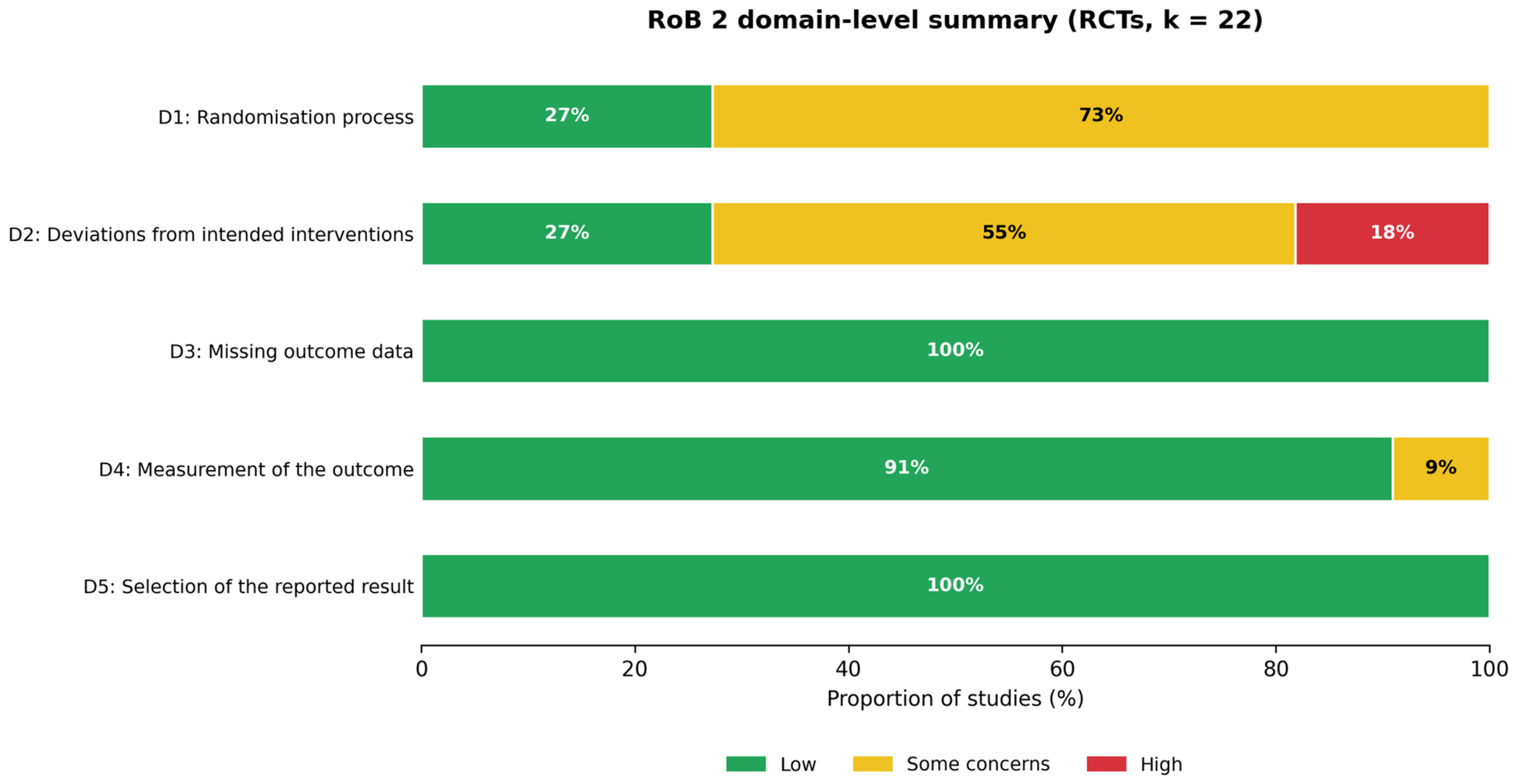
Among the 22 RCTs, 27% (6/22) were at low risk of bias for the randomization process, whereas 73% (16/22) raised some concerns, mainly because the method of allocation concealment was not described. Deviations from the intended interventions raised some concerns in 55% (12/22) of the trials and were at high risk in 18% (4/22), primarily reflecting the open-label nature of surgical interventions. All trials (100%) were at low risk of bias for missing outcome data and for selection of the reported result, and 91% (20/22) were at low risk for measurement of the outcome. Only 4 of the 22 randomized trials (18%) achieved an overall judgement of low risk of bias; in each of these the allocation sequence was concealed in sealed opaque envelopes or generated by an independent party, and the outcome assessors were explicitly blinded. The remaining 18 trials (82%) raised either some concerns or a high risk of bias overall, driven by incomplete reporting of allocation concealment and by the impossibility of blinding the operating surgeon in an open surgical procedure (Figure 2).
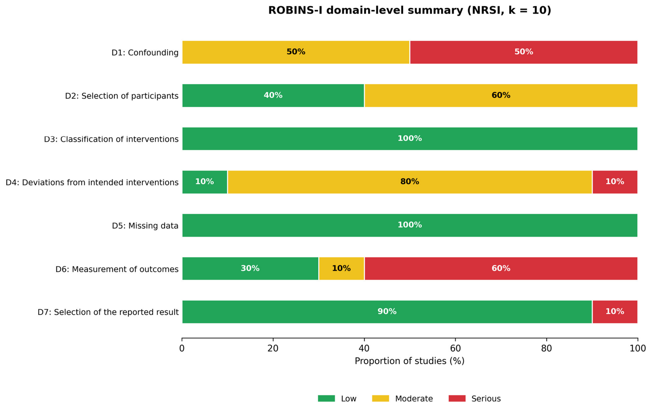
The ROBINS-I assessment revealed a serious overall risk of bias in 7 of the 10 non-randomized and observational studies. Half of the studies were at serious risk of confounding and half at moderate risk; 60% were at moderate risk for selection of participants; 60% were at serious risk for measurement of outcomes; and one study was at serious risk for selection of the reported result (Figure 3).
Francisco et al. [48] was judged at moderate risk of confounding (the within-patient design removes patient-level confounding, but the intervention was deterministically confounded with anatomical side and no side-wise baseline comparison is presented), at low risk for selection of participants, classification of interventions, deviations from intended interventions and missing data, at moderate risk for measurement of the outcome (a single non-blinded operator performed all histomorphometric analyses), and at serious risk for selection of the reported result. The overall judgement was serious risk of bias.

3.4. Primary Outcomes: Histomorphometric Analysis

3.4.1. New Bone Formation (NFB%)

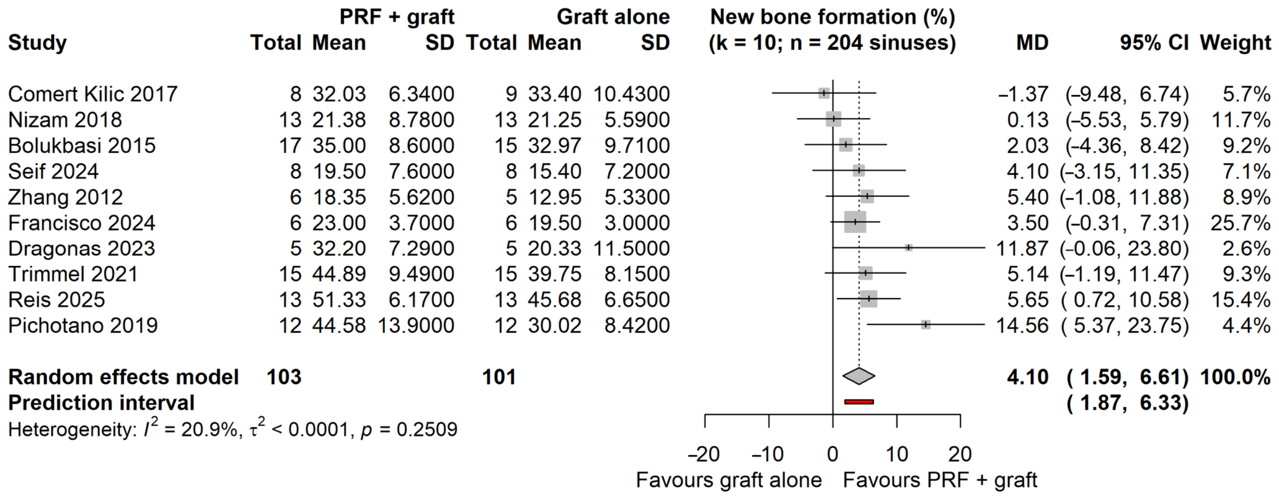
Ten studies contributing 204 augmented sinuses provided extractable histomorphometric data on NFB. The pooled RE estimate favoured the combination of PRF with a particulate bone graft over the graft alone (MD = 4.10%; 95% CI: 1.59 to 6.61; p = 0.005). Between-study heterogeneity was low and not statistically significant (I2 = 20.9%; τ2 < 0.0001; Q p = 0.25), and the 95% PI (1.87 to 6.33) excluded the null, indicating that a benefit of comparable magnitude would be expected in a comparable future study (Figure 4).

3.4.2. Residual Graft Material (RG%)

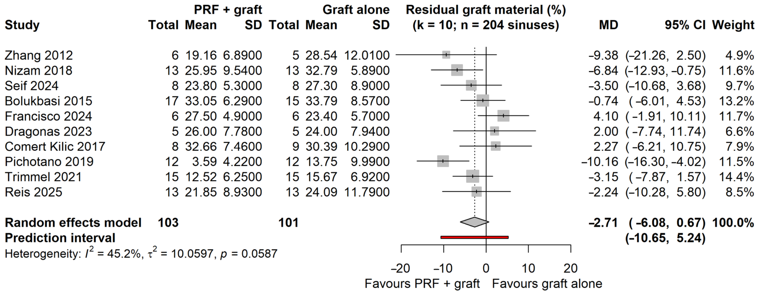
In the comparative evaluation, 10 studies (n = 204 augmented sinuses) contributed data to the analysis of residual graft material. The pooled estimate favoured the PRF-supplemented arm in direction but did not reach statistical significance (MD = −2.71%; 95% CI: −6.08 to 0.67; p = 0.103; I2 = 45.2%). The 95% PI (−10.65 to 5.24) was wide and encompassed both a clinically relevant reduction and an increase in residual graft. Leave-one-out analysis confirmed that no single study drove this null result, although omission of Francisco et al. [48] produced a significant estimate (MD = −3.65%; 95% CI: −6.81 to −0.50; p = 0.028). Accordingly, the present data do not support a firm conclusion that the addition of PRF accelerates graft resorption (Figure 5).

3.5. Subgroup Analyses

To explore the influence of PRF preparation, graft origin, and healing intervals, pre-planned subgroup analyses were conducted (Supplementary Figures S3–S8).

3.5.1. PRF Formulation

For NFB%, no significant subgroup differences were observed among the five PRF formulation strata (p = 0.707). For RG%, a statistically significant between-subgroup difference was detected (p = 0.002), the point estimate for L-PRF indicating the largest numerical reduction in residual graft material (MD = −8.49%; 95% CI: −29.58 to 12.60) relative to standard PRF (MD = −1.08%) and A-PRF (MD = −2.52%). These estimates must, however, be interpreted with considerable caution. The strata are very unequal in size (standard PRF, k = 3; L-PRF, k = 2; A-PRF, k = 3; liquid PRF, k = 1; H-PRF, k = 1), the confidence interval of the L-PRF estimate is very wide and includes the null, and the between-subgroup significance is generated by the opposing direction of the single liquid-PRF study (MD = +4.10%) rather than by the precision of any stratum estimate. The L-PRF confidence interval spans values from a large reduction to a clinically meaningful increase, and the corresponding L-PRF estimate for NFB% was similarly imprecise (MD = 6.87%; 95% CI: −84.61 to 98.35; I2 = 85%). Moreover, the significant subgroup p-value quantifies the contrast between strata and not the precision of the L-PRF estimate itself. Therefore, these subgroup findings are reported as hypothesis-generating and do not support a recommendation for any specific PRF preparation.

3.5.2. Graft Origin

When stratified by graft type (xenograft, k = 7; alloplast, k = 2; allograft, k = 1), a statistically significant reduction in RG% was confined to the xenograft subgroup (MD = −4.43%; 95% CI: −8.42 to −0.44), with a significant between-subgroup difference (p < 0.0001), which is driven by the single alloplast-and-liquid-PRF study lying in the opposite direction. Given the small number of studies contributing to each stratum, this observation is likewise regarded as exploratory and hypothesis-generating rather than as evidence of a differential treatment effect. For NFB%, graft origin did not significantly affect the results (p = 0.593).

3.5.3. Healing Interval

Stratification by healing time (<6 months vs. ≥6 months) did not reveal any statistically significant modifying effects for either NFB% (p = 0.129) or RG% (p = 0.196).

3.6. Meta-Regression Analysis

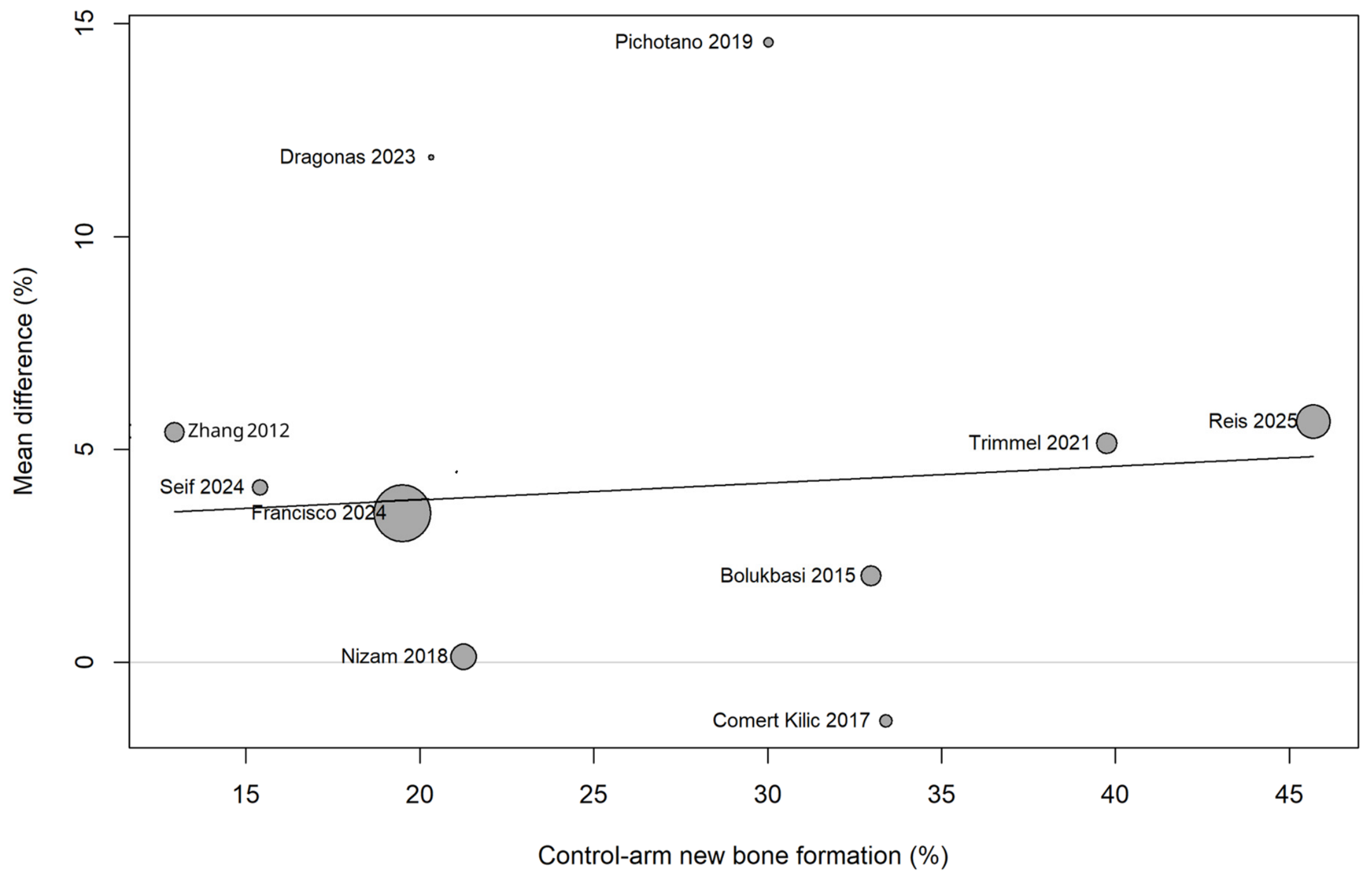
Meta-regression of the mean difference in NFB on the control-arm NFB showed no association (slope = 0.04% per percentage point; 95% CI: −0.20 to 0.28; F(1,8) = 0.14; p = 0.718) (Figure 6), indicating that the benefit associated with PRF was not systematically larger in studies whose control arms healed less well. Residual heterogeneity after adjustment was zero, but this reflects the very low heterogeneity of the corrected dataset rather than explanatory power of the covariate, and the analysis is underpowered with ten studies.
Because Francisco et al. [48] carried the largest single weight in the primary new-bone analysis (25.7%) while being the only included study judged at serious risk of bias for selection of the reported result, a further sensitivity analysis omitting it was performed. The pooled estimate was essentially unchanged (MD = 4.36%; 95% CI: 1.14 to 7.57; p = 0.014), confirming that the primary finding does not depend on this study.

3.7. Secondary Outcomes: Clinical and Radiographic Parameters

3.7.1. Implant Stability Quotient (ISQ)

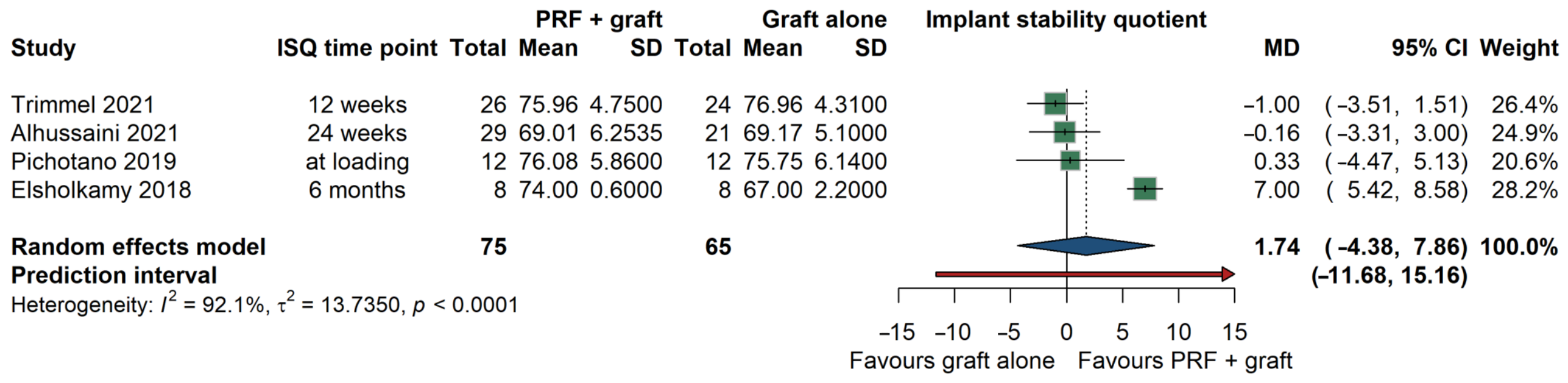
Data from four studies contributing 140 implants evaluated implant stability by resonance frequency analysis. The addition of PRF was associated with a directionally favourable but imprecise increase in ISQ (MD = 1.74; 95% CI: −4.38 to 7.86; p = 0.432), accompanied by substantial heterogeneity (I2 = 92.1%; τ2 = 13.74) (Figure 7). The confidence interval is compatible both with a clinically irrelevant difference and with a moderate benefit; these data are therefore inconclusive rather than demonstrating the absence of an effect.

3.7.2. Vertical Bone Height Gain (mm)

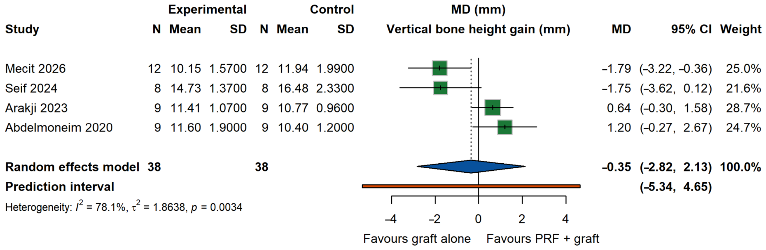
The volumetric and linear gains in vertical bone height were assessed across four studies (n = 76). The pooled MD showed no significant difference between the PRF-augmented and control groups (MD = −0.35 mm; 95% CI: −2.82 to 2.13; p = 0.687). Substantial heterogeneity was observed (I2 = 78.1%, p = 0.003) (Figure 8).

3.7.3. Radiographic Bone Density (HU)

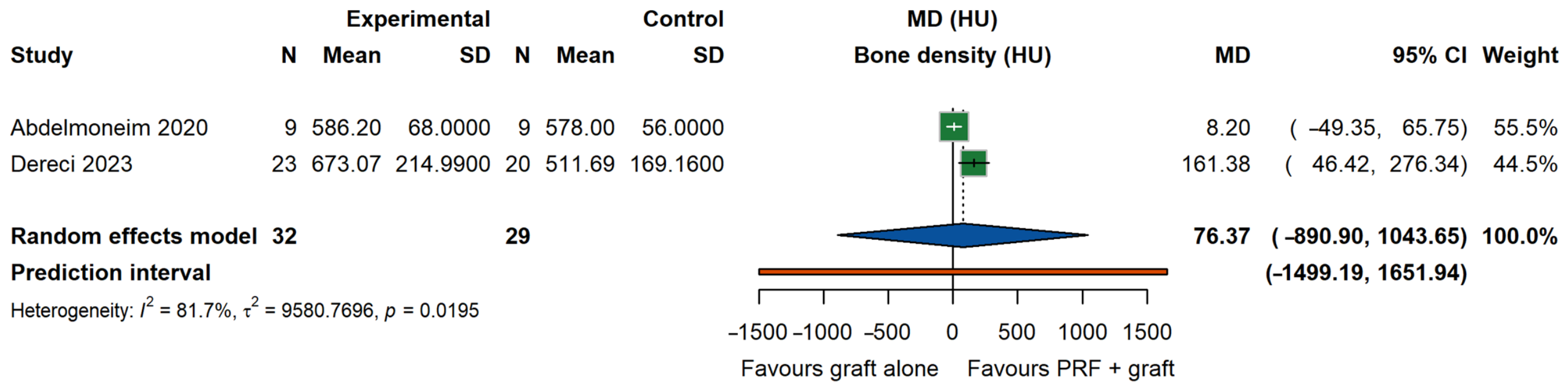
Analysis of radiographic bone density (Hounsfield Units) from two studies (n = 61) demonstrated a non-significant MD of 76.37 HU (95% CI: −890.90 to 1043.65; p = 0.499), burdened by substantial imprecision and heterogeneity (I2 = 81.7%, p = 0.019) (Figure 9).

3.8. Sensitivity Analyses and Publication Bias

Leave-one-out sensitivity analyses were performed for primary histomorphometric outcomes (Supplementary Figures S9–S11). For NFB%, the pooled estimate proved robust: omission of any single study left the effect size between 3.61% and 4.62% and statistically significant in every iteration (p = 0.003 to p = 0.016), and the residual between-study variance fell to zero when Pichotano et al. [55] was omitted.
In contrast, for RG%, the pooled estimate was not statistically significant in the primary analysis and remained non-significant in nine of the ten iterations; statistical significance was reached only when Francisco et al. [48] was omitted (MD = −3.65%; p = 0.028). Influence diagnostics identified Pichotano et al. [55] as the most influential study for new bone formation, the omission of which reduced both τ2 and I2 to zero, and Francisco et al. [48] as the most influential study for residual graft material. Therefore, the reduction in residual graft material should be interpreted as directionally consistent but statistically unconfirmed, and as being driven substantially by a single trial; this fragility, together with a 95% prediction interval that crosses the null, is reflected in the GRADE rating for this outcome. For ISQ, the exclusion of the study by Elsholkamy et al. [38] completely collapsed the heterogeneity to I2 = 0%, identifying it as the driver of inconsistency in this parameter, although the pooled effect remained non-significant.
Influence diagnostics (Supplementary Figures S12–S15), Baujat (Supplementary Figures S16–S18), Drapery (Supplementary Figure S19), and radial (Galbraith) (Supplementary Figure S20) plots confirmed the directional consistency and proportional precision of the included trials. Contour-enhanced funnel plots and Egger’s linear regression test were applied to both outcomes, each of which was informed by exactly ten studies. Neither showed statistically significant funnel plot asymmetry (NFB: t = 1.18, df = 8, p = 0.272; RG material: t = 0.03, df = 8, p = 0.977) (Supplementary Figures S21 and S22). Trim-and-fill analysis imputed two hypothetical studies for new bone formation; the adjusted pooled estimate was attenuated but remained statistically significant (MD = 3.31%; 95% CI: 0.20 to 6.43; p = 0.039) (Supplementary Figures S23 and S24). No studies were imputed for RG material and that estimate was unchanged. With only ten contributing studies these procedures have limited power, and the possibility of small-study effects cannot be excluded on this basis alone.

3.9. Certainty of Evidence (GRADE)

The certainty of the evidence was appraised using the GRADE framework (Table 2). For NFB, the certainty was rated as moderate: the evidence originates from randomized trials and therefore started at high certainty, but was downgraded one level for risk of bias, since only 4 of the 22 randomized trials achieved an overall judgement of low risk of bias and the majority of the pooled weight derives from trials that did not. For residual graft material, the certainty was rated as very low, having been downgraded one level for risk of bias and two further levels for very serious imprecision; the 95% CI includes the null, the pooled estimate reaches statistical significance in only one of ten leave-one-out iterations, and the 95% PI spans both a clinically relevant reduction and an increase. The certainty of evidence for the secondary outcomes (ISQ, vertical bone height gain, and radiographic bone density) was rated as very low, having been downgraded for serious risk of bias, substantial inconsistency (I2 > 75%), very serious imprecision (confidence intervals compatible with both no effect and a clinically relevant benefit), and an inability to exclude publication bias owing to the small number of contributing studies.

4. Discussion

This systematic review and meta-analysis, encompassing 32 studies and 728 augmented maxillary sinuses, provides a robust synthesis of the evidence regarding the adjunctive use of PRF in MSFA. The findings demonstrated that combining PRF with particulate bone grafts yielded a statistically significant enhancement in histological outcomes, specifically a 4.10% increase in NFB, without a statistically significant difference in residual graft material. However, these microscopic biological advantages did not translate into statistically significant differences in macroscopic clinical or radiographic parameters, such as the ISQ or vertical bone height gain.
Several systematic reviews have already addressed platelet concentrates in maxillary sinus floor augmentation [1,2,4,5,8,9]; therefore, the current systematic review is positioned as an update and a methodological extension rather than a repetition, and it differs from previous syntheses in four respects. First, it is the most extensive synthesis to date, comprising 32 studies published between 2006 and 2026 and 728 augmented sinuses, and it incorporates recent trials employing horizontal-centrifugation PRF and sticky-bone formulations that were not available to earlier reviews. Second, all pooled estimates were derived using the REML estimator with the HKSJ adjustment and are reported together with 95% PIs, which convey the range of effects expected in a future study rather than the precision of the mean alone; this is what allows the residual graft finding to be identified here as statistically fragile, a nuance not previously reported. Third, a meta-regression of the treatment effect on control-arm baseline new bone formation was performed to test whether the benefit of PRF depends on the intrinsic osteogenic performance of the graft used alone, a question not previously examined. Fourth, the certainty of the evidence was formally rated for each outcome using GRADE, which permits the histological and the clinical conclusions to be weighted rather than presented as equivalent.
The significant increase in NFB is consistent with the osteopromotive properties of PRF. The three-dimensional fibrin scaffold of PRF acts as a biomimetic matrix that traps platelets and leukocytes, facilitating the sustained release of key cytokines and growth factors, such as PDGF, VEGF, and TGF-β, over a period of 7–14 days [1,34,35]. This sustained release promotes neoangiogenesis, enhances the migration and proliferation of mesenchymal stem cells, and upregulates the expression of osteogenic markers such as RUNX2 and osteocalcin [35,45]. PRF accelerates biological turnover (creeping substitution) of the graft, shifting the microenvironment toward active bone remodelling earlier in the healing phase [40,47].
Subgroup analyses were undertaken to explore possible sources of heterogeneity and to generate hypotheses regarding protocol optimization; they were not powered to establish the superiority of any single preparation. Numerically, the reduction in residual graft material was most pronounced when L-PRF was used, and the between-subgroup contrast reached statistical significance (p = 0.002). Biologically, this would be consistent with a role for the leukocytes retained within the L-PRF matrix in the local immune response and in osteoclastogenesis, thereby accelerating degradation of the graft scaffold [5,26]. However, the L-PRF stratum comprised only two studies and its confidence interval was extremely wide (MD = −8.49%; 95% CI: −29.58 to 12.60), encompassing values from a large reduction to a clinically meaningful increase, and the corresponding NFB% estimate was equally imprecise (MD = 6.87%; 95% CI: −84.61 to 98.35). Therefore, the significant subgroup p-value reflects the contrast between strata rather than a precise estimate for L-PRF, and it does not license a recommendation for this preparation over A-PRF or standard PRF. The same caution applies to the graft-origin analysis, in which accelerated resorption reached significance only within the xenograft stratum (MD = −4.43%; 95% CI: −8.42 to −0.44; between-subgroup p < 0.0001). The proposed mechanism is biologically plausible, since DBBM is osteoconductive but devoid of osteoinductive proteins and exhibits a very slow resorption profile [7,55], and the addition of PRF may confer osteopromotive properties and a faster turnover rate that is advantageous when early implant placement is planned [33,55]; nevertheless, with so few studies per stratum this remains a hypothesis to be tested in trials designed for that purpose rather than an established finding.
Our meta-analysis did not detect a statistically significant difference in implant stability as measured by ISQ; the pooled estimate favoured PRF numerically (MD = +1.74 ISQ units) but was too imprecise to exclude either a null effect or a moderate benefit, and it should not be interpreted as evidence of equivalence. Both the PRF-augmented and the graft-alone groups achieved ISQ values approaching or exceeding the threshold generally regarded as compatible with successful osseointegration (approximately 70) [25,38]. One plausible explanation for the absence of a demonstrable difference is a ceiling effect: modern rough-surfaced implants placed in adequately condensed particulate grafts achieve excellent primary mechanical stability irrespective of the addition of biologics [42]. In addition, ISQ reflects the mechanical interface between the implant and the surrounding bone at a single time point and may be relatively insensitive to the histological changes in graft maturation demonstrated in the present analysis. Similarly, the vertical bone height gain and radiographic bone density did not differ significantly between the groups. Vertical bone gain in MSFA is dictated by the surgical space created by the elevation of the Schneiderian membrane and the physical volume and dimensional stability of the particulate scaffold, rather than the osteoinductive biological properties of the graft mixture [27,32].
From a clinical standpoint, the pattern of results supports a specific and limited indication rather than the routine addition of PRF to every sinus augmentation. Because the demonstrable effect of PRF is exerted on graft turnover and histological maturation rather than on the volume or the mechanical stability of the augmented site, its principal clinical value lies in protocols in which the graft maturation interval is the rate-limiting step, most notably when a slowly resorbing xenograft is used and earlier implant placement (approximately four months rather than six to eight months) is planned. Conversely, when the clinical objective is to achieve a defined vertical bone height, to increase radiographic density, or to enhance primary implant stability, the current evidence does not support the expectation of an additional benefit, and the associated venipuncture, chairside time, and cost should be weighed accordingly. The absolute magnitude of the histological benefit should be kept in perspective: a mean difference of approximately four percentage points in new bone formation is of uncertain clinical relevance in isolation, and it was not accompanied by a demonstrable acceleration of graft resorption. Given that only 4 of 22 trials were at low overall risk of bias and that the residual graft estimate is statistically unconfirmed, PRF is best presented to patients and clinicians as a low-risk, low-cost autologous adjunct with a probable biological benefit, rather than as an intervention of demonstrated clinical superiority.
Although this meta-analysis provides a comprehensive synthesis of the currently available evidence, several limitations must be acknowledged. First, and most importantly, the evidence base is affected by severe clinical and methodological heterogeneity. PRF preparation was not standardized across trials: relative centrifugal force, spin duration, rotor angulation, and tube type differed, these parameters are now recognized as principal determinants of the cellular and growth-factor content of the final product [5,49], yet relative centrifugal force was reported in only 10 of the 32 studies and rotor geometry in only 1. The comparator grafts were equally diverse, ranging from slowly resorbing xenografts to rapidly resorbing alloplasts, and the healing intervals ranged from 3 to 9 months. Consequently, the pooled estimates should be interpreted as an average effect across a family of related but non-identical interventions rather than as the effect of a single reproducible protocol. Subgroup analyses, meta-regression, and prediction intervals were used to explore and to express this heterogeneity, but they cannot eliminate it; the prediction intervals in particular indicate that the effect observed in a future individual study may differ appreciably from the pooled mean, and for residual graft material the prediction interval crosses the null.
Second, the risk of bias of the randomized evidence deserves particular emphasis: only 4 of the 22 included trials reached an overall judgement of low risk of bias, reflecting an intrinsic constraint of surgical research, since the operator cannot be blinded to whether a fibrin clot is incorporated into the graft, and it was compounded by incomplete reporting of allocation concealment. The consequences are not uniform across outcomes. Histomorphometric endpoints are measured on coded biopsy specimens by an examiner who can be, and in most studies was, blinded; performance bias is therefore less likely to have inflated the NFB% and RG% estimates. In contrast, ISQ readings and radiographic measurements were generally obtained by unblinded operators and are more vulnerable to detection bias. Therefore, readers should regard all effect estimates reported here as originating from the evidence at moderate to high risk of bias, and should weigh the histological findings, which are supported by moderate-certainty evidence, differently from the clinical and radiographic findings, for which certainty is very low. The GRADE certainty of evidence was appraised as moderate for histological outcomes and very low for clinical and radiographic outcomes. Third, most studies evaluated short- to medium-term outcomes (4–9 months). Long-term data evaluating implant survival and marginal bone loss over 5–10 years remain scarce.
Fourth, full-text reports could not be retrieved for 17 of the 51 records (33.3%) that progressed to the retrieval stage. Retrieval failure was concentrated in conference abstracts, academic theses, and articles published in non-indexed or non-archived local journals, and repeated attempts through institutional subscriptions, interlibrary loan, and direct correspondence with the authors were unsuccessful. Because these records could not be formally assessed against the eligibility criteria, an unquantified risk of selection bias remains: it cannot be determined how many would have proved eligible, nor whether their results would have differed systematically from those of the retrieved studies. Two observations partially mitigate, but do not remove, this concern: formal small-study effect testing for the two outcomes with at least ten studies showed no funnel plot asymmetry (Egger’s test p = 0.272 for NFB% and p = 0.977 for RG%); trim-and-fill imputed two studies for NFB%, and the adjusted estimate remained significant, while no studies were imputed for RG%. Nevertheless, this limitation should be weighed when interpreting the strength of the present conclusions, and it constitutes a further reason for regarding the secondary clinical outcomes as inconclusive.

5. Conclusions

The adjunctive use of PRF with a particulate bone graft in MSFA is associated with a modest but consistent increase of approximately four percentage points in NFB at histomorphometric analysis, a finding supported by moderate-certainty evidence and by a prediction interval that excludes the null. This histological advantage did not translate into a demonstrable difference in residual graft material, implant stability quotient, vertical bone height gain, or radiographic bone density, all of which were supported by low or very low certainty evidence with CIs compatible with clinically relevant effects in either direction. Therefore, PRF may be considered a low-cost autologous adjunct where accelerated graft maturation is the specific objective, but the current evidence does not justify a general recommendation for its routine use, nor does it support a preference for any particular preparation protocol.

Supplementary Materials

The following supporting information can be downloaded at: https://www.mdpi.com/article/10.3390/surgeries7030102/s1, Table S1: Platelet-rich fibrin preparation protocols (centrifuge device, relative centrifugal force, rotational speed, spin duration, rotor geometry, tube type and coating, and final physical form of the product) and graft characteristics (origin, particle size and expected resorption profile) of the 32 included studies; Figure S1: Study-level (traffic-light) risk-of-bias summary for the 22 randomized controlled trials assessed with the Cochrane RoB 2 tool; Figure S2: Study-level (traffic-light) risk-of-bias summary for the 10 non-randomized studies assessed with the ROBINS-I tool; Figure S3: Subgroup analysis of new bone formation (NFB%) stratified by PRF formulation; Figure S4: Subgroup analysis of residual graft material (RG%) stratified by PRF formulation; Figure S5: Subgroup analysis of NFB% stratified by graft origin; Figure S6: Subgroup analysis of RG% stratified by graft origin; Figure S7: Subgroup analysis of NFB% stratified by healing interval (<6 months vs. ≥6 months); Figure S8: Subgroup analysis of RG% stratified by healing interval (<6 months vs. ≥6 months); Figure S9: Leave-one-out sensitivity analysis for NFB%; Figure S10: Leave-one-out sensitivity analysis for RG%; Figure S11: Leave-one-out sensitivity analysis for the implant stability quotient (ISQ); Figure S12: Influence diagnostics (externally standardized residuals, Cook’s distances, leverage values, DFFITS and DFBETAS) for NFB%; Figure S13: Influence diagnostics for RG%; Figure S14: Influence diagnostics for ISQ; Figure S15: Influence diagnostics for vertical bone height gain; Figure S16: Baujat plot for NFB%; Figure S17: Baujat plot for RG%; Figure S18: Baujat plot for ISQ; Figure S19: Drapery plot for NFB%; Figure S20: Radial (Galbraith) plot for NFB%; Figure S21: Contour-enhanced funnel plot with Egger’s regression test for NFB%; Figure S22: Contour-enhanced funnel plot with Egger’s regression test for RG%; Figure S23: Trim-and-fill adjusted funnel plot for NFB%; Figure S24: Trim-and-fill funnel plot for RG%.

Author Contributions

Conceptualization, A.A.D.; methodology, A.A.D. and R.A.A.; software, A.S.A. and R.J.A.; validation, R.S., J.A.A. and N.A.A.; formal analysis, A.A.D. and S.M.M.H.; investigation, W.F.A. and A.M.A.H.; resources, M.A.A. and A.R.M.A.A.; data curation, A.M.H. and N.A.; writing—original draft preparation, A.A.D., R.A.A. and A.S.A.; writing—review and editing, all authors; visualization, R.J.A. and L.S.A.; supervision, A.A.D.; project administration, A.A.D. All authors have read and agreed to the published version of the manuscript.

Funding

This research received no external funding.

Institutional Review Board Statement

Not applicable. This study is a systematic review and meta-analysis of previously published data and did not involve new studies on humans or animals.

Informed Consent Statement

Not applicable.

Data Availability Statement

The data supporting the findings of this study are available within the article and its Supplementary Material. Additional data are available from the corresponding author upon request.

Conflicts of Interest

The authors declare no conflicts of interest.

Abbreviations

The following abbreviations are used in this manuscript:
MSFAMaxillary sinus floor augmentation
PRFPlatelet-rich fibrin
L-PRFLeukocyte- and platelet-rich fibrin
A-PRFAdvanced platelet-rich fibrin
i-PRFInjectable platelet-rich fibrin
T-PRFTitanium-prepared platelet-rich fibrin
H-PRFHorizontal centrifugation platelet-rich fibrin
PRPPlatelet-rich plasma
P-PRPPure platelet-rich plasma
CGFConcentrated growth factors
PRGFPlasma rich in growth factors
MPMMineralized plasmatic matrix
DBBMDeproteinized bovine bone mineral
FDBAFreeze-dried bone allograft
β-TCPBeta-tricalcium phosphate
BCPBiphasic calcium phosphate
SACBASerum albumin-coated bone allograft
NFBNewly formed bone
RGResidual graft material
ISQImplant stability quotient
MBLMarginal bone loss
CBCTCone-beam computed tomography
HUHounsfield units
IHCImmunohistochemistry
RFAResonance frequency analysis
PDGFPlatelet-derived growth factor
VEGFVascular endothelial growth factor
TGF-βTransforming growth factor-beta
BV/TVBone volume/total volume
RBHResidual bone height
RCTRandomized controlled trial
CCTControlled clinical trial
IRRInter-rater reliability
RoB 2Cochrane Risk of Bias 2 tool
ROBINS-IRisk of Bias in Non-randomized Studies-of Interventions
GRADEGrading of Recommendations Assessment, Development, and Evaluation
PRISMAPreferred Reporting Items for Systematic Reviews and Meta-Analyses
PICOSPopulation, Intervention, Comparator, Outcomes, Study design
PROSPEROInternational Prospective Register of Systematic Reviews
MeSHMedical Subject Headings
WHO-ICTRPWHO International Clinical Trials Registry Platform
MDMean difference
SMDStandardized mean difference
WMDWeighted mean difference
RRRisk ratio
CIConfidence interval
RERandom-effects
REMLRestricted maximum likelihood
HKSJHartung-Knapp-Sidik-Jonkman
PROMsPatient-reported outcome measures
VASVisual analog scale

References

  1. Mendes, V.V.; Martins, F.V.; Santana, C.M.M.; Santana, R.B. Do Recombinant, Purified, and Concentrated Growth Factors Enhance the Regenerative Potential of Particulate Bone Graft Substitutes in Maxillary Sinus Floor Augmentation? A Systematic Review and Meta-analysis. Int. J. Oral Maxillofac. Implants 2024, 39, e87–e101. [Google Scholar] [CrossRef] [Scilit] [PubMed]
  2. Babich, O.; Lugassy, E.; Babich, M.; Abayov, P.; Haimov, E.; Juodzbalys, G. The Use of Platelet-Rich Fibrin in Sinus Floor Augmentation Surgery: A Systematic Review. J. Oral Maxillofac. Res. 2024, 15, e1. [Google Scholar] [CrossRef] [Scilit] [PubMed]
  3. Costa, R.; Carvalho, A.; López-Jarana, P.; Costa, V.; Relvas, M.; Salazar, F.; da Câmara, T.I.; Vasques, M.N.; da Câmara, M.I. The Use of Platelet-Rich Fibrin in Combination with Synthetic Bone Grafting: A Systematic Review. Biomedicines 2025, 13, 2266. [Google Scholar] [CrossRef] [Scilit] [PubMed]
  4. Zhang, Y.; Yang, S.S.; Zhang, N.N.; Huang, G.L. Effect of platelet-derived bone enhancers used as adjuncts to deproteinized bovine bone matrix in maxillary sinus floor elevation: A systematic review and meta-analysis. BMC Oral Health 2025, 25, 1120. [Google Scholar] [CrossRef] [Scilit] [PubMed]
  5. Malcangi, G.; Patano, A.; Palmieri, G.; Di Pede, C.; Latini, G.; Inchingolo, A.D.; Hazballa, D.; de Ruvo, E.; Garofoli, G.; Inchingolo, F.; et al. Maxillary Sinus Augmentation Using Autologous Platelet Concentrates (Platelet-Rich Plasma, Platelet-Rich Fibrin, and Concentrated Growth Factor) Combined with Bone Graft: A Systematic Review. Cells 2023, 12, 1797. [Google Scholar] [CrossRef] [Scilit] [PubMed]
  6. de Azevedo, M.W.C.; Matos, L.A.d.S.; Silva, T.B.L.; e Silva, W.A.; Nascimento, F.C.D.; Silva, T.M.; Leal, L.P.; Paiva, L.M.; Brígido, J.A. Use of platelet- and leukocyte-rich fibrin (L-PRF) as an alternative for tissue regeneration in sinus lift surgery. Res. Soc. Dev. 2022, 11, e43111225751. [Google Scholar] [CrossRef] [Scilit]
  7. Pichotano, E.C.; de Molon, R.S.; de Paula, L.G.F.; de Souza, R.V.; Marcantonio, E., Jr.; Zandim-Barcelos, D.L. Early placement of dental implants in maxillary sinus grafted with leukocyte and platelet-rich fibrin (L-PRF) and deproteinized bovine bone mineral. J. Oral Implantol. 2018, 44, 199–206. [Google Scholar] [CrossRef] [Scilit] [PubMed]
  8. Liu, R.; Yan, M.; Chen, S.; Huang, W.; Wu, D.; Chen, J. Effectiveness of Platelet-Rich Fibrin as an Adjunctive Material to Bone Graft in Maxillary Sinus Augmentation: A Meta-Analysis of Randomized Controlled Trials. BioMed Res. Int. 2019, 2019, 7267062. [Google Scholar] [CrossRef] [Scilit] [PubMed]
  9. Shinohara, É.H.; de Santana, I.H.G.; Viana, M.R.M.; de Almeida, E.J.R.; Ferreira, A.J.; Júnior, J.M.P.; Andrade, A.F.d.B.; Horikawa, F.K.; Ribeiro, E.D. Efficacy of Platelet and Leukocyte Rich Fibrin (L-PRF) in the healing process and bone repair in maxillary sinus lift surgeries: A systematic review. Cell Tissue Bank. 2025, 26, 36. [Google Scholar] [CrossRef] [Scilit] [PubMed]
  10. Page, M.J.; McKenzie, J.E.; Bossuyt, P.M.; Boutron, I.; Hoffmann, T.C.; Mulrow, C.D.; Shamseer, L.; Tetzlaff, J.M.; Akl, E.A.; Brennan, S.E.; et al. The PRISMA 2020 statement: An updated guideline for reporting systematic reviews. BMJ 2021, 372, n71. [Google Scholar] [CrossRef] [Scilit] [PubMed]
  11. Higgins, J.P.T.; Thomas, J.; Chandler, J.; Cumpston, M.; Li, T.; Page, M.J.; Welch, V.A. (Eds.) Cochrane Handbook for Systematic Reviews of Interventions, 2nd ed.; John Wiley & Sons: Chichester, UK, 2019. [Google Scholar] [CrossRef] [Scilit]
  12. McHugh, M.L. Interrater reliability: The kappa statistic. Biochem. Med. 2012, 22, 276–282. [Google Scholar] [CrossRef] [Scilit]
  13. Sterne, J.A.C.; Savović, J.; Page, M.J.; Elbers, R.G.; Blencowe, N.S.; Boutron, I.; Cates, C.J.; Cheng, H.Y.; Corbett, M.S.; Eldridge, S.M.; et al. RoB 2: A revised tool for assessing risk of bias in randomised trials. BMJ 2019, 366, l4898. [Google Scholar] [CrossRef] [Scilit] [PubMed]
  14. Sterne, J.A.C.; Hernán, M.A.; Reeves, B.C.; Savović, J.; Berkman, N.D.; Viswanathan, M.; Henry, D.; Altman, D.G.; Ansari, M.T.; Boutron, I.; et al. ROBINS-I: A tool for assessing risk of bias in non-randomised studies of interventions. BMJ 2016, 355, i4919. [Google Scholar] [CrossRef] [Scilit] [PubMed]
  15. Guyatt, G.H.; Oxman, A.D.; Vist, G.E.; Kunz, R.; Falck-Ytter, Y.; Alonso-Coello, P.; Schünemann, H.J. GRADE: An emerging consensus on rating quality of evidence and strength of recommendations. BMJ 2008, 336, 924–926. [Google Scholar] [CrossRef] [Scilit] [PubMed]
  16. R Core Team. R: A Language and Environment for Statistical Computing; R Foundation for Statistical Computing: Vienna, Austria, 2026. [Google Scholar] [CrossRef] [Scilit]
  17. Balduzzi, S.; Rücker, G.; Schwarzer, G. How to perform a meta-analysis with R: A practical tutorial. Evid. Based Ment. Health 2019, 22, 153–160. [Google Scholar] [CrossRef] [Scilit] [PubMed]
  18. Borenstein, M.; Hedges, L.V.; Higgins, J.P.T.; Rothstein, H.R. Introduction to Meta-Analysis; John Wiley & Sons: Chichester, UK, 2009. [Google Scholar] [CrossRef] [Scilit]
  19. IntHout, J.; Ioannidis, J.P.; Borm, G.F. The Hartung-Knapp-Sidik-Jonkman method for random effects meta-analysis is straightforward and considerably outperforms the standard DerSimonian-Laird method. BMC Med. Res. Methodol. 2014, 14, 25. [Google Scholar] [CrossRef] [Scilit] [PubMed]
  20. Higgins, J.P.T.; Thompson, S.G.; Deeks, J.J.; Altman, D.G. Measuring inconsistency in meta-analyses. BMJ 2003, 327, 557–560. [Google Scholar] [CrossRef] [Scilit] [PubMed]
  21. Viechtbauer, W. Conducting meta-analyses in R with the metafor package. J. Stat. Softw. 2010, 36, 1–48. [Google Scholar] [CrossRef] [Scilit]
  22. Egger, M.; Davey Smith, G.; Schneider, M.; Minder, C. Bias in meta-analysis detected by a simple, graphical test. BMJ 1997, 315, 629–634. [Google Scholar] [CrossRef] [Scilit] [PubMed]
  23. Duval, S.; Tweedie, R. Trim and fill: A simple funnel-plot-based method of testing and adjusting for publication bias in meta-analysis. Biometrics 2000, 56, 455–463. [Google Scholar] [CrossRef] [Scilit] [PubMed]
  24. Gassling, V.; Purcz, N.; Braesen, J.H.; Will, M.; Gierloff, M.; Behrens, E.; Açil, Y.; Wiltfang, J. Comparison of two different absorbable membranes for the coverage of lateral osteotomy sites in maxillary sinus augmentation: A preliminary study. J. Craniomaxillofac. Surg. 2013, 41, 76–82. [Google Scholar] [CrossRef] [Scilit] [PubMed]
  25. Alhussaini, A.H.A.; Hassan, T.A.L.; Mohammed, H.B. Evaluation of Implant Stability Following Sinus Augmentation Utilizing Bovine Bone Mixed with Platelet-Rich Fibrin. Indian J. Forensic Med. Toxicol. 2021, 15, 390–399. [Google Scholar] [CrossRef] [Scilit]
  26. Nizam, N.; Eren, G.; Akcalı, A.; Donos, N. Maxillary sinus augmentation with leukocyte and platelet-rich fibrin and deproteinized bovine bone mineral: A split-mouth histological and histomorphometric study. Clin. Oral Implants Res. 2018, 29, 67–75. [Google Scholar] [CrossRef] [Scilit] [PubMed]
  27. Dereci, Ö.; Saruhan, N.; Tekin, G.; Uğurlu, M. Volumetric Comparison of New Bone Formation in Sinus Floor Augmentation Using Allografts with/without Platelet-Rich Fibrin Cohort Research (Retrospective). Turk. Klin. J. Dent. Sci. 2023, 29, 409–418. [Google Scholar] [CrossRef] [Scilit]
  28. Seif, S.A.; Elhadidy, M.S.; Emara, A.S. Xenograft with Advanced Platelet Rich Fibrin Compared to Xenograft for Maxillary Sinus Floor Augmentation: Randomized Clinical Trial. Egypt. Dent. J. 2024, 70, 3183–3192. [Google Scholar] [CrossRef] [Scilit]
  29. Powell, C.A.; Casarez-Quintana, A.; Zellner, J.; Al-Bayati, O.; Font, K. The application of leukocyte- and platelet-rich fibrin (L-PRF) in maxillary sinus augmentation. Clin. Adv. Periodontics 2022, 12, 277–286. [Google Scholar] [CrossRef] [Scilit] [PubMed]
  30. Arakji, H.; Osman, E.; Aboelsaad, N.; Shokry, M. Clinical, Radiographic, and Histological Evaluation of the Mineralized Plasmatic Matrix/Xenograft Mixture in Maxillary Sinus Floor Augmentation (A Randomized Controlled Clinical Trial). Eur. J. Dent. Oral Health 2023, 4, 7–13. [Google Scholar] [CrossRef] [Scilit]
  31. Mecit, M.; Koşar, Y.Ç. Evaluation of I-PRF-Enriched Bone Graft Matrix (Sticky Bone) Among Patients Requiring Sinus Lift Operation: A Retrospective Comparative Study. Med. Sci. Monit. 2026, 32, e952138. [Google Scholar] [CrossRef] [Scilit] [PubMed]
  32. Gadipelly, S.; Sultana, S.; Venkatesh, V.V.; Praveen, P. Comparative Radiological Analysis of Efficacy of Beta-tricalcium Phosphate and Beta-tricalcium Phosphate with Platelet-rich Fibrin in Maxillary Sinus Augmentation—A Clinical Study. Indian J. Dent. Adv. 2019, 10, 171–175. [Google Scholar] [CrossRef] [Scilit]
  33. Reis, G.G.D.; Denardi, R.J.; de Souza, S.L.S.; Silva, P.H.F.; Furlaneto, F.; Miron, R.J.; Mourão, C.F.; Messora, M.R. Sinus Floor Elevation with Platelet-Rich Fibrin from Horizontal Centrifugation and Xenograft: Randomized Clinical Trial. Clin. Implant Dent. Relat. Res. 2025, 27, e70093. [Google Scholar] [CrossRef] [Scilit] [PubMed]
  34. Choukroun, J.; Diss, A.; Simonpieri, A.; Girard, M.O.; Schoeffler, C.; Dohan, S.L.; Dohan, A.J.J.; Mouhyi, J.; Dohan, D.M. Platelet-rich fibrin (PRF): A second-generation platelet concentrate. Part V: Histologic evaluations of PRF effects on bone allograft maturation in sinus lift. Oral Surg. Oral Med. Oral Pathol. Oral Radiol. Endod. 2006, 101, 299–303. [Google Scholar] [CrossRef] [Scilit] [PubMed]
  35. Malzoni, C.M.A.; Pichotano, E.C.; de Paula, L.G.F.; de Souza, R.V.; Okamoto, R.; Austin, R.S.; Marcantonio, E., Jr.; de Molon, R.S.; Zandim-Barcelos, D.L. Combination of leukocyte and platelet-rich fibrin and demineralized bovine bone graft enhanced bone formation and healing after maxillary sinus augmentation: A randomized clinical trial. Clin. Oral Investig. 2023, 27, 5485–5498. [Google Scholar] [CrossRef] [Scilit] [PubMed]
  36. Zhang, Y.; Tangl, S.; Huber, C.D.; Lin, Y.; Qiu, L.; Rausch-Fan, X. Effects of Choukroun’s platelet-rich fibrin on bone regeneration in combination with deproteinized bovine bone mineral in maxillary sinus augmentation: A histological and histomorphometric study. J. Craniomaxillofac. Surg. 2012, 40, 321–328. [Google Scholar] [CrossRef] [Scilit] [PubMed]
  37. Khalfalla, A.E.; Fahmy, M.H.; El-Ashwah, A.A. Evaluation of Combination of Biphasic Calcium Phosphate and Platelet-Rich Fibrin as Grafting Material for Sinus Lift Augmentation. Alex. Dent. J. 2015, 40, 140–147. [Google Scholar] [CrossRef] [Scilit]
  38. Elsholkamy, M.A.; Eldesouky, G. Monitoring of Implant Stability When Placed Simultaneously in Platelet Rich Fibrin Enhanced Maxillary Sinus Bone Augmentation. Egypt. Dent. J. 2018, 64, 3149–3163. [Google Scholar] [CrossRef] [Scilit][Green Version]
  39. Bölükbaşı, N.; Ersanlı, S.; Keklikoğlu, N.; Başeğmez, C.; Özdemir, T. Sinus augmentation with platelet-rich fibrin in combination with bovine bone graft versus bovine bone graft in combination with collagen membrane. J. Oral Implantol. 2015, 41, 586–595. [Google Scholar] [CrossRef] [Scilit] [PubMed]
  40. Cömert Kılıç, S.; Güngörmüş, M.; Parlak, S.N. Histologic and histomorphometric assessment of sinus-floor augmentation with beta-tricalcium phosphate alone or in combination with pure-platelet-rich plasma or platelet-rich fibrin: A randomized clinical trial. Clin. Implant Dent. Relat. Res. 2017, 19, 959–967. [Google Scholar] [CrossRef] [Scilit] [PubMed]
  41. Soliman, O.H. Maxillary Sinus Lift to Enable One Stage Implant Placement Using Nanobone and PRF with or Without Simvastatin. Egypt. Dent. J. 2021, 67, 1229–1239. [Google Scholar] [CrossRef] [Scilit]
  42. Ibrahim, R.M.K.; El Ashwah, A.A.; Swedan, A.O.; Khalil, N.M.; Metawie, D.M.N. Evaluation of Titanium Prepared Platelet Rich Fibrin in Bone Regeneration in Lateral Window Maxillary Sinus Augmentation with Delayed Implant Placement (Randomized Controlled Clinical Trial). Alex. Dent. J. 2024, 50, 16–27. [Google Scholar] [CrossRef] [Scilit]
  43. Abdelmoneim, H.S.; Elfaramawi, T.I.; Meguid, I.M.A.; Elsharkawy, R.T. Allogenic Sticky Bone and Xenogenic Sticky Bone Versus Conventional Sticky Bone Preparations in Maxillary Sinus Floor Augmentation. Radiographic and Histological Evaluation. (Randomized Clinical Trial). Egypt. Dent. J. 2026, 72, 311–321. [Google Scholar] [CrossRef] [Scilit]
  44. Abdelmoneim, H.S.; Elsharkawy, R.T. Flowable Platelet Rich Fibrin a New Trend for Enhancing Bone Augmentation Results in Maxillary Sinus Floor Augmentation. (Radiographic and Histologic Evaluation). Egypt. Dent. J. 2020, 66, 873–881. [Google Scholar] [CrossRef] [Scilit]
  45. Arumugam, K.; Narayanan, V.; Chandran, S.; Ramakrishnan, K.; Subramanian, A. A qualitative and quantitative assessment of bone regeneration after sinus augmentation. A randomized comparative study. J. Osseointegr. 2021, 13, 13–20. [Google Scholar] [CrossRef] [Scilit]
  46. Barbu, H.M.; Andreescu, C.F.; Comaneanu, M.R.; Referendaru, D.; Mijiritsky, E. Maxillary Sinus Floor Augmentation to Enable One-Stage Implant Placement by Using Bovine Bone Substitute and Platelet-Rich Fibrin. BioMed Res. Int. 2018, 2018, 6562958. [Google Scholar] [CrossRef] [Scilit] [PubMed]
  47. Dragonas, P.; Prasad, H.S.; Yu, Q.; Mayer, E.T.; Fidel, P.L., Jr. Bone Regeneration in Maxillary Sinus Augmentation Using Advanced Platelet-Rich Fibrin (A-PRF) and Plasma Rich in Growth Factors (PRGF): A Pilot Randomized Controlled Trial. Int. J. Periodontics Restor. Dent. 2023, 43, 319–327. [Google Scholar] [CrossRef] [Scilit] [PubMed]
  48. Francisco, L.; Francisco, M.; Costa, R.; Vasques, M.N.; Relvas, M.; Rajão, A.; Monteiro, L.; Rompante, P.; Guerra, F.; Infante da Câmara, M. Sinus Floor Augmentation with Synthetic Hydroxyapatite (NanoBone®) in Combination with Platelet-Rich Fibrin: A Case Series. Biomedicines 2024, 12, 1661. [Google Scholar] [CrossRef] [Scilit] [PubMed]
  49. Trimmel, B.; Gyulai-Gaál, S.; Kivovics, M.; Jákob, N.P.; Hegedűs, C.; Szabó, B.T.; Dobó-Nagy, C.; Szabó, G. Evaluation of the Histomorphometric and Micromorphometric Performance of a Serum Albumin-Coated Bone Allograft Combined with A-PRF for Early and Conventional Healing Protocols after Maxillary Sinus Augmentation: A Randomized Clinical Trial. Materials 2021, 14, 1810. [Google Scholar] [CrossRef] [Scilit] [PubMed]
  50. Arafat, S.W.; Chehata, I.M.; Hossam, A.M. Sinus Floor Augmentation with Simultaneous Implant Placement Using Bio-Oss with and without PRF: Clinical Study. Egypt. Dent. J. 2017, 63, 355–364. [Google Scholar] [CrossRef] [Scilit]
  51. Kumar, M.; Chopra, S.; Das, D.; Gupta, M.; Memoalia, J.; Verma, G. Direct Maxillary Sinus Floor Augmentation for Simultaneous Dental Implant Placement. Ann. Maxillofac. Surg. 2018, 8, 188–192. [Google Scholar] [CrossRef] [Scilit] [PubMed]
  52. Xie, H.; Yongfu, L.; Qin, S.; Lingyan, C.; Minzhen, C. Application effect of injectable platelet-rich fibrin in extramaxillary sinus lift surgery. Shanghai Stomatol. 2019, 28, 71–75. [Google Scholar] [CrossRef]
  53. Tatullo, M.; Marrelli, M.; Cassetta, M.; Pacifici, A.; Stefanelli, L.V.; Scacco, S.; Dipalma, G.; Pacifici, L.; Inchingolo, F. Platelet Rich Fibrin (P.R.F.) in Reconstructive Surgery of Atrophied Maxillary Bones: Clinical and Histological Evaluations. Int. J. Med. Sci. 2012, 9, 872–880. [Google Scholar] [CrossRef] [Scilit] [PubMed]
  54. Dereci, Ö.; Saruhan, N.; Tekin, G.; Koşar, Y.Ç. Volumetric Evaluation of Sinus Floor Augmentation Using Platelet-Rich Fibrin and Allogenic Bone Graft Mixture with Simultaneous Dental Implant Placement in Severely Atrophic Posterior Maxillae in a Geriatric Patient Population Cohort Research (Retrospective). Turk. Klin. J. Dent. Sci. 2022, 28, 511–518. [Google Scholar] [CrossRef] [Scilit]
  55. Pichotano, E.C.; de Molon, R.S.; de Souza, R.V.; Austin, R.S.; Marcantonio, E., Jr.; Zandim-Barcelos, D.L. Evaluation of L-PRF combined with deproteinized bovine bone mineral for early implant placement after maxillary sinus augmentation: A randomized clinical trial. Clin. Implant Dent. Relat. Res. 2019, 21, 253–262. [Google Scholar] [CrossRef] [Scilit] [PubMed]
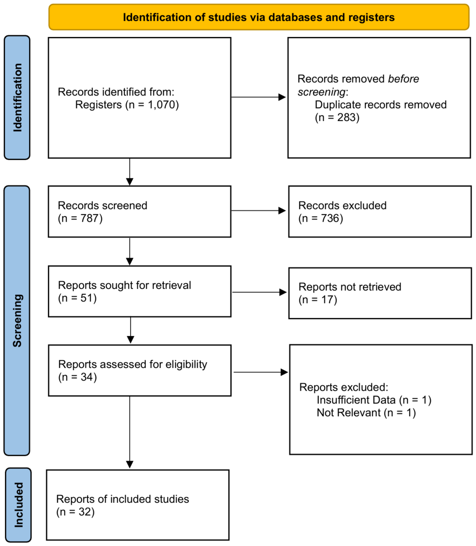
Figure 1. PRISMA 2020 flow diagram of the study selection process. The colours are used only to separate the three stages of the selection process (the orange bar identifies the search source; the blue side bars denote the identification, screening and included stages) and carry no analytical meaning.
Surgeries 07 00102 g001
Figure 2. Domain-level summary for Randomized Controlled Trials using the Cochrane RoB 2 tool.
Surgeries 07 00102 g002
Figure 3. Domain-level summary for Non-Randomized Studies of Interventions using the ROBINS-I tool.
Surgeries 07 00102 g003
Figure 4. Forest plot of the mean difference in new bone formation (NFB%) between PRF + bone graft and bone graft alone (10 studies: Nizam et al., 2018 [26]; Seif et al., 2024 [28]; Reis et al., 2025 [33]; Zhang et al., 2012 [36]; Bölükbaşı et al., 2015 [39]; Cömert Kılıç et al., 2017 [40]; Dragonas et al., 2023 [47]; Francisco et al., 2024 [48]; Trimmel et al., 2021 [49]; Pichotano et al., 2019 [55]). Pooled MD +4.10% (95% CI 1.59 to 6.61; I2 = 20.9%); positive values favour PRF + graft. The squares represent the study-level mean differences, with area proportional to the weight of the study, and the horizontal lines their 95% confidence intervals; the diamond is the pooled random-effects estimate (REML with Hartung–Knapp adjustment) and the red bar is the 95% prediction interval.
Surgeries 07 00102 g004
Figure 5. Forest plot of the mean difference in residual graft material (RG%) between PRF + bone graft and bone graft alone (10 studies: Nizam et al., 2018 [26]; Seif et al., 2024 [28]; Reis et al., 2025 [33]; Zhang et al., 2012 [36]; Bölükbaşı et al., 2015 [39]; Cömert Kılıç et al., 2017 [40]; Dragonas et al., 2023 [47]; Francisco et al., 2024 [48]; Trimmel et al., 2021 [49]; Pichotano et al., 2019 [55]). Negative values (less unresorbed biomaterial) favour PRF + graft. The squares represent the study-level mean differences, with area proportional to the weight of the study, and the horizontal lines their 95% confidence intervals; the diamond is the pooled random-effects estimate and the red bar is the 95% prediction interval.
Surgeries 07 00102 g005
Figure 6. Bubble plot of the random-effects meta-regression of the new bone formation mean difference on the control-arm baseline NFB% (10 studies: Nizam et al., 2018 [26]; Seif et al., 2024 [28]; Reis et al., 2025 [33]; Zhang et al., 2012 [36]; Bölükbaşı et al., 2015 [39]; Cömert Kılıç et al., 2017 [40]; Dragonas et al., 2023 [47]; Francisco et al., 2024 [48]; Trimmel et al., 2021 [49]; Pichotano et al., 2019 [55]). Each bubble is one study, with area proportional to its weight in the random-effects model; the fitted line shows the predicted change in effect size across baseline values (slope = 0.04% per percentage point; p = 0.718).
Surgeries 07 00102 g006
Figure 7. Forest plot of the mean difference in implant stability quotient (4 studies; 140 implants: Alhussaini et al., 2021 [25]; Elsholkamy and Eldesouky, 2018 [38]; Trimmel et al., 2021 [49]; Pichotano et al., 2019 [55]). Pooled MD +1.74 ISQ units (95% CI −4.38 to 7.86; I2 = 92.1%). The two arms of Alhussaini et al. [25] were combined into a single PRF arm. The green squares represent the study-level mean differences, with area proportional to the weight of the study, and the horizontal lines their 95% confidence intervals; the blue diamond is the pooled random-effects estimate and the bar beneath it is the 95% prediction interval. The confidence interval crosses the line of no effect; the point estimate favours PRF + graft, but the estimate is imprecise and establishes neither benefit nor equivalence.
Surgeries 07 00102 g007
Figure 8. Forest plot of the mean difference in vertical bone height gain (mm; 4 studies: Seif et al., 2024 [28]; Arakji et al., 2023 [30]; Mecit and Koşar, 2026 [31]; Abdelmoneim and Elsharkawy, 2020 [44]). Pooled MD −0.35 mm (95% CI −2.82 to 2.13; I2 = 78.1%); no significant difference between groups. Positive values favour PRF + graft. The green squares represent the study-level mean differences, with area proportional to the weight of the study, and the horizontal lines their 95% confidence intervals; the blue diamond is the pooled random-effects estimate and the red bar is the 95% prediction interval.
Surgeries 07 00102 g008
Figure 9. Forest plot of the mean difference in radiographic bone density (Hounsfield units) (2 studies: Dereci et al., 2023 [27]; Abdelmoneim and Elsharkawy, 2020 [44]). Pooled MD +76.4 HU (95% CI −890.9 to 1043.7; I2 = 81.7%); the estimate is highly imprecise and is reported for completeness only. Positive values favour PRF + graft. The green squares represent the study-level mean differences, with area proportional to the weight of the study, and the horizontal lines their 95% confidence intervals; the blue diamond is the pooled random-effects estimate and the bar beneath it is the 95% prediction interval.
Surgeries 07 00102 g009
Table 1. Characteristics of the 32 eligible studies included in the systematic review.
Study/YearStudy DesignSample Size (Patients/Sinuses)Intervention Group (s)Control GroupHealing TimeMain Outcomes Evaluated
Gassling et al., 2013 [24]Split-mouth RCT6 pts/12 sinusesAutologous bone/Bio-Oss + PRF membraneAutologous bone/Bio-Oss + Collagen membrane5 monthsVital bone %, residual bone substitute %
Alhussaini et al., 2021 [25]RCT25 pts/34 sinuses (50 implants)Grp B: DBBM + L-PRF
Grp C: DBBM + A-PRF
Grp A: DBBM alone6 monthsISQ at implant placement (T1) and at 24 weeks (T2)
Nizam et al., 2018 [26]Split-mouth RCT13 pts/26 sinusesDBBM + L-PRFDBBM alone6 monthsNFB%, Residual graft %, CBCT
Dereci et al., 2023 [27]Retrospective30 pts/43 sinusesFDBA + PRFFDBA alone6 monthsBone volume gain, Bone density (HU)
Seif et al., 2024 [28]RCT8 pts/16 sinusesXenograft + A-PRFXenograft alone6 monthsBone height gain, NFB%, Bone density
Powell et al., 2022 [29]Case Series3 pts/3 sinusesL-PRF + Allograft/Xenograft (or L-PRF alone)None6–8 monthsBone height gain (CBCT), Histology
Arakji et al., 2023 [30]RCT18 pts/18 sinusesMPM (Flowable PRF) + XenograftXenograft alone6 monthsEarly healing score, Bone height, Histology
Mecit and Koşar, 2026 [31]Retrospective24 pts/24 sinusesSticky bone (I-PRF + Allograft)Allograft alone6 monthsBone height gain, Bone resorption %
Gadipelly et al., 2019 [32]Split-mouth RCT20 pts/40 sinusesβ-TCP + PRFβ-TCP alone6 monthsBone height gain, Bone density (HU)
Reis et al., 2025 [33]Split-mouth RCT13 pts/26 sinusesDBBM + H-PRF (solid & liquid)DBBM alone4 monthsNFB%, Residual graft %, ISQ, Micro-CT (BV/TV), Implant survival
Choukroun et al., 2006 [34]CCTNot reported/9 sinusesFDBA + PRFFDBA alone4 vs. 8 monthsHistology, NFB%, Residual graft %
Malzoni et al., 2023 [35]RCT24 pts/36 sinusesDBBM + L-PRF (Evaluated at 4 & 8 mos)DBBM alone (Evaluated at 8 mos)4 & 8 monthsNFB%, Residual graft %, ISQ, IHC
Zhang et al., 2012 [36]RCT10 pts/11 sinusesBio-Oss + PRFBio-Oss alone6 monthsNFB%, Residual graft %, Bone contact
Khalfalla et al., 2015 [37]Single-arm Cohort10 pts/10 sinusesBCP + PRFNone9 monthsBone height gain, Bone density, Pain
Elsholkamy and Eldesouky, 2018 [38]RCT16 pts/16 sinusesBio-Oss + PRFBio-Oss alone6 monthsISQ, Bone density (CBCT)
Bölükbaşı et al., 2015 [39]RCT25 pts/32 sinusesBovine bone + PRFBovine bone + Collagen membrane6 monthsNFB%, Biomaterial remnant %, BL/IL ratio
Cömert Kılıç et al., 2017 [40]RCT26 pts/26 sinusesGrp 1: β-TCP + P-PRP
Grp 2: β-TCP + PRF
β-TCP alone6 monthsNFB%, Residual graft %, Soft tissue %
Soliman, 2021 [41]RCT20 pts/20 sinusesNanoBone + PRF + SimvastatinNanoBone + PRF6 monthsBone height gain, Bone density
Ibrahim et al., 2024 [42]RCT18 pts/18 sinusesT-PRF aloneT-PRF + Xenograft4 & 7 monthsISQ, Bone height, Bone density, Bone volume
Abdelmoneim et al., 2026 [43]RCT13 pts/18 sinusesGrp I: Allogenic sticky bone
Grp II: Xenogenic sticky bone
Grp III: Xenograft + Flowable PRF6 monthsNFB%, Bone density, Bone loss (Stability)
Abdelmoneim and Elsharkawy, 2020 [44]RCT10 pts/18 sinusesXenograft + Flowable PRFXenograft alone4 monthsBone height gain, Bone density, Histology
Arumugam et al., 2021 [45]RCT14 pts/14 sinusesGrp 2: Alloplastic + PRF
Grp 3: Autogenous + PRF
Grp 1: PRF alone (Abandoned)6 monthsBone height, Bone density
Barbu et al., 2018 [46]Case Series14 pts/14 sinusesBio-Oss + PRFNone6 monthsVertical bone height gain, Implant survival
Dragonas et al., 2023 [47]Pilot RCT15 pts/15 sinusesGrp 1: DBBM + A-PRF
Grp 2: DBBM + PRGF
DBBM alone6 monthsNFB% (Mineralized tissue), Residual graft
Francisco et al., 2024 [48]Case series (non-randomized split-mouth)6 pts/12 sinusesNanoBone + liquid PRFNanoBone alone6 monthsNFB%, Inert bone particles, Connective tissue
Trimmel et al., 2021 [49]RCT26 pts/30 sinusesSACBA + A-PRF (Evaluated at 3 mos)SACBA + A-PRF (Evaluated at 6 mos)3 vs. 6 monthsNFB%, ISQ, Micro-CT
Arafat et al., 2017 [50]RCT10 pts/12 sinusesBio-Oss + PRF + PRF membraneBio-Oss + Bio-Oss collagen memb.6 & 12 monthsBone height, Bone density
Kumar et al., 2018 [51]Case Series14 pts/14 sinusesBio-Oss + PRFNone1, 6, 12 monthsBone level (BL/IL ratio)
Xie et al., 2019 [52]RCT46 pts/46 sinusesBio-Oss + I-PRFBio-Oss alone6 & 12 monthsResidual bone height (RBH), ISQ
Tatullo et al., 2012 [53]Case Series60 pts/72 sinusesBio-Oss + PRFBio-Oss alone (in bilateral cases)106, 120, 150 daysHistology, ISQ
Dereci et al., 2022 [54]Retrospective14 pts/19 sinusesFDBA + PRFNone6 monthsBone volume, Ridge height
Pichotano et al., 2019 [55]Split-mouth RCT12 pts/24 sinusesDBBM + L-PRF (Evaluated at 4 mos)DBBM alone (Evaluated at 8 mos)4 vs. 8 monthsNFB%, Residual graft %, ISQ, CBCT volume
Abbreviations: RCT: randomized controlled trial; CCT: controlled clinical trial; PRF: platelet-rich fibrin; L-PRF: leukocyte- and platelet-rich fibrin; A-PRF: advanced platelet-rich fibrin; I-PRF: injectable platelet-rich fibrin; T-PRF: titanium-prepared platelet-rich fibrin; H-PRF: horizontal centrifugation platelet-rich fibrin; P-PRP: pure platelet-rich plasma; DBBM: deproteinized bovine bone mineral; FDBA: freeze-dried bone allograft; β-TCP: beta-Tricalcium phosphate; BCP: biphasic calcium phosphate; SACBA: serum albumin-coated bone allograft; MPM: mineralized plasmatic matrix; NFB: newly formed bone; ISQ: implant stability quotient; CBCT: cone-beam computed tomography; IHC: immunohistochemistry; HU: Hounsfield units. Arafat et al. [50] reported cone-beam computed tomographic bone height and bone density only and contributed no histomorphometric data to either primary synthesis. Of the studies reporting an implant stability quotient, four contributed to the quantitative synthesis [25,38,49,55]. Ibrahim et al. [42] was not pooled because its comparator was T-PRF alone rather than a graft-alone arm; Malzoni et al. [35] evaluated its arms at different healing intervals with no concurrent graft-alone comparator; Tatullo et al. [53] reported a single overall ISQ mean without group-level dispersion; and only a translated abstract was available for Xie et al. [52].
Table 2. GRADE summary of findings.
Outcome
(№ Studies; Participants)
Anticipated Effect
MD (95% CI)
Heterogeneity
I22)
Risk of BiasInconsistencyIndirectnessImprecisionPublication BiasCertainty (GRADE)
New bone formation (%)
k = 10; N = 204
+4.10 (1.59 to 6.61) PI 1.87 to 6.33
Favours PRF + graft
20.9% (τ2 < 0.0001)Serious 1Not seriousNot serious 2Not seriousUndetected 3
MODERATE
Residual graft material (%)
k = 10; N = 204
−2.71 (−6.08 to 0.67) PI −10.65 to 5.24
No clear difference
45.2% (τ2 = 10.06)Serious 1Not seriousNot seriousVery serious 4Undetected 3
VERY LOW
Implant stability quotient (ISQ)
k = 4; N = 140 implants
+1.74 (−4.38 to 7.86) PI −11.68 to 15.16
No clear difference
92.1% (τ2 = 13.74)Serious 1Serious 5Not seriousVery serious 6Suspected 7
VERY LOW
Vertical bone height gain (mm)
k = 4; N = 76
−0.35 (−2.82 to 2.13)
No clear difference
78.1% (τ2 = 1.86)Serious 1Serious 8Not seriousVery serious 6Suspected 7
VERY LOW
Radiographic bone density (HU)
k = 2; N = 61
+76.37 (−890.90 to 1043.65)
No clear difference
81.7% (τ2 = 9580.8)Very serious 9Serious 8Not seriousVery serious 6Suspected 7
VERY LOW
1 Only 4 of 22 contributing trials achieved an overall low risk of bias on RoB 2; the majority were rated some concerns because of open-label deviations (D2) and undescribed allocation concealment (D1), and these trials carry the majority of the pooled weight; downgraded one level. 2 Histomorphometric new bone formation is an accepted surrogate for graft maturation and was not downgraded for indirectness. 3 Funnel plot symmetry was formally assessable (k = 10) and Egger’s test showed no significant asymmetry; trim-and-fill imputed two studies for this outcome and the adjusted estimate remained statistically significant, so the domain was not downgraded, although power to detect small-study effects with ten studies is limited. 4 The 95% confidence interval includes no effect and is compatible with both a clinically relevant reduction in residual graft and a small increase; the 95% prediction interval (−10.65 to 5.24) is correspondingly wide, and statistical significance is reached in only one of ten leave-one-out iterations; downgraded two levels for very serious imprecision. 5 The I2 of 92% is attributable almost entirely to Elsholkamy et al. [38], which reports a standard deviation of 0.60 for a mean of 74 with an observed range of 71–78 in eight implants, a value that is arithmetically incompatible with the reported range. Omitting that study yields MD = −0.53 (95% CI −2.10 to 1.05; p = 0.285; I2 = 0%). 6 95% confidence interval includes no effect and/or few participants below the optimal information size; downgraded one (serious) or two (very serious) levels. 7 Fewer than 10 studies; therefore, funnel-based assessment of publication bias is unreliable and was downgraded by one level. 8 Substantial statistical heterogeneity (I2 ≥ 75%); downgraded by one level. 9 Evidence base includes non-randomized/retrospective studies (ROBINS-I serious) and was downgraded by two levels. GRADE certainty of evidence: moderate; very low.
Disclaimer/Publisher’s Note: The statements, opinions and data contained in all publications are solely those of the individual author(s) and contributor(s) and not of MDPI and/or the editor(s). MDPI and/or the editor(s) disclaim responsibility for any injury to people or property resulting from any ideas, methods, instructions or products referred to in the content.