Review

Management of the Patella in Revision Total Knee Arthroplasty: A Narrative Review of Available Techniques


by
Giorgio Carrozzi
1,2,
Luca Saccone
2,
Luca La Verde
2,*,
Angelo Baldari
2,3,
Antonio Caldaria
2,
Gian Mauro De Angelis D’Ossat
2,
Alessio Palumbo
1,2,
Matteo Guzzini
1,† and
Francesco Franceschi
2,3,†
1
Faculty of Medicine and Surgery, UniCamillus-Saint Camillus International University of Health and Medical Sciences, 00131 Rome, Italy
2
Department of Orthopaedic and Trauma Surgery, San Pietro Fatebenefratelli Hospital, 00189 Rome, Italy
3
Faculty of Medicine and Surgery, Link Campus University, 00165 Rome, Italy
*
Author to whom correspondence should be addressed.
Shared senior authorship between Guzzini and Franceschi.
Osteology 2026, 6(2), 6; https://doi.org/10.3390/osteology6020006
Submission received: 7 January 2026 / Revised: 23 March 2026 / Accepted: 23 March 2026 / Published: 25 March 2026
(This article belongs to the Special Issue New Trends in Arthroplasty)

Abstract

Background: In revision total knee arthroplasty (rTKA), clinical focus is mainly on the femoral and tibial components, while patellar complications are often underemphasized. However, patellar bone deficiency is a significant issue that can negatively affect surgical outcomes, especially in septic revisions. Objective: This review provides an overview of contemporary approaches to the evaluation and treatment of patellar bone loss in rTKA, emphasizing preoperative planning, classification frameworks, and treatment options. Methods: A narrative review of the literature was conducted. Studies published between 2000 and 2024 addressing preoperative assessment, surgical techniques, and outcomes in the management of patellar bone loss in rTKA were screened. Discussion: A recent consensus has addressed the evaluation, classification, and treatment of significant bone loss after total knee arthroplasty (TKA), providing specific recommendations for the management of patellar bone loss and extensor mechanism involvement in cases of severe bone deficiency. In this review, the principal surgical strategies described in the literature are summarized, including patellar component retention or revision, the use of biconvex inlay and trabecular metal implants, bone grafting, various augmentation techniques, resection arthroplasty, gull-wing osteotomy, and patellectomy. Conclusions: Despite various surgical strategies, no consensus exists on a universally superior approach. Ongoing research is essential to establishing standardized, evidence-based protocols for effective management of patellar bone loss in rTKA.

1. Introduction

The number of rTKA procedures is steadily increasing and is expected to rise significantly in the coming years: the number of rTKA procedures is expected to increase by 149% by 2040 and by 520% by 2060 [1]. Similarly, the incidence of cases requiring revision of the patellar component is also projected to increase. The patella plays a fundamental role in knee kinematics: it serves as a critical biomechanical structure that enhances the mechanical advantage of the knee extensor mechanism. Although the patella is anatomically classified as a sesamoid bone, multiple studies have highlighted that the absence of its proper reconstruction within the extensor mechanism results in suboptimal clinical outcomes, both in short- and long-term follow-up assessments [2,3].
In rTKA, most of the attention is given to the femoral and tibial components, while problems related to the patella and patellar bone loss are often given less attention. Revision of the patellar component must be considered in the following cases: aseptic patellar component loosening, periprosthetic patellar fracture with a loose patella implant, severe patellar component polyethylene wear, and TKA infection requiring removal of implants [4]. Approximately 10% of rTKAs involve a patella with severe bone loss, particularly in cases of septic revision [5]. The decision-making process regarding patellar management in rTKA remains inherently complex, with the current literature often lacking conclusive evidence to support a universally optimal surgical strategy. It is noteworthy that the recent consensus on the management of bone loss in rTKA dedicated considerable attention to the challenges associated with the patella [6]. To provide a thorough understanding of management of the patella in rTKA, the present article offers an updated narrative review encompassing all treatment modalities currently documented in the literature.

2. Materials and Methods

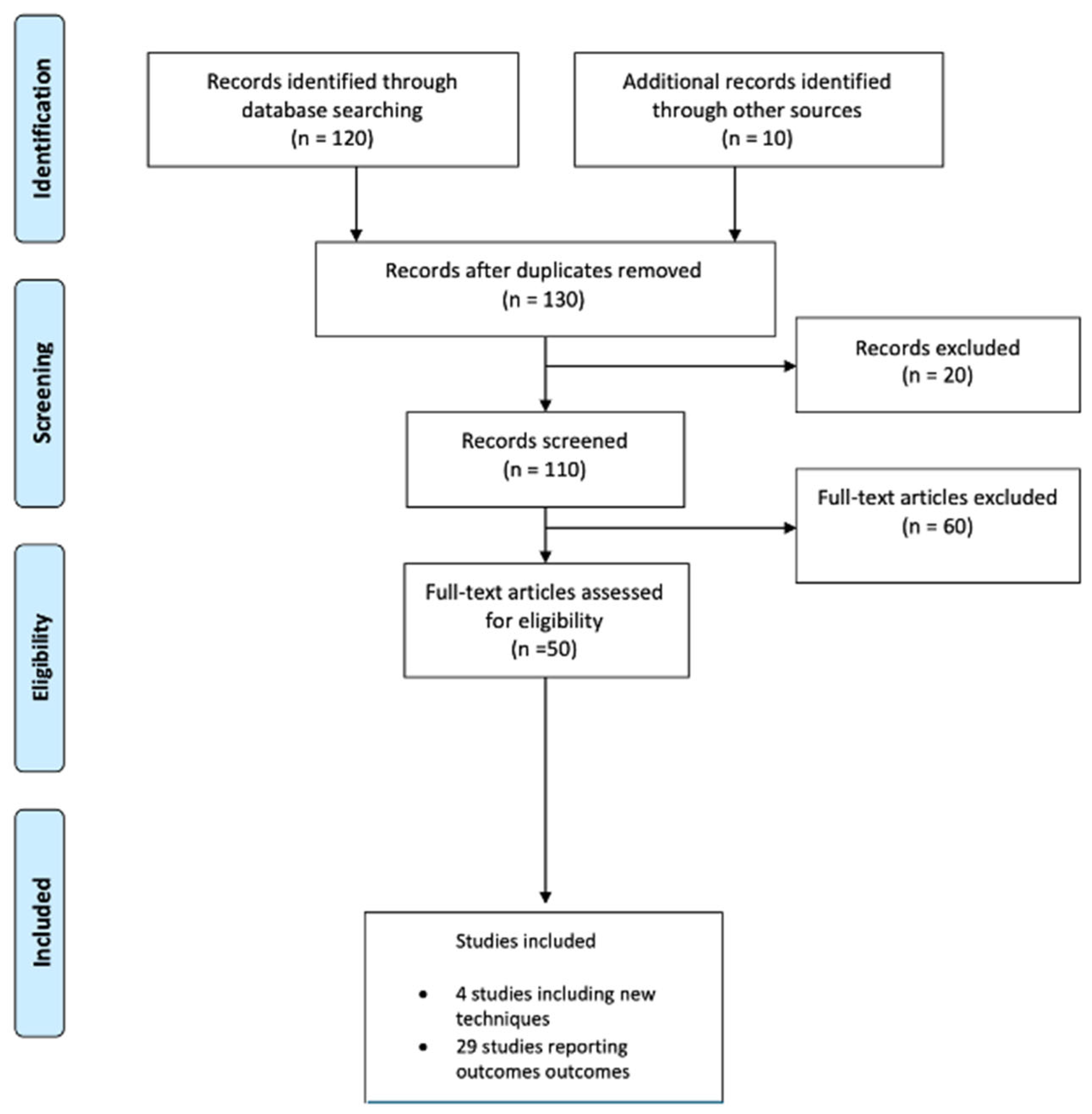
A comprehensive literature search was performed across PubMed–Medline, Cochrane Central, Scopus, and Google Scholar databases. The search strategy included the following keywords: “total knee arthroplasty,” “patella,” “bone loss,” and “revision”. Studies published in English and Italian between 2000 and 2024 that addressed preoperative assessment, surgical techniques, and clinical outcomes pertaining to the management of patellar bone loss in rTKA were included in the review. Additionally, the reference lists of pertinent articles were manually reviewed to identify further eligible studies. The literature selection process for this narrative review is illustrated in Figure 1.

3. Diagnostics

Preoperative planning is essential for distinguishing non-patellar sources of anterior knee pain and for establishing an accurate diagnosis prior to rTKA. Radiographic assessment plays a pivotal role in evaluating patellar bone stock, which is a critical factor in determining the most appropriate surgical strategy. Additionally, imaging facilitates the identification of patellar fractures or maltracking in relation to the femoral component [4].
A comprehensive and standardized preoperative radiographic evaluation should include anteroposterior (AP), true lateral, and Merchant views. The AP radiograph is optimally acquired in a weight-bearing position with the knee in full extension.
The true lateral projection should be performed with the patient positioned laterally on the affected side and the knee flexed to approximately 30 degrees. A properly executed true lateral image is characterized by superimposition of the medial and lateral femoral condyles.
Merchant views are acquired with the patient in the supine position, the knee flexed to 45 degrees and supported by a stable platform to ensure relaxation of the quadriceps muscle [6]. Moreover, an additional axial weight-bearing radiographic view, as described by Baldini et al., may offer supplementary diagnostic information beyond that provided by conventional radiographic assessments in the evaluation step [7].
Computed tomography (CT) is recommended to evaluate femoral and tibial component rotation, as malrotation is a well-recognized cause of patellofemoral maltracking [6].
Moreover, magnetic resonance Imaging (MRI) or CT scans should be considered when standard X-rays suggest the presence of osteolysis [6].
Technetium-99m (99mTc)-labeled scintigraphy can also be used in cases of suspected avascular necrosis; however, especially in the first two years following implantation, the risk of false-positive results remains high [6].

4. Classifications

Many classification systems have been developed to assess patellar bone deficiencies and to provide guidance on appropriate management strategies based on specific anatomical and implant-related characteristics.
A classification method proposed by Tetreault et al. [8] incorporates multiple factors including the stability, size, and positioning of the patellar component; the thickness and quality of the remaining patellar bone stock; and the functional integrity of the extensor mechanism (Table 1).
McPherson et al. [9] developed a classification system to help guide surgical management of patellar bone loss, based on two main criteria: the integrity of the cortical rim and the extent of cavitary bone loss (Table 2).
Peter Keyes Sculco et al., in a recent consensus on the management of bone loss in rTKA [6], proposed a novel classification system aimed at providing a simplified assessment of the patella to assist surgeons in selecting the most appropriate treatment strategy. A key addition in this classification is the evaluation of the surface of the remaining patella, which the authors identified as an underestimated factor in previous classification systems (Table 3).

5. Treatment Options

5.1. Retention of the Patellar Component

Retention of the patellar component during rTKA is indicated when the implant remains well-fixed, demonstrates no signs of wear, maintains satisfactory tracking, and shows no radiographic or clinical evidence of loosening, osteolysis, or instability [10].
Preservation of the patellar component is also considered appropriate in cases where femoral and tibial components from a different manufacturer are being revised, provided that implant compatibility is ensured [10,11,12]. This strategy helps to conserve patellar bone stock and mitigates the risk of iatrogenic patellar fracture [11,13].
Intraoperative verification of implant stability and fixation is critical, as radiographic findings may not reliably reflect the actual intraoperative condition of the component [14].

5.2. Patellar Component Revision

Patellar component revision is indicated in cases of loosening, significant wear, bone loss, maltracking, malpositioning, mechanical instability, or structural damage.
The management of the patella in rTKA is primarily guided by the quantity of remaining patellar bone stock in both the central and peripheral zones, as well as the condition and integrity of the previously implanted patellar component [4].
Multiple options are available: a standard all-poly patella, a biconvex implant, bone grafting, trabecular metal implants, reinforced cement augmentation techniques, and gull-wing osteotomy. The choice primarily depends on the severity of patellar bone loss. Patelloplasty and patellectomy are performed in cases where patellar resurfacing is contraindicated or not recommended.

5.3. Standard All-Polyethylene Onlay-Type Patella

A patella with adequate surface area and a remaining thickness of at least 10 mm allows for reimplantation of a standard all-poly patella, as indicated by the consensus group [6].
After carefully removing the patellar component while minimizing bone loss, the residual patellar thickness and surface area are measured, and if the anatomical dimensions are deemed adequate, button reimplantation can be performed.
A patellar component from the same manufacturer and with the same design as the femoral component should be used, or at least a compatible one [10,11].

5.4. All-Polyethylene Biconvex Patella

In cases of a central cavitary defect with a thickness between 5 mm and 10 mm, and a preserved peripheral cortical rim, the use of a biconvex all-poly patella is feasible [6,13].
This allows for the preservation of the peripheral bony rim of the patella and the implantation of a prosthetic component with a decreased outer circumference and increased central thickness [15].

5.5. Trabecular Metal Implants

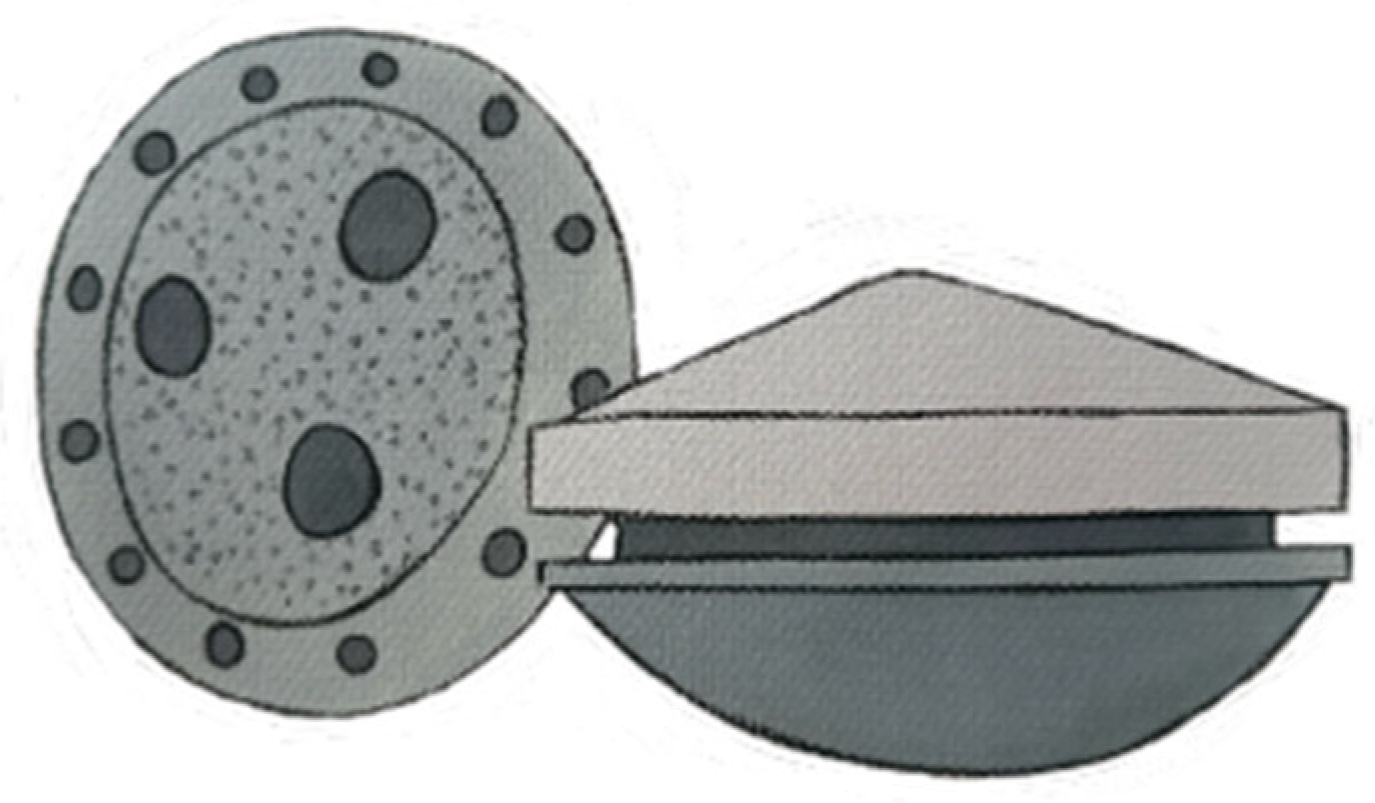
If the bone defect is severe enough patella with thickness < 5 mm according to Sculco et al. [6], to preclude the implantation of a standard all-polyethylene onlay-type patella or an all-polyethylene biconvex patella, a trabecular metal implant (Figure 2) may be used to promote bone ingrowth [4,10].
The porous metal implant is inserted into the residual patellar bone to facilitate osseointegration, while its peripheral rim is sutured to the adjacent soft tissues [16], thereby enabling the subsequent cementation of the polyethylene component onto the secured metal base.

5.6. Bone Grafting

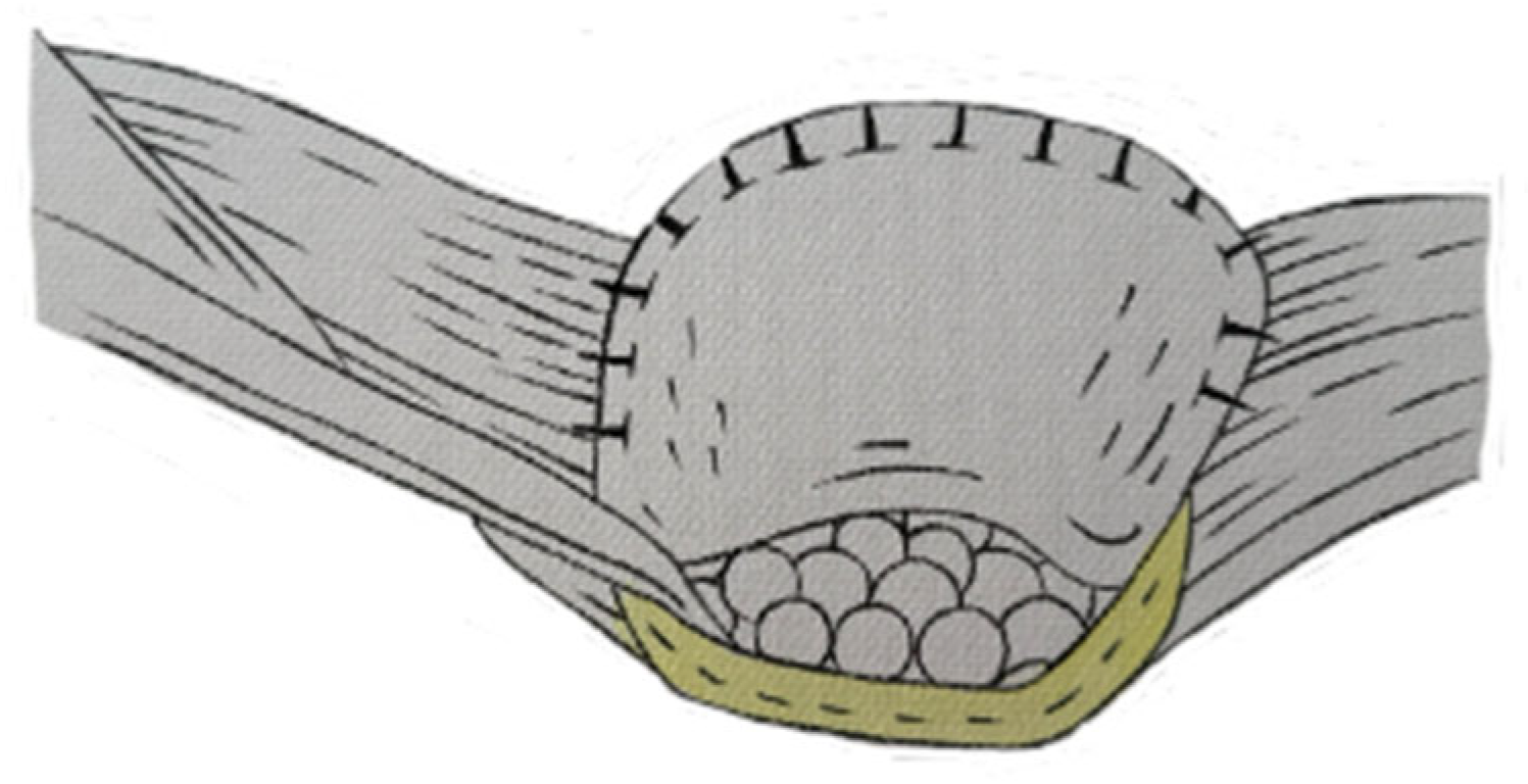
In cases of a patella with adequate surface area and intermediate (thickness: 5–10 mm) and severe (thickness < 5 mm) bone loss [6], reconstruction can be performed using autologous bone fragments. This technique, first described by Hanssen et al. [17], involves the harvest of morselized cancellous bone during revision surgery, specifically during femoral metaphyseal preparation. The bone chips should range in diameter from 5 to 8 mm [18].
Patellar reconstruction is achieved by utilizing a fibrous tissue disk harvested from the peripatellar region (or alternatively from the suprapatellar recess or the lateral femoral condyle). This disk is positioned to cover the residual patellar shell and is circumferentially sutured to the bony patellar rim to achieve a watertight closure, leaving only a small opening. This opening facilitates the introduction of the previously harvested morselized bone graft into the cavity [18] (Figure 3).
Meticulous handling of the soft tissue during flap development is essential to preserve the integrity of the quadricep tendon and its attachment to the superior pole of the patella [4]. The bone chips are subsequently impacted into the defect until a construct height exceeding 20 mm is achieved. The fibrous tissue flap is then completely sealed to prevent graft extrusion [17]. Postoperatively, the reconstructed patellar articular surface undergoes remodeling and adaptation to the femoral trochlea. The fibrous tissue flap functions analogously to an interpositional arthroplasty, facilitating congruence with the trochlear groove. This remodeling process is driven by compressive forces generated during knee flexion-extension, allowing the patella to progressively conform to its new anatomical configuration [18].

5.7. Reinforced Cement Augmentation Techniques

In cases of severe patellar bone loss, reconstruction using a cement-based technique reinforced with fixation devices may offer an effective, cost-efficient, and biomechanically reliable method for securing the patellar implant [19].

5.7.1. Augmentation with Transcortical Wiring of an Onlay-Type Prosthesis

Jai-Gon Seo et al. [20] described a technique involving augmentation with transcortical wiring of an onlay-type patellar prosthesis in twelve knees (nine patients) after conventional resurfacing was deemed inadequate. This approach was indicated by the authors when the residual patellar thickness was less than 8 mm and the cortical rim was compromised. Patellar revision was performed due to infection (five knees), loosening of the patellar component (four knees), and polyethylene wear associated with a metal-backed patella (three knees). The technique involved fixing wires to the three pegs of the patellar component, passing them through drill holes created in the anterior cortex, and, following compression of the cemented prosthesis, securing fixation by twisting the wires over the anterior surface.

5.7.2. Patellar Rebar Augmentation

In cases of severe patellar bone loss, when a patella has a residual thickness less than 5 mm [6], the patellar rebar augmentation technique, as described by McPherson et al. [9], may be employed. McPherson et al. recommend the use of this technique in cases of patellae with significant cavitary deficiencies (resurfacing should be considered only if the residual patellar thickness is ≥9 mm) and limited segmental rim deficiencies (less than 25%) [9].
This technique, as McPherson describes it, consists of using 2.0 mm titanium cortical screws vertically inserted in the dorsal patellar cortex, to augment cement fixation during revision surgery (Figure 4).
All cement should first be removed, ensuring careful extraction to prevent patellar fracture when detaching well-fixed cement, while also removing damaged cancellous bone. A hole is drilled with a 1.5 mm drill through the ventral surface, and 2.0 mm cortical screws are then placed and fixed in the dorsal patellar cortex, without crossing it. The patellar implant is then cemented.

5.7.3. Crossed Screw Patellar Reconstruction Technique

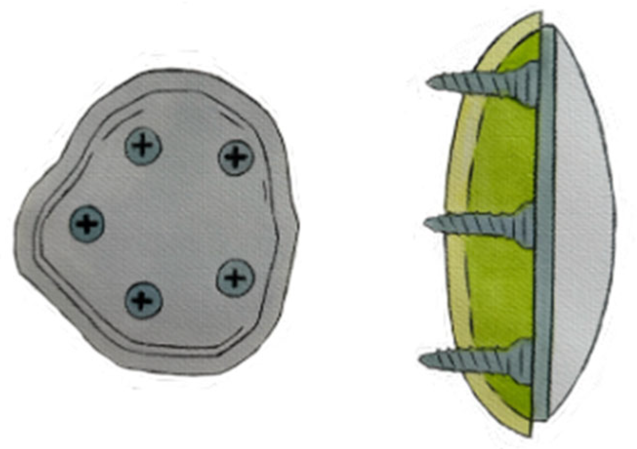
Poon et al. [19] recently proposed a new technique for the management of deficient patellae exhibiting severe bone loss with residual thickness of less than 10–12 mm.
Successful application of this method depends on the presence of an intact cortical rim to ensure reliable screw fixation and optimal cement fixation [19].
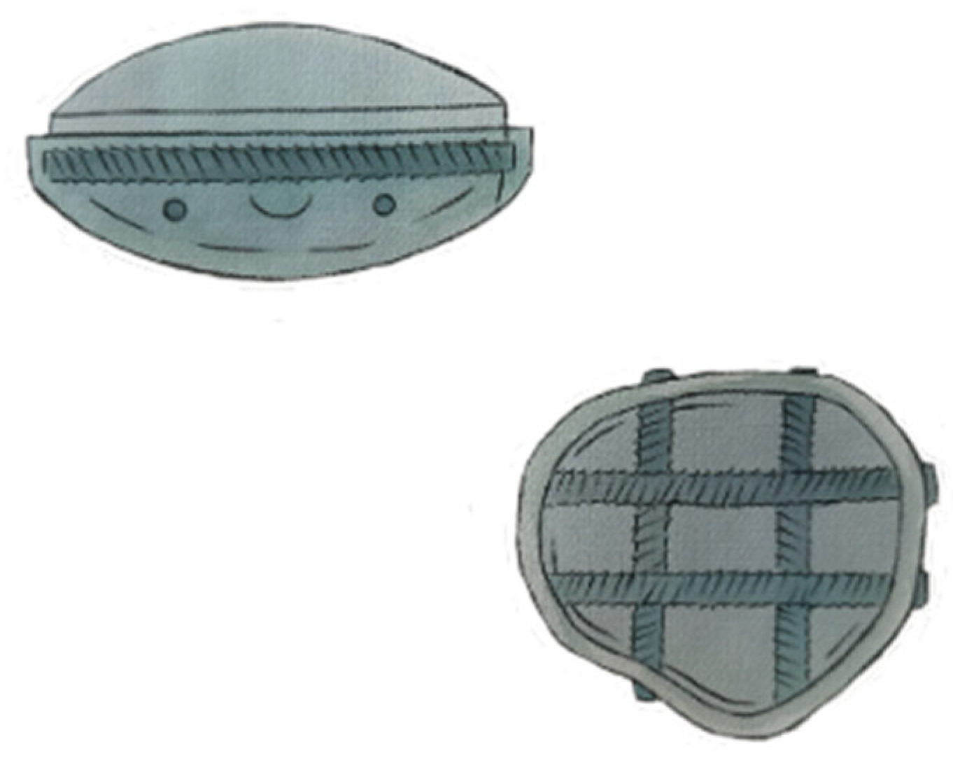
Using this technique, patellar reconstruction is achieved by placing four 2.7 mm titanium cortical screws in a crossed configuration, with two oriented horizontally and two vertically. This arrangement creates an internal, hash-like scaffold within the patellar cavity (Figure 5), providing structural support for subsequent reconstruction.
Nevertheless, the often markedly thinned cortical rim increases the likelihood of iatrogenic fracture, particularly during instrumentation such as drilling [19].
This technique offers two primary advantages over the approach described by McPherson et al. [19]: first, it enables bicortical fixation rather than limiting the construct to anterior cortical anchorage alone; second, it reduces the risk of implant-induced irritation to the prepatellar soft tissues [19].

5.8. Salvage Procedures

5.8.1. Patellar Resection Arthroplasty (Patelloplasty)

In cases of intermediate (thickness: 5–10 mm) or severe (<5 mm) patellar bone loss, patelloplasty is one of the available surgical options [6].
It is typically employed in cases where revision of the patellar component is not feasible due to extensive bone stock loss.
This technique involves removal of the patellar component, debridement of cement and any loose bone, followed by resection and smoothing of prominent areas of the remaining patella.

5.8.2. Gull-Wing Osteotomy

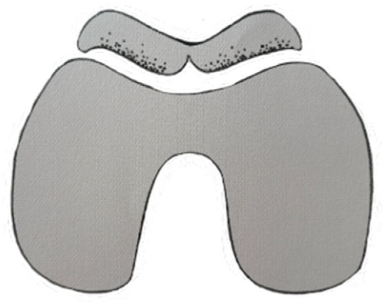
This technique serves as a salvage procedure in cases where reimplantation of a patellar component is not feasible. Instead of performing a patellectomy, a sagittal osteotomy is carried out to reshape the residual patellar bone into a V-shaped (gull-wing) configuration (Figure 6), thereby optimizing its tracking within the femoral trochlea [21].

5.8.3. Patellectomy

Patellectomy is a salvage surgical technique, largely abandoned due to its consistently poor functional outcomes [22], and is generally considered a treatment of last resort, recommended only in severe cases where all other therapeutic options are unsuitable [23].
Table 4 illustrates the relationship between residual patellar thickness, classified according to the Sculco classification, and the recommended treatment options, providing a practical framework to guide surgical decision-making in cases of patellar bone deficiency.

6. Discussion

Patellar bone loss in rTKA often slips into the background, overshadowed by the focus on femoral and tibial fixation. Yet the patella is central to knee mechanics, and neglecting it raises the risk of extensor-mechanism problems and poorer function. In this review, we bring the patella back to the foreground, explaining how to size the problem—residual bone stock, implant status, and associated complications—and how to match these factors to the available techniques. The literature offers several viable options, but the results are uneven and no single solution clearly prevails.
Whenever feasible, retaining the existing patellar component during revision procedures is always preferable, as it is associated with lower complication rates compared to component removal or replacement [11,24,25,26]. Nonetheless, in cases where revision becomes necessary, several techniques have been proposed.
Garcia et al. [27] reported favorable outcomes following the revision of failed metal-backed patellar components using cemented all-polyethylene patellar implants. Postoperative improvements were observed, with the mean Knee Society Score increasing from 73 to 89 points and the mean function score improving from 56 to 65 points.
An alternative technique for patellar component revision is the use of a biconvex inlay patellar component. The routine use of a biconvex inlay patellar component in TKA is associated with excellent long-term durability and a minimal incidence of patella-related complications. In a retrospective analysis conducted by Adit R. Maniar et al., a cohort of 2530 patients who underwent cemented TKA with routine patellar resurfacing using a cemented biconvex inlay component was evaluated. The study demonstrated a 15-year implant survivorship of 97.1% (95% CI: 96.1–98.1%) when considering revision surgery for any cause as the endpoint. Additionally, the survivorship at 15 years specifically related to patellar complications requiring revision was 99.7% (95% CI: 99.4–100%) [28].
Among a series of 262 rTKA recipients, Hines et al. showed that the application of biconvex patellar components in the context of significant patellar bone loss has favorable long-term performance. The 10-year survivorship without rerevision for aseptic loosening was 96%, while survivorship free of any revision of the patellar component at 10 years was 87%. These components proved to be reliable, as reflected by notable improvements in clinical outcome measures and a low incidence of associated complications [29].
Trabecular metal implants represent an alternative strategy. A key limitation of this technique, as highlighted by Ries et al., Kamath et al., and D. Tigani et al. [30,31,32], lies in the extent of the fixation interface between the implant and the residual patellar bone. Specifically, it is critical that more than 50% of the total fixation surface is in direct contact with bone. When most of the fixation relies on soft tissue rather than osseous support, there is a significantly increased risk of implant migration and loosening. This risk is particularly pronounced in patients with a history of patellectomy [4,30,31,32]. A final consideration is the economic impact, as this prosthetic system introduces an additional cost to an already inherently expensive surgical procedure [33].
An alternative approach involves patellar reconstruction using autologous bone fragments. This technique, known as bone grafting, effectively alleviates knee pain and enhances patient-reported outcome measures, with documented improvements in knee joint range of motion, including mean increases of 7° in flexion and 2° in extension [27,34,35,36].
In cases of severe patellar bone loss, reconstruction using a cement-based technique reinforced with fixation devices can be performed, and augmentation with transcortical wiring of an onlay-type prosthesis serves as a specific method. This technique has raised concerns among various authors [20], particularly in cases involving thin patellae: they had a case of patellar fracture occurred during flexion exercises one week postoperatively and was managed with partial patellectomy. To date, no other studies have evaluated the outcomes of this procedure, including those conducted by the original proponents of the technique.
Another approach is patellar rebar augmentation, designed to simplify the surgical procedure, minimize the risk of peripatellar fractures, and enhance mechanical stability of the reconstruction. It allows surgeons to broaden the indications for patellar revision procedures [9].
McPherson et al. [9] treated 128 patellae using this technique during rTKA procedures. Only four cases required subsequent patellar revision, among which there was a single incident of implant irritation. In an extremely thin patella shell, to achieve good screw fixation with this technique, the screw tips may protrude out of the anterior surface of the patella and cause irritation to the surrounding tissues. However, the authors noted significant difficulty in removing the patellar implants intraoperatively, indicating that the components were well-fixed to the bone. Furthermore, patient-reported outcomes were favorable, with postoperative improvements observed in both the Knee Society Score (KSS) and the flexion–extension arc.
A fourth cement-based technique reinforced with fixation devices is the crossed screw patellar reconstruction described by Poon G. et al. [19]. In the reported case, the patient treated with the crossed screw patellar reconstruction technique remained asymptomatic at the 3-year follow-up, demonstrating stable implant positioning, absence of patellar maltracking, and an improved knee range of motion (from 10–100° preoperatively to 5–110° postoperatively). She had fully resumed work activities without functional limitations.
To date, no other studies have evaluated the outcomes of this procedure, including the authors.
Salvage procedures are indicated in cases where reimplantation of a patellar component is not feasible as their clinical outcomes are poor or, in any case, inferior to those achieved with patellar component revision [8,13]. One such approach is patellar resection arthroplasty (patelloplasty), employed when patellar bone stock is inadequate. In a series of 1401 primary TKAs, Carlos J. Lavernia et al. reported a subset of patients who required revision surgery [37]. Among these, eight patients—accounting for a total of nine patellae—had insufficient bone stock to permit reimplantation of a new patellar component.
The Hospital for Special Surgery (HSS) scores were good to excellent in seven knees and fair in two. Notably, the commonly reported complications associated with patellectomy, such as quadriceps lag, extension weakness, and anterior knee pain, were not observed in these cases.
Although patelloplasty may be considered a surgical option for managing deficient patellae and can lead to postoperative improvement in clinical outcomes [37], patients frequently report persistent anterior knee pain [38].
Among salvage procedures, the gull-wing osteotomy demonstrated favorable functional and radiographic outcomes, although potential complications have been reported: Klein et al. [39] reported on a cohort of twelve consecutive patients who underwent rTKA in which a non-resurfaced patella was managed using a gull-wing osteotomy. The study showed significant improvement in pain, patellofemoral function, and Knee Society Scores postoperatively. All patients regained the ability to ascend and descend stairs, and radiographic assessments confirmed successful osteotomy healing with central patellar tracking. Gililland et al. [40] performed a gull-wing osteotomy in a cohort of 238 patients undergoing rTKA, where the remaining patellar bone was either avascular or measured less than 12 mm in thickness. Gull-wing patellar osteotomy resulted in significant improvement in knee extension (median 0°, p = 0.015), while the median flexion arc remained stable. The mean Knee Society Score improved from 86 to 142 (p < 0.001). Radiographic union was achieved in 12 of 13 patients, with nine showing central patellar tracking. One achieved fibrous union. Complications included three recurrent infections, and one case of avascular necrosis with fragmentation.
Potential complications associated with this procedure include disruption of the extensor mechanism, vascular disruption, and nonunion [19].
Finally, patellectomy should be regarded as a last resort. Chang et al. [41] analyzed eight patients who underwent TKA and subsequently sustained a patellar fracture treated with patellectomy. The results demonstrated relief from pain following the fracture; however, functional outcomes were suboptimal. Four patients exhibited mild extensor lag (less than 10%), two patients were unable to ascend or descend stairs, and two experienced complications, including quadriceps tendon rupture and secondary joint instability.
Yao et al. [2] conducted a comparative study involving 50 patients (52 primary TKAs) who had previously undergone patellectomy and a control group of 52 patients (52 primary TKAs) with preserved patellae. They evaluated both preoperative and postoperative outcomes using standardized measures, including the SF-12, WOMAC, and KSS. While TKA provided pain relief and functional improvement in patients with a history of patellectomy, their overall functional results were generally lower than those achieved by patients with an intact patella. In addition, knees with a history of patellectomy present an increased risk of postoperative instability and may warrant the use of more constrained TKA implants [42]

7. Conclusions

Patellar bone loss in rTKA is frequently underestimated and often perceived as a secondary concern, even by implant manufacturers, who predominantly concentrate on enhancing femoral and tibial component fixation. This comprehensive review addresses the full spectrum of concerns related to this complex and often overlooked issue. Various surgical techniques have been described, each tailored to the specific condition of the patella. While these approaches generally yield acceptable outcomes, the results remain suboptimal, and no single technique has yet emerged as definitively superior.
Future studies are warranted to establish an evidence-based algorithm for the surgical management of the patella in rTKA.

Author Contributions

G.C.: Writing—original draft. L.S.: Conceptualization, Writing—review and editing, Supervision, Validation, Visualization. L.L.V.: Conceptualization, Supervision, Validation. A.B.: Supervision, Validation. A.C.: Supervision, Validation. G.M.D.A.D.: Supervision, Validation. A.P.: Supervision, Validation. M.G.: Formal analysis, Supervision, Validation. F.F.: Formal analysis, Supervision, Validation. All authors have read and agreed to the published version of the manuscript.

Funding

This research received no external funding.

Institutional Review Board Statement

Not applicable.

Informed Consent Statement

Not applicable.

Data Availability Statement

No new data were created or analyzed in this study.

Conflicts of Interest

The authors declare no conflicts of interest.

Abbreviations

The following abbreviations are used in this manuscript:
rTKARevision total knee arthroplasty
APAnteroposterior
CTComputed tomography
MRIMagnetic resonance imaging
CIConfidence interval
TKATotal knee arthroplasty
KSSKnee Society Score
HSSHospital for Special Surgery

References

  1. Shichman, I.; Askew, N.; Habibi, A.; Nherera, L.; Macaulay, W.; Seyler, T.; Schwarzkopf, R. Projections and Epidemiology of Revision Hip and Knee Arthroplasty in the United States to 2040–2060. Arthroplast. Today 2023, 21, 101152. [Google Scholar] [CrossRef]
  2. Yao, R.; Lyons, M.C.; Howard, J.L.; McAuley, J.P. Does patellectomy jeopardize function after TKA? Clin. Orthop. Relat. Res. 2013, 471, 544–553. [Google Scholar] [CrossRef]
  3. Asadollahi, S.; Sorial, R.; Coffey, S.; Gupta, M.; Eslick, G.D. Total knee arthroplasty after patellectomy: A meta-analysis of case–control studies. Knee 2017, 24, 191–196. [Google Scholar] [CrossRef]
  4. Burnett, R.S.J.; Franco, M.; Baldini, A.; Barrack, R.L. Patellar treatment in revision total knee arthroplasty. In Total Knee Revision: Surgical Techniques; Edizioni Minerva Medica: Turin, Italy, 2012; pp. 127–144. [Google Scholar]
  5. Glynn, A.; Huang, R.; Mortazavi, J.; Parvizi, J. The impact of patellar resurfacing in two-stage revision of the infected total knee arthroplasty. J. Arthroplast. 2014, 29, 1439–1442. [Google Scholar] [CrossRef]
  6. Sculco, P.K.; Flevas, D.A.; Jerabek, S.A.; Jiranek, W.A.; Bostrom, M.P.; Haddad, F.S.; Fehring, T.K.; Della Valle, A.G.; Berry, D.J.; Brenneis, M.; et al. Management of Bone Loss in Revision Total Knee Arthroplasty: An International Consensus Symposium. HSS J. 2024, 20, 141–181. [Google Scholar] [CrossRef] [PubMed]
  7. Baldini, A.; Anderson, J.A.; Cerulli-Mariani, P.; Kalyvas, J.; Pavlov, H.; Sculco, T.P. Patellofemoral evaluation after total knee arthroplasty: Validation of a new weight-bearing axial radiographic view. J. Bone Jt. Surg. 2007, 89, 1810–1817. [Google Scholar] [CrossRef]
  8. Tetreault, M.W.; Gross, C.E.; Yi, P.H.; Bohl, D.D.; Sporer, S.M.; Della Valle, C.J. A classification-based approach to the patella in revision total knee arthroplasty. Arthroplast. Today 2017, 3, 264–268. [Google Scholar] [CrossRef] [PubMed]
  9. McPherson, E.J.; Sherif, S.M.; Dipane, M.V.; Arshi, A. Patellar Rebar Augmentation in Revision Total Knee Arthroplasty. J. Arthroplast. 2021, 36, 670–675. [Google Scholar] [CrossRef] [PubMed]
  10. Garcia, R.M.; Kraay, M.J.; Conroy-Smith, P.A.; Goldberg, V.M. Management of the deficient patella in revision total knee arthroplasty. Clin. Orthop. Relat. Res. 2008, 466, 2790–2797. [Google Scholar] [CrossRef]
  11. Lonner, J.H.; Mont, M.A.; Sharkey, P.F.; Siliski, J.M.; Rajadhyaksha, A.D.; Lotke, P.A. Fate of the unrevised all-polyethylene patellar component in revision total knee arthroplasty. J. Bone Jt. Surg. 2003, 85, 56–59. [Google Scholar] [CrossRef]
  12. Patil, N.; Lee, K.; Huddleston, J.I.; Harris, A.H.; Goodman, S.B. Patellar management in revision total knee arthroplasty: Is patellar resurfacing a better option? J. Arthroplast. 2010, 4, 589–593. [Google Scholar] [CrossRef]
  13. Maheshwari, A.V.; Tsailas, P.G.; Ranawat, A.S.; Ranawat, C.S. How to address the patella in revision total knee arthroplasty. Knee 2009, 16, 92–97. [Google Scholar] [CrossRef]
  14. Hanssen, A.D.; Pagnano, M.W. Revision of failed patellar components. Instr. Course Lect. 2004, 53, 201–206. [Google Scholar]
  15. Maheshwer, C.B.; Mitchell, E.; Kraay, M.; Goldberg, V.M. Revision of the patella with deficient bone using a biconvex component. Clin. Orthop. Relat. Res. 2005, 440, 126–130. [Google Scholar] [CrossRef] [PubMed]
  16. Nelson, C.L.; Lonner, J.H.; Lahiji, A.; Kim, J.; Lotke, P.A. Use of a trabecular metal patella for marked patella bone loss during revision total knee arthroplasty. J. Arthroplast. 2003, 18, 37–41. [Google Scholar] [CrossRef] [PubMed]
  17. Hanssen, A.D. Bone-grafting for severe patellar bone loss during revision knee arthroplasty. J. Bone Jt. Surg. 2001, 83, 171–176. [Google Scholar] [CrossRef]
  18. Baldini, A.; Franceschini, V.; Lamberti, A.; Balato, G. Gestione della perdita ossea rotulea nelle revisioni di protesi di ginocchio. Management of patellar bone loss in revision total knee arthroplasty. GIOT 2016, 39, 38–43. [Google Scholar]
  19. Poon, G.; Siow, W.M.; Poon, K.B. A Crossed Screw Patellar Reconstruction Technique for the Treatment of the Severely Deficient Scaphoid Shaped Patella in Total Knee Arthroplasty. Arthroplast. Today 2023, 24, 101259. [Google Scholar] [CrossRef]
  20. Seo, J.G.; Moon, Y.W.; Lim, S.J.; Lim, J.S.; Kim, S.M. Augmentation with transcortical wiring of an onlay-type prosthesis for a deficient patella during revision total knee arthroplasty. Clin. Orthop. Surg. 2012, 4, 163–166. [Google Scholar] [CrossRef]
  21. Vince, K.; Roidis, N.; Blackburn, D. Gull-Wing Sagittal Patellar Osteotomy in Total Knee Arthroplasty. Tech. Knee Surg. 2002, 1, 106–112. [Google Scholar] [CrossRef]
  22. Pang, H.N.; Sathappan, S.S. Patellar Reconstruction Using Distal Femoral Autograft in a Patellectomized Patient Undergoing Total Knee Arthroplasty. J. Arthroplast. 2008, 23, 939.e1–939.e7. [Google Scholar] [CrossRef] [PubMed]
  23. Günal, I.; Karatosun, V. Patellectomy: An overview with reconstructive procedures. Clin. Orthop. Relat. Res. 2001, 389, 74–78. [Google Scholar] [CrossRef] [PubMed]
  24. Barrack, R.L.; Rorabeck, C.; Partington, P.; Sawhney, J.; Engh, G. The results of retaining a well-fixed patellar component in revision total knee arthroplasty. J. Arthroplast. 2000, 15, 413–417. [Google Scholar] [CrossRef]
  25. Shield, W.P.; Greenwell, P.H.; Chapman, D.M.; Dalury, D.F. Ignore the Patella in Revision Total Knee Surgery: A Minimum 5-Year Follow-Up With Patella Component Retention. J. Arthroplast. 2019, 34, S262–S265. [Google Scholar] [CrossRef] [PubMed]
  26. Tetreault, M.W.; Della Valle, C.J.; Bohl, D.D.; Lodha, S.J.; Biswas, D.; Wysocki, R.W. What Factors Influence the Success of Medial Gastrocnemius Flaps in the Treatment of Infected TKAs? Clin. Orthop. Relat. Res. 2016, 474, 752–763. [Google Scholar] [CrossRef]
  27. Garcia, R.M.; Kraay, M.J.; Goldberg, V.M. Isolated all-polyethylene patellar revisions for metal-backed patellar failure. Clin. Orthop. Relat. Res. 2008, 466, 2784–2789. [Google Scholar] [CrossRef][Green Version]
  28. Maniar, A.R.; Luo, T.D.; Somerville, L.E.; MacDonald, S.J.; Naudie, D.D.; McCalden, R.W. Minimum 15-Year Survival of a Biconvex Inlay Patellar Component in Primary Total Knee Arthroplasty: An Analysis of 2530 Total Knee Arthroplasties From a Single Institution. J. Arthroplast. 2024, 39, S80–S85. [Google Scholar] [CrossRef] [PubMed]
  29. Hines, J.T.; Lewallen, D.G.; Perry, K.I.; Taunton, M.J.; Pagnano, M.W.; Abdel, M.P. Biconvex Patellar Components: 96% Durability at 10 Years in 262 Revision Total Knee Arthroplasties. J. Bone Jt. Surg. 2021, 103, 1220–1228. [Google Scholar] [CrossRef]
  30. Ries, M.D.; Cabalo, A.; Bozic, K.J.; Anderson, M. Porous tantalum patellar augmentation: The importance of residual bone stock. Clin. Orthop. Relat. Res. 2006, 452, 166–170. [Google Scholar] [CrossRef]
  31. Kamath, A.F.; Gee, A.O.; Nelson, C.L.; Garino, J.P.; Lotke, P.A.; Lee, G.C. Porous Tantalum Patellar Components in Revision Total Knee Arthroplasty. Minimum 5-Year Follow-Up. J. Arthroplast. 2012, 27, 82–87. [Google Scholar] [CrossRef]
  32. Tigani, D.; Trentani, P.; Trentani, F.; Andreoli, I.; Sabbioni, G.; Del Piccolo, N. Trabecular metal patella in total knee arthroplasty with patella bone deficiency. Knee 2009, 16, 46–49. [Google Scholar] [CrossRef] [PubMed]
  33. Balla, V.K.; Banerjee, S.; Bose, S.; Bandyopadhyay, A. Direct laser processing of a tantalum coating on titanium for bone replacement structures. Acta Biomater. 2010, 6, 2329–2334. [Google Scholar] [CrossRef]
  34. Erak, S.; Bourne, R.B.; MacDonald, S.J.; McCalden, R.W.; Rorabeck, C.H. The cemented inset biconvex patella in revision knee arthroplasty. Knee 2009, 16, 211–215. [Google Scholar] [CrossRef] [PubMed]
  35. Boettner, F.; Monsef, J.B. Achilles tendon allograft for augmentation of the Hanssen patellar bone grafting. Knee Surg. Sports Traumatol. Arthrosc. 2015, 23, 1035–1038. [Google Scholar] [CrossRef]
  36. Abdel, M.P.; Petis, S.M.; Taunton, M.J.; Perry, K.I.; Lewallen, D.G.; Hanssen, A.D. Long-Term Results of Patellar Bone-Grafting for Severe Patellar Bone Loss during Revision Total Knee Arthroplasty. J. Bone Jt. Surg. 2019, 101, 1636–1644. [Google Scholar] [CrossRef] [PubMed]
  37. Lavernia, C.J.; Alcerro, J.C.; Drakeford, M.K.; Tsao, A.K.; Krackow, K.A.; Hungerford, D.S. Resection arthroplasty for failed patellar components. Int. Orthop. 2009, 33, 1591–1596. [Google Scholar] [CrossRef]
  38. Rosenberg, A.G.; Jacobs, J.J.; Saleh, K.J.; Kassim, R.A.; Christie, M.J.; Lewallen, D.G.; Rand, J.A.; Rubash, H.E. The patella in revision total knee arthroplasty. J. Bone Jt. Surg. 2003, 85, S63–S70. [Google Scholar] [CrossRef]
  39. Klein, G.R.; Levine, H.B.; Ambrose, J.F.; Lamothe, H.C.; Hartzband, M.A. Gull-Wing Osteotomy for the Treatment of the Deficient Patella in Revision Total Knee Arthroplasty. J. Arthroplast. 2010, 25, 249–253. [Google Scholar] [CrossRef]
  40. Gililland, J.M.; Swann, P.; Pelt, C.E.; Erickson, J.; Hamad, N.; Peters, C.L. What Is the Role for Patelloplasty With Gullwing Osteotomy in Revision TKA? Clin. Orthop. Relat. Res. 2016, 474, 101–106. [Google Scholar] [CrossRef]
  41. Chang, M.A.; Rand, J.A.; Trousdale, R.T. Patellectomy after total knee arthroplasty. Clin. Orthop. Relat. Res. 2005, 440, 175–177. [Google Scholar] [CrossRef]
  42. Rodriguez-Merchan, E.C. Instability Following Total Knee Arthroplasty. HSS J. 2011, 7, 273–278. [Google Scholar] [CrossRef] [PubMed]
Figure 1. Flow diagram of narrative review of literature.
Osteology 06 00006 g001
Figure 2. Trabecular metal implant.
Osteology 06 00006 g002
Figure 3. Bone grafting.
Osteology 06 00006 g003
Figure 4. Patellar rebar augmentation.
Osteology 06 00006 g004
Figure 5. Crossed screw patellar reconstruction technique.
Osteology 06 00006 g005
Figure 6. Gull-wing osteotomy.
Osteology 06 00006 g006
Table 1. Classification of the patella in rTKA proposed by Tetreault et al. [8].
TypeDescriptionManagement
Type 1Component is well-fixed, appropriately sized, and properly positionedRetain the component
Type 2Component is loose or requires revision due to malpositioning, incorrect sizing, or infectionSee subtypes below
Type 2A>10 mm patellar remnant and adequate cancellous boneRevision using standard, cemented 3-peg component
Type 2B<10 mm patellar remnant and/or deficient cancellous bone precluding standard 3-peg componentSpecialized technique to reconstruct; impaction grafting, porous metal patella, or patellar osteotomy
Type 3Patellar fragmentation that precludes reconstructionTubularization or centralization of the extensor mechanism
Type 4Incompetent extensor mechanismReconstruction of the extensor mechanism
Table 2. McPherson Classification System of Patellar Defects in rTKA.
TypeDescriptionABC
Type 1Cortical Rim Intact<25% cavitary bone loss25–75% cavitary bone loss>75% cavitary bone loss (Eggshell Patella)
Type 2Rim Deficiency ≤ 25%
(Dorsal Cortex Intact)
<25% cavitary bone loss25–75% cavitary bone loss>75% cavitary bone loss (Eggshell Patella)
Type 3Rim Deficiency 25–50%
(Dorsal Cortex Intact)
<25% cavitary bone loss25–75% cavitary bone loss>75% cavitary bone loss (Eggshell Patella)
Table 3. Classification system proposed by the International Consensus Symposium.
TypeDescriptionManagement
1Patella with adequate surface area and thickness for button reimplantation
(Thickness > 10 mm)
Button reimplantation
2Patella with adequate surface area and intermediate thickness
(Thickness: 5–10 mm)
Patelloplasty
Bone grafting
Bi-convex patella
3Patella with thickness < 5 mmPatelloplasty
Bone grafting
Tantalum
Rebar technique
4Patella that presents avascular necrosis or fragmentationRetain the patella as is
Patellectomy if fragmentation is symptomatic
0Stable patella implant with presence of wearRetain the patellar implant
Table 4. Correlation between residual patellar thickness and surgical management strategies.
TypeDescriptionManagement
1Patella with adequate surface area and thickness for button reimplantation
(Thickness > 10 mm)
  • Reimplantation of a standard onlay-type patella
2Patella with adequate surface area and intermediate thickness
(Thickness: 5–10 mm)
  • Patelloplasty
  • Bone grafting
  • Bi-convex patella
  • Augmentation with transcortical wiring of an onlay-type prosthesis
  • Crossed screw patellar reconstruction technique
3Patella with thickness < 5 mm
  • Patelloplasty
  • Bone grafting
  • Tantalum
  • Rebar technique
4Patella that presents avascular necrosis or fragmentation
  • Retain the patella as is
  • Patellectomy if fragmentation is symptomatic
0Stable patella implant with presence of wear
  • Retain the patellar implant
Disclaimer/Publisher’s Note: The statements, opinions and data contained in all publications are solely those of the individual author(s) and contributor(s) and not of MDPI and/or the editor(s). MDPI and/or the editor(s) disclaim responsibility for any injury to people or property resulting from any ideas, methods, instructions or products referred to in the content.