Next Issue
Volume 4, September
Previous Issue
Volume 4, March


Osteology, Volume 4, Issue 2 (June 2024) – 5 articles

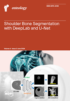
Cover Story (view full-size image): Evaluation of the 3D bone morphology of the glenohumeral joint is crucial for medicine. Zero echo time (ZTE) magnetic resonance imaging (MRI) provides excellent bone contrast and can potentially be used in the place of computed tomography. Segmentation of the shoulder bone anatomy is helpful for unobstructed assessment and measurements. We compared two popular convolutional deep learning models of DeepLab and U-Net for automated segmentation of ZTE MRI of human shoulders. While both models were satisfactory, U-Net slightly over-estimated, while DeepLab under-estimated the segmented area compared to the ground truth. The Dice score was significantly higher for U-Net than DeepLab for the humeral segmentation. We also implemented the U-Net model on an MRI console for push-button processing. This study has the potential to improve shoulder MR evaluation hindered by manual post-processing. View this paper
  • Issues are regarded as officially published after their release is announced to the table of contents alert mailing list.
  • You may sign up for e-mail alerts to receive table of contents of newly released issues.
  • PDF is the official format for papers published in both, html and pdf forms. To view the papers in pdf format, click on the "PDF Full-text" link, and use the free Adobe Reader to open them.
Order results
Result details
Select all
Export citation of selected articles as:
13 pages, 3051 KiB  
Article
Shoulder Bone Segmentation with DeepLab and U-Net
by Michael Carl, Kaustubh Lall, Darren Pai, Eric Y. Chang, Sheronda Statum, Anja Brau, Christine B. Chung, Maggie Fung and Won C. Bae
Osteology 2024, 4(2), 98-110; https://doi.org/10.3390/osteology4020008 - 11 Jun 2024
Viewed by 739
Abstract
Evaluation of the 3D bone morphology of the glenohumeral joint is necessary for pre-surgical planning. Zero echo time (ZTE) magnetic resonance imaging (MRI) provides excellent bone contrast and can potentially be used in the place of computed tomography. Segmentation of the shoulder anatomy, [...] Read more.
Evaluation of the 3D bone morphology of the glenohumeral joint is necessary for pre-surgical planning. Zero echo time (ZTE) magnetic resonance imaging (MRI) provides excellent bone contrast and can potentially be used in the place of computed tomography. Segmentation of the shoulder anatomy, particularly the humeral head and the acetabulum, is needed for the detailed assessment of each anatomy and for pre-surgical preparation. In this study, we compared the performance of two popular deep learning models based on Google’s DeepLab and U-Net to perform automated segmentation on ZTE MRI of human shoulders. Axial ZTE images of normal shoulders (n = 31) acquired at 3-Tesla were annotated for training with DeepLab and 2D U-Net, and the trained model was validated with testing data (n = 13). While both models showed visually satisfactory results for segmenting the humeral bone, U-Net slightly over-estimated while DeepLab under-estimated the segmented area compared to the ground truth. Testing accuracy quantified by Dice score was significantly higher (p < 0.05) for U-Net (88%) than DeepLab (81%) for the humeral segmentation. We have also implemented the U-Net model onto an MRI console for push-button DL segmentation processing. Although this is an early work with limitations, our approach has the potential to improve shoulder MR evaluation hindered by manual post-processing and may provide clinical benefit for quickly visualizing bones of the glenohumeral joint. Full article
Show Figures

Figure 1

10 pages, 1366 KiB  
Article
Reliability of Measuring the Proximal Humeral Bone Mineral Density Using Dual-Energy X-ray Absorptiometry
by Masataka Kamiyama, Hitoshi Shitara, Tsuyoshi Tajika, Daisuke Shimoyama, Shogo Hashimoto, Tsuyoshi Ichinose, Tsuyoshi Sasaki, Noritaka Hamano and Hirotaka Chikuda
Osteology 2024, 4(2), 88-97; https://doi.org/10.3390/osteology4020007 - 22 May 2024
Viewed by 844
Abstract
We established a protocol for assessing the areal bone mineral density (BMD) of the proximal humerus using dual-energy X-ray absorptiometry (DXA). We also investigated the correlation between the BMD of the proximal humerus and that of the lumbar spine and proximal femur to [...] Read more.
We established a protocol for assessing the areal bone mineral density (BMD) of the proximal humerus using dual-energy X-ray absorptiometry (DXA). We also investigated the correlation between the BMD of the proximal humerus and that of the lumbar spine and proximal femur to predict the BMD of the proximal humerus. We included female patients aged >60 years who underwent bone density evaluation using DXA. The BMD of the proximal humerus was calculated at seven regions of interest (ROIs): the head of the humerus, lesser tubercle, greater tubercle in two locations, and proximal metaphysis in three locations. The intra- and inter-examiner reliabilities in the setting of the ROIs were examined using intraclass correlation coefficients (ICCs) (1.1) and (2.1), respectively, and the intra-examiner reliability in DXA was examined using ICCs (1.1). The intra- and inter-examiner reliabilities in the setting of ROIs and the intra-examiner reliability in DXA were high in all regions. The BMD of the lumbar spine and proximal femur correlated weakly with that of the humeral head and diaphysis. Our method for measuring the BMD of the proximal humerus was found to be reliable and may be applied in future studies. Full article
Show Figures

Figure 1

24 pages, 3892 KiB  
Systematic Review
Sociodemographic and Lifestyle Risk Factors Associated with Fragility Hip Fractures: A Systematic Review and Meta-Analysis
by Diana Yeritsyan, Kaveh Momenzadeh, Amin Mohamadi, Sharri J. Mortensen, Indeevar R. Beeram, Daniela Caro, Nadim Kheir, Megan McNichol, John J. Wixted, Paul Appleton, Arvind von Keudell and Ara Nazarian
Osteology 2024, 4(2), 64-87; https://doi.org/10.3390/osteology4020006 - 14 May 2024
Viewed by 827
Abstract
Hip fractures inflict heightened morbidity and mortality upon older adults. Although previous studies have explored the impact of individual demographic factors on hip fracture risk, a comprehensive review can help reconcile disparities among these factors. This meta-analysis encompassed 69 studies involving 976,677 participants [...] Read more.
Hip fractures inflict heightened morbidity and mortality upon older adults. Although previous studies have explored the impact of individual demographic factors on hip fracture risk, a comprehensive review can help reconcile disparities among these factors. This meta-analysis encompassed 69 studies involving 976,677 participants and 99,298 cases of hip fractures. We found that age ≥ 85 (OR = 1.75), BMI < 18.5 (OR 1.72), female sex (OR = 1.23), history of falls (OR = 1.88), previous fractures (OR = 3.16), menopause (OR 7.21), history of maternal hip fractures (OR = 1.61), single and unmarried status (OR = 1.70), divorced status (OR 1.38), residing in a residential care facility (OR = 5.30), and living alone (OR = 1.47) were significantly associated with an increased incidence of hip fracture. Conversely, BMI ranging from 25 to 30 (OR = 0.59), BMI > 30 (OR = 0.38), parity (OR = 0.79), non-Caucasian descent (overall OR = 0.4, Asian OR 0.36, Black OR = 0.39, and Hispanic OR = 0.45), and rural residence (OR = 0.95) were significantly associated with a diminished risk of hip fracture. Hip fracture patients exhibited significantly lower weight and BMI than the non-fracture group, while their age was significantly higher. However, age at menopause and height did not significantly differ between the two groups. Full article
Show Figures

Figure 1

12 pages, 569 KiB  
Article
Pre-Operative Adiposity and Synovial Fluid Inflammatory Biomarkers Provide a Predictive Model for Post-Operative Outcomes Following Total Joint Replacement Surgery in Osteoarthritis Patients
by Dominika E. Nanus, Edward T. Davis and Simon W. Jones
Osteology 2024, 4(2), 53-63; https://doi.org/10.3390/osteology4020005 - 22 Apr 2024
Cited by 1 | Viewed by 955
Abstract
A proportion of osteoarthritis (OA) patients are unsatisfied with post-operative outcomes following total joint replacement surgery (TJR), with insufficient pain relief or poor functional improvement. Predicting those who will have poor outcomes would be beneficial for patients and clinicians. The aim of this [...] Read more.
A proportion of osteoarthritis (OA) patients are unsatisfied with post-operative outcomes following total joint replacement surgery (TJR), with insufficient pain relief or poor functional improvement. Predicting those who will have poor outcomes would be beneficial for patients and clinicians. The aim of this study was to determine the relationship between baseline anthropometric data and the concentration of pre-operative serum and peri-operative synovial fluid (SF) cytokines and 7-month post-operative outcomes in a cohort of knee and hip OA patients. 160 OA patients were recruited who were scheduled for TJR. The concentration of 24 cytokines was measured in blood and SF by multiplex assay. EQ5D index health status was assessed pre-operatively and at 7 months post-operatively. 13% of patients were identified as non-responders based on EQ5D index. Compared to responders, non-responders were of higher body mass index (BMI), had greater waist and hip circumference, and had higher levels of SF leptin but lower levels of SF resistin (p < 0.05). Linear regression analysis found a significant but weak relationship between pre-operative body weight and post-operative response (ΔEQ5D index; r = 0.222, p = 0.049). The combination of body weight with SF amphiregulin and SF IL-6 provided an improved predictive model of post-operative response (r = 0.470, p = 0.035). Full article
(This article belongs to the Special Issue New Trends in Arthroplasty)
Show Figures

Figure 1

8 pages, 4726 KiB  
Case Report
Type I Monteggia Fracture with Associated Ipsilateral Capitellar and Humeral Diaphyseal Fractures in an Adult
by Casey McDonald, Matt Kannenberg, Jason Goodrum, John Eakin, Paul Ryan and Anil Dutta
Osteology 2024, 4(2), 45-52; https://doi.org/10.3390/osteology4020004 - 4 Apr 2024
Viewed by 708
Abstract
Background: Monteggia fractures entail a proximal ulnar fracture with associated radial head dislocation. Primarily observed as a fracture in the pediatric population, there have been rare occurrences in adults. In rare instances, various associated fractures have been reported with Monteggia fractures. However, during [...] Read more.
Background: Monteggia fractures entail a proximal ulnar fracture with associated radial head dislocation. Primarily observed as a fracture in the pediatric population, there have been rare occurrences in adults. In rare instances, various associated fractures have been reported with Monteggia fractures. However, during our literature review, a type I Monteggia fracture had not been reported along with ipsilateral diaphyseal humerus and capitellar fractures. Here, we present a successful post-operative outcome for a unique fracture distribution and form of fixation that has yet to be reported in the literature. Full article
Show Figures

Figure 1

Previous Issue
Next Issue
Back to TopTop