Article

Reliability of Measuring the Proximal Humeral Bone Mineral Density Using Dual-Energy X-ray Absorptiometry

Department of Orthopaedic Surgery, Gunma University Graduate School of Medicine, 3-39-15 Showa-machi, Maebashi 371-8511, Gunma, Japan
*
Author to whom correspondence should be addressed.
Osteology 2024, 4(2), 88-97; https://doi.org/10.3390/osteology4020007
Submission received: 21 February 2024 / Revised: 29 April 2024 / Accepted: 20 May 2024 / Published: 22 May 2024

Abstract

:
We established a protocol for assessing the areal bone mineral density (BMD) of the proximal humerus using dual-energy X-ray absorptiometry (DXA). We also investigated the correlation between the BMD of the proximal humerus and that of the lumbar spine and proximal femur to predict the BMD of the proximal humerus. We included female patients aged >60 years who underwent bone density evaluation using DXA. The BMD of the proximal humerus was calculated at seven regions of interest (ROIs): the head of the humerus, lesser tubercle, greater tubercle in two locations, and proximal metaphysis in three locations. The intra- and inter-examiner reliabilities in the setting of the ROIs were examined using intraclass correlation coefficients (ICCs) (1.1) and (2.1), respectively, and the intra-examiner reliability in DXA was examined using ICCs (1.1). The intra- and inter-examiner reliabilities in the setting of ROIs and the intra-examiner reliability in DXA were high in all regions. The BMD of the lumbar spine and proximal femur correlated weakly with that of the humeral head and diaphysis. Our method for measuring the BMD of the proximal humerus was found to be reliable and may be applied in future studies.

1. Introduction

According to a World Health Organization (WHO) report, osteoporosis affects over 75 million people in the United States (U.S.), Europe, and Japan.
In the last few years, the prevalence of osteoporosis has increased among older adults, leading to disability, reduced quality of life, and even death of patients [1,2,3]. The high costs associated with inpatient and outpatient treatment, reduced quality of life, and increased mortality are major problems. Despite this burden, patients with fragility fractures rarely receive treatment [4].
Fractures caused by osteoporosis cost the U.S. healthcare system USD 17 billion annually. These costs are projected to reach USD 50 billion by 2040, and the number of hip fractures is estimated to double worldwide by 2050 [5]. Osteoporosis is defined by bone mineral density (BMD), and dual-energy X-ray absorptiometry (DXA) is the most widely accepted method for measuring BMD [6,7]. According to the WHO criteria, osteoporosis is defined as a BMD < 2.5 standard deviations below the mean in young, healthy females. This criterion is accepted worldwide as the standard for the diagnosis of osteoporosis.
Osteoporosis-related fractures frequently occur in the proximal femur, spinal compression region, distal radius, and proximal humerus. The BMD of the proximal femur, spine, and distal radius can be measured directly using DXA and is used for the treatment planning of fractures [8,9]. Although proximal humeral fractures are so frequent that they account for 5% of all fractures, a method for measuring BMD using DXA in the proximal humerus has not yet been established.
This technical difficulty in measuring the BMD of the proximal humerus arises because the humeral head overlaps with the glenoid fossa during routine anterior–posterior (AP) view roentgenography. Thus, we hypothesized that true AP view roentgenography, called “Grashey view”, would allow us to measure the BMD of the proximal humerus without interfering with the glenoid fossa. Measuring the BMD of the proximal humerus would allow us to investigate the correlation between the BMD of the humerus and that of the lumbar spine and femur. A strong correlation between these values may help predict the proximal humeral BMD and provide valuable insight into treatment, even when direct measurement is impossible. Reduced humeral BMD has been implicated in complications during open reduction and internal fixation, postoperative implant failure, and nonunion. Knowledge of humeral BMD loss can help anticipate and manage these risks in advance. Although previous studies have compared the BMD of the proximal humerus to that of the lumbar spine and femur, in this study, we further subdivided the regions of interest (ROIs) for comparison [10].
The primary purpose of this study was to evaluate the reliability of each imaging and analysis method for the areal BMD determination of the proximal humerus using DXA-based proximal humerus analysis. The secondary purpose was to investigate the correlation between the BMD values of the proximal humerus and those of the lumbar spine and femur.

2. Materials and Methods

2.1. Participants

We recruited patients who visited the orthopedic clinic at Usui Hospital between 2013 and 2018. An opt-out method was used to obtain informed consent, including for the publication of their images. The inclusion criteria were as follows: age ≥ 60 years; females; DXA examination of the left proximal humerus, left proximal femur, and lumbar spine performed on the same day for suspected osteoporosis; not being treated for osteoporosis; no history of major trauma involving the left proximal humerus, left proximal femur, or lumbar spine; and no history of surgery involving the left proximal humerus, left proximal femur, or lumbar spine. The exclusion criteria were as follows: osteoarthritis, lumbar spondylosis, comorbid neurological or psychiatric disorders, and difficulty communicating.
All participants provided informed consent for their inclusion before study participation. The study was conducted in accordance with the Declaration of Helsinki, and the protocol was approved by the Institutional Review Board of our hospital (Project identification code: 30-1, Date of approval: 10 August 2018).

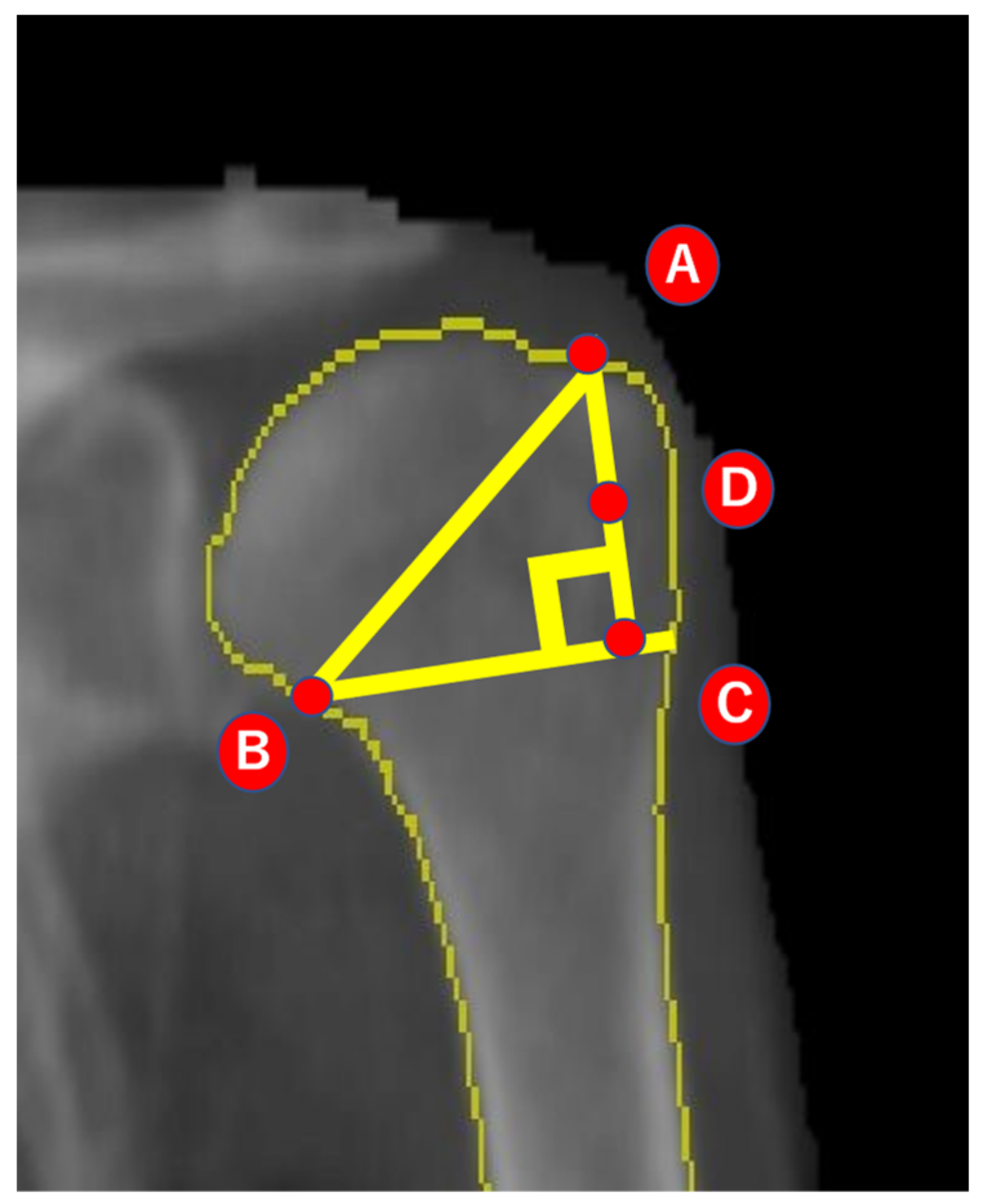
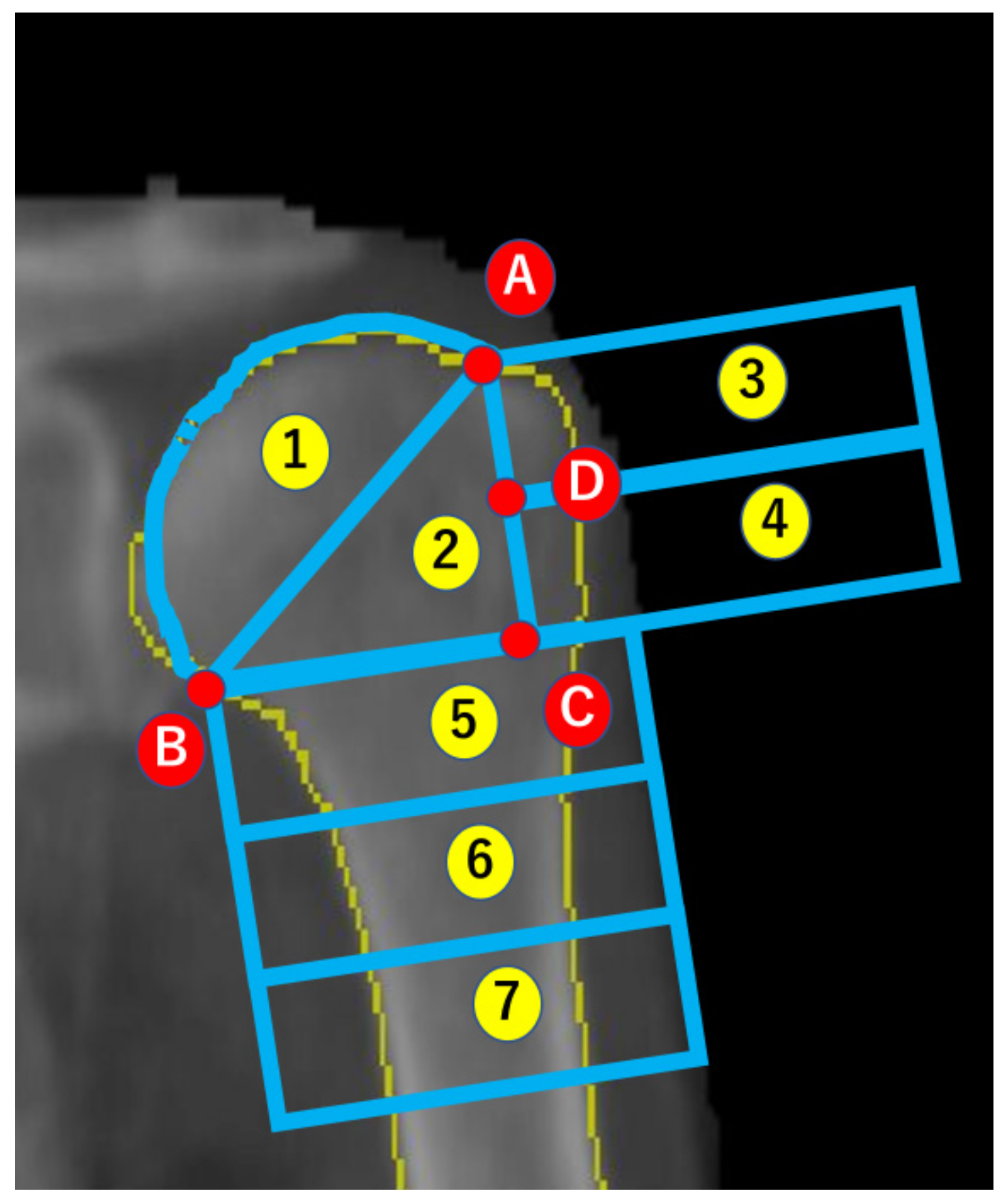
2.2. Analysis of BMD

Whole-body DXA (Prodigy C; General Electric Company, Boston, MA, USA) was used to measure BMD, and the procedure was performed by a certified radiologist with over 15 years of experience in musculoskeletal imaging. All examinations were performed with the patients in the supine position on an examination bed. The lower limbs were maintained in a neutral position to obtain images of the proximal femur, and the knee was flexed at 90° for obtaining images of the lumbar spine. To obtain images of the left proximal humerus, a positioning sponge was placed under the back with the arm positioner on the left forearm to maintain a left posterior oblique position, which enabled images to be obtained in the same manner as the true AP shoulder radiographs in neutral rotation (Figure 1). To ensure image quality, all images were visually inspected by a radiologist immediately after scanning.
All BMD analyses were performed using the ENCORE software program (version 17; General Electric Company, Boston, MA, USA). The automatic mode was applied for the analysis of the first to fourth lumbar vertebrae and the proximal femur. The proximal femur was assessed in three regions: the total hip and femoral neck, Ward’s triangle, and trochanter and intertrochanter.
A semi-automatic mode was used for the analysis of the proximal humerus, and the procedure was performed by two shoulder surgeons with 10 years of experience. The regions of bone, soft tissue, air, and artifacts were automatically separated, and a visual inspection was performed to ensure that the separation was accurate and to exclude osteoarthritis and spondylosis. The ROIs were then set.
Seven ROIs were included using the following methods: First, the medial edge of the greater tuberosity was designated as point A, and the intersection of the anatomical neck with the medial cortical bone was designated as point B. The intersection of a line parallel to the bone axis from point A and a straight line passing through point B and perpendicular to the bone axis was designated as point C. The midpoint of the straight-line AC was designated as point D (Figure 2). The area bounded by the straight-line AB and the cartilage of the humeral head was defined as region 1, triangle ABC was defined as region 2, and a rectangle with a straight-line AC on one side was created. The rectangle was placed outside the straight-line AC, and this area was defined (from above) as regions 3 and 4. A rectangle of the same size was placed underneath the straight-line BC, and this area was defined (from above) as regions 5−7 (Figure 3). Thus, a default ROI type was created and semi-automatically adapted for all participants as a template, with only slight fitting modifications, such as scaling up or down.
The medial edge of the greater tuberosity was designated as point A, and the intersec-tion of the anatomical neck with the medial cortical bone was designated as point B. The intersection of a line parallel to the bone axis from point A and a straight line passing through point B and perpendicular to the bone axis was designated as point C. The midpoint of the straight-line AC was designated as point D.
The area bounded by the straight-line AB and the cartilage of the humeral head was defined as region 1, triangle ABC was defined as region 2, and a rectangle with a straight-line AC on one side was created. The rectangle was placed outside the straight-line AC, and this area was defined (from above) as regions 3 and 4. A rectangle of the same size was placed underneath the straight-line BC, and this area was defined (from above) as regions 5−7.

2.3. Reliability of Imaging and Analysis

The first step of the study was to examine the intraclass correlation coefficient (ICC) (1.1) for assessing the intra-rater reliability of the DXA imaging method. The radiologist, who had over 10 years of experience in musculoskeletal imaging, conducted two imaging sessions on 10 patients on separate days, and the ROI was set by the same shoulder surgeon.
To assess the ICC (1.1) of the ROI setting, the shoulder surgeon analyzed 10 patients twice on separate days, and another shoulder surgeon analyzed the same 10 patients to assess the ICC (2.1) for inter-rater reliability. The patient names and BMD of the other sites were blinded to the two shoulder surgeons.

2.4. Statistical Analysis

Except for the post hoc power analysis of correlation, all statistical analyses were performed using IBM SPSS Statistics software (version 26, IBM Japan, Tokyo, Japan). The correlation between the BMD in areas 1–7 of the humerus, the BMD of the lumbar spine, and the BMD of the proximal femur and age was tested using Pearson’s correlation coefficient. Correlations were categorized as poor (r < 0.29), weak (r = 0.30–0.59), moderate (r = 0.60–0.79), or strong (r = 0.80–1.00) [11]. ICC assesses reliability, with high reliability defined as ≥0.75, moderate reliability defined as ≥0.40 to <0.75, and low reliability defined as <0.40 [12]. The significance level was set at <5%. Finally, the post hoc power analysis of correlation was performed using G*Power 3.1.9.7 to calculate the statistical power of this study.

3. Results

A total of 110 patients who met the criteria were included in this study. The mean age, height, and weight of the patients were 78.2 years, 147.0 cm, and 47.1 kg, respectively (Table 1). The BMDs of the lumbar spine, total hip, femoral neck, Ward’s triangle, trochanter, intertrochanter, and proximal humerus are shown in Table 2.

3.1. Correlation of the BMD with the ROIs and Age

There was a significant positive correlation between the BMD of each region of the proximal humerus and that of the lumbar spine and proximal femur. The correlation of the BMD of the lumbar spine with that of regions 1, 2, 3, and 5 was poor, whereas the correlation of the BMD of the lumbar spine with that of regions 4, 6, and 7 was weak. The correlation of the BMD of the total hip with that of region 1 was poor and with that of regions 2−7 was weak. The correlation of the BMD of the femoral neck with that of regions 1 and 3 was poor, and with that of regions 2, 4, 5, 6, and 7 was weak. The correlation of the BMD of Ward’s triangle with that of regions 1, 3, and 4 was poor, and with that of regions 2, 5, 6, and 7 was weak. The correlation of the BMD of the trochanter with that of region 1 was poor, and with that of regions 2−7 was weak. The correlation of the BMD of the intertrochanter with that of region 1 was poor and with that of regions 2−7 was weak (Table 3).
However, there was no significant correlation between the BMD of a specific region and age (Table 4).

3.2. Reliability

3.2.1. DXA Imaging Method

The ICC (1.1) for the intra-examiner reliability for regions 1−7 was 0.93, 0.96, 0.93, 0.88, 0.85, 0.87, and 0.92, respectively (Table 5).

3.2.2. BMD Analysis According to ROI

The ICC (1.1) for the BMD analysis according to the ROI of regions 1−7 was 0.98, 0.98, 0.98, 0.99, 0.99, 0.99, and 0.99, respectively. The ICC (2.1) for the BMD analysis according to the ROI of regions 1−7 was 0.98, 0.98, 0.98, 0.99, 0.98, 0.99, and 0.99, respectively (Table 6).

3.2.3. Post hoc Power Analysis of the Correlation

The post hoc power analysis of the correlation was performed to test the statistical power of this study. Using an effect size of 0.51 for the correlation coefficient between region 7 and the total hip, the study had a statistical power of 0.999. These results indicate that the sample size was sufficiently large to validate the purpose of this study.

4. Discussion

The BMD analysis method and ROIs used in this study were reliable. Because the correlation between the BMD of the proximal humerus and that of the lumbar spine and proximal femur was not strong, the BMD of the proximal humerus should be measured directly, as in this study.

4.1. Reliability

The ICC (1.1) and ICC (2.1) values in this study were highly reliable for all regions. Moreover, they were equal to those of previously reported DXA methods for the measurement of the BMD at other sites [13,14]. Krappinger et al. used computed tomography (CT) data to calculate the BMD of the proximal humerus, and the inter- and intra-observer reliability was ≥0.95 [15]. Thus, the reliability of the measurement of the BMD of the proximal humerus by CT was comparable to that of our results. In the present study, the intra- and inter-examiner reliabilities were high. The method used in this study was found to be reliable and applicable for future research.

4.2. BMD of the Humerus

Barvencik et al. measured the percentage of bone tissue in 12 ROIs of the proximal humerus in 30 cadavers [16]. They showed that the percentage of bone tissue decreased from the medial to lateral and proximal to distal parts of the proximal humerus. The area corresponding to regions 3 and 4 in our study was divided into three sections, and the proportion of bone tissue decreased toward the periphery, which is similar to our results. Tingart et al. measured the BMD of the humerus in 19 cadavers using DXA. The results showed that the BMD was 0.51 g/cm2 for sites corresponding to regions 1 and 2, 0.36 g/cm2 for sites corresponding to regions 3 and 4, and 0.52 g/cm2 for sites corresponding to regions 5−7. These values are close to those obtained in our analysis, indicating the accuracy of our analysis [17]. Doetsch et al. measured the BMD of the proximal humerus with a foam cushion fixed on the shoulder joint that was equivalently rotated and angled to a standard AP view and revealed that the BMD of the proximal humerus was significantly lower than the BMD of the hip [18], which was consistent with our findings. The highest BMD of the humerus was 0.60 g/cm2, which was lower than the BMD of the total hip (0.67 g/cm2).

4.3. Correlations of BMD

In our study, the BMD of all regions of the proximal humerus correlated more strongly with the total hip BMD than with that of the lumbar spine. Krappinger et al. converted the Hounsfield Unit values of the humeral head into BMD using a calibration device and investigated the correlation between the BMD of the proximal humerus and the BMD of the lumbar spine and proximal femur measured using the DXA method [15]. The correlation coefficient for the lumbar spine was 0.35, and that for the femur was 0.58. This correlation coefficient is similar to that obtained in our study, supporting the adequacy of our analytical results.
In the present study, regions 5–7 (the diaphyseal ends) were more strongly associated with the lumbar spine and proximal femur than regions 1–4 (the humeral head).
James et al. demonstrated a positive correlation between the diaphyseal cortical bone thickness in the proximal humerus and the BMD of the proximal femur and lumbar spine; moreover, the correlation with the BMD of the femur was stronger [19]. Our study also demonstrated that the diaphyseal end correlated more strongly with the femur than with the lumbar spine. This was possibly because of the greater proportion of cortical bone in the diaphysis. Oh et al. categorized the healthy and affected sides of the proximal humerus in patients with rotator cuff tears into seven ROIs and calculated the BMD of each region using quantitative CT to assess the correlation with the BMD of the lumbar spine and proximal humerus [20]. Their results regarding the healthy side were similar to those of our study; the BMD of a region equivalent to region 2 in our study positively correlated with the BMD of both the lumbar spine and proximal femur and more strongly correlated with the proximal femur.
In the present study, there was no correlation between age and bone density. Diseases such as rotator cuff tears and osteoarthritis have been reported to affect BMD, but such conditions were not excluded in this study. Therefore, we consider that there was no correlation between age and bone density [21,22]. Thus, the correlation trend between the bone density of each humeral site and that of the lumbar spine and proximal femur was identical in the previous and present studies.

4.4. Limitations

The present study had some limitations. First, the target population was limited to older adults suspected of having osteoporosis who underwent BMD analysis, which led to a selection bias. Second, diseases that cause localized bone loss, such as rotator cuff tears, were not excluded and may have affected the BMD. Third, there were cases in which the coracoid processes and glenoid fossae overlapped in region 1, which may have affected the BMD. Fourth, the ICC (2.1) of the DXA could not be measured because it was performed by a single radiologist. Finally, the results of this study were not compared with those of other modalities, such as CT, under the same conditions.

5. Conclusions

The method used to measure the BMD of the proximal humerus in this study was found to be reliable and may be applied in future studies. Because the correlation between the BMD of the proximal humerus and that of the lumbar spine and proximal femur was not strong, it is recommended that the BMD of the proximal humerus be measured directly using a reliable method, as in the present study. This could provide a more accurate assessment of osteoporosis and fracture risk in older adults.

Author Contributions

Conceptualization, H.S.; methodology, M.K. and H.S.; software, M.K.; validation, M.K. and H.S.; formal analysis, M.K.; investigation, H.S., T.T., D.S., S.H., T.I., T.S. and N.H.; resources, H.S.; data curation, M.K.; writing—original draft preparation, M.K.; writing—review and editing, H.S.; visualization, M.K.; supervision, H.C.; project administration, H.S.; funding acquisition, H.S. All authors have read and agreed to the published version of the manuscript.

Funding

This research was partially supported by grants from the Japan Osteoporosis Foundation Medicine Foundation 2018 to Hitoshi Shitara. The funding sources were not involved in the study design; collection, analysis, and interpretation of the data; writing of the report; or decision to submit the article for publication.

Institutional Review Board Statement

The Institutional Review Board of Usui Hospital (approval number: 30-1) approved this study.

Informed Consent Statement

An opt-out method was used to obtain informed consent, including for the publication of their images.

Data Availability Statement

The raw data supporting the conclusions of this article will be made available by the authors on request.

Acknowledgments

We thank the radiology department staff of Usui Hospital, who provided technical help, and the outpatient nurses who provided patient care. We also thank Takeaki Kawai and Yuu Yamanaka for their assistance with the statistical analysis.

Conflicts of Interest

The authors declare no conflict of interest.

References

  1. Yin, L.; Xu, Z.; Wang, L.; Li, W.; Zhao, Y.; Su, Y.; Sun, W.; Liu, Y.; Yang, M.; Yu, A.; et al. Associations of Muscle Size and Density With Proximal Femur Bone in a Community Dwelling Older Population. Front. Endocrinol. 2020, 11, 503. [Google Scholar] [CrossRef]
  2. Miyamoto, K.; Hirayama, A.; Sato, Y.; Ikeda, S.; Maruyama, M.; Soga, T.; Tomita, M.; Nakamura, M.; Matsumoto, M.; Yoshimura, N.; et al. A Metabolomic Profile Predictive of New Osteoporosis or Sarcopenia Development. Metabolites 2021, 11, 278. [Google Scholar] [CrossRef]
  3. Checa-Betegón, P.; Luque-Pérez, R.; Oñate Martínez-Olascoaga, D.; Pérez-González, J.L.; Domínguez-Esteban, I. Osteoporotic Vertebral Fractures: Natural History and Impact. Rev. Esp. Cir. Ortop. Traumatol. 2024, in press. [Google Scholar] [CrossRef]
  4. Gielen, E.; Aldvén, M.; Kanis, J.A.; Borgström, F.; Senior, E.; Willems, D. Cost-effectiveness of Romosozumab for the Treatment of Postmenopausal Women with Osteoporosis at High Risk of Fracture in Belgium. Osteoporos. Int. 2024, ahead of print. [Google Scholar] [CrossRef]
  5. Sing, C.W.; Lin, T.C.; Bartholomew, S.; Bell, J.S.; Bennett, C.; Beyene, K.; Bosco-Levy, P.; Bradbury, B.D.; Chan, A.H.Y.; Chandran, M.; et al. Global Epidemiology of Hip Fractures: Secular Trends in Incidence Rate, Post-Fracture Treatment, and All-Cause Mortality. J. Bone Miner. Res. 2023, 38, 1064–1075. [Google Scholar] [CrossRef]
  6. Genant, H.K.; Engelke, K.; Fuerst, T.; Glüer, C.C.; Grampp, S.; Harris, S.T.; Jergas, M.; Lang, T.; Lu, Y.; Majumdar, S.; et al. Noninvasive Assessment of Bone Mineral and Structure: State of the Art. J. Bone Miner. Res. 1996, 11, 707–730. [Google Scholar] [CrossRef]
  7. Mirsky, E.C.; Einhorn, T.A. Bone Densitometry in Orthopaedic Practice. J. Bone Jt. Surg. Am. 1998, 80, 1687–1698. [Google Scholar] [CrossRef]
  8. Court-Brown, C.M.; Caesar, B. Epidemiology of Adult Fractures: A Review. Injury 2006, 37, 691–697. [Google Scholar] [CrossRef]
  9. Court-Brown, C.M.; Garg, A.; McQueen, M.M. The Epidemiology of Proximal Humeral Fractures. Acta. Orthop. Scand. 2001, 72, 365–371. [Google Scholar] [CrossRef] [PubMed]
  10. Oh, J.H.; Song, B.W.; Kim, S.H.; Choi, J.-A.; Lee, J.W.; Chung, S.W.; Rhie, T.-Y. The Measurement of Bone Mineral Density of Bilateral Proximal Humeri Using DXA in Patients with Unilateral Rotator Cuff Tear. Osteoporos. Int. 2014, 25, 2639–2648. [Google Scholar] [CrossRef] [PubMed]
  11. Chan, Y.H. Biostatistics 104: Correlational Analysis. Singap. Med. J. 2003, 44, 614–619. [Google Scholar]
  12. Shrout, P.E.; Fleiss, J.L. Intraclass Correlations: Uses in Assessing Rater Reliability. Psychol. Bull. 1979, 86, 420–428. [Google Scholar] [CrossRef] [PubMed]
  13. Bayar, A.; Sarikaya, S.; Keser, S.; Ozdolap, S.; Tuncay, I.; Ege, A. Regional Bone Density Changes in Anterior Cruciate Ligament Deficient Knees: A DEXA Study. Knee 2008, 15, 373–377. [Google Scholar] [CrossRef] [PubMed]
  14. Fuller, J.T.; Archer, J.; Buckley, J.D.; Tsiros, M.D.; Thewlis, D. The Reliability of Dual-Energy X-ray Absorptiometry Measurements of Bone Mineral Density in the Metatarsals. Skelet. Radiol. 2016, 45, 135–140. [Google Scholar] [CrossRef]
  15. Krappinger, D.; Roth, T.; Gschwentner, M.; Suckert, A.; Blauth, M.; Hengg, C.; Kralinger, F. Preoperative Assessment of the Cancellous Bone Mineral Density of the Proximal Humerus using CT data. Skelet. Radiol. 2012, 41, 299–304. [Google Scholar] [CrossRef] [PubMed]
  16. Barvencik, F.; Gebauer, M.; Beil, F.T.; Vettorazzi, E.; Mumme, M.; Rupprecht, M.; Pogoda, P.; Wegscheider, K.; Rueger, J.M.; Pueschel, K.; et al. Age- and Sex-related Changes of Humeral Head Microarchitecture: Histomorphometric Analysis of 60 Human Specimens. J. Orthop. Res. 2010, 28, 18–26. [Google Scholar] [CrossRef] [PubMed]
  17. Tingart, M.J.; Apreleva, M.; von Stechow, D.; Zurakowski, D.; Warner, J.J. The Cortical Thickness of the Proximal Humeral Diaphysis Predicts Bone Mineral Density of the Proximal Humerus. J. Bone Jt. Surg. Br. 2003, 85, 611–617. [Google Scholar] [CrossRef] [PubMed]
  18. Doetsch, A.M.; Faber, J.; Lynnerup, N.; Wätjen, I.; Bliddal, H.; Danneskiold-Samsøe, B. Bone Mineral Density Measurement over the Shoulder Region. Calcif. Tissue Int. 2002, 71, 308–314. [Google Scholar] [CrossRef]
  19. Mather, J.; MacDermid, J.C.; Faber, K.J.; Athwal, G.S. Proximal Humerus Cortical Bone Thickness Correlates with Bone Mineral Density and can Clinically Rule Out Osteoporosis. J. Shoulder Elb. Surg. 2013, 22, 732–738. [Google Scholar] [CrossRef]
  20. Oh, J.H.; Song, B.W.; Lee, Y.S. Measurement of Volumetric Bone Mineral Density in Proximal Humerus using Quantitative Computed Tomography in Patients with Unilateral Rotator Cuff Tear. J. Shoulder Elb. Surg. 2014, 23, 993–1002. [Google Scholar] [CrossRef]
  21. Kawashima, I.; Ishizuka, S.; Oba, H.; Sakaguchi, T.; Nakashima, H.; Takegami, Y.; Imagama, S. Prevalence and Treatment Rates of Osteoporosis Among Individuals with Rotator Cuff Tears. J. Shoulder Elb. Surg. 2024, in press. [Google Scholar] [CrossRef]
  22. Chan, M.Y.; Center, J.R.; Eisman, J.A.; Nguyen, T.V. Bone Mineral Density and Association of Osteoarthritis with Fracture Risk. Osteoarthr. Cartil. 2014, 22, 1251–1258. [Google Scholar] [CrossRef]
Figure 1. Positioning of patients during left proximal humerus DXA examination. DXA, dual-energy X-ray absorptiometry.
Osteology 04 00007 g001
Figure 2. Point setting for determining ROIs. ROI, region of interest.
Osteology 04 00007 g002
Figure 3. Seven ROIs. ROI, region of interest.
Osteology 04 00007 g003
Table 1. Baseline characteristics of the patients (n = 110).
Age (years)78.2 ± 6.8
Height (cm)147.0 ± 6.1
Weight (kg)47.1 ± 7.6
Body Mass Index (kg/m2)21.8 ± 3.0
Data are presented as mean ± standard deviation.
Table 2. BMD of each region (g/cm2).
Lumbar spine (L1–L4)0.84 ± 0.17
Proximal femurTotal hip0.67 ± 0.14
Femoral neck0.63 ± 0.13
Ward’s triangle0.44 ± 0.17
Trochanter0.54 ± 0.12
Intertrochanter0.80 ± 0.17
Proximal humerusRegion 10.60 ± 0.15
Region 20.35 ± 0.13
Region 30.42 ± 0.12
Region 40.35 ± 0.13
Region 50.40 ± 0.14
Region 60.54 ± 0.16
Region 70.64 ± 0.14
Data are presented as mean ± standard deviation (n = 110). BMD, bone mineral density.
Table 3. Correlation between the BMD of the proximal humerus and that of the lumbar spine and proximal femur.
Proximal Humerus
Region 1Region 2Region 3Region 4Region 5Region 6Region 7
Lumbar spine
(L1–L4)
r0.190.270.280.340.290.330.33
p<0.05<0.01<0.01<0.01<0.01<0.01<0.01
Total hipr 0.27 0.38 0.34 0.39 0.38 0.49 0.51
p <0.01 <0.01 <0.01 <0.01 <0.01 <0.01 <0.01
Femoral neckr 0.21 0.34 0.28 0.31 0.32 0.41 0.44
p <0.05 <0.01 <0.01 <0.01 <0.01 <0.01 <0.01
Ward’s triangler 0.22 0.32 0.24 0.25 0.31 0.39 0.41
p <0.05 <0.01 <0.01 <0.01 <0.01 <0.01 <0.01
Trochanterr 0.28 0.38 0.40 0.44 0.35 0.44 0.46
p <0.01 <0.01 <0.01 <0.01 <0.01 <0.01 <0.01
Intertrochanterr 0.25 0.35 0.30 0.37 0.38 0.49 0.49
p <0.01 <0.01 <0.01 <0.01 <0.01 <0.01 <0.01
r: Pearson’s correlation coefficient, p: p value, degree of correlation: poor (r < 0.29), weak (r = 0.30–0.59),moderate (r = 0.60–0.79), and strong (r = 0.80–1.00), BMD: bone mineral density.
Table 4. Correlation between the BMD of a specific region and age.
Proximal Humerus
Region 1Region 2Region 3Region 4Region 5Region 6Region 7
Ager−0.09−0.12−0.14−0.07−0.11−0.14−0.12
p0.3530.2000.1330.4800.2680.1350.233
Lumber spine (L1–L4)Total hipFemoral neckWard’s triangleTrochanterIntertrochanter
Ager−0.030.100.100.090.070.11
p0.760.3160.2950.3660.4740.316
r: Pearson’s correlation coefficient, p: p value, degree of correlation: poor (r <0.29), weak (r = 0.30–0.59), moderate (r = 0.60–0.79), and strong (r = 0.80–1.00), BMD: bone mineral density.
Table 5. ICC (1.1) for intra-examiner reliability of DXA.
ICC (1.1)95% CI
Region 10.930.76–0.98
Region 20.960.87–0.99
Region 30.930.76–0.98
Region 40.880.60–0.97
Region 50.850.58–0.97
Region 60.870.71–0.98
Region 70.920.72–0.98
95% CI, 95% confidence interval; ICC, intraclass correlation coefficient; DXA, dual-energy X-ray absorptiometry.
Table 6. ICC (1.1) for intra-examiner reliability and ICC (2.1) for inter-examiner reliability of BMD analysis according to ROI.
ICC (1.1)95% CIICC (2.1)95% CI
Region 10.980.93–0.990.980.93–0.99
Region 20.980.94–0.990.980.94–0.99
Region 30.980.93–0.990.980.90–0.99
Region 40.990.96–0.990.990.96–0.99
Region 50.990.96–0.990.980.92–0.99
Region 60.990.98–0.990.990.96–0.99
Region 70.990.98–0.990.990.97–0.99
95% CI, 95% confidence interval; ICC, intraclass correlation coefficient; BMD, bone mineral density; ROI, region of interest.
Disclaimer/Publisher’s Note: The statements, opinions and data contained in all publications are solely those of the individual author(s) and contributor(s) and not of MDPI and/or the editor(s). MDPI and/or the editor(s) disclaim responsibility for any injury to people or property resulting from any ideas, methods, instructions or products referred to in the content.

