Review

Polycyclic Tetramate Macrolactams and Their Potential as Anticancer Agents

by
Alexandria Montavon
1,2,
M. Raquel Marchán-Rivadeneira
1,2,3,4 and
Yong Han
1,2,5,*
1
Translational Biomedical Sciences Program, Ohio University, Athens, OH 45701, USA
2
Edison Biotechnology Institute, Ohio University, Athens, OH 45701, USA
3
Department of Biological Sciences, Ohio University, Athens, OH 45701, USA
4
Center for Research on Health in Latinamerica (CISeAL)-Biological Science Department, Pontificia Universidad Católica del Ecuador (PUCE), Quito 170143, Ecuador
5
Department of Chemistry and Biochemistry, Ohio University, Athens, OH 45701, USA
*
Author to whom correspondence should be addressed.
Organics 2024, 5(4), 361-377; https://doi.org/10.3390/org5040019
Submission received: 12 August 2024 / Revised: 11 September 2024 / Accepted: 18 September 2024 / Published: 27 September 2024

Abstract

:
Natural products have been a reliable source of drug compounds in medical research. Technological advances have led to the discovery and characterization of many compounds that were previously difficult to isolate. However, when searching for anticancer drugs, finding natural compounds that can bind to specific targets is a daunting task. Polycyclic tetramate macrolactams (PoTeMs), specifically, have been a source of antibiotics for a long time, though they possess certain cytotoxic properties that make them attractive candidates for anticancer drug discovery. This review covers the structural diversity and widespread availability of PoTeM compounds and the past research that demonstrates their effects on human cancer cell lines. Additionally, this review documents the known receptors and molecular mechanisms of these compounds in mammalian cells.

1. Introduction

Natural products have been an important source of drug discovery for more than 70 years, ever since the first discovery of penicillin in 1928 [1]. The search for natural products derived from microbes, plants, and animals has become one of the foremost endeavors of research for medicine and agriculture. Since the 1990s, natural product use in the pharmaceutical industry has been due to, in part, efficient isolation techniques and automation improvements. The desire to use naturally occurring and produced secondary metabolites from various organisms, known as green materials, dominated much of the pharmaceutical research scene between 1990 and 2000. However, in the early 2000s, this new “green initiative” was dropped by many companies and research groups [2].
The use of natural products in drug development has been extensively studied in the past and is currently gaining attention in many facets of drug discovery research. One of the main reasons for a renewed interest in natural products is the urgent need for new drugs to fight against infections, particularly antimicrobial resistance [3]. The development of resistance to a drug used against a bacterium, parasite, virus, or fungi has become a major challenge in medical research and drug discovery, particularly in the last decade.
Aside from the potential role of natural products in the discovery of new and more effective antibiotics, natural products have also been an invaluable source for the development of agents against cancer, which has been one of the major health crises afflicting society since ancient times. According to the American Cancer Society, cancer is a health issue that has been documented since 3000 B.C. in written records and has been found among fossilized bone tumors and mummified remains of Ancient Egypt. Incidence and mortality data in the United States forecast that there will be over 2 million new cancer cases in 2024 [4]. Though cancer treatment has come a long way since ancient times, there are still some cancers that are inoperable, motivating the discovery of new therapeutics [5]. Hence, natural products have arisen as a promising avenue for uncovering the potential of nature-derived compounds for cancer therapy [6].
The exploration and discovery of natural products and their synthetic derivatives for alternative therapies and treatments has become a massive target for research [7]. The fast development of synthetic biology, advanced genome mining, and new engineering strategies have offered innovative approaches to the design and construction of biological systems exploring alternative cancer and antitumor therapies. This has been accompanied by the development of new methods for the isolation of natural products from plants, animals, marine organisms, and microorganisms such as bacteria [8]. The combination of synthetic biology and the exploration of natural products, however, is less traveled in terms of potential antitumor therapy research.
When developing small molecule antitumor drugs, one of the acknowledged difficulties is targeting. Drugs that target “difficult” proteins, such as proteins with extended binding sites, are acutely difficult to develop [9]. In 2012, natural product macrocycles and their derivatives emerged as the focus in the medicinal chemistry scene due to the search for drugs that could be used for challenging targets [10].
A particular group of natural products known as polycyclic tetramate macrolactams (PoTeMs) is a good potential candidate for drug discovery in combination with synthetic biology techniques. Natural products containing a tetramic acid moiety have been reported with a broad spectrum of biological activities, including antimicrobial, antifungal, and anticancer. This review paper outlines the potential use of PoTeMs in drug discovery for anticancer therapy research. The focal points for this review are the availability and inducibility of PoTeMs in phylogenetically diverse species, their chemical configurations and unique scaffolding that individually possess anticancer properties, and the past research conducted to give a basis of support in their anticancer behaviors, along with the specific receptors and molecular mechanisms through which they target and bind in the body.

2. Availability and Inducibility

2.1. Availability

PoTeMs are a group of bioactive compounds that are found in bacteria, among other systems. These compounds have been isolated in a variety of species, including Streptomyces, Lysobacter, Actinoalloteichus [11], and certain species associated with terrestrial organisms. Many of these compounds can be isolated using similar activation techniques and are often found in relation to a common gene cluster element known as polyketide synthase nonribosomal peptide synthetase (PKS-NRPS). This element resembles the polyketide synthases found in fungi [12,13].
The majority of antibiotics used in medicine are produced by actinomycete bacteria [10]. Though these bacteria commonly live in symbionts with marine and terrestrial sediments and are a common source of antibiotics, insect-associated strains have become popular for novel products. A common example is the southern pine beetle (Dendroctonus frontalis), which hosts two strains of Streptomyces SPB74 and SPB78 that promote the survival of their associated fungi. Streptomyces sp. SPB78 has been investigated, and under specific culturing conditions, it produces two compounds named frontalamides A and B, and they closely resemble other known PoTeMs (alteramide, cylindramide, dihydromaltophilin (HSAF), and discodermide) (Figure 1). This conserved biosynthetic locus was discovered in genomes from proteobacteria to actinomycetes.
Plants are another source of PoTeM compounds in response to soilborne pathogens. This production is considered an evolutionary mechanism. In 1999, Nakayama et al. [12] investigated the role of Stenotrophomonas sp. strain SB-K88 in the suppression of damping-off disease in sugar beets. After culturing sugar beet seedlings with SB-K88, researchers were able to successfully isolate three antifungal compounds, which they named Xanthobaccins A, B, and C (Figure 2).
The chemical structure of Xanthobaccin A (XB-A) contains a 5/5/6-tricyclic skeleton and a tetramic acid moiety, similar to a commonly found PoTeM maltophilin. Remarkably, the SB-K88 strain was initially isolated from the roots of sugar beet plants. However, further analysis revealed that bacterized seedlings produced XB-A, and non-bacterized seedlings showed no production of XB-A. Additionally, these PoTeMs were naturally produced by the rhizosphere of the seedlings with the strain Stenotrophomonas sp. SB-K88, which showed an antifungal defense for the growth of the plant.
The symbiotic microbial production of PoTeMs to combat fungi and other pathogenic antagonists has been observed in marine sponges, terrestrial plants, and insects, which carries an implication of conserved biosynthesis among genetically diverse and geographically unrelated organisms [10]. The fact that conserved biosynthetic pathways are found in organisms from different environments suggests that they may confer significant adaptive advantages, driving their retention across evolutionary time scales. Studying the mechanisms behind the production of PoTeMs and their role in mediating symbiotic relationships could offer valuable insights into ecological dynamics, the evolution of defensive strategies against pathogens, and potentially even applications in biotechnology or medicine. Further research in this area could illuminate the extent of this phenomenon, its ecological significance, and its potential practical implications.

2.2. Inducibility

While PoTeMs’ biosynthetic pathways can be found across species inhabiting widespread environments and representing phylogenetically diverse groups, these compounds are not always naturally produced. However, this is a conquerable obstacle, as many of the diverse species containing these compounds possess conserved biosynthetic gene clusters (BGCs) that can be “activated” to produce the PoTeMs more readily. Therefore, the ability to induce the expression of these compounds is also an attractive feature for research.
One method to induce the expression of PoTeMs combines genome mining with genetic engineering techniques. This approach relies on computational tools and bioinformatics to explore genomic information and identify biosynthetic pathways. An example of the value of genome mining to induce the expression of BGCs comes from Streptomyces coelicolor A3(2). This model organism was the first Streptomycete to be sequenced in 2002. The genome was published in 2003. Prior to the genome sequence being available, there were approximately a dozen kinds of secondary metabolites isolated from this strain. After the genome was published, however, seven more metabolites were discovered via genome mining. These additional seven had been “silent” until the genome mining tools and novel genetic engineering techniques allowed targeting or the BGCs to express and isolate them [14].
Alternatively, the expression of BGCs can be achieved by silencing the secondary metabolites that are not polycyclic tetramate macrolactam representatives, exemplified by the mutant strain of deep-sea-derived microbe Streptomyces koyangensis SCSIO 5802. The BGCs for two different types of secondary metabolites were inactivated, and two PoTeMs were then isolated from the strain [15]. Not only can certain biosynthetic gene clusters be activated by genome mining tools, but they can also be inactivated to help induce expression of PoTeMs.
Another method that has been employed successfully to activate cryptic PoTeM BGCs is a synthetic biology strategy known as plug-and-play. Under this method, Luo et al. [13] first analyzed the gene clusters of SGR810-815 from S. griseus, which contains the evolutionary conserved hybrid PKS-NRPS gene. Then, activation of the identified PTM gene cluster was conducted by gene cluster reconstruction, amplification via PCR, or chemical synthesis of corresponding fragments of DNA and inserting promoters that are suitable for the expression-host. The researchers then assembled the reconstructed BGC in yeast, isolated it, and re-transformed it into E. coli for verification via restriction digestion. Afterwards, they transferred the reconstructed BGC into the target expression-host (S. lividans) to grow in media. Finally, they extracted the metabolites of the strain carrying the reconstructed gene cluster and characterized the compounds using LC-MS and NMR. The method is called plug-and-play because there were many different promoters and controls used in this experiment to see which one was suitable and would produce the desired results. The authors activated a silent polycyclic tetramate macrolactam gene cluster using this plug-and-play biosynthetic technique and reconstructed the gene cluster in a host that was able to express it heterologously. The fact that they were able to produce these compounds in a heterologous host is indicative of the PKS-NRPS gene’s evolutionary conservation.
The continued development of novel methods to activate silent or cryptic gene clusters using genome-editing techniques will increase our ability to study natural products and unlock hidden chemical diversity.

3. Chemical Configuration, Biosynthetic Pathway, and Anticancer Properties

3.1. Basic Structure

PoTeMs are a unique class of natural products with a characterized structure. The structural configurations differ slightly from compound to compound, but they all have a “polycyclic” system with more than one ring of atoms and a tetramic acid moiety. In general, tetramic acids have a 2,4-diketo form (Figure 3). However, more acidic variations may exist in an enolic form [16], as seen in Figure 1. Tetramic acids contain a 2,4-pyrrolidinedione ring system with antibacterial, mycotoxic, antifungal, antiviral, and anticancer biological properties [17]. Macrolactams, the nitrogen analogs of cyclic carboxylic esters called lactones, are closed rings of more than twelve atoms.
Polycyclic tetramate macrolactams are structurally diverse as well as complex. These compounds are comprised of a macrocyclic lactam ring which has a 3-acyltetramic acid, characterized by a low pKa and their potential for metal chelation, attached to C-3 and C-5. So far, there are three sets of carbocycles that can be featured on the macrocycle: 5/5-bicycle, 5/6/5-tricycle, and 5/5/6-tricycle [18]. The 5/5-bicycle ring system can be formed via a C12-C19 cyclization, such as alteramide A, or a C13-C20 cyclization, which appears in cylindramide. The numerical motif refers to the order or position of the five-membered and six-membered rings (Figure 4).
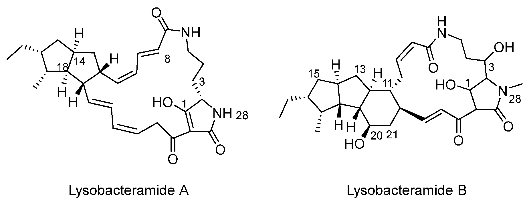
In 2015, researchers were able to isolate HSAF and analogues from various microorganisms, but the exact structures had not been elucidated. Xu et al. [19] isolated lysobacteramides A and B (Figure 5), HSAF, 3-dehydroxy HSAF, and alteramide A from L. enzymogenes C3. They used NMR and MS analysis to reveal the structures of these compounds and then assigned absolute configurations via theoretical ECD calculations. ECD, or electronic circular dichroism, uses circular polarized light to identify the absolute configurations of chiral compounds and is methodologically similar to UV spectroscopy. The theoretical absolute configurations helped advance our understanding of the biosynthetic formation of PoTeMs.
Xu et al. [19] analyzed cultures of L. enzymogenes C3 by HPLC-MS and noticed new peaks in glycerol-rich medium cultures. The new peaks were identified as metabolites with molecular masses similar to, but different from, HSAF, 3-dehydroxy HSAF, and alteramide A, which were the three previously isolated PoTeMs from L. enzymogenes C3. After EtOAc extracts of glycerol rich cultures had been fractioned by ODS chromatography and HPLC, two new PoTeMs dubbed lysobacteramides A and B were isolated. HSAF, 3-dehydroxy HSAF, and alteramide A structures were confirmed by spectroscopic data.
Lysobacteramide A shares the same carbon skeleton with alteramide A, but its lack of C3 and C20 hydroxy groups is one of its differences, as well as it having a 20,22-diene rather than a 21,23-diene, found in alteramide A. Lysobacteramide A possessing a 20,22-diene means the diene and conjugation system of the tetramic acid ring are separated. This is an uncommon feature for a polycyclic tetramate macrolactam compound. Lysobacteramide B is structurally similar to 3-dehydroxy HSAF, with the two exceptions of an additional methyl group and 28-NH lacking a proton.
In an attempt to establish the absolute configurations of the isolated compounds, they calculated theoretical ECD spectra of lysobacteramides A and B. They calculated two possible stereoisomers for the sake of comparative analysis. HSAF and 3-dehydroxy HSAF were similar to lysobacteramide B skeletally and in the relative configuration, so they measured their ECD spectra as well to assign their absolute configurations. Both of these compounds had an ECD spectrum similar to that of lysobacteramide B, so their absolute configurations assigned were the same. After performing a chiroptical analysis of the ECD data, lysobacteramides A and B, HSAF, and 3-dehydroxy HSAF have the same absolute configurations at the first cyclopentyl ring. It was assumed that this is a common feature in this group of compounds; therefore, alteramide A would have the same configuration in its 5/5 bicyclic ring.
After the ECD calculations of alteramide A, it was found that its 5/5 bicyclic ring is enantiomeric to the one in lysobacteramide A. This clarified that the absolute configuration of the first cyclopentyl ring in alteramide A is enantiomeric to the first cyclopentyl ring in HSAF. They proposed that HSAF and its related analogues achieve structural diversity via multiple cyclization and/or alkene isomerization pathways of polyene intermediates. Alteramide A and lysobacteramide A are byproducts of the main biosynthetic pathway.
Lysobacteramides A and B, HSAF, and 3-dehydroxy HSAF were also evaluated for growth inhibitory activity against human carcinoma A549, Hep-G2, and MCF-7 cells. Lysobacteramide B, HSAF, and 3-dehydroxy HSAF exhibited activity against all three cell lines with an IC50 range of 0.26–4.1 µM. Lysobacteramide A had an IC50 value range of 7.6 to 10.3 µM.

3.2. Biosynthetic Pathway

Although the chemical structures of many polycyclic tetramate macrolactams have been elucidated, their biosynthetic pathway has remained unknown for a long time. Luo et al. [13] identified a precursor compound and the subsequent cyclization cascade that leads to the formation of various polycyclic tetramate macrolactam structures. However, while they proposed a reasonable reaction, the biosynthesis of other PoTeMs and their absolute configurations still needed to be identified and studied properly.
Blodgett et al. uncovered common biosynthetic origins in PoTeMs across various species and organisms [12]. The findings showed PoTeMs’ cluster architecture is conserved and the associated genes of their BGCs are still unexplored. Studying them could be the key to identifying new members of the polycyclic tetramate macrolactam family.
PKS-NRPS, coupled with a myriad of redox enzymes, generates the common polyene tetramate precursor found at the beginning of the biosynthetic pathway for all PoTeMs. The redox enzymes turn this polyene tetramate precursor into diverse ring arrangements. The redox enzymes typically include one or a couple of flavin-dependent oxidoreductases, which can include alcohol dehydrogenase (such as IkaC, PtmC, and OX4). These enzymes have been characterized for generating the inner rings in the tricyclic ring systems through a Michael addition reaction [20].
Specifically, the PKS module produces two polyketide chains that are linked with an L-ornithine. This L-ornithine is then connected to the NRPS module via its α-amino group and δ-amino group (Figure 6). This linkage produces the common polyene tetramate precursor. The PKS/NRPS BGCs from S. koyangensis, S. griseus, and Lysobacter enzymogenes C3 show high identity and similarity to each other [15].
Downstream of the BGCs are two oxidoreductases that, in S. koyangensis and S. griseus, correspond heavily to one another, skoB1 and skoB2, and SGR813 and SGR812, respectively. From the polyene tetramate precursor, the diverse carbocyclic ring structure is formed after catalyzation by a set of oxidoreductases. In ikarugamycin, one FAD-dependent oxidoreductase (IkaB) catalyzes the ring formation [15]. The one oxidoreductase is responsible for catalyzing the formation of the 5/6 structure, but the presence of two or more oxidoreductases is considered to be required for the 5/5 structure [21]. An example here is that PtmB2, OX3, and SGR812 demonstrably catalyzed the first five-membered ring, while PtmB1, OX1/2, and SGR812 catalyzed the second five-membered ring [20,22].
Similar to the five-membered ring formation, in S. koyangensis, a gene for alcohol dehydrogenase, SkoC, flanks skoB1 and skoB2. IkaC in ikarugamycin, PtmC in the ptm gene cluster, OX4 in the HSAF gene cluster, and SGR811 in the SGR810-815 gene cluster show a high rate of identity and similarity to SkoC. OX4 was demonstrated by Li et al. [23] to facilitate the six-membered ring via Michael addition reaction, similar to SkoC.
The ring formation is carried out by different genes in unrelated bacteria, but the genes involved serve the same purpose in the pathway. In the five- and six-membered ring formations across species, the interactions between the gene clusters and oxidoreductases/dehydrogenases demonstrate high similarity.
The complexity that exists in the structure of PoTeMs has been acknowledged, and indeed, it has been proposed that this poses a challenge to synthesizing these compounds [20]. In nature, a conserved BGC gives rise to a biosynthetic pathway that successfully and efficiently builds these complex structures. The conserved origin of PoTeMs, PKS-NRPS, is responsible for the simple biosynthesis of the compounds. PKS produces two polyketide chains that are condensed to an ornithine and then attached to NRPS. This pathway was exemplified by a three-gene cassette-mediated genesis of ikarugamycin. The complexity arises from the mechanism belying the cyclization cascades that turn a key polyene tetramate precursor compound into diverse ring structures. This mechanism has remained mysterious.
Harper et al. conducted a comprehensive analysis of PoTeM biosynthetic gene clusters (BGCs) and demonstrated that although the BGCs are relatively simple in terms of gene content, the complexity of PoTeM structures arises from diverse ring systems formed through the action of flavin-dependent dehydrogenases and alcohol dehydrogenases like IkaC and OX4. Their study emphasized that the variability in ring arrangements and oxidative modifications across PoTeM family members complicates the prediction of their structures based solely on genomic data [24].
After the genome mining of S. pactum SCSIO 02999, Saha et al. [20] discovered a biosynthetic gene cluster consisting of five genes (ptmD, ptmA, ptmB1, ptmB2, and ptmC), which were homologous to the gene clusters flanking BGC. They concluded that ring formation in ikarugamycin was due to a 1,6-Michael addition reaction and could be catalyzed by IkaC, an alcohol dehydrogenase. The authors used the same strategy to test the reaction mechanism of PtmC.
Biosynthesis of heat-stable antifungal factor (HSAF), frontalamides, and ikarugamycin had gained understanding after Saha et al.’s assays. The in-frame deletion of ptmB1, ptmB2, and ptmC individually led to a cyclization cascade of six separate polycyclic tetramate macrolactam compounds, pactamides A–F, in a reductive cyclization reaction. Through this reaction, the 5, 5/5, and 5/5/6 ring system is created. By inducing reductive cyclization reactions in the polyene tetramate, a general strategy was created for the biosynthetic approach to synthesizing PoTeMs (Figure 7). When tested against cancer cell lines Hep-G2, SF-268, MCF-7, and NCI-H460, pactamides A–F showed in vitro antiproliferative activities. The IC50 values had a range of 0.24–0.26 µM.
In 2021, for the first time, Yan et al. [25] demonstrated substrate specificity of PoTeM cytochrome P450 enzymes (CYPs) in vivo. The biosynthetic gene clusters are typically composed of a PKS/NRPS gene and two to four oxidoreductase genes. A hydroxylase can add an additional hydroxy group at C-3 and CYP enzymes can add diverse oxidative modifications around the ring structures. Jiang et al. further demonstrated how distinct conformations of cytochrome P450 enzymes like IkaD and CftA lead to regio- and chemoselectivity, controlling the oxidation patterns in PoTeMs [26]. Yan et al. also reported the generation of new PoTeMs, combamides G, H, J, and I, by replacing the CYP genes. After being tested against HeLa cervical carcinoma cells and HCT116 and SW480 colorectal carcinoma cells in an MTT assay, combamides H, G, and I were active. The IC50 values for H, G, and I were 5.8 ± 0.9, 4.4 ± 0.9, and 4.7 ± 0.5 µM, respectively. This was the first time a combinatorial approach of producing new PoTeMs by swapping CYP genes had been reported.

3.3. Past Research to Support Anticancer Potential

The history of cancer treatments and potential “cures” is a long and complex process, marked by many milestones, breakthroughs, and obstacles. The initial milestone of cancer treatment was the use of surgery, which was seen as the only available option in 1809 when Dr. McDowell successfully removed an ovarian tumor from a patient, albeit without any anesthesia. However, it was not until the turn of the 20th century that chemotherapy and chemical treatments for cancer were established by Paul Ehrlich. Despite criticism for insufficient evidence, chemotherapy became a staple of cancer treatment besides radiation and surgery [27].
To this day, surgery is still necessary in the treatment of cancer patients. According to the Lancet Oncology Commission on Global Cancer surgery, an estimated 80% of cancer patients will need to undergo a surgical procedure, and some of these may require more than one surgery [28]. However, there are barriers to surgery as a treatment for cancer.
Aside from these barriers, there is, of course, the question of inoperable cancers. Inoperable cancers have been a question in the medical field for a long time, with proposals of radiation therapy and chemotherapy remaining the focal point of possibility since 1952 [29]. Under the umbrella of chemotherapy, bacterial polysaccharides, viruses, and Coley’s toxins are mentioned by Craver. These barriers have been in place for decades, making the treatment and cure for cancer elusive. This is why the field of anticancer drug research has continued to become more popular in recent years.
It has been well documented that PoTeMs have antifungal, antibacterial, antiprotozoal, and antiviral properties and behaviors. however, the possibility of antitumor and anticancer properties has not been completely explored. With the discovery of this potential, PoTeMs were cemented as a target for manipulation into potential antitumor and anticancer therapies. The next section is dedicated to some of the past experiments that have shown some sort of antitumor or anticancer properties in PoTeMs and their key chemical configurations.

3.3.1. PoTeMs Significantly Inhibit Tumor Cell Proliferation

Aoki et al. [30] conducted research searching for what they coined “ideal anticancer agents”—anticancer agents and treatments that target cancer cells without harming normal cells around the tumor. The chemotherapy currently used in clinical treatments can have side effects on normal cells surrounding the tumor, which can be quite serious and detrimental. Therefore, the need to discover novel drugs that can serve as antitumor agents without severe side effects is still pressing.
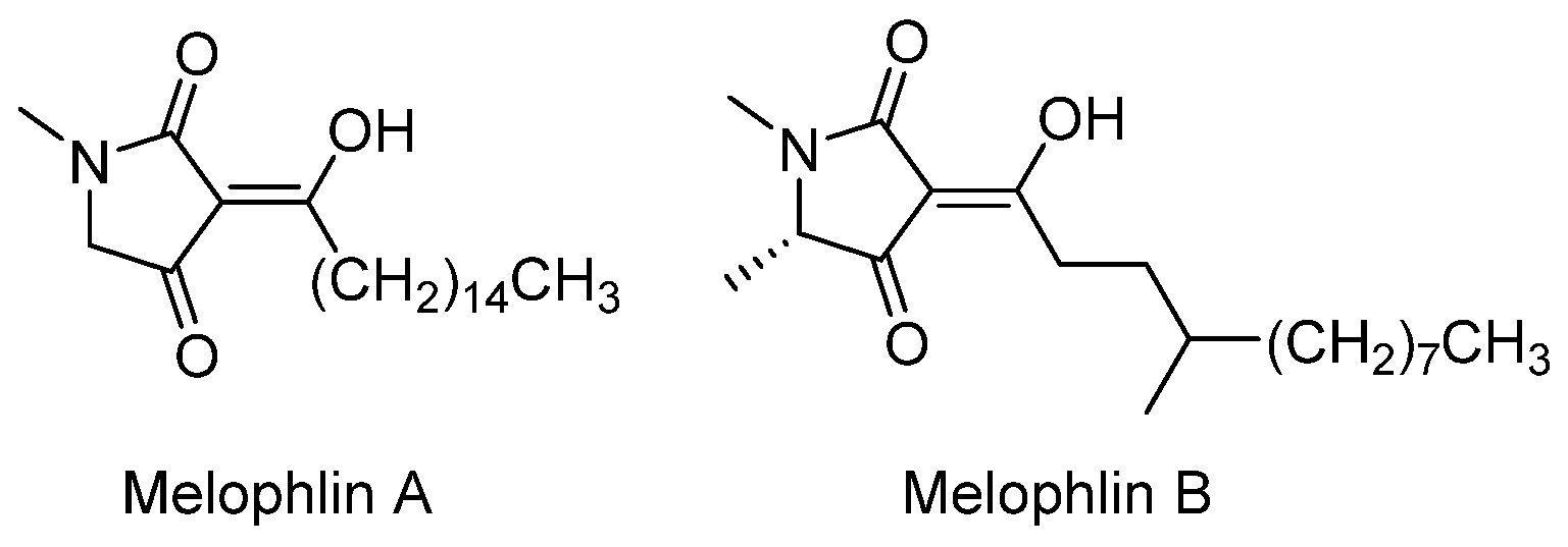
Recent studies have shown that marine creatures are a promising source of bioactive molecules that can be used to search for target-specific agents. Aoki et al. [30] found bioactive compounds derived from marine sources that can target ras oncogenes. Ras oncogenes are crucial in cell development and proliferation, meaning any agent that could take the destructive phenotype resulting from a ras-transformed oncogene and reverse that phenotype would be a good potential anticancer agent. To narrow down their search for bioactive molecules, they selectively chose a molecule that could reverse the phenotype, and successfully isolated melophlins A and B (Figure 8) from Melophlus sarassinorum.
Melophlins A and B, while not PoTeMs, are tetramic acids. Tetramic acids are an important, constant structural characteristic of PoTeMs and their anticancer functions. Aoki et al. provided evidence of this using melophlins A and B to reverse the phenotype of ras oncogenes. This study further supported the understanding of how every structural part of PoTeMs is crucial for their unique anticancer properties and behaviors.
HL60 cells, a cell line from 1977 isolated from a myeloid leukemia patient [31], were placed in incubation with melophlins A and B. Melophlins A and B exhibited cytotoxicity at a moderate level at low concentrations (0.2 and 0.4 µg/mL respectively) against the HL60 cells. Aoki et al. had used parental NIH3T3 cells as well as NIH3T3 cells that had been transformed with c-H-ras gene (human) and cultured both in the same broth and medium [30]. They inoculated the same number of cells into a 96-well plate. After 48 h, Aoki et al. were able to see that cell morphology was affected by the ras oncogene. Melophlins A and B, at a higher concentration (5 µg/mL), were able to reverse the phenotype of H-ras NIH3T3 fibroblasts back to normal. Additionally, these compounds also froze the NIH3T3 cells in the G1 phase at a concentration of 1 µg/mL.
In 2023, Li et al. [32] discovered three new PoTeMs, pseudoamides A, B, and C expressed by the pel cluster from Pseudoalteromonas elyakovii. Pel1 and Pel3, FMN-dependent oxidoreductases, form the 5/5-bicyclic ring system in pseudoamides A–C. No biosynthetic pathway for the C13-C20 cyclization of the 5/5-bicycle ring system had been established previously. With the discovery of pseudoamides A–C, the pel cluster appeared to be the first C13-C20 5/5-cyclization biosynthetic pathway. Pseudoamides A–C, when tested in vitro for cytotoxicity against A549 lung and MIA-Paca2 pancreatic cancer, had an IC50 value range of 1.3–19.4 µM.
The C13-C20 and C12-C19 cyclization is reported to have influence over antitumor activity. In 2024, Chen et al. [33] isolated the known PoTeM aburatubolactam C and three new PoTeMs, aburatubolactams D–F, from Streptomyces sp. SCSIO 40070. X-ray analysis established the absolute configuration of aburatubolactam C. Chen et al. used AntiSMASH analysis to discover the atl biosynthetic gene cluster, which encodes four enzymes, PKS/NRPS AtlA and FAD-dependent oxidoreductases AtlB1, AtlB2, and AtlB3. AtlB3 is proposed to be responsible for the cyclization of the first five-membered ring, and AtlB1 and AtlB2 are proposed to be responsible for the formation of the second five-membered ring. Aburatubolactams C, D, and E have a C13-C20 cyclization pattern, while aburatubolactam F has a C12-C19 cyclization pattern. Against C4-2B, A549, and HepG2 cancer cell lines, aburatubolactams C-E had an IC50 range of 2.43–16.16 µM, while aburatubolactam F was inactive against all tested cell lines. Chen et al. reported that the cyclization pattern will influence the antitumor effects of 5/5-bicycle PoTeMs.

3.3.2. Polycyclic Tetramate Macrolactam with Potential Anticancer Properties from S. zhaozhouensis Subspecies

Actinomycetes are one of the most common microorganisms used to isolate bioactive secondary metabolites for drug discovery. In a study by Dhaneesha et al. [34], they isolated an actinomycete from an Indian Ocean sponge-associated and designated it as strain MCCB267. By using 16S RNA gene sequencing, the authors revealed that MCCB267 had high similarity to S. zhaozhouensis. However, MCCB267 showed unique biochemical and physiological characteristics, which led to the recognition of the subspecies status for this strain named S. zhaozhouensis subsp. mycale subsp. nov.
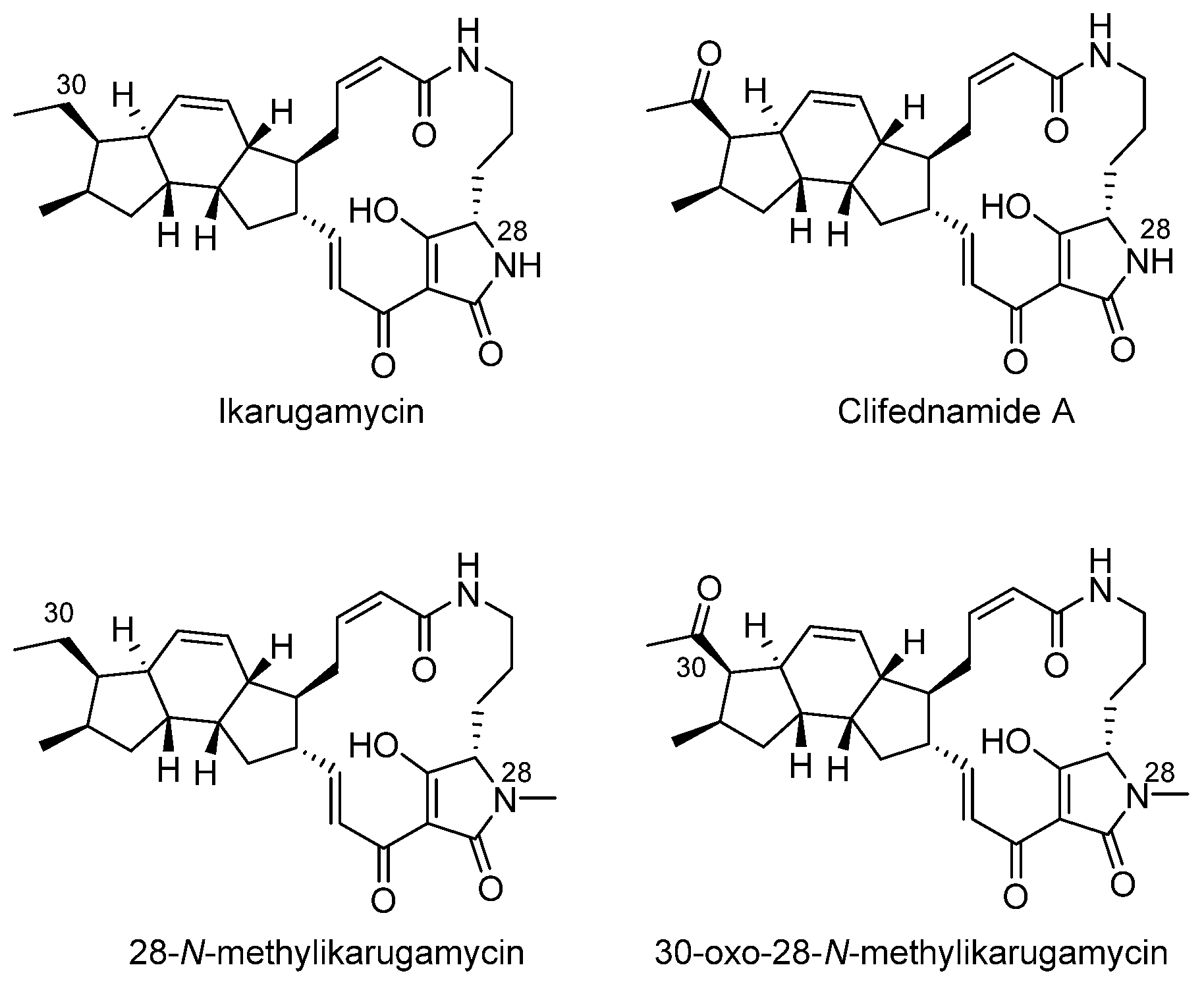
Ikarugamycin, clifednamide A, 30-oxo-28-N-methylikarguamycin, and 28-N-mthylikarugamycin, all members of the polycyclic tetramate macrolactam family, were retrieved from ethyl acetate extracts of MCCB267 and exhibited cytotoxicity against NCI-H460 lung carcinoma cells in vitro via the induction of apoptosis (Figure 9). Cell cycle arrest in the G1 phase in NCI-H460 carcinoma cells occurred due to ikarugamycin, 30-oxo-28-N-methylikarugamycin, and 28-N-methylikarguamycin; clifednamide A induced cell cycle arrest in the S phase. All four compounds showed concentration-dependent cells accumulating in the sub-G1 phase. These in vitro results were supported by molecular docking and dynamic simulation analysis.
The docking studies, in silico, were conducted using Autodock 4.2 software due to the allowance for estimation of interaction energies between the ligands and the DNA target. The PoTeMs can bind to the minor groove of DNA, which will then lead to cell death via apoptosis. Ikarugamycin and its derivatives underwent a blind docking study in the binding pocket to predict the ligands’ possible binding modes. The docking energies for ikarugamycin (−7.971 kcal/mol), 30-oxo-28-N-methylikarugamycin (−7.149 kcal/mol), 28-N-methylikarugamycin (−5.580 kcal/mol), and clifednamide A (−3.689 kcal/mol) were determined by clustering analyses. The structures of the compounds, from the analysis, had a clear effect on the binding affinity.
Ikarugamycin and 30-oxo-28-N-methylikarugamycin had similar docking energies; however, 28-N-methylikarugamycin and clifednamide A were less specific in binding to DNA. Dhaneesha et al. [34] suggested, from their MD simulations, that all the compounds may be potential anticancer agents, although ikarugamycin would be the most potent candidate. MM-PBSA-based calculations showed electrostatic interactions contributed to interaction energy and polar solvation energy provided stabilization in terms of ikarugamycin. However, with the other three compounds, van der Waals interactions had greater influence in the initial interaction than electrostatic interactions, and polar solvation energy did not contribute to the same degree as in the case of ikarugamycin.
In the in vitro MTT Assay, the IC50 values of ikarugamycin (1.43 µg/mL), clifednamide A (16.29 µg/mL), 30-oxo-28-N-methylikarugamycin (7.17 µg/mL), and 28-N-methylikarugamycin (1.78 µg/mL) demonstrated that all four compounds inhibited growth depending on the concentration of treatment. NCI-H460 cells were also stained with Hoechst 33,342 in order to directly observe apoptosis hallmarks microscopically. All four compounds had induced apoptosis characteristics (shrinking and cellular nuclei fragmentation) after a treatment period of 24 h, and the control cells were still round and intact. Nuclear DNA fragmentation was confirmed by a TUNEL assay.

4. Anticancer Mechanistic Pathways of PoTeMs

Identifying new potential treatment options for any disease, not just cancer, is challenging because it requires understanding of how exactly the alternative potential therapy will act not only on the actual target, but also on the body’s surrounding cells and tissues. In the case of PoTeMs, the most established mechanistic research appears to have a focus on the identification of the biosynthetic pathway and genome mining. However, research has been conducted to elucidate the methods through which PoTeMs target certain cells and pathways to exhibit the cytotoxic behaviors that make them interesting candidates for further anticancer research.
Indeed, previous research to establish the mechanism of ikarugamycin, a very common polycyclic tetramate macrolactam, focused on how ikarugamycin inhibits the uptake of oxidized low-density lipoprotein (LDL) by macrophages in mammalian cells. The findings are an important contribution because a hallmark of atherosclerosis is the appearance of cholesterol-ester foam cells, which are derived from macrophages [35]. In the early stages of atherosclerosis, multiple forms of lipids are retained and trapped within arterial walls, where macrophages will infiltrate and take them up [36]. The pathogenesis of atherosclerosis is characterized by the dysregulation of macrophages, specifically their atypical activation due to mediators, cholesterol crystals, and modified lipids [37]. There are many modifications of lipids that can induce such a state of macrophages. Hasumi et al. [38] utilized oxidation as the particular physiological modification.
During the incubation of mouse macrophage cell line J774 A.1 with oxidized LDL, the cells showed an accumulation of stained lipid droplets in their cytoplasm. However, this accumulation was inhibited by ikarugamycin at 50% concentrations when used at a concentration of 2 µm and nearly entirely at a concentration of 4 µm. When tested for its ability to suppress the synthesis of neutral or polar lipids from [14C]oleate, ikarugamycin was found to inhibit the synthesis of cholesteryl ester, but not tri-acylglycerol and polar lipids. Although ikarugamycin did not inhibit the cell-surface binding of oxidized LDL, microsomal acyl-CoA: cholesterol acyltransferase (ACAT), and lysosomal hydrolysis of internalized oxidized LDL, it reduced the internalization of oxidized LDL in the macrophages to 50% when used at lower concentrations [38].
These results showed the uptake of oxidized LDL was successfully inhibited by ikarugamycin, which in turn inhibited cholesteryl ester accumulation. Moreover, according to Hasumi et al., this inhibition had been specific to the uptake process of the pathway and not the cell-surface binding or re-esterification of cholesterol by ACAT. Incubation of J774 at 37 °C with ikarugamycin resulted in a slight decrease in cell-surface receptor activity for oxidized LDL, but this reduction appeared to be due to the intracellular trapping of part of the receptors since total receptor activity was not affected.
In order to gauge the efficacy of PoTeMs as potential cancer treatments, it is important to understand the mechanisms behind PoTeMs’ interactions in mammalian cells, especially when interacting with cells that are either hallmarks of disease or directly part of it, such as tumor cells. Hasumi et al. provided insight into ikarugamycin’s inhibitory mechanism in the prevention of the transformation of macrophages into foam cells. More recently, there have been studies of ikarugamycin’s involvement in endocytosis.
Specifically, Hasumi et al. identified that ikarugamycin can act as an inhibitor of clathrin-mediated endocytosis. Clathrin-mediated endocytosis (CME) is a process through which cell surface receptors and their cargo or bound ligands are internalized by the cell. While there are a few ways to inhibit CME, every method has limitations.
Elkin et al. [39] identified ikarugamycin as a potential selective inhibitor of the pathway. They found that ikarugamycin could inhibit CME of multiple receptors in different cell lines. To determine the inhibitory abilities of ikarugamycins, they tested a range of concentrations of ikarugamycin on clathrin-mediated uptake of transferrin receptor TfnR in pre-treated non-small cell lung cancer (H1299) cells and revealed a dose-dependent decrease in TfnR uptake. They also found the IC50 of 2.7 ± 0.3 µM.
Once it was determined that ikarugamycin could inhibit CME of multiple receptors, they determined the efficacy of ikarugamycin inhibition of CME in different cell lines. H1299 and other non-small cell lung cancer lines, HCC366 and H1437, were tested as well as HBEC3KT and ARPE-19 cell lines (bronchial epithelial cells and retinal pigment epithelial cells, respectively). After preincubating cells for 3 h with 4 µM ikarugamycin, TfnR uptake was measured after five minutes. In H1299, HCC366, and ARPE-19 cells, TfnR uptake had been inhibited by approximately 80%. In H1437 and HBEC3KT cells, TfnR uptake was inhibited by approximately 50%. Elkin et al. suggested, with these results, that ikarugamycin is able to inhibit TfnR uptake in multiple human cell lines.
These findings created another question: Is ikarugamycin inhibiting CME in a receptor-specific manner? TfnR was tested alongside other known receptors that were known to traffic in a CME-selective manner, such as LDL receptor (LDLR) and epidermal growth factor receptor (EGFR). TfnR, LDLR, and EGFR all use CME to traffic, although the adaptor molecules they each require are different. TfnR and EGFR uptake experiments were conducted in H1299 but, because most cells express low levels of LDLR, CD8-chimeras encoding the LDLR FxNPxY internalization motif [40] were expressed in ARPE-19 cells in order for the LDLR uptake experiments to be carried out. In cells pretreated with ikarugamycin at a concentration of 4 µM for 3 h, CME of each of the receptors had been inhibited.
Last, the specificity of ikarugamycin’s inhibitory behaviors had been tested by measuring its effects on the other endocytic pathways for which Elkin et al. used their previously developed assays: CavME (caveolae-mediated endocytosis) measured by albumin uptake and multiple CIE (clathrin and caveolae-independent endocytosis) measured by CD44 and CD59 uptake. Over a range of 1–4 µM, ikarugamycin showed no inhibition, although the TfnR uptake was inhibited significantly. Ikarugamycin showed selectivity of CME over other endocytosis pathways.
In 2001, Luo et al. [35] also investigated a clathrin-related endocytosis pathway using ikarugamycin. The nef genes of human and simian immunodeficiency virus (HIV and SIV, respectively) play a crucial role in HIV replication. Nef can remove CD4 from the cell surface by speeding up the internalization and degradation process of CD4 [41]. It had been suggested in previous studies that this Nef-induced CD4 internalization had been achieved by a higher association with clathrin-coated pits, or CCPs. Luo et al. investigated ikarugamycin on CD4 cell surface expression in stable human cells that express HIV type 1 Nef. Ikarugamycin had been able to restore CD4 surface expression without affecting Nef or CD4 synthesis. Luo et al. concluded that ikarugamycin could be a promising inhibitor of CCP-dependent endocytosis.
A study published in 2011 by Popescu et al. [42] elucidated the mechanism of cytotoxicity of ikarugamycin. In the study, ikarugamycin induced apoptosis in HL-60 cells (human promyelocytic leukemia cells) via DNA damage and activation of caspases-9, -8, and -3. Caspase cleavage was found to have a connection to intracellular calcium increase and p38 MAP kinase activation. Popescu et al. found that nanomolar concentrations of ikarugamycin had decreased viability of p53-null HL-60 cells. Prior to this paper, Bertasso et al. [43] had reported that ikarugamycin was active against p53-positive MCF-7 breast cancer cells. Popescu et al. used these two results to suggest that this is a cell-line and p53-independent cell death effect. Popescu et al. found ikarugamycin only contributed small changes to S and G2-M phases, which had served as an indication that ikarugamycin could induce apoptosis in HL-60 cells without a cell cycle arrest. However, they suggested extensive analyses over time were needed to effectively rule out inhibition of DNA replication occurring before apoptosis.
Ikarugamycin was confirmed to cause DNA strand breaks, either directly or indirectly, followed by checkpoint kinase Chk2 and caspase-9 and -3 activation, and finally ending in apoptosis. Checkpoint kinase Chk2 and γ-H2AX are essential for protection of genome integrity and become active through DNA double-stranded breaks. Ikarugamycin induced caspase activation via intracellular calcium levels increasing.
There are many reports identifying intracellular calcium overload as a trigger for apoptosis. Ikarugamycin is an inhibitor of endocytosis, as discussed previously, co-regulated by calcium. Popescu et al. referenced cylindramide, a polycyclic tetramate macrolactam that exhibits cytotoxic behaviors, which has been shown to reduce free intracellular calcium for a short period and interact with the endoplasmic reticulum for a long period. Popescu et al. suggested that there are different mechanisms behind the cytotoxicity of PoTeMs.
In a study focusing on cancer cell metabolism, ikarugamycin isolated from S. xiamenensis 318 exhibited antiproliferative qualities against PDAC, or pancreatic ductal adenocarcinoma, which is one of the most aggressive malignancies in humans. Unfortunately, the protein target for ikarugamycin is not clearly elucidated. However, Jiang et al. [44] used metabolomic studies and transcriptome-based profiling to prove that ikarugamycin causes a drop in glucose-6-phosphate (G6P) and an increase in glucose level in the cell. SPR (surface plasmon resonance) experiments and docking studies demonstrated that hexokinase 2 (HK2), the key enzyme in glycolysis, was a target of ikarugamycin. Ikarugamycin reduced tumor size in PDAC mice and increased the response to gemcitabine chemotherapy.
PDAC cells depend on glycolysis, specifically the resulting glucose flux, in contrast to normal cells’ dependence on ATP generation via oxidative phosphorylation by the mitochondria. Cancer cells can be distinguished from normal cells by the HK2 expression. Due to this dependence on glycolysis, cancer cell proliferation is extra sensitive to an altered glucose metabolism. Ikarugamycin caused metabolic changes in glucose metabolism, specifically the immensely decreased production of G6P and ATP. The ikarugamycin binding protein was revealed to be HK2, and the binding affinity was measured by SPR. A possible binding site was revealed via docking studies, although Jiang et al. encouraged further studies to support the direct interaction between ikarugamycin and the amino acids Phe602, Ser603, Pro605, Cys606, Asn608, and Val654. They concluded that ikarugamycin could inhibit glycolysis in cancer cells by selectively targeting HK2, and that ikarugamycin could successfully be used in combination with established chemotherapy drugs.
Although ikarugamycin had been established as an inhibitor of CME, whether it influences cytokine production was yet to be understood. Minamidate et al. [45] investigated the relationship between tumor necrosis factor-α (TNF) due to TNF’s crucial role in the pathogenesis of many diseases. The functions of membrane TNF (mTNF) have not been fully revealed. It was reported that mTNF and anti-TNF agents, in a complex, are endocytosed via a clathrin-dependent pathway. Minamidate et al. hypothesized that ikarugamycin’s inhibition of CME results in the accumulation of TNF on the surface of the cell. They called for further studies to prove this hypothesis. Ikarugamycin now has a new reported function, increasing soluble TNF and surface expression of mTNF, which results in persistent inflammation over an extended period of time. Minamidate et al. suggested that their results indicated ikarugamycin could be a fundamental tool for the future research of TNF signaling.

5. Conclusions

PoTeMs are a massive resource in synthetic biology and drug discovery. These compounds have been used for a long time as antibiotics and in the treatment of plant pathogens. However, the increasing demand for alternative anticancer and antitumor drugs has motivated the interest in PoTeMs as viable candidates for research. From the discovery of the first PoTeM, ikarugamycin, in 1972, to now, over 80 new PoTeMs have been discovered, and potentially thousands are out there waiting to be isolated and characterized. PoTeMs have unique properties and interactions and their availability and inducibility are important features that aid in their consideration in natural product research. Now that technology has advanced, the search for and isolation of novel PoTeMs has become more possible.
The elucidation of the biosynthetic pathway and gene clusters has proven indispensable. The chemical and structural configurations of PoTeMs have been identified, as well as their availability and inducibility. For those that have not been fully confirmed, there are now published methodologies that could help future researchers elucidate the structures of other PoTeMs and their derivatives. It is crucial to understand the chemical configurations of these compounds in order to understand how and why they interact with various cells and tissues the way they do.
While recent advancements and methods have made the discovery and isolation of new PoTeMs more accessible, key challenges still remain, particularly in two areas highlighted by this review. First, obtaining sufficient quantities of PoTeMs is a significant challenge. Although synthetic biology technologies enable the cloning and heterologous expression of large biosynthetic gene clusters, scaling up biosynthesis to produce enough material for further studies remains difficult. Total synthesis is another potential solution, but it is rarely reported due to the complex stereocenters present in many PoTeMs, making efficient total synthesis development a major hurdle. Second, identifying the molecular targets of PoTeMs remains an ongoing challenge. Techniques such as activity-based protein profiling (ABPP), drug affinity responsive target stability (DARTs), and comparative genomics or proteomics are critical tools in addressing this issue, but their application to PoTeMs requires further development. Overcoming these challenges is essential for advancing PoTeM research and unlocking their full potential for therapeutic applications.

Author Contributions

A.M.: literature research, manuscript writing, and original draft preparation. M.R.M.-R.: reviewing and editing. Y.H.: reviewing and supervision. All authors have read and agreed to the published version of the manuscript.

Funding

This work is supported by the Konneker Research Fund, Edison Biotechnology Institute, and the Translational Biomedical Sciences program at Ohio University.

Institutional Review Board Statement

Not applicable.

Informed Consent Statement

Not applicable.

Data Availability Statement

Not applicable.

Conflicts of Interest

The authors declare that they have no known competing financial or personal interests.

References

  1. Demain, A.L. Importance of microbial natural products and the need to revitalize their discovery. J. Ind. Microbiol. Biotechnol. 2014, 41, 185–201. [Google Scholar] [CrossRef] [PubMed]
  2. David, B.; Wolfender, J.-L.; Dias, D.A. The pharmaceutical industry and natural products: Historical status and new trends. Phytochem. Rev. 2015, 14, 299–315. [Google Scholar] [CrossRef]
  3. Atanasov, A.G.; Zotchev, S.B.; Dirsch, V.M.; Supuran, C.T. Natural products in drug discovery: Advances and opportunities. Nat. Rev. Drug Discov. 2021, 20, 200–216. [Google Scholar] [CrossRef]
  4. Siegel, R.L.; Giaquinto, A.N.; Jemal, A. Cancer statistics, 2024. CA Cancer J. Clin. 2024, 74, 12–49. [Google Scholar] [CrossRef] [PubMed]
  5. Sun, Y.; Zheng, H.; Qian, L.; Liu, Y.; Zhu, D.; Xu, Z.; Chang, W.; Xu, J.; Wang, L.; Sun, B.; et al. Targeting GDP-Dissociation Inhibitor Beta (GDI2) with a Benzo[a]quinolizidine Library to Induce Paraptosis for Cancer Therapy. JACS Au 2023, 3, 2749–2762. [Google Scholar] [CrossRef]
  6. Huang, M.; Lu, J.J.; Ding, J. Natural Products in Cancer Therapy: Past, Present and Future. Nat. Prod. Bioprospect. 2021, 11, 5–13. [Google Scholar] [CrossRef]
  7. Zheng, H.; Dong, Y.; Li, L.; Sun, B.; Liu, L.; Yuan, H.; Lou, H. Novel Benzo[a]quinolizidine Analogs Induce Cancer Cell Death through Paraptosis and Apoptosis. J. Med. Chem. 2016, 59, 5063–5076. [Google Scholar] [CrossRef]
  8. Wang, H.; He, Y.; Jian, M.; Fu, X.; Cheng, Y.; He, Y.; Fang, J.; Li, L.; Zhang, D. Breaking the Bottleneck in Anticancer Drug Development: Efficient Utilization of Synthetic Biology. Molecules 2022, 27, 7480. [Google Scholar] [CrossRef]
  9. Surade, S.; Blundell, T.L. Structural biology and drug discovery of difficult targets: The limits of ligandability. Chem. Biol. 2012, 19, 42–50. [Google Scholar] [CrossRef]
  10. Mallinson, J.; Collins, I. Macrocycles in new drug discovery. Future Med. Chem. 2012, 4, 1409–1438. [Google Scholar] [CrossRef]
  11. Liu, W.; Zhang, W.; Jin, H.; Zhang, Q.; Chen, Y.; Jiang, X.; Zhang, G.; Zhang, L.; Zhang, W.; She, Z.; et al. Genome Mining of Marine-Derived Streptomyces sp. SCSIO 40010 Leads to Cytotoxic New Polycyclic Tetramate Macrolactams. Mar. Drugs 2019, 17, 663. [Google Scholar] [CrossRef] [PubMed]
  12. Blodgett, J.A.; Oh, D.C.; Cao, S.; Currie, C.R.; Kolter, R.; Clardy, J. Common biosynthetic origins for polycyclic tetramate macrolactams from phylogenetically diverse bacteria. Proc. Natl. Acad. Sci. USA 2010, 107, 11692–11697. [Google Scholar] [CrossRef] [PubMed]
  13. Luo, Y.; Huang, H.; Liang, J.; Wang, M.; Lu, L.; Shao, Z.; Cobb, R.E.; Zhao, H. Activation and characterization of a cryptic polycyclic tetramate macrolactam biosynthetic gene cluster. Nat. Commun. 2013, 4, 2894. [Google Scholar] [CrossRef]
  14. Medema, M.H.; de Rond, T.; Moore, B.S. Mining genomes to illuminate the specialized chemistry of life. Nat. Rev. Genet. 2021, 22, 553–571. [Google Scholar] [CrossRef] [PubMed]
  15. Ding, W.; Tu, J.; Zhang, H.; Wei, X.; Ju, J.; Li, Q. Genome Mining and Metabolic Profiling Uncover Polycyclic Tetramate Macrolactams from Streptomyces koyangensis SCSIO 5802. Mar. Drugs 2021, 19, 440. [Google Scholar] [CrossRef]
  16. Mo, X.; Li, Q.; Ju, J. Naturally occurring tetramic acid products: Isolation, structure elucidation and biological activity. RSC Adv. 2014, 4, 50566–50593. [Google Scholar] [CrossRef]
  17. Lowery, C.A.; Park, J.; Gloeckner, C.; Meijler, M.M.; Mueller, R.S.; Boshoff, H.I.; Ulrich, R.L.; Barry, C.E., 3rd; Bartlett, D.H.; Kravchenko, V.V.; et al. Defining the mode of action of tetramic acid antibacterials derived from Pseudomonas aeruginosa quorum sensing signals. J. Am. Chem. Soc. 2009, 131, 14473–14479. [Google Scholar] [CrossRef]
  18. Bodenschatz, K.; Stoeckl, J.; Winterer, M.; Schobert, R. A synthetic approach to 5/5/6-polycyclic tetramate macrolactams of the discodermide type. Tetrahedron 2022, 104, 132113. [Google Scholar] [CrossRef]
  19. Xu, L.; Wu, P.; Wright, S.J.; Du, L.; Wei, X. Bioactive Polycyclic Tetramate Macrolactams from Lysobacter enzymogenes and Their Absolute Configurations by Theoretical ECD Calculations. J. Nat. Prod. 2015, 78, 1841–1847. [Google Scholar] [CrossRef]
  20. Saha, S.; Zhang, W.; Zhang, G.; Zhu, Y.; Chen, Y.; Liu, W.; Yuan, C.; Zhang, Q.; Zhang, H.; Zhang, L.; et al. Activation and characterization of a cryptic gene cluster reveals a cyclization cascade for polycyclic tetramate macrolactams. Chem. Sci. 2017, 8, 1607–1612. [Google Scholar] [CrossRef]
  21. Jin, H.; Zhang, W.; Zhang, G.; Zhang, L.; Liu, W.; Zhang, C. Engineered Biosynthesis of 5/5/6 Type Polycyclic Tetramate Macrolactams in an Ikarugamycin (5/6/5 Type)-Producing Chassis. Org. Lett. 2020, 22, 1731–1735. [Google Scholar] [CrossRef] [PubMed]
  22. Li, Y.; Wang, H.; Liu, Y.; Jiao, Y.; Li, S.; Shen, Y.; Du, L. Biosynthesis of the Polycyclic System in the Antifungal HSAF and Analogues from Lysobacter enzymogenes. Angew. Chem. (Int. Ed. Engl.) 2018, 57, 6221–6225. [Google Scholar] [CrossRef] [PubMed]
  23. Li, X.; Wang, H.; Shen, Y.; Li, Y.; Du, L. OX4 Is an NADPH-Dependent Dehydrogenase Catalyzing an Extended Michael Addition Reaction to Form the Six-Membered Ring in the Antifungal HSAF. Biochemistry 2019, 58, 5245–5248. [Google Scholar] [CrossRef] [PubMed]
  24. Harper, C.P.; Day, A.; Tsingos, M.; Ding, E.; Zeng, E.; Stumpf, S.D.; Qi, Y.; Robinson, A.; Greif, J.; Blodgett, J.A.V. Critical analysis of polycyclic tetramate macrolactam biosynthetic gene cluster phylogeny and functional diversity. Appl. Environ. Microbiol. 2024, 90, e0060024. [Google Scholar] [CrossRef]
  25. Yan, Y.; Wang, H.; Song, Y.; Zhu, D.; Shen, Y.; Li, Y. Combinatorial Biosynthesis of Oxidized Combamides Using Cytochrome P450 Enzymes from Different Polycyclic Tetramate Macrolactam Pathways. ACS Synth. Biol. 2021, 10, 2434–2439. [Google Scholar] [CrossRef]
  26. Jiang, P.; Jin, H.; Zhang, G.; Zhang, W.; Liu, W.; Zhu, Y.; Zhang, C.; Zhang, L. A Mechanistic Understanding of the Distinct Regio- and Chemoselectivity of Multifunctional P450s by Structural Comparison of IkaD and CftA Complexed with Common Substrates. Angew. Chem. (Int. Ed. Engl.) 2023, 62, e202310728. [Google Scholar] [CrossRef]
  27. DeVita, V.T., Jr.; Rosenberg, S.A. Two hundred years of cancer research. N. Engl. J. Med. 2012, 366, 2207–2214. [Google Scholar] [CrossRef]
  28. Sullivan, R.; Alatise, O.I.; Anderson, B.O.; Audisio, R.; Autier, P.; Aggarwal, A.; Balch, C.; Brennan, M.F.; Dare, A.; D’Cruz, A.; et al. Global cancer surgery: Delivering safe, affordable, and timely cancer surgery. Lancet Oncol. 2015, 16, 1193–1224. [Google Scholar] [CrossRef] [PubMed]
  29. Craver, L.F. Recent advances in treatment of inoperable cancer. Bull. N. Y. Acad. Med. 1952, 28, 385–407. [Google Scholar]
  30. Aoki, S.; Higuchi, K.; Ye, Y.; Satari, R.; Kobayashi, M. Melophlins A and B, novel tetramic acids reversing the phenotype of ras-transformed cells, from the marine sponge Melophlus sarassinorum. Tetrahedron 2000, 56, 1833–1836. [Google Scholar] [CrossRef]
  31. Birnie, G.D. The HL60 cell line: A model system for studying human myeloid cell differentiation. Br. J. Cancer. Suppl. 1988, 9, 41–45. [Google Scholar] [PubMed]
  32. Li, X.; Liu, Q.; Zou, H.; Luo, J.; Jiao, Y.; Wang, H.; Du, L.; Shen, Y.; Li, Y. Discovery and biosynthesis of pseudoamides reveal enzymatic cyclization of the polyene precursor to 5–5 bicyclic tetramate macrolactams. ACS Catal. 2023, 13, 4760–4767. [Google Scholar] [CrossRef]
  33. Chen, Y.; Jin, H.; Xiong, W.; Fang, Z.; Sun, W.; Zhu, Y.; Zhang, L.; Zhang, Y.; Zhang, W.; Zhang, C. Discovery of Aburatubolactams Reveals Biosynthetic Logic for Distinct 5/5-Type Polycyclic Tetramate Macrolactams. Org. Lett. 2024, 26, 1677–1682. [Google Scholar] [CrossRef] [PubMed]
  34. Dhaneesha, M.; Hasin, O.; Sivakumar, K.C.; Ravinesh, R.; Naman, C.B.; Carmeli, S.; Sajeevan, T.P. DNA Binding and Molecular Dynamic Studies of Polycyclic Tetramate Macrolactams (PTM) with Potential Anticancer Activity Isolated from a Sponge-Associated Streptomyces zhaozhouensis subsp. mycale subsp. nov. Mar. Biotechnol. 2019, 21, 124–137. [Google Scholar] [CrossRef] [PubMed]
  35. Khoo, J.C.; Miller, E.; McLoughlin, P.; Steinberg, D. Enhanced macrophage uptake of low density lipoprotein after self-aggregation. Arteriosclerosis 1988, 8, 348–358. [Google Scholar] [CrossRef]
  36. Gui, Y.; Zheng, H.; Cao, R.Y. Foam Cells in Atherosclerosis: Novel Insights into Its Origins, Consequences, and Molecular Mechanisms. Front. Cardiovasc. Med. 2022, 9, 845942. [Google Scholar] [CrossRef] [PubMed]
  37. Poznyak, A.V.; Nikiforov, N.G.; Markin, A.M.; Kashirskikh, D.A.; Myasoedova, V.A.; Gerasimova, E.V.; Orekhov, A.N. Overview of OxLDL and its impact on cardiovascular health: Focus on atherosclerosis. Front. Pharmacol. 2021, 11, 613780. [Google Scholar] [CrossRef]
  38. Hasumi, K.; Shinohara, C.; Naganuma, S.; Endo, A. Inhibition of the uptake of oxidized low-density lipoprotein in macrophage J774 by the antibiotic ikarugamycin. Eur. J. Biochem. 1992, 205, 841–846. [Google Scholar] [CrossRef]
  39. Elkin, S.R.; Oswald, N.W.; Reed, D.K.; Mettlen, M.; MacMillan, J.B.; Schmid, S.L. Ikarugamycin: A Natural Product Inhibitor of Clathrin-Mediated Endocytosis. Traffic 2016, 17, 1139–1149. [Google Scholar] [CrossRef]
  40. Motley, A.; Bright, N.A.; Seaman, M.N.; Robinson, M.S. Clathrin-mediated endocytosis in AP-2-depleted cells. J. Cell Biol. 2003, 162, 909–918. [Google Scholar] [CrossRef]
  41. Luo, T.; Fredericksen, B.L.; Hasumi, K.; Endo, A.; Garcia, J.V. Human immunodeficiency virus type 1 Nef-induced CD4 cell surface downregulation is inhibited by ikarugamycin. J. Virol. 2001, 75, 2488–2492. [Google Scholar] [CrossRef] [PubMed]
  42. Popescu, R.; Heiss, E.H.; Ferk, F.; Peschel, A.; Knasmueller, S.; Dirsch, V.M.; Krupitza, G.; Kopp, B. Ikarugamycin induces DNA damage, intracellular calcium increase, p38 MAP kinase activation and apoptosis in HL-60 human promyelocytic leukemia cells. Mutat. Res. 2011, 709–710, 60–66. [Google Scholar] [CrossRef] [PubMed]
  43. Bertasso, M.; Holzenkampfer, M.; Zeeck, A.; Stackebrandt, E.; Beil, W.; Fiedler, H.-P. Ripromycin and other polycyclic macrolactams from Streptomyces sp. Tu 6239: Taxonomy, fermentation, isolation and biological properties. J. Antibiot. 2020, 56, 364–371. [Google Scholar] [CrossRef] [PubMed]
  44. Jiang, S.H.; Dong, F.Y.; Da, L.T.; Yang, X.M.; Wang, X.X.; Weng, J.Y.; Feng, L.; Zhu, L.L.; Zhang, Y.L.; Zhang, Z.G.; et al. Ikarugamycin inhibits pancreatic cancer cell glycolysis by targeting hexokinase 2. FASEB J. Off. Publ. Fed. Am. Soc. Exp. Biol. 2020, 34, 3943–3955. [Google Scholar] [CrossRef]
  45. Minamidate, A.; Onizawa, M.; Saito, C.; Hikichi, R.; Mochimaru, T.; Murakami, M.; Sakuma, C.; Asakawa, T.; Hiraoka, Y.; Oshima, S.; et al. A potent endocytosis inhibitor Ikarugamycin up-regulates TNF production. Biochem. Biophys. Rep. 2021, 27, 101065. [Google Scholar] [CrossRef]
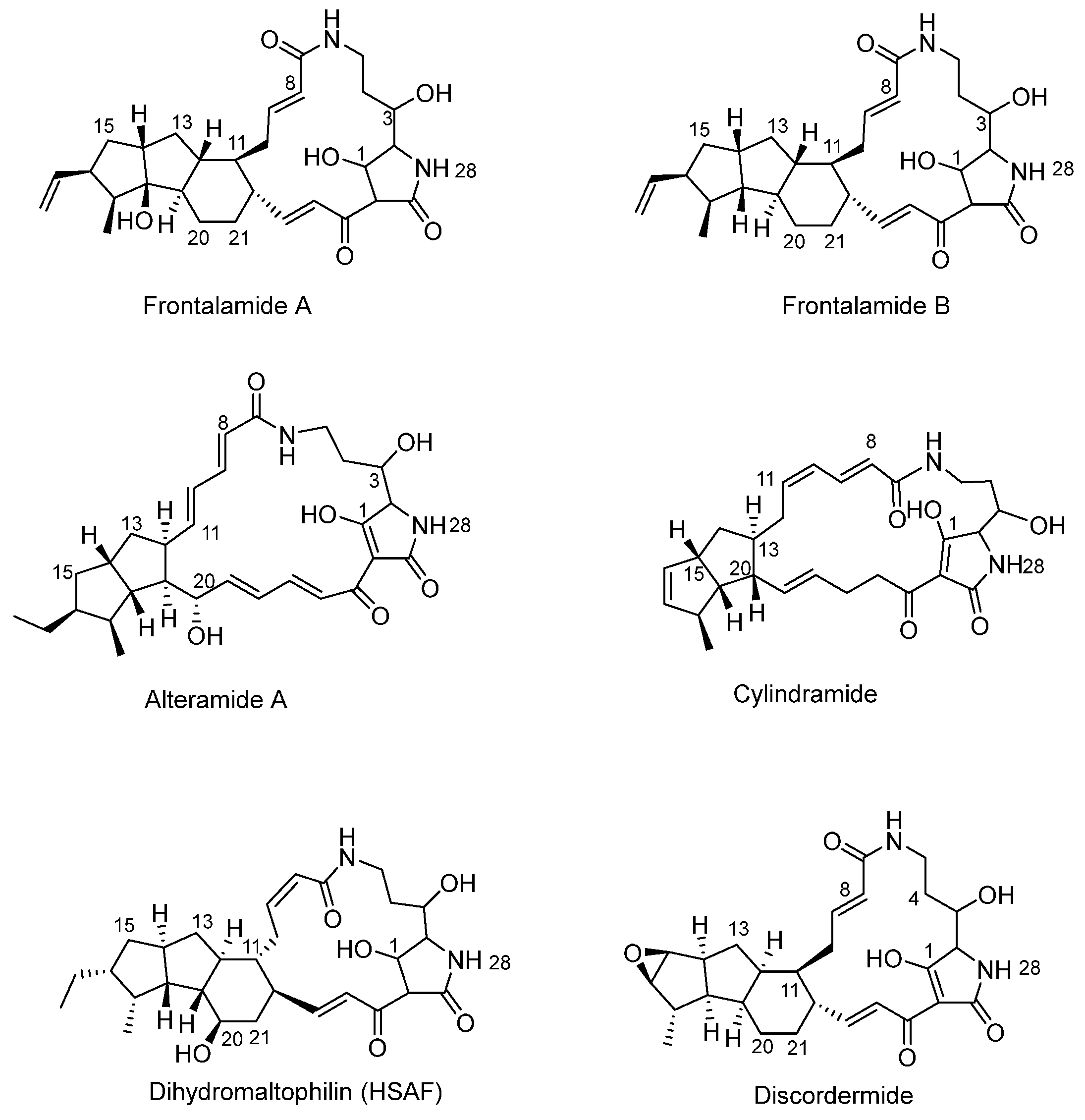
Figure 1. Frontalamide A, frontalamide B, alteramide A, cylindramide, dihydromaltophilin (HSAF), and discodermide.
Organics 05 00019 g001
Figure 2. Xanthobaccins A, B, and C.
Organics 05 00019 g002
Figure 3. (A): The basic 2,4-diketo form of tetramic acid. (B): The basic enolic form of tetramic acid.
Organics 05 00019 g003
Figure 4. (A) Alteramide B with the fused 5/5 ring structure (blue). (B) HSAF with a 5/5/6 ring structure. (C) Ikarugamycin with the 5/6/5 ring structure. The tetramic acid moiety is color coded pink.
Organics 05 00019 g004
Figure 5. The chemical structures of lysobacteramides A and B.
Organics 05 00019 g005
Figure 6. The actions of PKS/NRPS and L-ornithine on the polyene precursor to generate a PoTeM intermediate, which is then characterized into various distinct and specific compounds depending on the subsequent oxidoreductases and dehydrogenases from the particular biosynthetic gene clusters.
Organics 05 00019 g006
Figure 7. The chemical structures of pactamides A–F.
Organics 05 00019 g007
Figure 8. The chemical structure of melophlin A and B.
Organics 05 00019 g008
Figure 9. Several PoTeMs which have 5/6/5 tricycle structures.
Organics 05 00019 g009
Disclaimer/Publisher’s Note: The statements, opinions and data contained in all publications are solely those of the individual author(s) and contributor(s) and not of MDPI and/or the editor(s). MDPI and/or the editor(s) disclaim responsibility for any injury to people or property resulting from any ideas, methods, instructions or products referred to in the content.