Article

Quantitative and Sensitive Mid-Infrared Frequency Modulation Detection of HCN behind Shock Waves

Institut für Physikalische Chemie, Christian-Albrechts-Universität zu Kiel, Max-Eyth-Straße 1, 24118 Kiel, Germany
*
Author to whom correspondence should be addressed.
Fuels 2021, 2(4), 437-447; https://doi.org/10.3390/fuels2040025
Submission received: 21 September 2021 / Revised: 18 October 2021 / Accepted: 19 October 2021 / Published: 29 October 2021

Abstract

:
Despite its key role for the study and modeling of nitrogen chemistry and NO x formation in combustion processes, HCN has only rarely been detected under high-temperature conditions. Here, we demonstrate quantitative detection of HCN behind incident and reflected shock waves using a novel sensitive single-tone mid-infrared frequency modulation (mid-IR-FM) detection scheme. The temperature-dependent pressure broadening of the P(26) line in the fundamental CH stretch vibration band was investigated in the temperature range 670 K T 1460 K , yielding a pressure broadening coefficient for argon of 2 γ Ar 296 K = ( 0.093 ± 0.007 ) cm 1 atm 1 and a temperature exponent of n Ar = 0.67 ± 0.07 . The sensitivity of the detection scheme was characterized by means of an Allan analysis, showing that HCN detection on the ppm mixing ratio level is possible at typical shock wave conditions. In order to demonstrate the capability of mid-IR-FM spectroscopy for future high-temperature reaction kinetic studies, we also report the first successful measurement of a reactive HCN decay profile induced by its reaction with oxygen atoms.

1. Introduction

Hydrogen cyanide (HCN) is formed by gas phase combustion reactions and during devolatilization of fuels containing organic nitrogen, e.g., biomass residues [1,2]. The present work, which is based on the authors’ contribution to the virtual 10th European Combustion Meeting 2021 [3], is motivated by the important role of HCN in thermochemical processes and as a key intermediate for modeling nitrogen chemistry in combustion, in particular for prompt-NO formation [1,4]. Prompt-NO formation is initiated by the reaction CH + N 2 NCN + H [5] and the fate of the NCN radical is largely determined by the branching ratio of its subsequent reaction with H atoms [4]:
NCN + H CH + N 2 ( a ) HCN + N ( b )
Whereas the reverse association/rearrangement channel (a) to CH + N 2 exhibits a slightly negative temperature dependence, the forward channel (b) yields the so-called Fenimore products HCN + N in an activation controlled reaction step [6,7,8]. As a result, the branching becomes highly temperature dependent and switches from the HCN + N channel at high temperatures to the CH + N 2 channel at low temperatures, going along with other non-Fenimore routes for NCN consumption by reactions with H 2 and O atoms [4,9,10]. Although there is general agreement about the importance of this so-called prompt-NO switch and the HCN intermediate for NO x modeling, significant disagreement between experiment and flame modeling studies on the one hand and theoretical rate constant predictions on the other hand remains. For example, at a typical flame temperature of 2000 K, experimental data and flame modeling results [7,11] are consistent with a forward branching fraction of about 70%, but theory [6,8] predicts a much lower value of about 20%. In order to resolve this issue, measurements of the branching ratio by monitoring HCN concentration-time profiles as a direct product of the reaction NCN + H are required. However, in contrast to quantitative detection schemes for the educt species NCN [12,13,14], reliable high-temperature detection of HCN is difficult and suitable detection schemes are sparse. Ex-situ detection of absolute HCN concentrations in low-pressure flames using near-infrared cavity ring down spectroscopy has been performed by Lamoureux et al. [15,16]. Similarly, Gersen et al. determined HCN concentrations in fuel-rich methane/air flames using near-infrared wavelength modulation spectroscopy and microprobe gas sampling [17]. Measurements in the mid-infrared (mid-IR) spectral region are preferable, since the absorption linestrengths in the fundamental band of the CH stretch vibration enable considerably higher sensitivity, which is especially relevant for single-shot shock tube measurements that require high time resolution and sensitivity. In-situ detection of HCN in the mid-IR range was realized with polarization spectroscopy in flames at atmospheric pressure by Sun et al. [18] and with degenerate four-wave mixing spectroscopy during straw gasification by Hot et al. [19], respectively. Both groups have chosen the P(20) rovibrational line in the ν 1 band for their measurements.
Early shock tube studies by Hanson and coworkers [20,21] investigated the pressure broadening of several rovibrational transitions of HCN in the ν 1 band at room temperature and of the P(10) line behind shock waves using tunable diode laser spectroscopy. For species detection in the mid-IR, pressure broadening often becomes comparable to Doppler broadening at typical shock wave conditions such that the determination of accurate pressure broadening coefficients is crucial for quantitative HCN detection. However, to the best of our knowledge and due to the limited sensitivity of conventional detection schemes, kinetic profiles of HCN have not been measured behind shock waves so far.
Frequency modulation (FM) spectroscopy offers high sensitivity and µs time resolution even under harsh experimental conditions [22]. FM detection schemes for shock tube applications have been successfully implemented for shock tube experiments targeting the detection of NH 2 [23], 1 CH 2 [24], HCO [25], and OH [26] on electronic transitions in the VIS and UV spectral range. Very recently, we established single-tone frequency modulation spectroscopy in the mid-IR [27] aiming at sensitive detection of various species on their fundamental rovibrational transitions in shock tube experiments.
In this work, we report the first FM detection of HCN behind shock waves. As a prerequisite for quantitative HCN detection to measure the branching fraction of the reaction NCN + H, the high-temperature pressure broadening coefficient of argon was determined. The spectroscopically isolated and high-J P(26) absorption line at 3228.049 cm 1 in the ν 1 CH stretch vibration band was selected to ensure high sensitivity under high-temperature conditions. Moreover, to demonstrate the capability of µs time-resolved detection behind shock waves, first kinetic profiles of HCN during its oxidation by oxygen atoms were recorded as well. For sure, future studies on this and other bimolecular reactions of HCN as well as on the important isomerization step of HCN to hydrogen isocyanide (HNC) [28] will benefit from the availability of a sensitive HCN detection scheme as outlined in this work.

2. Experimental

The cw-OPO-laser based FM spectrometer with external modulation of the laser beam using a resonant, single-frequency electro-optic modulator and analog demodulation electronics has been described in Ref. [27]. For an outline of the operation principle of the time-resolved FM spectrometer we refer to Ref. [22]. Important for a sensitive FM detection scheme for high-temperature shock tube applications is a high modulation frequency (comparable to the Doppler- and pressure-broadened linewidth of the probed absorption line) in combination with strong modulation (expressed in terms of the modulation index M, with a preferable value of M 1 ). Both these requirements have only recently become technically feasible in the MIR with the availability of efficient single-frequency electro-optic modulators and fast HgCdTe heterostructure photovoltaic detectors, the latter also offering a sufficiently high optical saturation power to allow for near shot-noise limited detection. Compared to the earlier version of the setup in Ref. [27], by now we have implemented several modifications to further enhance the spectrometer performance; these will be described in more detail elsewhere. Briefly, a new temperature-stabilized electro-optic modulator (EO-500T3-MWIR1, Qubig, München, Germany) was used to modulate the laser beam in double-pass configuration at a frequency of ν m = 489 MHz, yielding a modulation index of M = 1.0 and thus a very high value for a system operating in the mid-IR. Reduction of thermal noise contributions was achieved by a photodetector with two additional thermo-electric cooling stages (PVI-4TE-5, Vigo System, Ozarow, Poland), increasing the specific detectivity of the photodetector by 70% to D * = 1.41 × 10 11 cm Hz 0.5 W 1 . Finally, the analog demodulation electronics have been replaced by an all-digital 600 MHz lock-in amplifier (UHFLI, Zurich Instruments, Zürich, Switzerland) that also served as the modulation frequency source. The low-pass of the demodulated signal was set to a bandwidth of 790 kHz, equivalent to a 10%-to-90% signal rise time of about 500 ns, hence ensuring a sufficiently fast time response to temporally resolve μ s concentration transients.
The pressure-driven stainless steel Kiel shock tube with an inner diameter of 81 mm has been described in detail elsewhere [29]. Shock wave conditions were calculated with a frozen chemistry/real gas code with shock wave velocities and shock wave damping measured with four fast piezoelectric sensors. Gas mixtures were prepared in a stainless steel gas mixing system equipped with glass storage flasks and were further diluted with argon using calibrated mass flow controllers. Before the shock tube experiment, the low pressure section of the shock tube was flushed for about 6 min with the sample gas mixture to reduce possible adsorption effects on the shock tube wall. The following gases were used as supplied (Air Liquide): HCN (calibrated mixture of 969 ppm ± 29 ppm in N 2 ), argon as the main buffer gas (99.999%) to dilute the HCN stock mixture using mass flow controllers, variable mixtures of H 2 /N 2 as driver gas (both 99.999%), and N 2 O (99.999%) as high-temperature source for oxygen atoms.

3. Results and Discussion

The P(26) transition at 3228.049 cm 1 in the ν 1 CH stretch band of HCN was selected for sensitive detection. The curves in Figure 1 illustrate the temperature dependent linestrengths of selected rovibrational transitions with different rotational quantum number J as calculated using the parameters included in the HITRAN database [30]. While at temperatures below 1100 K some HCN lines, including the P(8), P(15), and P(19) transitions shown in Figure 1, offer somewhat higher intensities than the selected P(26) line, due to several hot band transitions located close to the respective line centers, they are not fully spectroscopically isolated. In fact, selecting the high-J P(26) line represents a compromise between a high linestrength at high temperatures and spectroscopic isolation to ensure that the recorded FM signal originates only from the absorption line of interest.

3.1. Pressure Broadening Coefficient

Three sets of shock tube experiments were conducted to derive the temperature-dependent pressure broadening coefficient 2 γ Ar for argon. For each set with HCN mole fractions of 200 ppm, 350 ppm, and 500 ppm, respectively, the pressure and temperature behind the incident or reflected wave were kept approximately constant ( Δ p / p = 3.0 % and Δ T / T = 3.0 % ). The experimental conditions are summarized in Table 1.
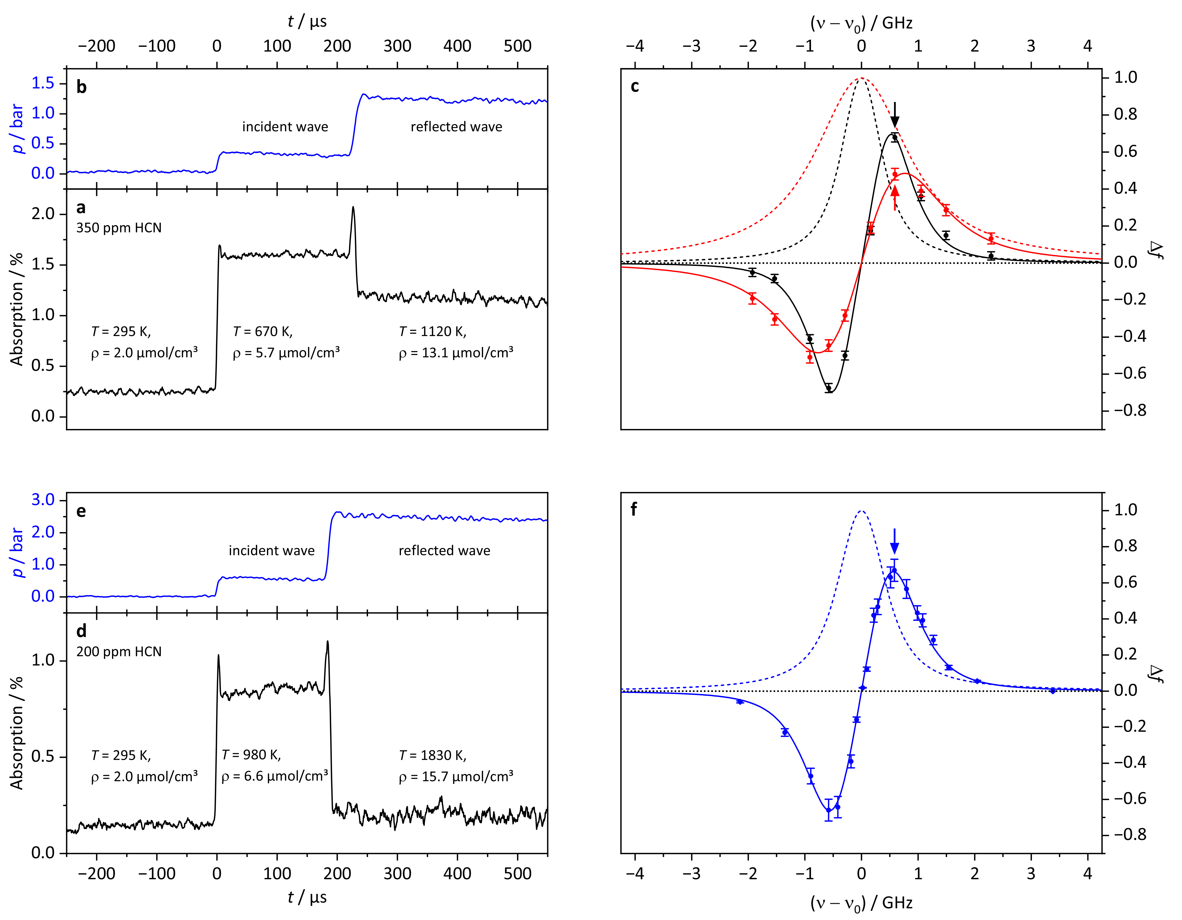
In order to measure the complete FM profile of the absorption line under the respective conditions, several shock tube experiments at different fixed wavelengths around the line center were performed. The linewidth of the single-frequency OPO laser system was about 2 × 10 6 cm 1 , much below the Doppler width of 0.014 cm 1 (FWHM at T = 1000 K). Therefore, the resulting FM profile can be derived from a purely Doppler and pressure broadened Voigt lineshape, since convolution with the laser lineshape function is not necessary. Example traces of HCN absorption during two shock tube experiments are shown in Figure 2. The blue traces in Figure 2b,e depict the measured pressures during the shock tube experiments and the black traces in Figure 2a,d represent the corresponding absorption signals. Note that, for the sake of clarity, FM equivalent absorption (as calculated from the finally derived lineshape parameters of this work) is shown instead of the raw FM signal.
Following the arrival of the incident and reflected shock wave, the signals exhibits step-like features reflecting the temperature and pressure dependent absorption cross section and the change of total density. The stable plateau signals indicate that HCN is thermally stable under the experimental conditions. For experiments with a laser wavelength close to the maximum of the absorption line pre-shock absorption was observed as well and needed to be properly taken into account to correctly set the baseline of the signal. In contrast, due to the much narrower linewidth at pre-shock conditions, for measurements in the outer wings of the absorption line no such baseline correction was necessary. Figure 2c shows the measured FM profiles for data set 2 with 350 ppm HCN under incident ( T 670 K, p 0.32 bar) and reflected ( T 1120 K, p 1.22 bar) shock wave conditions as well as the corresponding simulated derivative-like FM lineshapes (solid curves) and the underlying conventional Voigt absorption line profiles (dashed curves). The errors for the individual data points were estimated from the uncertainties of pressure and temperature as well as from the FM signal noise. Figure 2f shows equivalent results for the experiments performed with an HCN mole fraction of 200 ppm. For this data set, the measured FM signals behind the reflected wave in Figure 2d were too weak to yield reliable lineshape data, leaving in total five distinct FM profiles with a given pressure/temperature combination to determine the pressure broadening by argon.
All data in Figure 2c,f are shown in terms of the FM factor Δ f as defined in Ref. [22]:
I FM = I 0 2 × Δ f × α c × l × G
Here, I 0 and I FM correspond to the measured signal levels of the total laser intensity and the demodulated FM intensity, respectively. l is the absorption length and G an electronic gain factor accounting for the amplification and losses of the FM signal in the demodulation circuitry. Briefly, the FM factor Δ f can be understood as the relative FM signal measured at a given detection wavelength, normalized to the conventional narrow bandwidth absorption coefficient α c at the line center. Its actual value depends on the modulation index M, the ratio of the modulation frequency ν m and absorption linewidth, and on the demodulation phase angle. The latter was set to a value corresponding to a pure absorption signal in our case. As it is outlined in more detail in Ref. [22], for a given laser frequency ν and a known modulation index M, the FM factor can be directly calculated from the corresponding absorption line profile in a straightforward manner. For a phase angle set to pure absorption, the FM factor is given as follows:
Δ f = 2 × [ n = 0 J n ( M ) J n + 1 ( M ) ( α n 1 α n + 1 + α n α n ) ] / α c
Here, the α i correspond to the narrow-bandwidth value of the absorption coefficient at the spectral position of the n-th sideband of the frequency-modulated laser beam at ν 0 ± n v m , weighted according to their corresponding Bessel function values J n ( M ) . α c is the absorption coefficient at line center, which is introduced for the purpose of normalization. As becomes clear from Equation (2), for higher M values, the resulting FM signal exhibits contributions from higher sidebands. In the limiting case M 1 , however, it simply represents the difference in absorption measured at the first order sidebands at ( ν 0 + ν m ) and ( ν 0 ν m ) which explains the overall derivative nature of the observed FM profile.
By combining the absorption values of all HCN traces for each data set in Table 1, the FM line profiles for incident and reflected wave conditions were fitted iteratively by a Voigt lineshape model. The Lorentzian width 2 Γ total was used as the sole adjustable parameter, which sensitively depends on total pressure and temperature. Contributions of N 2 and HCN to the Lorentzian linewidth were subtracted using literature values for their pressure broadening coefficients and temperature exponents [31,32] and assuming the usual mole fraction weighted additivity rule, Γ total = Σ i x i Γ i . The temperature dependence of the full-width broadening coefficient 2 γ is expressed as
2 γ i ( T ) = 2 γ i 296 K 296 K T n i
In the case of HCN, the unknown temperature exponent was assumed to be n HCN = 1 , which is consistent with earlier results reported for the P(10) line [21].
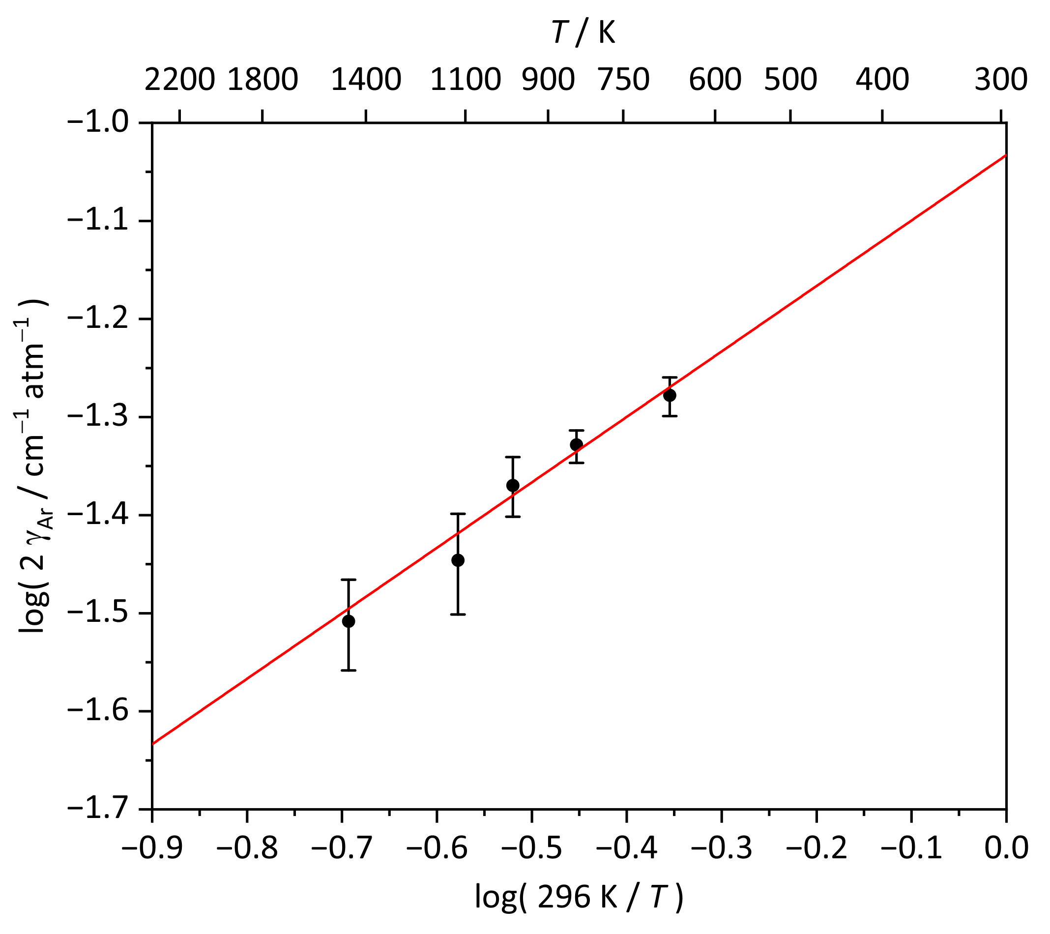
As it turns out, the significant increase of the broadening of the absorption line behind the reflected wave (see Figure 2c) is predominantly due to the higher pressure behind the reflected wave instead of the temperature related increase of the Doppler broadening contribution. Figure 3 shows a log-log plot of the determined argon broadening coefficients. A linear fit of the data according to Equation (3) yields the temperature exponent of n Ar = 0.67 ± 0.07 from the slope and the full-width room temperature pressure broadening coefficient of 2 γ Ar 296 K = ( 0.093 ± 0.007 ) cm 1 atm 1 from the intercept. These results compare well with values for the temperature exponent of the P(10) line ( 0.63 ± 0.06 ) and the argon broadening coefficients for the lines between P(1) ( 0.162 cm 1 atm 1 ) and P(18) ( 0.099 cm 1 atm 1 ) reported in Refs. [20,21].
Altogether, these data reveal the expected clear trend of lower pressure broadening with increasing rotational excitation, which is typically ascribed to the lower rotational transition probabilities resulting from the more widely spaced rotational levels at higher J values [33]. The calculated integrated linestrengths S for the P(26) transition are included in Figure 1 as symbols. On average, these data are 17% higher than the values predicted from the HITRAN database [30] with the highest deviation occurring for the experiments with the largest HCN mole fractions of 500 ppm. Note that higher HCN mole fractions go along with higher N 2 content as well, corresponding to a mole fraction of up to 52 % N 2 in the shock-heated gas mixtures in case of the 500 ppm HCN experiments. The reason for using an HCN/N 2 stock gas mixture instead of HCN/argon was simply due to its commercial availability. Handling of the highly toxic HCN is difficult and therefore we have refrained from preparing our own stock gas mixtures from pure HCN gas. In order to validate the HCN content in the gas mixtures, complementary measurements of the undiluted HCN/ N 2 mixture were performed at room temperature for five intense lines between P(8) to P(21) using standard FTIR spectroscopy (Vertex 80, Bruker). On average, the linestrengths derived from the FTIR data were 6 % higher than the HITRAN values, which, however, cannot fully account for the difference between our data and the HITRAN prediction as seen in Figure 1. We tentatively ascribe the remaining moderate discrepancy to non-idealities and uncertainties in the calculated shock wave parameters. Although non-idealities resulting from vibrational relaxation effects of shock-heated N 2 -containing gas mixtures should remain small for the conditions used in this work [34], the clear trend of higher deviations for increasing N 2 content is evident. Whereas the data point with 200 ppm HCN is in agreement with the HITRAN prediction within error limits, the experiments at 500 ppm deviate by up to 30%.

3.2. HCN Detection Limit

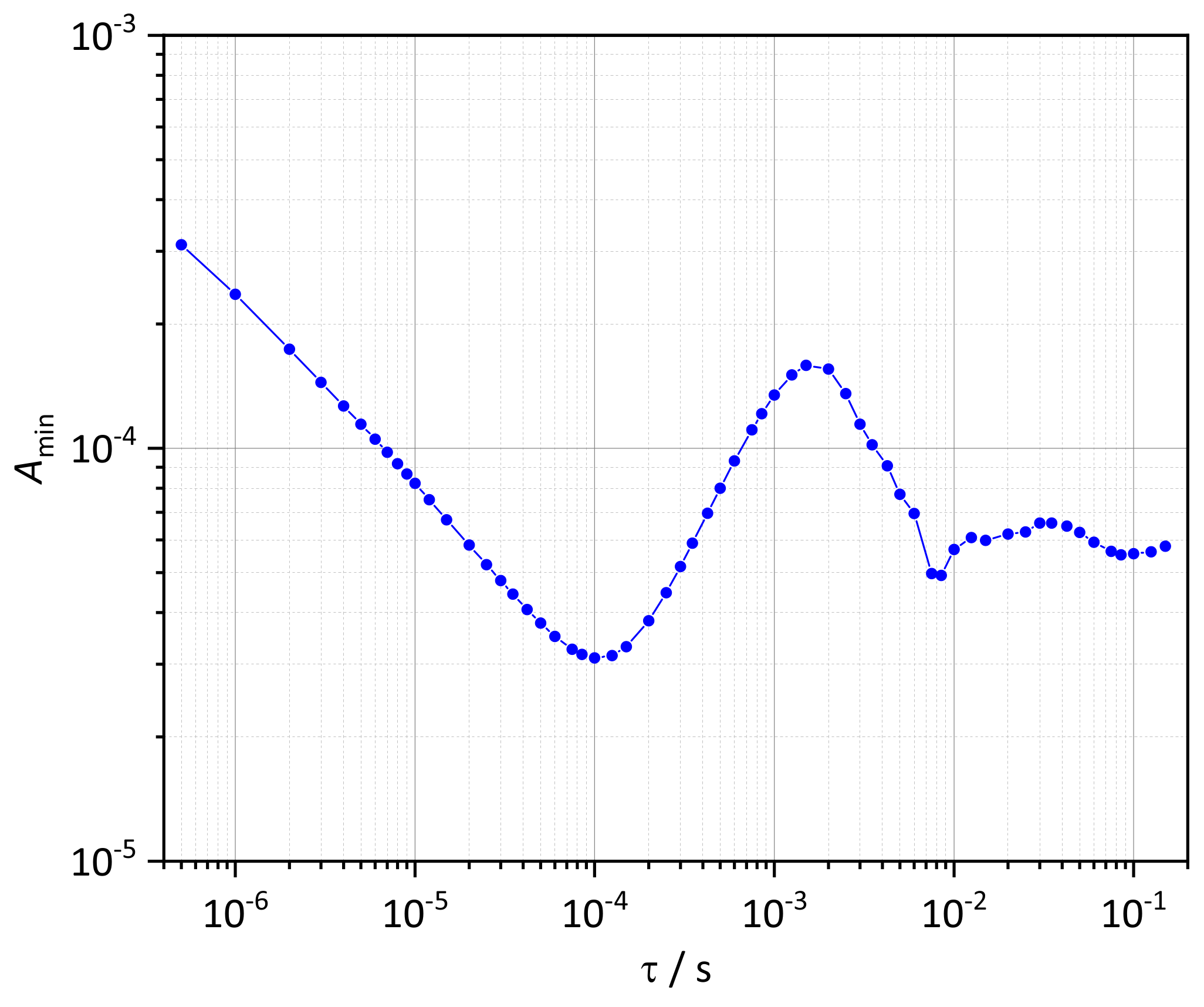
The HCN detection limit of the mid-IR-FM setup can be estimated from an Allan analysis of the FM signal baseline noise using the overlapping Allan deviation as the preferred measure of sensitivity [35]. To this end, pure FM signal noise was recorded under normal lab conditions without shock tube operation and was then subjected to an overlapping Allan analysis procedure. The calculated values for the overlapping Allan deviation as function of the averaging period τ , corresponding to the time resolution of the detection system, were converted to the respective absorption values A = α c × l using the standard FM Equation (1) with the FM factor Δ f calculated using Equation (2). As shown in Ref. [22], FM factors on the order of Δ f 1 correspond to the best sensitivity that can be achieved with an FM detection scheme. With Δ f 0.6 as obtained in this work (see Figure 2c,f), thanks to the combination of high modulation index and high modulation frequency, the current version of our FM spectrometer already comes close to this optimum. Figure 4 shows the resulting Allan plot, assuming Δ f = 0.6 .
For an effective time resolution of τ = 500 ns, the minimum detectable absorption is A min = 3 × 10 4 . With increasing averaging time, A min initially decreases with τ 0.5 , indicating that statistical noise components such as thermal and/or shot noise of the photodetector prevails. The lowest detectable absorption of A min = 3 × 10 5 is achieved at a time response of τ = 100 µs. These favorable values demonstrate the high detection sensitivity of FM spectroscopy even on the short timescales, as they are required for experiments behind shock waves. Another advantage of the derivative nature of FM spectroscopy for shock tube applications stems from the fact that typical shock tube induced acoustic and pressure induced noise components are significantly reduced, resulting in a more stable baseline compared to conventional difference amplification laser absorption schemes [36].
Note that the increase of the Allan deviation for time resolutions τ > 100 µs, and hence the loss of sensitivity on longer timescales, is due to well-known residual amplitude modulation (RAM) effects in FM spectroscopy [37] that typically cause baseline undulations in the kHz range. These are of minor concern for shock tube applications since the necessary time resolutions are in the range of 1–20 µs and the total experimental test time window is limited to at most a few ms anyway.
Using the linestrengths from the HITRAN database and the argon broadening parameters derived in this work, the HCN detection limit at T = 1000 K and p = 0.4 bar is about 3 ppm for τ = 10 µs and single-pass shock tube detection with l = 81 mm. This demonstrates the high potential of mid-IR-FM spectroscopy for quantitative detection of HCN under shock wave conditions. However, towards higher temperatures and pressures, the rapidly decreasing Boltzmann population of individual rotational states (going along with lower linestrengths, see Figure 1) as well as the increasing linewidths (see Figure 2c) significantly reduce the attainable detection limit. For example, at T = 2500 K and p = 1.6 bar the HCN detection limit increases to 28 ppm.
A second overlapping Allan analysis was conducted using FM signal noise measured behind the reflected wave in pure argon. For realistic time resolutions of τ 20 µs, this analysis yielded detection limits that are a factor of 2 higher than the A min values shown in Figure 4, most likely due to additional technical noise contributions caused by the propagating shock wave.

3.3. Kinetic HCN Profile

To further demonstrate the capability of mid-IR-FM spectroscopy to enable sensitive kinetic studies behind shock waves, first reactive concentration-time profiles of HCN during the reaction HCN + O were recorded. Here, the thermal decomposition of N 2 O according to N 2 O + M     N 2 + O + M served as an effective source of O atoms at temperatures T > 1500 K.
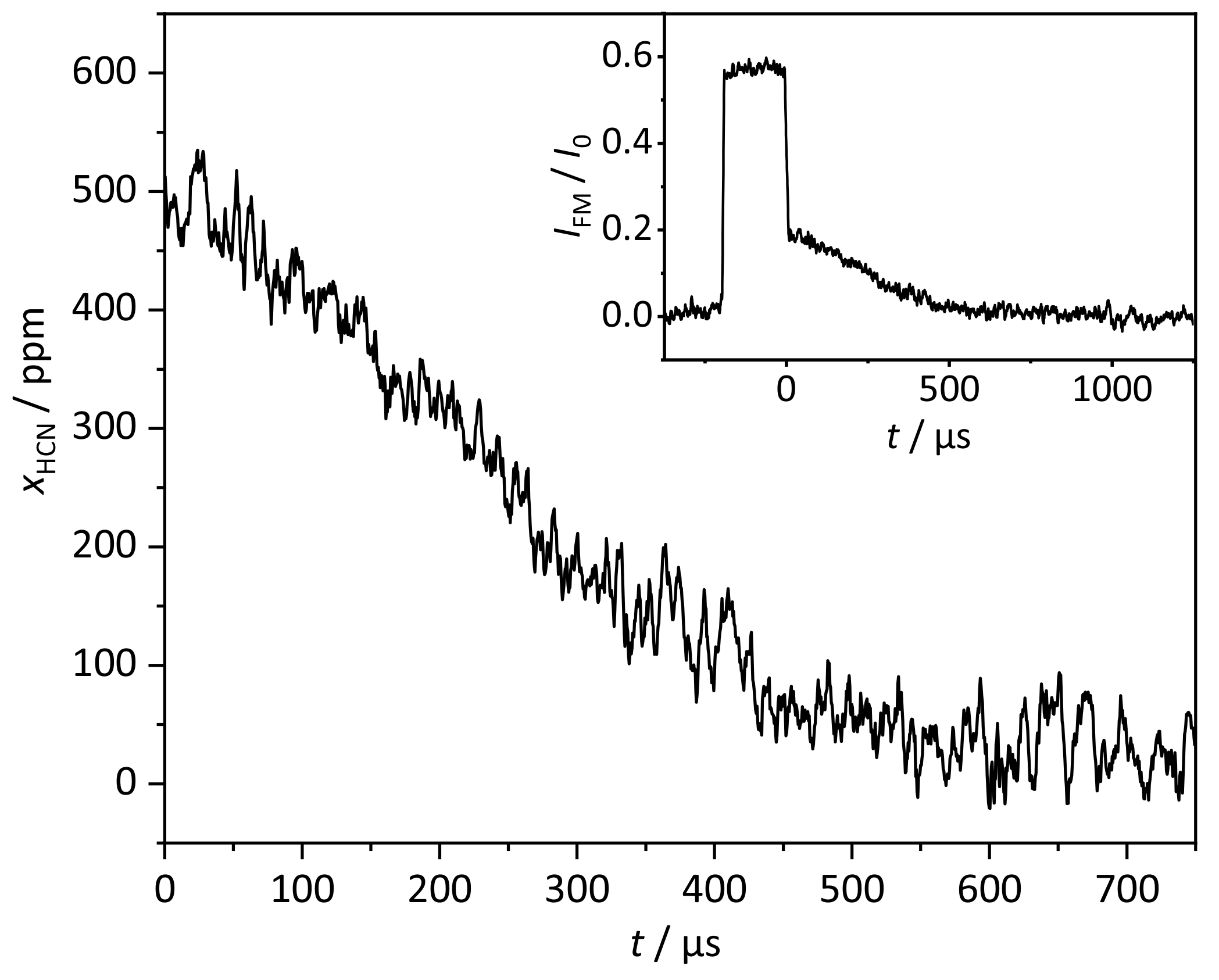
Figure 5 illustrates, to the best of our knowledge for the first time, a kinetic profile of HCN behind shock waves. The signal was obtained with a gas mixture of 500 ppm HCN and 2.0 % N 2 O in N 2 /argon at a temperature of 1638 K and a pressure of 1.24 bar behind the reflected shock wave. The sigmoidal HCN profile with accelerated loss towards longer reaction times is due to the ever-increasing O atom concentration during the experiment resulting from the rather slow N 2 O decomposition. For this particular kinetic HCN profile, the standard deviation of the noise behind the reflected wave corresponds to a detection limit of about 20–25 ppm. This is consistent with the detection limit of 21 ppm derived from the Allan analysis for reflected wave conditions mentioned in the previous section.
A more systematic study targeting the determination of the high temperature rate constant of the reaction HCN + O is currently underway and will be published elsewhere.

4. Conclusions

Single-tone mid-IR-FM spectroscopy has been demonstrated as a suitable and sensitive laser absorption method for the detection of HCN in shock tube experiments. As a prerequisite for quantitative high temperature detection of HCN, we have determined the argon pressure broadening parameters for the P(26) line in the ν 1 band of HCN. A detection limit as low as 3 ppm at T = 1000 K and p = 400 mbar for single-pass shock tube detection ( l = 81 mm) was achieved. Furthermore, the first reactive profiles of HCN behind reflected shock waves were recorded. The outcome of this study serves as a basis for future reaction kinetic studies of HCN reactions and, in particular, paves the way for the determination of the HCN yield and with it the temperature-dependent branching ratio of the reaction NCN + H, which is key for prompt-NO modeling.

Author Contributions

Conceptualization, M.S. and G.F.; methodology, all authors; formal analysis, M.S.; investigation, M.S. and S.H.; writing—original draft preparation, M.S.; writing—review and editing, S.H. and G.F.; visualization, M.S.; supervision, G.F.; funding acquisition, G.F. All authors have read and agreed to the published version of the manuscript.

Funding

The authors like to acknowledge the financial support of the German Science Foundation (DFG) through projects FR1529/4-2 and FR1529/7-1, as well as the Cluster of Excellence EC-80 “Future Ocean” at Kiel University for making available the OPO laser system.

Institutional Review Board Statement

Not applicable.

Informed Consent Statement

Not applicable.

Data Availability Statement

Not applicable.

Conflicts of Interest

The authors declare no conflict of interest.

References

  1. Glarborg, P.; Miller, J.A.; Ruscic, B.; Klippenstein, S.J. Modeling nitrogen chemistry in combustion. Prog. Energy Combust. 2018, 67, 31–68. [Google Scholar] [CrossRef] [Green Version]
  2. Schmid, D.; Karlström, O.; Yrjas, P. Release of NH3, HCN and NO during devolatilization and combustion of washed and torrefied biomass. Fuel 2020, 280, 118583. [Google Scholar] [CrossRef]
  3. Stuhr, M.; Hesse, S.; Friedrichs, G. Quantitative and Sensitive Mid-Infrared Frequency Modulation Detection of HCN behind Shock Waves. In Proceedings of the European Combustion Meeting, Online. 14–15 April 2021; 10, pp. 767–772. [Google Scholar]
  4. Lamoureux, N.; Desgroux, P.; Olzmann, M.; Friedrichs, G. The story of NCN as a key species in prompt-NO formation. Prog. Energ. Combust. 2021, 87, 100940. [Google Scholar] [CrossRef]
  5. Moskaleva, L.; Lin, M. The spin-conserved reaction CH + N2→ H + NCN: A major pathway to prompt NO studied by quantum/statistical theory calculations and kinetic modeling of rate constant. Proc. Combust. Inst. 2000, 28, 2393–2401. [Google Scholar] [CrossRef]
  6. Teng, W.S.; Moskaleva, L.V.; Chen, H.L.; Lin, M.C. Ab Initio Chemical Kinetics for H + NCN: Prediction of NCN Heat of Formation and Reaction Product Branching via Doublet and Quartet Surfaces. J. Phys. Chem. A 2013, 117, 5775–5784. [Google Scholar] [CrossRef]
  7. Faßheber, N.; Dammeier, J.; Friedrichs, G. Direct measurements of the total rate constant of the reaction NCN+ H and implications for the product branching ratio and the enthalpy of formation of NCN. Phys. Chem. Chem. Phys. 2014, 16, 11647–11657. [Google Scholar] [CrossRef] [Green Version]
  8. Klippenstein, S.J.; Pfeifle, M.; Jasper, A.W.; Glarborg, P. Theory and modeling of relevance to prompt-NO formation at high pressure. Combust. Flame 2018, 195, 3–17. [Google Scholar] [CrossRef]
  9. Faßheber, N.; Lamoureux, N.; Friedrichs, G. The rate constant of the reaction NCN + H2 and its role in NCN and NO modeling in low pressure CH4/O2/N2-flames. Phys. Chem. Chem. Phys. 2015, 17, 15876–15886. [Google Scholar] [CrossRef] [Green Version]
  10. Dammeier, J.; Faßheber, N.; Friedrichs, G. Direct measurements of the high temperature rate constants of the reactions NCN + O, NCN + NCN, and NCN + M. Phys. Chem. Chem. Phys. 2012, 14, 1030–1037. [Google Scholar] [CrossRef]
  11. Lamoureux, N.; Merhubi, H.E.; Pillier, L.; de Persis, S.; Desgroux, P. Modeling of NO formation in low pressure premixed flames. Combust. Flame 2016, 163, 557–575. [Google Scholar] [CrossRef]
  12. Dammeier, J.; Friedrichs, G. Thermal Decomposition of NCN3 as a High-Temperature NCN Radical Source: Singlet-Triplet Relaxation and Absorption Cross Section of NCN(3Σ). J. Phys. Chem. A 2010, 114, 12963–12971. [Google Scholar] [CrossRef]
  13. Lamoureux, N.; Western, C.M.; Mercier, X.; Desgroux, P. Reinvestigation of the spectroscopy of the transition of the NCN radical at high temperature: Application to quantitative NCN measurement in flames. Combust. Flame 2013, 160, 755–765. [Google Scholar] [CrossRef]
  14. Faßheber, N.; Bornhorst, L.; Hesse, S.; Sakai, Y.; Friedrichs, G. The Reaction NCN + H2: Quantum Chemical Calculations, Role of 1NCN Chemistry, and 3NCN Absorption Cross Section. J. Phys. Chem. A 2020, 124, 4632–4645. [Google Scholar] [CrossRef]
  15. Lamoureux, N.; Merhubi, H.E.; Mercier, X.; Pauwels, J.; Desgroux, P. HCN quantitative measurement in a laminar low pressure flame at 1036 nm using pulsed CRDS technique. Proc. Combust. Inst. 2013, 34, 3557–3564. [Google Scholar] [CrossRef]
  16. Lamoureux, N.; Merhubi, H.E.; Gasnot, L.; Schoemaecker, C.; Desgroux, P. Measurements and modelling of HCN and CN species profiles in laminar CH4/O2/N2 low pressure flames using LIF/CRDS techniques. Proc. Combust. Inst. 2015, 35, 745–752. [Google Scholar] [CrossRef]
  17. Gersen, S.; Mokhov, A.; Levinsky, H. Diode laser absorption measurement and analysis of HCN in atmospheric-pressure, fuel-rich premixed methane/air flames. Combust. Flame 2008, 155, 267–276. [Google Scholar] [CrossRef]
  18. Sun, Z.; Li, Z.; Konnov, A.; Aldén, M. Quantitative HCN measurements in CH4/N2O/O2/N2 flames using mid-infrared polarization spectroscopy. Combust. Flame 2011, 158, 1898–1904. [Google Scholar] [CrossRef]
  19. Hot, D.; Pedersen, R.L.; Weng, W.; Zhang, Y.; Aldén, M.; Li, Z. Spatially and temporally resolved IR-DFWM measurement of HCN released from gasification of biomass pellets. Proc. Combust. Inst. 2019, 37, 1337–1344. [Google Scholar] [CrossRef]
  20. Varghese, P.L.; Hanson, R.K. Tunable diode laser measurements of spectral parameters of HCN at room temperature. J. Quant. Spectrosc. Radiat. Transf. 1984, 31, 545–559. [Google Scholar] [CrossRef]
  21. Chang, A.; Hanson, R. Shock-tube study of HCN self-broadening and broadening by argon for the P(10) line of the ν1 band at 3 µm. J. Quant. Spectrosc. Radiat. Transf. 1985, 33, 213–217. [Google Scholar] [CrossRef]
  22. Friedrichs, G. Sensitive Absorption Methods for Quantitative Gas Phase Kinetic Measurements. Part 1: Frequency Modulation Spectroscopy. Z. Phys. Chem. 2008, 222, 1–30. [Google Scholar] [CrossRef]
  23. Deppe, J.; Friedrichs, G.; Ibrahim, A.; Römming, H.J.; Wagner, H.G. The thermal decomposition of NH2 and NH radicals. Ber. Bunsenges. Phys. Chemi 1998, 102, 1474–1485. [Google Scholar] [CrossRef]
  24. Friedrichs, G.; Wagner, H.G. Quantitative FM spectroscopy at high temperatures: The detection of 1CH2 behind shock waves. Z. Phys. Chem. 2000, 214, 1723–1746. [Google Scholar] [CrossRef]
  25. Friedrichs, G.; Herbon, J.T.; Davidson, D.F.; Hanson, R.K. Quantitative detection of HCO behind shock waves: The thermal decomposition of HCO. Phys. Chem. Chem. Phys. 2002, 4, 5778–5788. [Google Scholar] [CrossRef]
  26. Wang, S.; Hanson, R.K. Ultra-sensitive spectroscopy of OH radical in high-temperature transient reactions. Opt. Lett. 2018, 43, 3518–3521. [Google Scholar] [CrossRef]
  27. Stuhr, M.; Faßheber, N.; Friedrichs, G. Single-tone mid-infrared frequency modulation spectroscopy for sensitive detection of transient species. Opt. Express 2019, 27, 26499–26512. [Google Scholar] [CrossRef]
  28. Glarborg, P.; Marshall, P. Importance of the Hydrogen Isocyanide Isomer in Modeling Hydrogen Cyanide Oxidation in Combustion. Energy Fuels 2017, 31, 2156–2163. [Google Scholar] [CrossRef]
  29. Colberg, M.; Friedrichs, G. Room temperature and shock tube study of the reaction HCO + O2 using the photolysis of glyoxal as an efficient HCO source. J. Phys. Chem. A 2006, 110, 160–170. [Google Scholar] [CrossRef]
  30. Gordon, I.; Rothman, L.; Hill, C.; Kochanov, R.; Tan, Y.; Bernath, P.; Birk, M.; Boudon, V.; Campargue, A.; Chance, K.; et al. The HITRAN2016 molecular spectroscopic database. J. Quant. Spectrosc. Radiat. Transf. 2017, 203, 3–69. [Google Scholar] [CrossRef]
  31. Devi, V.M.; Benner, D.; Smith, M.; Rinsland, C.; Sharpe, S.W.; Sams, R. A multispectrum analysis of the ν1 band of H12C14N: Part I. Intensities, self-broadening and self-shift coefficients. J. Quant. Spectrosc. Radiat. Transf. 2003, 82, 319–341. [Google Scholar] [CrossRef]
  32. Rinsland, C.; Devi, V.M.; Smith, M.; Benner, D.C.; Sharpe, S.W.; Sams, R.L. A multispectrum analysis of the ν1 band of H12C14N: Part II. Air- and N2-broadening, shifts and their temperature dependences. J. Quant. Spectrosc. Radiat. Transf. 2003, 82, 343–362. [Google Scholar] [CrossRef]
  33. Rao, D.R.; Oka, T. Dicke narrowing and pressure broadening in the infrared fundamental band of HCl perturbed by Ar. J. Mol. Spectrosc. 1987, 122, 16–27. [Google Scholar] [CrossRef]
  34. Appleton, J.P. Shock-Tube Study of the Vibrational Relaxation of Nitrogen Using Vacuum-Ultraviolet Light Absorption. J. Chem. Phys. 1967, 47, 3231–3240. [Google Scholar] [CrossRef]
  35. Riley, W.J. Handbook of Frequency Stability Analysis; US Department of Commerce, National Institute of Standards and Technology (NIST): Gaithersburg, MD, USA, 2008.
  36. Votsmeier, M.; Song, S.; Davidson, D.; Hanson, R. Sensitive detection of NH2 in shock tube experiments using frequency modulation spectroscopy. Int. J. Chem. Kinet. 1999, 31, 445–453. [Google Scholar] [CrossRef]
  37. Silver, J.A. Frequency-modulation spectroscopy for trace species detection: Theory and comparison among experimental methods. Appl. Opt. 1992, 31, 707–717. [Google Scholar] [CrossRef] [PubMed]
Figure 1. Temperature-dependent linestrengths S for several rovibrational transitions in the P-branch of the ν 1 band of HCN. HITRAN database [30] predictions (curves) are compared to experimental data obtained for the P(26) transition (symbols).
Fuels 02 00025 g001
Figure 2. Typical measured HCN traces (a,d, 2 MHz sampling rate, 20 points moving average), the corresponding pressure traces (b,e), and the resulting line profiles (c,f) for the P(26) absorption line at ν ˜ = 3228.049 cm 1 . The data points extracted from the experiments shown in the left plots are highlighted in the right plots with arrows. The solid and dashed curves in the right plots correspond to the simulated FM profiles and their respective underlying absorption lineshapes for T 670 K and p 0.32 bar (black), for T 1120 K and p 1.22 bar (red), and for T 980 K and p 0.54 bar (blue).
Fuels 02 00025 g002
Figure 3. log-log plot for the temperature dependence of the argon broadening coefficient 2 γ Ar . Experimental data (black symbols) including errors estimated from the Voigt fits and a linear fit according to Equation (3) (red line).
Fuels 02 00025 g003
Figure 4. Minimum detectable absorption as function of the time resolution τ for typical measurement conditions ( 790 kHz bandwidth, 700 µW light power on the detector, I 0 = 1.2 V, Δ f = 0.6 , G = 200 , ν ˜ = 3228.05 cm 1 ).
Fuels 02 00025 g004
Figure 5. Kinetic profile of HCN during the reaction HCN + O behind the reflected shock wave ( 500 ppm HCN, 2.0 % N 2 O, T = 1638 K, p = 1.24 bar, 2 MHz sampling rate, 10 points moving average). The inset shows the baseline-corrected and I 0 -normalized FM signal for pre-shock, incident wave, and reflected wave conditions. t = 0 marks the arrival of the reflected shock wave.
Fuels 02 00025 g005
Table 1. Experimental conditions for FM line profile measurements behind incident (inc.) and reflected (refl.) shock waves and the corresponding total Lorentzian linewidths 2 Γ total (FWHM).
Data Set x HCN /ppmT/Kp/bar 2 Γ total /cm 1
1inc.2009800.540.0279
refl.20018302.39— *
2inc.3506700.320.0240
refl.35011201.220.0631
3inc.5008400.210.0254
refl.50014600.980.0484
* Not used for analysis, see text.
Publisher’s Note: MDPI stays neutral with regard to jurisdictional claims in published maps and institutional affiliations.