Next Issue
Volume 4, December
Previous Issue
Volume 4, June


Transplantology, Volume 4, Issue 3 (September 2023) – 7 articles

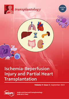
Cover Story (view full-size image): A huge barrier to transplantation is the separate location of donors and recipients leading to increased cold ischemic time and resulting ischemia-reperfusion injury. Due to the major shortage of transplantable organs, many patients need a transplant every year, and it is vital that these limited organs are not squandered. Understanding and overcoming ischemia-reperfusion injury is an important step towards improving the outcomes of transplanted patients and increasing the number of viable transplantable organs. Partial heart transplantation is still poorly understood but could expand the number of viable heart tissue transplants nationwide. Will this new addition to the transplant toolkit face the same scrutiny for cold ischemic time as other organs? View this paper
  • Issues are regarded as officially published after their release is announced to the table of contents alert mailing list.
  • You may sign up for e-mail alerts to receive table of contents of newly released issues.
  • PDF is the official format for papers published in both, html and pdf forms. To view the papers in pdf format, click on the "PDF Full-text" link, and use the free Adobe Reader to open them.
Order results
Result details
Section
Select all
Export citation of selected articles as:
7 pages, 2139 KiB  
Case Report
Diagnostic Challenge in Renal Transplantation: Splenosis vs. Post-Transplant Lymphoproliferative Disorder—A Case Report
by Jes M. Sanders, Daniel Galvez, Xiaoqi Lin and Joseph Leventhal
Transplantology 2023, 4(3), 178-184; https://doi.org/10.3390/transplantology4030017 - 21 Sep 2023
Viewed by 1018
Abstract
Splenosis is a benign, acquired condition characterized by the auto-implantation of focal deposits of splenic tissue throughout the peritoneal cavity, most commonly occurring after splenic injury and/or splenectomy. Post-Transplant Lymphoproliferative Disorder (PTLD) is a well-known complication of solid organ transplantation that results from [...] Read more.
Splenosis is a benign, acquired condition characterized by the auto-implantation of focal deposits of splenic tissue throughout the peritoneal cavity, most commonly occurring after splenic injury and/or splenectomy. Post-Transplant Lymphoproliferative Disorder (PTLD) is a well-known complication of solid organ transplantation that results from unregulated B-cell proliferation due to chronic immunosuppression. Given their clinical and radiologic similarities, these two entities may pose a diagnostic dilemma in select solid-organ transplant recipients. We present the case of a 54-year-old kidney-transplant recipient presenting with abdominal pain and found to have a retroperitoneal soft-tissue mass concerning for PTLD. He underwent a CT-guided biopsy of the mass, and histopathological studies revealed lymphoid tissue consistent with splenic tissue, thus ruling out PTLD. The patient subsequently underwent symptomatic management, with the eventual resolution of his symptoms. The early diagnosis of PTLD is paramount, as prompt intervention has a substantial impact on the high rate of morbidity and mortality associated with this condition. Additionally, the diagnosis of splenosis in the setting of a retroperitoneal mass is critical in order to avoid invasive diagnostic and therapeutic procedures that may result in significant complications. A detailed surgical history, including prior splenic trauma and/or splenectomy, should raise clinical suspicion for splenosis and guide further diagnostic and therapeutic decision making. Full article
(This article belongs to the Section Special Clinical Cases and Videos)
Show Figures

Figure 1

17 pages, 345 KiB  
Review
Recurrent Immunoglobulin A Nephropathy after Kidney Transplant—An Updated Review
by Hwarang S. Han, Michelle L. Lubetzky, Nidharshan S. Anandasivam, Rebecca A. Cox and Brian K. Lee
Transplantology 2023, 4(3), 161-177; https://doi.org/10.3390/transplantology4030016 - 6 Sep 2023
Viewed by 2698
Abstract
Immunoglobulin A nephropathy (IgAN) is the commonest glomerulonephritis worldwide, a category that represents the third most frequent cause of end-stage kidney disease (ESKD) in the United States. Kidney transplantation remains the optimal treatment of ESKD, and yet the prospects of IgAN recurrence post-transplant [...] Read more.
Immunoglobulin A nephropathy (IgAN) is the commonest glomerulonephritis worldwide, a category that represents the third most frequent cause of end-stage kidney disease (ESKD) in the United States. Kidney transplantation remains the optimal treatment of ESKD, and yet the prospects of IgAN recurrence post-transplant dampens the enthusiasm for living kidney donation in some instances, in addition to limiting the longevity of the kidney allograft. Moreover, the lack of a standardized method for detecting IgAN recurrence, since not all centers perform protocol allograft biopsies, has led to an underestimation of the extent of the issue. The pathogenesis of de novo IgAN remains conjectural, let alone the pathways for recurrent disease, but is increasingly recognized as a multi-hit injury mechanism. Identification of recurrent disease rests mainly on clinical symptoms and signs (e.g., hematuria, proteinuria) and could only be definitively proven with histologic evidence which is invasive and prone to sampling error. Treatment had relied mainly on nonspecific goals of proteinuria reduction, and in some cases, immunosuppression for active, crescentic disease. More recently, newer targets have the potential to widen the armamentarium for directed therapies, with more studies on the horizon. This review article provides an update on recurrent IgAN post-transplant. Full article
10 pages, 2110 KiB  
Case Report
Upfront Normothermic Machine Perfusion for a Liver Graft with Severe Macrovesicular Steatosis: A Proof-of-Concept Case
by Damiano Patrono, Ana Lavinia Apostu, Giorgia Rizza, Davide Cussa, Antonella Barreca, Selene Limoncelli, Stefano Mirabella and Renato Romagnoli
Transplantology 2023, 4(3), 151-160; https://doi.org/10.3390/transplantology4030015 - 23 Aug 2023
Cited by 1 | Viewed by 1442
Abstract
Graft steatosis has been associated with inferior outcomes after liver transplantation. Given the rising prevalence of obesity and fatty liver disease, strategies allowing safe and successful utilization of fatty liver grafts are needed. Liver preservation by normothermic machine perfusion (NMP) allows reducing ischemia-reperfusion [...] Read more.
Graft steatosis has been associated with inferior outcomes after liver transplantation. Given the rising prevalence of obesity and fatty liver disease, strategies allowing safe and successful utilization of fatty liver grafts are needed. Liver preservation by normothermic machine perfusion (NMP) allows reducing ischemia-reperfusion injury, extending preservation time and assessing graft viability prior to implantation into the recipient. NMP can be initiated at the donor hospital using a transportable device (referred to as upfront NMP or normothermic machine preservation) or after a period of cold ischemia (known as back-to-base). In this report, we present the case of a graft from an HCV-positive DBD donor with 70% macrovesicular steatosis, which was successfully preserved and transplanted using upfront NMP. This approach was key to minimize initial injury to the graft and allowed assessing its viability before transplantation, while improving transplant logistics. Upfront NMP represents a promising approach to enhance the transplantation of fatty liver grafts. Full article
(This article belongs to the Section Organ and Tissue Donation and Preservation)
Show Figures

Figure 1

12 pages, 521 KiB  
Review
Update on Desensitization Strategies and Drugs on Hyperimmune Patients for Kidney Transplantation
by Maurizio Salvadori
Transplantology 2023, 4(3), 139-150; https://doi.org/10.3390/transplantology4030014 - 8 Aug 2023
Cited by 2 | Viewed by 2763
Abstract
The presence in a recipient of antibodies directed against donor-specific antigens represents a major obstacle to transplantation. Removal of these antibodies represents a challenge for physicians dealing with kidney transplantation. Several strategies, techniques, and old and new drugs are currently used for desensitizing [...] Read more.
The presence in a recipient of antibodies directed against donor-specific antigens represents a major obstacle to transplantation. Removal of these antibodies represents a challenge for physicians dealing with kidney transplantation. Several strategies, techniques, and old and new drugs are currently used for desensitizing these patients. Desensitization may either occur before transplantation, at the time of transplantation, or after transplantation according to whether physicians are dealing with living or deceased donors. Different techniques may be used to reveal the presence of antibodies in the recipients; each technique has different sensitivities and specificities, and different advantages and drawbacks. The targets of the drugs used to desensitize are B cells, plasma cells, the antibodies themselves, and, finally, the complement that is the final actor causing tissue disruption. B cells are relatively easy to target; targeting the plasma cell is more difficult. Indeed, several new drugs are also used in randomized trials to defeat plasma cells. Antibodies may be removed easily, but their removal is often followed by antibody rebound. The complement is not easy to defeat and new drugs are currently used for this aim. Overall, despite difficulties, desensitization is currently possible in many cases, to obtain a safe and successful transplantation. Full article
Show Figures

Figure 1

15 pages, 1189 KiB  
Review
Mechanisms of Cold Preservation and Reperfusion Injury for Solid Organ Transplantation: Implications for Partial Heart Transplantations
by Corey Mealer, Haley Konsek, Zachary Travis, Rebecca N. Suk and Taufiek Konrad Rajab
Transplantology 2023, 4(3), 124-138; https://doi.org/10.3390/transplantology4030013 - 18 Jul 2023
Cited by 1 | Viewed by 2225
Abstract
Cold preservation is a key component to organ procurement and transplantation. Cold preservation functions by slowing metabolic activity of procured organs and begins the period known as cold ischemic time (CIT). Reducing CIT and warm ischemic time (WIT) are paramount to minimizing donor [...] Read more.
Cold preservation is a key component to organ procurement and transplantation. Cold preservation functions by slowing metabolic activity of procured organs and begins the period known as cold ischemic time (CIT). Reducing CIT and warm ischemic time (WIT) are paramount to minimizing donor organ damage from ischemia and the build-up of waste products and signals that drive reperfusion injury prior to transplantation into a matching recipient. Preventing damage from CIT and WIT and extending the amount of time that organs can tolerate has been a major goal of organ transplantation since donors and recipients are frequently not located within the same hospital, region, or state. Meanwhile, the amount of CIT that a transplant center is willing to accept differs based on the organ, the institution receiving the organ offer, and the doctor receiving the offer for that institution. With the introduction of a partial heart transplantation conducted last year at Duke University, it is important to discuss how much CIT transplant centers conducting a partial heart transplantation (pHT) are willing to accept. This article will review the physiology of WIT and CIT, associated organ damage, CIT variation among transplant centers and organ types, and provide a brief discussion of the future of pHT-accepted CIT and the need for research in this field. Full article
Show Figures

Figure 1

13 pages, 707 KiB  
Article
Lower Extremity Peripheral Arterial Disease and Its Relationship with Adverse Outcomes in Kidney Transplant Recipients: A Retrospective Cohort Study
by Luis Carlos Alvarez-Perdomo, John Ubeimar Cataño-Bedoya, Maribel Plaza-Tenorio, Ana María Botero-Mora, Isabel del Pilar Cardozo-Moreno, Luis Manuel Barrera-Lozano, Jaime Alberto Ramírez-Arbeláez and Carlos M. Ardila
Transplantology 2023, 4(3), 111-123; https://doi.org/10.3390/transplantology4030012 - 14 Jul 2023
Viewed by 1496
Abstract
The purpose of the study was to characterize lower extremity peripheral arterial disease (LEPAD) in a series of kidney transplant patients and to assess the impact on adverse outcomes. A retrospective cohort study was conducted including kidney transplant recipient patients who underwent screening [...] Read more.
The purpose of the study was to characterize lower extremity peripheral arterial disease (LEPAD) in a series of kidney transplant patients and to assess the impact on adverse outcomes. A retrospective cohort study was conducted including kidney transplant recipient patients who underwent screening for LEPAD. The outcomes evaluated were classified as perioperative and post-transplant, including cardiovascular events, amputation, mortality, and loss of the graft. A total of 141 renal transplant patients screened for LEPAD were identified, with an average follow-up of 3 years. LEPAD occurred in 14.2% (20/141). No differences in cardiovascular risk factors were found between the groups, except for smoking (45% vs. 24%, p < 0.05). In the group with LEPAD, the most compromised anatomical segment was the infrapopliteus, with no iliac involvement found. The Cox proportional hazards model indicated that the variables age, gender, and weight were significant in patients with LEPAD. There were no differences between the groups in terms of graft loss and death. The infrapopliteal segment is the area of greatest stenosis in kidney transplant patients with LEPAD. Together with smoking, they can explain the presence of major amputations in kidney transplant patients; however, they had no impact on graft functionality or death. Full article
(This article belongs to the Special Issue Recent Advances in Dialysis and Kidney Transplantation)
Show Figures

Figure 1

9 pages, 721 KiB  
Article
A Combination of Cytological Biomarkers as a Guide in the Diagnosis of Acute Rejection in Lung Transplant Recipients
by Silvia Aguado Ibáñez, Rosalía Laporta Hernández, Myriam Aguilar Pérez, Christian García Fadul, Cristina López García Gallo, Gema Díaz Nuevo, Sonia Salinas Castillo, Raquel Castejón Diaz, Clara Salas Anton, Ana Royuela Vicente, Francisco Antonio Bernabeu Andreu and María Piedad Ussetti Gil
Transplantology 2023, 4(3), 102-110; https://doi.org/10.3390/transplantology4030011 - 25 Jun 2023
Viewed by 1214
Abstract
The usefulness of bronchoalveolar lavage fluid (BALF) to support the diagnosis of acute cellular (ACR) rejection in lung transplant (LTX) recipients remains controversial. ACR has been associated with blood eosinophil counts (EOS) in other solid organ recipients, but there are few studies in [...] Read more.
The usefulness of bronchoalveolar lavage fluid (BALF) to support the diagnosis of acute cellular (ACR) rejection in lung transplant (LTX) recipients remains controversial. ACR has been associated with blood eosinophil counts (EOS) in other solid organ recipients, but there are few studies in relation to lung transplants. Our aim was to assess the usefulness of the combined analysis of BALF cellularity and EOS for the diagnosis of ACR in lung transplant recipients. This is a retrospective study of findings observed simultaneously in 887 transbronchial biopsies (TBB), BALF, and blood samples obtained from 363 LTx patients transplanted between 2014 and 2020. The variables collected were: demographics, ACR degree, BALF cellularity, and simultaneous blood EOS counts. The lymphocyte count in BALF was significantly higher in patients with ACR than in those without (11.35% vs. 6.11%; p < 0.001). In parallel, EOS counts were also significantly higher in patients with ACR than in the non-ACR group (EOS 213 ± 206/mm3 vs. 83 ± 129/mm3; p < 0.001). Increases in both parameters were associated with an increased risk of ACR (lymphocytes OR 1.100; 95% CI 1.080–1.131; EOS OR 1.460; 95% CI 1.350–1.580). The diagnostic specificity of ACR for a lymphocyte count > 12% was 71.1%, which increased to 95.8% when taking into account a simultaneous blood EOS count > 200/mm3. Simultaneous assessment of BALF lymphocyte counts and blood eosinophil counts may be useful for diagnosing ACR in patients with risk factors for TBB or in the presence of inconclusive histological samples. Full article
Show Figures

Figure 1

Previous Issue
Next Issue
Back to TopTop