
Case Report

Lipschutz Ulcer—A Case Report with a Narrative Literature Review

1
Department of Obstetrics and Gynecology, MHAT “Saint Anna”, 9002 Varna, Bulgaria
2
Medical University “Prof. Dr. Paraskev Stoyanov”, 9002 Varna, Bulgaria
3
Department of Gynecologic Oncology, Medical University Pleven, 5800 Pleven, Bulgaria
4
Research Institute, Medical University Pleven, 5800 Pleven, Bulgaria
*
Author to whom correspondence should be addressed.
Reprod. Med. 2026, 7(3), 33; https://doi.org/10.3390/reprodmed7030033
Submission received: 3 May 2026 / Revised: 10 July 2026 / Accepted: 13 July 2026 / Published: 17 July 2026

Abstract

Background/Objectives: Lipschutz ulcers were first described in 1912 by the Austrian dermatologist Benjamin Lipschutz. The onset presents with flu-like symptoms with consecutive development of painful vulvar ulcers. The term is currently used to describe painful genital lesions associated with an immunological reaction to an extragenital source of infection, usually viral or bacterial infections or certain vaccines. This disease affects predominantly adolescent non-sexually active females and is usually self-limiting. Methods: We searched PubMed, Research Gate and Google Scholar using the keywords “Lipschutz ulcer”, “Ulcus vulvae acutum Lipschutz”, “Acute genital ulcer”, and “Reactive non-sexually related acute genital ulcers”. A total of 24 articles in English between the years 2000 and 2025 were selected in order to mark the latest advancements towards diagnostic and treatment plans. Only 19 provided thorough information about the treatment and follow-up of the patients, encompassing a total of 69 girls and women. Results: This review synthesizes the most common etiological factors for Lipschutz ulcers and explores the mechanism behind their development. Different treatment approaches were also investigated. Based on our findings, we propose a diagnostic and treatment algorithm depending on the patient’s general condition and severity of the ulcers. We reported a 12-year-old girl’s case, diagnosed and treated at our department. Despite our efforts, conservative therapy proved inefficient. Surgery was performed. Conclusions: Lipschutz’s ulcer is a diagnosis often overlooked. Therefore, further research is needed to address the best treatment strategies. Surgery should be reserved only for patients who show no improvement with conservative treatment.

1. Introduction

Lipschutz ulcer was first described by the eponymous Austrian dermatologist Benjamin Lipschutz in 1912 [1]. It describes the onset of acute genital ulcers (AGUs) without evidence of sexually transmitted disease. Currently, the term is used to characterize the appearance of acute vulvar lesions linked to an extragenital site of infection. These vulvar ulcers have been associated with several bacterial or viral infections, such as Epstein–Barr virus (EBV) [2], Mycoplasma pneumoniae [3], Cytomegalovirus (CMV) [4], Salmonella paratyphi [5], influenza [6], streptococcal infection [7], COVID-19 vaccines, etc. [8].
The incidence of the disease remains unknown, possibly due to its rare recognition. The pathogenesis remains incompletely understood. The lesions have a predilection for the labia minora but also appear at other sites, such as the labia majora, perineum, or introitus. The disease typically presents with flu-like symptoms such as fever, asthenia, and lymphadenomegaly, particularly claiming young girls and adolescent women who have not yet begun having sexual intercourse. It is a diagnosis of exclusion, as it mimics a wide variety of diseases, sexually transmitted diseases (STDs), trauma, and autoimmune conditions. We present a case report of a 12-year-old premenarcheal girl with a Lipschutz ulcer, who was treated at our department.

2. Search Strategy

A narrative literature review was conducted using PubMed, Google Scholar, and ResearchGate. The search terms included “Lipschutz ulcer”, “Ulcus vulvae acutum Lipschutz”, “acute genital ulcer”, and “reactive non-sexually related acute genital ulcers”. Articles published in English between 2000 and 2025 were considered. Publications were selected based on their relevance to clinical presentation, pathogenesis, differential diagnosis, treatment, and follow-up. Reports lacking sufficient information regarding therapeutic management or clinical outcome were excluded from the analysis of treatment strategies. Of the 24 articles identified, 19 provided detailed information on treatment and follow-up and were therefore included in the therapeutic analysis. Given the narrative design of this review, no formal assessment of the risk of bias was undertaken. Consequently, the possibility of selection bias should be acknowledged as a limitation of this review.

3. Case Report

A 12-year-old girl, Virgo intacta, presented in the ED with chief complaints of swelling, yellowish discharge on her underwear, and changes in the color and size of her labia, which were painful to the touch, as well as vulvar aphthae. A few days before the lesions appeared, her temperature rose to 39.6 degrees, and she reported tonsillitis. At the time of examination, the patient was subfebrile. The flu test was negative. The patient had no relevant medical history, and no oral aphthae were noted. The mother of the patient denied a family history of autoimmune diseases.
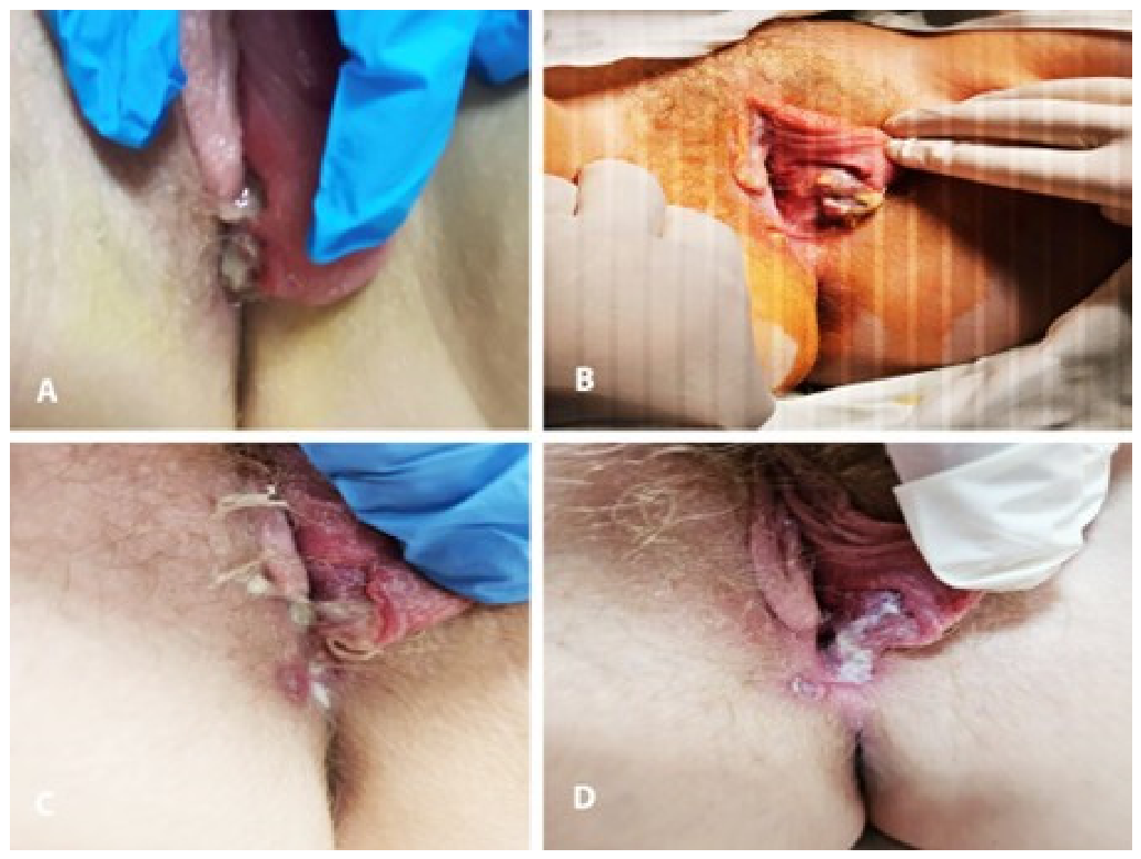
Gynecological examination revealed symmetrical ulcerated areas in a kissing pattern on the labia minora at the level of the introitus and posterior commissure. The lesions were a violet color, covered with fibrinous plaque, and painful to the touch (Figure 1A). No enlarged lymph nodes were detected.
Given that Lipschutz ulcer is a diagnosis of exclusion, a comprehensive diagnostic workup was undertaken to exclude infectious, autoimmune, traumatic, and malignant causes of acute genital ulceration. This included laboratory investigations, microbiological and serological testing, and multidisciplinary assessment by dermatology and infectious disease specialists.
Upon admission, the laboratory findings were not specific. We noted that leukocytes (19.1 × 10⁹/L) and CRP (217 mg/L) were elevated. No other changes in the blood count and biochemistry were observed. Hemoculture was negative. The swab taken from the ulcer surface did not detect pathological microorganisms. Serological tests for EBV and CMV and the Venereal Disease Research Laboratory test (VDRL) were also performed, which were negative. Venereal causes of the UVAL were excluded. The patient had a recent febrile illness accompanied by tonsillitis and elevated antistreptolysin-O titers. However, no microbiological confirmation of streptococcal infection was available, and the association with Lipschutz ulcer should be regarded as presumptive. We consulted the patient with a dermatologist and an infectious disease specialist. The patient met all of the criteria for the diagnosis of Lipschutz ulcer.
The patient was treated with oral amoxicillin/clavulanic acid (1000 mg, two tablets twice daily for 7 days) and intravenous metronidazole (500 mg twice daily for 5 days). In addition, inosine pranobex (500 mg, two tablets three times daily for 10 days), ibuprofen, paracetamol, and vitamin C were administered. Local treatment consisted of ethacridine lactate compresses (Rivanol), topical liniment, and corticosteroid cream. By hospital day 6, no clinical improvement had been observed. Inflammatory markers remained elevated despite broad-spectrum antimicrobial therapy, and the patient continued to have fever (38.3 °C). Therefore, surgical necrotomy was performed because of persistent symptoms and failure of conservative treatment. After the surgical intervention, an improvement in the local status of the patient was observed: the swelling of the labia disappeared, and wound granulation tissue appeared (Figure 1B–D). The patient remained permanently afebrile, and the values of the laboratory parameters were within the reference ranges. The patient was discharged, and so far, no recurrence of the disease has been reported.
Parental consent was obtained for presenting the case and using the photos.

4. Discussion

4.1. Epidemiology

The disease is typical mostly in the pediatric and adolescent populations. In a large case series, the mean age of onset was 16.6 years [9]. According to the aforementioned article, 38.46% of the patients were aged 19 years, and 15.38% were 18 years old. The rest of the patients were 11 years old, 12 years old, 14 years old, 15 years old, and 17 years old, each category amounting to 7.69% of all cases studied. The exact incidence of the disease is currently unknown because it is often overlooked and misdiagnosed.

4.2. Clinical Manifestations

As mentioned, the disease begins with asthenia, fever, and enlarged lymph nodes. Due to the location of the lesions, sometimes dysuria may also be present [6]. It is important to know that it can sometimes result in bladder retention requiring catheterization. Diarrhea, sickness, and vomiting are concomitant with gastrointestinal bacterial or viral agents [10]. The ulcerations typically appear on the labia minor but may also involve the labia major, perineum, vestibule, and lower vagina [11]. Macroscopically, the lesions appear with a round shape, with sharply demarcated borders, covered by fibrinous exudate or eschar. Edema and erythema of the labia, as well as inguinal lymphadenopathy, are also often described [12].

4.3. Pathogenesis

Pathogenesis remains enigmatic. However, it is hypothesized that the cytotoxic T-lymphocytes, found in the infiltrate, play a crucial role in the formation of the ulcers [13].
Another possible scenario is a type III hypersensitivity reaction during which immune complexes deposit in the dermal vessels, further activating the complement system, thus leading to tissue necrosis [14].
A deficit in the local immune response, particularly partial Ig A, has also been mentioned as a possible cause by Kinyó Á et al. [15].

4.4. Forms of Lu

Torok et al. [16] described three forms of the disease which we present in the table below (Table 1).

4.5. Histopathological Findings

Due to the fact that LU usually occurs in younger age groups and the lesions are generally self-resolving, biopsy is rarely required. As neoplasms are part of the differential diagnosis for LU, the latter must be excluded.
Therefore, with lesions lasting more than a month or in patients with significant risk factors for neoplasia, biopsy may be appropriate [17,18,19].
Barnwal et al. documented a case of UVAL following CMV infection in a 19-year-old patient with histological findings consistent with dysplasia [20].
We give a quick reminder that the risk factors for the development of childhood cancers include radiation exposure, previous cancer treatment, Down syndrome, secondhand smoke, infections, and certain inherited conditions.
The histological picture of LU is nonspecific: dilated capillaries with lymphocytic, histiocytic, plasmocytic, and fibroblastic infiltration. Proliferation and local thickening of the vascular wall are also observed. In advanced cases, the infiltration contains predominantly polymorphic neutrophils, with the subsequent formation of miliary pseudoabscess and ulceration [16].
Major and minor diagnostic criteria have been proposed by Sadoghi et al. [18] to help evaluate the condition (Table 2).
According to Sadoghi, if both major and at least two minor criteria are present, the diagnosis of UVAL is warranted.

4.6. Differential Diagnosis

The differential diagnosis is vast: mechanical and chemical trauma and sexual abuse should be excluded. Venereal diseases ought to be ruled out with complementary tests. Autoimmune diseases such as Behcet’s disease and Crohn’s ulcerative colitis also cause lesions in the perineal and vulvar regions. Pyoderma gangrenosum and some malignancies can mimic Lipschutz ulcers as well. Because Lipschutz ulcer is a diagnosis of exclusion, a thorough diagnostic evaluation is mandatory. Infectious causes such as herpes simplex virus, syphilis, Epstein–Barr virus, cytomegalovirus, Mycoplasma pneumoniae, influenza, streptococcal infection, and other sexually transmitted diseases should be excluded. Non-infectious causes, including Behçet’s disease, Crohn’s disease, pyoderma gangrenosum, trauma, sexual abuse, and vulvar malignancies, should also be considered. Multidisciplinary assessment involving gynecologists, dermatologists, and infectious disease specialists may facilitate the diagnostic process.

4.7. Treatment

It is crucial to remember that Lipschutz ulcers are self-limiting and tend to resolve spontaneously. While recognizing that the etiology behind the appearance of LU is important regarding the overall health of the patient, there is no evidence that the treatment of the main disease promotes faster healing of the ulcers. The treatment aims to relieve the pain and help epithelialize the ulcer. Patients in many case reports have been treated symptomatically with analgesics, disinfectant compresses, topical steroids, and antibiotics when superinfection was suspected. Lipschutz ulcer is generally considered a self-limiting condition, and surgical treatment is not part of standard management. Conservative treatment consisting of analgesics, topical anesthetics, local wound care, and, in selected severe cases, systemic corticosteroids remains the preferred approach. In our patient, treatment failure was defined by persistent severe pain, absence of clinical improvement after six days of multimodal therapy, and the presence of tightly adherent necrotic tissue. Under these circumstances, surgical necrectomy was considered a salvage procedure. Potential benefits include removal of necrotic tissue and acceleration of wound healing, whereas possible risks comprise pain, bleeding, secondary infection, and scar formation. Therefore, surgery should be reserved for carefully selected patients who do not respond to conservative measures and should not be regarded as routine treatment. In addition, treatment with a short course of systemic corticosteroids (0.5–1 mg/kg prednisolone for 7 to 10 days) may help heal severely painful, multiple, or necrotic ulcers [21].
Some authors claim positive results using colchicine for faster healing of lesions. This is likely linked to the fact that colchicine blocks the chemotaxis and phagocytic activity of neutrophils [22].
Surgical treatment is indicated in patients who have tightly adherent necrotic material covering the ulcer and show no improvement with conservative therapy [23].
Typically, the necrotic material is removed with a scalpel, followed by a saline lavage. Then the ulcer is covered by a gel containing antiseptics as well as moisturizers. Finally, alginate dressing is used to bandage the wound [24].
The authors observed a quick recovery with the aforementioned treatment.
Out of the 24 publications used as sources, 18 provide thorough information about the treatment and follow-up of the patients. The data is shown in Table 3, encompassing a total of 69 patients.
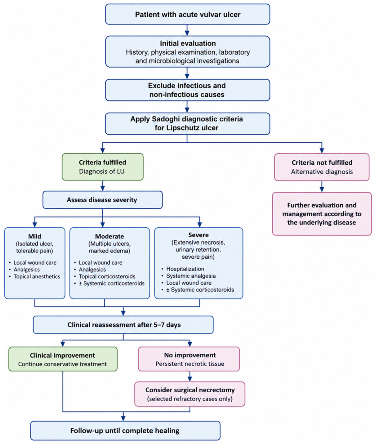
Based on research results, we propose the following algorithm leading to the diagnosis and treatment of patients with UVAL.
For practical purposes, disease severity was categorized according to clinical presentation. Mild disease was defined as isolated ulceration with tolerable pain and no urinary symptoms. Moderate disease included multiple ulcers, marked edema, and pain requiring systemic analgesics. Severe disease was characterized by extensive necrotic lesions, severe pain impairing ambulation or urination, urinary retention, or lack of response to conservative treatment. These categories are based on clinical judgment and have not been validated (Figure 2).

4.8. Follow-Up

It is important to know that most of the time, the ulcers resolve over a period of between 2 and 6 weeks without sequelae. Relapse of the disease has been reported in nearly a third of the patients over a period of one year [13]. Physicians should always remember that recurrent genital aphthosis may harbor Behcet’s disease and maintain close monitoring, especially with patients suffering from recurrent oral and genital ulceration, and ocular inflammation [24]. Therefore, we consider that a gynecological exam has to be conducted once a year to rule out progression of UVAL.

4.9. Limitations of the Study

This manuscript has several limitations that should be acknowledged when interpreting its findings. First, as this study describes the clinical course of a single patient, the findings have limited generalizability and may not be applicable to broader patient populations. Second, as a narrative review, it did not employ a predefined systematic search protocol or standardized study selection process, which may have resulted in the exclusion of relevant literature sources. Third, the inclusion and interpretation of the literature relied on the authors’ discretion, which may have introduced selection and interpretation bias. Moreover, as the review was carried out primarily using the literature in English, relevant evidence from other languages might have been omitted.
The review relied on published studies available through selected databases, potentially introducing publication bias by underrepresenting studies with negative or inconclusive findings. Differences in the methodological approaches, participant populations, research settings, and measured outcomes across the included studies posed challenges in comparing and synthesizing the findings.
As this review did not include a formal assessment of the methodological quality of the selected studies, the reliability and strength of the evidence may differ across the included literature. Therefore, the findings should be interpreted with caution, as they represent a narrative synthesis of the available evidence rather than conclusive proof.
Future studies employing systematic review methodologies and quantitative analyses are recommended to provide more robust and comprehensive evidence.

5. Conclusions

Lipschutz ulcer is a diagnosis often overlooked. Therefore, critical thinking as well as careful examination and evaluation are needed to recognize the clinical entity. A false diagnosis in this case causes a great distress to both patients and family. More effort is needed to spread awareness for UVAL diagnosis and treatment throughout different medical specialties that may come in contact with such patients.

Author Contributions

Conceptualization and writing—original draft, D.G.; methodology and visualization, S.K.; software, S.S.; resources and data analysis, Y.K. and Y.I.; supervision and review and editing, A.Y. All authors contributed to the interpretation of the results, critically revised the manuscript, approved the final version, and agreed to be accountable for all aspects of the work. All authors have read and agreed to the published version of the manuscript.

Funding

This research received no external funding.

Institutional Review Board Statement

According to the policies of the Ethics Committee of Medical University Pleven, formal ethics committee approval is not mandatory for the publication of individual case reports. Consequently, no such approval was sought or obtained.

Informed Consent Statement

Written informed consent has been obtained from the patient’s guardianto publish this paper.

Data Availability Statement

The original contributions presented in this study are included in the article. Further inquiries can be directed to the corresponding author.

Acknowledgments

Artificial intelligence (OpenAI) was used solely to assist with the graphical design of Figure 2. The scientific content, clinical reasoning, and final version of the figure were created, reviewed, and approved by the authors.

Conflicts of Interest

The authors declare no conflicts of interest.

Abbreviations

AGUAcute genital ulcer
ASTAntistreptolysin titer
CMVCytomegalovirus
EBVEpstein–Barr virus
LULipschutz ulcer
NSAIDNon-steroidal anti-inflammatory drug
PtPatient
RNRAGUReactive non-sexually-related acute genital ulcer
STDSexually transmitted disease
VDRLVenereal Disease Research Laboratory test
UVALUlcus vulvae acutum

References

  1. Lipschütz, B. Über eine eigenartige Geschwürsform des weiblichen Genitales (Ulcus vulvae acutum). Arch. Dermatol. Syph. 1912, 114, 363–396. [Google Scholar] [CrossRef] [Scilit]
  2. Lorenzo, C.V.; Robertson, W.S. Genital ulcerations as presenting symptom of infectious mononucleosis. J. Am. Board Fam. Pract. 2005, 18, 67–68. [Google Scholar] [CrossRef] [Scilit] [PubMed]
  3. Kos, L.; Galbraith, S.S.; Lyon, V.B. Vaginal ulcerations with acute mycoplasma infection. J. Am. Acad. Dermatol. 2007, 56, S117–S118. [Google Scholar] [CrossRef] [Scilit] [PubMed]
  4. Martín, J.M.; Godoy, R.; Calduch, L.; Villalon, G.; Jordá, E. Lipschütz acute vulval ulcers associated with primary cytomegalovirus infection. Pediatr. Dermatol. 2008, 25, 113–115. [Google Scholar] [CrossRef] [Scilit] [PubMed]
  5. Pelletier, F.; Aubin, F.; Puzenat, E.; Deprez, P.; Blanc, D.; Estavoyer, J.M.; Humbert, P. Lipschütz genital ulceration: A rare manifestation of paratyphoid fever. Eur. J. Dermatol. 2003, 13, 297–298. [Google Scholar] [PubMed]
  6. Limperg, T.; Bledsoe, M.; Strickland, J.; Jackson, M.A. Respiratory Pathogen Evaluation for Lipschütz Ulcer. J. Pediatr. Adolesc. Gynecol. 2018, 31, 212. [Google Scholar] [CrossRef] [Scilit]
  7. Limão, S.; Ventura, A.; Queirós, G.; Cunha, F. Lipschütz ulcer and group A streptococcal tonsillitis. BMJ Case Rep. 2018, 2018, bcr2017223579. [Google Scholar] [CrossRef] [Scilit] [PubMed]
  8. Hsu, T.; Sink, J.R.; Alaniz, V.I.; Zheng, L.; Mancini, A.J. Acute Genital Ulceration After Severe Acute Respiratory Syndrome Coronavirus 2 Vaccination and Infection. J. Pediatr. 2022, 246, 271–273. [Google Scholar] [CrossRef] [Scilit] [PubMed]
  9. Farhi, D.; Wendling, J.; Molinari, E.; Raynal, J.; Carcelain, G.; Morand, P.; Avril, M.F.; Francès, C.; Rozenberg, F.; Pelisse, M.; et al. Non-sexually related acute genital ulcers in 13 pubertal girls: A clinical and microbiological study. Arch. Dermatol. 2009, 145, 38–45. [Google Scholar] [CrossRef] [Scilit] [PubMed]
  10. Skeens, K.; Walker, L. Lipschütz Ulcers In 12-year-old Premenarchal Female Days After A Gastrointestinal Illness: A Case Report. Clin. Pract. Cases Emerg. Med. 2025, 9, 146–148. [Google Scholar] [CrossRef] [Scilit] [PubMed]
  11. Moise, A.; Nervo, P.; Doyen, J.; Kridelka, F.; Maquet, J.; Vandenbossche, G. Ulcer of Lipschutz, a rare and unknown cause of genital ulceration. Facts Views Vis. Obgyn. 2018, 10, 55–57. [Google Scholar] [PubMed]
  12. Visentin, D.; Driul, L.; Buligan, C.; Angarkhayeva, A.; Pinzani, C.; Martina, M.D.; Londero, A.; Stinco, G. Ulcus vulvae acutum—A case of genital ulcers in adolescent girl. Case Rep. Women’s Health 2015, 9, 4–6. [Google Scholar] [CrossRef] [Scilit] [PubMed]
  13. Lehman, J.S.; Bruce, A.J.; Wetter, D.A.; Ferguson, S.B.; Rogers, R.S., 3rd. Reactive nonsexually related acute genital ulcers: Review of cases evaluated at Mayo Clinic. J. Am. Acad. Dermatol. 2010, 63, 44–51. [Google Scholar] [CrossRef] [Scilit] [PubMed]
  14. Huppert, J.S. Lipschutz ulcers: Evaluation and management of acute genital ulcers in women. Dermatol. Ther. 2010, 23, 533–540. [Google Scholar] [CrossRef] [Scilit] [PubMed]
  15. Kinyó, Á.; Nagy, N.; Oláh, J.; Kemény, L.; Bata-Csörgő, Z. Ulcus vulvae acutum Lipschütz in two young female patients. Eur. J. Dermatol. 2014, 24, 361–364. [Google Scholar] [CrossRef] [Scilit] [PubMed]
  16. Török, L.; Domján, K.; Faragó, E. Ulcus vulvae acutum. Cutis 2000, 65, 387–389. [Google Scholar] [PubMed]
  17. Gómez Aguilar, D.L. Lipschütz ulcer: Report and follow-up of a clinical case: Case Report. Am. J. Med. Clin. Res. Rev. 2024, 3, 1–5. [Google Scholar]
  18. Sadoghi, B.; Stary, G.; Wolf, P.; Komericki, P. Ulcus vulvae acutum Lipschütz: A systematic literature review and a diagnostic and therapeutic algorithm. J. Eur. Acad. Dermatol. Venereol. 2020, 34, 1432–1439. [Google Scholar] [CrossRef] [Scilit] [PubMed]
  19. Brambilla, I.; Moiraghi, A.; Guarracino, C.; Pistone, C.; Tondina, E.; Riccipetitoni, G.; Raffaele, A.; Cavaiuolo, S.; Bertozzi, M.; Brazzelli, V. Recurrent reactive non-sexually related acute genital ulcers: A risk factor for Behcet’s disease? Acta Biomed. 2022, 93, e2022196. [Google Scholar] [CrossRef] [Scilit] [PubMed]
  20. Barnwal, S.; Priya, D.; Kala, P.S.; Sangal, B.; Pant, A. Dysplastic changes secondary to cytomegalovirus-induced Lipschutz ulcer: Unraveling complexity. Indian J. Sex. Transm. Dis. AIDS 2024, 45, 69–71. [Google Scholar] [CrossRef] [Scilit] [PubMed]
  21. Mourinha, V.; Costa, S.; Urzal, C.; Guerreiro, F. Lipschütz ulcers: Uncommon diagnosis of vulvar ulcerations. BMJ Case Rep. 2016, 2016, bcr2015214338. [Google Scholar] [CrossRef] [Scilit] [PubMed]
  22. Nouchi, A.; Monsel, G.; Lafon-Desmurs, B.; Meng, L.; Burrel, S.; Moyal-Barracco, M.; Caumes, E. Epstein-Barr Virus-related Acute Genital Ulcer Successfully Treated with Colchicine. Acta Derm.-Venereol. 2018, 98, 134–135. [Google Scholar] [CrossRef] [Scilit] [PubMed]
  23. Delgado-García, S.; Palacios-Marqués, A.; Martínez-Escoriza, J.C.; Martín-Bayón, T.A. Acute genital ulcers. BMJ Case Rep. 2014, 2014, bcr2013202504. [Google Scholar] [CrossRef] [Scilit] [PubMed]
  24. Calles Sastre, L.; Serrano González, L.; Hernández Sánchez, L.; García Espantaleón, M.J.; Pérez Medina, T. Nuevas vías de tratamiento en la úlcera de Lipschütz. Prog. Obstet. Ginecol. 2018, 61, 176–178. [Google Scholar]
Figure 1. (A)—Lipschutz ulcer during admission. (B)—Ulcers just before surgery. (C)—Appearance of Lipschutz ulcer 3 days after the surgery. (D)—Ulcers 5 days after the surgery.
Reprodmed 07 00033 g001
Figure 2. Proposed diagnostic and therapeutic algorithm for Lipschutz ulcer.
Reprodmed 07 00033 g002
Table 1. Three forms of Lipschutz ulcer.
TypeGangrenous Form
(Most Common)
Chronic Form
(Pseudo Venereal)
Miliary Form
Macroscopic characteristicsUlcers covered with gray-yellowish exudateRecurrent round superficial ulcers with significant swellingUlcers the size of a pinhead on the edges of the labia and perineum
Systemic symptomsProminent systemic symptoms Subtle symptomsNo systemic complaints
Table 2. Diagnostic criteria of Lipschutz ulcer.
Major CriteriaMinor Criteria
Acute onset of ≥1 painful ulcerous lesions in the vulvar regionLocalization of ulcer at vestibule or labia minora
Exclusion of infectious and other non-infectious causes for the ulcerNo sexual intercourse ever or within the last 3 months
Flu-like symptoms
Systemic infection within 2–4 weeks prior to onset of vulvar ulcer
Table 3. Summary of published case reports and case series included in this narrative review, presenting patient characteristics, presumed etiology, treatment, follow-up, and clinical outcomes of Lipschutz ulcer.
ReferenceNumber of
patients
AgeEtiologyTreatmentFollow-Up/Outcome
Lorenzo et al. (2005) [2]218EBVPt 1—acyclovir (discontinued), narcotic analgesics
Pt 2—acyclovir, ciprofloxacin (discontinued), prednisone
  • P1 healed in 16 days, no sequelae
  • P2 healed in 1 week, no sequelae
Kos et al. (2007) [3]212, 11Mycoplasma
pneumoniae
Azithromycin for 5 daysResolved in a few weeks, no scarring
Limão et al. (2018) [7]111Strep. A10-day course of oral amoxicillin (500 mg twice a day), topical anesthetic (lidocaine) and antiseptic healing ointment (dexpanthenol and chlorhexidine)Healed in 2 weeks, no recurrence
Hsu et al. (2022) [8]312, 14, 29COVID-19 vaccination
and infection
Pt 1—topical clobetasol 0.05% ointment twice daily, lidocaine 2% jelly every 4-6 h as needed, oral acetaminophen and ibuprofen as needed, and sitz baths
Pt 2—same as patient 1 + oral nortriptyline and oral prednisone taper (20 mg twice daily for 5 days followed by 20 mg once daily for 5 days)
Pt 3—clobetasol 0.05% ointment twice daily
Pt 1—healed in 2 weeks
Pt 2—this was a second episode of AGU, third occurrence of ulcers after SARS-CoV-2 infection, both healed in 4 weeks
Pt 3—healed quickly
Farhi et al. (2009) [9]1311–19 years,
mean 16.6
EBV in 4 patients,
Behçet’s disease in 1 patient, idiopathic AGU in 8 patients
Five patients with no specific treatment; three received antibiotics only (cefpodoxime/amoxicillin);
four received valacyclovir; one patient received valacyclovir, cefpodoxime, josamycine
10 patients healed in 16 days,
3 patients (23%) experienced 1 relapse of AGU during the following year, one diagnosed with Behçet’s disease
Skeens and Walker (2025) [10]112Viral gastroenteritisIce packs once every hour to the area, topical clobetasol ointment twice daily, and topical lidocaine gel, oral oxycodone, oral prednisone taperHealed in 4 weeks
Moise et al. (2018) [11]113Suspected viral illnessParacetamol, tramadol, wound care, acyclovir 200 mg 5X/day (discontinued)Healed in a few weeks
Visentin et al. (2015) [12]116EBVEmollients and analgesicsHealed in 20 days
Lehman et al. (2010) [13]109–16 years,
mean 11.5
Various viral
pathogens
Analgesics, topical corticosteroids, NSAIDsComplete resolution
Kinyó et al. (2014) [15]23, 25Partial IgA deficiencyPt 1—local steroid and antiseptic ointment
Pt 2—amoxicillin/clavulanic acid, steroids
Pt 1—healed within 2 weeks
Pt 2—healed in 3 weeks
Gómez Aguilar (2024) [17]117Idiopathic AGUDoxycycline, sitz bath with potassium permanganate, betadine dressingsHealed in 3 weeks
Sadoghi et al. (2020) [18]272 months–30 years,
mean 15.5
VariousTopical and systemic antibiotics in 17 patients, topical and systemic cortisone in 10 patients, analgesics in 6 patients, surgery in 1 patientMost healed in 2–3 weeks, two of which with scarring
Brambilla et al. (2022) [19]112Idiopathic AGUAmoxicillin–clavulanic acid, Teicoplanin, Metronidazole, topical Betamethasone, Gentamicin, Clotrimazole, VaselineRelapse after 15 months, HLA-B51 positivity
Barnwal et al. (2024) [20]119CMVAcyclovir (400 mg 3x day for 7 days), oral corticosteroids (Tab prednisolone 40 mg for 3 days), additional anti-inflammatory medication (Tab paracetamol 650 mg to be taken as required)Healed in 3 weeks
Mourinha et al. (2016) [21]122Idiopathic AGUAzithromycin (no improvement), Doxycycline, acyclovir and analgesia (local and intravenous), topical ClobetasolHealed in 3 weeks
Nouchi et al. (2018) [22]118EBVOpioids and topical lidocaine, topical and systemic corticosteroids (0.5 mg/kg/day for 3 days), topical Imiquimod, Colchicine, 1 mg per dayComplete healing by day 7
Delgado-García et al. (2014) [23]113Idiopathic AGUNSAIDs, analgesics, prophylactic broad-spectrum antibiotics (Amoxicillin/Clavulanic acid and Azithromycin), surgical removal of adherent necrotic materialHealed in 3 weeks; this was a second episode of AGU
Calles Sastre et al. (2018) [24]124Idiopathic AGUNSAIDs, surgical debridement, followed by alginate wound dressingHealed completely in 7 days
Abbreviations: AGU, acute genital ulcer; CMV, cytomegalovirus; COVID-19, coronavirus disease 2019; EBV, Epstein–Barr virus; IgA, immunoglobulin A; NSAIDs, non-steroidal anti-inflammatory drugs; Pt, patient; SARS-CoV-2, severe acute respiratory syndrome coronavirus 2; Strep. A, Group A Streptococcus.
Disclaimer/Publisher’s Note: The statements, opinions and data contained in all publications are solely those of the individual author(s) and contributor(s) and not of MDPI and/or the editor(s). MDPI and/or the editor(s) disclaim responsibility for any injury to people or property resulting from any ideas, methods, instructions or products referred to in the content.