Review

Protein-Bound Nano-Injectable Suspension: Unveiling the Promises and Challenges

1
MET’s Institute of Pharmacy, Nashik 422003, Maharashtra, India
2
School of Pharmacy, Gujarat Technological University, Gandhinagar 382028, Gujarat, India
3
Vidman Pharmaceuticals Pvt Ltd., Ahmedabad 380061, Gujarat, India
4
National Institute of Pharmaceutical Education and Research (NIPER), Ahmedabad 382355, Gujarat, India
5
Dynamic Biologics Inc., Monmouth Junction, NJ 08852, USA
6
Centre for Research Impact & Outcome, Chitkara College of Pharmacy, Chitkara University, Rajpura 140401, Punjab, India
7
Faculty of Pharmacy, Silpakorn University, Nakhon Pathom 73000, Thailand
*
Authors to whom correspondence should be addressed.
Current address: Department of Pharmaceutical Chemistry, Vaccine Analytics and Formulation Center, University of Kansas, Multidisciplinary Research Building, 2030 Becker Dr., Lawrence, KS 66047, USA.
Appl. Nano 2025, 6(2), 9; https://doi.org/10.3390/applnano6020009
Submission received: 20 April 2025 / Revised: 28 May 2025 / Accepted: 28 May 2025 / Published: 30 May 2025
(This article belongs to the Collection Review Papers for Applied Nano Science and Technology)

Abstract

:
Protein-bound nano-injectable solutions represent a cutting-edge advancement in nanomedicine, offering a versatile platform for precise and controlled drug delivery. By leveraging the biocompatibility and functional versatility of proteins such as albumin, gelatin, and casein, these nano systems enhance drug solubility, prolong circulation time, and improve site-specific targeting, which are particularly beneficial in cancer and inflammatory diseases. This review provides a comprehensive overview of their formulation strategies, physicochemical characteristics, and biological behavior. Emphasis is placed on therapeutic applications, regulatory considerations, fabrication techniques, and the underlying mechanisms of drug–protein interactions. This review also highlights improved pharmacokinetics and reduced systemic toxicity, while also critically addressing challenges like immunogenicity, protein instability, and production scalability. Recent FDA-approved formulations and emerging innovations in precision medicine and theranostics underscore the transformative potential of protein-based nanosuspensions in next-generation drug delivery systems.

1. Introduction

Nanotechnology has allowed for the creation of more efficient, tailored, and patient-friendly medicines in recent decades, transforming the landscape of drug delivery systems. When it comes to treating complicated diseases like cancer, autoimmune disorders, and infectious diseases, nano-injectable suspensions have proven to be a potent modality among the several platforms in nanomedicine [1]. Bypassing the first-pass metabolism and promoting quick commencement of action, these methods offer the distinct benefit of delivering therapeutic medicines directly into systemic circulation [2,3].
Innovations in materials science, especially the incorporation of biocompatible and biodegradable components into nano-formulations, have played a crucial role in the development of injectable nanomedicines. Because of their adaptability and usefulness as carriers, proteins have attracted a lot of interest in this field [4,5]. Some naturally occurring proteins have advantageous properties, such as being biodegradable, not being immunogenic, and having the potential to bind and stabilize a variety of medicines. These include transferrin, gelatin, casein, and human serum albumin. Because of these characteristics, they can be used to create nano-injectable systems that are more stable, more soluble, and more targetable [6,7].
Abraxane®, an albumin-bound paclitaxel formulation that revolutionized nano-oncology, brought the idea of protein-bound nanoparticles into mainstream therapeutics [4,5]. This formulation has two benefits: first, it improves medication accumulation in tumor tissues through the increased permeability and retention (EPR) effect; second, it eliminates the requirement for hazardous solvents. Numerous protein-based micro-injectable solutions have been the subject of research due to these promising case studies, which aim to enhance therapeutic indices while reducing off-target effects [8,9]. Notwithstanding these encouraging features, there are still a number of obstacles to overcome in the creation and implementation of protein-bound micro-injectable solutions. Concerns over immunogenicity, batch-to-batch variability, scalability, and protein denaturation during formulation are among them. Another major obstacle to broad clinical translation is the persistence of regulatory challenges related to the safety, effectiveness, and quality control of these complex biologic–nano hybrids [3,10,11].
The purpose of this review is to present a synopsis of protein-bound nano-injectable suspensions, touching on topics such as their methods of action, therapeutic benefits, clinical uses, and methods of formulation. In addition to outlining current developments and potential future paths in the subject, it provides a critical analysis of the constraints and technical difficulties linked with these systems. The goal of this review is to help in the rational design and development of safer, more effective, and precision therapy-tailored nanomedicines based on proteins by outlining the potential benefits and drawbacks of this technology.

2. Fundamentals of Protein-Bound Nano-Injectable Suspensions

Sterile nanoparticle suspensions developed for intravenous delivery are known as nano-injectable suspensions. They are composed of nanocarriers that are suspended in injectable or water-based media, with sizes typically ranging from 10 to 1000 nm [9]. Rapid systemic distribution, site-specific drug accumulation, and bypassing of hepatic first-pass metabolism are all features of these formulations, which are designed for intravenous, intramuscular, or subcutaneous administration [2,4,12]. Typical uses include treating cancer, infectious diseases, and chronic inflammatory disorders. When a medicinal substance interacts with either naturally occurring or artificially produced proteins utilized as carriers in nanosuspensions, this process is known as protein binding. Medicine remains stable in its nanoscale form due to these interactions, which also inhibit aggregation and improve systemic circulation by imitating endogenous proteins. The structural integrity of medications, the protection of fragile components from degradation, and the provision of controlled release profiles are all enhanced by protein-bound systems. The protein also helps with tailored distribution by way of receptor-mediated transport that is particular to certain tissues [4].

2.1. Types of Carrier Proteins

Proteins play an essential role in micro-injectable suspensions because of their versatility in encapsulating, stabilizing, and targeting drugs. Of these proteins, human serum albumin (HSA) is by far the most popular [13]. The fact that it is non-immunogenic, abundant in plasma, and remarkably biocompatible has led to its extensive use. In particular, hydrophobic medications can benefit from HSA’s many binding domains, and the compound shows remarkable water solubility [13]. It plays an important role in receptor-mediated endocytosis, particularly via the increased gp60 and SPARC receptors found in tumor microenvironments. It is perfect for prolonged systemic drug delivery due to this feature and its extended plasma half-life [13]. The FDA-approved albumin-bound paclitaxel formulation, Abraxane®, is a pioneering use of this carrier in the treatment of certain malignancies [5]. Protein types that serve as carriers are shown in Figure 1.
Casein, a phosphoprotein derived from milk, is another beneficial protein. Casein can encapsulate hydrophobic medicinal molecules because of its amphiphilic nature, which allows it to self-assemble into micellar structures [14,15,16]. Particularly in nutraceutical and oncological uses, its inherent biodegradability and lack of adverse effects make it an ideal candidate for administration by injection or oral means. Another protein-based substance with excellent biocompatibility and adaptability is gelatin [17], which is a denatured version of collagen. It can encapsulate a variety of medicinal substances in thermosensitive gels and nanoparticles. To further enhance their selectivity to diseased tissues or cells, gelatin nanoparticles can have their surfaces chemically changed to contain targeting ligands [14].
The ability of glycoprotein transferrin to transport iron is well known. Since transferrin receptors are overexpressed in a large number of cancer cells, it has found application in cancer-targeted delivery [18]. To improve cellular uptake and therapeutic effectiveness, nanoparticles functionalized with transferrin can engage in receptor-mediated endocytosis. Finally, serum proteins and globulins, such as immunoglobulins, have powerful drug-binding and immunomodulatory properties. Nanoparticle systems that incorporate them have demonstrated potential to improve therapeutic targeting, especially in vaccine delivery and immunotherapy [19,20]. The proteins utilized as carrier in nano-injectable solutions are listed in Table 1.

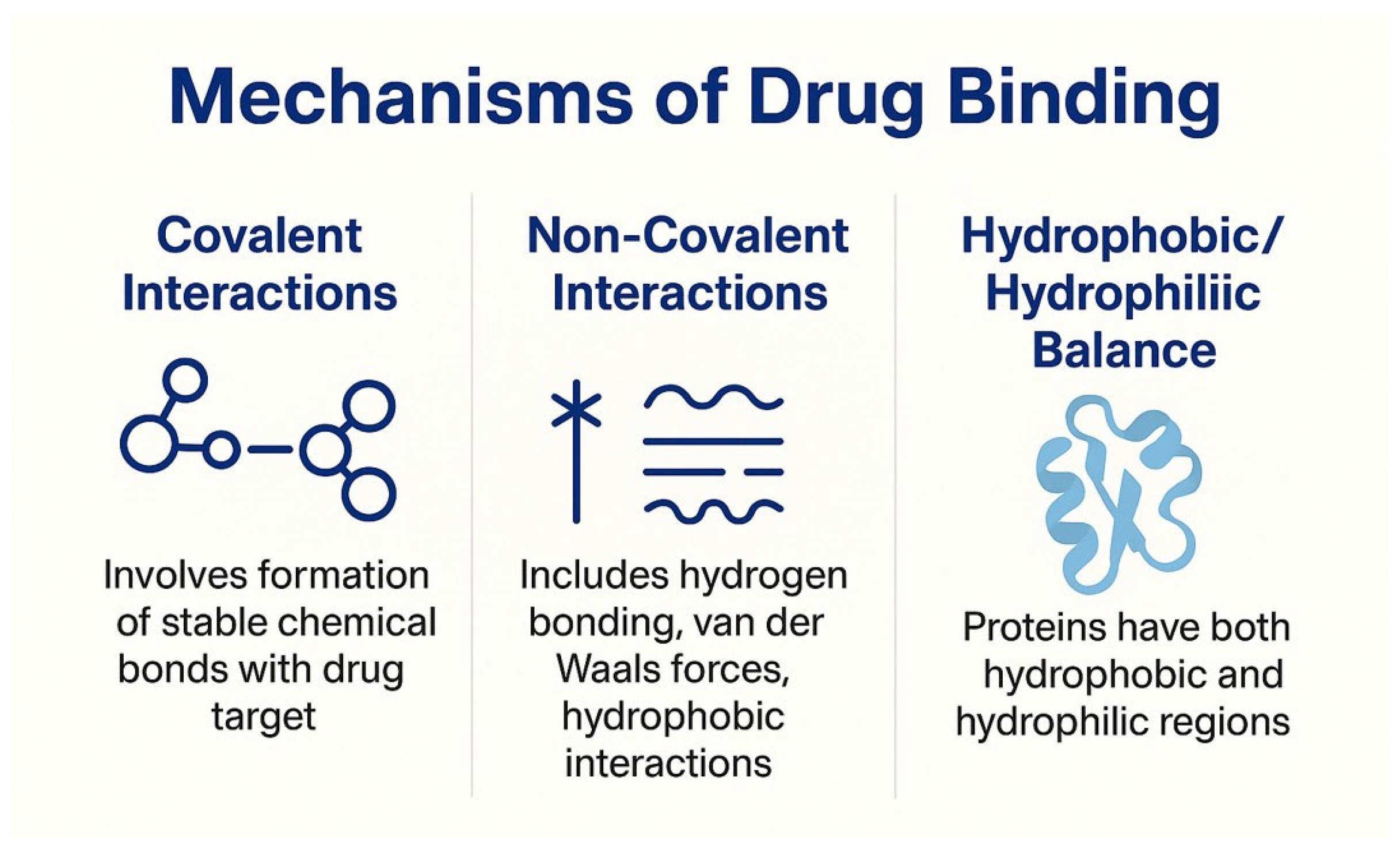
2.2. Mechanisms of Drug Binding

2.2.1. Covalent Interactions

One of the most stable and robust ways for medicinal chemicals to be attached to carrier proteins in micro-injectable solutions is through covalent bonds. The drug molecule forms chemical interactions with reactive functional groups on the surface or within the protein’s structure as part of this mechanism. Amino acid residues like cysteine, glutamic acid, and lysine contain thiol (-SH) groups, carboxyl (-COOH) groups, and amine (-NH2) groups, which are typical reactive groups on proteins [26]. To aid the bond formation without severely damaging the drug or protein’s integrity, coupling agents or activation procedures are commonly used to generate these covalent connections. Because the bond keeps the drug firmly bound to the carrier until enzymatic or pH-mediated cleavage happens in the targeted tissue, it is an advantageous binding mechanism for medicines that need extended retention, gradual release, or site-specific activation [24]. But there is a price to pay for this stability. Proteins’ biological function, receptor recognition, and immunogenic profile can all be impacted by covalent modifications that change their natural structure [27]. For instance, albumin’s capacity to target tumors can be diminished if it is over-modified, as this can prevent it from attaching to the gp60 receptor [4,13]. To prevent protein denaturation or aggregation, careful regulation of reaction conditions is frequently necessary during covalent conjugation [26]. One popular use of this approach is PEGylation, which involves covalently attaching chains of polyethylene glycol (PEG) to a protein or peptide. By enhancing medication solubility and stability and hiding immunogenic epitopes, this mechanism prolongs systemic circulation and prevents quick clearance through the reticuloendothelial system (RES) [28]. One additional example is drug–linker conjugates, which are embedded in antibody–drug conjugates (ADCs) and involve the covalent binding of a cytotoxic drug to a targeted moiety via a cleavable or non-cleavable linker [29,30].
Ke et al. (2023) [31] investigated covalent and non-covalent interaction mechanisms for drug binding in protein-based nanosuspensions using casein and quercetin as model systems [31]. Both interaction modes altered the structural and functional behavior of casein; however, covalent binding demonstrated superior binding efficiency and stability enhancement. Covalent conjugation occurred under alkaline conditions (pH 9.0), facilitated by oxygen, leading to irreversible bond formation between nucleophilic amino acid residues and quercetin’s oxidized quinone intermediates. This resulted in a substantial reduction of free sulfhydryl groups from 8.12 μmol/g (native casein) to 4.35 μmol/g, confirming effective covalent linkage. Drug loading increased proportionally with quercetin concentration, reaching 3.89 mg/g at 160 μmol/g, compared to 2.31 mg/g at 10 μmol/g. FTIR structural analysis showed significant conformational rearrangement: α-helix increased from 24.5% to 29.8%, β-turn from 15.2% to 18.7%, and random coil from 22.1% to 26.4%, indicating enhanced molecular stability. These changes contributed to improved solubility, thermal stability, and oxidative resistance—key attributes for sustained drug retention in protein-bound nanosuspensions. Non-covalent binding, in contrast, was governed primarily by hydrophobic interactions and hydrogen bonding, offering reversible attachment but comparatively lower stability under physiological conditions.

2.2.2. Non-Covalent Interactions

Protein-bound nano-injectable solutions rely heavily on non-covalent interactions because of their reversibility and the fact that they can keep the carrier protein and therapeutic component structurally intact [32]. Electric fields, hydrogen bonds, hydrophobic interactions, van der Waals forces, and electrostatic attractions are examples of these interactions, which are often physical than chemical. These forces may not be as strong as covalent connections, but when combined, they can produce stable drug–protein complexes, particularly in cases when there are numerous interaction sites [2].
To improve binding specificity and orientation, hydrogen bonds are formed between polar functional groups on the medication and corresponding locations on the protein. The protein–drug interface is especially densely packed; thus, the van der Waals forces add even more stability [33]. The interaction between the drug’s nonpolar regions and the protein’s hydrophobic amino acid residues is crucial for medications that are poorly soluble in water. Encapsulation and solubilization are facilitated by these interactions, which eliminate the necessity for strong solvents or chemical alterations [34].
For example, when cationic amine groups interact with anionic carboxyl groups on the medication or protein, electrostatic attractions form between the two groups. During formulation development, the interactions can be fine-tuned by altering the ionic strength and pH of the medium [35,36,37]. By avoiding changes in the protein carrier’s original structure or biological function, non-covalent interactions reduce the likelihood of immunogenicity and maintain targeting capability [27]. In addition, the reversibility of these connections enables localized and regulated drug release in response to environmental cues, such as changes in pH, enzyme activity, or temperature. Since the chemical changes needed for covalent conjugation might cause labile or sensitive medications to lose activity or degrade, this method is especially useful for them. These medications can be more effectively and safely formulated using non-covalent binding, which allows for a stable yet bio-responsive formulation [38].
Ke et al. (2023) [31] also explored the role of non-covalent interactions in drug binding within protein-based nanosuspensions using casein–quercetin complexes [31]. Unlike covalent interactions, non-covalent binding occurs under neutral pH and is mediated by hydrogen bonding, hydrophobic forces, and electrostatic interactions. This reversible nature resulted in a moderately decreased sulfhydryl content—yet higher than covalent systems—indicating weaker but more dynamic protein–polyphenol associations. The drug loading efficiency was lower, increasing from 2.31 mg/g at 10 μmol/g to 3.12 mg/g at 160 μmol/g, owing to the non-permanent nature of binding. Structural analysis via FTIR showed moderate secondary structure changes, with α-helix increasing from 24.5% to 27.3%, β-turn from 15.2% to 17.5%, and random coil from 22.1% to 24.8%, suggesting improved conformational flexibility. Although these complexes showed lower solubility and thermal stability compared to covalent forms, they exhibited enhanced surface hydrophobicity, foaming capacity, and emulsifying activity. In contrast, non-covalent interactions provide dynamic binding, better protein flexibility, and functional benefits such as enhanced emulsifying and foaming properties, which may be advantageous in formulations requiring responsive or stimuli-sensitive drug release.

2.2.3. Hydrophobic/Hydrophilic Balance

The design and performance of protein-bound nano-injectable solutions are greatly influenced by the hydrophobic/hydrophilic balance of proteins. Most proteins found in nature are amphiphilic, meaning they have separate domains that are hydrophobic and hydrophilic, respectively [39]. Because of its two-fold nature, it can interact with a wide variety of pharmacological molecules, including those that are soluble in water and those that are extremely lipophilic and thus poorly soluble. Nonpolar amino acid residues like valine, phenylalanine, leucine, and isoleucine make up most hydrophobic domains in proteins. Lipophilic medicines, which are difficult to administer because they are poorly soluble in water, find a suitable microenvironment in these areas [39]. As an example, formulations such as Abraxane® can administer paclitaxel a hydrophobic anticancer drug without the need of harmful organic solvents because of its strong binding affinity to the hydrophobic pockets of human serum albumin [4,12,40].
However, areas that are rich in polar or charged amino acids like glutamine, lysine, and aspartic acid are known to be hydrophilic, and they interact well with the water environment. To keep protein–drug nanoparticles from aggregating in biological fluids, keep them colloidally stable, and increase their systemic circulation, these areas are crucial. Their addition improves the nano-formulations’ dispersibility and solubility, two properties that are critical for intravenous administration [41,42].
There are several reasons why it is critical to achieve the ideal hydrophobic/hydrophilic equilibrium. To start with, it establishes the carrier’s drug loading capacity, which affects the amount of active pharmaceutical ingredients that may be successfully added without lowering stability. Secondly, it influences the nanoparticle system’s physical stability, which might cause precipitation or early medication release if there are abnormalities. Finally, this equilibrium determines surface features that impact biodistribution, cellular uptake, and immune system recognition. Excessive hydrophilicity, on the one hand, can impede cellular uptake, while excessive hydrophobicity, on the other, might prompt quick clearance by the mononuclear phagocyte system [43]. Thus, a well-balanced hydrophobic/hydrophilic ratio is an essential factor in therapeutic efficacy and not just a physicochemical property. To customize the distribution profile for clinical uses, advanced formulation procedures frequently alter this balance by modifying surfaces, altering proteins, or co-forming with stabilizers and surfactants [44]. Figure 2 shows the processes involved in drug binding.
The hydrophobic/hydrophilic balance (HHB) critically influences drug loading in protein-based nanocarriers. Xu et al. (2024) [45] demonstrated that co-assembly of BSA, SPIONs, and hydrophobic DOX achieved ultrahigh drug loading of 58% and stable particle size (~90 nm) over one year. Encapsulation efficiency increased with rising DOX concentrations (from 0.5 to 10 mg/mL), with the co-assembly method outperforming solvent diffusion and adsorption. Zeta potential varied significantly across methods (−24 mV for co-assembly vs. −6 mV for adsorption), reflecting differences in drug localization. Notably, replacing BSA with ovalbumin or SPIONs with hydrophobic quantum dots preserved the high loading (>50%), confirming that surface hydrophobicity is a key determinant. These findings affirm that optimizing HHB enables high drug incorporation without compromising colloidal stability.

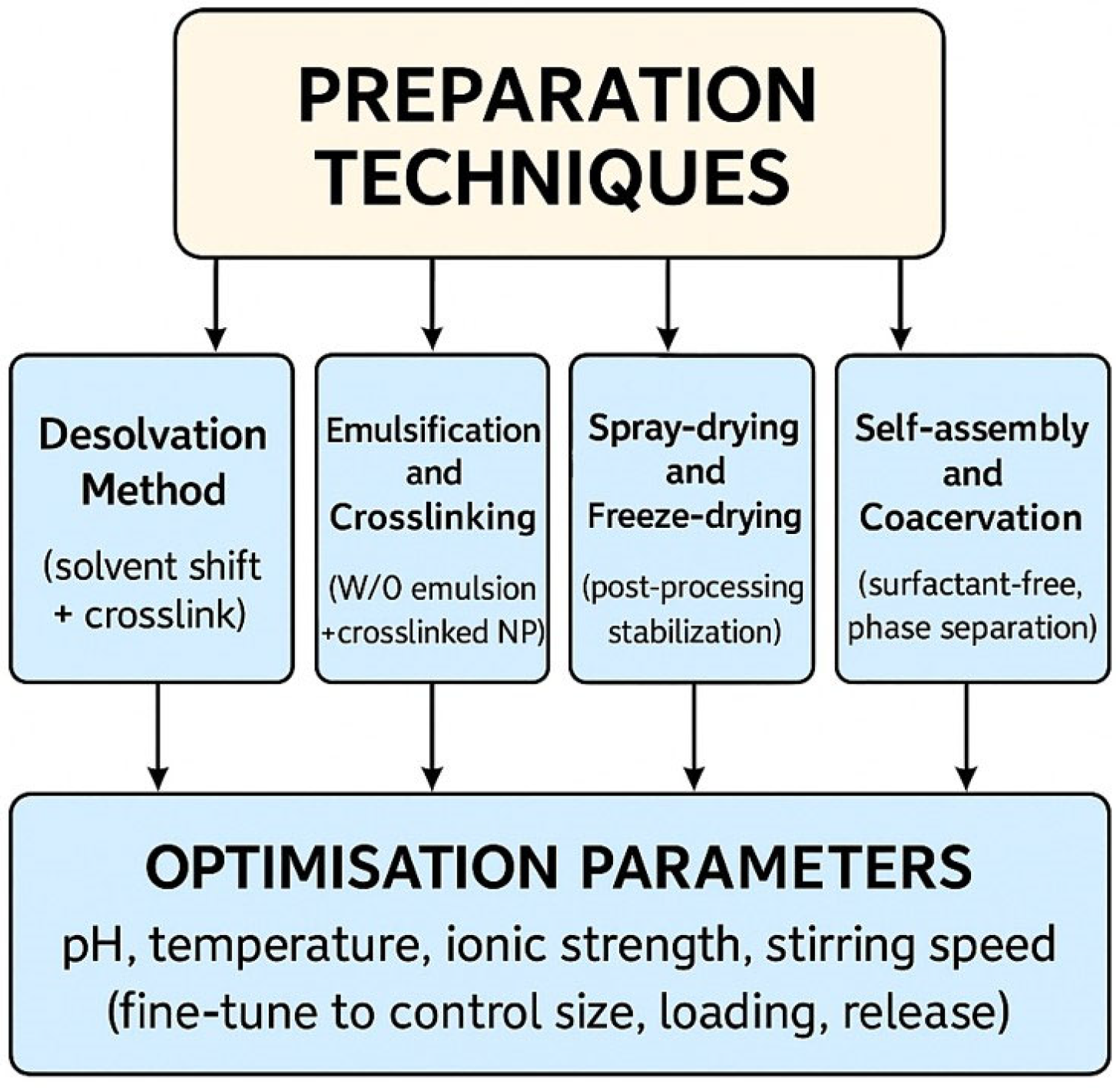
3. Preparation Techniques

3.1. Desolvation Method

A desolvating substance, like acetone or ethanol, is slowly added to a protein solution using this method. Protein precipitates out as nanoparticles when the polarity of the solvent changes. To further secure the nanoparticle structure, crosslinking chemicals such as glutaraldehyde are frequently employed thereafter [4,46,47]. This approach is highly effective for producing albumin-based nanoparticles and is known for its simplicity, reproducibility, and ease of scaling up. Nanoparticles derived from HSA are ideal candidates for this method because albumin is biocompatible, non-immunogenic, and may bind a diverse array of therapeutic chemicals [17,48]. There has been a lot of research on using HSA nanoparticles made by desolvation to transport diagnostic chemicals, anti-inflammatory medicines, and chemotherapeutics [19]. Surface modification or ligand attachment, made possible by desolvation, also permits active targeting of particular cells or organs. In conclusion, the desolvation method is a reliable, repeatable, and scalable way to make nano-injectable solutions containing bound proteins. This lays the groundwork for future drug delivery methods that are more effective and safer [49].

3.2. Emulsification and Crosslinking

This process creates a water-in-oil emulsion by introducing an organic phase (often oil) into a protein solution that is in an aqueous phase. To create nanoemulsions, the size of the droplets is reduced by mechanical stirring or ultrasonication. The droplets are transformed into stable nanoparticles by adding crosslinking agents, such as glutaraldehyde or genipin [50]. This approach enables surface functionalization and is well suited for encapsulating hydrophobic medicines. This method involves making a water-in-oil (W/O) emulsion by dispersing a protein solution (often water-based) containing the medicinal agent into a non-miscible organic phase, commonly an oil like paraffin or vegetable oil. To create nanoparticles, this emulsion is first needed. Applying high-shear mechanical stirring, ultrasonication, or homogenization can improve the emulsification process and decrease the size of droplets to the nanoscale. In these procedures, future nanoparticles are represented by individual droplets of the dispersed water phase, which results in fine, homogeneous nanoemulsions [50,51].

3.3. Spray-Drying and Freeze-Drying

Protein-based nano-injectable suspensions rely on two crucial post-processing techniques: spray-drying and freeze-drying, also known as lyophilization. Although they work on separate principles and serve different purposes, both are commonly employed to make nano-formulations more stable, extend their shelf life, and make them easier to handle. Spray-drying is faster and more cost-effective, whereas freeze-drying offers better preservation of sensitive materials [52,53]. A rotary atomizer or specialized nozzle is used in spray-drying, a continuous, one-step drying process, to atomize a liquid formulation into a hot air stream. This liquid formulation is usually a protein–drug solution or nanoparticle suspension. The solvent is quickly evaporated as the tiny droplets move through the drying chamber, turning them into dry powder particles. Filters or cyclone separators gather the produced particles at the chamber’s base [54].
The quick drying process reduces the amount of time that thermo-stable medications and proteins are exposed to heat, making this procedure ideal for them. Particle size, shape, and moisture content can be precisely controlled by adjusting variables including inlet/outlet temperature, feed rate, atomization speed, and nozzle type. When it comes to the scalable manufacture of nanoparticle formulations for parenteral or pulmonary administration, spray-drying powders are frequently the way to go because of the better aerosolization, reconstitution characteristics, and dose consistency [52,53,55]. However, heat-sensitive biomolecules such as biologics, peptides, and proteins can be delicately dehydrated by freeze-drying, also known as lyophilization. Sublimation, the direct transformation of ice to vapor without passing through the liquid phase, is induced by subjecting the protein-based nanoparticle suspension to vacuum conditions after it has been frozen at low temperatures. By avoiding the aggregation and degradation that can happen with traditional drying procedures, this ensures that labile medicinal medicines retain their native structure and biological function [56].
In a conventional freeze-drying process, there are three steps: freezing, sublimation, and desorption. The final product is a dry, porous cake or powder that can be mixed with an appropriate solvent for injection. Furthermore, it remains stable at room temperature for an extended period, eliminating the requirement for cold-chain storage. When the end product is meant for transportation, commercial distribution, or long-term storage, both methods are commonly used as post-processing procedures following nanoparticle manufacturing. To further ensure the nanoparticles’ integrity throughout drying and reconstitution, they also make it easier to incorporate cryoprotectants or stabilizers such as trehalose, mannitol, or sucrose [57].

3.4. Self-Assembly and Coacervation

Protein-based nanoparticles can be generated in an elegant, surfactant-free manner using either self-assembly or coacervation; these methods excel at handling biologics, delicate biomolecules, and labile medications [38]. These methods build persistent, functional nanostructures using proteins’ inherent physicochemical qualities and their responsiveness to environmental cues, rather than using harsh chemical treatments or mechanical forces. Proteins can spontaneously assemble themselves into complex nanoscale structures under certain environmental or physiological stimuli, a process known as self-assembly. Many other types of non-covalent interactions, including electrostatic attractions, van der Waals forces, hydrogen bonding, and hydrophobic interactions, are responsible for this phenomenon. By adjusting variables like temperature, solvent polarity, ionic strength, and pH, the process can be fine-tuned. When the pH nears their isoelectric point or when the ionic strength changes, proteins may change electrostatic connections and unfold and reassemble into nanostructures [56,58].
According to the drug’s affinity for particular domains within the protein, self-assembled protein nanoparticles can encapsulate hydrophobic or hydrophilic pharmaceuticals within their matrix or core. This technology is used in medication delivery. Therapeutic peptides, antibodies, and nucleic acids are ideal candidates for the self-assembly method due to their reversibility, biocompatibility, and ease of application. The ability to release drugs into target tissues in response to external stimuli is another benefit of such systems, which are typically sensitive to physiological signals [59]. In contrast, coacervation is a method of liquid–liquid phase separation that, when applied to a homogeneous protein solution, causes dense colloidal droplets, or coacervates, to form. The separation of proteins into two insoluble liquid phases, a protein-rich coacervate phase and a dilute supernatant, require meticulous control over several variables, such as pH, ionic concentration, temperature, and the presence of a counter-polyelectrolyte. The desired medication is encased in coacervate droplets, which can then be dried or crosslinked to form stable nanoparticles [60,61,62]. There are two types of coacervation: simple, which involves only one polymer (like albumin or gelatin), and complicated, which involves interactions between two polymers with opposite charges (like gum arabic and gelatin) [60,61,62]. The gentle conditions used in this method are highly regarded because they aid in the preservation of the functional integrity of bioactive or delicate therapeutic molecules during the nanoparticle creation process.
Methods for fabricating nanoparticles can be controlled, cleaned up, and repeated using either self-assembly or coacervation. Particularly for injectable formulations meant for parenteral use, these methods are ideal for regulatory compliance and clinical translation because they do not include surfactants, high-energy input, or organic solvents [59].
Figure 3 displays the various methods of preparation.

3.5. Optimization Parameters

Precision in controlling and optimizing critical formulation and process parameters is just as important as the technique choice when it comes to the effective formulation of protein-based nano-injectable suspensions. The resulting nanoparticles’ physicochemical characteristics, therapeutic efficacy, and biocompatibility are all affected by these criteria. Particle size uniformity, appropriate drug loading, and desired release characteristics can only be achieved by meticulously balancing these variables [61].
During the production of nanoparticles, pH is a key factor in deciding the solubility, net surface charge, and protein structure. At their isoelectric point (pI), proteins often aggregate or precipitate because their net charge is zero [63,64]. Proteins can be optimized for nanoparticle production and made more soluble by changing the pH away from the pI. Encapsulation efficiency and protein–drug interactions are both affected by variations in pH, which is particularly important when working with excipients or pharmaceuticals that are sensitive to changes in pH [17]. Another important consideration is temperature, since many proteins and bioactive compounds are heat sensitive. The capacity of proteins to self-assemble or coacervate is influenced by how they fold and unfold. While heated crosslinking agents, such as glutaraldehyde, may improve efficiency, there is a chance that proteins may denature and lose their biological activity if the temperature is too high. Hence, temperature regulation is crucial, with specific requirements for different types of proteins and encapsulated drugs [46,65].
The formulation’s salt or electrolyte concentration determines the ionic strength, which in turn affects the electrostatic interactions between medicines and proteins. Because phase separation occurs under certain ionic circumstances in coacervation-based formulations, it is of paramount importance in certain cases [60]. Sheng et al. (2025) [66] demonstrated that apigenin–gelatin nanoparticles (AP-GNPs), prepared via a pH-driven method, exhibited strong stability in high-salt environments, a critical parameter for oral delivery applications. When exposed to NaCl concentrations ranging from 50 to 1000 mmol/L, AP-GNPs showed no significant changes in particle size (365.3 ± 7.7 nm) or PDI (0.209 ± 0.022) (p  >  0.05), indicating robust resistance to ion-induced aggregation. This stability arises from effective electrostatic shielding, where Na⁺ ions neutralize surface charges, minimizing repulsion without compromising colloidal integrity. Such salt tolerance is vital for maintaining nanoparticle dispersion in the gastrointestinal tract, ultimately enhancing bioavailability.
In contrast to low ionic strength, which may encourage greater dispersion and more uniform particle creation, high ionic strength can mask electrostatic repulsions, encouraging aggregation [46,47]. For nanoparticle production processes like emulsification and self-assembly, the shear force and mixing efficiency are affected by the stirring speed. When it comes to injectable formulations, smaller particles with narrower size distributions are usually the result of higher stirring rates. Excessive churning, on the other hand, might cause turbulence, trapped air, or mechanical protein breakdown [67]. Therefore, it is necessary to determine the ideal stirring speed to strike a balance between energy input and product stability. To guarantee repeatability, scalability, and compliance with regulatory standards, it is necessary to methodically fine-tune these optimization parameters. This is commonly achieved through experimental design methodologies like Design of Experiments (DoE) or Response Surface Methodology (RSM [68]). To produce stable nanoparticles with high drug entrapment effectiveness, biocompatibility, and homogeneity, all of which are necessary for safe and successful clinical applications, it is necessary to achieve the appropriate pH, temperature, ionic strength, and stirring speed [49].

4. Physicochemical and Biological Characterization

Ensuring the efficacy, stability, safety, and regulatory compliance of protein-bound nano-injectable solutions requires comprehensive characterization. A wide range of analytical methods must be applied to each formulation in order to determine its physicochemical properties, drug loading capacity, release behavior, and carrier protein structural integrity [69].

4.1. Particle Size and Zeta Potential

Nanoparticle behavior in biological systems can be indicated by particle size and zeta potential. Changes in biodistribution, cellular uptake, and clearance rates are influenced by particle size. The average hydrodynamic diameter and the polydispersity index (PDI), which show size homogeneity, are often measured using Dynamic Light Scattering (DLS). Values beyond ±30 mV usually indicate good electrostatic repulsion, which minimizes aggregation, and zeta potential reflects surface charge and predicts colloidal stability [35,70].

4.2. Surface Morphology

Scanning electron microscopy (SEM) and transmission electron microscopy (TEM) are two examples of high-resolution imaging methods widely used to study surface morphology. These methods reveal details about the structure, surface roughness, and particle form. Because of their improved flow characteristics and decreased immunogenicity, spherical, smooth-surfaced nanoparticles are typically chosen for injectable formulations [69].

4.3. Drug Loading and Entrapment Efficiency

Whether the formulation is therapeutically effective and cost-efficient depends on its drug loading and entrapment efficiency. The loading of drugs into nanoparticles is measured in milligrams per nanoparticle, whereas the entrapment efficiency is the ratio of the amount of drug recovered to the amount that was initially applied. Normal methods for determining these values following free drug separation include UV-Vis spectrophotometry and high-performance liquid chromatography [71].

4.4. Stability Studies

To find out how well the nanoparticles keep their characteristics when exposed to various environmental factors like light, pH, and temperature, researchers conduct stability studies. In both short-term and long-term storage, the physical and chemical stability of the substance is tracked to ensure it does not clump or degrade (drug degradation, protein denaturation, etc.). The formulation’s durability and longevity are guaranteed by these tests [71].

4.5. In Vitro Release Profiles

The rate of medication release from nanoparticles can be learned using in vitro release profiles. Release studies mimic physiological circumstances to examine the drug’s release over time; they are commonly conducted in buffer solutions utilizing dialysis or sample-and-separate techniques. When looking to decrease dosage frequency and increase therapeutic efficacy, controlled and sustained release is frequently the way to go [71].

4.6. Protein Structure Integrity

Denatured proteins lose their functioning and can potentially provoke immunological responses; therefore, maintaining their structural integrity is crucial. Protein degradation and purity can be evaluated using analytical methods such as SDS-PAGE (sodium dodecyl sulfate–polyacrylamide gel electrophoresis). Circular dichroism (CD) and Fourier transform infrared spectroscopy (FTIR) are two methods that can be used to identify changes in the secondary structure of proteins. These techniques verify if the protein’s native shape has been preserved throughout processing and formulation. Taken as a whole, these characterization methods illuminate the formulation’s performance in great detail, paving the way for further optimization and guaranteeing repeatability in clinical settings [71,72]. Table 2 enlists the physiological and biological characterization techniques for nanostructured formulations

5. Advantages and Therapeutic Promises

Advancements in nanomedicine have led to the development of protein-bound nano-injectable solutions, which overcome many of the drawbacks of traditional drug delivery methods. They are highly suitable as adaptable vehicles for the precise and effective administration of medicines because of their distinctive physicochemical characteristics and high degree of biocompatibility. Hydrophobic medications, which have low bioavailability and water solubility, are able to be more soluble, which is a major benefit. It is possible to disperse lipophilic compounds in physiological fluids without using harmful solvents or surfactants, thanks to the hydrophobic binding sites found in proteins such as HSA [73,74].
Enhanced biocompatibility and lower toxicity are further benefits of these formulations. Many of the carrier proteins come from harmless biological sources or occur naturally in the body, making them less likely to cause immunological responses or other negative effects. A favorable safety profile is further enhanced by the absence of harsh excipients, which allows for repeated dosage when needed. Their capacity to offer regulated and sustained release of encapsulated medications is another important advantage. Protein–drug interactions that are neither covalent nor hydrophobic provide slow release at the target site, keeping therapeutic levels stable for longer periods with less dosing frequency. The treatment efficacy is increased, and patient compliance is improved [74,75].
Nano-formulations coupled to proteins demonstrate the ability to target passively or actively. Particularly helpful in cancers and inflammatory areas, their nanoscale size enables passive targeting via the EPR (enhanced permeability and retention) effect. Active targeting through ligand–receptor interactions ensures site-specific delivery and minimizes systemic exposure. Surface modifications, such as folic acid, antibodies, and peptides, further facilitate this process [41]. This method has therapeutic potential and has shown clinical viability in several examples approved by the FDA. By providing solvent-free administration with reduced hypersensitivity and improved tumor accumulation, Abraxane®, an albumin-bound paclitaxel formulation, has transformed chemotherapy [4,5,12]. At-007 (govorestat), an aldose reductase inhibitor coupled to protein nanocarriers being studied for metabolic and neurological disorders, and Aldoxorubicin, a pH-sensitive albumin–drug combination with enhanced tumor targeting, are two other potential candidates [76].

6. Challenges and Limitations

The formation of nanoparticles attached to proteins raises serious problems regarding the denaturation and aggregation of the proteins. Proteins become functionally useless when they undergo denaturation, a process in which their normal structure is disrupted by environmental conditions such as high heat, acidity, or mechanical stress [3,77,78]. Protein aggregation adds another layer of formulation complexity since clumped proteins cannot work as expected and could potentially cause unwanted immunological reactions. Protein stability during formulation and storage is of utmost importance for the nanoparticles’ therapeutic potential to remain intact. Nanoparticles can become immunogenic if they include foreign proteins or have changed protein structures, which causes the immune system to mistake them for hazardous substances. This poses a risk to the formulation’s safety because it can cause allergic reactions or other hypersensitivity responses. To guarantee the efficacy and safety of protein-based medication delivery systems, it is essential to minimize immunogenicity [27,78]. When discussing nanoparticle formulations, the term “drug–protein binding variability” describes the variation in drug interactions with proteins. The drug’s release profile, bioavailability, and therapeutic effects can be impacted by these changes. Nanoparticle drug delivery systems are susceptible to inconsistencies in drug binding, which can cause unpredictable release patterns. To optimize the formulation, it is crucial to comprehend and manage drug–protein interactions [27,78].
There are obstacles to achieving and sustaining quality and consistency when producing protein-bound nanoparticles on an industrial scale from their current laboratory settings. Problems with repeatability, batch-to-batch variability, and maintaining particle size and medication loading consistency can emerge while scaling up. Reliability and consistent large-scale production of the formulation for clinical and commercial uses depend on resolving these issues. Because protein-bound nanoparticle formulations must adhere to rigorous quality, safety, and efficacy standards, navigating the regulatory environment for these materials can be challenging [63]. It may be expensive and time-consuming to ensure compliance with regulatory requirements in many areas. To further guarantee the end product satisfies all requirements and continues to be safe and effective for patients, stringent quality control must be maintained throughout the manufacturing process.
It can be quite expensive to produce protein-bound nanoparticles, especially when using sophisticated formulations and technology. Particularly in less developed countries or for larger patient populations, the high production costs of these formulations can make them inaccessible or too expensive [79,80]. A major economic hurdle that may affect the commercial feasibility of protein-based medication delivery systems is finding a balance between production costs and the price of the finished product. For these formulations to be widely used, it is crucial to address cost-effectiveness without sacrificing quality. Complexity is inherent in the development and marketing of drug delivery systems involving protein-bound nanoparticles, as these constraints and obstacles demonstrate. To guarantee the formulation is safe and effective for clinical usage, each concern must be thoroughly addressed [80]. Table 3 contains the constraints and difficulties.
Protein-bound nano-injectable suspensions represent a promising frontier in targeted cancer therapy, offering advantages such as improved solubility, reduced reliance on toxic solvents, and potential for enhanced tumor accumulation via the enhanced permeability and retention (EPR) effect [81,82]. However, their clinical translation faces significant challenges. The heterogeneity of the EPR effect across different tumor types and patient populations leads to inconsistent nanoparticle accumulation and failure of therapeutic efficacy. Additionally, the dense extracellular matrix and elevated interstitial fluid pressure within tumors can hinder nanoparticle penetration, limiting drug delivery to cancer cells. Furthermore, the stability of protein-bound nanoparticles is a double-edged sword; while sufficient stability is necessary to prevent premature drug release, excessive stability can hinder drug release at the tumor site, reducing therapeutic effectiveness. These complexities underscore the need for a tailored approach in designing protein-bound nano-injectable suspensions, balancing stability and release kinetics and considering tumor-specific characteristics to optimize clinical outcomes [82,83].
The enhanced permeability and retention (EPR) effect, once indicated as a breakthrough in nanomedicine, has faced significant challenges in clinical translation. Studies have demonstrated that the EPR effect is more useful in rodent models than in humans, leading to limited tumor-specific accumulation of nanoparticles in clinical settings. For example, a study by Qin Dai et al. (2018) revealed that less than 14 out of 1 million intravenously administered nanoparticles reached targeted cancer cells, with the majority being trapped in the extracellular matrix or taken up by tumor-associated macrophages [84]. Similarly, research by Christin P. Hollis et al. (2013) found that drug accumulation in tumors was less than 1% of the administered dose, raising questions about the effectiveness of the EPR effect in human tumors [83].
The challenges faced by the EPR effect emphasize the importance of learning from past formulation failures. For example, Abraxane®, an albumin-bound paclitaxel formulation, while approved and used clinically, has limitations such as high cost, the need for premedication, and potential off-target effects like bone marrow suppression and nausea. These issues highlight the need for more effective targeting strategies and formulation designs. Furthermore, a review by Gillian Murphy et al. (2025) [84,85] indicates that no new albumin-based drug delivery approaches are currently in the clinical development pipeline, suggesting a stagnation in the advancement of albumin-based systems. This stagnation emphasizes the necessity to revisit and refine formulation strategies to overcome the limitations of the EPR effect and improve therapeutic outcomes.

7. Clinical and Preclinical Applications

Nano-injectable solutions coupled to proteins have demonstrated encouraging results in numerous therapeutic domains, including cancer, infectious illnesses, and neurological disorders. As a result of their potential to improve drug solubility, stability, targeted administration, and controlled release, they are excellent experimental and clinical translational possibilities [12].

7.1. Oncology

The capacity of protein-bound nanoparticles to reduce systemic toxicity while selectively delivering chemotherapeutic drugs to tumor sites has led to remarkable progress in cancer treatment. These formulations can be further tailored with ligands for active targeting and take advantage of the tumors’ increased EPR effect. One famous albumin-bound paclitaxel formulation, nab-paclitaxel (Abraxane®), outperformed more conventional formulations in treating metastatic breast cancer [12]. The delivery of gemcitabine and other medicines has been explored using protein-based nanocarriers, which enhance penetration of thick stromal tissues. Because of its improved tumor targeting and less hypersensitivity reactions, nab-paclitaxel has shown improved efficacy in NSCLC (non-small cell lung cancer), SDSuV, particularly in combination treatments [12]. Danışman-Kalındemirtaş et al. (2022) [86] developed a novel IR light-assisted, one-step self-assembly method to fabricate carboplatin-loaded gelatin nanoparticles (CP-NPs) with size-tunable properties, showcasing significant anticancer potential. Particle size varied with temperature—ranging from ~10 nm at 30 °C to ~190 nm at 40 °C—due to the gelatin’s thermo-reversible gelation. The CP-NPs synthesized at 50 °C exhibited optimal stability (PDI 0.607) and the highest cytotoxicity against HCT116 colon cancer cells (IC50 = 39.39 µM), demonstrating 2.2-fold greater potency than free carboplatin (IC50 = 87.75 µM), with reduced toxicity to normal cells. Additionally, CP-NPs-50 showed efficient drug release (94% in 20 h) and enhanced apoptosis without increasing MDR1 resistance. This highlights the promising role of protein-bound nanosuspensions in targeted and effective cancer therapy.
Khatun et al. (2024) [87] demonstrated the promising clinical and preclinical potential of casein nanoparticles in oncology, specifically against highly invasive triple negative breast cancer (TNBC). Their glutathione-conjugated casein nanoparticles (CCNG NPs) showed enhanced targeted delivery of camptothecin and IR 797 dye, resulting in approximately 85 ± 7% cancer cell death in vitro. In vivo studies in 4T1 BALB/c mice revealed a significant tumor volume reduction of up to 68% post-treatment with CCNG NPs combined with 808 nm NIR laser irradiation. Additionally, treated groups showed normalization of spleen size and weight (reduction from ~0.30 g in disease controls to normal levels), indicating reduced systemic toxicity and immunomodulatory effects. These quantitative findings substantiate the high efficacy and safety profile of casein nanoparticle-based formulations, supporting their potential as a robust therapeutic platform in oncology.

7.2. Anti-Inflammatory and Autoimmune Disorders

Nanoparticles attached to proteins allow for the targeted distribution and regulated release of anti-inflammatory drugs, which improves the therapeutic index while decreasing systemic side effects. Inflammatory bowel disease (IBD), psoriasis, and rheumatoid arthritis are among the conditions that have been studied. A more targeted approach to immune response modulation would involve focusing on inflammatory regions while ignoring healthy cells [88,89]. Tang et al. (2024) [90] demonstrated that casein-based chrysin nanoparticles (CCPs) significantly enhance the anti-inflammatory efficacy in a pulmonary infection model caused by Acinetobacter baumannii. CCPs exhibited high encapsulation efficiency (79.84% ± 1.81%) and improved oral bioavailability, with an AUC0–∞ value of 18.60 μg/L·h, which is 2.01-fold higher than free chrysin (9.14 μg/L·h). In vivo, CCP treatment markedly attenuated lung inflammation, normalizing lung index values and reducing lung bacterial load significantly compared to controls. Importantly, CCPs elevated antioxidant enzyme levels such as SOD and CAT, as well as decreasing oxidative stress marker MDA in lung tissues, with SOD levels significantly higher than in free chrysin-treated groups. Furthermore, CCPs suppressed key pro-inflammatory cytokines IL-6, IL-1β, and TNF-α in both serum and lung tissues, with statistical significance (p < 0.01 to p < 0.0001) indicating potent anti-inflammatory activity. These data collectively validate casein nanoparticles as an effective anti-inflammatory and therapeutic delivery system in infectious pulmonary inflammation.

7.3. Antimicrobial and Antiviral Delivery

Antibiotic and antiviral drug resistance is on the rise, so scientists are working to find ways to make protein-bound nanoparticles more bioavailable and more targeted. By delivering medications directly to affected tissues, these methods can increase efficacy and decrease resistance by avoiding biological barriers. Amphotericin B and antiviral medications, used for both systemic and localized infections, are examples of such treatments [91,92,93].
Tong et al. (2022) [93] demonstrated both the safety and efficacy of gelatin-stabilized ferrous sulfide nanoparticles (Gel-FeS NPs) as antiviral agents against PRRSV. The nanoparticles showed good biocompatibility, with over 90% cell viability observed at concentrations up to 255 µg/mL and moderate cytotoxicity only at 340 µg/mL. In contrast, free ferrous ions (Fe2+) exhibited significant cytotoxicity, reducing cell viability below 75% at just 60 µg/mL, highlighting the improved safety profile of the gelatin-stabilized formulation. In terms of efficacy, Gel-FeS NPs caused a dose-dependent antiviral effect, achieving approximately a 1000-fold reduction in viral titer at 340 µg/mL (p < 0.001), as confirmed by a plaque reduction assay. RT-qPCR quantification of ORF7 gene expression also showed significant viral RNA suppression across the tested concentration range. Additionally, mechanistic analysis revealed that Gel-FeS NPs inhibited PRRSV at multiple stages—adsorption, invasion, and replication—reducing viral entry by about 10-fold. These findings support the dual advantages of Gel-FeS NPs: enhanced antiviral efficacy and reduced toxicity compared to free ferrous ions, affirming their potential as a safe and effective antiviral nanocarrier.

7.4. Neurological Disorders

One of the biggest obstacles in neurotherapeutics is bypassing the blood–brain barrier (BBB). Neuroactive medications for diseases including Alzheimer’s, Parkinson’s, and glioblastoma could be delivered using protein-based nanoparticles, particularly those modified with targeting ligands or cell-penetrating peptides. Researchers have investigated albumin-based carriers because of their inherent affinity for BBB receptors, which could make drug delivery to the brain easier [94,95,96].
Annu et al. (2025) [97] developed transferrin (Tf)-conjugated chitosan nanoparticles (CH-NPs) for the intranasal delivery of lurasidone hydrochloride (LH) aimed at enhancing brain targeting in schizophrenia. Pharmacokinetic studies revealed that Tf-conjugated NPs achieved a significantly higher brain concentration (Cmax: 1479.3 ± 37.12 ng/mL) compared to unconjugated NPs (1257.15 ± 49.67 ng/mL), indicating a 1.17-fold increase. The AUC0–24 also improved significantly (p < 0.005), with a 1.06-fold rise in relative bioavailability. Tf-conjugated NPs showed lower plasma levels and higher drug targeting efficiency (%DTE) and direct transport percentage (%DTP), confirming enhanced nose-to-brain delivery via receptor-mediated endocytosis. Pharmacodynamic evaluations in ketamine-induced schizophrenic rats demonstrated a significant (p < 0.001) reduction in hyperlocomotor activity, anxiety behavior, and cataleptic response in Tf-NP-treated rats compared to those treated with LH-solution, suggesting superior therapeutic efficacy and safety of the targeted formulation.

7.5. Case Studies and Ongoing Clinical Trials

Clinical trials and preclinical evaluations of multiple protein-bound nanoparticle formulations are ongoing at different levels. Oncology case studies of Abraxane® demonstrate the therapeutic efficacy of such platforms [4]. Nanoparticles coupled with albumin or transferrin are being studied in ongoing studies as a potential medication delivery mechanism for cancer, autoimmune disorders, and central nervous system diseases. In order to facilitate their wider clinical usage, these studies seek to confirm the effectiveness, safety, and pharmacokinetics of such sophisticated delivery systems. The clinical and preclinical uses are listed in Table 4.
Yoneshima et al. (2021) [98] conducted a 3-phase randomized trial comparing nanoparticle albumin-bound (nab-) paclitaxel with docetaxel in 503 previously treated patients with advanced non-small cell lung cancer (NSCLC). Nab-paclitaxel demonstrated noninferiority in overall survival (OS) with a median OS of 16.2 months versus 13.6 months for docetaxel (hazard ratio [HR] = 0.85, 95.2% CI: 0.68–1.07). Progression-free survival (PFS) was significantly longer with nab-paclitaxel (4.2 vs. 3.4 months; HR = 0.76, 95% CI: 0.63–0.92, p = 0.0042), and the objective response rate (ORR) was higher (29.9% vs. 15.4%, p = 0.0002). Safety profiles differed markedly: febrile neutropenia occurred in only 2% of nab-paclitaxel patients compared to 22% with docetaxel, though peripheral sensory neuropathy was more common with nab-paclitaxel (10% vs. 1%). These findings support nab-paclitaxel as an effective and tolerable treatment option for previously treated advanced NSCLC patients, especially for those at higher risk of neutropenia or with contraindications with other therapies.
Vojdani, A. (2021) [99] demonstrated the efficacy of human serum albumin (HSA) as a carrier by forming immunogenic conjugates with aluminum salts—aluminum hydroxide, citrate, and potassium sulfate. In ELISA tests using sera from 94 healthy individuals and 47 patients each with Crohn’s disease, celiac disease, Alzheimer’s, and MCTD, HSA alone showed minimal reactivity (OD 0.156–0.231), confirming its inert nature. In contrast, aluminum–HSA conjugates elicited strong IgG responses; for example, aluminum sulfate showed OD 2.26 at 1:200 dilution, dropping to 0.43 at 1:1600 (81% decrease). Inhibition studies confirmed up to 85% binding specificity. Clinical data revealed high aluminum–HSA IgG positivity in celiac (81%, AUC = 0.92), Crohn’s (65%, AUC = 0.87), and Alzheimer’s (63%, AUC = 0.80) groups, supporting the safety and functional utility of albumin as a stable and immunologically active carrier.
Table 4. Clinical and preclinical applications.
Application AreaTarget Diseases/ConditionsKey BenefitsExamples
OncologyBreast cancer, pancreatic cancer, non-small cell lung cancerEnhanced tumor targeting, reduced toxicity, improved efficacyNab-paclitaxel (Abraxane®), Albumin-bound gemcitabine [4,5,12]
Anti-inflammatory and Autoimmune DisordersRheumatoid arthritis, IBD, PsoriasisControlled release, targeted delivery, reduced systemic side effectsAlbumin-based anti-inflammatory agents [88,100]
Antimicrobial and Antiviral DeliveryBacterial infections, viral infections (e.g., COVID-19)Improved bioavailability, site-specific delivery, reduced resistanceProtein-bound amphotericin B [92], remdesivir-loaded nanoparticles [101]
Neurological DisordersAlzheimer’s, Parkinson’s, glioblastomaBBB penetration, sustained CNS (central nervous system) delivery, reduced peripheral toxicityAlbumin nanoparticles with BBB-targeting ligands [94,96]
Case Studies and Clinical TrialsVarious cancers, autoimmune, CNS disordersValidates efficacy and safety, supports regulatory approval and broader clinical applicationAbraxane® trials, Albumin/transferrin nanoparticle trials [102]

8. Regulatory Landscape and Safety Concerns

As a product that lies at the crossroads of nanotechnology, biotechnology, and pharmaceuticals, protein-bound nano-injectable solutions are subject to intricate regulatory systems [103,104]. To achieve successful clinical translation and gain public trust, it is essential to ensure their safety, efficacy, and consistent quality. Development, preclinical testing, clinical trials, approval, and post-marketing surveillance are all parts of a product’s lifecycle that are subject to regulatory oversight [71].

8.1. Guidelines from FDA, EMA and ICH [105,106]

Several international bodies have released fundamental documents and concepts to govern the development of protein-bound nanoparticles; however, the regulatory guidelines are constantly evolving. Drugs containing nanomaterials are subject to particular guidelines from the FDA. The guideline titled “Drug Products, Including Biological Products, that Contain Nanomaterials” lays out the standards for identification, production controls, and safety evaluations [71]. Because these formulations are innovative and complicated, the FDA advocates for a risk-based, science-driven approach [71].
Regarding the progress of nanomedicine product development, the EMA (European Medicines Agency) publishes reflection papers. It promotes a case-by-case evaluation approach and early scientific guidance. Research on the immunogenicity of nano–bio interfaces, as well as biodistribution studies and comprehensive physicochemical characterization, are essential according to EFDAMA [6]. For nanomedicines, the ICH (International Council for Harmonization) standards for pharmaceutical development (Q8) [107], quality risk management (Q9) [108], and pharmaceutical quality systems (Q10) [109] lay forth broad principles for QbD, process control, and risk management. Timelines for non-clinical safety investigations required for regulatory submissions are also provided by ICH M3(R2) [110]. The development of complex drug delivery systems, such as protein-bound nanoparticles, is emphasized by these standards, which aim to enhance transparency, consistency, and scientific rigor.

8.2. Preclinical Safety Requirements

Thorough non-clinical research is required to assess the nano-formulation’s safety profile before beginning human trials [105]. To ensure the safety of patients, these investigations must identify any possible toxicity or biological interaction. They must establish appropriate dose limits and detect potential toxicity to specific organs through the use of animal models. Furthermore, they must make sure the formulation will not cause any mutations or DNA (Deoxyribonucleic acid) damage [110]. The risks to fertility and embryo–fetal development are assessed, with a focus on long-term therapy. Critical for systems based on proteins, this evaluates the possibility of immune system activation, monitors the trajectory of administered nanoparticles, and shed light on patterns of accumulation and systemic exposure. Clinical trials rely on these IND (Investigational New Drug) submissions, which in turn are supported by preclinical research, which helps determine the No Observed Adverse Effect Level (NOAEL) [111].

8.3. Risk Assessment: Toxicokinetic and Immunotoxicity

Comprehensive toxicological evaluation is required for protein-bound nanoparticles because of the special dangers they pose as physiologically active nanocarriers. These investigations examine the ADME (absorption, distribution, metabolism, and excretion) of the nanoparticles [71]. To comprehend the dangers of exposure, one must know parameters like half-life, bioavailability, and organ retention. Any abnormal buildup in the kidneys, spleen, or liver could be a sign of potential long-term harm. These formulations have the ability to elicit immunological responses due to the protein content. Epitopes that function as antigens may be exposed as a result of structural alterations that occur during formulation, such as protein denaturation [105,106]. Consequently, to identify any negative immune reactions, in vivo models are used in conjunction with in vitro tests (such as cytokine release assays). To forecast dose–response relationships, safety margins, and long-term consequences, a thorough risk assessment incorporates data from immunotoxicity and toxicokinetic investigations [112].

8.4. Pharmacovigilance Post-Approval

The continuation of safety once the formulation reaches the public is ensured through post-marketing surveillance, often known as pharmacovigilance. Data about uncommon or long-term side effects not picked up in clinical trials can be found through spontaneous adverse event reporting systems like FDA MedWatch [113] and EMA EudraVigilance [114]. To proactively find, assess, and reduce risks that may arise during a product’s lifetime, Risk Management Plans (RMPs) are created [105,106]. To keep regulatory bodies informed of any new safety concerns, companies must file periodic safety update reports (PSURs) at predetermined intervals. Patient registries and observational studies are examples of real-world evidence (RWE) sources used in pharmacovigilance. In doing so, we may evaluate the safety, efficacy, and long-term effects in larger patient populations, including those with off-label uses and unusual problems. If manufacturers identified safety signals, they must take corrective actions such as updating the label, withdrawing the product from the market, or undertaking clinical studies after the product has been marketed [115]. Issues with safety and the regulatory environment are summarized in Table 5.

9. Future Perspectives

Improving therapeutic accuracy, safety, and efficacy is a promising future for protein-bound nano-injectable suspensions, which are undergoing fast evolution. Their incorporation into precision and personalized medicine is one of the most game-changing avenues. These nano systems can be customized for personalized therapy by utilizing a patient’s genetic, phenotypic, and metabolic characteristics [18]. This level of personalization improves treatment outcomes while decreasing toxicity by delivering medications precisely to sick tissues with little off-target effects. To further improve treatment precision, biomarkers can be used to drive formulation design and dosage methods [116].
Concurrently, novel approaches to generating bespoke proteins for use as carriers are becoming available thanks to developments in recombinant technologies and protein engineering. Albumin, transferrin, and antibody fragments are examples of naturally occurring proteins that can be engineered via recombinant technologies to make them more stable, more specific to certain tissues, and more difficult for the immune system to detect. This improves the medication’s compatibility with biological systems, enables controlled release, and increases the efficiency of drug binding. Oncology and inflammatory illnesses are two areas where engineered proteins can be very helpful when functionalized with ligands or peptides to enable active targeting of particular cellular receptors [42,117].
In addition, the process of developing new drugs is being transformed by the use of AI (artificial intelligence) and ML (machine learning) into the design of nanoparticle formulations. Size of particles, zeta potential, drug release kinetics, and protein–drug compatibility are some of the important formulation properties that can be predicted by AI-driven models. Optimal formulation parameters can be found by analyzing large datasets using machine learning algorithms. This also simplifies the screening of stabilizers, excipients, and process conditions. The development period is shortened, and the reproducibility and scalability are guaranteed, which are essential for clinical success and regulatory approval [118].
Another development that is generating platforms with synergistic features is the rise of hybrid nano systems. These systems integrate proteins with various nanocarriers, including polymers, inorganic nanoparticles, or liposomes. The drug loading capacity, stimulation-responsive release, and circulation times can all be increased with these hybrid systems. Researchers can more efficiently transport medications to difficult destinations, including the brain or tumor microenvironments, by combining the benefits of many delivery mechanisms in a single formulation, thereby overcoming biological barriers [11]. Finally, the area is heading toward theragnostic and real-time monitoring, where a single nano-formulation can serve as both a diagnostic tool and a therapeutic agent. To observe medication biodistribution and monitor therapy success in real time, imaging agents like fluorescent dyes, MRI (magnetic resonance imaging) contrast agents, or radionuclides can be co-loaded with protein-bound nanoparticles [119]. More tailored and timely care is the end result of this method’s facilitation of guided therapy, adaptive dosing, and quicker response detection. Taken as a whole, these innovative approaches represent a sea change in the development and implementation of protein-bound nano-injectable systems, leading the way for therapies that are smarter, more adaptive, and focused on the patient.
Despite these exciting advancements, one critical but often underemphasized consideration is the cost-effectiveness and global accessibility of protein-bound nano-injectable systems. The integration of recombinant protein engineering, AI-guided optimization, and hybrid nanotechnology, while scientifically compelling, often entails high development and manufacturing costs [120,121]. Proteins such as transferrin or monoclonal antibody fragments require stringent production under Good Manufacturing Practice (GMP) conditions, including complex purification and validation protocols, which can significantly elevate the cost of goods (COGs). Moreover, the need for cold-chain storage and specialized handling for protein-based therapeutics poses logistical challenges, particularly in low-resource settings. These economic and infrastructural constraints can hinder widespread clinical adoption, despite strong scientific merit. Therefore, future work must include cost–benefit analyses, scalable manufacturing platforms (e.g., microbial expression systems or cell-free synthesis), and techno-economic modeling to ensure that such advanced systems are not limited to elite healthcare centers but can be translated into accessible and sustainable therapies worldwide. Integration of health economics and implementation science into nano-formulation development pipelines will be essential to bridge the gap between laboratory innovation and public health impact.

10. Conclusions

In conclusion, protein-bound nano-injectable solutions are a promising enhanced drug delivery technology. Physicochemical and biological characterization, rigorous safety studies, and strategic design techniques have shown promise in improving therapeutic precision, bioavailability, and systemic toxicity. Their wide range of clinical applications in oncology, infectious diseases, autoimmune problems, and neurological disorders shows their relevance in modern medicine. Further research confirms these nano-formulations revolutionary potential. Advances in protein engineering, AI-assisted formulation design, and hybrid nanocarrier methods allow for individualized and precision medicine. These improvements extend therapeutic possibilities and may solve drug transport issues such as low solubility, off-target effects, and immune reactions. Despite its benefits, a balanced perspective is needed. Protein denaturation, immunogenicity, scalability, and regulatory complexity remain substantial obstacles. In large-scale production and worldwide accessibility, development and manufacturing costs limit economic opportunity. For instance, Abraxane and Fyarro offer clinical benefits but are often not cost-effective in low- and middle-income countries due to high manufacturing costs and limited generic availability. Out-of-pocket healthcare systems and constrained public budgets make these therapies largely unaffordable. Their modest clinical gains rarely justify their expenses, leading to their exclusion from essential medicine lists. Improving affordability may require local production, tiered pricing, or outcome-based reimbursement strategies. For discipline to expand, multidisciplinary collaboration, strong regulatory frameworks, and technological improvement must address these obstacles. Our future development plan includes improving protein–drug interaction understanding, characterization tools, and translational research that connects laboratory success and clinical utility. Real-time monitoring, adaptive dosing, and patient-specific delivery strategies should be prioritized. Protein-bound nano-injectable systems will power next-generation therapies with sustained innovation and rigorous scientific and regulatory management.

Author Contributions

E.D.A., N.S., K.V.M., S.S., M.K.B., B.B., D.K. and B.G.P.; writing—review and editing, S.S., D.K. and B.G.P.; visualization, D.K.; supervision, S.S.; funding acquisition. All authors have read and agreed to the published version of the manuscript.

Funding

This research received no external funding.

Data Availability Statement

Not applicable.

Conflicts of Interest

Kalarav V Makwana and Bhavesh Bhavsar was employed by the company Vidman Pharmaceuticals Pvt Ltd and Dynamic Biologics INC USA respectively. The remaining authors declare that the research was conducted in the absence of any commercial or financial relationships that could be construed as a potential conflict of interest.

Abbreviations

The following abbreviations are used in this manuscript:
ADCsAntibody–drug conjugates
ADMEAbsorption, distribution, metabolism, and excretion
AlArtificial intelligence
BBBBlood–brain barrier
CDCircular dichroism
CNSCentral nervous system
DLSDynamic Light Scattering
DNADeoxyribonucleic acid
DoEDesign of Experiments
EMAEuropean Medicines Agency
EPREnhanced permeability and retention
FDAFood and Drug Administration
FTIRFourier transform infrared spectroscopy
HPLCHigh-performance liquid chromatography
HSAHuman serum albumin
IBDInflammatory bowel disease
ICHInternational Council for Harmonization
INDInvestigational new drug
MLMachine learning
MRIMagnetic resonance imaging
NOAELNo Observed Adverse Effect Level
NSCLCNon-small cell lung cancer
PDIPolydispersity index
PEGPolyethylene glycol
pIIsoelectric point
PSURsPeriodic safety update reports
RMPsRisk Management Plans
RSMResponse surface methodology
RWEReal-world evidence
SDS-PAGESodium dodecyl sulfate–polyacrylamide gel electrophoresis
SEMScanning electron microscopy
TEMTransmission electron microscopy

References

  1. Zhang, Q.; Zhang, J.; Song, J.; Liu, Y.; Ren, X.; Zhao, Y. Protein-Based Nanomedicine for Therapeutic Benefits of Cancer. ACS Nano 2021, 15, 8001–8038. [Google Scholar] [CrossRef] [PubMed]
  2. Elsabahy, M.; Wooley, K.L. Design of Polymeric Nanoparticles for Biomedical Delivery Applications. Chem. Soc. Rev. 2012, 41, 2545–2561. [Google Scholar] [CrossRef]
  3. Aljabali, A.A.A.; Rezigue, M.; Alsharedeh, R.H.; Obeid, M.A.; Mishra, V.; Serrano-Aroca, Á.; Tambuwala, M.M. Protein-Based Drug Delivery Nanomedicine Platforms: Recent Developments. Pharm. Nanotechnol. 2022, 10, 257–267. [Google Scholar] [CrossRef] [PubMed]
  4. Desai, N. Nanoparticle Albumin-Bound Paclitaxel (Abraxane®). In Albumin in Medicine: Pathological and Clinical Applications; Springer: Singapore, 2016; pp. 101–119. [Google Scholar]
  5. U.S. Food and Drug Administration. Highlights of Prescribing Information for Paclitaxel Protein-Bound Particles for Injectable Suspension (Albumin-Bound); U.S. Food and Drug Administration (FDA): Silver Spring, MD, USA, 2022. [Google Scholar]
  6. European Medicines Agency (EMEA). Reflection Paper on Nanotechnology-Based Medicinal Products for Human Use; European Medicines Agency (EMEA): Amsterdam, The Netherlands, 2006. [Google Scholar]
  7. Jain, S.; Hirst, D.G.; O’Sullivan, J.M. Gold Nanoparticles as Novel Agents for Cancer Therapy. Br. J. Radiol. 2012, 85, 101–113. [Google Scholar] [CrossRef]
  8. Mishra, V.; Kesharwani, P.; Jain, N.K. Biomedical Applications and Toxicological Aspects of Functionalized Carbon Nanotubes. Crit. Rev. Ther. Drug Carr. Syst. 2018, 35, 293–330. [Google Scholar] [CrossRef] [PubMed]
  9. Rabinow, B.E. Nanosuspensions in Drug Delivery. Nat. Rev. Drug Discov. 2004, 3, 785–796. [Google Scholar] [CrossRef] [PubMed]
  10. Xu, X.; Wu, J.; Liu, Y.; Yu, M.; Zhao, L.; Zhu, X.; Bhasin, S.; Li, Q.; Ha, E.; Shi, J.; et al. Ultra-PH-Responsive and Tumor-Penetrating Nanoplatform for Targeted SiRNA Delivery with Robust Anti-Cancer Efficacy. Angew. Chem.—Int. Ed. 2016, 55, 7091–7094. [Google Scholar] [CrossRef]
  11. Zhang, Y.; Chan, H.F.; Leong, K.W. Advanced Materials and Processing for Drug Delivery: The Past and the Future. Adv. Drug Deliv. Rev. 2013, 65, 104–120. [Google Scholar] [CrossRef]
  12. Yardley, D.A. Nab-Paclitaxel Mechanisms of Action and Delivery. J. Control. Release 2013, 170, 365–372. [Google Scholar] [CrossRef]
  13. Cho, H.; Jeon, S.I.; Ahn, C.H.; Shim, M.K.; Kim, K. Emerging Albumin-Binding Anticancer Drugs for Tumor-Targeted Drug Delivery: Current Understandings and Clinical Translation. Pharmaceutics 2022, 14, 728. [Google Scholar] [CrossRef]
  14. Mustafai, A.; Zubair, M.; Hussain, A.; Ullah, A. Recent Progress in Proteins-Based Micelles as Drug Delivery Carriers. Polymers 2023, 15, 836. [Google Scholar] [CrossRef] [PubMed]
  15. Patil, G.B.; Borse, D.M.; More, M.P.; Patil, D.A. Gossypol-Embedded Casein Nanoparticles for Potential Targeting of Ovarian Cancer: Formulation, Characterization, and Anticancer Activity. J. Pharm. Innov. 2023, 18, 563–574. [Google Scholar] [CrossRef]
  16. Spanu, C.; Camorani, S.; Tortorella, S.; Agnello, L.; Maturi, M.; Comes Franchini, M.; Cerchia, L.; Locatelli, E. Synthesis and Functionalization of Casein Nanoparticles with Aptamers for Triple-Negative Breast Cancer Targeting. New J. Chem. 2022, 46, 21995–21999. [Google Scholar] [CrossRef]
  17. Ahsan, S.M.; Rao, C.M. The Role of Surface Charge in the Desolvation Process of Gelatin: Implications in Nanoparticle Synthesis and Modulation of Drug Release. Int. J. Nanomed. 2017, 12, 795–808. [Google Scholar] [CrossRef] [PubMed]
  18. Lammers, T.; Kiessling, F.; Hennink, W.E.; Storm, G. Drug Targeting to Tumors: Principles, Pitfalls and (Pre-) Clinical Progress. J. Control. Release 2012, 161, 175–187. [Google Scholar] [CrossRef]
  19. Sleep, D. Albumin and Its Application in Drug Delivery. Expert Opin. Drug Deliv. 2015, 12, 793–812. [Google Scholar] [CrossRef]
  20. Salave, S.; Rana, D.; Benival, D. Peptide Functionalised Nanocarriers for Bone Specific Delivery of PTH (1-34) in Osteoporosis. Curr. Nanomed. 2021, 11, 142–148. [Google Scholar] [CrossRef]
  21. Wang, X.; Liang, Y.; Fei, S.; He, H.; Zhang, Y.; Yin, T.; Tang, X. Formulation and Pharmacokinetics of HSA-Core and PLGA-Shell Nanoparticles for Delivering Gemcitabine. AAPS PharmSciTech 2018, 19, 812–819. [Google Scholar] [CrossRef]
  22. Elzoghby, A.O.; Helmy, M.W.; Samy, W.M.; Elgindy, N.A. Novel Ionically Crosslinked Casein Nanoparticles for Flutamide Delivery: Formulation, Characterization, and in Vivo Pharmacokinetics. Int. J. Nanomed. 2013, 8, 1721–1732. [Google Scholar] [CrossRef]
  23. Xu, J.; Gattacceca, F.; Amiji, M. Biodistribution and Pharmacokinetics of EGFR-Targeted Thiolated Gelatin Nanoparticles Following Systemic Administration in Pancreatic Tumor-Bearing Mice. Mol. Pharm. 2013, 10, 2031–2044. [Google Scholar] [CrossRef]
  24. Zeeshan, M.; Ain, Q.U.; Sunny, A.; Raza, F.; Mohsin, M.; Khan, S.; Weigmann, B.; Ali, H. QbD-Based Fabrication of Transferrin-Anchored Nanocarriers for Targeted Drug Delivery to Macrophages and Colon Cells for Mucosal Inflammation Healing. Biomater. Sci. 2023, 11, 1373–1397. [Google Scholar] [CrossRef] [PubMed]
  25. Chauhan, G.; Chopra, V.; Alvarado, A.G.; Gómez Siono, J.A.; Madou, M.J.; Martinez-Chapa, S.O.; Kulkarni, M.M. Doxorubicin Conjugated γ-Globulin Functionalised Gold Nanoparticles: A PH-Responsive Bioinspired Nanoconjugate Approach for Advanced Chemotherapeutics. Pharmaceutics 2024, 16, 208. [Google Scholar] [CrossRef]
  26. Schaefer, D.; Cheng, X. Recent Advances in Covalent Drug Discovery. Pharmaceuticals 2023, 16, 663. [Google Scholar] [CrossRef] [PubMed]
  27. Wang, W.; Singh, S.K.; Li, N.; Toler, M.R.; King, K.R.; Nema, S. Immunogenicity of Protein Aggregates—Concerns and Realities. Int. J. Pharm. 2012, 431, 1–11. [Google Scholar] [CrossRef]
  28. Wang, Y.; Cheetham, A.G.; Angacian, G.; Su, H.; Xie, L.; Cui, H. Peptide–Drug Conjugates as Effective Prodrug Strategies for Targeted Delivery. Adv. Drug Deliv. Rev. 2017, 110–111, 112–126. [Google Scholar] [CrossRef]
  29. Okajima, D.; Yasuda, S.; Maejima, T.; Karibe, T.; Sakurai, K.; Aida, T.; Toki, T.; Yamaguchi, J.; Kitamura, M.; Kamei, R.; et al. Datopotamab Deruxtecan, a Novel TROP2-Directed Antibody-Drug Conjugate, Demonstrates Potent Antitumor Activity by Efficient Drug Delivery to Tumor Cells. Mol. Cancer Ther. 2021, 20, 2329–2340. [Google Scholar] [CrossRef]
  30. Fu, Z.; Li, S.; Han, S.; Shi, C.; Zhang, Y. Antibody Drug Conjugate: The “Biological Missile” for Targeted Cancer Therapy. Signal Transduct. Target Ther. 2022, 7, 93. [Google Scholar] [CrossRef] [PubMed]
  31. Ke, C.; Liu, B.; Dudu, O.E.; Zhang, S.; Meng, L.; Wang, Y.; Wei, W.; Cheng, J.; Yan, T. Modification of Structural and Functional Characteristics of Casein Treated with Quercetin via Two Interaction Modes: Covalent and Non-Covalent Interactions. Food Hydrocoll. 2023, 137, 108394. [Google Scholar] [CrossRef]
  32. Zhang, J.; Sun, Y.; Qu, Q.; Li, B.; Zhang, L.; Gu, R.; Zuo, J.; Wei, W.; Ma, C.; Liu, L.; et al. Engineering Non-Covalently Assembled Protein Nanoparticles for Long-Acting Gouty Arthritis Therapy. J. Mater. Chem. B 2021, 9, 9923–9931. [Google Scholar] [CrossRef]
  33. Abdel-Bar, H.M.; Tulbah, A.S.; Darwish, H.W.; Salama, R.; Naguib, I.A.; Yassin, H.A.; Abo El-Enin, H.A. Quetiapine Albumin Nanoparticles as an Efficacious Platform for Brain Deposition and Potentially Improved Antipsychotic Activity. Pharmaceutics 2023, 15, 1785. [Google Scholar] [CrossRef]
  34. Qing, R.; Hao, S.; Smorodina, E.; Jin, D.; Zalevsky, A.; Zhang, S. Protein Design: From the Aspect of Water Solubility and Stability. Chem. Rev. 2022, 122, 14085–14179. [Google Scholar] [CrossRef] [PubMed]
  35. Wang, W.; Huang, Z.; Li, Y.; Wang, W.; Shi, J.; Fu, F.; Huang, Y.; Pan, X.; Wu, C. Impact of Particle Size and PH on Protein Corona Formation of Solid Lipid Nanoparticles: A Proof-of-Concept Study. Acta Pharm. Sin. B 2021, 11, 1030–1046. [Google Scholar] [CrossRef]
  36. Guo, X.; Wu, X.; Sun, Z.; Li, D.; Jia, H.; Zhang, K.; Zhao, Y.; Zheng, H. Preparation, Characterization, and Binding Mechanism of PH-Driven Gliadin/Soy Protein Isolate Nanoparticles. Food Res. Int. 2025, 208, 116289. [Google Scholar] [CrossRef]
  37. Brancolini, G.; Rotello, V.M.; Corni, S. Role of Ionic Strength in the Formation of Stable Supramolecular Nanoparticle–Protein Conjugates for Biosensing. Int. J. Mol. Sci. 2022, 23, 2368. [Google Scholar] [CrossRef]
  38. Chen, B.; He, X.Y.; Yi, X.Q.; Zhuo, R.X.; Cheng, S.X. Dual-Peptide-Functionalized Albumin-Based Nanoparticles with PH-Dependent Self-Assembly Behavior for Drug Delivery. ACS Appl. Mater. Interfaces 2015, 7, 15148–15153. [Google Scholar] [CrossRef]
  39. Zhang, M.; Cai, H.; Zhang, H. Protein Nanospheres and Nanofibers Prepared by Ice-Templating for the Controlled Release of Hydrophobic Drugs. ACS Appl. Nano Mater. 2024, 7, 21692–21704. [Google Scholar] [CrossRef] [PubMed]
  40. Bae, Y.H.; Park, K. Targeted Drug Delivery to Tumors: Myths, Reality and Possibility. J. Control. Release 2011, 153, 198–205. [Google Scholar] [CrossRef] [PubMed]
  41. Salave, S.; Rana, D.; Kumar, H.; Kommineni, N.; Benival, D. Anabolic Peptide-Enriched Stealth Nanoliposomes for Effective Anti-Osteoporotic Therapy. Pharmaceutics 2022, 14, 2417. [Google Scholar] [CrossRef]
  42. Allen, T.M.; Cullis, P.R. Liposomal Drug Delivery Systems: From Concept to Clinical Applications. Adv. Drug Deliv. Rev. 2013, 65, 36–48. [Google Scholar] [CrossRef]
  43. Fang, D.; Pi, M.; Pan, Z.; Song, N.; He, X.; Li, J.; Luo, F.; Tan, H.; Li, Z. Stable, Bioresponsive, and Macrophage-Evading Polyurethane Micelles Containing an Anionic Tripeptide Chain Extender. ACS Omega 2019, 4, 16551–16563. [Google Scholar] [CrossRef]
  44. Petros, R.A.; Desimone, J.M. Strategies in the Design of Nanoparticles for Therapeutic Applications. Nat. Rev. Drug Discov. 2010, 9, 615–627. [Google Scholar] [CrossRef] [PubMed]
  45. Xu, Z.; Lin, H.; Dai, J.; Wen, X.; Yu, X.; Xu, C.; Ruan, G. Protein-Nanoparticle Co-Assembly Supraparticles for Drug Delivery: Ultrahigh Drug Loading and Colloidal Stability, and Instant and Complete Lysosomal Drug Release. Int. J. Pharm. 2024, 658, 124231. [Google Scholar] [CrossRef] [PubMed]
  46. Weber, C.; Coester, C.; Kreuter, J.; Langer, K. Desolvation Process and Surface Characterisation of Protein Nanoparticles. Int. J. Pharm. 2000, 194, 91–102. [Google Scholar] [CrossRef] [PubMed]
  47. Tanjung, Y.P.; Dewi, M.K.; Gatera, V.A.; Barliana, M.I.; Joni, I.M.; Chaerunisaa, A.Y. Factors Affecting the Synthesis of Bovine Serum Albumin Nanoparticles Using the Desolvation Method. Nanotechnol. Sci. Appl. 2024, 17, 21–40. [Google Scholar] [CrossRef]
  48. Sadeghi, R.; Kalbasi, A.; Moosavi-Movahedi, A.A.; Karimi, M.; Kokini, J. Preparation of BSA Nanoparticles by Desolvation Method as a Delivery System for Nutraceuticals. In Proceedings of the 2013 NSTI Nanotechnology Conference and Expo, NSTI-Nanotech 2013, Washington, DC, USA, 12–16 May 2013; Volume 3. [Google Scholar]
  49. Salehiabar, M.; Nosrati, H.; Javani, E.; Aliakbarzadeh, F.; Kheiri Manjili, H.; Davaran, S.; Danafar, H. Production of Biological Nanoparticles from Bovine Serum Albumin as Controlled Release Carrier for Curcumin Delivery. Int. J. Biol. Macromol. 2018, 115, 83–89. [Google Scholar] [CrossRef]
  50. Lin, J.; Meng, H.; Yu, S.; Wang, Z.; Ai, C.; Zhang, T.; Guo, X. Genipin-Crosslinked Sugar Beet Pectin-Bovine Serum Albumin Nanoparticles as Novel Pickering Stabilizer. Food Hydrocoll. 2021, 112, 106306. [Google Scholar] [CrossRef]
  51. Long, X.; Ren, J.; Zhang, C.; Ji, F.; Jia, L. Facile and Controllable Fabrication of Protein-Only Nanoparticles through Photo-Induced Crosslinking of Albumin and Their Application as Dox Carriers. Nanomaterials 2019, 9, 797. [Google Scholar] [CrossRef]
  52. Zimmermann, C.M.; Baldassi, D.; Chan, K.; Adams, N.B.P.; Neumann, A.; Porras-Gonzalez, D.L.; Wei, X.; Kneidinger, N.; Stoleriu, M.G.; Burgstaller, G.; et al. Spray Drying SiRNA-Lipid Nanoparticles for Dry Powder Pulmonary Delivery. J. Control. Release 2022, 351, 137–150. [Google Scholar] [CrossRef] [PubMed]
  53. Mahalakshmi, L.; Choudhary, P.; Moses, J.A.; Anandharamakrishnan, C. Emulsion Electrospraying and Spray Drying of Whey Protein Nano and Microparticles with Curcumin. Food Hydrocoll. Health 2023, 3, 100122. [Google Scholar] [CrossRef]
  54. Khramtsov, P.; Burdina, O.; Lazarev, S.; Novokshonova, A.; Bochkova, M.; Timganova, V.; Kiselkov, D.; Minin, A.; Zamorina, S.; Rayev, M. Modified Desolvation Method Enables Simple One-Step Synthesis of Gelatin Nanoparticles from Different Gelatin Types with Any Bloom Values. Pharmaceutics 2021, 13, 1537. [Google Scholar] [CrossRef]
  55. Nelemans, L.C.; Melo, V.A.; Buzgo, M.; Bremer, E.; Simaite, A. Antibody Desolvation with Sodium Chloride and Acetonitrile Generates Bioactive Protein Nanoparticles. PLoS ONE 2024, 19, e0300416. [Google Scholar] [CrossRef] [PubMed]
  56. Luebbert, C.C.E.; Mansa, R.; Rahman, R.; Jakubek, Z.J.; Frahm, G.E.; Zou, S.; Johnston, M.J.W. Influence of Bound Dodecanoic Acid on the Reconstitution of Albumin Nanoparticles from a Lyophilized State. Sci. Rep. 2021, 11, 4768. [Google Scholar] [CrossRef] [PubMed]
  57. Wu, J.; Shi, M.; Li, W.; Zhao, L.; Wang, Z.; Yan, X.; Norde, W.; Li, Y. Pickering Emulsions Stabilized by Whey Protein Nanoparticles Prepared by Thermal Cross-Linking. Colloids Surf. B Biointerfaces 2015, 127, 96–104. [Google Scholar] [CrossRef]
  58. Olshefsky, A.; Richardson, C.; Pun, S.H.; King, N.P. Engineering Self-Assembling Protein Nanoparticles for Therapeutic Delivery. Bioconjug. Chem. 2022, 33, 2018–2034. [Google Scholar] [CrossRef]
  59. Zhang, T.; Xu, J.; Chen, J.; Wang, Z.; Wang, X.; Zhong, J. Protein Nanoparticles for Pickering Emulsions: A Comprehensive Review on Their Shapes, Preparation Methods, and Modification Methods. Trends Food Sci. Technol. 2021, 113, 26–41. [Google Scholar] [CrossRef]
  60. Hedayati, R.; Jahanshahi, M.; Attar, H. Fabrication and Characterization of Albumin-Acacia Nanoparticles Based on Complex Coacervation as Potent Nanocarrier. J. Chem. Technol. Biotechnol. 2012, 87, 1401–1408. [Google Scholar] [CrossRef]
  61. Vogelaar, T.D.; Agger, A.E.; Reseland, J.E.; Linke, D.; Jenssen, H.; Lund, R. Crafting Stable Antibiotic Nanoparticles via Complex Coacervation of Colistin with Block Copolymers. Biomacromolecules 2024, 25, 4267–4280. [Google Scholar] [CrossRef] [PubMed]
  62. Habibi, N.; Mauser, A.; Ko, Y.; Lahann, J. Protein Nanoparticles: Uniting the Power of Proteins with Engineering Design Approaches. Adv. Sci. 2022, 9, 2104012. [Google Scholar] [CrossRef]
  63. Desai, N.; Nayi, S.; Khunt, D.; Kapoor, D.U.; Salave, S.; Prajapati, B.; Vora, C.; Malviya, R.; Maheshwari, R.; Patel, R. Zein: Potential Biopolymer in Inflammatory Bowel Diseases. J. Biomed. Mater. Res. A 2025, 113, e37785. [Google Scholar] [CrossRef]
  64. Doan, C.D.; Ghosh, S. Formation and Stability of Pea Proteins Nanoparticles Using Ethanol-Induced Desolvation. Nanomaterials 2019, 9, 949. [Google Scholar] [CrossRef]
  65. Khramtsov, P.; Kalashnikova, T.; Bochkova, M.; Kropaneva, M.; Timganova, V.; Zamorina, S.; Rayev, M. Measuring the Concentration of Protein Nanoparticles Synthesized by Desolvation Method: Comparison of Bradford Assay, BCA Assay, Hydrolysis/UV Spectroscopy and Gravimetric Analysis. Int. J. Pharm. 2021, 599, 120422. [Google Scholar] [CrossRef] [PubMed]
  66. Sheng, Y.; Li, Y.; Hu, H.; Zhong, J.; Zhong, X.; Wang, F.; Pan, H.; Bi, Y.; Kong, F. Characterization, Stability and Activity of Apigenin-Gelatin Nanoparticles Prepared by PH-Driven Method. Colloids Surf. A Physicochem. Eng. Asp. 2025, 718, 136962. [Google Scholar] [CrossRef]
  67. Gimondi, S.; Ferreira, H.; Reis, R.L.; Neves, N.M. Microfluidic Devices: A Tool for Nanoparticle Synthesis and Performance Evaluation. ACS Nano 2023, 17, 14205–14228. [Google Scholar] [CrossRef]
  68. Dawoud, M.H.S.; Abdel-Daim, A.; Nour, M.S.; Sweed, N.M. A Quality by Design Paradigm for Albumin-Based Nanoparticles: Formulation Optimization and Enhancement of the Antitumor Activity. J. Pharm. Innov. 2023, 18, 1395–1414. [Google Scholar] [CrossRef]
  69. Nadkarni, A.; Rana, D.; Desai, N.; Benival, D.; Joshi, V.; Salave, S.; Khunt, D. Advanced Characterization and Sample Preparation Strategies for Nanoformulations. J. Nanotheranostics 2024, 5, 104–127. [Google Scholar] [CrossRef]
  70. Bhattacharjee, S. DLS and Zeta Potential—What They Are and What They Are Not? J. Control. Release 2016, 235, 337–351. [Google Scholar] [CrossRef]
  71. FDA; CDER; YEATON; AYSE. Drug Products, Including Biological Products, That Contain Nanomaterials—Guidance for Industry; U.S. Food and Drug Administration: Rockville, MD, USA, 2022. [Google Scholar]
  72. Wu, S.; Xia, J.; Wei, Z.; Sun, W.; Zhang, X.; Xiang, N. Preparation, Characterization, and Foaming Properties of Soy Protein Nanoparticles by the Cross-Linking Reaction Induced by Microbial Transglutaminase. Food Hydrocoll. 2023, 140, 108627. [Google Scholar] [CrossRef]
  73. Cheow, W.S.; Hadinoto, K. Factors Affecting Drug Encapsulation and Stability of Lipid-Polymer Hybrid Nanoparticles. Colloids Surf. B Biointerfaces 2011, 85, 214–220. [Google Scholar] [CrossRef]
  74. Makadia, H.K.; Siegel, S.J. Poly Lactic-Co-Glycolic Acid (PLGA) as Biodegradable Controlled Drug Delivery Carrier. Polymers 2011, 3, 1377–1397. [Google Scholar] [CrossRef]
  75. Nguyen, T.H.; Kim, S.H.; Decker, C.G.; Wong, D.Y.; Loo, J.A.; Maynard, H.D. A Heparin-Mimicking Polymer Conjugate Stabilizes Basic Fibroblast Growth Factor. Nat. Chem. 2013, 5, 221–227. [Google Scholar] [CrossRef]
  76. Kumar, S.; Anselmo, A.C.; Banerjee, A.; Zakrewsky, M.; Mitragotri, S. Shape and Size-Dependent Immune Response to Antigen-Carrying Nanoparticles. J. Control. Release 2015, 220, 141–148. [Google Scholar] [CrossRef] [PubMed]
  77. Manning, M.C.; Chou, D.K.; Murphy, B.M.; Payne, R.W.; Katayama, D.S. Stability of Protein Pharmaceuticals: An Update. Pharm Res 2010, 27, 544–575. [Google Scholar] [CrossRef] [PubMed]
  78. Wang, W.; Nema, S.; Teagarden, D. Protein Aggregation-Pathways and Influencing Factors. Int. J. Pharm. 2010, 390, 89–99. [Google Scholar] [CrossRef]
  79. Rasmussen, K.; Rauscher, H.; Mech, A.; Riego Sintes, J.; Gilliland, D.; González, M.; Kearns, P.; Moss, K.; Visser, M.; Groenewold, M.; et al. Physico-Chemical Properties of Manufactured Nanomaterials—Characterisation and Relevant Methods. An Outlook Based on the OECD Testing Programme. Regul. Toxicol. Pharmacol. 2018, 92, 8–28. [Google Scholar] [CrossRef]
  80. Bosetti, R. Cost-Effectiveness of Nanomedicine: The Path to a Future Successful and Dominant Market? Nanomedicine 2015, 10, 1851–1853. [Google Scholar] [CrossRef] [PubMed]
  81. Maeda, H.; Khatami, M. Analyses of Repeated Failures in Cancer Therapy for Solid Tumors: Poor Tumor-selective Drug Delivery, Low Therapeutic Efficacy and Unsustainable Costs. Clin. Transl. Med. 2018, 7, e11. [Google Scholar] [CrossRef]
  82. Park, K. Questions on the Role of the EPR Effect in Tumor Targeting. J. Control. Release 2013, 172, 391. [Google Scholar] [CrossRef]
  83. Hollis, C.P.; Weiss, H.L.; Leggas, M.; Evers, B.M.; Gemeinhart, R.A.; Li, T. Biodistribution and Bioimaging Studies of Hybrid Paclitaxel Nanocrystals: Lessons Learned of the EPR Effect and Image-Guided Drug Delivery. J. Control. Release 2013, 172, 391. [Google Scholar] [CrossRef]
  84. Murphy, G.; Brayden, D.J.; Cheung, D.L.; Liew, A.; Fitzgerald, M.; Pandit, A. Albumin-Based Delivery Systems: Recent Advances, Challenges, and Opportunities. J. Control. Release 2025, 380, 375–395. [Google Scholar] [CrossRef]
  85. Bristol Myers Squibb Abraxane® Side Effects & Expectations for Metastatic Breast Cancer, Labelling Information. Available online: https://www.abraxane.com/mbc/expectations-and-side-effects-of-abraxane (accessed on 27 May 2025).
  86. Danışman-Kalındemirtaş, F.; Kariper, İ.A.; Erdemir, G.; Sert, E.; Erdem-Kuruca, S. Evaluation of Anticancer Effects of Carboplatin–Gelatin Nanoparticles in Different Sizes Synthesized with Newly Self-Assembly Method by Exposure to IR Light. Sci. Rep. 2022, 12, 1–11. [Google Scholar] [CrossRef]
  87. Khatun, S.; Pebam, M.; Sankaranarayanan, S.A.; Pogu, S.V.; Bantal, V.S.; Rengan, A.K. Glutathione—IR 797 Coupled Casein Nano-Trojan for Augmenting the Therapeutic Efficacy of Camptothecin in Highly Invasive Triple Negative Breast Cancer. Biomater. Adv. 2024, 159, 213802. [Google Scholar] [CrossRef] [PubMed]
  88. Kim, M.; Sahu, A.; Hwang, Y.; Kim, G.B.; Nam, G.H.; Kim, I.S.; Chan Kwon, I.; Tae, G. Targeted Delivery of Anti-Inflammatory Cytokine by Nanocarrier Reduces Atherosclerosis in Apo E−/- Mice. Biomaterials 2020, 226, 119550. [Google Scholar] [CrossRef] [PubMed]
  89. Lamprecht, A.; Ubrich, N.; Yamamoto, H.; Schäfer, U.; Takeuchi, H.; Maincent, P.; Kawashima, Y.; Lehr, C.M. Biodegradable Nanoparticles for Targeted Drug Delivery in Treatment of Inflammatory Bowel Disease. J. Pharmacol. Exp. Ther. 2001, 299, 775–781. [Google Scholar] [CrossRef]
  90. Tang, H.; Dong, L.; Xia, X.; Chen, X.; Ren, M.; Shu, G.; Fu, H.; Lin, J.; Zhao, L.; Zhang, L.; et al. Preparation, Optimization, and Anti-Pulmonary Infection Activity of Casein-Based Chrysin Nanoparticles. Int. J. Nanomed. 2024, 19, 5511–5522. [Google Scholar] [CrossRef]
  91. Grela, E.; Stączek, S.; Nowak, M.; Pawlikowska-Pawlega, B.; Zdybicka-Barabas, A.; Janik, S.; Cytryńska, M.; Grudzinski, W.; Gruszecki, W.I.; Luchowski, R. Enhanced Antifungal Activity of Amphotericin B Bound to Albumin: A “Trojan Horse” Effect of the Protein. J. Phys. Chem. B 2023, 127, 3632–3640. [Google Scholar] [CrossRef]
  92. Park, H.H.; Kim, H.; Lee, H.S.; Seo, E.U.; Kim, J.E.; Lee, J.H.; Mun, Y.H.; Yoo, S.Y.; An, J.; Yun, M.Y.; et al. PEGylated Nanoparticle Albumin-Bound Steroidal Ginsenoside Derivatives Ameliorate SARS-CoV-2-Mediated Hyper-Inflammatory Responses. Biomaterials 2021, 273, 120827. [Google Scholar] [CrossRef]
  93. Tong, T.; Deng, S.; Zhang, X.; Fang, L.; Liang, J.; Xiao, S. Inhibitory Effect and Mechanism of Gelatin Stabilized Ferrous Sulfide Nanoparticles on Porcine Reproductive and Respiratory Syndrome Virus. J. Nanobiotechnol. 2022, 20, 70. [Google Scholar] [CrossRef]
  94. Lin, T.; Zhao, P.; Jiang, Y.; Tang, Y.; Jin, H.; Pan, Z.; He, H.; Yang, V.C.; Huang, Y. Blood–Brain-Barrier-Penetrating Albumin Nanoparticles for Biomimetic Drug Delivery via Albumin-Binding Protein Pathways for Antiglioma Therapy. ACS Nano 2016, 10, 9999–10012. [Google Scholar] [CrossRef] [PubMed]
  95. Li, X.; Oh, J.S.; Lee, Y.; Lee, E.C.; Yang, M.; Kwon, N.; Ha, T.W.; Hong, D.Y.; Song, Y.; Kim, H.K.; et al. Albumin-Binding Photosensitizer Capable of Targeting Glioma via the SPARC Pathway. Biomater. Res. 2023, 27, 23. [Google Scholar] [CrossRef]
  96. Zensi, A.; Begley, D.; Pontikis, C.; Legros, C.; Mihoreanu, L.; Wagner, S.; Büchel, C.; von Briesen, H.; Kreuter, J. Albumin Nanoparticles Targeted with Apo E Enter the CNS by Transcytosis and Are Delivered to Neurones. J. Control. Release 2009, 137, 78–86. [Google Scholar] [CrossRef]
  97. Annu; Sartaj, A.; Qamar, Z.; Aldosari, E.; Baboota, S.; Ali, J. Intranasal Delivery of Transferrin Conjugated Chitosan Nanoparticles to the Brain: A Comparative Analysis of Conjugated and Un-Conjugated Nanoparticles in Vivo. J. Drug Deliv. Sci. Technol. 2025, 104, 106550. [Google Scholar] [CrossRef]
  98. Yoneshima, Y.; Morita, S.; Ando, M.; Nakamura, A.; Iwasawa, S.; Yoshioka, H.; Goto, Y.; Takeshita, M.; Harada, T.; Hirano, K.; et al. Phase 3 Trial Comparing Nanoparticle Albumin-Bound Paclitaxel With Docetaxel for Previously Treated Advanced NSCLC. J. Thorac. Oncol. 2021, 16, 1523–1532. [Google Scholar] [CrossRef] [PubMed]
  99. Vojdani, A. Elevated IgG Antibody to Aluminum Bound to Human Serum Albumin in Patients with Crohn’s, Celiac and Alzheimer’s Disease. Toxics 2021, 9, 212. [Google Scholar] [CrossRef]
  100. Nogueira-Librelotto, D.R.; Codevilla, C.F.; Farooqi, A.; Rolim, C.M.B. Transferrin-Conjugated Nanocarriers as Active-Targeted Drug Delivery Platforms for Cancer Therapy. Curr. Pharm. Des. 2016, 23, 454–466. [Google Scholar] [CrossRef]
  101. Jeon, W.J.; Lee, H.K.; Na, Y.G.; Jung, M.; Han, S.C.; Hwang, J.H.; Jung, E.; Hwang, D.; Shin, J.S.; Cho, C.W. Antiviral Lipid Nanocarrier Loaded with Remdesivir Effective Against SARS-CoV-2 in Vitro Model. Int. J. Nanomed. 2023, 18, 1561–1575. [Google Scholar] [CrossRef]
  102. Gianni, L.; Mansutti, M.; Anton, A.; Calvo, L.; Bisagni, G.; Bermejo, B.; Semiglazov, V.; Thill, M.; Chacon, J.I.; Chan, A.; et al. Comparing Neoadjuvant Nab-Paclitaxel vs Paclitaxel Both Followed by Anthracycline Regimens in Women with ERBB2/HER2-Negative Breast Cancer-the Evaluating Treatment with Neoadjuvant Abraxane (ETNA) Trial a Randomized Phase 3 Clinical Trial. JAMA Oncol. 2018, 4, 302–308. [Google Scholar] [CrossRef]
  103. Rodríguez-Gómez, F.D.; Monferrer, D.; Penon, O.; Rivera-Gil, P. Regulatory Pathways and Guidelines for Nanotechnology-Enabled Health Products: A Comparative Review of EU and US Frameworks. Front. Med. 2025, 12, 1544393. [Google Scholar] [CrossRef]
  104. Tahir, D.; Syarifuddin, S.; Noor, E.E.M.; Heryanto, H.; Mohamed, M.A. Advancements in Protein-Based Bionanocomposites for Targeted and Controlled Drug Delivery Systems: A Comprehensive Review. J. Drug Deliv. Sci. Technol. 2025, 106, 106698. [Google Scholar] [CrossRef]
  105. US Food and Drug Administration, Department of Health and Human Services. The Biologics License Application (BLA) Process Explained; 2024. Available online: https://www.thefdagroup.com/blog/2014/07/test-the-biologics-license-application-bla-process/ (accessed on 19 April 2025).
  106. US Food and Drug Administration, Department of Health and Human Services.21 CFR PART 600—Biological Products: General. Available online: https://www.ecfr.gov/current/title-21/chapter-I/subchapter-F/part-600 (accessed on 19 April 2025).
  107. International Conference on Harmonization ICH. Q8 (R2): Pharmaceutical Development Q8; International Council for Harmonisation of Technical Requirements for Pharmaceuticals for Human Use (ICH): Geneva, Switzerland, 2009. [Google Scholar]
  108. International Conference on Harmonization ICH. Q9: Quality Risk Management; International Council for Harmonisation of Technical Requirements for Pharmaceuticals for Human Use (ICH): Geneva, Switzerland, 2005. [Google Scholar]
  109. International Conference on Harmonization ICH. Q10: Pharmaceutical Quality System; International Council for Harmonisation of Technical Requirements for Pharmaceuticals for Human Use (ICH): Geneva, Switzerland, 2008. [Google Scholar]
  110. International Conference on Harmonization ICH. M3(R2): Guidance on Nonclinical Safety Studies for the Conduct of Human Clinical Trials and Marketing Authorization for Pharmaceuticals; International Council for Harmonisation of Technical Requirements for Pharmaceuticals for Human Use (ICH): Geneva, Switzerland, 2009. [Google Scholar]
  111. Dorato, M.A.; Engelhardt, J.A. The No-Observed-Adverse-Effect-Level in Drug Safety Evaluations: Use, Issues, and Definition(s). Regul. Toxicol. Pharmacol. 2005, 42, 265–274. [Google Scholar] [CrossRef]
  112. Pita, R.; Ehmann, F.; Papaluca, M. Nanomedicines in the EU—Regulatory Overview. AAPS J. 2016, 18, 1576–1582. [Google Scholar] [CrossRef]
  113. Food and Drug Administration. USA MedWatch: The FDA Safety Information and Adverse Event Reporting Program. Available online: https://www.fda.gov/safety/medwatch-fda-safety-information-and-adverse-event-reporting-program (accessed on 21 May 2025).
  114. European Medicines Agency. EudraVigilance. Available online: https://www.ema.europa.eu/en/human-regulatory-overview/research-development/pharmacovigilance-research-development/eudravigilance (accessed on 21 May 2025).
  115. Arora, S.; Rajwade, J.M.; Paknikar, K.M. Nanotoxicology and in Vitro Studies: The Need of the Hour. Toxicol. Appl. Pharmacol. 2012, 258, 151–165. [Google Scholar] [CrossRef] [PubMed]
  116. Torchilin, V.P. Multifunctional, Stimuli-Sensitive Nanoparticulate Systems for Drug Delivery. Nat. Rev. Drug Discov. 2014, 13, 813–827. [Google Scholar] [CrossRef] [PubMed]
  117. Gabizon, A.; Shmeeda, H.; Barenholz, Y. Pharmacokinetics of Pegylated Liposomal Doxorubicin: Review of Animal and Human Studies. Clin. Pharmacokinet 2003, 42, 419–436. [Google Scholar] [CrossRef]
  118. Wang, A.Z.; Farokhzad, O.C. Current Progress of Aptamer-Based Molecular Imaging. J. Nucl. Med. 2014, 55, 353–356. [Google Scholar] [CrossRef] [PubMed]
  119. Gervits, N.E.; Gippius, A.A.; Tkachev, A.V.; Demikhov, E.I.; Starchikov, S.S.; Lyubutin, I.S.; Vasiliev, A.L.; Chekhonin, V.P.; Abakumov, M.A.; Semkina, A.S.; et al. Magnetic Properties of Biofunctionalized Iron Oxide Nanoparticles as Magnetic Resonance Imaging Contrast Agents. Beilstein J. Nanotechnol. 2019, 10, 1964–1972. [Google Scholar] [CrossRef]
  120. Chen, L.; Li, Q.; Nasif, K.F.A.; Xie, Y.; Deng, B.; Niu, S.; Pouriyeh, S.; Dai, Z.; Chen, J.; Xie, C.Y. AI-Driven Deep Learning Techniques in Protein Structure Prediction. Int. J. Mol. Sci. 2024, 25, 8426. [Google Scholar] [CrossRef]
  121. Kopac, T. Leveraging Artificial Intelligence and Machine Learning for Characterizing Protein Corona, Nanobiological Interactions, and Advancing Drug Discovery. Bioengineering 2025, 12, 312. [Google Scholar] [CrossRef]
Figure 1. Types of carrier proteins. Carrier proteins play a vital role in drug delivery systems by enhancing the stability, solubility, and targeting of therapeutic agents. Human serum albumin (HSA) is the most commonly used carrier due to its multiple binding sites and long plasma half-life, making it ideal for prolonged circulation. Casein, with its amphiphilic properties and micelle-forming ability, is biodegradable and useful for encapsulating hydrophobic drugs. Gelatin, derived from collagen, offers thermosensitive gel formation and a modifiable surface for functionalization. Transferrin is a glycoprotein known for its ability to bind to receptors, making it effective in tumor-targeted delivery. Lastly, globulins contribute to drug binding, immune modulation, and serve important roles as serum proteins. These proteins collectively provide diverse and efficient platforms for therapeutic delivery.
Applnano 06 00009 g001
Figure 2. Mechanisms of drug binding. Illustration of covalent interactions (stable bonds with protein residues), non-covalent interactions (hydrogen bonding, van der Waals forces, electrostatic and hydrophobic effects), and hydrophobic/hydrophilic balance (amphiphilic protein domains facilitating drug encapsulation and delivery).
Applnano 06 00009 g002
Figure 3. Different preparation techniques; the image outlines various preparation techniques such as desolvation, emulsification, spray-drying, and self-assembly. It also highlights key optimization parameters, including pH, temperature, ionic strength, and stirring speed.
Applnano 06 00009 g003
Table 1. Carrier proteins used in nano-injectable suspensions.
ProteinSourceKey PropertiesApplicationsExamplePharmacokinetics/Pharmacodynamics Performance
HSAHuman plasmaBiocompatible, non-immunogenic, multiple binding sites, long circulation half-lifeCancer therapy, targeted deliveryAbraxane® (paclitaxel)↑ AUC, ↑ Cmax, ↑ t1/2 vs. free gemcitabine; sustained in vitro release via diffusion and erosion [21].Extended therapeutic window; improved efficacy due to sustained drug levels; reduced dosing frequency
CaseinMilkAmphiphilic, forms micelles, biodegradableOral/injectable delivery, nutraceuticalsFlutamide NPs↑ Half-life from 0.88 h to 14.64 h, ↓ clearance, sustained release up to 4 days [22].Extended circulation and retention, improved therapeutic window for poorly soluble anticancer drugs
GelatinCollagen (animal origin)Thermoresponsive, surface modifiable, biodegradableInjectable delivery, tissue targetingEGFR-targeted gelatin nanoparticles EGFR-targeted gelatin nanoparticles showed higher blood AUC (19.56% ID/mL·h) and tumor AUC (322% ID/g·h) vs. unmodified particles (10.71 and 138, respectively); PEG-modified NPs had intermediate values [23].EGFR-targeted gelatin NPs achieved 2× greater tumor accumulation and sustained tumor retention, confirming the success of active tumor targeting and enhancing therapeutic potential in pancreatic cancer models.
TransferrinBlood plasma glycoproteinTumor targeting via transferrin receptors, receptor-mediated uptakeCancer, gene deliveryTransferrin-conjugated NPsThe Tofa-P/tfr NCs demonstrated sustained drug release at colon-relevant pH 7.4, enhancing site-specific retention [24].In vivo, Tofa-P/tfr NCs significantly reduced pro-inflammatory cytokines and STAT-1/TFR-1 expression, restored histopathology and vascular integrity, and normalized hematological and microbial markers in DSS-induced ulcerative colitis.
GlobulinsBlood serumImmune recognition, drug binding, receptor specificityImmunotherapy, vaccine deliveryDoxorubicin-γ-globulin-AuNPsγG-AuNPs improved biostability in harsh serum, pH-sensitive release at acidic pH (5.5) enhanced drug delivery in tumor microenvironment [25].Dox-γG-AuNPs showed 10-fold higher cytotoxic potency via p53-mediated ROS apoptosis pathway; enhanced uptake and targeted cell death in cancer cells through pH-triggered release.
↑ = Increase; ↓ = Decrease.
Table 2. Physicochemical and biological characterization.
ParameterDescriptionTechniques/Methods
Particle Size and Zeta PotentialKey indicators of nanoparticle behavior (biodistribution, stability).DLS: Measures size and PDI.
Zeta Potential: Assesses stability.
Surface MorphologyAnalyzes shape and texture of nanoparticles for injectable formulations.SEM, TEM: High-resolution imaging of surface and internal structure.
Drug Loading and Entrapment EfficiencyMeasures drug amount and encapsulation efficiency in nanoparticles.UV-Vis spectrophotometry, HPLC (high-performance liquid chromatography): Quantifies drug concentration and entrapment.
Stability StudiesAssesses physical and chemical stability under various conditions.Monitoring of aggregation, degradation, and sedimentation over time.
In Vitro Release ProfilesExamines drug release kinetics under simulated physiological conditions.Dialysis or Sample-and-Separate: Measures release rate and pattern.
Protein Structure IntegrityEnsures protein maintains its natural conformation to prevent denaturation.SDS-PAGE, FTIR, CD spectroscopy: Analyzes protein purity, structure, and conformation.
Table 3. Challenges and limitations.
Challenge/LimitationsDescription
Protein Denaturation and AggregationProtein instability can lead to loss of functionality and aggregation, affecting efficacy and safety.
Immunogenicity and HypersensitivityPotential immune responses or allergic reactions due to the presence of foreign proteins.
Drug–Protein Binding VariabilityVariability in how drugs bind to proteins can impact drug release, bioavailability, and therapeutic outcomes.
Scale-up and Reproducibility IssuesDifficulties in scaling up production while maintaining consistency and quality of nanoparticles.
Regulatory and Quality Control HurdlesChallenges in meeting regulatory standards and maintaining rigorous quality control during formulation and production.
Cost-effectiveness and Economic ConstraintsHigh production costs may limit the economic feasibility and accessibility of the final product.
Table 5. Regulatory landscape and safety concerns.
SectionKey FocusDetails/Highlights
Guidelines from FDA, EMA, ICHRegulatory framework and guidance
-
FDA: Nanomaterials guidance (quality, safety, efficacy)
-
EMA: Reflection papers on nanomedicines
-
ICH: Q8–Q10 for quality and risk management
Preclinical Safety RequirementsSafety evaluation before human trials
-
Acute, sub-chronic toxicity
-
Genotoxicity, reproductive toxicity
-
Immunogenicity testing
-
ADME studies
Risk AssessmentToxicokinetic and immunotoxicity evaluation
-
Absorption, distribution, metabolism, excretion (ADME)
-
Organ accumulation studies
-
Immunological response (e.g., hypersensitivity, cytokine release)
Pharmacovigilance Post-approvalOngoing safety monitoring after marketing
-
Adverse event reporting (e.g., MedWatch, EudraVigilance)
-
Risk Management Plans (RMPs)
-
Periodic safety update reports (PSURs)
-
Real-world evidence collection
Disclaimer/Publisher’s Note: The statements, opinions and data contained in all publications are solely those of the individual author(s) and contributor(s) and not of MDPI and/or the editor(s). MDPI and/or the editor(s) disclaim responsibility for any injury to people or property resulting from any ideas, methods, instructions or products referred to in the content.

