Article

Electrochemical Detection of Adrenaline Using Nafion–Trimethylsilyl and Nafion–Trimethylsilyl–Ru2+-Complex Modified Electrodes

by
R. Aguilar-Sánchez
1,2,*,
D. A. Durán-Tlachino
1,
S. L. Cabrera-Hilerio
1 and
J. L. Gárate-Morales
1
1
Facultad de Ciencias Químicas, Benemérita Universidad Autónoma de Puebla, Puebla 72570, Mexico
2
Institute of Inorganic Chemistry and Electrochemistry, RWTH Aachen University, 52074 Aachen, Germany
*
Author to whom correspondence should be addressed.
Electrochem 2025, 6(2), 10; https://doi.org/10.3390/electrochem6020010
Submission received: 3 February 2025 / Revised: 16 March 2025 / Accepted: 20 March 2025 / Published: 27 March 2025

Abstract

:
The preparation and properties of Nafion–TMS (Nafion–trimethylsilyl) and Nafion–TMS–Ru2+-complex modified GC electrodes are reported for the electrochemical oxidation reaction of adrenaline (AD). The structure of Nafion–TMS was studied by atomic force microscopy. The incorporation of [Ru(bpy)3]2+ and [Ru(phen)3]2+ complexes into Nafion–TMS was investigated by UV-vis spectroscopy, providing information about the interaction of the modified Nafion–TMS–Ru2+-complex composite. According to electrochemical studies, the electrodes modified with this composite polymer showed a faster electron transfer and greatly improved kinetics for the redox reaction of AD in standard solutions when compared to bare and Nafion–TMS modified electrodes. The oxidation current increased linearly with adrenaline concentration in the range from 1 to 20 mM and 1 to 100 mM for Nafion–TMS and the modified Nafion–TMS–Ru2+ complex, respectively. A strong pH dependence on the electroanalytical parameters was found for adrenaline detection, indicating that electron transfer reaction occurs in tandem with proton transfer.

1. Introduction

Epinephrine, also known as adrenaline (3,4-dihydroxy-α-[(methylamino)methyl]benzyl alcohol, AD), is a crucial neuromediator in the mammalian central nervous system. This catecholamine and hormone acts as a chemical facilitator, transmitting nerve impulses to various organs, and regulating biological functions and neural chemical processes. Adrenaline is biologically synthetized in the adrenal medulla, and abnormal concentration levels may lead to several diseases, including myocardial infarction and hypoglycemia [1]. Additionally, it plays a key role in the proper functioning of the central nervous and cardiovascular systems. Consequently, its detection and quantification are crucial in clinical analysis since it can indicate the occurrence of important diseases. Several analytical methods have been used to detect and quantify adrenaline, including chemiluminescence [2,3], spectrophotometry [4,5], fluorometry [6,7], and chromatography [8]. However, these techniques often suffer from limitations such as low sensitivity and high detection limits, as well as high cost. To overcome these drawbacks, electroanalytical approaches offer a more appropriate strategy. Since catecholamines are redox active species, electrochemical methods are adequate for studying their electron transfer oxidation reactions [9,10]. The electrochemical detection of adrenaline has been explored over a wide range of materials and catalysts, such as molecularly imprinted microspheres stabilized in chitosan/Nafion [11], ruthenium oxide nanoparticles [12], carbon nanotubes [13], carbon quantum dots [14], and reduced graphene oxide (rGO) functionalized with a porphyrin-based polymer [15], just to name a few. The common factor in all these strategies is to enhance electron transfer efficiency for improved electrochemical detection of AD. Due to slow transfer kinetics of most materials, achieving high sensitivity and low detection limits remains a challenge.
In this context, the modification of electrodes by polymers represents a powerful strategy to immobilize catalysts and extend their electrochemical capabilities to promote charge transport, avoid surface fouling, and prevent undesirable reactions in the detection of biological systems. Perfluorinated-ionomer membranes are promising as matrixes to bound electroactive catalysts quite readily. The surface immobilization of redox catalysts in perfluorosulfonated polymers is an excellent strategy to improve charge transfer and to fabricate composite materials with suitable electrocatalytical and optical properties. The immobilization of Ru2+ redox species has shown potential applications in light-harvesting materials [16], catalysts [17], electroluminescent materials [18], selective and sensitive sensors for dopamine [19,20], and histidine in living bodies [21]. In addition, Ru(III) Schiff base complex has been explored as an adrenaline sensor [22]. Among the family of perfluorinated membranes, Nafion® has been widely researched as an electrode modifier in voltammetric sensors [23,24]. Nevertheless, it has one unfortunate drawback related to the sluggish diffusion process of biological chemical species through it. Fortunately, Nafion® contains highly acidic sulfonic groups, providing sites for the growth of inorganic phases [25]. Considering this, Nafion–trimethylsilyl (Nafion–TMS) was first introduced by Murata and Noyori [26], who reported the use of trimethylsilyl trifluoromethanesulfonate as a silylating agent for Nafion®. Since then, its application has been limited to serving as a catalyst for the synthesis of ethylene acetals from haloketones [27]. Apart from our studies [28], no further reports exist regarding its use as an electrode modifier for electrochemical sensing. By introducing the trimethylsilyl group, we found that the Nafion–TMS polymer-modified electrodes exhibit excellent ion permeation and enhanced diffusion for electroactive species [28] compared to Nafion®. In electrochemical sensing, molecular and charge transport, as well as rapid diffusion, are fundamental and compulsory phenomena for efficient performance. The synergistic effects of combining Nafion–TMS and Ru2+ species provide, on one hand, chemical stability and rapid diffusion, and on the other hand, enhanced electrocatalytic activity.
In this work, we use our previously reported method to dissolve the Nafion–TMS polymer [28] to prepare Nafion–TMS-modified electrodes for the electroanalytical detection of adrenaline in standard solutions. We aim to overcome the slow diffusion limitation that modifiers such as Nafion® typically exhibit. Additionally, we investigate the electrochemical properties of composite membranes of Nafion–TMS modified with Ru2+ complexes containing bipyridyl and phenanthroline ligands, to construct electrodes. Our main goal is to study the electrochemical performance of these modified electrodes in standard solutions to assess their possible application for adrenaline sensing in biological systems or pharmaceutical products. To the best of our knowledge, there are no reports on the use of Nafion–TMS and Nafion–TMS–Ru2+ complexes as sensors for adrenaline. Moreover, the unique structure of Nafion–TMS, which enables both hydrophilic and hydrophobic interactions, represents an excellent opportunity to develop composite hybrid materials for catalysts with improved electrochemical performance. Further, over the past year, Nafion®, a trademark from DuPontTM, has become increasingly scarce from common suppliers. This emphasizes the need to explore suitable alternatives for electrode modifiers and catalyst supports, further justifying research in this direction.

2. Experimental

2.1. Chemicals

All chemicals used were of analytical reagent grade and were utilized without further purification. Adrenaline (3,4-dihydroxy-α-[(methylamino)methyl]benzyl alcohol), CH3CH2OH (Baker, Saddle Brook, NJ, USA), H2SO4 (Merck, Darmstadt, Germany), HNa2PO4·12H2O (Merck, Darmstadt, Germany), and H2NaPO4·H2O (Merck, Darmstadt, Germany) were obtained from commercial suppliers. Nafion–TMS was purchased from Sigma-Aldrich (St. Louis, MO, USA) in the form of polymer beads. Ruthenium complexes, including C36H24Cl2N6Ru·xH2O ([Ru(phen)3]2+, dichlorotris (1,10-phenanthroline) ruthenium (II) hydrate, 98%) and C30H24Cl2N6Ru·6H2O ([Ru(bpy)3]2+ Tris (2,2′-bipyridyl) ruthenium (II) chloride hexahydrate) were also obtained from Sigma-Aldrich. All solutions were freshly prepared with ultrapure water (18.2 MΩ) immediately before every experiment. The corresponding measuring solutions were prepared by appropriately diluting the stock solutions in the electrolyte at a given pH using either H2SO4 or a phosphate buffer solution.

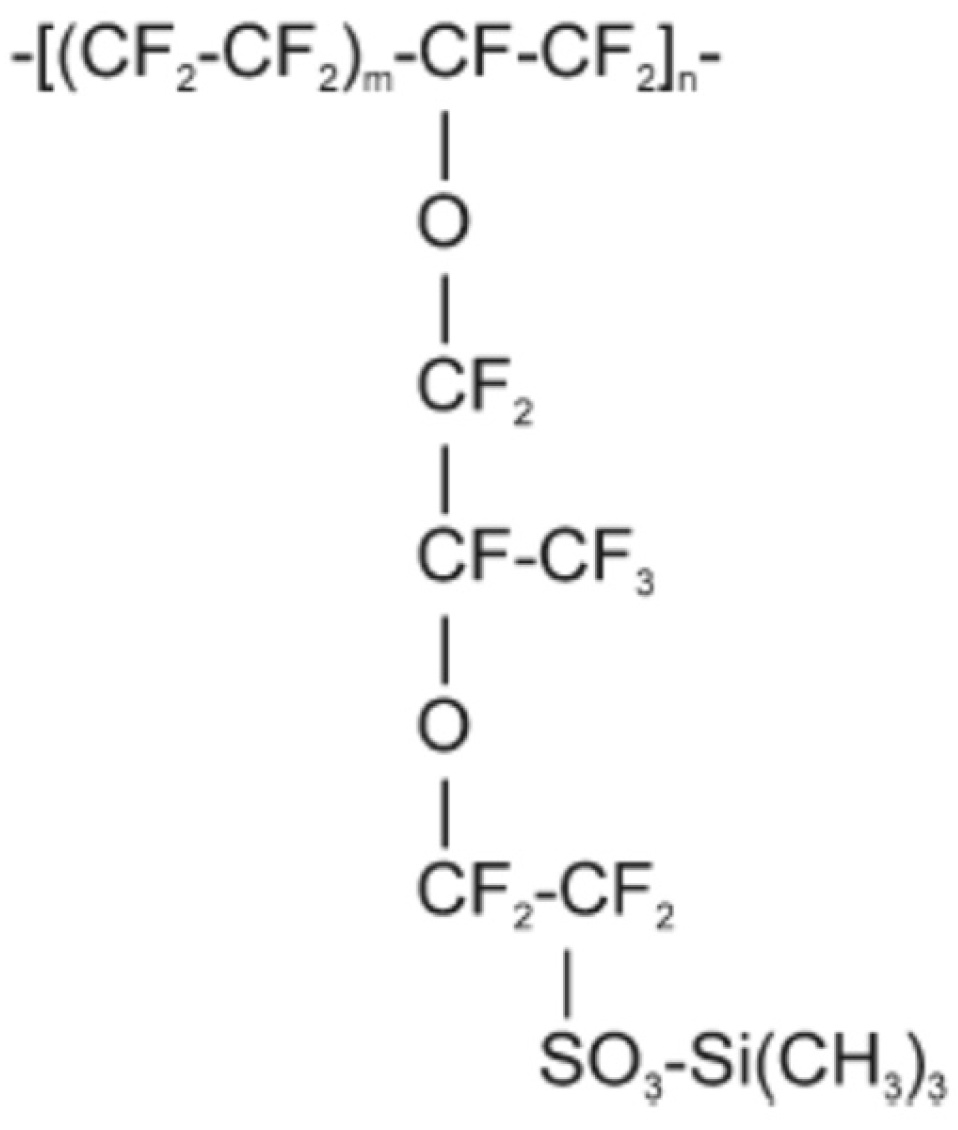
2.2. Nafion–TMS Dissolution Process and Preparation of Nafion–TMS–Ru2+ Complex Films

Previously, we have reported the dissolution of Nafion–TMS [28]. Accordingly, to prepare a 5% Nafion–TMS solution, the corresponding amount of clean solid polymer pellets was placed in a beaker containing 20 mL of a water–ethanol mixture (50:50), then transferred to a high-pressure reactor. The reactor was purged with ultrahigh-purity Ar and heated at 250 °C for 2 h. The obtained solution was transparent and slightly viscous. To prepare Nafion–TMS films, an aliquot of the polymeric solution was deposited onto an electrode or a glass substrate, and the solvent was left to evaporate naturally at room temperature.
On one hand, we aim to investigate the type of interaction between the polymer and the catalyst. On the other hand, we seek to incorporate the catalyst into the membrane at a controlled concentration. To achieve this, two methods of insertion were employed to incorporate the Ru2+ complexes (Ru(phen)3]2+ and [Ru(bpy)3]2+) into the Nafion–TMS polymer. Method 1 involved incorporating the Ru2+ complex through electrostatic and hydrophobic interactions by simply immersing the polymer films in a 1 mM Ru2+-complex solution. Method 2 involved a one-pot incorporation approach, following the same procedure used for the dissolution process of Nafion–TMS [28], but with the addition of the corresponding amount of solid Ru2+ complex to the high-pressure reactor to achieve a concentration of 1 mM. The insertion of the Ru2+ complex using method 1 was monitored by UV-vis spectroscopy.

2.3. Surface Characterization

Structural analysis of Nafion–TMS was performed by Atomic Force Microscopy (AFM) employing a Nanosurf Naio-AFM microscope (Nanosurf, Liestal, Switzerland) provided with silicon carbide tips. The measurements were conducted in contact mode. The measurements were performed in air, under a static force operating mode with a set point of 55 nN.

2.4. Spectroscopic Measurements

The mechanism for the insertion of the Ru2+ complex into the Nafion–TMS polymer was monitored using absorption spectroscopy. Surface bound UV-vis spectra were recorded employing a UV-Vis-NIR Cary Varian spectrophotometer using a wavelength from 300 to 800 nm. All measurements were carried out with a film at ambient conditions, employing the transmission technique. The measurements were performed under absorption mode.

2.5. Electrode Preparation and Electrochemical Measurements

To deposit Nafion–TMS or Nafion–TMS–Ru2+ complex, a glassy carbon (GC) surface was used as the substrate. Prior to deposition, the electrode was successively polished with diamond paste of 3 and 0.5 μm diameters. The polished electrode was then rinsed with acetone and thoroughly cleaned multiple times with ultrapure (18 MΩ) deionized water using an ultrasonic bath. After cleaning, the electrode was coated by depositing a 10 μL volume of the corresponding Nafion–TMS or Ru2+-complex modified Nafion–TMS–Ru2+ complex solution, followed by evaporation of the solvent at room temperature.
All electrochemical studies were performed with an Epsilon potentiostat–galvanostat from Bioanalytical Systems. The BASi-Epsilon (ver. 2.13.77) software was employed for control and data acquisition. All experiments were accomplished using a typical three electrode glass cell with GC, Nafion–TMS modified GC, or Nafion–TMS–Ru2+-complex modified GC as the working electrode. The counter electrode was a Pt coiled wire, and saturated Ag/AgCl functioned as the reference electrode.

3. Results and Discussion

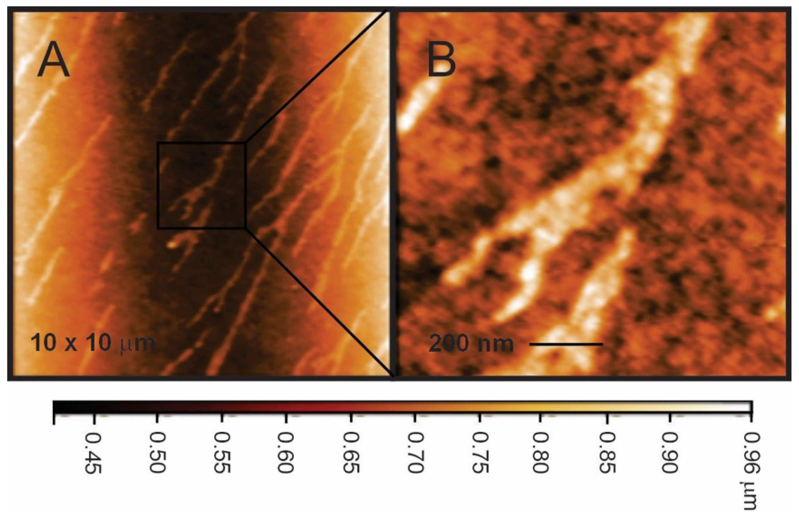
3.1. Surface Structure of Nafion–TMS

One of the merits of Nafion–TMS is its capability to incorporate certain amounts of catalysts through electrostatic and hydrophobic interactions [28]. This capability can be attributed to its structural properties, which are expected to be similar to those of its relative polymer, Nafion [29,30]. The chemical structure (Chart 1) of Nafion–TMS [26] features a Teflon-like backbone with an acidic side chain containing a trimethylsilyl group. Consequently, it is anticipated that the morphological structure of Nafion–TMS would be consistent with aggregated semi-crystalline regions (hydrophilic) combined with non-crystalline areas (hydrophobic). These components undergo phase separation, forming a network of hydrophilic channels embedded within a hydrophobic fluorocarbon matrix at a nanometer scale. Figure 1 shows representative AFM images of Nafion–TMS films deposited on glass substrates and dried in air for 24 h. The morphology of Nafion–TMS showed a bidimensional-type microstructure, in which the polar microphase, consisting of clustered ionic groups, forms long-branched, oriented channels approximately 80–150 nm wide, embedded in a continuous matrix. The contrast observed in the images can be attributed to differences in material stiffness; dark regions would correspond to “softer” areas, while bright regions would correspond to rich crystalline ionic domains. From the line fit (color intensity bar) at the bottom of Figure 1 and from the height profiles, it can be inferred that the height of the clusters are around 400 nm. Currently, we are conducting a detailed structural study of Nafion–TMS and the modified Nafion–TMS–Ru2+-complex polymer.

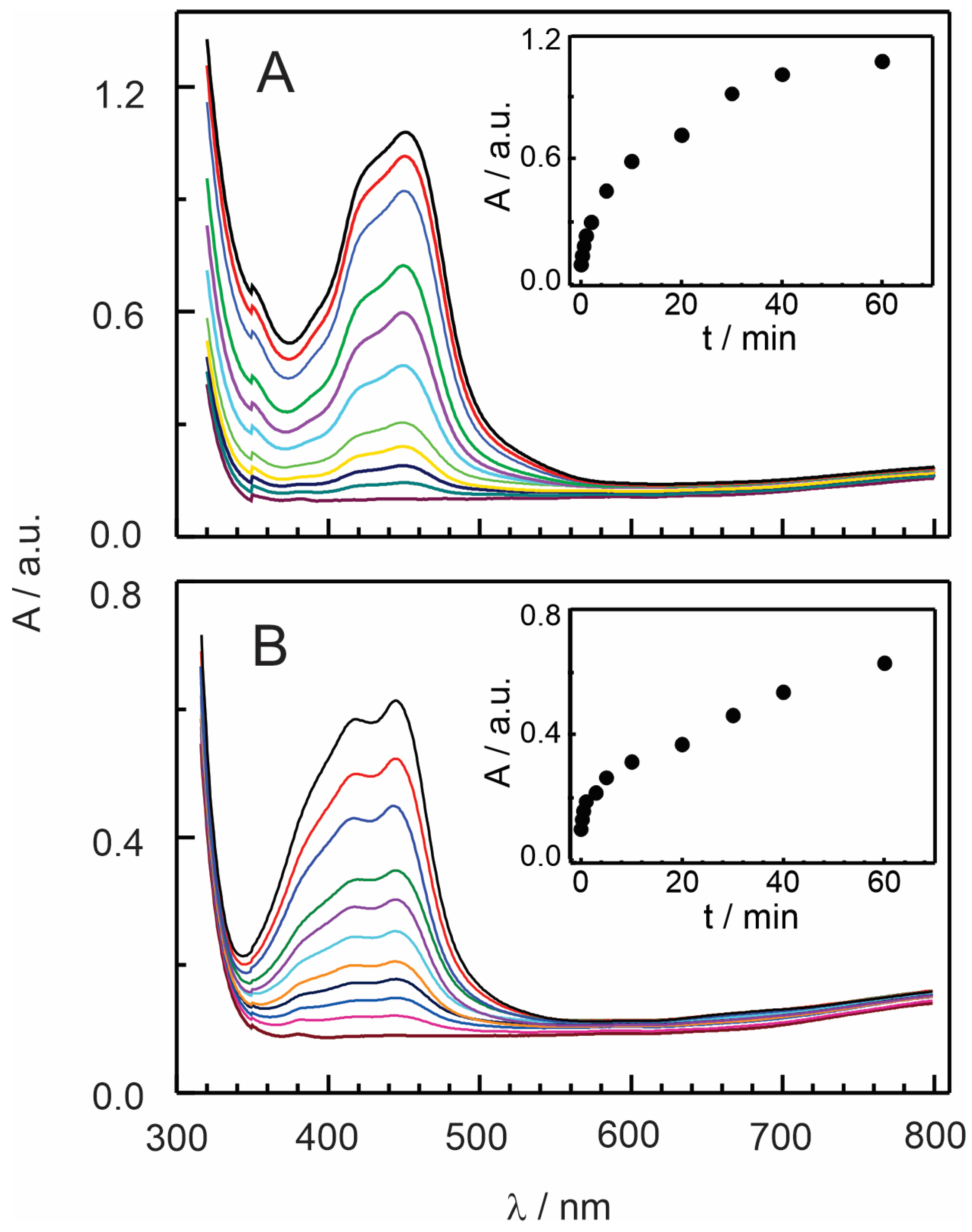
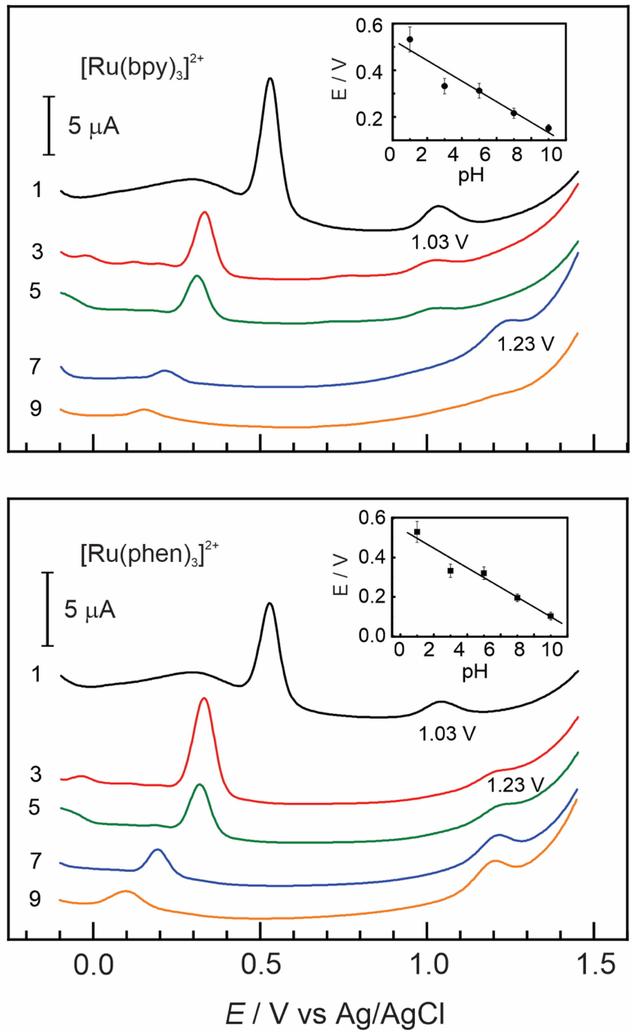
3.2. Insertion of [Ru(bpy)3]2+ and [Ru(phen)3]2+ Complexes in Nafion–TMS

The immobilization of Ru2+ complex into Nafion–TMS membranes was performed via a one-step method (see Experimental Section). To better understand the interactions and mechanisms involved in the immobilization process, we carried out a spectrophotometric study during the incorporation of [Ru(bpy)3]2+ and [Ru(phen)3]2+ complexes into Nafion–TMS polymer over time. Figure 2 shows the UV-vis spectra of [Ru(bpy)3]2+ and [Ru(phen)3]2+ while being incorporated into Nafion–TMS. The study was carried out with Nafion–TMS membranes in contact with a 1 mM Ru2+-complex aqueous solution for a specified time. The UV-vis spectra display a low-energy band positioned at approximately 455 nm which can be attributed to the metal-to-ligand charge transfer (MLCT) transition [31], as confirmed by comparing with the spectra of standard Ru complexes. As observed, the signal continuously increased with longer immersion times, indicating that the complex was being loaded into the film.
The incorporation of [Ru(bpy)3]2+ occurred significantly faster and in larger quantities compared to [Ru(phen)3]2+, as evidenced by a more rapid and higher absorbance response. This can be attributed to the faster diffusion of [Ru(bpy)3]2+ in comparison to [Ru(phen)3]2+ in the Nafion–TMS polymer, which correlates well with differences in their size, shape, and hydrophobicity [28,32,33,34]. The more hydrophobic ligand in the [Ru(phen)3]2+ complex leads to stronger interactions with the polymer, resulting in slower diffusion through the film. This strengths the assumption that the interactions within Nafion–TMS are both hydrophilic and hydrophobic.

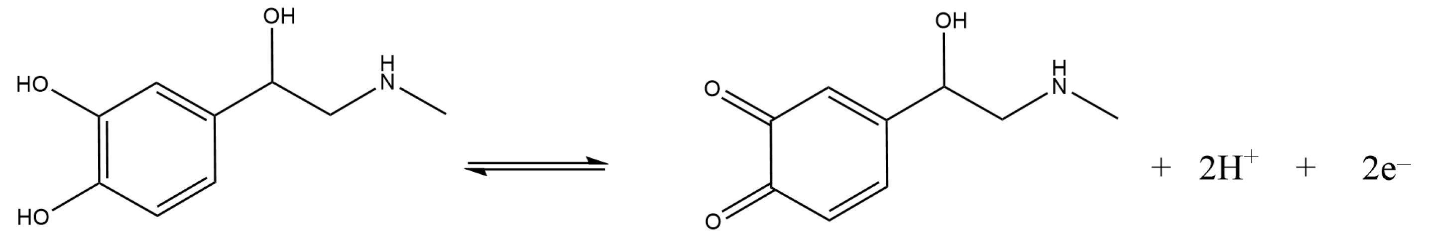
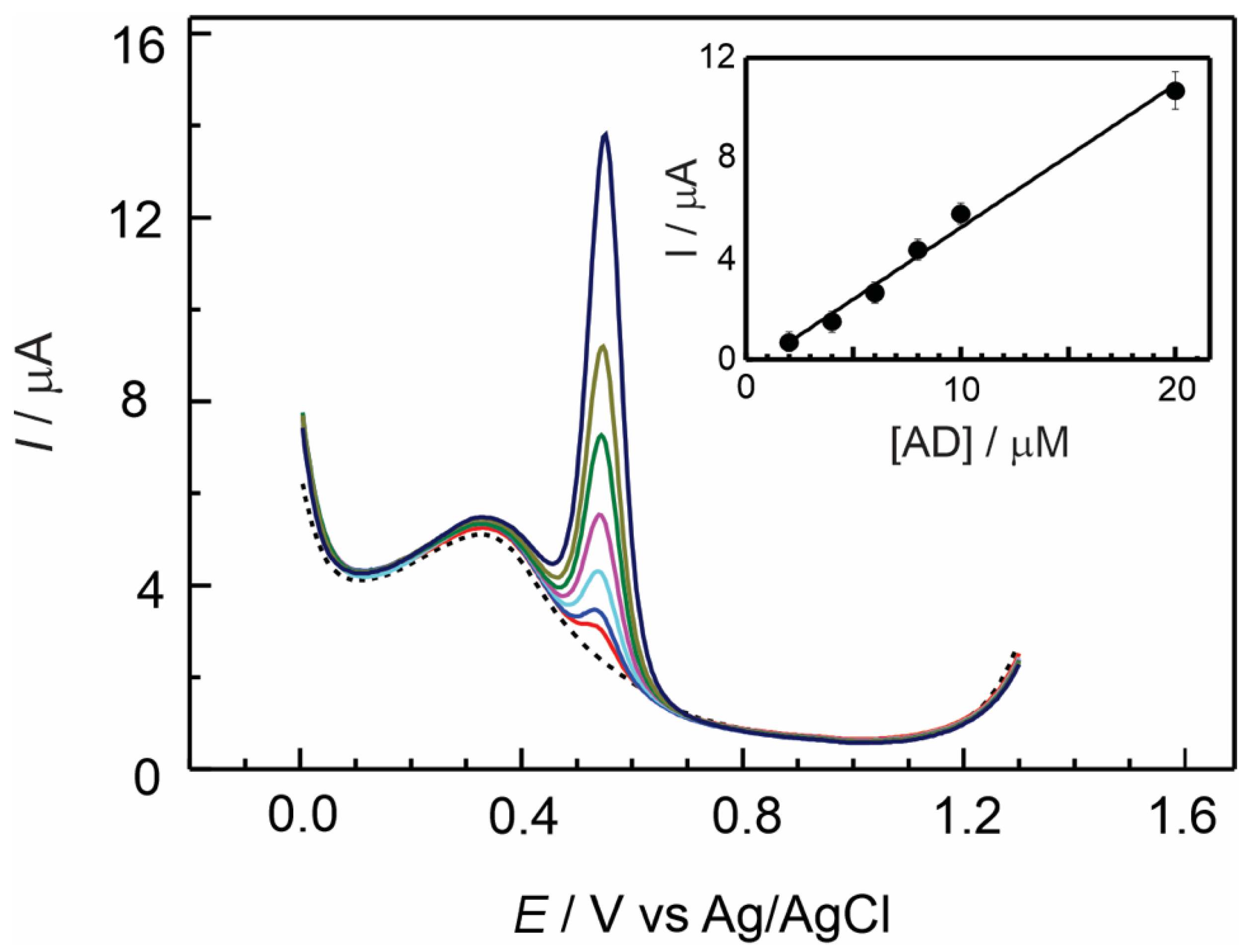
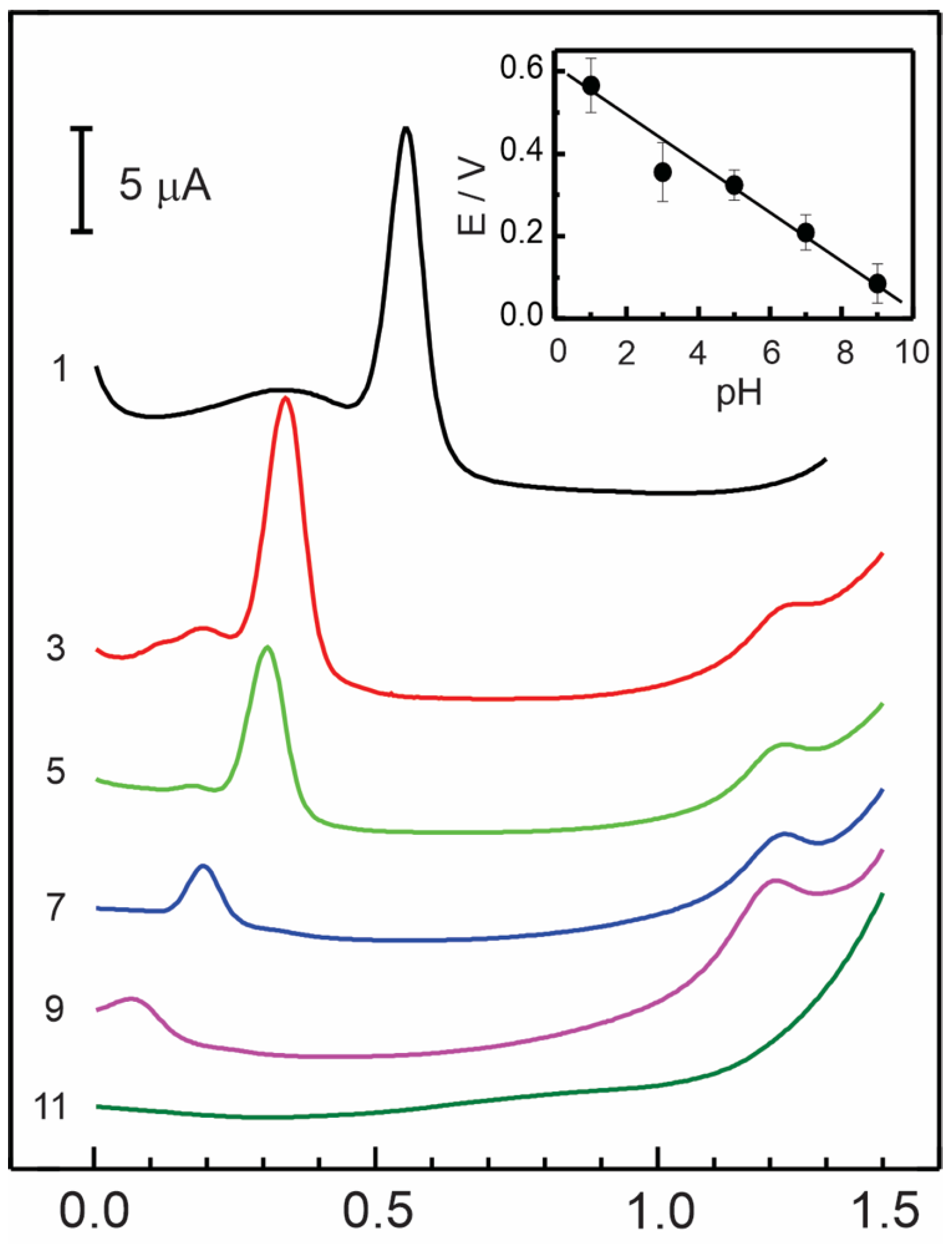
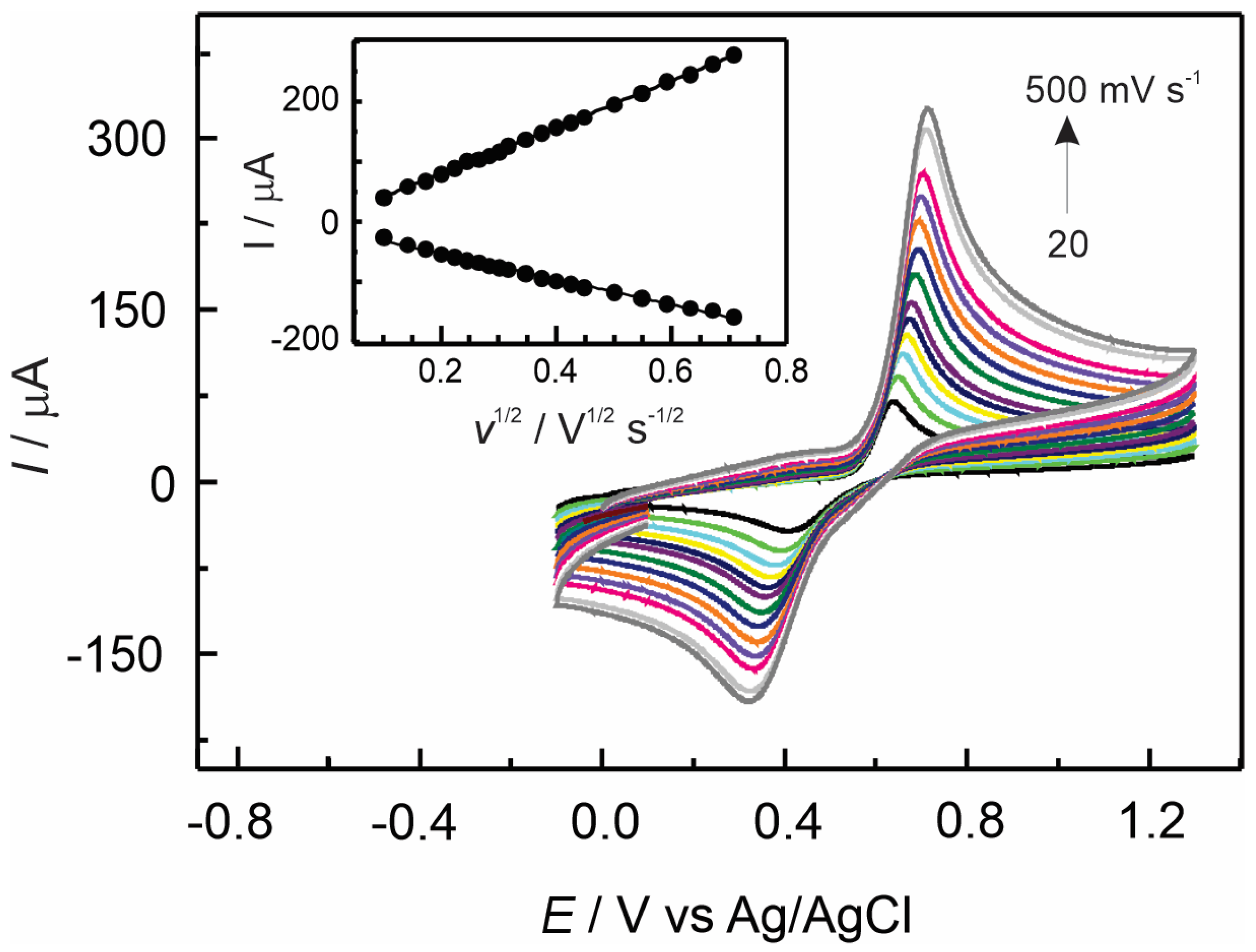
3.3. Electrochemical Detection of Adrenaline at GC/Nafion–TMS Modified Electrodes

The analytical properties of Nafion–TMS as an electrode modifier were studied through the electrooxidation reaction of adrenaline (AD), by measuring the peak current as correlated with the analyte concentration in standard solutions at several pH values [35].
Figure 3 presents a series of differential pulse voltammograms recorded at a Nafion–TMS modified GC electrode at micromolar concentrations of AD in 0.1 M sulfuric acid, within a potential window from 0 to 1.3 V versus the Ag/AgCl reference electrode. A sharp well-defined redox signal was observed at approximately 550 mV, which can be attributed to the redox reaction shown in Reaction 1. The electrode reaction follows a two-electron process, accompanied by the transfer of two protons, and is highly dependent on the solution pH. The inset of Figure 3, presents the current linear response of AD with increasing concentration in the range from 2 to 20 micromolar, yielding a sensitivity of 0.57 ± 0.027 μA/μM. The analytical parameters are reported in Table 1.

Effect of pH

It is widely recognized that electrochemical characteristics of catecholamines are highly dependent on pH. The effect of pH on the electrochemical detection of AD was examined across a range of pH values ranging from 1 to 11. Figure 4 shows differential pulse voltammograms registered for 2 × 10⁻⁵ mol L−1 AD at several pH values using a Nafion–TMS-modified GC electrode. The peak potential of adrenaline oxidation presents a linear dependence with pH. A negative shift in the oxidation peak potential occurred with increasing pH, following a Nernstian behavior as evidenced by the slope of the linear correlation shown in the inset of Figure 4. This slope was −56 mV/pH-unit, which is nearly identical to the ideal Nernstian behavior (−59 mV/pH), and similar to the performance registered for dopamine at several pH values [28]. Usually, this phenomenon is due to pure thermodynamic conditions, but it may also indicate that the electron transfer reaction occurs in tandem with proton transfer and depends on proton concentration.
In the pH interval from 3 to 9, a small oxidation peak was observed at around 1.22 V, with its current increasing as the pH value rose. Above pH 3, the presence of the unprotonated form of adrenaline quinone allows for the cyclization reaction of adrenaline, leading to the formation of adrenochrome [36]. Under strongly acidic (pH 1) or basic (pH 11) conditions, the oxidation signal at 1.22 V completely disappeared. Additionally, the mean oxidation peak of adrenaline is not observed at high pH values; this is understandable since adrenaline tends to be totally oxidized in aqueous solution at a pH higher than 10 [37]. From the above results, the variability in the voltammetric signal with pH for adrenaline sensing provides valuable insights into the analytical characteristics of the Nafion–TMS membrane and its potential applications in real-world scenarios, such as pharmaceutical products or biological samples. For example, under physiological conditions (pH 7.4), the signal is well-defined and appears at low overpotentials, but exhibits a low current. In contrast, at pH 3, in a mildly acidic environment, the signal is stronger, but the overpotential is higher than at physiological pH. This study is useful when considering the analytical conditions for adrenaline sensing across several experimental environments.
The effect of scan rate on adrenaline oxidation was studied by cyclic voltammetry using a Nafion–TMS modified GC electrode at a concentration of 2 × 10⁻⁴ mol L−1 AD in 0.1 M H2SO4. As shown in Figure 5, both the anodic and cathodic peak currents rise linearly with square root of the scan rate (v1/2), indicating that diffusion is the dominant transport mechanism through the layer. By applying the Randles–Sevcik equation (Equation (1)) [38] and analyzing the slope of the inset shown in Figure 5, the diffusion coefficient was calculated to be 7.9 (± 0.5) × 10−5 cm2 s−1, as reported in Table 1.
I p = 2.69 × 10 5 A D 1 / 2 n 3 / 2 C v 1 / 2
In this equation, A is the electrode area, D is the diffusion coefficient of the analyte, n is the number of electrons involved in the redox reaction, C is the bulk concentration of the analyte, and v is the scan rate used in cyclic voltammetry. The results suggest that the electrochemical behavior of AD is predominantly a diffusion-controlled process on Nafion–TMS-modified electrodes [39,40].

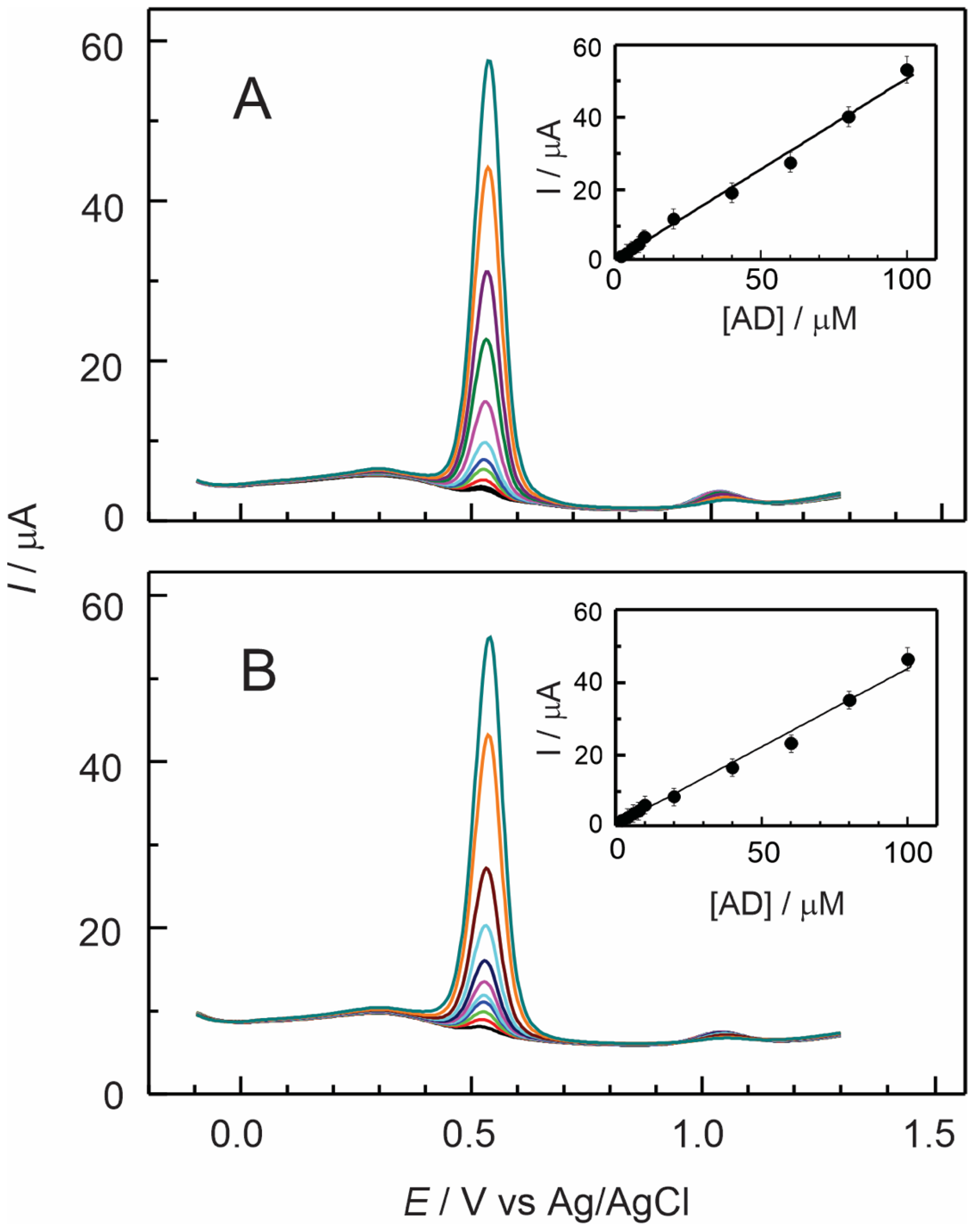
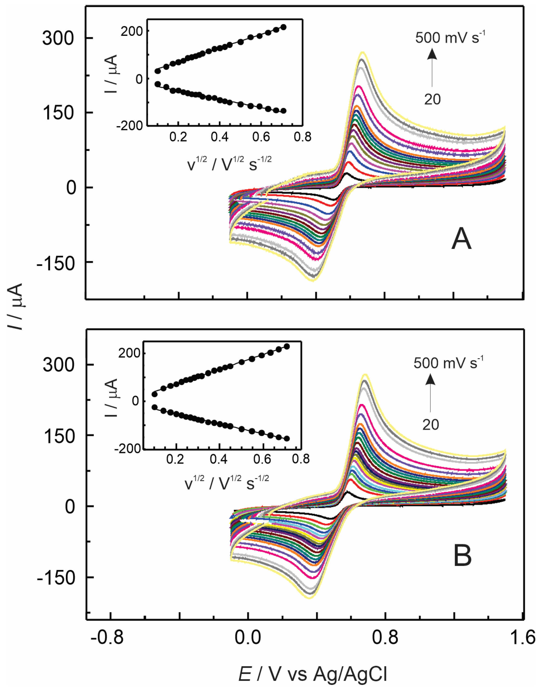
3.4. Electrochemical Detection of Adrenaline Using Nafion–TMS–Ru2+-Complex Modified GC Electrodes

By combining the properties of polymers with the electrocatalytic activity of the Ru2+ complex, composite membranes help to improve the rate and mechanism of electrochemical reactions while improving electrode surfaces for the development of efficient electrochemical sensors. The analytical performance of Nafion–TMS–Ru2+ complex-modified GC electrodes was assessed by measuring the peak current as a function of adrenaline concentration in standard solutions. Figure 6A,B presents the first scan of differential pulse voltammograms recorded at Nafion–TMS–[Ru(bpy)3]2+ and Nafion–TMS–[Ru(phen)3]2+-modified GC electrodes, respectively, for several adrenaline concentrations ranging from 2 × 10⁻⁶ to 1 × 10⁻⁴ mol L−1 in 0.1 M H2SO4. An oxidation signal was observed at approximately 0.53 V for both Ru2+-complex modified electrodes, corresponding to the electrooxidation of adrenaline. The large oxidation currents recorded on these electrodes confirm the enhanced electrocatalytic activity of Ru2+ complexes toward adrenaline oxidation. While the sensitivity and detection limit were similar to those for Nafion–TMS electrodes (Table 1), the Nafion–TMS–Ru2+ complex-modified electrodes exhibited a more distinct and better-defined peak, along with a significant improvement in linear dependence over a broader concentration range. These characteristics can be attributed to the fast electron transfer [41] mediated by Ru, as also observed in cyclic voltammetry (see below).
The analytical properties of Nafion–TMS and Ru2+ complex-modified Nafion–TMS, compared to other modified electrodes, are summarized in Table 2. The achieved features, such as low detection limit and high sensitivity, suggest its potential for further exploration as a sensor for real sample analysis, including pharmaceutical products and biological fluids [42,43]. In addition, these polymer composite membranes could find wider applicability and possible use in ion transport membranes. Both of these applications represent other relevant aspects that are worth investigating.

Effect of pH on Nafion–TMS–Ru2+ Complex

The oxidation potential values, as well as the peak current, were found to largely depend on the pH of the electrolyte. Figure 7 shows the electrochemical responses measured at different pH values using Nafion–TMS–[Ru(bpy)3]2+ and Nafion–TMS–[Ru(phen)3]2+ modified GC electrodes in the presence of 2 × 10⁻⁵ M adrenaline. For both electrodes, the current for adrenaline oxidation decreases, while the peak potential displaces to more negative values. Additionally, the peak potential presents a quasi-linear dependence with increasing pH (insets of Figure 7) of the solution, as shown in the insets of Figure 7. Although the plots of potential versus pH for both electrodes show a small deviation from the thermodynamic Nernstian behavior, the corresponding slope values of −44 mV/pH-unit and −49 mV/pH-unit, respectively, suggest a proton-dependent rate limiting process. Notably, at pH 3, the adrenaline oxidation potential shifts to more negative values than the expected for a Nernstian dependence for both Nafion–TMS and Nafion–TMS–Ru2+ complex-modified electrodes (insets of Figure 4 and Figure 7). This observation indicates that at this pH, the oxidation reaction is significantly influenced by proton gradients [6] and the energy required for the oxidation process is slightly improved in a mildly acidic environment. Apparently, the coupling of the electro-oxidation reaction to proton gradients in the bulk solution decreases the electrochemical potential required for the process. This occurs particularly at a gradient concentration where the liberation of phenolic protons is facilitated, which seems to be approximately at pH 3. The autooxidation of adrenaline and the formation of its cyclization product, related to the amino group, proceeds slowly at this pH [44], as can be inferred from the low current signal at 1.23 V for all modified electrodes. This behavior aligns well with the chemical stability of adrenaline, which is optimal between 3 and 4 [45].
At pH 1, the redox signal corresponding to the Ru2+ to Ru3+ oxidation reaction is observed at around 1.03 V for both Ru-modified electrodes (Figure 7). For the [Ru(bpy)3]2+ electrode, the current of this signal decreases as pH increases, and remains detectable up to pH 5, whereas it disappears for the [Ru(phen)3]2+ modified electrode. At the same time, the autooxidation signal of adrenaline at 1.23 V, linked to the unprotonated form of adrenaline quinone and the formation of its corresponding cyclization product [46], is absent between pH 1 and 5 for the [Ru(bpy)3]2+ electrode. However, for the [Ru(phen)3]2+ electrode, the cyclization wave of adrenaline starts to emerge from pH 3. This suggests that energy transfer from [Ru(phen)3]2+ to adrenaline and its subsequent oxidation product is more efficient than in the [Ru(bpy)3]2+ system [47], facilitating the oxidation reaction of the adrenochrome. This is reflected in the slightly higher current observed for both oxidation signals in [Ru(phen)3]2+. Since the phenantroline ligand is more basic than the bipyridine ligand at room temperature [48], it facilitates the redox reactions of adrenaline and its cyclization product, by a mechanism of a proton acceptor. In contrast, bipyridine causes sluggish cyclization of adrenaline in mildly acidic media through a pH-controlled cross-linking mechanism between the oxidation product of adrenaline and [Ru(bpy)3]2+. Cross-linking between catechols and metal ions can significantly enhance mechanical strength and stability through pH control. This represents a promising approach for the formation of self-healing polymer composite networks [49]. However, this topic is beyond the scope of the current work, and we plan to explore it further. We aim to explore the possibility of maintaining the activity of the composite Nafion–TMS–Ru2+ complex-modified polymer at acidic pH. The above results are significant to find out the best analytical conditions for the electrochemical sensing of biologically significant catecholamines.
The nature of the electrochemical oxidation of adrenaline on Nafion–TMS–Ru2+-complex GC electrodes in H2SO4 was studied by cyclic voltammetry at different scan rates. It is worthwhile to note that for Nafion–TMS electrodes, the potential difference between oxidation and reduction signals obtained by cyclic voltammetry is far (approximately 312 mV, Figure 5) from expected Nernst reversible behavior. However, for the Ru2+-complex modified electrodes, this difference is considerably smaller (see Table 1), indicating a faster electron transfer and greatly different kinetic parameters for the redox reaction.
For both Ru2+-complex-modified electrodes, both the cathodic and anodic currents followed a linear relationship with the square root of scan rate within an interval from 10 to 500 mV s−1 at a constant adrenaline concentration of 2 × 10⁻⁴ mol L−1. This behavior is consistent with a process that is limited by the molecular diffusion of the reactant from the solution in bulk to the electrode surface. The apparent diffusion coefficient of adrenaline through Nafion–TMS–Ru2+-complex films was estimated from the peak current of the voltammograms using the Randles–Sevcik equation [38]. It is worthwhile to note that the diffusion coefficient of adrenaline obtained with Nafion–TMS–Ru2+ complex is of the same order of magnitude as that found for Nafion–TMS-modified GC electrodes. It is clear that the presence of cationic species does not hinder the diffusion of cationic adrenaline [50], despite the high possibility of electrostatic repulsion. This suggests charge-hopping, mediated by Ru2+-complex redox species. Electron transfer from adrenaline to the electrode seems to be particularly promoted by the Ru2+-complexes, possibly due to the delocalization of pi electrons from the ligands. As expected, these results indicate that the diffusion of adrenaline is slightly slower in the presence of the more voluminous [Ru(phen)3]2+ ion compared to [Ru(bpy)3]2+. However, its motion inside the film remains sufficiently fast to enhance adrenaline charge transfer relative to the Nafion–TMS-modified GC electrode. Table 1 reports the potential differences between anodic and cathodic peaks (ΔE values) for all modified electrodes, obtained from Figure 5 and Figure 8 at a scan rate of 100 mV s−1. This suggests that the diffusion mechanism through the film involves both hydrophobic and electrostatic interactions. It can be concluded that the electron transfer process of adrenaline electrooxidation is accelerated by the Ru2+ complex-modified Nafion–TMS polymer while maintaining Nernstian behavior across various pH values.
The obtained results raise several open questions that remain. Currently, we are investigating the influence of catalyst concentration on the analytical performance and the electron transfer mechanism of the composite Nafion–TMS–Ru2+ complex membranes. Additionally, the impact of interfering substances on adrenaline detection and the potential use of the sensor in biological samples are being studied using electroanalytical techniques such as impedance spectroscopy, quartz crystal microbalance, chronoamperometry, and rotating disk electrode measurements.

4. Conclusions

The electrodes modified with Nafion–TMS and Nafion–TMS–Ru2+ complex exhibited high sensitivity for adrenaline detection. The incorporation of Ru2+ complexes into Nafion–TMS membranes significantly improves the electron transfer during the redox reaction of adrenaline, likely due to a charge-hopping mechanism facilitated by Ru redox species, as demonstrated by the reduced difference between oxidation and reduction potential values. The strong effect of pH in the redox reaction of adrenaline offers a unique opportunity to fine-tune the catalytic and analytical properties of polymer/Ru2+-complex composites by simply adjusting the pH. Electrodes modified with Nafion–TMS and the Nafion–TMS–Ru2+ complexes maintained a Nernstian behavior at various pH values, indicating stable and predictable electrochemical performance. In basic environments, the predominance of the adrenaline cyclization reaction correlated well with the acid-base properties of the Ru2+-complex ligands. The structural properties of Nafion–TMS are comparable to those of the more commercial and expensive Nafion® polymer. Hydrophobic and hydrophilic interactions play a significant role in the ionic properties of Nafion–TMS, as evidenced by its ability to insert different ions into its structure. Given the limited availability of commercial Nafion® polymer solutions from usual suppliers, Nafion–TMS represents an excellent alternative for applications in electrochemical sensors.

Author Contributions

Conceptualization, R.A.-S.; methodology, R.A.-S., S.L.C.-H. and D.A.D.-T.; validation, S.L.C.-H.; formal analysis, R.A.-S. and D.A.D.-T. investigation, R.A.-S.; writing original—review and editing, R.A.-S., S.L.C.-H. and J.L.G.-M.; project administration, R.A.-S. and J.L.G.-M.; funding acquisition, R.A.-S. All authors have read and agreed to the published version of the manuscript.

Funding

The authors acknowledge VIEP-BUAP for the financial support of the project VIEP 2025 (532-PV/2025). This work was funded by CONAHCyT-México through the projects 243030 and 104361. CONAHCyT Mexico provided the Research Fellowship “Estancia Sabática al Extranjero 2024-Reference 121841”.

Institutional Review Board Statement

Not applicable.

Informed Consent Statement

Not applicable.

Data Availability Statement

Data are contained within the article.

Acknowledgments

The authors acknowledge the Instituto de Física BUAP for providing access the UV-Vis-NIR Cary Varian spectrophotometer for spectroscopic measurements.

Conflicts of Interest

The authors declare no conflicts of interest.

References

  1. Schweigert, N.; Zehnder, A.J.B.; Eggen, R.I.L. Chemical properties of catechols and their molecular modes of toxic action in cells, from microorganisms to mammals. Environ. Microbiol. 2001, 3, 81–91. [Google Scholar] [PubMed]
  2. Su, Y.; Wang, J.; Chen, G.N. Determination of epinephrine based on its enhancement for electrochemiluminescence of lucigenin. Talanta 2005, 65, 531–536. [Google Scholar] [PubMed]
  3. Michalowski, J.; Halabura, P. Flow-injection chemiluminescence determination of epinephrine in pharmaceutical preparations using raw apple juice as enzyme source. Talanta 2001, 55, 1165–1171. [Google Scholar]
  4. Yadav, T.; Sahu, R.; Mukherjee, V. Molecular modeling and spectroscopic investigation of a neurotransmitter: Epinephrine. J. Mol. Struct. 2019, 1176, 94–109. [Google Scholar]
  5. Solich, P.; Polydorou, C.K.; Koupparis, M.A.; Efstathiou, C.E. Automated flow injection spectrophotometric determination of catecholamines (epinephrine and isoproterenol) in pharmaceutical formulations based on ferrous complex formation. J. Pharm. Biomed. 2000, 22, 781–789. [Google Scholar]
  6. Adeniyi, W.K.; Wright, A.R. Novel fluorometric assay of trace analysis of epinephrine in human serum. Spectrochim. Acta Part A 2009, 74, 1001–1004. [Google Scholar]
  7. Zhang, Y.; Ren, W.; Fan, Y.Z.; Dong, J.X.; Zhang, H.; Luo, H.Q.; Li, N.B. Label-free fluorescent discrimination and detection of epinephrine and dopamine based on bioinspired in situ copolymers and excitation wavelength switch. Anal. Chim. Acta 2019, 1054, 167–175. [Google Scholar]
  8. Fotopoulou, M.A.; Ioannou, P.C. Post-column terbium complexation and sensitized fluorescence detection for the determination of norepinephrine, epinephrine and dopamine using high-performance liquid chromatography. Anal. Chim. Acta. 2002, 462, 179–185. [Google Scholar]
  9. Özel, R.E.; Hayat, A.; Andreescu, S. Recent developments in electrochemical sensors for the detection of neurotransmitters for applications in biomedicine. Anal. Lett. 2015, 48, 1044–1069. [Google Scholar]
  10. Madhurantakama, S.; Babua, K.J.; Rayappana, J.B.B.; Krishnan, U.M. Nanotechnology-based electrochemical detection strategies for hypertension markers. Biosen. Bioelectron. 2018, 116, 67–80. [Google Scholar]
  11. Tadi, K.K.; Motghare, R.V.; Ganesh, V. Electrochemical detection of epinephrine using a biomimic made up of hemin modified molecularly imprinted microspheres. RSC Adv. 2015, 5, 99115–99124. [Google Scholar]
  12. Zare, H.R.; Ghanbari, Z.; Nasirizadeh, N.; Benvidi, A. Simultaneous determination of adrenaline, uric acid, and cysteine using bifunctional electrocatalyst of ruthenium oxide nanoparticles. Comptes Rendus Chim. 2013, 16, 287–295. [Google Scholar]
  13. Charithra, M.M.; Manjunatha, J.G. Electrochemical sensing of adrenaline using surface modified carbon nanotube paste electrode. Mater. Chem. Phys. 2021, 262, 124293. [Google Scholar] [CrossRef]
  14. Canevaria, T.C.; Nakamura, M.; Cincotto, F.H.; de Melo, F.M.; Toma, H.E. High performance electrochemical sensors for dopamine and epinephrine using nanocrystalline carbon quantum dots obtained under controlled chronoamperometric conditions. Electrochim. Acta 2016, 209, 464–470. [Google Scholar]
  15. Liu, X.; Ji, H.; Tang, J.; Tao, F.; Zhang, X.; Yao, Z.; Song, H.; Li, C.; Wang, F. Electrochemical activation and renewal of pyrrole nitrogen sites in porphyrin-based conjugated polymer for simultaneous determination of uric acid and adrenaline. J. Electroanal. Chem. 2021, 884, 115055. [Google Scholar] [CrossRef]
  16. Feng, Z.; Zhou, J.; Xi, Y.; Lan, B.; Guo, H.; Chen, H.; Zhang, Q.; Lin, Z. Solid-state hybrid photovoltaic cells with a novel redox polymer and nanostructured inorganic semiconductors. J. Power Sources 2009, 194, 1142–1149. [Google Scholar] [CrossRef]
  17. Thirunavukkarasu, V.S.; Kozhushkov, S.I.; Ackermann, L. C–H nitrogenation and oxygenation by ruthenium catalysis. Chem. Commun. 2014, 50, 29–39. [Google Scholar] [CrossRef]
  18. Cai, M.; Loague, Q.R.; Zhu, J.; Lin, S.; Usov, P.M.; Morris, A.J. Ruthenium (II)-polypyridyl doped zirconium (IV) metal–organic frameworks for solid-state electrochemiluminescence. Dalton Trans. 2018, 47, 16807–16812. [Google Scholar]
  19. Sheth, S.; Li, M.; Song, Q. New luminescent probe for the selective detection of dopamine based on in situ prepared Ru(II) complex-sodium dodecyl benzyl sulfonate assembly. J. Photochem. Photobiol. A Chem. 2019, 371, 128–135. [Google Scholar] [CrossRef]
  20. Tian, L.; Wang, X.; Wu, K.; Hu, Y.; Wang, Y.; Lu, J. Ultrasensitive electrochemiluminescence biosensor for dopamine based on ZnSe, graphene oxide@multi walled carbon nanotube and Ru(bpy)32+. Sens. Actuators B Chem. 2019, 286, 266–271. [Google Scholar] [CrossRef]
  21. Gao, Q.; Song, B.; Ye, Z.; Yang, L.; Liu, R.; Yuan, J. A highly selective phosphorescence probe for histidine in living bodies. Dalton Trans. 2015, 44, 18671–18676. [Google Scholar] [CrossRef] [PubMed]
  22. Turkušić, E.; Redžić, S.; Kahrović, E.; Zahirović, A. Electrochemical Determination of Adrenaline at Ru(III) Schiff Base Complex Modified Carbon Electrodes. Croat. Chem. Acta 2017, 90, 345–352. [Google Scholar]
  23. Ruifang, W.S.; Jiao, G.K. Electrochemistry and Electrocatalysis of Hemoglobin in Nafion/nano-CaCO3 Film on a New Ionic Liquid BPPF6 Modified Carbon Paste Electrode. J. Phys. Chem. B 2007, 111, 4560–4567. [Google Scholar]
  24. Toh, R.J.; Peng, W.K.; Han, J.; Pumera, M. Direct In Vivo Electrochemical Detection of Haemoglobin in Red Blood Cells. Sci. Rep. 2014, 4, 6209. [Google Scholar] [CrossRef] [PubMed]
  25. Mauritz, K.A.; Payne, J.T. [Perfluorosulfonate ionomer]/silicate hybrid membranes via base-catalyzed in situ sol–gel processes for tetraethylorthosilicate. J. Membr. Sci. 2000, 168, 39–51. [Google Scholar]
  26. Murata, S.; Noyori, R. Silylation with a perfluorinated resinsulfonic acid trimethylsilyl ester. Tetrahedron Lett. 1980, 21, 767–768. [Google Scholar]
  27. Carlson, R.; Gautun, H.; Westelund, A. A convenient procedure for the synthesis of acetals from α-halo ketones. Adv. Synth. Catal. 2002, 344, 57–60. [Google Scholar] [CrossRef]
  28. Aguilar-Sánchez, R.; Díaz-Caballeros, R.J.; Méndez-Bermúdez, J.A.; Gárate-Morales, J.L.; Domínguez-Meneses, G. Electrochemical studies of nafion–trimethylsilyl and nafion–trimethylsilyl/Ru complex-modified electrodes. J. Solid State Electrochem. 2012, 16, 2867–2876. [Google Scholar]
  29. Yeager, H.L.; Steck, A. Cation and water diffusion in Nafion ion-exchange membranes: Influence of polymer structure. J. Electrochem. Soc. 1981, 128, 1880–1884. [Google Scholar]
  30. Hsu, W.Y.; Gierke, T.D. Ion transport and clustering in nafion perfluorinated membranes. J. Membr. Sci. 1983, 13, 307–326. [Google Scholar]
  31. Juris, A.; Balzani, V.; Barigelletti, F.; Campagna, S.; Belser, P.; von Zelewsky, A. Ru(II) polypyridine complexes: Photophysics, photochemistry, eletrochemistry, and chemiluminescence. Coord. Chem. Rev. 1988, 84, 85–277. [Google Scholar]
  32. Barton, J.K.; Goldberg, J.M.; Kumar, C.V.; Turro, N.J. Binding modes and base specificity of tris (phenanroline) ruthenium (II) enantiomers with nucleic acids: Tuning the stereoselectivity. J. Am. Chem. Soc. 1986, 108, 2081–2088. [Google Scholar]
  33. Yonemoto, E.H.; Saupe, G.B.; Schmeh, R.H.; Hubig, S.M.; Riley, R.L.; Iverson, B.L.; Mallouk, T.E. Electron-Transfer Reactions of Ruthenium Trisbipyridyl-Viologen Donor-Acceptor Molecules: Comparison of the Distance Dependence of Electron Transfer-Rates in the Normal and Marcus Inverted Regions. J. Am. Chem. Soc. 1994, 116, 4786–4795. [Google Scholar]
  34. Jenkins, Y.; Friedman, A.E.; Turro, N.J.; Barton, J.K. Characterization of dipyridophenazine complexes of ruthenium(II): The light switch effect as a function of nucleic acid sequence and conformation. Biochemistry 1992, 31, 10809–10816. [Google Scholar] [PubMed]
  35. Aguilar Sánchez, R.; Durán Tlachino, D.A.; Cabrera Hilerio, S.L.; Gárate Morales, J.L.; Cerna Cortez, J.R. Electrochemical detection of adrenaline employing nafion-tms modified electrodes. Tend. Doc. Inv. Quim. 2018, 4, 421–425. [Google Scholar]
  36. De Pelichy, L.G.; Smith, E.T. A study of the oxidation pathway of adrenaline by cyclic voltammetry: An undergraduate analytical chemistry laboratory exercise. Chem. Educ. 1997, 2, 1–13. [Google Scholar]
  37. Bretti, C.; Cigala, R.M.; Crea, F.; de Stefano, C.; Vianelli, G. Solubility and modeling acid–base properties of adrenaline in NaCl aqueous solutions at different ionic strengths and temperatures. Eur. J. Pharmac. Sci. 2015, 78, 37–46. [Google Scholar]
  38. Bard, A.J.; Fundamentals, L.R. Electrochemical Methods: Fundamentals and Applications, 2nd ed.; Wiley: New York, NY, USA, 1980; p. 218. [Google Scholar]
  39. Cui, F.; Zhang, X. Electrochemical sensor for epinephrine based on a glassy carbon electrode modified with graphene/gold nanocomposites. J. Electroanal. Chem. 2012, 669, 35–41. [Google Scholar]
  40. Li, X.; Chen, M.; Ma, X. Selective determination of epinephrine in the presence of ascorbic acid using a glassy carbon electrode modified with graphene. Anal. Sci. 2012, 28, 147–151. [Google Scholar]
  41. Zhang, C.; Wang, G.; Liu, M.; Feng, Y.; Zhang, Z.; Fang, B. Hydroxylamine electrochemical sensor based on electrodeposition of porous ZnO nanofilms onto carbon nanotubes films modified electrode. Electrochim. Acta 2010, 55, 2835–2840. [Google Scholar]
  42. Raoof, J.B.; Ojani, R.; Baghayeri, M. A selective sensor based on a glassy carbon electrode modified with carbon nanotubes and ruthenium oxide/hexacyanoferrate film for simultaneous determination of ascorbic acid, epinephrine and uric acid. Anal. Meth. 2011, 3, 2367–2373. [Google Scholar]
  43. Tsele, T.P.; Adekunle, A.S.; Fayemi, O.E.; Ebenso, E.E. Electrochemical detection of Epinephrine using Polyaniline nanocomposite films doped with TiO2 and RuO2 Nanoparticles on Multi-walled Carbon Nanotube. Electrochim. Acta 2017, 243, 331–348. [Google Scholar]
  44. Volkov, V.; Lobanov, A.; Voronkov, M.; Baygildiev, T.; Misin, V.; Tsivileva, O. Kinetics and Mechanism of Epinephrine Autoxidation in the Presence of Plant Superoxide Inhibitors: A New Look at the Methodology of Using a Model System in Biological and Chemical Research. Antioxidants 2023, 12, 1530. [Google Scholar] [CrossRef] [PubMed]
  45. Guzov, E.A.; Kazin, V.N.; Moshareva, V.A.; Zhukova, A.A. Application of electronic spectroscopy and quantum-chemical modelling for analysis of products of autoxidation of adrenaline. J. Appl. Spectros. 2019, 85, 1107–1113. [Google Scholar]
  46. Umek, N. Cyclization step of noradrenaline and adrenaline autoxidation: A quantum chemical study. RSC Adv. 2020, 10, 16650. [Google Scholar]
  47. Li, F.; Cui, H.; Lin, X.-Q. Determination of adrenaline by using inhibited Ru(bpy)32+ electrochemiluminescence. Anal. Chim. Acta 2002, 471, 187–194. [Google Scholar]
  48. Bretti, C.; Crea, F.; De Stefano, C.; Sammartano, S. Solubility and activity coefficients of 2,2-bipyridyl, 1,10-phenanthroline and 2,2′,6′,2″-terpyridine in NaCl(aq) at different ionic strengths and T = 298.15 K. Fluid Phase Equilibria 2008, 272, 47–52. [Google Scholar]
  49. Holten-Andersen, N.; Harrington, M.J.; Birkedal, H.; Lee, B.P.; Messersmith, P.B.; Lee, K.Y.C.; Waite, J.H. pH-induced metal-ligand cross-links inspired by mussel yield self-healing polymer networks with near-covalent elastic moduli. Proc. Nat. Acad. Sci. USA 2011, 108, 2651–2655. [Google Scholar]
  50. Álvarez-Diduk, R.; Galano, A. Adrenaline and Noradrenaline: Protectors against Oxidative Stress or Molecular Targets? J. Phys. Chem. B 2015, 119, 3479–3491. [Google Scholar]
Chart 1. Chemical composition of Nafion–TMS polymer consisting of a backbone similar to Nafion® [26,28].
Electrochem 06 00010 ch001
Reaction 1. Electrooxidation reaction of adrenaline.
Electrochem 06 00010 re001
Figure 1. Representative height-mode atomic force microscopy (AFM) images of Nafion–TMS (A) at low and (B) higher resolution. The line fit (color intensity) represents the vertical profile of the sample, where the lightest region corresponds to the highest point.
Electrochem 06 00010 g001
Figure 2. A series of visible absorption spectral changes at different immersion times of Nafion–TMS membranes in 1 mM of (A) [Ru(bpy)3]2+ and (B) [Ru(phen)3]2+ complexes. The insets show the dependence of absorbance on immersion times.
Electrochem 06 00010 g002
Figure 3. Differential pulse response measured at different concentrations from 1 × 10⁻⁶ to 2 × 10⁻⁵ mol L−1 of adrenaline recorded in 0.1 M H2SO4 at a Nafion–TMS modified GC electrode. Sweep rate 20 mV s−1; pulse amplitude, 50 mV; pulse period, 200 ms. Inset: Peak current exhibiting a linear dependence on AD concentration.
Electrochem 06 00010 g003
Figure 4. Differential pulse voltammograms recorded at 2 × 10⁻⁵ mol L−1 adrenaline at several pH values (ranging from 1 to 11 as indicated in the figure), using a Nafion–TMS-modified GC electrode. DPV parameters: sweep rate, 20 mV s−1; pulse amplitude, 50 mV; pulse period, 200 ms. Inset: Relationship between the anodic peak potential and the pH of the electrolyte.
Electrochem 06 00010 g004
Figure 5. Cyclic voltammograms of the scan rate dependence (20–500 mV s−1) versus current, recorded for 2 × 10⁻⁴ mol L−1 adrenaline and Nafion–TMS-modified GC electrodes in 0.1 M H2SO4 [35]. Inset: Linear function of current versus square root of the scan rate.
Electrochem 06 00010 g005
Figure 6. Differential pulse voltammograms at several concentrations of adrenaline ranging from 2 × 10⁻⁶ to 1 × 10⁻⁴ M using (A) Nafion–TMS–[Ru(bpy)3]2+ and (B) Nafion–TMS–[Ru(phen)3]2+-modified GC electrodes in 0.1 M H2SO4. Inset: Linear response of current versus adrenaline concentration.
Electrochem 06 00010 g006
Figure 7. Differential pulse voltammograms recorded at 2 × 10⁻⁵ M adrenaline at several pH values ranging from 1 to 9, using Nafion–TMS–[Ru(bpy)3]2+ and Nafion–TMS–[Ru(phen)3]2+-modified GC electrodes. DPV parameters: v = 20 mV s−1; pulse amplitude, 50 mV; pulse period, 200 ms. Insets: Relationship between the anodic peak potential and the pH of the electrolyte.
Electrochem 06 00010 g007
Figure 8. Cyclic voltammograms at several scan rates (10–500 mV s−1) in the presence of 2 × 10⁻⁴ M AD, using (A) Nafion–TMS–[Ru(bpy)3]2+ and (B) Nafion–TMS–[Ru(phen)3]2+ modified GC electrodes in 0.1 M H2SO4. Inset: Linear relationship of current as a function of the square root of scan rate.
Electrochem 06 00010 g008
Table 1. Analytical parameters derived from cyclic and differential pulse voltammetry for Nafion–TMS and Nafion–TMS–Ru2+-complex modified GC electrodes in presence of adrenaline in 0.1 M H2SO4.
Electrode Modifier[AD]
10−6 M
Sensitivity
μA/μM
Detection Limit
μM
ΔE a
mV
D
cm2 s−1
Naf-TMS1–200.57 ± 0.020.52 ± 0.02312 ± 8(7.9 ± 0.5) × 10−5
Naf-TMS/[Ru(bpy)3]2+1–1000.51 ± 0.010.58 ± 0.04178 ± 5(4.6 ± 0.4) × 10−5
Naf-TMS/[Ru(phen)3]2+1–1000.45 ± 0.010.68 ± 0.05225 ± 5(5.1 ± 0.4) × 10−5
a Determined at 0.2 mM AD by CV. Scan rate: 100 mV s−1.
Table 2. Comparison of electroanalytical features between other modified electrodes and the present work.
Electrode ModifierConcentration Linear Range
μM
Limit of Detection
μM
Reference
Hemin modified MIP, DPV0.05–400.012[11]
RuO NP on GCE3–560.47[12]
56–750
POXMCNTPE10–1100.03[13]
GCE/C-dots0.05–2.00.0061[14]
P-TP/rGO/GCE0.05–1000.0045[15]
RuOHCF/MWCNT on GCE0.1–100.052[42]
MWCNT-PANI-RuO2/AuE4.9–76.90.18[43]
Nafion–TMS on GC1–200.52This work
Nafion–TMS–[Ru(bpy)3]2+1–1000.58
Nafion–TMS– [Ru(phen)3]2+1–1000.68
Disclaimer/Publisher’s Note: The statements, opinions and data contained in all publications are solely those of the individual author(s) and contributor(s) and not of MDPI and/or the editor(s). MDPI and/or the editor(s) disclaim responsibility for any injury to people or property resulting from any ideas, methods, instructions or products referred to in the content.

