Review

Multifunctional Bioceramic Coatings for Dental Implants: Advances in Antibacterial Activity, Corrosion Resistance and Osseointegration with Clinical Perspectives and a Focus on Zirconia-Based Systems

Odontological Biomaterials and Nanotechnology Research Laboratory, Faculty of Dental Medicine, Mohammed V University in Rabat, Rabat 10100, Morocco
*
Author to whom correspondence should be addressed.
Prosthesis 2026, 8(6), 56; https://doi.org/10.3390/prosthesis8060056

Submission received: 11 April 2026 / Revised: 2 June 2026 / Accepted: 4 June 2026 / Published: 8 June 2026

Abstract

Background/Objectives: Titanium alloy Ti6Al4V remains the gold standard in dental implantology due to its excellent mechanical properties, corrosion resistance, and biocompatibility. However, implant-associated infections and insufficient osseointegration continue to represent major clinical challenges, mainly related to bacterial biofilm formation and suboptimal surface–tissue interactions. Biofilm formation refers to the adhesion, accumulation, and growth of microbial communities embedded within a self-produced extracellular polymeric matrix on implant surfaces, which contributes to bacterial persistence and resistance to host defense mechanisms. This review aims to critically evaluate recent advances in multifunctional bioceramic coatings for dental implants, with a particular focus on zirconia (ZrO2)-based systems and their antibacterial mechanisms. Methods: A structured literature analysis was conducted using major scientific databases including PubMed, Scopus, and Web of Science, focusing mainly on studies published between 2015 and 2025 related to CaP, Ag, and ZrO2-based coatings for dental implants. The review examines their physicochemical properties, antibacterial strategies, ion release behavior, and biological responses, including osteogenic activity and biofilm inhibition. Particular attention is given to hybrid systems integrating multiple functional phases. Results: CaP coatings exhibit excellent osteoconductivity and promote early osseointegration but show limited intrinsic antibacterial activity. Ag-based coatings provide strong broad-spectrum antimicrobial effects through controlled Ag+ ion release, although concerns regarding cytotoxicity and dose-dependent responses remain. ZrO2 coatings significantly enhance corrosion resistance and surface stability, while their antibacterial performance can be improved through nanostructuring, laser surface modification, and ionic doping. Hybrid Ag–CaP–ZrO2 coatings demonstrate improved antibacterial activity, enhanced corrosion resistance, and better regulation of ion release kinetics and osteogenic response compared with single-component coating systems. Conclusions: Multifunctional bioceramic coatings represent a promising strategy for improving the performance of dental implants and addressing the dual challenge of infection control and tissue integration. However, challenges remain regarding long-term stability, controlled ion release, and limited clinical validation. Future research should focus on the development of smart, stimuli-responsive coatings and standardized evaluation protocols to facilitate clinical translation.

1. Introduction

Titanium and its alloys, particularly Ti6Al4V, remain the gold standard in dental implantology due to their favorable combination of mechanical properties, corrosion resistance, and well-established biocompatibility [1,2,3,4,5]. Despite this success, implant failure remains a significant clinical challenge, often associated with insufficient osseointegration and the development of peri-implant infections related to biofilm formation [6,7,8,9,10,11]. Microbial colonization of the implant surface, followed by biofilm maturation, promotes local dysbiosis (microbial imbalance of the oral microbiota), persistent inflammation of peri-implant tissues, and ultimately bone loss, compromising the long-term stability of the implant [6,7,8,11]. Recent studies have further demonstrated that implant surface properties, such as topography, surface energy, wettability, and chemical composition, play a critical role in modulating bacterial adhesion, cellular response, and the quality of the tissue implant interface [6,7,8,9,12,13,14,15]. Despite extensive research efforts, achieving a simultaneous optimization of antibacterial performance and long-term biocompatibility remains a major challenge.
To overcome these limitations, an increasing number of studies have focused on the development of multifunctional surface coatings capable of simultaneously improving bioactivity, corrosion resistance, and antibacterial performance of dental implants [12,13,16,17,18,19]. Current strategies rely on a combination of physical, chemical, and biological modifications, including nano- and microstructural engineering, bioactive coatings, and the incorporation of antimicrobial agents [12,13,16,17,18,19,20]. In the present review, the term ‘bioceramic coatings’ refers to bioactive or biocompatible ceramic-based coatings, particularly calcium phosphate and zirconia-based systems, developed to enhance the biological, antibacterial, and physicochemical performance of titanium dental implants [16,21,22]. Among the most extensively studied materials, CaP-based coatings occupy a central position due to their chemical similarity to the mineral phase of bone and their ability to stimulate osteoconduction and osseointegration [16,21,22]. However, CaP alone generally exhibits limited intrinsic antibacterial activity, which justifies its combination with antimicrobial agents or other ceramic phases [16,21,22,23].
In this context, the incorporation of metallic ions, particularly Ag, has attracted considerable attention due to its broad-spectrum antibacterial activity and its ability to reduce biofilm formation on implant surfaces [17,24,25,26]. Emerging evidence suggests that silver-based or hybrid coatings can enhance antibacterial efficacy while maintaining acceptable cytocompatibility, provided that ion release kinetics are carefully controlled [24,26]. In parallel, more complex systems combining nanoparticles, porous structures, and functional matrices have emerged as promising approaches to balance anti-infective activity and favorable tissue response [3,17,24,25].
Furthermore, ZrO2 has gained increasing interest in dental implantology due to its high chemical stability, hardness, corrosion resistance, aesthetic properties, and favorable interaction with soft tissues [5,27,28]. Beyond its passive protective role, recent investigations indicate that zirconia-based surfaces can exhibit enhanced biological and antibacterial properties when appropriately engineered, for instance through nanostructuring, laser surface modification, or ionic doping with elements such as Ga or Zn [27,28,29,30]. These modifications help reduce bacterial adhesion, limit biofilm formation, and improve cellular response, thereby paving the way for more biologically and microbiologically efficient implant surfaces [27,28,29,30,31].
Consequently, hybrid approaches combining Ag–CaP–ZrO2 have emerged as one of the most promising strategies for designing multifunctional dental implants capable of simultaneously providing electrochemical protection, enhanced bioactivity, and long-lasting antibacterial activity [18,22,25,29,30]. These systems aim to achieve a complementary balance between antibacterial efficacy, osteogenic potential, and physicochemical stability. However, despite encouraging in vitro and preclinical results, clinical translation remains limited due to heterogeneity in experimental protocols, variability in deposition techniques, challenges in controlling long-term ion release, and the lack of long-term clinical studies [7,13,17,26,30].
In this context, a structured literature search was conducted following PRISMA-inspired guidelines to improve methodological transparency and minimize potential selection bias. The bibliographic search was performed using PubMed, Scopus, and Web of Science databases and primarily included peer-reviewed articles published between 2015 and 2025 related to multifunctional bioceramic coatings for dental implant applications. The search strategy combined specific keywords and Boolean operators, including “dental implants”, “Ti6Al4V”, “calcium phosphate coatings”, “hydroxyapatite coatings”, “silver nanoparticles”, “Ag-based coatings”, “zirconia coatings”, “ZrO2 coatings”, “antibacterial activity”, “biofilm inhibition”, “corrosion resistance”, and “osseointegration”. Article selection was based on title, abstract, and full-text screening according to predefined inclusion and exclusion criteria. Included studies focused on CaP, Ag, ZrO2, or hybrid multifunctional coatings applied to titanium dental implants, particularly those evaluating antibacterial activity, bioactivity, corrosion resistance, osseointegration, ion release behavior, and related physicochemical or biological properties. Duplicate records, non-English publications, editorials, conference abstracts, studies unrelated to dental implantology or Ti6Al4V substrates, and studies lacking relevant biological or physicochemical evaluation were excluded. Although the present work is a narrative review rather than a full systematic review, the literature selection process was structured according to PRISMA-inspired recommendations in order to improve reproducibility, methodological rigor, and scientific reliability.

2. Comparative Analysis of Bioceramic Coatings

2.1. Calcium Phosphate (CaP) Coatings

CaP-based coatings are among the most extensively studied approaches for improving the biological performance of titanium dental implants due to their strong chemical and crystallographic similarity to the mineral phase of bone, primarily hydroxyapatite (HA) [32,33,34]. Hydroxyapatite (HA) possesses a hexagonal crystal structure that closely resembles the mineral phase of natural bone, contributing to its excellent bioactivity and osteoconductive properties. This similarity confers remarkable bioactivity, promoting protein adsorption, cell adhesion, and the activation of osteogenic signaling pathways [23,35,36].
From a biological perspective, CaP coatings play a critical role in modulating cell–material interactions [37,38]. They promote the adhesion, proliferation, and differentiation of osteoblasts, notably by enhancing the expression of osteogenic markers such as alkaline phosphatase (ALP), RUNX2, and osteocalcin. Furthermore, the partial dissolution of CaP in physiological environments leads to the release of Ca2+ and PO43− ions, which actively contribute to bone mineralization and the formation of a direct chemical bond between the implant and the surrounding bone tissue [32,33,34,39,40,41].
In terms of osseointegration, numerous studies have demonstrated that CaP-coated surfaces enable faster and more stable bone integration compared to unmodified Ti surfaces. This improvement is attributed not only to the intrinsic bioactivity of CaP but also to its ability to modulate key surface properties, such as roughness, wettability and surface energy, which directly influence cellular and tissue responses [16,21,22].
However, despite these advantages, CaP coatings exhibit limited intrinsic antibacterial activity, which represents a major drawback in the oral environment characterized by complex polymicrobial biofilms [42]. This limitation increases the risk of early bacterial colonization and peri-implant infection.
To address this issue, various functionalization strategies have been developed. The incorporation of antibacterial metallic ions, such as Ag, Zn, or Cu, into the CaP matrix represents one of the most effective approaches. These ions can be introduced either through ionic substitution within the crystal lattice or via the deposition of nanoparticles on the surface. Their controlled release enables the inhibition of bacterial adhesion, disruption of microbial cell membranes, and reduction in biofilm formation, while preserving the bioactive properties of CaP [8,43,44].
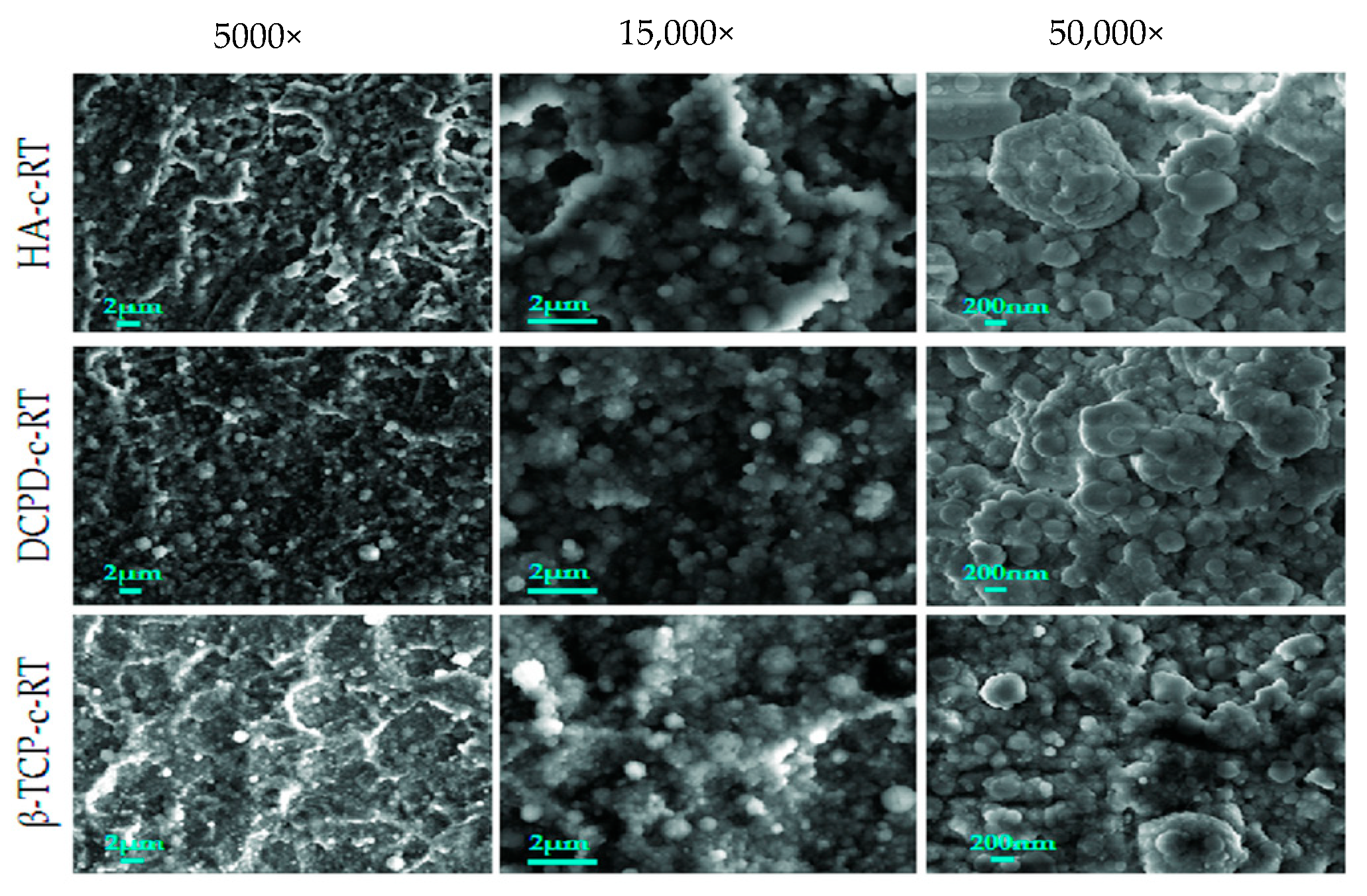
In addition, recent advances have highlighted the importance of nano- and microstructuring of CaP coatings. Nanostructured and nanoporous surfaces significantly increase the specific surface area, thereby enhancing protein adsorption and cell–material interactions. These architectures also enable controlled ion diffusion and may indirectly contribute to reducing bacterial adhesion [42]. The morphological characteristics of different CaP phases are illustrated in Figure 1 [11,14,15].
Another critical aspect concerns the mechanical stability and adhesion of the coating to the metallic substrate. CaP coatings, particularly those deposited by plasma spraying, may present a risk of delamination under mechanical loading or in physiological environments. To improve their stability, several deposition techniques have been developed, including electrodeposition, sol–gel processes and biomimetic approaches, which provide better cohesion and a more stable interface with the Ti6Al4V substrate [16,21,22]. To summarize the key physicochemical and biological properties of CaP coatings, Table 1 presents their main advantages, limitations, and functional characteristics.
Overall, CaP coatings exhibit excellent bioactivity and osteogenic potential; however, their limited antibacterial performance and potential mechanical instability remain key challenges.
Finally, current research is increasingly focused on the development of multifunctional CaP-based coatings integrating bioactive, antibacterial, and even therapeutic properties. In this context, CaP is often used as a matrix in hybrid systems, particularly in combination with Ag or ZrO2, in order to achieve an optimal balance between osseointegration and infection prevention [16,21,22].

2.2. Ag-Based Coatings

Ag is widely recognized as one of the most effective antimicrobial agents used in biomedical applications due to its broad-spectrum antibacterial activity against both Gram-positive and Gram-negative bacteria, particularly Staphylococcus aureus and Escherichia coli [45,46,47,48]. In the context of dental implants, silver-based coatings have been extensively investigated for their ability to prevent biofilm formation and reduce the risk of peri-implant infections [49].
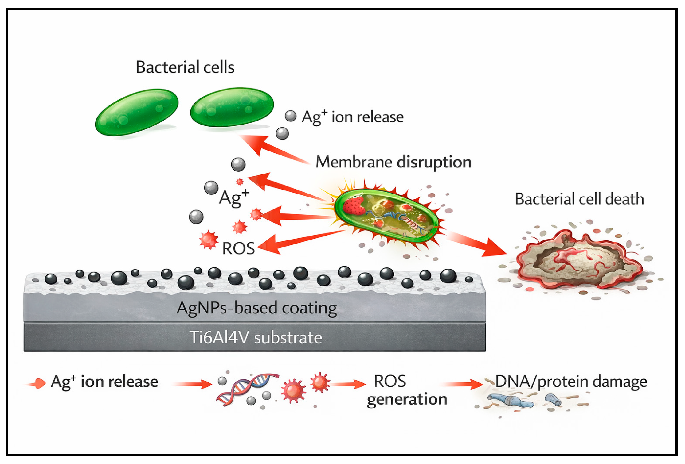
The antibacterial mechanism of silver is primarily based on the release of Ag+ ions, which interact with bacterial cellular components at multiple levels. Ag+ ions can disrupt the integrity of the bacterial cell membrane, increasing its permeability and leading to leakage of intracellular contents. They can also bind to proteins containing thiol groups, thereby inhibiting essential enzymatic functions involved in bacterial metabolism. In addition, silver ions can interact with bacterial DNA, impairing replication and ultimately leading to cell death [45,46,47,48]. Furthermore, silver nanoparticles (AgNPs) have been reported to induce the generation of reactive oxygen species (ROS), thereby enhancing their bactericidal effect.
Despite this remarkable antibacterial efficacy, the use of silver in implant coatings presents significant challenges. The main issue lies in controlling the release kinetics of Ag+ ions. Excessive ion release may induce cytotoxic effects on host cells, particularly osteoblasts, thereby compromising biocompatibility and osseointegration [50]. Conversely, insufficient ion release may reduce antibacterial effectiveness, allowing bacterial persistence and biofilm formation. Therefore, achieving an optimal balance between antibacterial activity and cytocompatibility remains a critical challenge.
To overcome these limitations, several strategies have been developed to optimize silver delivery. The incorporation of Ag into ceramic matrices such as CaP or ZrO2 enables modulation of ion release and improves coating stability [20,51,52,53,54]. These composite systems help minimize burst release effects while ensuring sustained antimicrobial activity.
Moreover, the use of AgNPs has attracted increasing interest due to their high specific surface area and enhanced interaction with bacterial cells [46,55].
Coatings incorporating AgNPs exhibit improved antibacterial performance even at low concentrations [56,57], thereby reducing the risk of cytotoxicity. However, their long-term stability, potential aggregation, and behavior in biological environments remain important challenges requiring further investigation [46,55,57].
A schematic illustration summarizing the morphology, surface distribution, and antibacterial mechanisms of silver nanoparticles on implant surfaces is presented in Figure 2.
Another aspect to consider is the aesthetic impact of silver, particularly in dental implantology. The presence of Ag may lead to discoloration of implant surfaces or surrounding tissues, which represents a potential limitation in esthetically sensitive areas.
Overall, silver-based coatings offer powerful antibacterial properties; however, their clinical applicability strongly depends on precise control of ion release, long-term stability, and cytocompatibility. Therefore, hybrid approaches combining Ag with bioactive materials such as CaP or ZrO2 represent particularly promising strategies for the development of multifunctional implant surfaces [20,51,52]. The main properties, antibacterial mechanisms, and limitations of Ag-based coatings are summarized in Table 2.

2.3. ZrO2-Based Coatings

ZrO2-based coatings have attracted increasing interest in dental implantology due to their excellent physicochemical properties, including high chemical stability, superior corrosion resistance, high hardness, and remarkable biocompatibility [5,27,28,31,58,59,60]. As a surface layer, ZrO2 acts as an effective protective barrier, limiting the electrochemical degradation of the Ti6Al4V substrate in the oral environment, which is characterized by pH fluctuations and the presence of aggressive ionic species [59,61].
Beyond its passive protective role, ZrO2-based surfaces also exhibit promising biological properties, particularly in terms of interactions with both soft and hard tissues [62,63]. Recent studies have demonstrated that ZrO2 promotes gingival fibroblast adhesion and reduces local inflammation, thereby contributing to improved peri-implant tissue integration [5,27,28,64,65,66]. In addition, its white color provides a significant aesthetic advantage over metallic surfaces, especially in anterior regions.
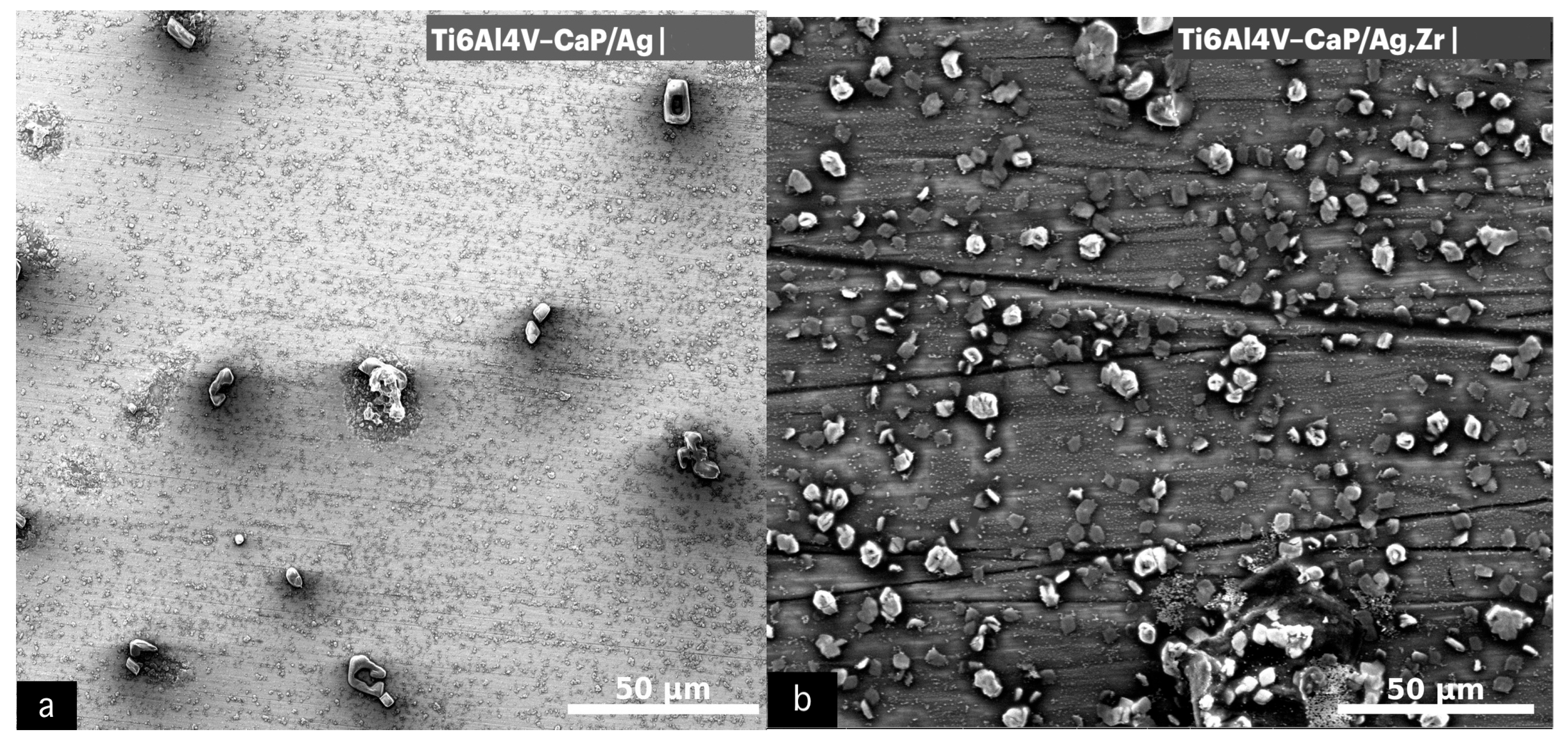
However, pure ZrO2 exhibits limited intrinsic antibacterial activity, which represents a major limitation for preventing peri-implant infections. Consequently, advanced surface modification strategies are required to enhance its antibacterial performance. Among these, surface nanostructuring (e.g., nanotubes, nanopillars, and hierarchical architectures) plays a crucial role in reducing bacterial adhesion by modulating surface roughness, wettability and surface energy [5,67,68,69,70,71]. These nanostructured surfaces can also exert mechanical bactericidal effects by physically disrupting bacterial membranes [70,72]. The incorporation of Ag promotes the formation of dispersed surface particles, whereas the addition of ZrO2 leads to a denser and more homogeneous coating morphology, as illustrated in Figure 3.
X-ray diffraction (XRD) analysis reported in the literature plays an important role in characterizing multifunctional coating systems by identifying crystalline phases, evaluating phase stability, and assessing coating crystallinity. In CaP-based systems, XRD commonly confirms the presence of hydroxyapatite, β-TCP, or other calcium phosphate phases depending on the deposition conditions. In Ag-containing coatings, additional diffraction peaks associated with metallic Ag or Ag-containing phases may be observed, while ZrO2-based coatings may exhibit tetragonal or monoclinic zirconia structures depending on processing parameters and thermal treatment. These structural characteristics may influence coating stability, ion release behavior, and overall biological and antibacterial performance [16,21,73,74].
In addition, ionic doping of ZrO2 with elements such as Ag, Zn, Cu, Ga, or Ce has demonstrated promising antibacterial performance. These ions can be released in a controlled manner and interact with bacterial cells by disrupting their metabolism and membrane integrity [28,29,75,76,77,78]. Notably, Ga can interfere with bacterial iron metabolism, while Zn contributes not only to antibacterial activity but also to osteogenic stimulation, highlighting the multifunctional potential of doped ZrO2 systems.
Recent advances have also highlighted the role of laser-functionalized surfaces and ZrO2–bioactive glass hybrid coatings in simultaneously enhancing bioactivity and antibacterial performance. These approaches enable the formation of super hydrophilic surfaces, which promote protein adsorption and cell adhesion while reducing bacterial attachment [5,27,28,29,30,79].
Furthermore, the combination of ZrO2 with CaP represents a particularly promising strategy. CaP provides high bioactivity and promotes osseointegration, while ZrO2 ensures mechanical stability and chemical durability of the coating. This complementary interaction enables the design of coatings with optimized biological and physicochemical performance [5,21,22,28,29]. The main physicochemical, biological, and antibacterial properties of ZrO2-based coatings are summarized in Table 3.
Overall, ZrO2-based coatings offer excellent corrosion resistance, biocompatibility, and aesthetic properties; however, their antibacterial performance requires additional functionalization through nanostructuring, ionic doping, or hybridization strategies.
Finally, the antibacterial mechanisms associated with modified ZrO2 surfaces involve multiple synergistic effects, including changes in wettability, reduction in bacterial adhesion, controlled release of antimicrobial ions, and, in some cases, the generation of ROS [69,80,81]. These synergistic mechanisms contribute to improving both antibacterial performance and the overall biological response of implant surfaces.
The crystalline structure and phase composition of CaP and ZrO2-based coatings may also influence their antibacterial and physicochemical behavior. Parameters such as crystallinity, grain size, surface defects, and phase stability can affect ion diffusion, surface reactivity, wettability, and bacterial adhesion. In ZrO2-based systems, phase organization and surface modifications may additionally influence local electrochemical interactions and ROS generation. Therefore, the antimicrobial performance of multifunctional coatings depends not only on their chemical composition but also on their crystalline and microstructural characteristics [42,69,72,74].

2.4. Ag–CaP–ZrO2 Composite Coatings

Hybrid coatings incorporating Ag, CaP and ZrO2 represent one of the most advanced and promising strategies for the development of multifunctional implant surfaces. These composite systems are designed to simultaneously address the main clinical requirements in dental implantology, namely infection prevention, enhancement of osseointegration, and long-term physicochemical stability of implants. From a functional perspective, these coatings rely on a synergistic interaction between their constituent components. Silver provides broad-spectrum antibacterial activity through the controlled release of Ag+ ions, which are capable of disrupting bacterial membranes, inhibiting enzymatic activity, and interfering with microbial DNA. Calcium phosphate acts as a bioactive phase, promoting cell adhesion and osteoblastic differentiation by mimicking the mineral composition of bone, thereby accelerating osseointegration. In parallel, ZrO2 serves as a stable matrix, enhancing corrosion resistance, mechanical strength, and structural integrity of the coating [21,73,74,82].
Beyond this complementarity, recent studies have demonstrated that the integration of these three phases within hierarchical nanocomposite structures enables fine-tuning of interfacial properties and functional performance. In particular, the presence of ZrO2 contributes to stabilizing the CaP matrix and modulating the diffusion of Ag+ ions, thereby reducing burst release effects responsible for cytotoxicity while maintaining prolonged antibacterial activity [83,84,85]. This controlled ion release behavior is critical for achieving an optimal balance between antimicrobial efficacy and biocompatibility.
However, the multifunctional behavior of Ag–CaP–ZrO2 coatings should not be interpreted as a simple additive combination of the individual properties of each component. In complex hybrid systems, interfacial reactions between phases, oxidation phenomena, phase instability, and modifications in ion release kinetics may significantly alter the expected biological and antibacterial performance. For example, the incorporation of Ag within CaP or ZrO2 matrices may modify Ag+ diffusion behavior and local electrochemical interactions, thereby influencing both antibacterial efficacy and cytocompatibility. Similarly, phase distribution, coating architecture, and long-term physicochemical stability under physiological conditions may affect coating durability and functional performance. Therefore, the overall multifunctional response of these systems results from complex synergistic and competitive interactions that require careful optimization and long-term evaluation [53,72,74].
It is also important to distinguish between the different architectures used in multifunctional coating systems, as their structural organization strongly influences their biological and physicochemical behavior. Ion-doped coatings generally involve the incorporation of antibacterial ions within a single matrix, allowing controlled ion release while preserving the original coating structure. In contrast, composite coatings combine multiple phases within the same layer, leading to more complex interfacial interactions and multifunctional responses. Multilayer systems consist of successive functional layers designed to separately optimize properties such as corrosion resistance, bioactivity, and antibacterial performance. Consequently, antibacterial efficacy, ion diffusion behavior, mechanical stability, and long-term durability may vary considerably depending on the coating architecture and fabrication strategy [53,72,74].
From an electrochemical standpoint, Ag–CaP–ZrO2 coatings significantly enhance the corrosion resistance of Ti6Al4V, owing to the barrier effect of ZrO2 combined with the compactness and bioactivity of CaP. In addition, modifications in surface topography and wettability induced by these coatings contribute to reducing initial bacterial adhesion, thereby limiting biofilm formation [73].
From a biological perspective, these hybrid systems promote more favorable cell–material interactions. Several studies have reported enhanced osteoblast adhesion and proliferation, as well as upregulation of osteogenic markers, suggesting improved osseointegration potential. At the same time, the combined antibacterial effect significantly reduces microbial colonization, contributing to the prevention of peri-implant infections.
Despite these promising results, several challenges remain. The complexity of fabrication processes, precise control over phase distribution, and reproducibility of coating properties represent major limitations. Furthermore, interfacial instability and long-term durability of multiphase coatings remain insufficiently investigated. In addition, most available studies are still limited to in vitro or preclinical models, and long-term clinical data remain scarce.
Overall, Ag–CaP–ZrO2 composite coatings represent a highly promising approach for next-generation dental implants, enabling the integration of sustained antibacterial activity, enhanced bioactivity, and improved physicochemical stability. However, their successful clinical translation will require further optimization of fabrication techniques, standardization of evaluation protocols, and validation through long-term in vivo and clinical studies. Table 4 summarizes the main physicochemical, biological, and antibacterial characteristics of CaP, Ag, ZrO2 and Ag–CaP–ZrO2 composite coatings.
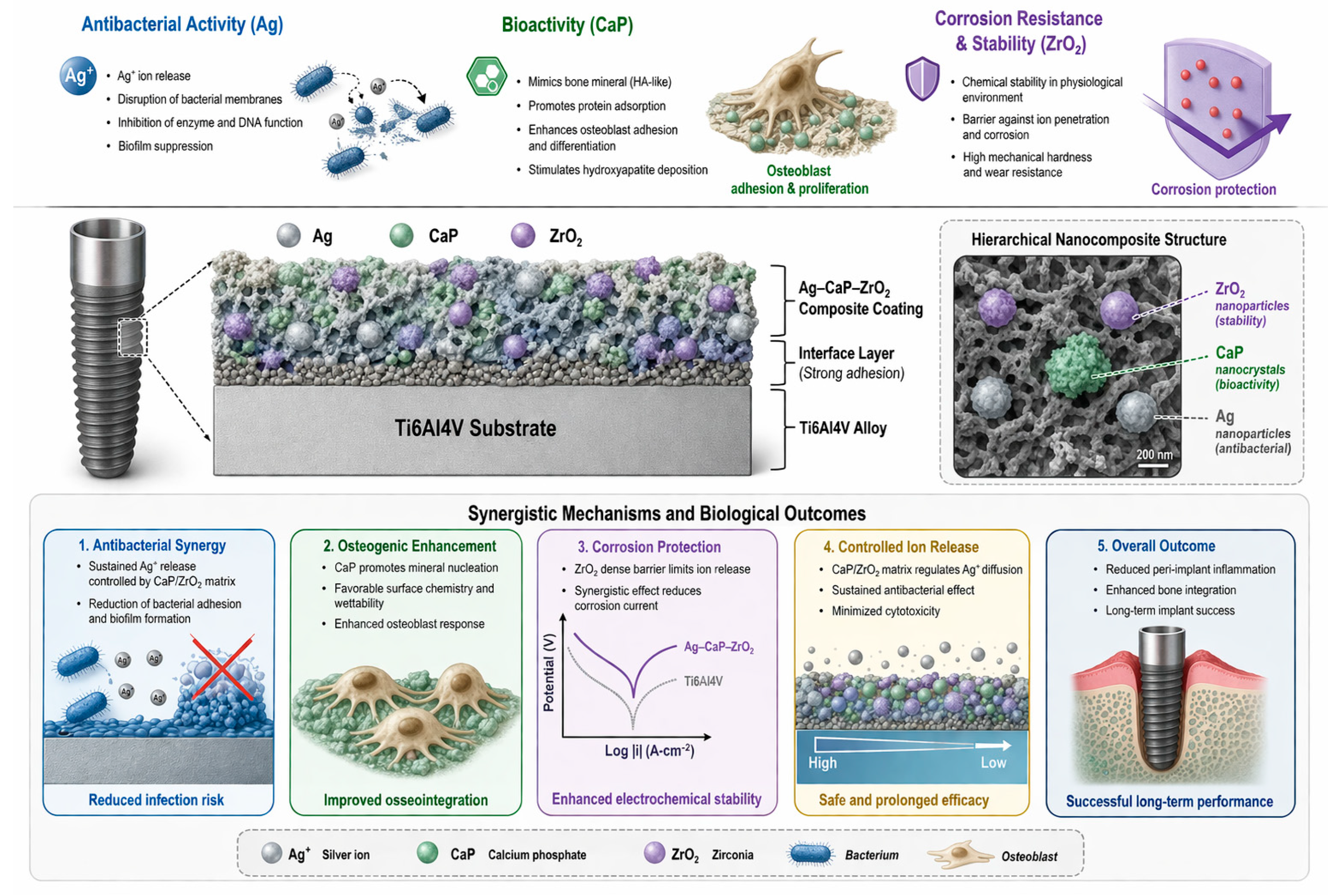
The multifunctional mechanisms and synergistic effects of Ag–CaP–ZrO2 composite coatings are schematically illustrated in Figure 4.
These mechanisms are supported by experimentally reported observations in the literature, including controlled Ag+ ion release, reduced bacterial adhesion, enhanced corrosion resistance, and improved osteoblastic response associated with multifunctional Ag–CaP–ZrO2 coatings [73,74,83].
At the molecular level, the antibacterial performance of multifunctional coatings is strongly influenced by ion release behavior and coating microstructure. Metal ions such as Ag+, Zn2+, Cu2+, and Ga3+ can interfere with bacterial membrane integrity, protein activity, enzymatic pathways, and microbial DNA replication. In addition, oxide-based surfaces and nanostructured architectures may promote reactive oxygen species (ROS) generation and modify local electrochemical interactions, thereby contributing to bacterial inactivation. Parameters such as coating porosity, crystallinity, grain size, and ion diffusion kinetics further regulate the accessibility and sustained release of antibacterial species, ultimately influencing biofilm inhibition and long-term antimicrobial efficacy [42,43,46,69,72,74].

3. Clinical Perspectives and Limitations

Multifunctional bioceramic coatings, particularly hybrid systems based on Ag–CaP–ZrO2, have demonstrated promising performance in numerous in vitro and preclinical studies, especially in terms of antibacterial activity, bioactivity, and corrosion resistance. However, their translation into clinical practice remains limited and represents a major challenge in dental implantology. One of the primary limitations lies in the relevance of the experimental models used. Most studies rely on simplified conditions, including mono-species bacterial cultures such as Staphylococcus aureus or Escherichia coli, which do not accurately reflect the complexity of the oral microbiome or the dynamic interactions of polymicrobial biofilms under real clinical conditions [86]. In addition, these models often fail to account for host immune responses, salivary factors, pH fluctuations, and mechanical loading conditions characteristic of the oral environment.
Another critical challenge concerns the long-term durability of antibacterial properties. The effectiveness of silver-based coatings strongly depends on the controlled release of Ag+ ions. Rapid ion release may induce cytotoxic effects toward host cells, particularly osteoblasts, whereas insufficient release may allow bacterial persistence and biofilm formation. Furthermore, potential bacterial adaptation or tolerance to modified surfaces may compromise long-term antimicrobial efficacy [50,87,88].
The physicochemical and mechanical stability of coatings also represents a major concern. In the oral environment, implants are subjected to complex conditions, including cyclic mechanical loading, temperature variations, pH fluctuations, and exposure to salivary enzymes. These factors may lead to progressive degradation of coatings, partial dissolution of bioactive phases, or delamination of surface layers, ultimately compromising their integrity and long-term performance [89].
In multifunctional hybrid coatings, long-term interfacial stability represents an additional challenge due to the coexistence of multiple phases with different physicochemical properties. Differences in thermal expansion behavior, ion diffusion, and local electrochemical activity may progressively induce interfacial stress, microstructural degradation, or partial delamination under physiological conditions. Furthermore, the reproducibility of multifunctional coating architectures remains difficult to achieve at an industrial scale because small variations in fabrication parameters may significantly affect coating composition, phase distribution, and functional performance. These limitations highlight the need for standardized fabrication protocols and long-term in vivo evaluation before routine clinical implementation [53,72,83,89,90].
Moreover, significant limitations remain regarding the reproducibility of fabrication processes and their scalability for industrial applications. Advanced deposition techniques (such as sol–gel, electrodeposition, and plasma spraying), although allowing precise control of surface properties, are often costly, sensitive to processing parameters and difficult to standardize, which hinders large-scale clinical implementation [90].
In addition, the lack of standardized experimental protocols represents a major obstacle to the comparison of results across studies. Variations in coating methods, testing conditions, bacterial strains, and evaluation criteria make it difficult to establish robust conclusions and slow down clinical validation of these technologies.
In this context, future research should focus on the development of “smart” or stimuli-responsive coatings capable of dynamically adapting to the biological environment. Systems with controlled or triggered release mechanisms (e.g., pH-responsive, enzyme-responsive, or mechanically activated systems) represent particularly promising strategies to optimize antibacterial efficacy while minimizing cytotoxic effects.
Recent advances in additive manufacturing have also stimulated growing interest in 3D-printed polymer- and composite-based materials for dental implant applications. These technologies allow the fabrication of patient-specific implants with complex architectures and controlled porosity, which may enhance tissue integration and biomechanical adaptation. In particular, polymer–ceramic composites incorporating bioactive phases such as hydroxyapatite or zirconia have shown promising biological performance. However, compared with Ti6Al4V implants modified by multifunctional bioceramic coatings, these systems may present limitations related to lower mechanical strength, long-term degradation behavior, wear resistance, and limited intrinsic antibacterial activity. Future research may explore the combination of additive manufacturing technologies with multifunctional antibacterial and bioactive coating strategies to develop next-generation implant systems with enhanced personalization and clinical performance [91].
Furthermore, the integration of multifunctional approaches combining antibacterial, immunomodulatory, and osteogenic properties may provide improved control of host–implant interactions. Finally, significant efforts should be directed toward the development of more representative biological models, including polymicrobial biofilms and relevant in vivo systems, as well as long-term clinical trials. The standardization of experimental protocols and rigorous evaluation of safety and efficacy will be essential for successful clinical translation [29,30].

4. Conclusions

Multifunctional bioceramic coatings applied to titanium dental implants represent a promising strategy for simultaneously improving corrosion resistance, bioactivity, and antibacterial performance of implant surfaces. This review highlights the complementary roles of CaP, Ag, and ZrO2-based coatings, as well as the growing importance of hybrid systems in the development of next-generation dental implants.
CaP coatings are distinguished by their excellent bioactivity and their ability to promote early osseointegration, although their intrinsic antibacterial activity remains limited. In contrast, silver-based systems provide strong antimicrobial efficacy through the release of Ag+ ions but require precise control to avoid cytotoxic effects. ZrO2 plays a crucial role as a stable matrix and protective barrier, significantly enhancing corrosion resistance and mechanical performance, while also serving as a versatile platform for advanced surface modifications.
In this context, hybrid Ag–CaP–ZrO2 coatings emerge as a particularly effective approach to address the major challenges in dental implantology. By combining antibacterial activity, osteogenic potential, and physicochemical stability, these multifunctional systems enable synergistic effects and optimized control of ion release kinetics. These properties position hybrid coatings as strong candidates for improving long-term implant performance and reducing the risk of peri-implant infections.
Nevertheless, the clinical translation of these technologies remains dependent on overcoming several key challenges, including long-term coating stability, precise control of ion release behavior, and the standardization of experimental protocols. Furthermore, the widespread reliance on simplified biological models continues to limit the clinical relevance of current findings.
Future research should focus on the development of smart, stimuli-responsive implant surfaces capable of dynamically interacting with their biological environment. The integration of additional functionalities, such as immunomodulation and host–biofilm interaction control, may represent a major advancement in the design of next-generation coatings. Furthermore, well-designed in vivo studies and long-term clinical trials will be essential to validate the safety, efficacy, and durability of these systems.
In conclusion, ZrO2-based coatings, particularly when combined with CaP and Ag, represent a strategic and highly promising direction for the development of multifunctional dental implants capable of meeting the complex biological, mechanical, and microbiological requirements of modern implantology. Their successful clinical translation will depend on continued advances in material design, process optimization, and rigorous clinical validation.

Author Contributions

Conceptualization: M.A. and A.E.-R.; Methodology: M.A. and A.E.-R.; Formal analysis: M.A. and A.E.-R.; Investigation: M.A.; Resources: A.E.-R. and N.M.; Writing—Original Draft Preparation: M.A.; Writing—Review & Editing: A.E.-R. and N.M.; Supervision: A.E.-R. and N.M. All authors have read and agreed to the published version of the manuscript.

Funding

This research received no external funding.

Institutional Review Board Statement

Not applicable.

Informed Consent Statement

Not applicable.

Data Availability Statement

The original contributions presented in this study are included in the article. Further inquiries can be directed to the corresponding author.

Acknowledgments

During the preparation of this manuscript, the authors used ChatGPT (OpenAI, ChatGPT Plus, https://chatgpt.com; access on 1 June 2026) for the generation of Figure 2 and Figure 4. The authors reviewed and edited the output and take full responsibility for the content of this publication.

Conflicts of Interest

The authors declare no conflicts of interest.

Abbreviations

The following abbreviations are used in this manuscript:
AgSilver
AgNPsSilver Nanoparticles
ALPAlkaline Phosphatase
ASArtificial Saliva
CaPCalcium Phosphate
CeCerium
CP-TiCommercially Pure Titanium
CuCopper
DCPDDicalcium Phosphate Dihydrate
EISElectrochemical Impedance Spectroscopy
GaGallium
HAHydroxyapatite
OCPOpen Circuit Potential
PO43−Phosphate Ion
ROSReactive Oxygen Species
RpPolarization Resistance
RUNX2Runt-Related Transcription Factor 2
SBFSimulated Body Fluid
SEMScanning Electron Microscopy
Ti6Al4VTitanium Alloy
ZnZinc
ZrO2Zirconium Dioxide

References

  1. Hanawa, T. Titanium–tissue interface reaction and its control with surface treatment. Front. Bioeng. Biotechnol. 2019, 7, 170. [Google Scholar] [CrossRef] [PubMed]
  2. Kligman, S.Z.; Ren, C.-H.; Chung, M.A.; Perillo, Y.-C.; Chang, H.; Zheng, K.Z.; Li, C. The impact of dental implant surface modifications on osseointegration and biofilm formation. J. Clin. Med. 2021, 10, 1641. [Google Scholar] [CrossRef]
  3. Bokobza, L. On the use of nanoparticles in dental implants. Materials 2024, 17, 3191. [Google Scholar] [CrossRef]
  4. Chen, Q.; Thouas, G.A. Metallic implant biomaterials. Mater. Sci. Eng. R Rep. 2015, 87, 1–57. [Google Scholar] [CrossRef]
  5. Aldhuwayhi, S. Zirconia in Dental Implantology: A Review of the Literature with Recent Updates. Bioengineering 2025, 12, 543. [Google Scholar] [CrossRef]
  6. de Campos Kajimoto, N.; de Paiva Buischi, Y.; Mohamadzadeh, M.; Loomer, P. The oral microbiome of peri-implant health and disease: A narrative review. Dent. J. 2024, 12, 299. [Google Scholar] [CrossRef]
  7. Di Spirito, F.; Pisano, M.; Di Palo, M.P.; Franci, G.; Rupe, A.; Fiorino, A.; Rengo, C. Peri-implantitis-associated microbiota before and after peri-implantitis treatment, the biofilm “competitive balancing” effect: A systematic review of randomized controlled trials. Microorganisms 2024, 12, 1965. [Google Scholar] [CrossRef]
  8. Gkioka, M.; Rausch-Fan, X. Antimicrobial effects of metal coatings or physical, chemical modifications of titanium dental implant surfaces for prevention of peri-implantitis: A systematic review of in vivo studies. Antibiotics 2024, 13, 908. [Google Scholar] [CrossRef]
  9. Li, Y.; Stewart, C.A.; Finer, Y. Advanced antimicrobial and anti-infective strategies to manage peri-implant infection: A narrative review. Dent. J. 2024, 12, 125. [Google Scholar] [CrossRef] [PubMed]
  10. Koo, H.; Allan, R.N.; Howlin, R.P.; Stoodley, P.; Hall-Stoodley, L. Targeting microbial biofilms: Current and prospective therapeutic strategies. Nat. Rev. Microbiol. 2017, 15, 740–755. [Google Scholar] [CrossRef]
  11. Marsh, P.; Zaura, E. Dental biofilm: Ecological interactions in health and disease. J. Clin. Periodontol. 2017, 44, S12–S22. [Google Scholar] [CrossRef] [PubMed]
  12. Zafar, M.S.; Fareed, M.A.; Riaz, S.; Latif, M.; Habib, S.R.; Khurshid, Z. Customized therapeutic surface coatings for dental implants. Coatings 2020, 10, 568. [Google Scholar] [CrossRef]
  13. Dong, H.; Liu, H.; Zhou, N.; Li, Q.; Yang, G.; Chen, L.; Mou, Y. Surface modified techniques and emerging functional coating of dental implants. Coatings 2020, 10, 1012. [Google Scholar] [CrossRef]
  14. Anselme, K.; Davidson, P.; Popa, A.M.; Giazzon, M.; Liley, M.; Ploux, L. The interaction of cells and bacteria with surfaces structured at the nanometre scale. Acta Biomater. 2010, 6, 3824–3846. [Google Scholar] [CrossRef]
  15. Hotchkiss, K.M.; Sowers, K.T.; Olivares-Navarrete, R. Novel in vitro comparative model of osteogenic and inflammatory cell response to dental implants. Dent. Mater. 2019, 35, 176–184. [Google Scholar] [CrossRef]
  16. Drevet, R.; Fauré, J.; Benhayoune, H. Bioactive calcium phosphate coatings for bone implant applications: A review. Coatings 2023, 13, 1091. [Google Scholar] [CrossRef]
  17. Bakitian, F.A. A comprehensive review of the contemporary methods for enhancing osseointegration and the antimicrobial properties of titanium dental implants. Cureus 2024, 16, e68720. [Google Scholar] [CrossRef]
  18. Aissi, M.; Tayyaba, Q.; Er-Ramly, A.; Hermawan, H.; Merzouk, N. Improving the clinical performance of dental implants through advanced surface treatments: The case of Ti and ZrO2 coatings. Metals 2025, 15, 320. [Google Scholar] [CrossRef]
  19. Wang, Z.; Wang, J.; Wu, R.; Wei, J. Construction of functional surfaces for dental implants to enhance osseointegration. Front. Bioeng. Biotechnol. 2023, 11, 1320307. [Google Scholar] [CrossRef]
  20. Liu, X.; Chu, P.K.; Ding, C. Surface modification of titanium, titanium alloys, and related materials for biomedical applications. Mater. Sci. Eng. R Rep. 2004, 47, 49–121. [Google Scholar] [CrossRef]
  21. Matys, J.; Rygus, R.; Kensy, J.; Okoniewska, K.; Zakrzewski, W.; Kotela, A.; Struzik, N.; Gerber, H.; Fast, M.; Dobrzyński, M. Enhancing osseointegration of zirconia implants using calcium phosphate coatings: A systematic review. Materials 2025, 18, 4501. [Google Scholar] [CrossRef]
  22. Montesissa, M.; Tommasini, V.; Rubini, K.; Boi, M.; Baldini, N.; Boanini, E. State of art and perspective of calcium phosphate-based coatings coupled with bioactive compounds for orthopedic applications. Nanomaterials 2025, 15, 1199. [Google Scholar] [CrossRef]
  23. Samavedi, S.; Whittington, A.R.; Goldstein, A.S. Calcium phosphate ceramics in bone tissue engineering: A review of properties and their influence on cell behavior. Acta Biomater. 2013, 9, 8037–8045. [Google Scholar] [CrossRef]
  24. Yu, C.; Yu, Y.; Lu, Y.; Quan, K.; Mao, Z.; Zheng, Y.; Qin, L.; Xia, D. UiO-66/AgNPs coating for dental implants in preventing bacterial infections. J. Dent. Res. 2024, 103, 516–525. [Google Scholar] [CrossRef] [PubMed]
  25. Alenezi, A. Effects of Implant Silver Coatings on Bone Formation in Animal Models: A Systematic Review and Meta-Analysis. J. Funct. Biomater. 2025, 16, 369. [Google Scholar] [CrossRef] [PubMed]
  26. Salaie, R.N.; Besinis, A.; Tredwin, C.; Handy, R.D. Low toxicity of dissolved silver from silver-coated titanium dental implants to human primary osteoblast cells. Toxicol. Rep. 2024, 13, 101776. [Google Scholar] [CrossRef]
  27. Fernandes, B.F.; Silva, N.; Da Cruz, M.B.; Garret, G.; Carvalho, Ó.; Silva, F.; Mata, A.; Francisco, H.; Marques, J.F. Cell biological and antibacterial evaluation of a new approach to zirconia implant surfaces modified with MTA. Biomimetics 2024, 9, 155. [Google Scholar] [CrossRef] [PubMed]
  28. D’AGostino, A.; Misiti, G.; Scalia, A.C.; Pavarini, M.; Fiorati, A.; Cochis, A.; Rimondini, L.; Borrini, V.F.; Manfredi, M.; Andena, L.; et al. Gallium-doped zirconia coatings modulate microbiological outcomes in dental implant surfaces. J. Biomed. Mater. Res. Part A 2024, 112, 2098–2109. [Google Scholar] [CrossRef]
  29. Odatsu, T.; Valanezhad, A.; Shinohara, A.; Takase, K.; Naito, M.; Sawase, T. Bioactivity and antibacterial effects of zinc-containing bioactive glass on the surface of zirconia abutments. J. Dent. 2024, 145, 105033. [Google Scholar] [CrossRef]
  30. Łosiewicz, B.; Osak, P.; Nowińska, D.; Maszybrocka, J. Developments in dental implant surface modification. Coatings 2025, 15, 109. [Google Scholar] [CrossRef]
  31. Denry, I.; Kelly, J.R. State of the art of zirconia for dental applications. Dent. Mater. 2008, 24, 299–307. [Google Scholar] [CrossRef] [PubMed]
  32. Dorozhkin, S.V. Calcium orthophosphates in nature, biology and medicine. Materials 2009, 2, 399–498. [Google Scholar] [CrossRef]
  33. Ghassemi, T.; Shahroodi, A.; Ebrahimzadeh, M.H.; Mousavian, A.; Movaffagh, J.; Moradi, A. Current concepts in scaffolding for bone tissue engineering. Arch. Bone Jt. Surg. 2018, 6, 90. [Google Scholar]
  34. LeGeros, R.Z. Calcium phosphate-based osteoinductive materials. Chem. Rev. 2008, 108, 4742–4753. [Google Scholar] [CrossRef]
  35. Dorozhkin, S.V. Calcium orthophosphates. J. Mater. Sci. 2007, 42, 1061–1095. [Google Scholar] [CrossRef]
  36. Yang, Y.; Kim, K.-H.; Ong, J.L. A review on calcium phosphate coatings produced using a sputtering process—An alternative to plasma spraying. Biomaterials 2005, 26, 327–337. [Google Scholar] [CrossRef] [PubMed]
  37. Dorozhkin, S.V. Bioactive and Protective Calcium Orthophosphate (CaPO4) Deposits on Magnesium and Its Alloys. Corros. Mater. Degrad. 2025, 6, 9. [Google Scholar] [CrossRef]
  38. Zhang, Y.; Shu, T.; Wang, S.; Liu, Z.; Cheng, Y.; Li, A.; Pei, D. The osteoinductivity of calcium phosphate-based biomaterials: A tight interaction with bone healing. Front. Bioeng. Biotechnol. 2022, 10, 911180. [Google Scholar] [CrossRef]
  39. Xu, C.; Sun, Y.; Jansen, J.; Li, M.; Wei, L.; Wu, Y.; Liu, Y. Calcium phosphate ceramics and synergistic bioactive agents for osteogenesis in implant dentistry. Tissue Eng. Part C Methods 2023, 29, 197–215. [Google Scholar] [CrossRef]
  40. Bohner, M. Calcium orthophosphates in medicine: From ceramics to calcium phosphate cements. Injury 2000, 31, D37–D47. [Google Scholar] [CrossRef]
  41. Wang, Y.; Wu, M.; Wang, X.; Wang, P.; Ning, Z.; Zeng, Y.; Liu, X.; Sun, H.; Zheng, A. Biodegradable MnO2-based gene-engineered nanocomposites for chemodynamic therapy and enhanced antitumor immunity. Mater. Today Bio 2023, 18, 100531. [Google Scholar] [CrossRef]
  42. Modaresifar, K.; Azizian, S.; Ganjian, M.; Fratila-Apachitei, L.E.; Zadpoor, A.A. Bactericidal effects of nanopatterns: A systematic review. Acta Biomater. 2019, 83, 29–36. [Google Scholar] [CrossRef]
  43. Xu, N.; Fu, J.; Zhao, L.; Chu, P.K.; Huo, K. Biofunctional elements incorporated nano/microstructured coatings on titanium implants with enhanced osteogenic and antibacterial performance. Adv. Healthc. Mater. 2020, 9, 2000681. [Google Scholar] [CrossRef] [PubMed]
  44. Barros, J.; Monteiro, F.J.; Ferraz, M.P. Bioengineering approaches to fight against orthopedic biomaterials related-infections. Int. J. Mol. Sci. 2022, 23, 11658. [Google Scholar] [CrossRef]
  45. Rai, M.; Yadav, A.; Gade, A. Silver nanoparticles as antimicrobial agents. Biotechnol. Adv. 2009, 27, 76–83. [Google Scholar] [CrossRef]
  46. Franci, G.; Falanga, A.; Galdiero, S.; Palomba, L.; Rai, M.; Morelli, G.; Galdiero, M. Silver nanoparticles as potential antibacterial agents. Molecules 2015, 20, 8856–8874. [Google Scholar] [CrossRef]
  47. Durán, N.; Nakazato, G.; Seabra, A.B. Antimicrobial activity of biogenic silver nanoparticles, and silver chloride nanoparticles: An overview and comments. Appl. Microbiol. Biotechnol. 2016, 100, 6555–6570. [Google Scholar] [CrossRef]
  48. Zhao, L.; Wang, H.; Huo, K.; Cui, L.; Zhang, W.; Ni, H.; Zhang, Y.; Wu, Z.; Chu, P.K. Antibacterial nano-structured titania coating incorporated with silver nanoparticles. Biomaterials 2011, 32, 5706–5716. [Google Scholar] [CrossRef]
  49. Zhao, L.; Chu, P.K.; Zhang, Y.; Wu, Z. Antibacterial coatings on titanium implants. J. Biomed. Mater. Res. Part B Appl. Biomater. 2009, 91, 470–480. [Google Scholar] [CrossRef] [PubMed]
  50. Chen, X.; Schluesener, H.J. Nanosilver: A nanoproduct in medical application. Toxicol. Lett. 2008, 176, 1–12. [Google Scholar] [CrossRef] [PubMed]
  51. Zhang, Y.; Dong, C.; Yang, S.; Chiu, T.-W.; Wu, J.; Xiao, K.; Huang, Y.; Li, X. Enhanced silver loaded antibacterial titanium implant coating with novel hierarchical effect. J. Biomater. Appl. 2018, 32, 1289–1299. [Google Scholar] [CrossRef] [PubMed]
  52. Shi, Q.; Qian, Z.; Liu, D.; Liu, H. Surface modification of dental titanium implant by layer-by-layer electrostatic self-assembly. Front. Physiol. 2017, 8, 574. [Google Scholar] [CrossRef]
  53. Nikolova, M.P.; Apostolova, M.D. Advances in multifunctional bioactive coatings for metallic bone implants. Materials 2022, 16, 183. [Google Scholar] [CrossRef]
  54. Birkett, M.; Zia, A.W.; Devarajan, D.K.; Panayiotidis, M.I.; Joyce, T.J.; Tambuwala, M.M.; Serrano-Aroca, Á. Multi-functional bioactive silver-and copper-doped diamond-like carbon coatings for medical implants. Acta Biomater. 2023, 167, 54–68. [Google Scholar] [CrossRef]
  55. Rai, M.K.; Deshmukh, S.D.; Ingle, A.P.; Gade, A.K. Silver nanoparticles: The powerful nanoweapon against multidrug-resistant bacteria. J. Appl. Microbiol. 2012, 112, 841–852. [Google Scholar] [CrossRef]
  56. Durán, N.; Durán, M.; de Jesus, M.B.; Seabra, A.B.; Fávaro, W.J.; Nakazato, G. Silver nanoparticles: A new view on mechanistic aspects on antimicrobial activity. Nanomed. Nanotechnol. Biol. Med. 2016, 12, 789–799. [Google Scholar] [CrossRef]
  57. Fernandez, C.C.; Sokolonski, A.R.; Fonseca, M.S.; Stanisic, D.; Araújo, D.B.; Azevedo, V.; Portela, R.D.; Tasic, L. Applications of silver nanoparticles in dentistry: Advances and technological innovation. Int. J. Mol. Sci. 2021, 22, 2485. [Google Scholar] [CrossRef] [PubMed]
  58. Piconi, C.; Sprio, S. Zirconia implants: Is there a future? Curr. Oral Health Rep. 2018, 5, 186–193. [Google Scholar] [CrossRef]
  59. Zhang, Y.; Lawn, B.R. Novel zirconia materials in dentistry. J. Dent. Res. 2018, 97, 140–147. [Google Scholar] [CrossRef]
  60. Osman, R.B.; Swain, M.V. A critical review of dental implant materials with an emphasis on titanium versus zirconia. Materials 2015, 8, 932–958. [Google Scholar] [CrossRef] [PubMed]
  61. Hanawa, T. Biocompatibility of titanium from the viewpoint of its surface. Sci. Technol. Adv. Mater. 2022, 23, 457–472. [Google Scholar] [CrossRef] [PubMed]
  62. Roehling, S.; Schlegel, K.A.; Woelfler, H.; Gahlert, M. Zirconia compared to titanium dental implants in preclinical studies—A systematic review and meta-analysis. Clin. Oral Implant. Res. 2019, 30, 365–395. [Google Scholar] [CrossRef] [PubMed]
  63. Kesim, B. Güncel Protetik Diş Tedavisi Çalışmaları IX; Akademisyen Kitabevi: Ankara, Turkey, 2025. [Google Scholar]
  64. Jin, H.W.; Noumbissi, S.; Wiedemann, T.G. Comparison of zirconia implant surface modifications for optimal osseointegration. J. Funct. Biomater. 2024, 15, 91. [Google Scholar] [CrossRef]
  65. Roehling, S.; Schlegel, K.A.; Woelfler, H.; Gahlert, M. Performance and outcome of zirconia dental implants in clinical studies: A meta-analysis. Clin. Oral Implant. Res. 2018, 29, 135–153. [Google Scholar] [CrossRef]
  66. Andreiotelli, M.; Wenz, H.J.; Kohal, R.J. Are ceramic implants a viable alternative to titanium implants? A systematic literature review. Clin. Oral Implant. Res. 2009, 20, 32–47. [Google Scholar] [CrossRef]
  67. Sun, W.; Tan, A.W.-Y.; Wu, K.; Yin, S.; Yang, X.; Marinescu, I.; Liu, E. Post-process treatments on supersonic cold sprayed coatings: A review. Coatings 2020, 10, 123. [Google Scholar] [CrossRef]
  68. Kuroda, C.; Ajima, K.; Ueda, K.; Sobajima, A.; Yoshida, K.; Kamanaka, T.; Sasaki, J.; Ishida, H.; Haniu, H.; Okamoto, M.; et al. Isolated lymphatic vessel lumen perfusion system for assessing nanomaterial movements and nanomaterial-induced responses in lymphatic vessels. Nano Today 2021, 36, 101018. [Google Scholar] [CrossRef]
  69. Ciszyński, M.; Chwaliszewski, B.; Simka, W.; Dominiak, M.; Gedrange, T.; Hadzik, J. Zirconia dental implant designs and surface modifications: A narrative review. Materials 2024, 17, 4202. [Google Scholar] [CrossRef]
  70. Linklater, D.P.; De Volder, M.; Baulin, V.A.; Werner, M.; Jessl, S.; Golozar, M.; Maggini, L.; Rubanov, S.; Hanssen, E.; Juodkazis, S.; et al. High aspect ratio nanostructures kill bacteria via storage and release of mechanical energy. ACS Nano 2018, 12, 6657–6667. [Google Scholar] [CrossRef]
  71. Šístková, J.; Fialová, T.; Svoboda, E.; Varmužová, K.; Uher, M.; Číhalová, K.; Přibyl, J.; Dlouhý, A.; Goldbergová, M.P. Insight into antibacterial effect of titanium nanotubular surfaces with focus on Staphylococcus aureus and Pseudomonas aeruginosa. Sci. Rep. 2024, 14, 17303. [Google Scholar] [CrossRef] [PubMed]
  72. Teulé-Trull, M.; Altuna, P.; Arregui, M.; Rodriguez-Ciurana, X.; Aparicio, C. Antibacterial coatings for dental implants: A systematic review. Dent. Mater. 2025, 41, 229–247. [Google Scholar] [CrossRef] [PubMed]
  73. Aissi, M.; Er-Ramly, A.; Merzouk, N. Electrochemical Evaluation of Ag–CaP–ZrO2 Composite Coatings on Ti6Al4V for Enhanced Corrosion Resistance in Dental Implants. Prosthesis 2025, 7, 141. [Google Scholar] [CrossRef]
  74. Chopra, D.; Jayasree, A.; Guo, T.; Gulati, K.; Ivanovski, S. Advancing dental implants: Bioactive and therapeutic modifications of zirconia. Bioact. Mater. 2022, 13, 161–178. [Google Scholar] [CrossRef]
  75. Cao, F.; Xiang, C.; Kong, S.; Guo, N.; Shang, H. Room Temperature Strengthening and High-Temperature Superplasticity of Mg-Li-Al-Sr-Y Alloy Fabricated by Asymmetric Rolling and Friction Stir Processing. Materials 2023, 16, 2345. [Google Scholar] [CrossRef]
  76. Shalamova, A.; Suntsov, A.Y. Thermodynamic analysis of defect equilibration in highly nonstoichiometric perovskites LnBaMn2O6–δ (Ln = Nd, Sm). Ceram. Int. 2024, 50, 12195–12202. [Google Scholar] [CrossRef]
  77. Kang, M.-H.; Lee, H.; Jang, T.-S.; Seong, Y.-J.; Kim, H.-E.; Koh, Y.-H.; Song, J.; Jung, H.-D. Biomimetic porous Mg with tunable mechanical properties and biodegradation rates for bone regeneration. Acta Biomater. 2019, 84, 453–467. [Google Scholar] [CrossRef]
  78. Sun, L.; Hong, G. Surface modifications for zirconia dental implants: A review. Front. Dent. Med. 2021, 2, 733242. [Google Scholar] [CrossRef]
  79. Singh, P.V.; Reche, A.; Paul, P.; Agarwal, S. Zirconia facts and perspectives for biomaterials in dental implantology. Cureus 2023, 15, e46828. [Google Scholar] [CrossRef]
  80. Dai, F.; Sun, P.; Liu, Y.; Liu, W. Redox-cleavable star cationic PDMAEMA by arm-first approach of ATRP as a nonviral vector for gene delivery. Biomaterials 2010, 31, 559–569. [Google Scholar] [CrossRef]
  81. Xin, L.; Lin, X.; Pan, Y.; Zheng, X.; Shi, L.; Zhang, Y.; Ma, L.; Gao, C.; Zhang, S. A collagen scaffold loaded with human umbilical cord-derived mesenchymal stem cells facilitates endometrial regeneration and restores fertility. Acta Biomater. 2019, 92, 160–171. [Google Scholar] [CrossRef]
  82. Negrescu, A.M.; Killian, M.S.; Raghu, S.N.V.; Schmuki, P.; Mazare, A.; Cimpean, A. Metal oxide nanoparticles: Review of synthesis, characterization and biological effects. J. Funct. Biomater. 2022, 13, 274. [Google Scholar] [CrossRef] [PubMed]
  83. Semisch-Dieter, O.K.; Choi, A.H.; Ben-Nissan, B.; Stewart, M.P. Modifying an implant: A mini-review of dental implant biomaterials. BIO Integr. 2021, 2, 12–21. [Google Scholar] [CrossRef]
  84. Qu, Y.; Liu, L. Zirconia materials for dental implants: A literature review. Front. Dent. Med. 2021, 2, 687983. [Google Scholar] [CrossRef]
  85. Anavadya, K.; Vijayalakshmi, U. Advanced Biomaterials as Bioimplants for Load-bearing Applications. In Advanced Manufacturing Technologies in Biomedical Science; CRC Press: Boca Raton, FL, USA, 2026; pp. 261–285. [Google Scholar]
  86. He, Y.; Li, F.; Jiang, P.; Cai, F.; Lin, Q.; Zhou, M.; Liu, H.; Yan, F. Remote control of the recruitment and capture of endogenous stem cells by ultrasound for in situ repair of bone defects. Bioact. Mater. 2023, 21, 223–238. [Google Scholar] [CrossRef] [PubMed]
  87. Gao, X.; You, Z.; Li, Y.; Kang, X.; Yang, W.; Wang, H.; Zhang, T.; Zhao, X.; Sun, Y.; Shen, H. Multifunctional hydrogel modulates the immune microenvironment to improve allogeneic spinal cord tissue survival for complete spinal cord injury repair. Acta Biomater. 2023, 155, 235–246. [Google Scholar] [CrossRef]
  88. Li, B.; Webster, T.J. Bacteria antibiotic resistance: New challenges and opportunities for implant-associated orthopedic infections. J. Orthop. Res. 2018, 36, 22–32. [Google Scholar] [CrossRef] [PubMed]
  89. Sazali, E.S.; Rais, A.N.; Amin, N.A.; Bhattacharya, N.A. Titania Additive Incorporation for Improving the Antibacterial Efficacy of Boron-Activated Mesoporous Bioactive Glass-Ceramics. In InGlobal Conference on Materials Science and Engineering; Springer Nature: Singapore, 2025; pp. 213–224. [Google Scholar]
  90. Liang, W.; Zhou, C.; Bai, J.; Zhang, H.; Long, H.; Jiang, B.; Dai, H.; Wang, J.; Zhang, H.; Zhao, J. Current developments and future perspectives of nanotechnology in orthopedic implants: An updated review. Front. Bioeng. Biotechnol. 2024, 12, 1342340. [Google Scholar] [CrossRef]
  91. Lepišová, M.; Tomášik, J.; Oravcová, Ľ.; Thurzo, A. Three-dimensional-printed elements based on polymer and composite materials in dentistry: A narrative review. Bratisl. Med. J. 2025, 126, 14–27. [Google Scholar] [CrossRef]
Figure 1. Scanning electron microscopy (SEM) images of different CaP coatings, including HA, dicalcium phosphate dihydrate (DCPD), and β-tricalcium phosphate (β-TCP), at various magnifications. The images highlight differences in surface morphology, particle size, and micro- and nanostructural organization depending on the CaP phase. Reproduced and adapted from [16,21,22] under the Creative Commons CC BY license.
Prosthesis 08 00056 g001
Figure 2. Schematic illustration of silver nanoparticle (AgNP)-based coatings on titanium implant surfaces and their antibacterial mechanisms. AgNPs deposited on the implant surface release Ag+ ions, which disrupt bacterial cell membranes, interfere with intracellular components, and induce ROS generation, ultimately leading to bacterial cell death. Created by the authors based on literature data [20,51,52].
Prosthesis 08 00056 g002
Figure 3. SEM micrographs of CaP-based coatings on Ti6Al4V surfaces illustrating the effect of Ag and ZrO2 incorporation on surface morphology. (a) Ag–CaP composite coating showing dispersed Ag-containing particles and a heterogeneous surface structure. (b) Ag–CaP–ZrO2 composite coating showing a denser and more homogeneous morphology due to the combined influence of Ag and ZrO2 incorporation. Reproduced and adapted from [73] under the Creative Commons CC BY 4.0 license.
Prosthesis 08 00056 g003
Figure 4. Schematic illustration of multifunctional Ag–CaP–ZrO2 composite coatings on Ti6Al4V dental implants. Ag provides antibacterial activity through controlled Ag+ ion release, leading to bacterial membrane disruption and inhibition of biofilm formation. CaP promotes osteoblast adhesion, proliferation, and bone regeneration by mimicking the mineral phase of bone. ZrO2 acts as a protective barrier, enhancing corrosion resistance and mechanical stability. The complementary interaction between these components enables improved antibacterial performance, enhanced osseointegration, and long-term implant stability.
Prosthesis 08 00056 g004
Table 1. Physicochemical and biological properties of CaP coatings for dental implants.
ParameterDescriptionBiological/Clinical ImpactLimitationsReferences
Chemical similarityMimics natural bone mineral (hydroxyapatite)Promotes bioactivity and bone bonding[32,33,34]
Osteogenic propertiesEnhances ALP, RUNX2, osteocalcin expressionAccelerates osseointegration[32,33,34]
Ion release behaviorRelease of Ca2+ and PO43− ionsStimulates bone mineralization and remodelingExcessive dissolution may occur[32,33,34]
Surface propertiesTunable roughness and wettabilityImproves cell adhesion and proliferation[16,21,22]
Antibacterial activityIntrinsically lowSusceptible to biofilm formation[42]
Ion doping capabilityIncorporation of Ag, Zn, Cu ionsProvides antibacterial functionalityCytotoxicity risk if uncontrolled[8,43,44]
Micro/nanostructurePorous or nanostructured surfacesEnhances protein adsorption and cell responseComplex fabrication processes[42]
Mechanical stabilityDepends on deposition techniqueInfluences coating durabilityRisk of delamination (plasma spray)[16,21,22]
Table 2. Antibacterial mechanisms, advantages, and limitations of Ag-based coatings for dental implants.
ParameterDescriptionBiological/Clinical ImpactLimitationsReferences
Antibacterial spectrumEffective against Gram-positive and Gram-negative bacteriaReduces peri-implant infections[45,46,47]
Ion release (Ag+)Release of silver ionsDisrupts bacterial membrane and metabolismRequires controlled release[45,46,47]
Mechanism of actionMembrane damage, protein denaturation, DNA interaction, ROS generationStrong bactericidal effectPotential cytotoxicity[45,46,47]
AgNPsHigh surface area and reactivityEnhanced antibacterial efficiency at low dosesStability concerns[45,46,47]
CytocompatibilityDose-dependent effectSafe at controlled concentrationsToxic at high concentrations[50]
Composite coatingsAg combined with CaP or ZrO2Controlled ion release and improved balanceComplex fabrication[20,51,52]
Table 3. Physicochemical, biological and antibacterial properties of ZrO2-based coatings for dental implants.
ParameterDescriptionBiological/Clinical ImpactLimitationsReferences
Corrosion resistanceHigh chemical stability in oral environmentProtects Ti6Al4V substrate[61]
BiocompatibilityFavorable interaction with soft and hard tissuesImproved tissue integrationLimited bioactivity alone[5,27,28]
Aesthetic propertyWhite colorSuitable for anterior implants[5]
Antibacterial activityIntrinsically lowRequires modification[5,27,28]
Surface nanostructuringNanotubes, nanopillars, rough surfacesReduces bacterial adhesionComplex fabrication[64,67]
Ion dopingAg, Zn, Cu, Ga, Ce incorporationEnhances antibacterial activityRisk of cytotoxicity[28,29,75,76,77,78]
Hybrid coatingsCombination with CaP or bioactive glassImproves bioactivity + stabilityMulti-step processing[5,21,22,28,29]
Laser surface modificationCreates micro/nano-texturesEnhances osseointegrationRequires optimization[27,28,29,30]
Table 4. Comparative analysis of CaP, Ag, ZrO2 and Ag–CaP–ZrO2 coatings for dental implant applications.
ParameterCaPAgZrO2Ag–CaP–ZrO2 (Composite)References
Main functionBioactivityAntibacterial agentProtective/stability layerMultifunctional (bioactive + antibacterial + protective)[14,29,30,31,36,48,49]
Antibacterial activityLowHigh (broad-spectrum)Low (requires modification)High (cooperative effect)[26,29,30,31,40,48]
BioactivityExcellent (osteoconductive)LimitedModerateExcellent[14,15,20,48]
OsseointegrationPromotes early bone integrationMay impair at high dosesGood soft tissue responseEnhanced osseointegration[14,17,37,48]
Corrosion resistanceModerateLimitedExcellentExcellent[36,48]
Mechanical stabilityModerate (risk of delamination)Depends on incorporationHighHigh[11,48,49]
Ion release behaviorCa2+, PO43− releaseAg+ releaseMinimalControlled multi-ion release[23,24,25,50,51,52]
CytocompatibilityHighDose-dependentHighOptimized (controlled release)[32,48]
Surface propertiesTunable roughness and porosityEnhanced reactivityStable surfaceTailored multifunctional surface[26,48]
Fabrication complexityModerateModerateModerateHigh[11,33,34,35,48]
Clinical maturityClinically usedLimited (controlled use)Increasing interestExperimental/emerging[7,22,48]
Disclaimer/Publisher’s Note: The statements, opinions and data contained in all publications are solely those of the individual author(s) and contributor(s) and not of MDPI and/or the editor(s). MDPI and/or the editor(s) disclaim responsibility for any injury to people or property resulting from any ideas, methods, instructions or products referred to in the content.