Article

Computational Mechanics of Polymeric Materials PEEK and PEKK Compared to Ti Implants for Marginal Bone Loss Around Oral Implants

1
Centre for Biomedical Engineering, Indian Institute of Technology (IIT), Delhi 110016, India
2
Faculty of Engineering and Technology, University Polytechnic, Jamia Millia Islamia, New Delhi 110025, India
3
Government of Uttar Pradesh, Community Health Centre, Ayodhya 224204, India
4
Department of Dental Sciences, Faculty of Dental Sciences, University of Medical Science, Saifai 206130, India
5
Department of Biomedical Engineering, All India Institute of Medical Sciences (AIIMS), Delhi 110029, India
*
Author to whom correspondence should be addressed.
Prosthesis 2025, 7(4), 93; https://doi.org/10.3390/prosthesis7040093 (registering DOI)
Submission received: 2 May 2025 / Revised: 8 July 2025 / Accepted: 11 July 2025 / Published: 1 August 2025

Abstract

Background/Objectives: Dental practitioners widely use dental implants to treat traumatic cases. Titanium implants are currently the most popular choice among dental practitioners and surgeons. The discovery of newer polymeric materials is also influencing the interest of dental professionals in alternative options. A comparative study between existing titanium implants and newer polymeric materials can enhance professionals’ ability to select the most suitable implant for a patient’s treatment. This study aimed to investigate material property advantages of high-performance thermoplastic biopolymers such as PEEK and PEKK, as compared to the time-tested titanium implants, and to find the most suitable and economically fit implant material. Methods: Three distinct implant material properties were assigned—PEEK, PEKK, and commercially pure titanium (CP Ti-55)—to dental implants measuring 5.5 mm by 9 mm, along with two distinct titanium (TI6AL4V) abutments. Twelve three-dimensional (3D) models of bone blocks, representing the mandibular right molar area with Osseo-integrated implants were created. The implant, abutment, and screw were assumed to be linear; elastic, isotropic, and orthotropic properties were attributed to the cancellous and cortical bone. Twelve model sets underwent a three-dimensional finite element analysis to evaluate von Mises stress and total deformation under 250 N vertical and oblique (30 degree) loads on the top surface of each abutment. Results: The study revealed that the time-tested titanium implant outperforms PEEK and PEKK in terms of marginal bone preservation, while PEEK outperforms PEKK. Conclusions: This study will assist dental practitioners in selecting implants from a variety of available materials and will aid researchers in their future research.

1. Introduction

Selecting the appropriate implant biomaterial is essential for the sustained vitality of the implants. Implants must be chosen to prevent undesirable biological reactions while providing adequate functionality and optimizing biological performance. A complete understanding of the various biomaterials utilized for dental implants is something that every physician should constantly strive to achieve. Alloplastics such as titanium, pHDPE (porous high-density polyethylene) [1], ePTFE (expanded polytetrafluoroethylene) [2], PMMA (Polymethyl methacrylate) [3], polyester, polyamide, polyacrylamide, polyalkylimide, hydroxyapatite [4], hyaluronic acid, P-poly-L-lactate, and polyaryletherketone [5] are among the most common and extensively used materials in the industry.
Dental implants have become more popular for the replacement of missing teeth since titanium alloys were first developed for this purpose in the 1980s [6,7]. Periodontal disease, dental caries, trauma, and orthodontic extractions are the main causes of tooth loss [8,9]. Untreated dental caries are thought to be the primary cause of tooth loss, except for old-aged people [9,10]. Titanium, being a bioinert material, is most widely used in the manufacturing of dental implants due to its high biocompatibility, bio-adhesion, bio-functionality, and corrosion resistance [11,12]. Insufficient stress shielding, bone resorption, and inadequate surface treatment of titanium implants exhibit poor integration with the bone and gingival tissue, which can result in dental implant failures [13]. One of the most prevalent issues is the Young’s modulus of elasticity of titanium implants, i.e., 110 GPa, compared to that of human bone, i.e., 14 GPa (Giga Pascal). As an added bonus, more and more people are opting for metal-free dental restorations [14]. Research into new dental implant materials has been accelerated due to the high demand from the general public and the limits of current options.
Orthopedics and trauma have begun to use polyaryletherketone (PAEK) more and more as a primary polymer material owing to its excellent biocompatibility, high mechanical strength, and radiolucency [15]. One common biopolymer material for implants is polyetheretherketone, or PEEK. Reinforced PEEK can have an elastic modulus that is similar to human bone, and it also has great aesthetic qualities. Commercial implantable PEEK was initially proposed by Thornton Cleveleys in April 1998. Ever since, PEEK has seen tremendous growth in its biomedical applications, displacing metal implants and replacing them as the material of choice in trauma and plastic surgeries [16,17]. Currently, PEEK is being refined for usage in a variety of surgical procedures, including dental implants, maxillofacial/cranial implants, general orthopedics, spinal surgery, and heart surgery [15]. The oral biomedical sciences rely on PEEK, a top-performing thermoplastic polymer belonging to the polyaryletherketone (PAEK) family [18,19]. Additional modifications like coating the PEEK implant with “hydroxyapatite (HA)” forms a “nanosized matter” layer similar to anthropoidal bone in size, structure, and transparency [20] and coatings of titanium dioxide (TiO2) on PEEK implants improves shear connection between the implant and bone, which speed up the process of new bone formation [20], while sandblasted PEEK was highly osseointegrated to its bone matrix, in addition to possessing outstanding osteogenic characteristics and biocompatibility [21]. Therefore, PEEK can also serve as the superstructure of an implant-retained prosthesis [22]. PEEK is structurally different from other implant materials, which allows for an optimal distribution of masticatory forces around the implant [23].
PEKK (polyetherketoneketone) is a high-performance thermoplastic which does not include methacrylate [24]. Restorative, prosthetic, and implant dentistry are just a few of the many areas where PEKK has found growing use as a biomaterial with dental and medically-suited characteristics. The presence of a second ketone group in PEKK raises the glass transition and melting temperatures by increasing the polarity and backbone rigidity [25]. Furthermore, PEKK’s additional ketone group has superior physical and mechanical characteristics, including compressive strength, and has robust polymer chains [26]. The PEKK has proven to be an effective biomaterial for dental prosthetics and implants [27]. Because of its improved stress distribution, shock absorption, fracture resistance, and mechanical properties, PEKK has found recent use in a number of dental applications [27,28]. As a substitute for metal and ceramics, PEKK provides metal-free restorations, which greatly improve its biocompatibility [29]. Throughout the past forty years, in silico predictions derived from finite element (FE) analysis have been widely employed to comprehend the processes by which implant-bone structures fail and to assess novel implant designs [30,31,32]. While FE predictions can be useful in the lab, their applicability to real-world situations is highly dependent on factors including model correctness, loading, and boundary conditions [33]. Consequently, it is difficult to draw a conclusion on which is better. In this study, we will compare and contrast the effects of von Mises stress and total deformation on CP Ti-55, PEEK, and PEKK implants, as well as on the cortical and cancellous bone around the implants.

2. Materials and Methods

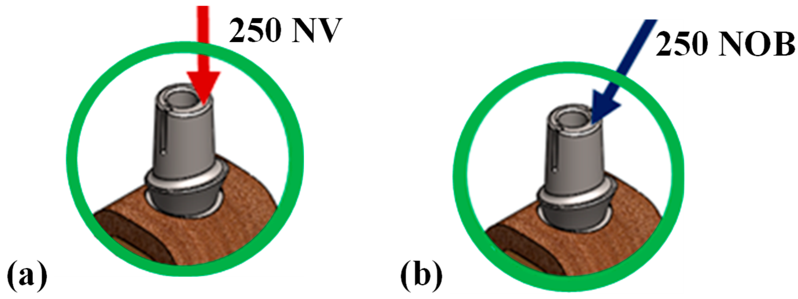
Through the utilization of the patient’s CBCT (Cone Beam Computed Tomography) data, a 3-dimensional model of a segment of the right mandibular bone was created. A section of the right mandibular region, which had a missing tooth, as 20 × 14 × 10 (L × W × T) was selected. The right mandibular portion comprised a central region of spongy cancellous bone, which was of height 20 mm and width 14 mm, and a thickness of 2 mm hard surrounding cortical bone, as shown in Figure 1a. For the analysis, we used a standard CP Ti-55 dental implant model (9 × 5 mm in length and diameter; ISPS1135; Adin Dental Implants) and two Ti alloy (Ti-6Al-4V) abutments (3.8 mm and 4.5 mm diameter, 46-2141/32462141/51, XIVE PLUS, Dentsply Sirona, Charlotte, NC, USA). The CAD models of the abutment, screw, and implant can be depicted in Figure 1b. The analysis was run over three implants made of CP Ti-55, PEEK (polyetheretherketone), and PEKK (polyetherketoneketone), each with two titanium alloy abutments of varying diameters. These models were subjected to both vertical and oblique compressive loads separately. The study was conducted on a total of 12 models mentioned in Table 1 to record von Mises stress and total deformation under two different compressive loading conditions as shown in Figure 2a,b. The Micro-CT dataset provided all the necessary dimensions for the implant and the abutment. The CT dataset was analyzed utilizing the medical image processing software MIMICS ver.-21 and 3-matic medical ver.-13.0 (Materialize, Leuven, Belgium), and 3D models of implants, abutments, and other components were designed using the SOLIDWORKS® software, v. 2023 (Dassault Systems, France). Finally, the meshing of these models was done using 10-node tetrahedral elements in ANSYS R17.2 FE software (Ansys Inc., Canonsberg, PA, USA). The assembled models from SOLIDWORKS were imported into the Ansys Workbench, ANSYS R17.2 FE software, and the file format was converted to an analysis-ready format. The mesh sizes utilized during the modeling procedure was 0.2 for the implant, abutment, and screw, and 0.3 for the cancellous and cortical bones. The finite element analysis tool’s bonded and surface-to-surface contact feature was used to look at how the implant and mandible section (cortical and cancellous bone), the implant and abutment, and the abutment and screw interfaces acted when they were loaded. Orthotropic mechanical properties to the cortical and cancellous bones were assigned to replicate the function of the bone in the mandibular region, and isotropic properties were defined to the abutment, implant, and screw, as shown in Table 2. These properties were considered from the literature: PEEK [27], CP Ti-55 [34], PEKK [16], Ti6Al4V [35], cancellous bone [36], cortical bone [37]. The literature review reports a range of different loading values, i.e., from 50 N to 500 N [38] and loading conditions, i.e., at 15-degree, 30-degree, and 45-degree from the vertical axis [38], as opted by the researchers. In this study, an axial compressive load of 250 N [39] vertically and obliquely at 30 degrees from vertical [39], as shown in Figure 2a,b, was applied separately on the top surface of the abutment, considering that the load acting on the cusp of the crown is transferred uniformly over the abutment surface. Alloy metals such as Ti and Zr, are the most utilized materials for traditional prostheses and implant restorations. To improve substructure properties, polymer materials with outstanding performance are at the leading edge of dental research, potentially decreasing the cost of prosthetic tooth rehabilitation [40]. Polymers, the most important constituents in dentistry, provide huge physical and mechanical qualities, as well as good biocompatibility. Polymers are employed in the manufacture of a diverse variety of transferable applications, restorations, and denture base materials [40,41]. Polyetheretherketone (PEEK) and polyetherketoneketone (PEKK) are novel polymers that have captured the interest of researchers because of their outstanding properties, which are suitable for diverse applications [42]. PEKK and PEKK have recently gained recognition as biomaterials with characteristics suitable for dental and medical applications [43]. They are utilized in various dental applications, encompassing therapeutic, prosthetic, and implant dentistry. They also serve multiple purposes in reconstructive, artificial, and implantable surgeries [44]. PEEK has gained attention for its potential use in orthopedic implants due to its exceptional biocompatibility, chemical resistance, sterilizability, and mechanical properties, such as stiffness and strength, which closely mimic those of human bone [45]. Additionally, PEEK does not present the same issues, with local inflammation and stress shielding commonly associated with metallic implants, making it a promising biomaterial [46]. Polyetherketoneketone (PEKK) is an advanced, high-performance polymer that has garnered widespread attention for its exceptional properties and versatility, particularly within the biomedical field [47]. PEKK shows outstanding mechanical strength, excellent chemical resistance, and high thermal stability, all of which position it as a leading material among thermoplastic composites. This methacrylate-free thermoplastic offers remarkable shock absorption and fracture resistance, making it a reliable choice for demanding environments [41]. A notable feature of PEKK is its similarity to human bone in terms of density and elastic modulus, which significantly reduces the risk of bone resorption when used in implants [48]. For the purpose of evaluating the biomechanical performance of orthodontic devices, fixed prosthetics, complete dentures, and dental implants, as well as to investigate the distribution of stress in peri-implant bone and natural or restored teeth [49,50,51,52], the finite element technique (FEM) is utilized extensively. One of the primary foci of the finite element method (FEM) is the distribution of stress, which refers to the variations in stress that occur throughout the solid structure. Stress also serves as an indicator of the magnitude of force that an item is subjected to [53], which is another function of stress. The induced stress and total deformation were assessed from the applied compressive stress across the abutment to the implant using the finite element method. Previous studies have observed that the biomechanical characteristics of the implant are dependent on the quality of the bone. Nonetheless, a gap persists in the literature concerning the material types of dental implants for different bone conditions. Consequently, the current investigation was carried out by providing implants with varying material properties under static loading conditions to determine the ideal implant material properties.

3. Results

The study determined the equivalent von Mises stress and total deformation values for the three implant materials, PEEK, PEKK, and CP Ti-55 (commercially pure titanium), in combination with 3.8 mm and 4.5 mm abutments under the specified vertical and oblique loading (250 N) conditions. Ansys Workbench software, v. 17.2, was used for static analysis of the components.

3.1. Von Mises Stress Analysis

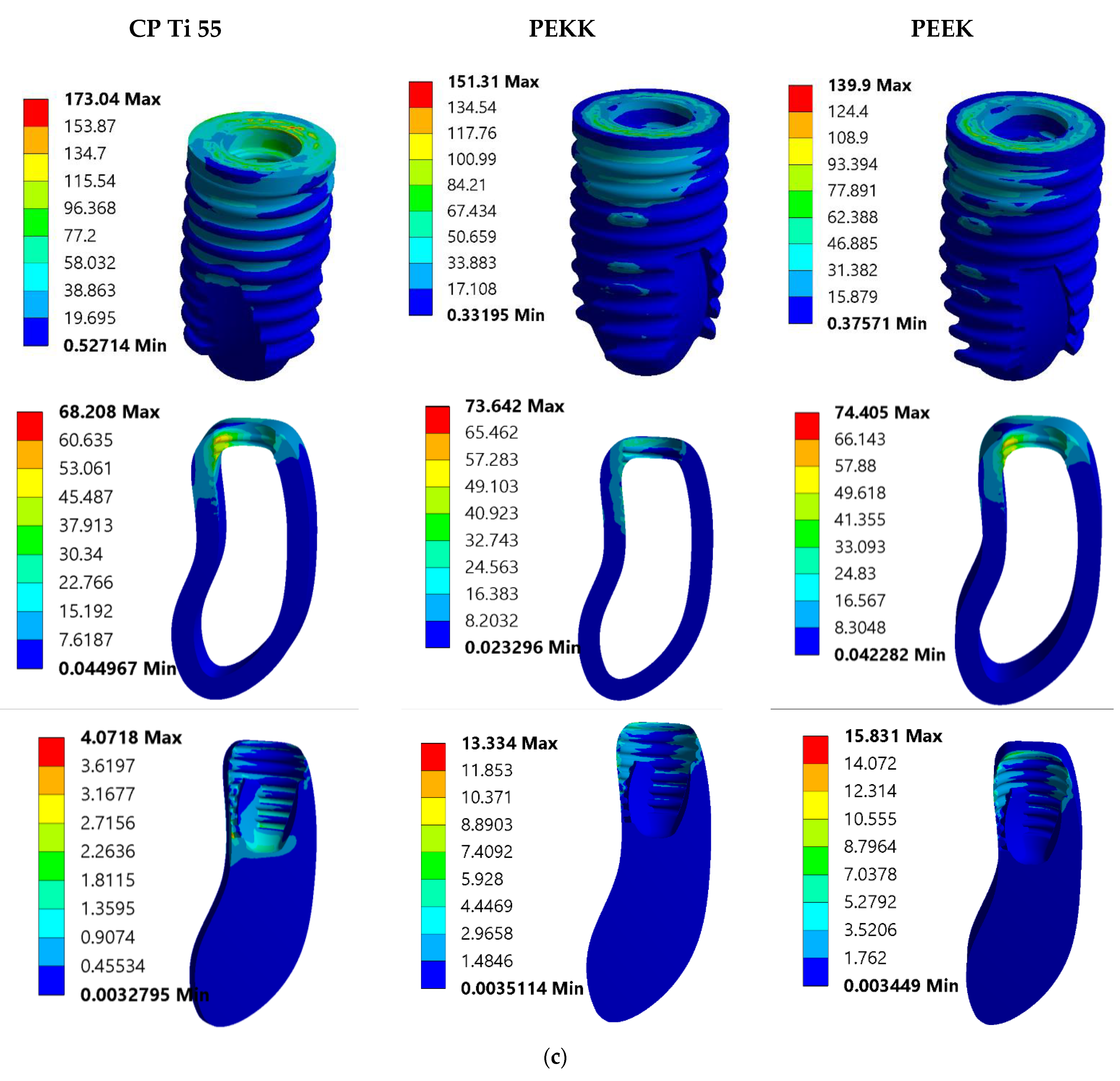
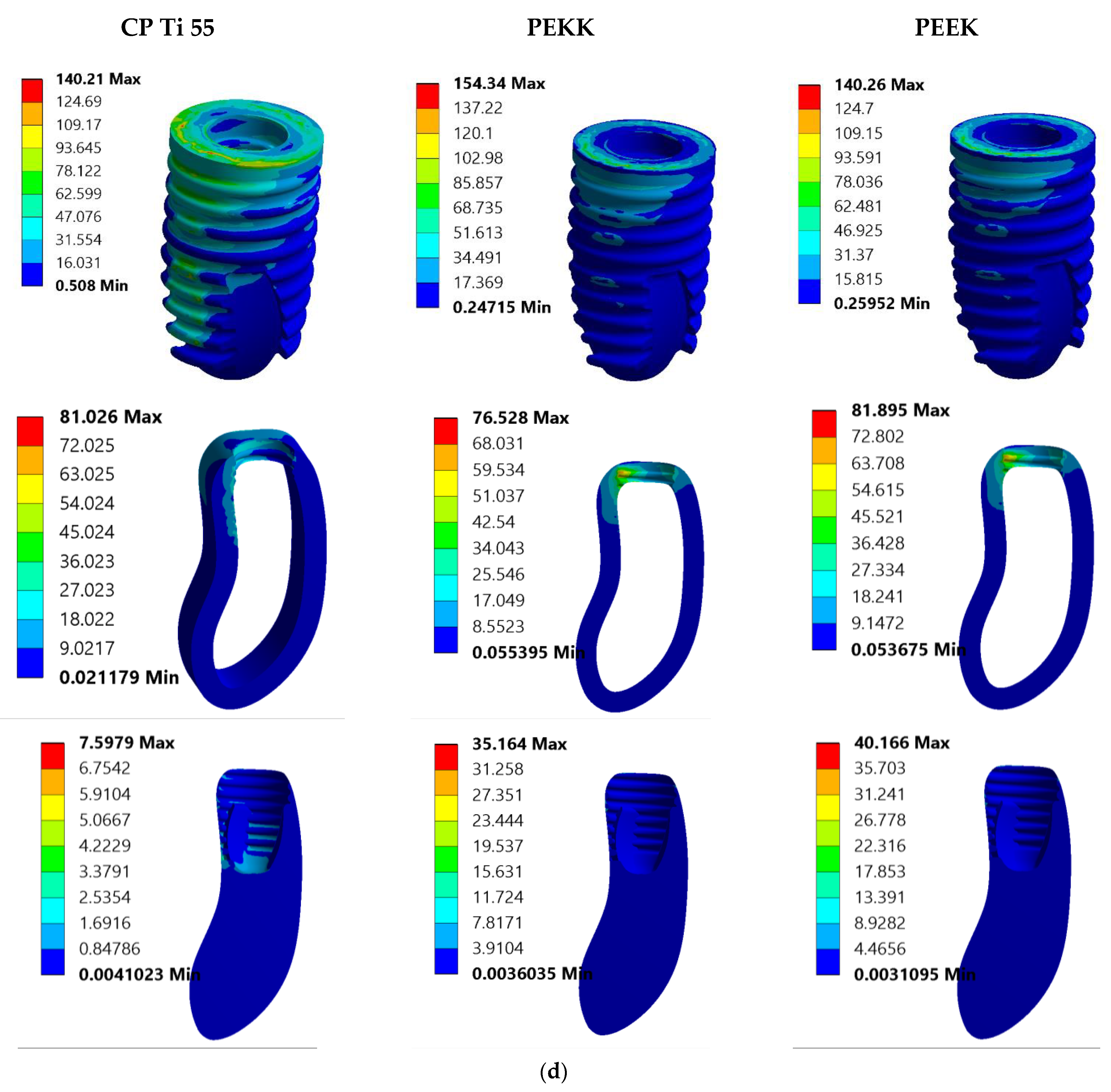
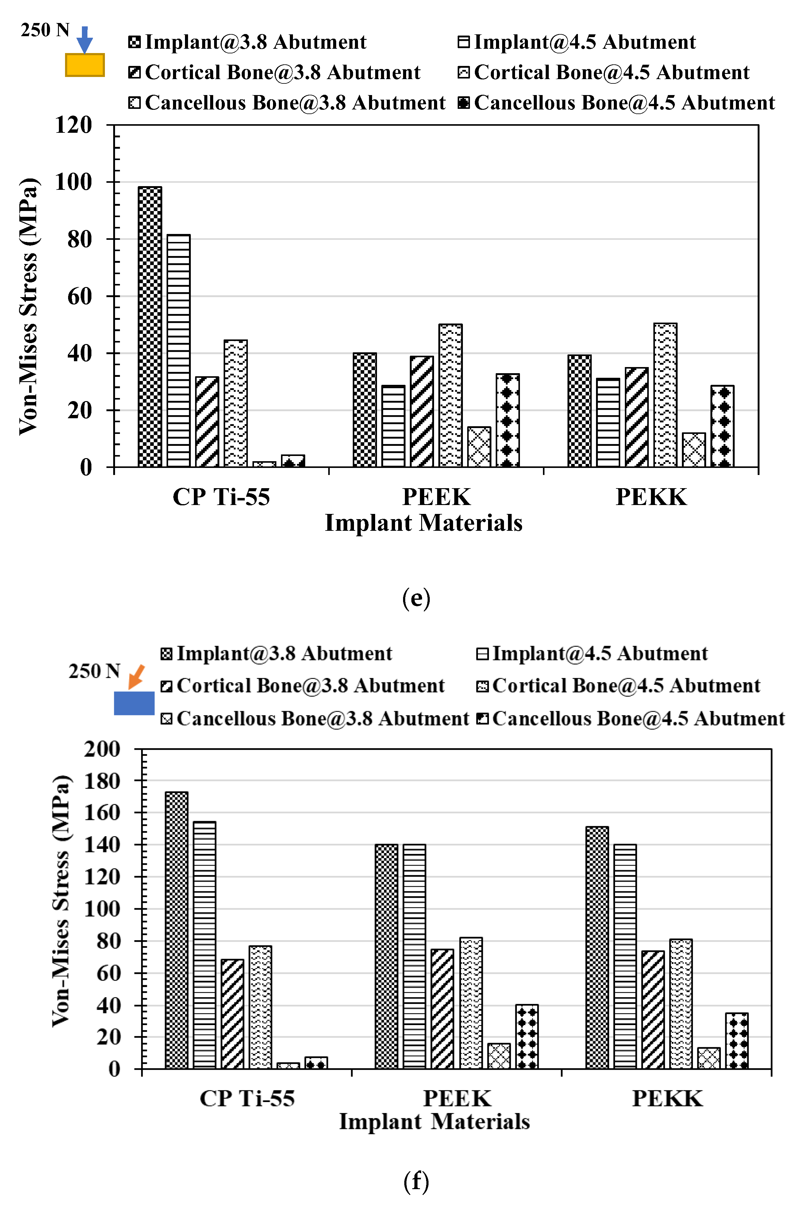
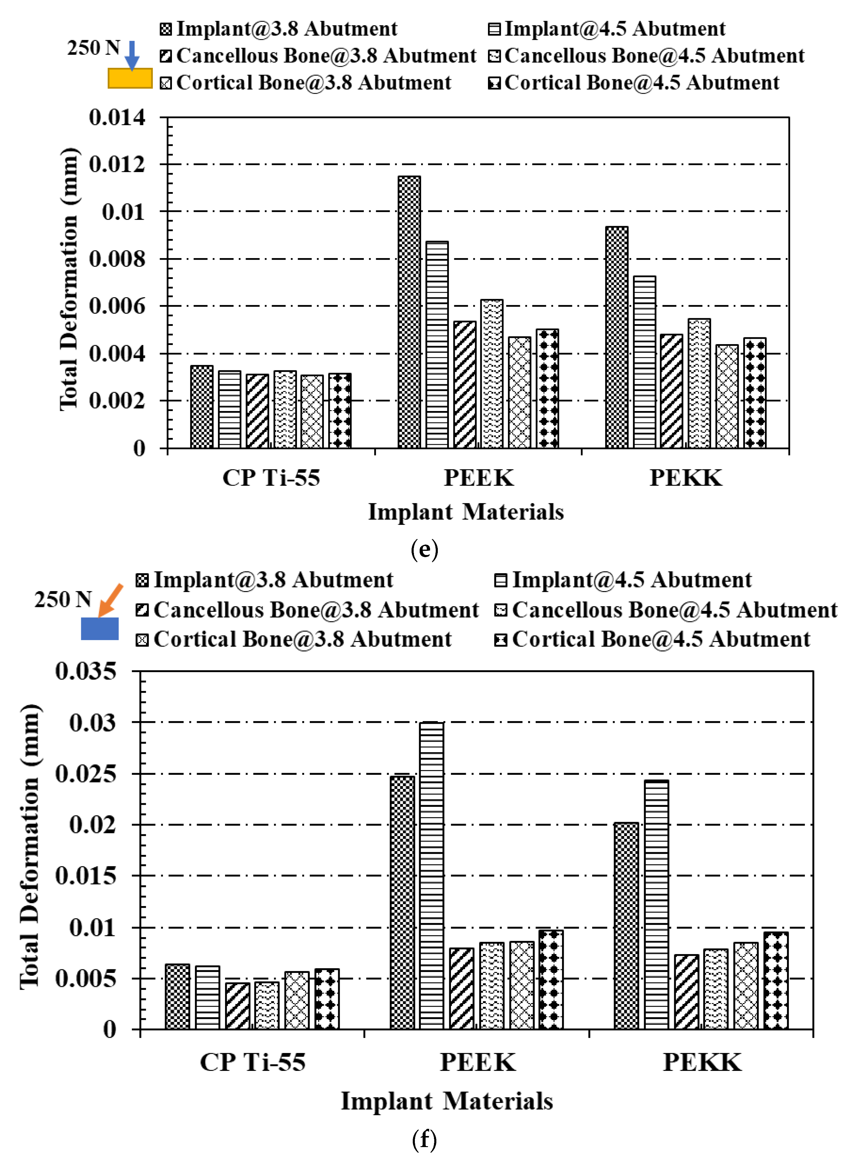
Figure 3a,b shows the effect of von Mises stress on implants and cortical and cancellous bone under 250 N vertical loadings on the occlusal surface of 3.8 mm and 4.5 mm abutments, respectively. The CP Ti-55 implant exhibits the highest strength of 98.243 MPa, with PEEK at 39.314 MPa, while the minimum is in PEKK at 34.958 MPa. Cortical bone experiences a minimum stress of 31.609 MPa when the titanium implant was used and a maximum of 38.847 MPa when the PEEK implant was used. Similarly, cancellous bone experiences minimum with a titanium implant and maximum with a PEEK implant, when a 3.8 mm abutment was placed over the implant. A similar trend for von Mises stress was recorded when a 4.5 mm abutment was placed over the implants. Under an oblique load of 250 N at 30 degrees from the vertical axis, maximum von Mises stress was recorded as 173.04 MPa for the CP Ti-55 implant and a minimum of 139.9 MPa in the PEEK implant. Cortical bone experiences a minimum stress of 68.208 MPa when the titanium implant was used, 73.642 MPa for the PEKK implant, and a maximum of 74.405 MPa for the PEEK implant. Cancellous bone experiences a minimum stress of 4.0718 MPa when a titanium implant was used, 13.334 MPa in a PEKK implant, and 15.831 MPa when a PEEK implant was used under a 3.8 mm abutment as shown in Figure 3c. Similar results were recorded for implants and cortical and cancellous bone when a 4.5 mm abutment was set over the implants, as shown in Figure 3d. The trend for von Mises stress in different components like implants and cortical and cancellous bone for different implant materials (CP Ti-55, PEEK, and PEKK) under two abutment diameters can be depicted from Figure 3e,f. When the von Mises stress was compared to under 3.8 mm and 4.5 mm abutment in different components, the values in implants (CP Ti-55, PEEK, and PEKK) were higher in 3.8 mm than 4.5 mm abutment, while in cortical and cancellous bone, values were lower in 3.8 mm than 4.5 mm under vertical loading. The stress value under oblique loading conditions was high in the CP Ti-55 implant in 3.8 mm than in 4.5 mm, but opposite results were found in PEEK and PEKK implants, as shown in Figure 3e. But cortical and cancellous bones exhibited higher stress values in the case of 4.5 mm abutment than 3.8 mm, when examined under oblique loading as shown in Figure 3f.

3.2. Total Deformation Analysis

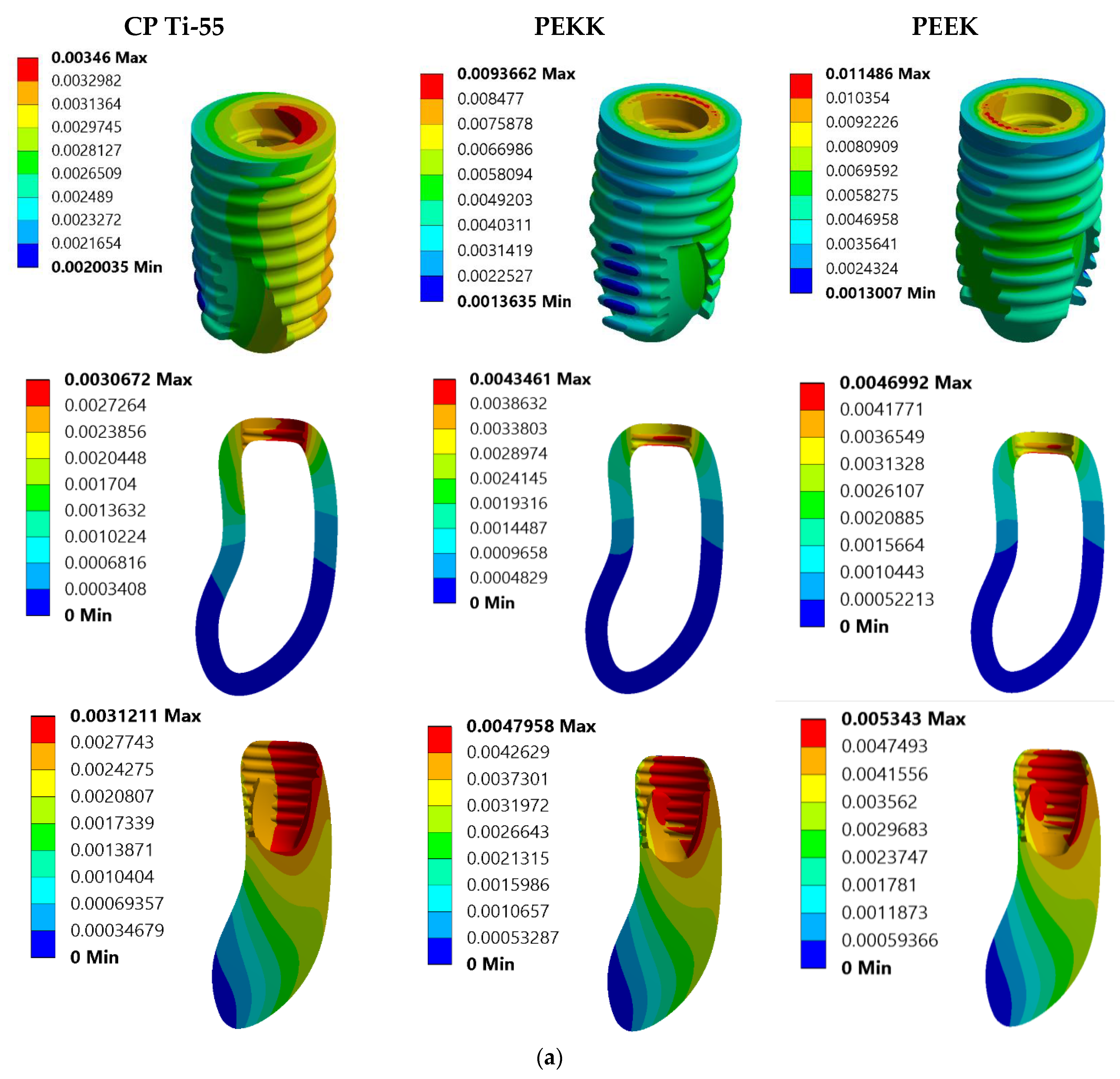
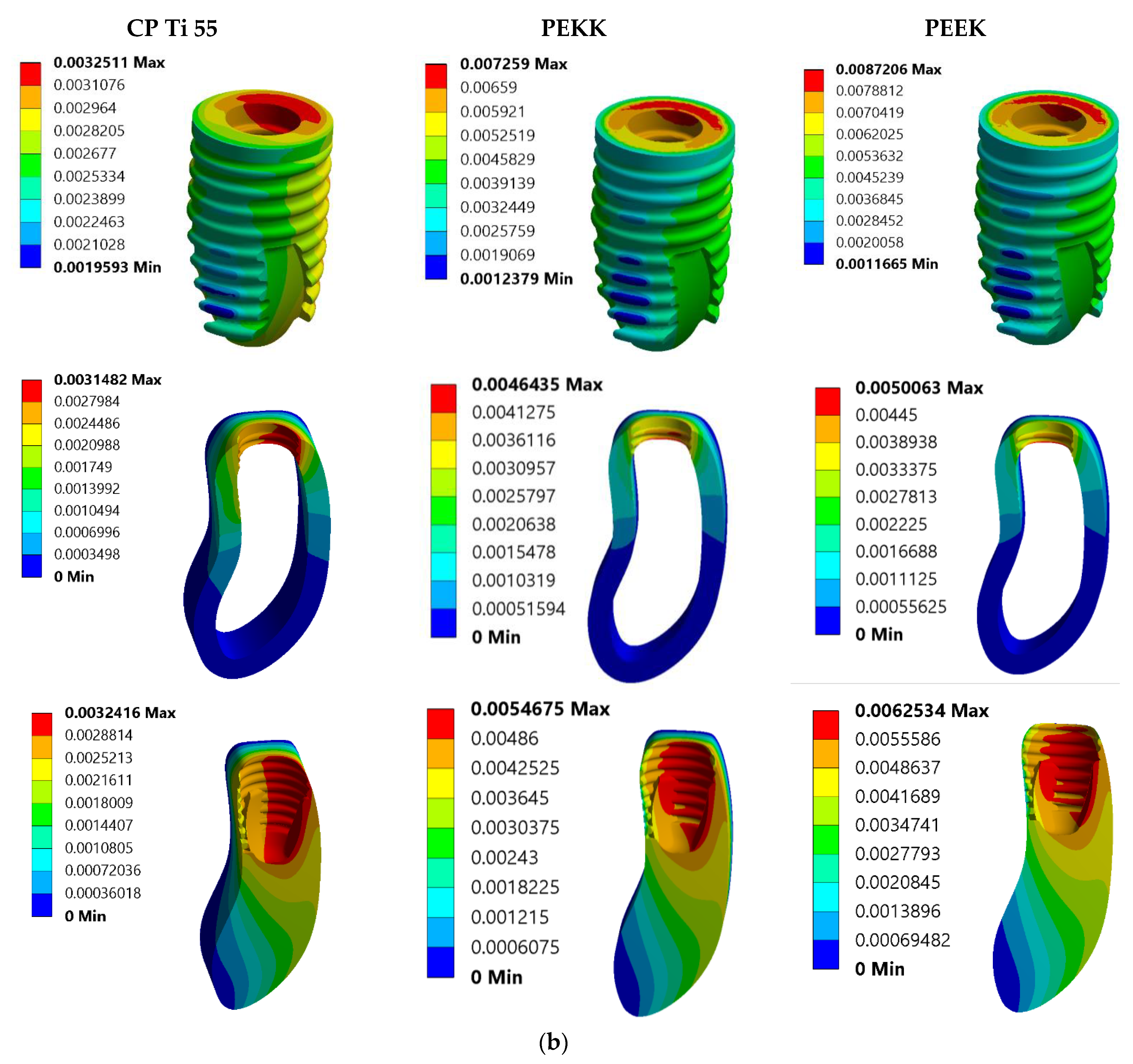
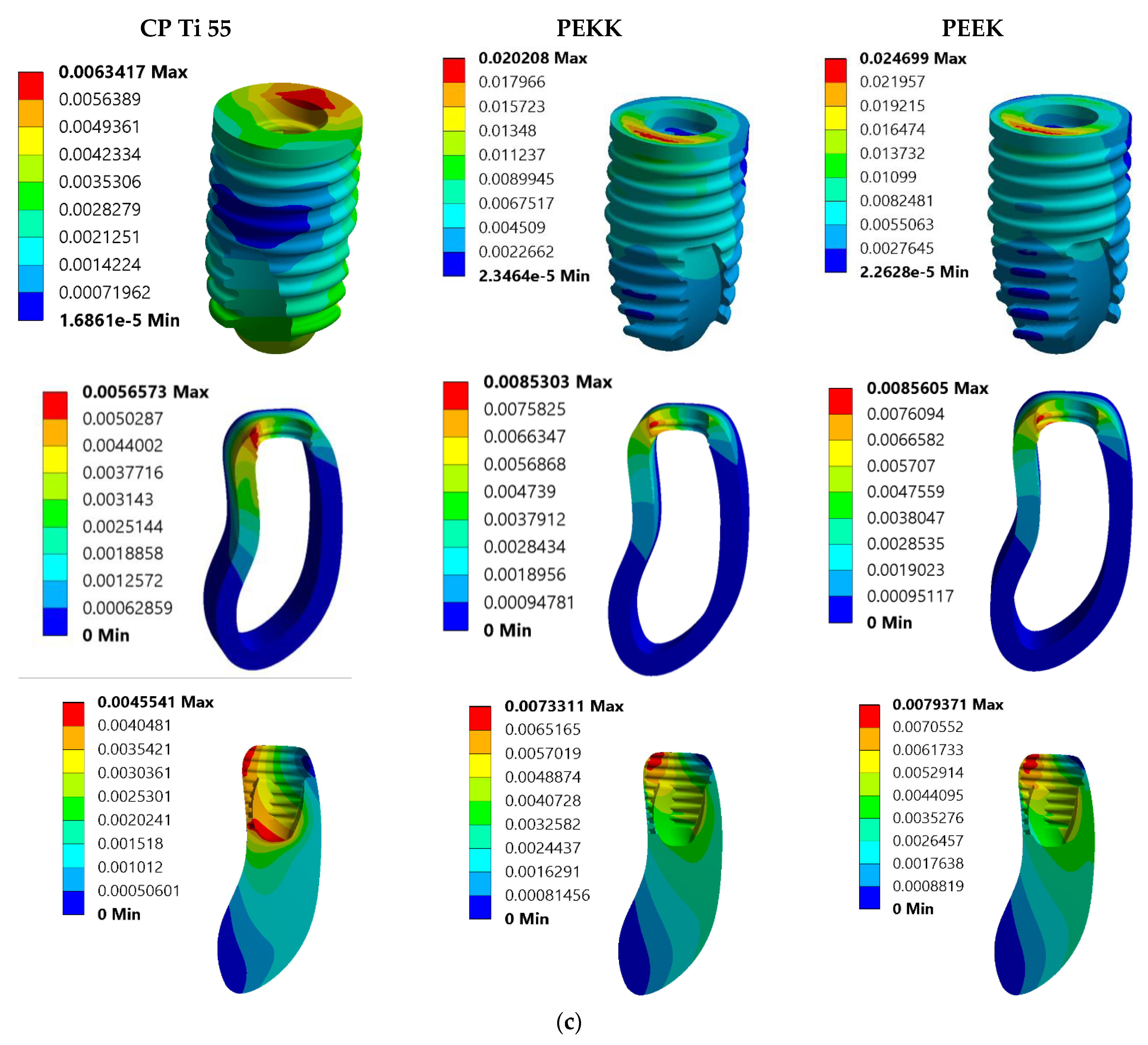
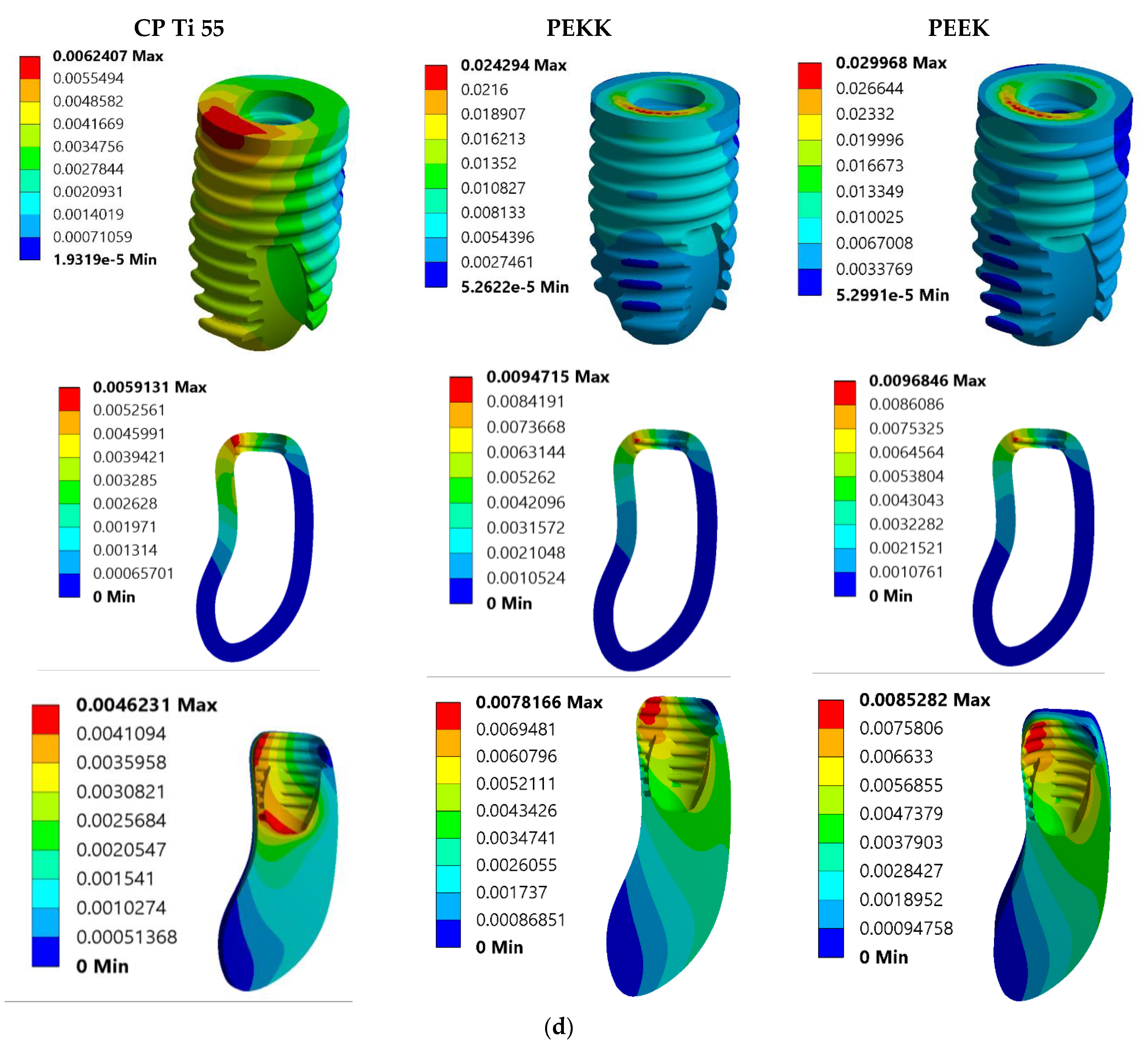
Figure 4a,b shows the effect of total deformation on implants and cortical and cancellous bone under 250 N vertical loadings on the occlusal surface of 3.8 mm and 4.5 mm abutment respectively. The PEEK implant exhibits the highest deformation of 0.011486 mm, and the PEKK implant is 0.0093662 mm while the minimum in CP Ti-55 is 0.00346 mm. Cortical bone experiences a minimum deformation of 0.0030672 mm when a titanium implant was used and a maximum 0.0046992 mm when a PEEK implant was used. Similarly, cancellous bone experiences minimum deformation with titanium implant and maximum deformation with PEEK implant, when 3.8 mm abutment was placed over the implant. A similar trend for total deformation was recorded when 4.5 mm abutment was placed over the implants. Under an oblique load of 250 N at 30 degrees from the vertical axis, maximum deformation was recorded as 0.024699 mm for the PEEK implant and minimum deformation was 0.006341 mm in the titanium implant. Cortical bone experienced a minimum deformation of 0.0056573 mm when the titanium implant was used, 0.0085303 mm for the PEKK implant, and a maximum of 0.0085605 mm for the PEEK implant. Cancellous bone experiences minimum deformation of 0.0045541 mm when a titanium implant was used, 0.0073311 mm in a PEKK implant, and 0.0079371 mm when a PEEK implant was used under a 3.8 mm abutment, as shown in Figure 4c. Similar results were recorded for implants and cortical and cancellous bone when a 4.5 mm abutment was set over the implants, as shown in Figure 4d. The trend for total deformation in different components like implants and cortical and cancellous bone for different implant materials (CP Ti-55, PEEK, and PEKK) under two abutment diameters can be depicted from Figure 4e,f. When the total deformation was compared to under 3.8 mm and 4.5 mm abutment in different components, the values in implants (CP Ti-55, PEEK, and PEKK) were higher in 3.8 mm than 4.5 mm abutment, while in cortical and cancellous bone, values were lower in 3.8 mm than 4.5 mm under vertical loading. The deformation value under oblique loading conditions was high in the CP Ti-55 implant at 3.8 mm, compared to 4.5 mm, but opposite results were found in PEEK and PEKK implants. Cortical and cancellous bone exhibited higher deformation values in the case of 4.5 mm abutment than 3.8 mm.

4. Discussion

In comparison to fixed partial dentures and removable partial dentures, dental implants have quickly become the treatment modality of choice for patients who are fully or partially edentulous. This is due to the comparatively high success rate that dental implants have in comparison to these other options. Numerous elements—which can be broken down into three categories: surgical factors, host-related factors, and implant or occlusion-related factors—all have a role in the successful combination of osseointegration and its maintenance. In order to effectively maintain implant osseointegration and promote good remodeling, it is imperative that the degree of mechanical stress that is applied to the neighboring bone remains within the limitations that are established [54]. Titanium was chosen for inclusion in the research project because of its reputation as the material of choice for the fabrication of dental implants, which are used in the rehabilitation of patients who are either partially or completely edentulous [55]. In light of the fact that multiple studies have indicated that PEEK is a superior option to titanium implants due to its modulus of elasticity being comparable to that of human jawbone, this study examined both PEEK and PEKK implants along with CP Ti-55 implants [56]. Even though implants are used in a wide variety of applications, there are still a great deal of aspects linked to their biomechanical qualities that are not fully understood. There is a considerable impact that biomechanical factors have on the preservation of the bone-implant contact [57]. In contrast to natural teeth, which have a stress-relieving mechanism that is evenly distributed by the periodontal ligament, implants do not have this mechanism [58]. Following that, a three-dimensional finite element analysis was performed to analyze the mechanical properties of three different implant materials in the mandible having missing teeth. The failure mechanism, comprising von Mises stresses and total deformation were some of the features identified.
This analysis assessed the von Mises stress and total deformation of all components, including implants, cortical bone, and cancellous bone. The study showed that the CP Ti-55 implant is much superior to the polymeric biomaterials PEEK and PEKK. Under 250 N vertical loading, the CP Ti-55 implant exhibited a 64.42% superior strength relative to PEEK and a 59.98% superiority over PEKK, whereas PEKK had an 11.08% greater strength than the PEEK implant. A reduction of 41.59% in distortion of the cancellous bone was observed with the CP Ti-55 implant in comparison to PEEK, and a 34.92% reduction was noted with PEKK. Cortical bone exhibits 34.73% less distortion when the CP Ti-55 implant, compared to PEEK and 29.43% compared to PEKK. Similar trends were observed in the strength of CP Ti-55, PEEK, and PEKK implants, as well as in deformations of cancellous and cortical bone under 250 N oblique (30-degree) loading conditions. Consequently, CP Ti-55 implants demonstrate reduced stress and deformation in peri-implant bone, leading to diminished marginal bone loss relative to PEEK and PEKK. Thus, PEEK transmitted slightly higher stress compared to CP Ti-55 and PEKK. This is similar to the study conducted by Andreas Schwitalla, where the CRF-PEEK implants presented higher load concentration in the cervical area and cortical bone than the titanium implants [59]. Due to its significantly low modulus of elasticity and lack of lateral contact with surrounding structures, the PEEK implant experienced higher stress values than the CP Ti-55 and PEKK implant. This is because the PEEK implant transfers stress to the implant as well as the bone tissue that surrounds the implant. This result is consistent with the findings of Jao Rodrigo and colleagues, who used a three-dimensional finite element method to analyze the stress distribution in CRF-PEEK and titanium dental implants. Their findings showed that CRF-PEEK exhibited a higher stress concentration at the implant neck than titanium did as a result of its decreased stiffness and increased deformation. The authors come to the conclusion that the titanium implant was superior to the CFR-PEEK implant in terms of characteristics [60]. A similar pattern was observed in the finite element analysis that was carried out by Harinee A. and her colleagues [61]. All the assessed parameters clearly indicate why titanium continues to be the gold standard material of choice for dental implant treatment. Despite the differences in outcome results between titanium and polymeric materials, further research and modifications suggest that PEEK and PEKK are preferable for patients with titanium hypersensitivity and for those seeking metal-free implants. BIC values of PEEK obtained in studies conducted on animals presented a desirable osseointegration in comparison to the BIC values of the titanium implants [62,63]. Recent studies and review articles aim at proving that newer polymeric biomaterials, such as PEEK and PEKK, are better than conventional CP Ti-55 used in dentistry. However, in our study, CP Ti-55 outperformed both the opted polymeric biomaterials, PEEK and PEKK. Thus, for better biological response of peri-implant bone, PEEK and PEKK require surface modifications and treatments. Current research is going on plasma treatment, laser treatment, etching, grafting, hydrogel coatings, antimicrobial coatings, composite polymer coatings, etc., to further enhance these polymeric biomaterials for use as dental implants [64,65,66].
It is essential to acknowledge certain intrinsic limitations of finite element analysis research [67,68].
  • The loading criteria in this study were distilled down to a singular force (vertical and oblique individually), and the boundary condition was established as fixed at designated sites.
  • Because they were not subject to any kind of flaw population, the virtual materials were thought of as orthotropic and isotropic.
  • There was no attempt to mimic sliding contacts, operator mistake, or vertical misalignment of the prosthesis. While the linear contact between screws and polymeric materials used in models may not provide an exact representation of the stress condition under loading, it is standardized and allows for easier comparison.
  • Computer-based finite element analysis predicts bone or implant component failure probabilistically. Tissue reaction, blood circulation, growth hormones, microbial colonization, and cleanliness habits are ignored.

5. Conclusions

The approach of this research article is towards the application of newer polymeric biomaterials of the PEAK family in dental implant therapy. Although PEEK and PEKK can help in reducing the manufacturing cost of dental implants, the finite element analysis fails to prove the outperformance of PEEK and PEKK implants over traditionally used Titanium implants, as shown by the maximum stress tolerated and total deformation experienced. Titanium implant exhibited the minimal degree of total deformation under all loading conditions, and bone response was also better with titanium. Various techniques like optimizing morphology, porosity, wettability, chemical composition, and surface bioactivity may enhance the biomechanical response of PEEK and PEKK. To confirm the differences of biomechanical characteristics and the resultant biological response among CP Ti-55, PEEK, and PEKK, additional clinical trials with long-term follow-ups are suggested.

Author Contributions

Conceptualization, M.A., A.C., V.A. and S.A.; methodology, M.A., S.A. and A.C.; software, M.A.; validation, M.A., V.A., S.A. and A.C.; formal analysis, M.A., V.A., S.A. and A.C.; investigation, M.A., V.A., S.A. and A.C.; data curation, M.A., S.A. and A.C.; writing—original draft preparation, M.A.; writing—review and editing, M.A., V.A., S.A. and A.C.; supervision, A.C. All authors have read and agreed to the published version of the manuscript.

Funding

This research received no external funding.

Institutional Review Board Statement

Not applicable.

Informed Consent Statement

Not applicable.

Data Availability Statement

The datasets generated during and/or analyzed during this current study are not publicly available due to the fact they are large datasets, but are available from the corresponding author upon reasonable request.

Conflicts of Interest

The authors declare no conflicts of interest.

References

  1. Paxton, N.C.; Dinoro, J.; Ren, J.; Ross, M.T.; Daley, R.; Zhou, R.; Bazaka, K.; Thompson, R.G.; Yue, Z.; Beirne, S.; et al. Additive manufacturing enables personalised porous high-density polyethylene surgical implant manufacturing with improved tissue and vascular ingrowth. Appl. Mater. Today 2021, 22, 100965. [Google Scholar] [CrossRef]
  2. Roina, Y.; Auber, F.; Hocquet, D.; Herlem, G. ePTFE-based biomedical devices: An overview of surgical efficiency. J. Biomed. Mater. Res. Part B Appl. Biomater. 2021, 110, 302–320. [Google Scholar] [CrossRef]
  3. Kaur, H.; Thakur, A. Applications of poly(methyl methacrylate) polymer in dentistry: A review. Mater. Today Proc. 2022, 50, 1619–1625. [Google Scholar] [CrossRef]
  4. Etinosa, P.O.; Osuchukwu, O.A.; Anisiji, E.O.; Lawal, M.Y.; Mohammed, S.A.; Ibitoye, O.I.; Oni, P.G.; Aderibigbe, V.D.; Aina, T.; Oyebode, D.; et al. In-depth review of synthesis of hydroxyapatite biomaterials from natural resources and chemical regents for biomedical applications. Arab. J. Chem. 2024, 17, 106010. [Google Scholar] [CrossRef]
  5. Zol, S.M.; Alauddin, M.S.; Said, Z.; Ghazali, M.I.M.; Hao-Ern, L.; Farid, D.A.M.; Zahari, N.A.H.; Al-Khadim, A.H.A.; Aziz, A.H.A. Description of Poly(aryl-ether-ketone) Materials (PAEKs), Polyetheretherketone (PEEK) and Polyetherketoneketone (PEKK) for Application as a Dental Material: A Materials Science Review. Polymers 2023, 15, 2170. [Google Scholar] [CrossRef]
  6. Hong, D.G.K.; Oh, J. Recent advances in dental implants. Maxillofac. Plast. Reconstr. Surg. 2017, 39, 33. [Google Scholar] [CrossRef]
  7. Shemtov-Yona, K.; Rittel, D. An Overview of the Mechanical Integrity of Dental Implants. BioMed Res. Int. 2015, 2015, 547384. [Google Scholar] [CrossRef]
  8. Ong, G. Periodontal disease and tooth loss. Int. Dent. J. 1998, 48, 233–238. [Google Scholar] [CrossRef]
  9. Gabiec, K.; Bagińska, J.; Łaguna, W.; Rodakowska, E.; Kamińska, I.; Stachurska, Z.; Dubatówka, M.; Kondraciuk, M.; Kamiński, K.A. Factors Associated with Tooth Loss in General Population of Bialystok, Poland. Int. J. Environ. Res. Public Health 2022, 19, 2369. [Google Scholar] [CrossRef]
  10. Wiener, R.C.; Shen, C.; Findley, P.A.; Sambamoorthi, U.; Tan, X. The association between diabetes mellitus, sugar-sweetened beverages, and tooth loss in adults. J. Am. Dent. Assoc. 2017, 148, 500–509.e4. [Google Scholar] [CrossRef]
  11. Kadhim, D.R.; Hamad, T.I.; Fatalla, A.A. Use of Eggshells as Bone Grafts around Commercially Pure Titanium Implant Screws Coated with Nano Calcium Sulfate. Int. J. Biomater. 2022, 2022, 8722283. [Google Scholar] [CrossRef]
  12. Țap, M.D.; Bîcleşanu, F.C.; Honțaru, O.-S.; Radu, A.-C. Patient Centricity-An Empirical Research on Titanium Dental Implants and Their Adverse Effects on Health Condition. Healthcare 2024, 12, 2207. [Google Scholar] [CrossRef]
  13. Silva, R.C.S.; Agrelli, A.; Andrade, A.N.; Mendes-Marques, C.L.; Arruda, I.R.S.; Santos, L.R.L.; Vasconcelos, N.F.; Machado, G. Titanium Dental Implants: An Overview of Applied Nanobiotechnology to Improve Biocompatibility and Prevent Infections. Materials 2022, 15, 3150. [Google Scholar] [CrossRef]
  14. Belkhode, V.; Godbole, S.; Nimonkar, S.; Nimonkar, P.; Pisulkar, S. Comparative evaluation of the efficacy of customized maxillary oral appliance with mandibular advancement appliance as a treatment modality for moderate obstructive sleep apnea patients-protocol for a randomized controlled trial. Trials 2022, 23, 159. [Google Scholar] [CrossRef]
  15. Panayotov, I.V.; Orti, V.; Cuisinier, F.; Yachouh, J. Polyetheretherketone (PEEK) for medical applications. J. Mater. Sci. Mater. Med. 2016, 27, 118. [Google Scholar] [CrossRef]
  16. AlOtaibi, N.; Naudi, K.; Conway, D.; Ayoub, A. The current state of PEEK implant osseointegration and future perspectives: A systematic review. Eur. Cells Mater. 2020, 40, 1–20. [Google Scholar] [CrossRef]
  17. Ma, T.; Zhang, J.; Sun, S.; Meng, W.; Zhang, Y.; Wu, J. Current treatment methods to improve the bioactivity and bonding strength of PEEK for dental application: A systematic review. Eur. Polym. J. 2023, 183, 111757. [Google Scholar] [CrossRef]
  18. Jung, H.-D.; Jang, T.-S.; Lee, J.E.; Park, S.J.; Son, Y.; Park, S.-H. Enhanced bioactivity of titanium-coated polyetheretherketone implants created by a high-temperature 3D printing process. Biofabrication 2019, 11, 045014. [Google Scholar] [CrossRef]
  19. de Araújo Nobre, M.; Moura Guedes, C.; Almeida, R.; Silva, A. Poly-ether-ether-ketone and implant dentistry: The future of mimicking natural dentition is now! Polym. Int. 2021, 70, 999–1001. [Google Scholar] [CrossRef]
  20. Johansson, P.; Jimbo, R.; Kozai, Y.; Sakurai, T.; Kjellin, P.; Currie, F.; Wennerberg, A. Nanosized Hydroxyapatite Coating on PEEK Implants Enhances Early Bone Formation: A Histological and Three-Dimensional Investigation in Rabbit Bone. Materials 2015, 8, 3815–3830. [Google Scholar] [CrossRef]
  21. Li, L.; Zhou, C.; Wei, J.; Ma, J. Quantitative analysis of nFA/PEEK implant interfaces in Beagle dogs. Shanghai Kou Qiang Yi Xue 2014, 23, 166–171. [Google Scholar] [PubMed]
  22. Belkhode, V.M.; Nimonkar, S.V.; Godbole, S.R.; Nimonkar, P.; Sathe, S.; Borle, A. Evaluation of the effect of different surface treatments on the bond strength of non-precious alloy—Ceramic interface: An SEM study. J. Dent. Res. Dent. Clin. Dent. Prospect. 2019, 13, 200–207. [Google Scholar] [CrossRef] [PubMed]
  23. Meningaud, J.P.; Spahn, F.; Donsimoni, J.M. After titanium, peek? Rev. De Stomatol. Et De Chir. Maxillo Faciale 2012, 113, 407–410. [Google Scholar] [CrossRef]
  24. Hoskins, T.J.; Dearn, K.D.; Kukureka, S.N. Mechanical performance of PEEK produced by additive manufacturing. Polym. Test. 2018, 70, 511–519. [Google Scholar] [CrossRef]
  25. Kewekordes, T.; Wille, S.; Kern, M. Wear of polyetherketoneketones—Influence of titanium dioxide content and antagonistic material. Dent. Mater. 2018, 34, 560–567. [Google Scholar] [CrossRef]
  26. Polymer Science: A Comprehensive Reference | ScienceDirect. Available online: https://www.sciencedirect.com/referencework/9780080878621/polymer-science-a-comprehensive-reference (accessed on 10 December 2024).
  27. Alsadon, O.; Wood, D.; Patrick, D.; Pollington, S. Fatigue behavior and damage modes of high performance poly-ether-ketone-ketone PEKK bilayered crowns. J. Mech. Behav. Biomed. Mater. 2020, 110, 103957. [Google Scholar] [CrossRef]
  28. Elkabbany, A.; Kern, M.; Elkhadem, A.H.; Wille, S.; Amer, A.A.; Chaar, M.S. Retention of metallic and non-metallic double-crown-retained mandibular overdentures on implants: An in-vitro study. J. Prosthodont. Res. 2020, 64, 384–390. [Google Scholar] [CrossRef]
  29. Schwitalla, A.D.; Spintig, T.; Kallage, I.; Müller, W.D. Flexural behavior of PEEK materials for dental application. Dent. Mater. 2015, 31, 1377–1384. [Google Scholar] [CrossRef] [PubMed]
  30. Erdemir, A.; Guess, T.M.; Halloran, J.; Tadepalli, S.C.; Morrison, T.M. Considerations for reporting finite element analysis studies in biomechanics. J. Biomech. 2012, 45, 625–633. [Google Scholar] [CrossRef]
  31. Pankaj, P. Patient-specific modelling of bone and bone-implant systems: The challenges. Int. J. Numer. Methods Biomed. Eng. 2013, 29, 233–249. [Google Scholar] [CrossRef]
  32. Cowin, S.C. Bone Mechanics Handbook; CRC Press: Boca Raton, FL, USA, 2001. [Google Scholar]
  33. Taylor, M.; Prendergast, P.J. Four decades of finite element analysis of orthopaedic devices: Where are we now and what are the opportunities? J. Biomech. 2015, 48, 767–778. [Google Scholar] [CrossRef]
  34. Afazal, M.; Gupta, S.; Tevatia, A.; Afreen, S.; Chanda, A. Computational Investigation of Dental Implant Restoration Using Platform-Switched and -Matched Configurations. Computation 2023, 11, 79. [Google Scholar] [CrossRef]
  35. Afazal, M.; Afreen, S.; Chanda, A. Computational modelling and analysis of hard tissue behavior around 0.5 mm and 0.85 mm platform switched abutment using 3D finite element analysis. Forces Mech. 2023, 13, 100243. [Google Scholar] [CrossRef]
  36. Gačnik, F.; Ren, Z.; Hren, N.I. Modified bone density-dependent orthotropic material model of human mandibular bone. Med Eng. Phys. 2014, 36, 1684–1692. [Google Scholar] [CrossRef]
  37. Dhatrak, P.; Girme, V.; Shirsat, U.; Sumanth, S.; Deshmukh, V. Significance of Orthotropic Material Models to Predict Stress Around Bone-Implant Interface Using Numerical Simulation. BioNanoScience 2019, 9, 652–659. [Google Scholar] [CrossRef]
  38. Falcinelli, C.; Valente, F.; Vasta, M.; Traini, T. Finite element analysis in implant dentistry: State of the art and future directions. Dent. Mater. 2023, 39, 539–556. [Google Scholar] [CrossRef] [PubMed]
  39. Barbosa, F.T.; Zanatta, L.C.S.; de Souza Rendohl, E.; Gehrke, S.A. Comparative analysis of stress distribution in one-piece and two-piece implants with narrow and extra-narrow diameters: A finite element study. PLoS ONE 2021, 16, e0245800. [Google Scholar] [CrossRef]
  40. Xu, X.; He, L.; Zhu, B.; Li, J.; Li, J. Advances in polymeric materials for dental applications. Polym. Chem. 2017, 8, 807–823. [Google Scholar] [CrossRef]
  41. Najeeb, S.; Zafar, M.S.; Khurshid, Z.; Siddiqui, F. Applications of polyetheretherketone (PEEK) in oral implantology and prosthodontics. J. Prosthodont. Res. 2016, 60, 12–19. [Google Scholar] [CrossRef]
  42. Rokaya, D.; Srimaneepong, V.; Sapkota, J.; Qin, J.; Siraleartmukul, K.; Siriwongrungson, V. Polymeric materials and films in dentistry: An overview. J. Adv. Res. 2018, 14, 25–34. [Google Scholar] [CrossRef] [PubMed]
  43. Mechanical Performances of PEKK Thermoplastic Composites Linked to Their Processing Parameters. Available online: https://www.researchgate.net/publication/327952758_Mechanical_performances_of_PEKK_thermoplastic_composites_linked_to_their_processing_parameters (accessed on 12 June 2025).
  44. Levy-Marchal, C.; Czernichow, P. Cyclosporin A in insulin-dependent diabetes mellitus of recent onset: A pilot study in children. Horm. Res. 1988, 29, 177–184. [Google Scholar] [CrossRef]
  45. PEEK vs PEKK: Which High Performance Material Should You Choose?—3Dnatives. Available online: https://www.3dnatives.com/en/peek-vs-pekk-240620214/ (accessed on 12 June 2025).
  46. McMullan, R.; Golbang, A.; Salma-Ancane, K.; Ward, J.; Rodzen, K.; Boyd, A.R. Review of 3D Printing of Polyaryletherketone/Apatite Composites for Lattice Structures for Orthopedic Implants. Appl. Sci. 2025, 15, 1804. [Google Scholar] [CrossRef]
  47. Alqurashi, H.; Khurshid, Z.; Syed, A.U.Y.; Habib, S.R.; Rokaya, D.; Zafar, M.S. Polyetherketoneketone (PEKK): An emerging biomaterial for oral implants and dental prostheses. J. Adv. Res. 2021, 28, 87–95. [Google Scholar] [CrossRef]
  48. Buck, E.; Li, H.; Cerruti, M. Surface Modification Strategies to Improve the Osseointegration of Poly(etheretherketone) and Its Composites. Macromol. Biosci. 2020, 20, 1900271. [Google Scholar] [CrossRef] [PubMed]
  49. Knop, L.; Gandini, L.G.; Shintcovsk, R.L.; Gandini, M.R.E.A.S. Scientific use of the finite element method in Orthodontics. Dent. Press J. Orthod. 2015, 20, 119–125. [Google Scholar] [CrossRef]
  50. Matsuoka, T.; Nakano, T.; Yamaguchi, S.; Ono, S.; Watanabe, S.; Sato, T.; Yatani, H. Effects of Implant–Abutment Connection Type and Inter-Implant Distance on Inter-Implant Bone Stress and Microgap: Three-Dimensional Finite Element Analysis. Materials 2021, 14, 2421. [Google Scholar] [CrossRef]
  51. Reddy, M.; Sundram, R.; Eid Abdemagyd, H. Application of finite element model in implant dentistry: A systematic review. J. Pharm. Bioallied Sci. 2019, 11, S85–S91. [Google Scholar] [CrossRef] [PubMed]
  52. Romanyk, D.L.; Vafaeian, B.; Addison, O.; Adeeb, S. The use of finite element analysis in dentistry and orthodontics: Critical points for model development and interpreting results. Semin. Orthod. 2020, 26, 162–173. [Google Scholar] [CrossRef]
  53. Menacho-Mendoza, E.; Cedamanos-Cuenca, R.; Díaz-Suyo, A. Stress analysis and factor of safety in three dental implant systems by finite element analysis. Saudi Dent. J. 2022, 34, 579–584. [Google Scholar] [CrossRef]
  54. Weinstein, A.M.; Klawitter, J.J.; Anand, S.C.; Schuessler, R. Stress Analysis of Porous Rooted Dental Implants. J. Dent. Res. 1976, 55, 772–777. [Google Scholar] [CrossRef]
  55. Winkelhoff, A.; Cune, M. Zirconia Dental Implants: A Clinical, Radiographic, and Microbiologic Evaluation up to 3 Years. Int. J. Oral Maxillofac. Implant. 2014, 29, 914–920. [Google Scholar] [CrossRef]
  56. Gowda, E.M.; Iyer, S.R.; Verma, K.; Murali Mohan, S. Evaluation of PEEK composite dental implants: A comparison of two different loading protocols. J. Dent. Res. Rep. 2018, 1. [Google Scholar] [CrossRef]
  57. Çehreli, M.C.; Akça, K.; İplikçioğlu, H. Force transmission of one- And two-piece morse-taper oral implants: A nonlinear finite element analysis. Clin. Oral Implant. Res. 2004, 15, 481–489. [Google Scholar] [CrossRef]
  58. Misch, C.E.; Qu, Z.; Bidez, M.W. Mechanical properties of trabecular bone in the human mandible: Implications for dental implant treatment planning and surgical placement. J. Oral Maxillofac. Surg. 1999, 57, 700–706. [Google Scholar] [CrossRef]
  59. Schwitalla, A.D.; Abou-Emara, M.; Spintig, T.; Lackmann, J.; Müller, W. Finite element analysis of the biomechanical effects of PEEK dental implants on the peri-implant bone. J. Biomech. 2015, 48, 1–7. [Google Scholar] [CrossRef]
  60. Sarot, J.R.; Contar, C.M.M.; Da Cruz, A.C.C.; De Souza Magini, R. Evaluation of the stress distribution in CFR-PEEK dental implants by the three-dimensional finite element method. J. Mater. Sci. Mater. Med. 2010, 21, 2079–2085. [Google Scholar] [CrossRef]
  61. Harinee, A.; Rajesh, C.; Anilkumar, S.; Indu, R.; Raghavan, S.M. Comparison of Mechanical Properties of the Implant Materials- Titanium, Zirconium and Peek Using Three Dimensional Finite Element Analysis. Int. J. Adv. Res. 2023, 11, 656–663. [Google Scholar] [CrossRef] [PubMed]
  62. Cook, S.D.; Rust-Dawicki, A.M. Preliminary evaluation of titanium-coated PEEK dental implants. J. Oral Implantol. 1995, 21, 176–181. [Google Scholar] [PubMed]
  63. Koch, F.P.; Weng, D.; Krämer, S.; Biesterfeld, S.; Jahn-Eimermacher, A.; Wagner, W. Osseointegration of one-Piece zirconia implants compared with a titanium implant of identical design: A histomorphometric study in the dog. Clin. Oral Implant. Res. 2010, 21, 350–356. [Google Scholar] [CrossRef] [PubMed]
  64. Majhi, R.; Patro, T.K.; Dhal, A.; Kumar, S.; Guha, P.; Goswami, L.; Goswami, C.; Majhi, R.K.; Garhnayak, L. Evaluation of Osteogenic Potential of a Polysaccharide-Based Hydrogel Coating on Titanium. Cureus 2024, 16, e57785. [Google Scholar] [CrossRef]
  65. Negi, S. Surfactants as antimicrobial nanocoatings for medical devices and implants. In Next-Generation Antimicrobial Nanocoatings for Medical Devices and Implants; Woodhead Publishing: Cambridge, UK, 2024; pp. 181–204. [Google Scholar] [CrossRef]
  66. Nedela, O.; Slepicka, P.; Švorcík, V. Surface modification of polymer substrates for biomedical applications. Materials 2017, 10, 1115. [Google Scholar] [CrossRef] [PubMed]
  67. El-Anwar, M.I.; El-Zawahry, M.M.; Ibraheem, E.M.; Nassani, M.Z.; ElGabry, H. New dental implant selection criterion based on implant design. Eur. J. Dent. 2017, 11, 186–191. [Google Scholar] [CrossRef] [PubMed]
  68. Hussein, M.O.; Alruthea, M.S. Evaluation of Bone–Implant Interface Stress and Strain Using Heterogeneous Mandibular Bone Properties Based on Different Empirical Correlations. Eur. J. Dent. 2021, 15, 454–462. [Google Scholar] [CrossRef] [PubMed]
Figure 1. (a) CAD model generated of cortical and cancellous bone, (b) CAD model generated of abutment, screw, and implant.
Prosthesis 07 00093 g001
Figure 2. Direction of loading on occlusal surface of abutment: (a) 250 N vertical loading (b) 250 N oblique loading.
Prosthesis 07 00093 g002
Figure 3. (a) Induced von Mises stress in implants and cortical and cancellous bone under 250 N vertical loading on occlusal surface of 3.8 mm abutment. (b) Induced von Mises stress in implants and cortical and cancellous bone under 250 N vertical loading on occlusal surface of 4.5 mm abutment. (c) Induced von Mises stress in implants and cortical and cancellous bone under 250 N oblique loading on occlusal surface of 3.8 mm abutment. (d) Induced von Mises stress in implant, cortical and cancellous bone under 250 N oblique loading on occlusal surface of 4.5 mm abutment. (e) Bar diagram for von Mises stress in implants and cortical and cancellous bone under 250 N vertical loading on the occlusal surface of 3.8 mm and 4.5 mm diameter of abutment, when using different implant materials. (f) Bar diagram for von Mises stress in implants and cortical and cancellous bone under 250 N oblique loading on the occlusal surface of 3.8 mm and 4.5 mm diameter of abutment, when using different implant materials.
Prosthesis 07 00093 g003aProsthesis 07 00093 g003bProsthesis 07 00093 g003cProsthesis 07 00093 g003dProsthesis 07 00093 g003e
Figure 4. (a) Induced total deformation in implants and cortical and cancellous bone under 250 N vertical loading on occlusal surface of 3.8 mm abutment. (b) Induced total deformation in implants and cortical and cancellous bone under 250 N vertical loading on occlusal surface of 4.5 mm abutment. (c) Induced total deformation in implants and cortical and cancellous bone under 250 N oblique loading on occlusal surface of 3.8 mm abutment. (d) Induced total deformation in implants and cortical and cancellous bone under 250 N oblique loading on occlusal surface of 4.5 mm abutment. (e) Bar diagram for total deformation in implants and cortical and cancellous bone under 250 N vertical loading on occlusal surface of 3.8 mm and 4.5 mm diameter of abutment, when using different implant materials. (f) Bar diagram for total deformation in implants and cortical and cancellous bone under 250 N oblique loading on occlusal surface of 3.8 mm and 4.5 mm diameter of abutment, when using different implant materials.
Prosthesis 07 00093 g004aProsthesis 07 00093 g004bProsthesis 07 00093 g004cProsthesis 07 00093 g004dProsthesis 07 00093 g004e
Table 1. Details of models of analysis.
Model No.Model SetLoad Condition
1.CP Ti-55, implant with 3.8 mm abutment250 N Vertical
2.CP Ti-55, implant with 3.8 mm abutment250 N Oblique
3.CP Ti-55, implant with 4.5 mm abutment250 N Vertical
4.CP Ti-55, implant with 4.5 mm abutment250 N Oblique
5.PEEK implant with 3.8 mm abutment250 N Vertical
6.PEEK implant with 3.8 mm abutment250 N Oblique
7.PEEK implant with 4.5 mm abutment250 N Vertical
8.PEEK implant with 4.5 mm abutment250 N Oblique
9.PEKK implant with 3.8 mm abutment250 N Vertical
10.PEKK implant with 3.8 mm abutment250 N Oblique
11.PEKK implant with 4.5 mm abutment250 N Vertical
12.PEKK implant with 4.5 mm abutment250 N Oblique
Table 2. Material properties of different components.
MaterialsComponentsDensity (Kg/m3)Elasticity (MPa)Rigidity (MPa)Poisson’s Ratio
CP Ti-55Implant4500110,000-0.37
PEEKImplant1.33700-0.4
PEKK Implant1.35100-0.4
Ti6AL4VAbutment4428.5105,000-0.32
Screw
Central BoneCancellous E1 = 210G12 = 68P12 = 0.06
E2 = 1148G13 = 68P21 = 0.11
E3 = 1148G23 = 434P13 = 0.06
--P31 = 0.09
--P23 = 0.32
--P32 = 0.33
Surrounding BoneCortical E1 = 12,700G12 = 5000P12 = 0.18
E2 = 17,900G13 = 5500P21 = 0.35
E3 = 22,800G23 = 7400P13 = 0.3
P31 = 0.5
P23 = 0.28
P32 = 0.3
Disclaimer/Publisher’s Note: The statements, opinions and data contained in all publications are solely those of the individual author(s) and contributor(s) and not of MDPI and/or the editor(s). MDPI and/or the editor(s) disclaim responsibility for any injury to people or property resulting from any ideas, methods, instructions or products referred to in the content.