Article

The Combined Role of Coronal and Toe Joint Compliance in Transtibial Prosthetic Gait: A Study in Non-Amputated Individuals

1
School of Integrative and Global Majors, University of Tsukuba, Tsukuba 305-8577, Japan
2
Center for Innovative Medicine and Engineering, University of Tsukuba Hospital, Tsukuba 305-8576, Japan
3
Institute of Systems and Information and Engineering, University of Tsukuba, Tsukuba 305-8577, Japan
*
Author to whom correspondence should be addressed.
Prosthesis 2025, 7(4), 82; https://doi.org/10.3390/prosthesis7040082
Submission received: 17 June 2025 / Revised: 6 July 2025 / Accepted: 10 July 2025 / Published: 14 July 2025
(This article belongs to the Section Orthopedics and Rehabilitation)

Abstract

Background/Objectives: The projected rise in limb amputations highlights the need for advancements in prosthetic technology. Current transtibial prosthetic designs primarily focus on sagittal plane kinematics but often neglect both the ankle kinematics and kinetics in the coronal plane, and the metatarsophalangeal joint, which play critical roles in gait stability and efficiency. This study aims to evaluate the combined effects of compliance in the coronal plane and a flexible toe joint on prosthetic gait using non-amputated participants as a model. Methods: We conducted gait trials on ten non-amputated individuals in the presence and absence of compliance in the coronal plane and toe compliance, using a previously developed three-degree-of-freedom (DOF) prosthetic foot with a prosthetic simulator. We recorded and analyzed sagittal and coronal kinematic data, ground reaction forces, and electromyographic signals from muscles involved in the control of gait. Results: The addition of compliance in the coronal plane and toe compliance had significant kinematic and muscular effects. Notably, this compliance combination reduced peak pelvis obliquity by 27%, preserved the swing stance/ratio, and decreased gluteus medius’ activation by 34% on the non-prosthetic side, compared to the laterally rigid version of the prosthesis without toe compliance. Conclusions: The results underscore the importance of integrating compliance in the coronal plane and toe compliance in prosthetic feet designs as they show potential in improving gait metrics related to mediolateral movements and balance, while also decreasing muscle activation. Still, these findings remain to be validated in people with transtibial amputations.

1. Introduction

The rate of limb amputations is expected to rise in the coming decades due to the increasing prevalence of diabetes, obesity, and trauma. By 2050, it is estimated that over 3.6 million people in the U.S. alone will be living with an amputation [1]. Current prosthetic leg designs primarily focus on sagittal plane movements, aiming for stable walking in that direction. However, amputation also significantly impacts coronal plane gait stability, which is greatly reduced when using a lower-limb transtibial prosthesis [2].
In individuals with amputations, inversion and eversion torques are either altered or absent. This leads to challenges in lateral balance control during walking, as the frontal adjustments of the center of mass and center of pressure are disrupted. Additionally, most prosthetic devices prescribed today have limited flexibility in the coronal plane, resulting in patients developing compensatory gait patterns to walk efficiently and safely [3]. These adaptations often include increased step width [4] and reduced stance time on the prosthetic side. Such gait changes increase the risk of long-term conditions like osteoarthritis, chronic pain, and deep tissue injuries due to the asymmetrical load distribution [5,6].
Similarly, the metatarsophalangeal (MTP) joint is crucial in walking, as it contributes significantly to push-off during the late stance phase. The extension and flexion of this joint during walking can affect stability, walking speed [7], and energy expenditure [8]. Despite its importance, the MTP joint is often excluded in many transtibial prosthetic designs, and only a few studies have explored its impact on lower limb prosthetic users [9,10]. Research has shown that incorporating a flexible forefoot or independent toe joint into prosthetic designs can improve user satisfaction and enhance gait stability [11]. Moreover, the absence of push-off, a consequence of amputation, increases the risk of asymmetrical gait and osteoarthritis, particularly in the non-amputated limb [9]. Interestingly, toe joint stiffness can have as much or even more influence in the center of mass dynamics compared to ankle stiffness [10], where much of the design optimization in prosthetics currently focuses.
Currently, the understanding of how lateral compliance, defined as compliance in the coronal plane, affects prosthetic users is limited, beyond its role in improving stability and reducing balance-related effort on uneven surfaces [12]. In spite of the development of a variety of passive prostheses with independent compliance in the lateral plane through multiaxial ankles paired with soft dampers [13,14,15], spherical joints that allow the three-dimensional movement of the ankle complex [16], cam designs to allow mobility in the coronal plane [17], or prosthetic devices with torsional bars and 3D-printed ankles [18,19], the effect of lateral compliance in transtibial gait has been evaluated only in tasks other than baseline ambulation in straight, even, and non-inclined walking, with studies focusing on tasks that functionally involve the use of ankle inversion and eversion such as turning [19], stair ascent [20], ambulation on uneven ground [20], or gait on crossed slopes [18].
Most importantly, all these studies evaluated the effect of lateral compliance in the absence of a toe joint, which also significantly affects gait in people with transtibial amputation due to its primary role in the sagittal plane, also playing an important role in controlling mediolateral balance and stability during gait, interacting directly with the ankle compliance in the coronal plane. Studies examining individuals with toe amputations have shown that removing the toe joint not only affects sagittal plane mechanics but also alters biomechanics in the coronal plane, specifically influencing the mediolateral center of pressure and knee adduction moments [21], which are known to be influenced by compliance in the coronal plane as well [18,19,20]. Moreover, both the toe joint and coronal ankle compliance are important for adaptability in the coronal plane, helping the foot adjust during stance, turning movements, precise foot placement, and gait perturbations [17,20,22,23]. This adaptability relies on coordinated rotations at both the toes and ankle, which enable changes in the center of gravity [23], the center of pressure, and the exertion of lateral torques [17,24]. Consequently, many prosthetic feet adopt a split-toe design to enhance the adaptability in the coronal plane [17,25,26], improve mediolateral ground reaction forces, and provide more stable foot contact [22].
Because of this, the objective of this work is to assess the combined effect of including lateral compliance and a toe joint in a passive transtibial prosthesis on human biomechanics.
Although prosthetic users are preferred for the evaluation of prosthesis, several studies have presented the use of non-amputated participants and prosthetic simulators as a common and valuable approach in exploratory studies of novel lower-limb prosthetic devices [10,27,28,29,30,31,32,33,34]. While it is acknowledged that people with and without amputation exhibit non-identical neuromotor responses to prosthesis due to the differences in sensory feedback and motor control strategies [28,30], previous studies have also shown that non-amputees wearing prosthetic simulators can still provide valuable insights into the adaptations of human locomotion to prosthetic properties [27]. Utilizing experimental subjects without amputation allows for controlling the high variability observed in people living with amputation [27,33]. Variables as residual limb length, socket fit, amputation type, specific prosthetic device, and secondary conditions can be controlled in the experimental design, contributing to the isolation and preliminary assessment of the effects of specific prosthetic features before validation with individuals with limb loss [27,31].
Using our previously developed multiaxis passive 3-DOF (three degree of freedom) transtibial prosthesis [35], we conducted experiments that explored the impact of compliance in the coronal plane and toe compliance on spatiotemporal variables, kinematics, ground reaction forces, and muscular activation in non-amputated participants through a set of gait trials under various compliance conditions. This paper presents the experiment’s design and results, provides a discussion of the obtained results in terms of gait biomechanics, and serves as a comparison point for future assessments in people with amputations.

2. Materials and Methods

We conducted a series of gait experiments on non-amputated individuals using a prosthetic simulator and a leg length-extending shoe, shown in Figure 1. The participants were asked to walk along a straight, even, non-inclined, 10-m course, at a self-selected speed and to perform two consecutive steps over two force plates while walking. The compliance of the ankle in the coronal plane and the compliance of the toe joint in the sagittal plane, defined as the passive angular motion of these joints under load, without active control or actuation, were changed systematically as the experimental participants wore four variants of the prosthetic foot combining either compliant or rigid toes, and an ankle joint with and without (rigid) compliance in the coronal plane. The participants performed four 10-m walks per prosthetic version. As seen in Figure 2, lower-limb kinematics, ground reaction forces and moments, and electromyographic data were collected to assess the effect of providing compliance in the coronal plane and toe joint over prosthetic gait. An overview of the experimental setup with its corresponding inputs and outputs can be found in Figure 2. A detailed explanation of the experimental hardware, protocol, and data processing can be found in the following subsections.

2.1. Experimental Hardware

We used a previously developed passive 3-DOF (three degree of freedom) prosthesis with compliance in the coronal plane and toe joint compliance in the experimental setup [35]. The 3D-printed prosthetic foot consists of 3 parts: the foot body, the ankle joint, and a shank. Compliant dorsiflexion and plantarflexion are achieved by attaching a single coil spring placed inside the foot and attached to both the shank and forefoot segments. The sagittal range of motion was set to 20° for both dorsiflexion and plantarflexion based on the average sagittal movement of the ankle during walking [36,37,38]. In the coronal plane, compliance is achieved by a pair of stainless-steel (SUS304 CSP) leaf springs attached laterally to the ankle main body creating a moment arm relative to the shank rotation point with a range of motion in the range [−10°,15°], which allows for compliance in the coronal plane and inversion and eversion movements within the physiological range of motion [36,37]. In addition, the prosthesis has a flexible toe joint composed of a flexible forefoot section 3D-printed from thermoplastic polyurethane (TPU) with an internal grid pattern at 50% infill that compresses during late stance and push-off. The central piece and the forefoot can be replaced by rigid parts to prevent the movement of the ankle joint in the coronal plane or the use of a toe joint. The prosthesis is shown in Figure 3.
To be used by non-amputated people, the prosthesis was mounted on a prosthetic simulator. This impedes the users’ ankle movements in the sagittal and coronal plane so the users ambulate using the prosthetic foot’s compliant mechanisms only. The prosthesis includes a slot for a male prosthetic pyramid adaptor (Otobock 4R112) to be attached to the simulator boots. The participants used the developed prosthesis in their dominant lower limb, and a leg length-extending shoe on the contralateral side to compensate for the height increment. This leg length-extending shoe incorporated a 3D-printed, high-density, incompressible polycarbonate platform to minimize any inherent compliance or cushioning effects from the shoe. The participants selected between two leg length-extending shoes with different lengths (23.5 cm or 27.5 cm) depending on their shoe size and individual preferences and used the same shoe for all trials. The prosthetic simulator boot and leg length-extending shoe are shown in Figure 1.
Lower-limb kinematics were acquired using an optical motion capture system. The system consists of an array of 16 infrared cameras with a sampling frequency of 100 Hz (Vicon, Oxford, UK) mounted on the ceiling of the experimental room. A set of 22 reflexive markers were attached to the participant’s lower limbs in the left and right spina iliaca anterior superior, spina iliaca posterior superior, thigh, knee, tibia, ankle, heel, and toe of the experimental device; in the ankle, heel, and toe of the prosthetic foot; and in the ankle heel and toe of the lower sole of the leg length-extending shoe.
Ground reaction forces and moments were recorded using two consecutive force plates (AMTI AccuGait, Watertown, MA, USA), placed along the walking path without being embedded in the floor at the same height as the walking surface. The force platforms were calibrated and synchronized to the motion capture system and were operating at a sampling frequency of 1000 Hz and 6 channels corresponding to three-dimensional forces and moments.
In addition, muscle activity was recorded with wireless dry electrode EMG sensors (Delsys Trigno, MA, USA). The sensors were attached bilaterally to the medial aspects of the gluteus medius, vastus lateralis, and gluteus maximus as they correspond to major muscles involved in sagittal and mediolateral control of balance and gait. The sensors were attached directly to the participant’s skin with the double-sided adhesive tape provided by the manufacturer. Before attachment, the skin was cleaned with an alcohol-wet tissue. The sensors were connected to and synchronized with the motion-capture system using the adapters provided by the manufacturer.

2.2. Experimental Protocol

A total of 10 healthy, non-amputated young adults (5 females and 5 males, age range 21–34 years old) were recruited for the gait trials. The inclusion criteria were as follows: (1) above 18 years old and below 50 years old, (2) no history of physical or neurological injuries with lasting effects, (3) body weight above 45 kg and below 80 kg, (4) body height above 150 cm and below 185 cm, and (5) good physical fitness condition, no muscle pain, joint pain, fatigue, or other issues at the time of the experiments. The age range of the participants was limited to minimize the inter-subject variability in terms of neuromuscular control, balance, and gait adaptation capacity, which are known to be influenced by age [39,40,41]. Height and weight were limited to ensure compatibility with the prosthetic simulator and to preserve the device’s integrity and participant safety. This study was conducted in accordance with the Declaration of Helsinki, and approved by the Institutional Review Board of the University of Tsukuba (approval number 2023R754). All participants provided written informed consent prior to participation.
Participants were asked to walk under five different conditions, four wearing the experimental device and a barefoot walking trial without the device. For the first four conditions, the compliance of the toe joint and the compliance of the ankle in the coronal plane were modified into a state of either complete rigidity or a stiffness of 1.1 Nm/deg (toes) and 2.54 Nm/deg (coronal). The dorsiflexion and plantarflexion stiffness was set at 5.63 Nm/deg for all participants.
The coronal stiffness was selected based on the values for the static physiological ankle coronal stiffness, reported within the range of 0.82–1.39 Nm/deg [42,43,44,45,46], and a combination of dynamic and safety factors. As the coronal compliance has been shown to be increased by the joint load including the muscular activation and weight bearing [42,43,44,47], we included a dynamic load factor of 1.35, based on the average ankle load during walking [48], and a safety factor of 1.5, to reduce the risk of falls for the participants [49] by elevating the center of mass of the leg length-extending shoe. This led to a final estimate in the range 1.65–2.8 Nm/deg. We selected 2.54 Nm/deg as it corresponded to the closest possible value offered by the prosthetic mechanism.
The toe joint stiffness, set at 1.1 Nm/deg, was determined through preliminary mechanical testing of the 3D-printed toe components [35]. This value was selected to approximate reported passive stiffness ranges of the human metatarsophalangeal joint [50], aiming to provide a realistic dorsiflexion resistance during toe-off. The stiffness level also provided sufficient toe deformation under physiological loading. Lastly, the sagittal stiffness, set at 5.63 Nm/deg, was derived from the average mass of the experimental participants (59.2 kg) in order for the mechanism to provide 1.4 Nm/kg at 15 degrees, imitating the average physiological torque requirements [51].
The conditions corresponded to rigid toes and no compliance in the coronal plane (NT-NL), compliant toes and no compliance in the coronal plane (T-NL), rigid toes and compliance in the coronal plane (NT-L), and both compliant toes and compliance in the coronal plane (T-L). The conditions were randomized for each participant, except for the barefoot walk, which was always performed last and used as a qualitative reference to assess for specific gait deviations that could limit participation in the experiment.
Each participant performed a 15 min practice session. The participants wore the experimental device with the prosthetic foot already set up in their corresponding randomized first condition. Then, they were instructed to walk the 10 m walkway at their preferred speed, making sure to perform two consecutive steps on the two force plates embedded in the walkway. The participants were always accompanied by at least one experimenter during the practice and main trial sessions. Following this, the participants went through the main trial consisting of 4 repetitions of a 10 m walk using the experimental device, performing one step per force plate over the 4 different randomized stiffness conditions. Lastly, the participants underwent the control trial, also a 4 × 10 m walk without the device. The participants were given 5 min rests between the conditions. The participants’ height, weight, inter anterior superior iliac spine distance, leg length, knee width, and ankle width were also measured.

2.3. Data Processing

2.3.1. Preprocessing

Raw marker data was processed in Nexus 2.12 to account for missing marker positions and trajectories. In addition, a unique plugin gait model was created and executed for each of the experimental participants using their corresponding anatomical measurements. The heel strikes were manually annotated in Mokka, an external visualizer for general motion capture files.

2.3.2. Kinematics

The kinematics for the hip, knee, and non-prosthetic ankle were obtained directly from the outputs of the plugin gait. For the prosthetic ankle kinematics, we computed the three-dimensional orientation of the prosthetic sole and each participant’s feet over time using the toe, heel, and ankle markers placed in both bodies. Then, the relative angles in the sagittal and coronal planes were calculated by subtracting the corresponding components of the unit vectors of the surfaces. The range of motion was extracted from the kinematic data as the difference between the maximum and minimum angles during each gait cycle. We tested for statistical differences between conditions in the ankle kinematics (peak dorsiflexion angle, peak plantarflexion angle, peak inversion angle, peak eversion angle), knee kinematics (peak flexion angle), hip kinematics (peak flexion angle, peak extension angle, peak abduction angle, peak adduction angle), and pelvis kinematics (peak tilt and peak obliquity). In addition, we evaluated the range of motion for all joints.

2.3.3. Spatiotemporal Variables

We computed the gait speed, step width, step length, and stride length from the marker positions and the annotated heel strikes. Also, we calculated the stance time, swing time, and swing–stance ratio. The start of the swing phase was detected from the vertical position of the toe marker and its derivative during each gait cycle. All variables were normalized in terms of mass, leg length, and time for inter-participant comparison following the equations from Hof et al. [52]. The normalized mass, length, and time correspond to m ^ = m / m 0 , l ^ = l / l 0 , and t ^ = t / g / l 0 where m 0 , l 0 , and g represent body mass, length (including leg length), and the acceleration of gravity, respectively. We tested for significant differences for all variables between gait conditions.

2.3.4. Ground Reaction Forces and Moments

Ground reaction forces, ground reaction moments, and the position of the center of pressure (COP) were obtained directly from the force plates and filtered using a low-pass 4th-order Butterworth filter with a cut frequency of 15 Hz (forces and moments) and 10 Hz for the COP. We recorded the anteroposterior and mediolateral forces and the sagittal (around the mediolateral axis) and coronal (around the anteroposterior axis) moments. The variables were normalized to the participant’s body weight and leg length following the equations from Hof et al. [52]. The normalized force and moments correspond to F ^ = F / ( m 0 g ) and M ^ = M / ( m 0 g l 0 ) where m 0 , l 0 , and g represent body mass, length (including leg length), and the acceleration of gravity, respectively. We tested for significant differences in the peak anteroposterior and mediolateral moments and for differences in the coronal plane variability of the trajectory of the center of pressure between gait conditions.

2.3.5. Electromyography

Electromyographical signals were filtered using a 6th-order Butterworth band-pass filter with cutoff frequencies of 20 Hz and 500 Hz [53]. Then, the DC component of the signals was removed by mean subtraction over a moving window of 500 ms. Then the signals were rectified, and each contraction was segmented using a threshold of 85% of the mean of the signal. Finally, we calculated the average root-mean-square (RMS) and coefficient of variability (COV) of the signals per condition and participant. We tested for significant differences in the RMS and COV for each of the muscles and participants between gait conditions.

2.4. Statistical Analysis

The normality of the data was rejected ( α < 0.05) through the Shapiro–Wilk test and confirmed by visual inspection. In spite of including all four combinations of toes and compliance in the coronal plane, due to the correlated measurements of the within-subject replications (as each participant tried all four versions of the prosthetic foot) and the sample size (n = 10), a full factorial two-level approach was not considered. Therefore, to assess the effect of the different conditions, the Friedman test was used. When significant differences were found, we used the Wilcoxon test post hoc for pairwise comparisons against the no toes and no lateral compliance condition (NT-NL), which was considered the baseline. In addition, we conducted a power analysis and fit a general linear mixed model over each of the significant variables to evaluate the sensitivity of the significant findings and to account for any potential interaction effects between toe compliance and compliance in the coronal plane.

3. Results

The results compare the biomechanical outcomes of walking on flat terrain with the following conditions: NT-NL: no toe compliance and no lateral compliance; T-NL: with toe compliance but no lateral compliance; NT-L: no toe compliance but with lateral compliance, and T-L: with both toe compliance and lateral compliance.

3.1. Spatiotemporal Variables

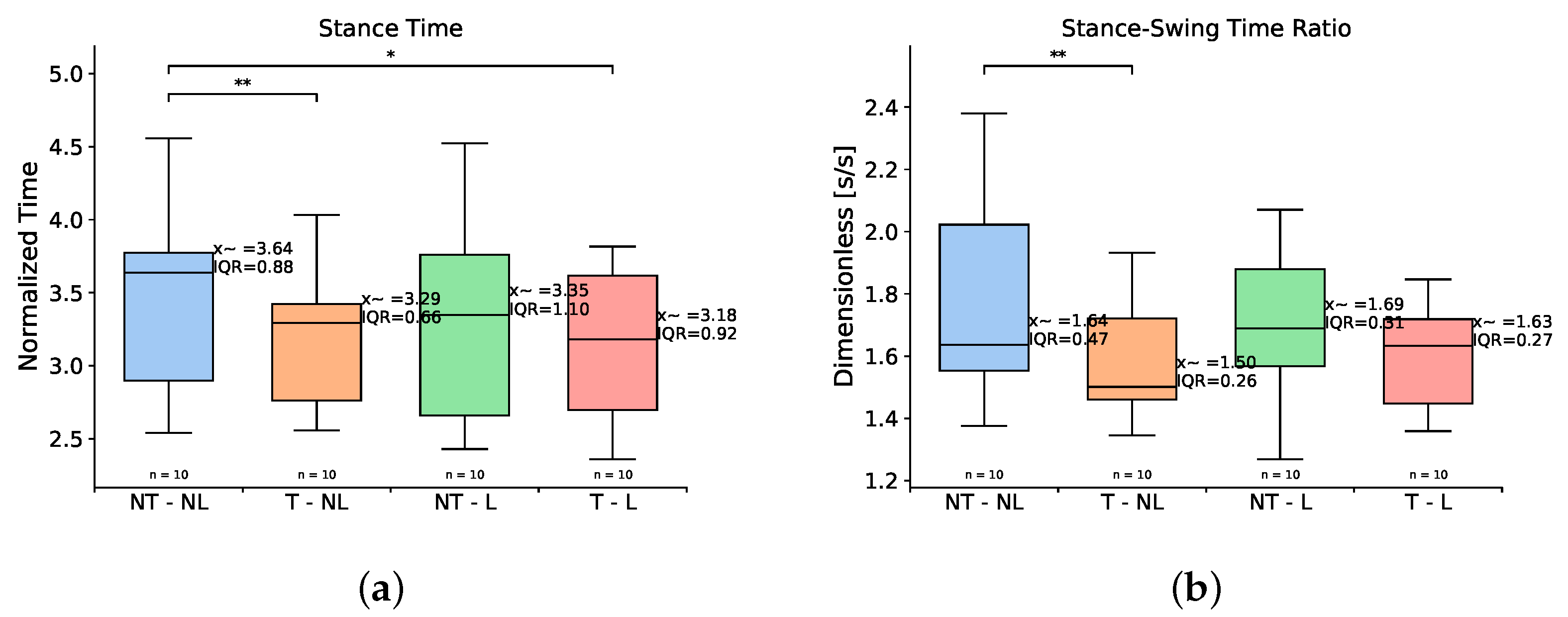
We found significant differences in both the stance time and stance/swing ratio between the baseline condition (NT-NL) and the conditions involving the use of toe compliance. As observed in Table 1 and Figure 4, there was a significant reduction in the stance time in both the T-NL and T-L conditions–9% and 12%, respectively. Moreover, the stance/swing ratio was also affected by the use of a compliant toe joint; however, a significant reduction of 8.5% was only observed when the toe joint was used alone. On the other hand, the addition of toe and/or lateral compliance did not affect gait speed, swing time, step width, step length, or stride length when compared to walking with a toe and laterally rigid prosthesis.

3.2. Kinematics

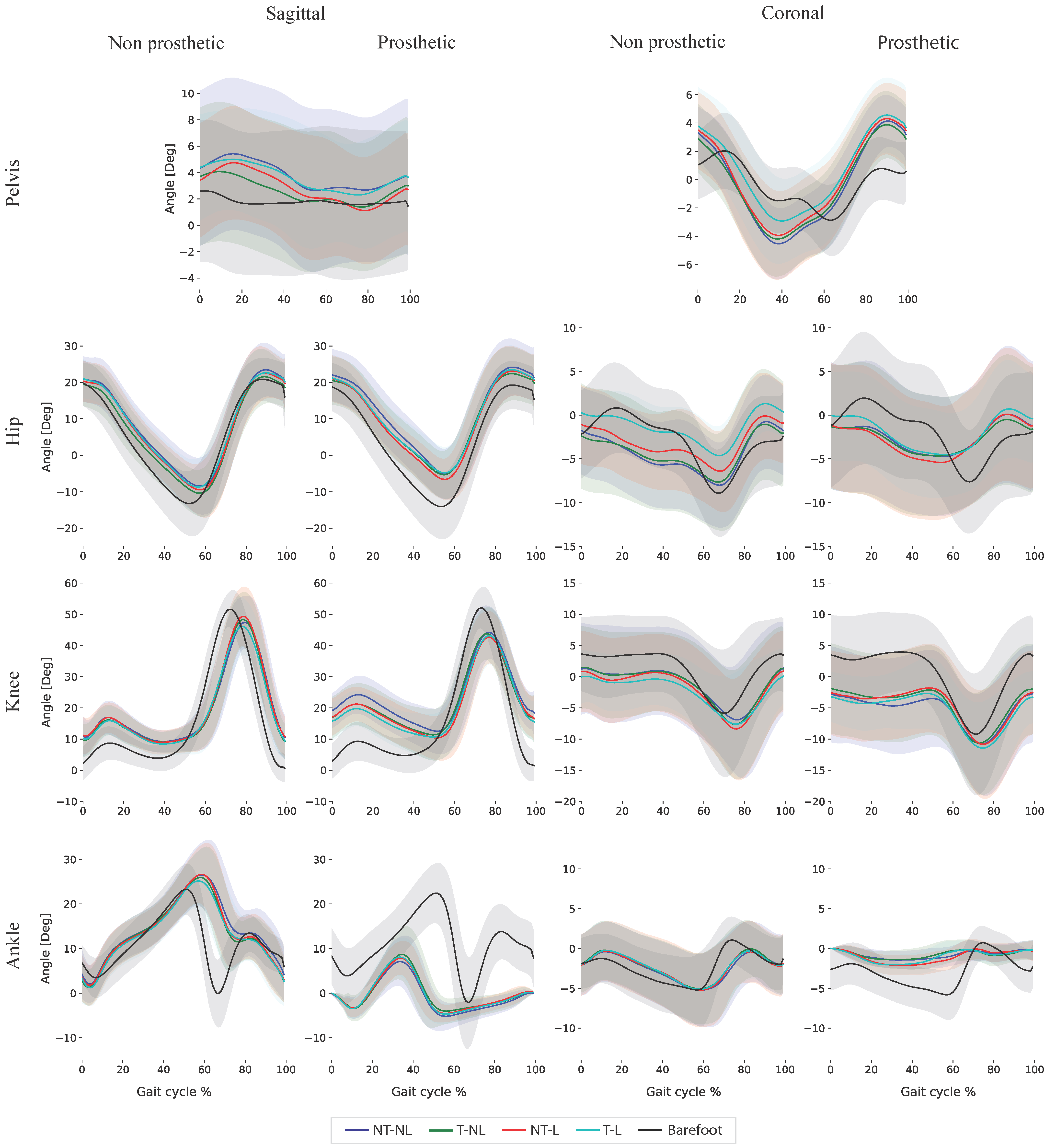
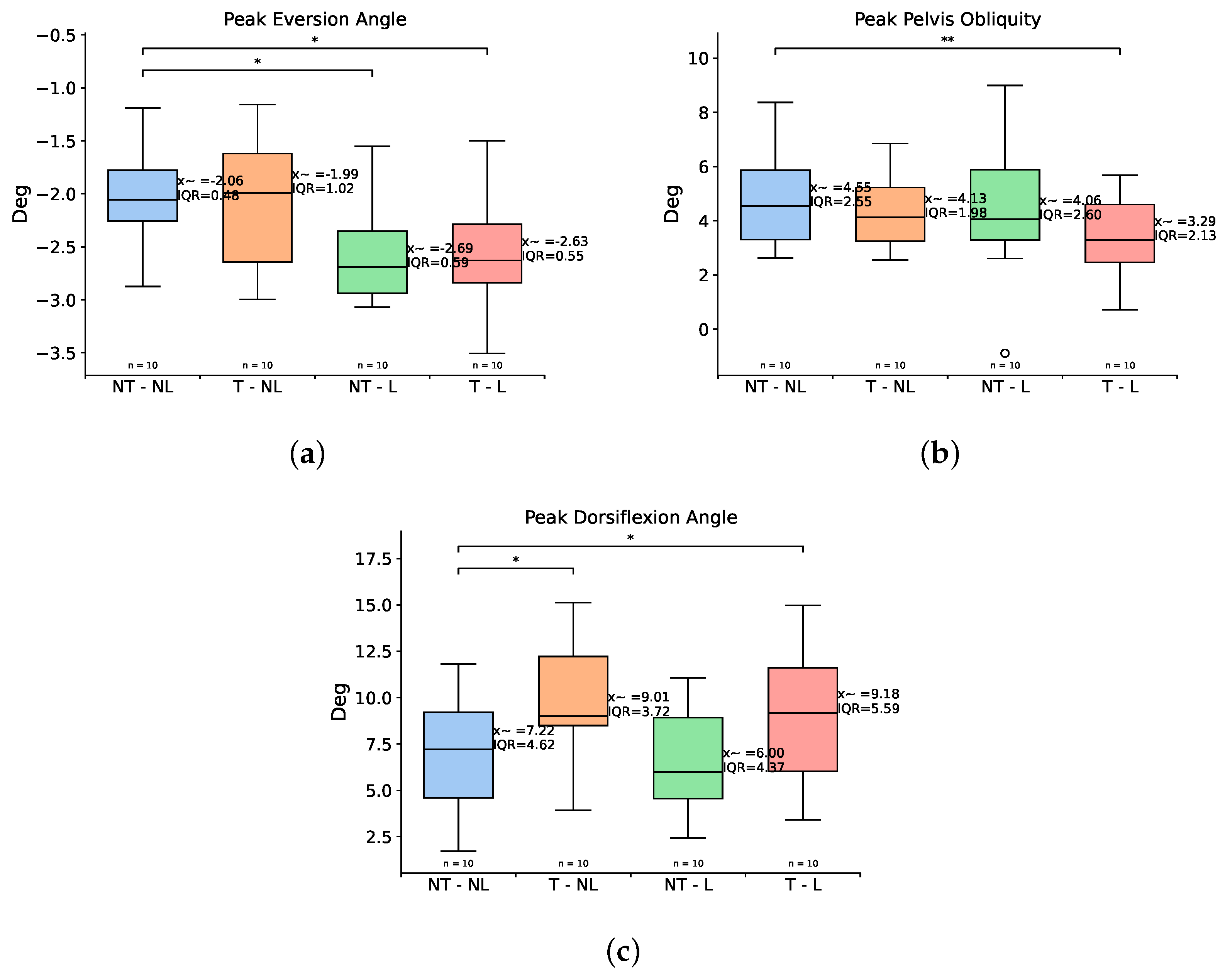
The obtained kinematics are presented in Figure 5. The addition of compliance in the coronal plane and toe compliance to the prosthetic foot mostly affected the ankle and pelvis kinematics of the participants. In the sagittal plane, the addition of compliant toes significantly increased the peak ankle dorsiflexion angle—24% and 27%, respectively for the T-NL and T-L conditions, compared to a completely rigid foot. Significant effects were also found in the coronal plane. As seen in Table 2 and Figure 6, the maximum ankle eversion angle was significantly higher for the T-L condition. In addition, this particular compliance combination significantly decreased maximum pelvis obliquity. We also found a main effect for peak plantarflexion angle across the conditions. Lastly, the flexible toes trials show a reduction in the peak plantarflexion angle regardless of the use of lateral compliance; nevertheless, this effect was not significant for the pairwise comparisons.
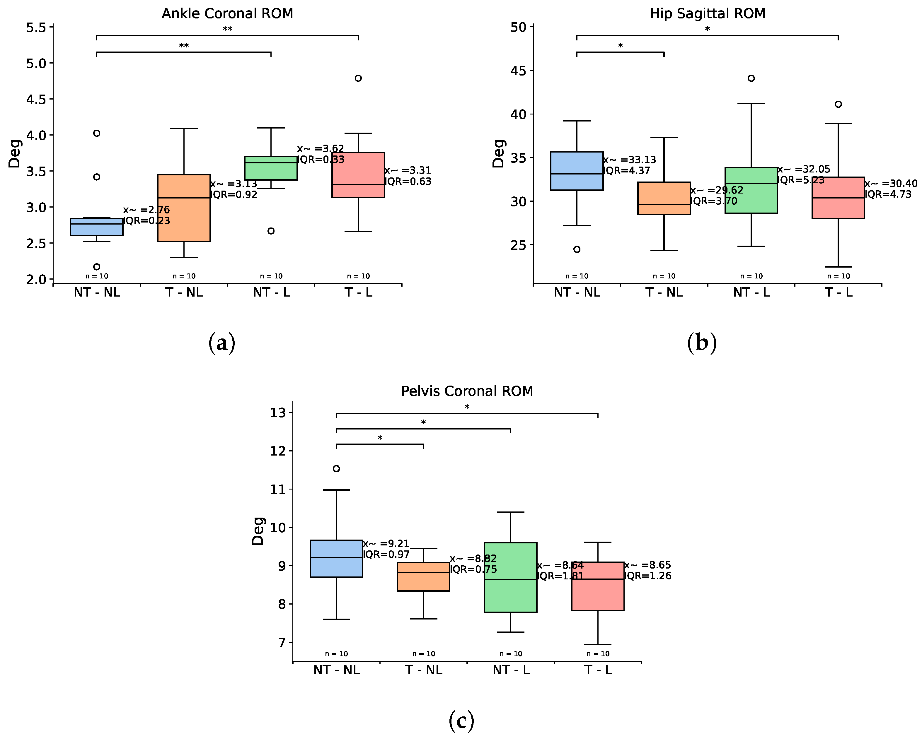
In accordance with other works, the knee and hip peak angles were not significantly affected by the use of toe or lateral compliance in the sagittal plane [10,11,54]. Only a decrease in the hip sagittal ROM was observed as a product of the use of flexible toes, as shown in Figure 7. In the coronal plane, significant effects were observed in the hip joint due to the addition of lateral compliance in transtibial prostheses, but only in tasks directly involving the use of the foot’s joint in the coronal plane as in the case of turning [19], unlike our experimental setup for straight, even terrain gait.

3.3. Range of Motion

Regarding the range of motion (ROM), there was a significant increment in the ankle coronal ROM for the conditions where lateral mobility was present, with an increase of 31% and 16% compared to the baseline condition (Figure 7). Additionally, the pelvis coronal range of motion tended to decrease with the addition of both lateral and toe compliance. Moreover, as seen in Table 3, the use of a compliant forefoot in the prosthesis (T-NL and T-L) significantly decreased the sagittal range of motion by about 3°, which was not observed for the NT-L condition. Lastly, the knee ROM was not unaffected by the use of lateral and/or toe compliance in the participants.

3.4. Ground Reaction Forces and Moments

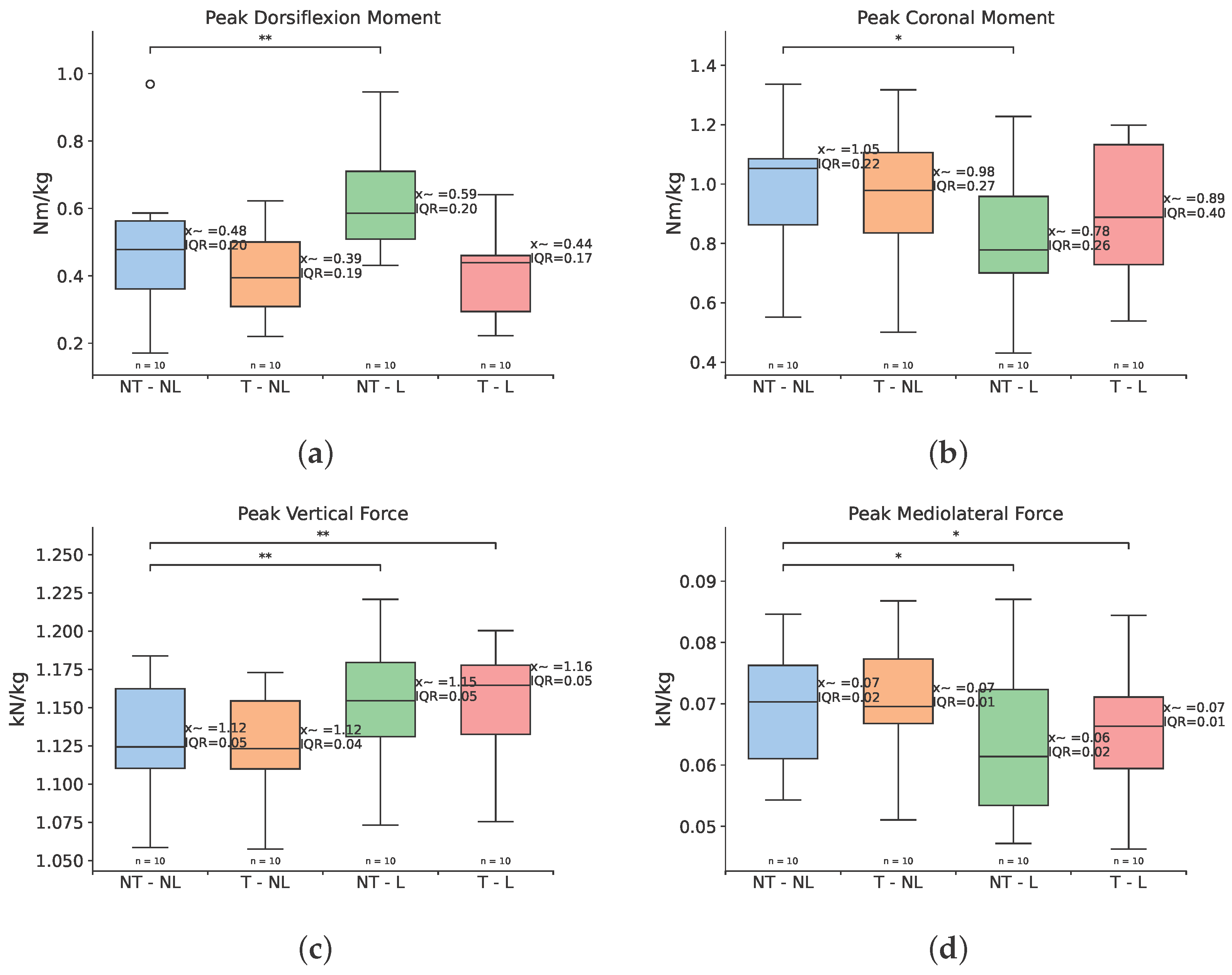
The obtained ground reaction forces and moments are presented in Figure 8. As seen in Table 4, the peak ground reaction moment in the mediolateral direction was found to be significantly smaller for the prosthetic foot that included lateral compliance only (NT-L) (Figure 9). In this condition the participants exerted 0.27 Nm/kg less than in the baseline condition (NT-NL), which represents a difference of 25.7%. Interestingly, the use of compliance in the coronal plane significantly affected the sagittal plane kinetics, producing an increase of 0.11 Nm/kg in the peak dorsiflexion moment. This effect was not perceived in the T-L condition, despite also showcasing compliance in the coronal plane.
It can also be observed that the use of toe compliance increased the peak mediolateral moment when comparing the NT-L vs. T-L conditions. This opposes the effect of lateral compliance in the T-L condition, which, as observed in Table 4, is not significantly different from the NT-NL condition despite also being laterally compliant. Lastly, we found no significant differences in the peak transversal moments.
With respect to the ground reaction forces, we found both a decrease in the peak mediolateral forces and an increase in the peak vertical forces for the conditions featuring compliance in the coronal plane (NT-L and T-L) as presented in Figure 9. In alignment with the ground reaction moments, the addition of toe compliance to the NT-L version of the foot tended to increase the peak lateral and vertical forces. This is consistent with other studies evidencing that decreased ankle stiffness or the use of a flexible forefoot section can significantly influence vertical forces, impulses, and breaking forces [55,56,57,58]. Surprisingly, there is no significant effect of the use of flexible toes between conditions on the peak sagittal moments or forces, even though the toes are compressed in this direction during gait.

3.5. Electromyography

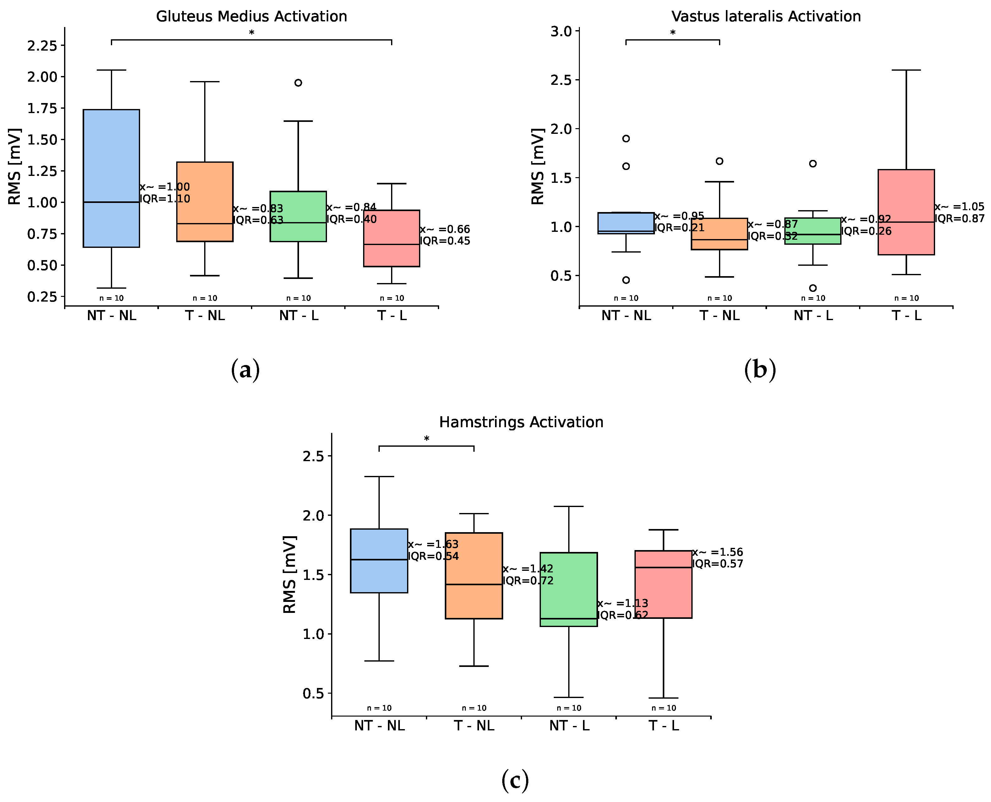
The different compliance and mobility conditions exerted a significant effect on the activity of the gluteus medius (Figure 10) on the non-prosthetic side and in the vastus lateralis and hamstrings on the prosthetic side (Table 5). The activity of the gluteus medius, which plays a major mediolateral role during gait and controls pelvis stabilization and posture, decreased as mobility increased, as observed in Figure 10. We observed a decrease of 0.17 µV in the T-NL condition, which seems to be enhanced by the use of lateral compliance, leading to a further decrease of 34% for the T-L condition compared to the baseline foot. However, the reduction effect is only significant for the T-L condition.
Regarding the prosthetic side, we found significant differences in muscles involved in the control of gait and balance in the sagittal plane. In particular, the vastus lateralis and hamstrings presented a significant reduction in their activity for the T-NL condition—8% and 12%, respectively. This decrease, which can be attributed to the compliant toes, was not observed in the T-L condition and might have been occluded by the joint use of lateral compliance. The activity of the gluteus medius and gluteus maximus did not show significant changes with increased compliance. Lastly, the variability of the muscular activity was not affected by the compliance and mobility combinations on either the prosthetic or non-prosthetic side.

4. Discussion

Contrary to other studies showing no effect of a compliant forefoot on spatiotemporal variables [11,54,57,59], this study found a significant reduction in the stance time due to the use of compliant toes. However, the combined use of toe and lateral compliance seems to prevent a subsequent decrease in the stance/swing ratio, found within the normal values for able-bodied participants of 1.61 ± 4% [60], except for compliant toes only (T-NL) (Table 1). While this does not correspond to a significant interaction effect between the two compliances according to our linear mixed models (p-value = 0.3761), this compliance combination has the potential to mitigate the asymmetric stance/swing ratio that characterizes prosthetic gait, which shows a significantly decreased ratio on the prosthetic side [61]. In line with other studies, we found no major changes in other spatiotemporal variables associated with flexible toes [11,57], in combination with lateral compliance.
For the T-L condition, the peak inversion angle did not show any significant changes, while the peak eversion angle was significantly increased. This matches the previously observed increase in mobility for other laterally compliant prosthetic feet and might cohere with the decrease in the overall foot stiffness [19]. In addition, the increase in the coronal ROM of the ankle for this condition indicates that the prosthetic users might be prone to naturally use additional compliance in the coronal plane, if available, even for straight non-inclined gait as previously investigated [20]. This can benefit the users in terms of stability and balance, as well as provide potential surface adaptation. In the sagittal plane, increased peak dorsiflexion angles in the T-L condition were also observed in other feet with flexible forefoot sections [10,54,57]. In this study, however, the increased peak dorsiflexion was not accompanied by an increase in the ankle sagittal range of motion, as can be seen in Table 1.
For the pelvis, providing either compliance in the coronal plane or toe mobility for prosthetic feet alone did not result in significant changes in the pelvis peak angles. Instead, we found a significant decrease in the pelvis obliquity as a product of the combined effect of flexible toes and lateral compliance, which showed no interaction effects in the linear mixed models (p-value = 0.21621). Pelvis obliquity was reduced by 27% in this condition compared to NT-NL and was closer to the median values for pelvis obliquity reported in non-amputees of about 2° [62]. This effect, in combination with the significant reduction in the activity of the gluteus medius for the same condition, suggests that both a flexible forefoot and a coronally compliant ankle capable of exerting mediolateral torque are required for improved pelvis stability in prosthetic gait. Reducing peak obliquity could impact the “hip hiking” strategy of lower-limb prosthetic gait [63,64] and also offer some benefits such as decreased vertical loads [64] and improved limb forward progression and energy recovery [63].
Regarding the ground reaction moments in the sagittal plane, we consider that the combined use of flexible toes and lateral compliance can decrease the overall foot effective stiffness and led to the observed increased in the anterior–posterior moment. As reported in other studies, the decrease in prosthetic stiffness can cause increased ground reaction propulsive and breaking impulses in the sagittal plane, which can be directly correlated with the exerted moments [19]. On the other hand, regarding the addition of a compliant toe joint, it is possible that the chosen stiffness for the toes is sufficiently stiff to avoid a decrease in the effective foot length [10], leading to no significant changes in the peak anterior–posterior moment previously reported for prosthetic feet with a flexible forefoot [11,54].
In the coronal plane, the significant decrease in the peak mediolateral moment in the NT-L condition but not in the L-T condition can be related to the properties of the toes. The material used in combination with its rigid attachment to the foot body might limit a fast mediolateral adaptation, increasing the resistance to lateral loads, in comparison with the coronal mechanism, which features a dedicated physical axis of rotation [16,18]. This should be considered in the design of multi-axial transtibial prostheses with multiple degrees of freedom.
With respect to the ground reaction forces, we found a significant decrease in the peak lateral forces together with a significant increase in the peak vertical forces when using compliance in the coronal plane. This could be explained similarly to the coronal moments, as the limited coronal mobility and adaptability of the prosthesis in the NT-NL and T-NL conditions might cause increased lateral ground loads [17], which are reduced in the presence of a coronal ankle joint (NT-L and T-L). In the vertical direction, our results are in disagreement with other studies investigating feet with either flexible forefoot sections or coronal adaptability that show that the peak vertical forces seem to be unaffected by compliance in the coronal plane [18], and studies that indicate that a flexible forefoot section either reduces [57] or exerts no effect [54] on peak vertical forces. The effect of multiple multiaxial compliance on vertical forces requires further investigation. Lastly, to the best of our knowledge, this is the first study to investigate the combined effect of lateral compliance and toe joint on anterior–posterior ground reaction forces and moments. In this study, we found no effect in the sagittal forces, and significant effects in the coronal and sagittal moments generated by the compliance in the coronal plane.
On the prosthetic side, and contrary to the expected increase in muscular activation due to increased prosthetic foot compliance [55,65], toe compliance resulted in decreasing the activity of the vastus lateralis and hamstrings. We attribute this effect to the additional push-off provided by the toes, which supports the role of both muscles in the generation of vertical and forward movements [66]. However, it is unclear why this effect was significant only for the T-NL condition and not the T-L condition as well. Still, we can observe a reduced median RMS value for the NT-L and T-L conditions. This toe effect could be favorable for transtibial prosthetic users, as it has been reported that they rely significantly more on their hamstrings and gluteus maximus to generate power in the prosthetic limb [65].
Regarding gluteus maximus and gluteus medius, these muscles’ activity seems not to be affected in the gait of transtibial prosthetic users who use prostheses without coronal mobility in either gait analysis or simulations [65], as their main role has been found to be in the coronal plane. In this plane, however, we found a significant reduction in the activity of the gluteus medius for the T-L condition on the non-prosthetic side, which has been reported to be significantly increased during all phases of the gait cycle for people with transtibial amputation [67]. As pelvis support is one of the roles of gluteus medius stance [68], our results support the idea that the combination of a flexible toe joint and lateral compliance can reduce the user’s effort in stabilizing the pelvis during stance.
These significant effects of combining lateral and toe compliance in the gait of participants could potentially have an effect in the development of compensatory gait mechanisms and secondary conditions observed in people living with transtibial amputation. It has been observed that people with transtibial amputation (TTA) experience increased trunk and pelvis movements in both the sagittal and coronal planes [69], which directly correlates with the development of pelvic drop and chronic low-back pain [70] due to increased trunk–pelvis–spine muscular effort [70,71]. We believe that the reduction in pelvis obliquity and pelvis coronal range of motion offered by combining lateral and toe compliance can potentially address this particular gait adaptation as well as reduce the risk of the back pain associated with it.
In addition, the observed effects in mediolateral ground reaction moment may help mitigate the mediolateral trunk sway commonly seen in individuals with transtibial amputation. More importantly, this sway is often linked to asymmetrical joint loading, particularly excessive loading on the intact limb, which has been associated with long-term tissue damage, joint degeneration, and pain [5,72]. We hypothesize that the observed decrease in the mediolateral GRM on the prosthetic side as a result of lateral compliance may help reduce the mediolateral ground reaction impulse asymmetry and also promote more balanced load distribution across limbs [25]. Moreover, the addition of compliance in the coronal plane can facilitate gait control in challenging surfaces [18], reduce the energy requirements for direction control [17], and reduce prosthetic limb discomfort and skin breakdown [17]
Similarly, reduced dorsiflexion on the prosthetic foot has been also associated with augmented vertical loadings in the intact limb, which increases the risk of secondary conditions derived from mechanical stress [73]. The use of toes with and without lateral compliance showed significant impact on this variable, potentially addressing this risk. Lastly, it has been observed that people with TTA have reduced hip adduction on the prosthetic side, partially attributed to gluteus medius insufficiency [74]. Moreover, this population also presents a significant increase in the activity of the gluteus medius in the intact limb compared to that of people without TTA [75]. The combination of these two characteristics increases the risk of the development of a positive Trendelenburg sign [74], a reduction in pelvis control [74], and lower-back pain [76]. The use of lateral and toe compliance in transtibial prosthesis might also potentially impact this compensatory gait strategy as it significantly reduces the muscular activity of the gluteus medius in the intact limb, while also tending to increase the activity of the same muscle in the prosthetic limb.

4.1. Power Analysis

Due to the exploratory nature of this study, we conducted a post hoc power analysis for each of the significant paired comparisons for all the studied biomechanical variables to assess the sample size. We used the observed Cohen’s d and a paired t-test approximation ( α = 0.05 , two-sided) as a conservative proxy for the nonparametric tests utilized [77,78]. The results of the power analysis are presented in Table 6.
The results show that despite our modest sample size (n = 10), most of the statistically significant comparisons had large observed effects (18/24) with the majority of them also presenting enough estimated power (>0.80, 11/18). The hip and pelvis sagittal range of motion as well as the peak eversion ankle presented an estimated power just below 0.8. On the other hand, while all the remaining significant comparisons presented moderated effects (d > 0.5), with the exception of the peak mediolateral GRF for the T-L condition, they did not reach enough statistical power, in particular for the hamstrings’ activity on the prosthetic side and the hip sagittal range of motion. These variables should be interpreted with caution and would require a larger cohort to verify their significance.

4.2. Limitations

Several limitations of this study must be acknowledged. First, due to practical design considerations of the prosthetic simulator and considerations related to controlling the number of experimental variables, the provided stiffness for both the sagittal and coronal planes were fixed for all participants regardless of their individual masses, potentially influencing sagittal plane responses and data variability, despite the normalization of kinetic variables to body weight.
Second, while the use of a prosthetic simulator allowed non-amputated individuals to walk with the mechanical properties of different versions of the prosthetic foot, this setup does not account for the differences in neuromuscular control, the absence of sensory feedback, and the unique muscle activation profiles observed in people with lower-limb amputations [61,79]. Adaptations such as compensatory pelvis and trunk movements or hip hiking are not reflected in the gaits of our participants and therefore limit the assessment of the impact of compliance in the coronal plane and of the toe joint [79]. Nevertheless, the use of non-amputated participants in the experimental protocol offered a mean to control for the considerable inter-subject variability typically observed among individuals with limb loss [27,33]. While this initial approach with non-amputees provides valuable insight into the effects of the mechanical design of prosthetics, the nature of the experimental participants impedes the direct applicability of our findings to the gaits of people with transtibial amputation.
In the case of our study, the use of prosthetic simulators inevitably altered the subjects’ anthropometric proportions and static balance [31]. Specifically, the use of simulators increased the center of mass (COM) height and added unequal masses to the lower limbs, which may influence kinematic outcomes [27]. In terms of height and weight, the prosthetic simulator and leg length-extending shoe added 24.5 cm to the experimental subject’s height and between 1.8 kg to 2.45 kg to the lower limbs, causing an interlimb mass discrepancy of up to 0.4 kg. Besides being less than 1% of the body weight of the participants, these combined mass and height alterations are expected to influence gait. Studies investigating stilt gait have shown that an artificial height increase between 60 cm and 100 cm can significantly affect spatiotemporal parameters, reducing gait speed and increasing step width and stance time [80,81,82,83]. Also, these studies show that while sudden height increases do not affect the joint’s kinematic patterns they can significantly reduce plantar flexion, knee flexion, and hip extension range of motion [80,82] without altering the mediolateral and anteroposterior ground reaction forces, even with the added mass of the stilts (roughly 3 kg), in our experimental setup [82].
With regard to the effect of the added mass and interlimb mass differences, previous studies involving unilateral mass additions to prosthesis in the range of 0.3 to 4.1 kg, both distally and proximally, have reported minor reductions in gait speed [84], while joint kinematics and joint work seem not to be altered [85,86,87,88]. Still, as the locations of the center of mass of the leg length-extending shoe and the prosthetic simulator are different, participants were also subjected to interlimb inertial differences. While the mediolateral and height locations of the center of mass for the prosthetic simulator and the leg length-extending shoe presented differences of less than 2.5 cm, the anteroposterior location of the center of mass varied between the simulator and the shoes in 5.5 cm (23.5 cm shoe) and 10.5 cm (27.5 cm shoe). As a consequence, participants wearing the 27.5 cm shoe experienced repercussions in both the anteroposterior and mediolateral moments of inertia, with an approximate interlimb inertial difference of 30%. While these interlimb inertial differences have been previously studied in other works showing no impact on spatiotemporal or kinematic patterns [86,89] or muscular activity [90], other studies show that inertial differences can generate a decrease in the symmetry of spatiotemporal variables [86] and an increase in the energy cost of walking [89], peak hip angle and moment [91], peak knee angle, and vertical ground reaction forces [92], which could have influenced our results.
Although we maintained a consistent device, height and mass addition, and mass distribution across participants to allow for the assessment of within-subject effects, hardware differences between participant limbs compromise the reliability of contralateral comparisons, wherefore the combined effects of toe and compliance in the coronal plane on gait symmetry require further investigation. Furthermore, while the mass, height, and alignment of the simulator were consistent across all experimental conditions and are not expected to interact between trials [28,29], they may still impose artificial constraints on natural gait patterns. In particular, the vertical offset in the location between the prosthetic (right) and human (left) ankle joint may have introduced deviations in the joint kinematics, natural foot–floor contact, and ground reaction forces, which limits the interpretation of the absolute values of these variables.
Lastly, while the simulator boots limit the generalization of the results to people living with amputation, they remain a valid tool for exploring how human gait adapts to controlled variations in toe and ankle properties [27]. The different versions of the experimental prosthesis shared the same base design, varying only in some components, which helps in isolating the impact of ankle stiffness on static balance [31]. It is also important to note that even if the effective stiffness values were not uniform across the participants due to weight differences, the spatiotemporal and kinetic variables were normalized to the total body weight and extended leg length to allow for intra-subject comparisons [10] and design consistency across trials.
For the coronal stiffness, while the selected 2.54 Nm/deg is higher than the estimated range for the ankle joint, the use of a higher physiological stiffness in the prosthetic ankle is not rare in prostheses. This is usually observed at reduced walking speeds, crossed slopes, and stairs [73,93,94,95] and is associated with a reduced risk of falls [49]. Still, this corresponds to a limitation of our study due manufacturing limits associated with the leaf springs used in the coronal mechanism. In comparison, other studies estimate the dynamic stiffness of this joint between 1.85 and 5.74 Nm/deg [96] or use stiffness control ranging in the coronal plane 0–5 Nm/deg [4]. However, the use of fixed stiffness values in both planes for all participants, regardless of their body mass, represents a practical limitation that likely introduced additional variability in the recorded parameters. Future work should include stiffness values scaled to the participant’s mass to better isolate the compliance effects
Another limitation is the generalizability of these findings. The participant recruitment was limited due to weight restrictions of the prosthetic materials and also the selection of young adults resulted in a reduced age range for the participants. While this improves the internal validity of the findings, it reduces the transferability of the results to other populations [27]. In addition, while previous studies have reported minimal impact of the use of prosthetic simulators on gait parameters aside from ground reaction forces [97], some others showed divergent results when compared to the gait of people with amputation [27]. This suggests that the mechanical effects of lateral and toe compliance may not be independent of the study population, as previously investigated [32]. Moreover, the added mass and leg length given by the prosthetic simulator might influence the effects of push-off provided by the prosthesis, being a factor not present in people with amputation [34].
Finally, it remains uncertain if results from non-amputated individuals will fully align with those with amputations, as people with simulated and real lower-limb amputations can display different responses to the same prosthetic device [30]. This gap between experimental designs, including people with and without amputation, could be potentially approached through musculoskeletal models and predictive simulations of gait [98,99]. Previous studies have not only created detailed musculoskeletal models to simulate and examine the unique kinematics and kinetics of people with amputation [100] but also utilized data acquired from people without amputation to assist in the development and optimization of prosthetic devices [101] and exoskeletons [102,103]. Future research including people with amputation and a larger sample size is needed to verify our results, allow for a full factorial experimental design, and explore the clinical implications of these findings for prosthetic design [27].

5. Conclusions

This study provides insights into the individual and combined effects of compliance in the coronal plane and a toe joint on transtibial prosthetic gait, using non-amputees as a preliminary model. The results suggest that the inclusion of these two features can influence spatiotemporal variables, joint kinematics, ground reaction forces, and muscular activity in both the sagittal and coronal planes. Notably, the addition of lateral compliance and a flexible toe joint showed potential in improving balance, decreasing pelvic obliquity, increasing the ankle coronal range of motion, and reducing muscle activation of the gluteus medius on the non-prosthetic side, compared to a laterally rigid foot without toe compliance. These results underscore the importance of integrating compliance in the coronal plane and toe compliance in prosthetic designs to address the biomechanical challenges faced by people with lower-limb amputation. Future studies involving people with amputation are necessary to validate these findings and explore their clinical relevance, particularly concerning long-term gait adaptations and the prevention of secondary musculoskeletal conditions such as osteoarthritis. This research lays the groundwork for advancing prosthetic technology with the goal of improving the utility of lower-limb prostheses.

Author Contributions

Conceptualization, S.G.-L. and K.S.; methodology, S.G.-L. and H.K.; software, S.G.-L.; validation, H.K. and M.H.; formal analysis, S.G.-L. and K.S.; investigation, S.G.-L. and K.S.; resources, K.S., M.H. and H.K.; data curation, M.H.; writing—original draft preparation, S.G.-L.; writing—review and editing, S.G.-L., H.K. and M.H.; visualization, S.G.-L.; supervision, K.S.; project administration, K.S.; funding acquisition, K.S. All authors have read and agreed to the published version of the manuscript.

Funding

This work was supported by the Japan Society for the Promotion of Science KAKENHI (grant number 23H00485).

Institutional Review Board Statement

This study was conducted in accordance with the Declaration of Helsinki and approved by the Institutional Review Board of the University of Tsukuba (Ethical Committee approval number 2023R754, 27 July 2023). All participants provided written informed consent prior to participation.

Informed Consent Statement

Informed consent was obtained from all subjects involved in the study.

Data Availability Statement

The data presented in this study are available on request from the corresponding author due to privacy and ethical restrictions. All motion capture data have been de-identified to protect participant anonymity. Access to the data can be obtained through a controlled access database, which is available to researchers who meet the criteria for access.

Acknowledgments

The authors thank Margaux Lafitte, Santiago Price, and Elia Pedrazzini for their support in the experimental setup and data acquisition.

Conflicts of Interest

The authors declare no conflicts of interest.

References

  1. Kathryn, Z.G.; Ellen J, M.; Patti L, E.; Thomas G, T.; Ron, B. Estimating the prevalence of limb loss in the United States: 2005 to 2050. Arch. Phys. Med. Rehabil. 2008, 89, 422–429. [Google Scholar] [CrossRef]
  2. Arifin, N.; Abu Osman, N.A.; Ali, S.; Gholizadeh, H.; Wan Abas, W.A.B. Postural stability characteristics of transtibial amputees wearing different prosthetic foot types when standing on various support surfaces. Sci. World J. 2014, 2014, 856279. [Google Scholar] [CrossRef] [PubMed]
  3. Askew, G.N.; McFarlane, L.A.; Minetti, A.E.; Buckley, J.G. Energy cost of ambulation in trans-tibial amputees using a dynamic-response foot with hydraulic versus rigid ‘ankle’: Insights from body centre of mass dynamics. J. Neuroeng. Rehabil. 2019, 16, 39. [Google Scholar] [CrossRef] [PubMed]
  4. Myunghee, K.; Hannah, L.; Tianjian, C.; Steven H, C. The Effects of Prosthesis Inversion/Eversion Stiffness on Balance-Related Variability During Level Walking: A Pilot Study. J. Biomech. Eng. 2020, 142, 091011. [Google Scholar] [CrossRef]
  5. Teater, R.H.; Wolf, D.N.; McDonald, K.A.; Zelik, K.E. Unilateral transtibial prosthesis users load their intact limb more than their prosthetic limb during sit-to-stand, squatting, and lifting. Clin. Biomech. 2023, 108, 106041. [Google Scholar] [CrossRef]
  6. Portnoy, S.; Siev-Ner, I.; Shabshin, N.; Kristal, A.; Yizhar, Z.; Gefen, A. Patient-specific analyses of deep tissue loads post transtibial amputation in residual limbs of multiple prosthetic users. J. Biomech. 2009, 42, 2686–2693. [Google Scholar] [CrossRef]
  7. Misu, S.; Doi, T.; Asai, T.; Sawa, R.; Tsutsumimoto, K.; Nakakubo, S.; Yamada, M.; Ono, R. Association between toe flexor strength and spatiotemporal gait parameters in community-dwelling older people. J. Neuroeng. Rehabil. 2014, 11, 143. [Google Scholar] [CrossRef]
  8. Zhu, J.; Wang, Q.; Wang, L. Effects of toe stiffness on ankle kinetics in a robotic transtibial prosthesis during level-ground walking. Mechatronics 2014, 24, 1254–1261. [Google Scholar] [CrossRef]
  9. Childers, W.L.; Takahashi, K.Z. Increasing prosthetic foot energy return affects whole-body mechanics during walking on level ground and slopes. Sci. Rep. 2018, 8, 5354. [Google Scholar] [CrossRef]
  10. Honert, E.C.; Bastas, G.; Zelik, K.E. Effect of toe joint stiffness and toe shape on walking biomechanics. Bioinspir. Biomimetics 2018, 13, 066007. [Google Scholar] [CrossRef]
  11. McDonald, K.A.; Teater, R.H.; Cruz, J.P.; Kerr, J.T.; Bastas, G.; Zelik, K.E. Adding a toe joint to a prosthesis: Walking biomechanics, energetics, and preference of individuals with unilateral below-knee limb loss. Sci. Rep. 2021, 11, 1924. [Google Scholar] [CrossRef] [PubMed]
  12. Brandt, A.; Huang, H.H. Effects of extended stance time on a powered knee prosthesis and gait symmetry on the lateral control of balance during walking in individuals with unilateral amputation. J. Neuroeng. Rehabil. 2019, 16, 151. [Google Scholar] [CrossRef] [PubMed]
  13. McMulkin, M.L.; Osebold, W.R.; Mildes, R.D.; Rosenquist, R.S. Comparison of Three Pediatric Prosthetic Feet During Functional Activities. JPO J. Prosthetics Orthot. 2004, 16, 78–84. [Google Scholar] [CrossRef]
  14. Leopaldi, M.; Brugo, T.M.; Tabucol, J.; Zucchelli, A. Parametric Design of an Advanced Multi-Axial Energy-Storing-and-Releasing Ankle–Foot Prosthesis. Prosthesis 2024, 6, 726–743. [Google Scholar] [CrossRef]
  15. Jin, J.; Wang, K.; Ren, L.; Qian, Z.; Liang, W.; Xu, X.; Zhao, S.; Lu, X.; Zhao, D.; Wang, X.; et al. Design of a Flexible Bionic Ankle Prosthesis Based on Subject-specific Modeling of the Human Musculoskeletal System. J. Bionic Eng. 2023, 20, 1008–1020. [Google Scholar] [CrossRef]
  16. Tabucol, J.; Kooiman, V.G.M.; Leopaldi, M.; Brugo, T.M.; Leijendekkers, R.A.; Tagliabue, G.; Raveendranathan, V.; Sotgiu, E.; Benincasa, P.; Oddsson, M.; et al. The Functionality Verification through Pilot Human Subject Testing of MyFlex-δ: An ESR Foot Prosthesis with Spherical Ankle Joint. Appl. Sci. 2022, 12, 4575. [Google Scholar] [CrossRef]
  17. Maitland, M.E.; Allyn, K.J.; Ficanha, E.M.; Colvin, J.M.; Wernke, M.M. Finite element simulation of frontal plane adaptation using full-foot, split-toe and cam-linkage designs in prosthetic feet. J. Prosthetics Orthot. JPO 2022, 34, 14. [Google Scholar] [CrossRef]
  18. Altenburg, B.; Ernst, M.; Maciejasz, P.; Schmalz, T.; Braatz, F.; Gerke, H.; Bellmann, M. Effects Of A Prosthetic Foot with Increased Coronal Adaptability On Cross-slope Walking. Can. Prosthetics Orthot. J. 2021, 4, 35206. [Google Scholar] [CrossRef]
  19. Shell, C.E.; Segal, A.D.; Klute, G.K.; Neptune, R.R. The effects of prosthetic foot stiffness on transtibial amputee walking mechanics and balance control during turning. Clin. Biomech. 2017, 49, 56–63. [Google Scholar] [CrossRef]
  20. Maitland, M.E.; Imsdahl, S.I.; Fogelberg, D.J.; Allyn, K.J.; Cain, K.C.; Humbert, A.T.; Albury, A.; Ficanha, E.M.; Colvin, J.M.; Wernke, M.M. Motion Analysis of a Frontal Plane Adaptable Prosthetic Foot. JPO J. Prosthetics Orthot. 2023, 36, 255–262. [Google Scholar] [CrossRef]
  21. Doerks, F.; Gempfer, C.; Reulbach, M.; Jakubowitz, E. Gait Characteristics of People with Hallux and Forefoot Amputations—A Case Series. J. Clin. Med. 2025, 14, 2140. [Google Scholar] [CrossRef] [PubMed]
  22. Maitland, M.E.; Allyn, K.J.; Ficanha, E.M.; Colvin, J.M.; Wernke, M.M. The effect of single and multiple split-toe designs on cross-slope adaptability of prosthetic feet: A finite element simulation study. J. Prosthetics Orthot. JPO 2023, 35, e24. [Google Scholar] [CrossRef] [PubMed]
  23. Saito, R.; Okamoto, S.; Sakamoto, M. Effect of toe contact condition during forward stepping on the ground reaction forces during turning movement. J. Phys. Ther. Sci. 2019, 31, 344. [Google Scholar] [CrossRef]
  24. Damavandi, M.; Dixon, P.C.; Pearsall, D.J. Kinematic adaptations of the hindfoot, forefoot, and hallux during cross-slope walking. Gait Posture 2010, 32, 411–415. [Google Scholar] [CrossRef]
  25. Tang, Y.W.; Murai, A.; Hobara, H. Mediation of the mediolateral ground reaction force profile to maintain straight running among unilateral transfemoral amputees. Sci. Rep. 2023, 13, 7823. [Google Scholar] [CrossRef]
  26. Ernst, M.; Altenburg, B.; Schmalz, T. Characterizing adaptations of prosthetic feet in the frontal plane. Prosthetics Orthot. Int. 2020, 44, 225–233. [Google Scholar] [CrossRef]
  27. Hedrick, E.A.; Malcolm, P.; Wilken, J.M.; Takahashi, K.Z. The effects of ankle stiffness on mechanics and energetics of walking with added loads: A prosthetic emulator study. J. Neuroeng. Rehabil. 2019, 16, 148. [Google Scholar] [CrossRef]
  28. Kim, M.; Collins, S.H. Once-per-step control of ankle-foot prosthesis push-off work reduces effort associated with balance during walking. J. Neuroeng. Rehabil. 2015, 12, 43. [Google Scholar] [CrossRef]
  29. Jacobson, M.; Kantharaju, P.; Jeong, H.; Ryu, J.K.; Park, J.J.; Chung, H.J.; Kim, M. Foot contact forces can be used to personalize a wearable robot during human walking. Sci. Rep. 2022, 12, 10947. [Google Scholar] [CrossRef]
  30. Caputo, J.M.; Collins, S.H. Prosthetic ankle push-off work reduces metabolic rate but not collision work in non-amputee walking. Sci. Rep. 2014, 4, 7213. [Google Scholar] [CrossRef]
  31. Louessard, A.; Bonnet, X.; Catapano, A.; Pillet, H. Quantification of the Influence of Prosthetic Ankle Stiffness on Static Balance Using Lower Limb Prosthetic Simulators. Prosthesis 2022, 4, 636–647. [Google Scholar] [CrossRef]
  32. Groothuis, A.; Houdijk, H. The Effect of Prosthetic Alignment on Prosthetic and Total Leg Stiffness While Running with Simulated Running-Specific Prostheses. Front. Sport. Act. Living 2019, 1, 466153. [Google Scholar] [CrossRef] [PubMed]
  33. Zelik, K.E.; Collins, S.H.; Adamczyk, P.G.; Segal, A.D.; Klute, G.K.; Morgenroth, D.C.; Hahn, M.E.; Orendurff, M.S.; Czerniecki, J.M.; Kuo, A.D. Systematic variation of prosthetic foot spring affects center-of-mass mechanics and metabolic cost during walking. IEEE Trans. Neural Syst. Rehabil. Eng. 2011, 19, 411–419. [Google Scholar] [CrossRef] [PubMed]
  34. Malcolm, P.; Quesada, R.E.; Caputo, J.M.; Collins, S.H. The influence of push-off timing in a robotic ankle-foot prosthesis on the energetics and mechanics of walking. J. Neuroeng. Rehabil. 2015, 12, 21. [Google Scholar] [CrossRef]
  35. Galindo Leon, S.A.; Hassan, M.; Suzuki, K. A Passive Three Degree of Freedom Transtibial Prosthesis with Adjustable Coronal Compliance and Independent Toe Joint. In Proceedings of the Annual International Conference of the IEEE Engineering in Medicine and Biology Society, EMBS, Glasgow, UK, 11–15 July 2022; pp. 4330–4333. [Google Scholar] [CrossRef]
  36. Brockett, C.L.; Chapman, G.J. Biomechanics of the ankle. Orthop. Trauma 2016, 30, 232. [Google Scholar] [CrossRef]
  37. Wolf, P.; Moor, R.; Lundberg, A.; Nester, C.; Arndt, A.; Graf, E. Human ankle joint movements during walking are probably not determined by talar morphology. Sci. Rep. 2022, 12, 13856. [Google Scholar] [CrossRef]
  38. Vaca, M.; Stine, R.; Hammond, P.; Cavanaugh, M.; Major, M.J.; Gard, S.A. The Effect of Prosthetic Ankle Dorsiflexion Stiffness on Standing Balance and Gait Biomechanics in Individuals with Unilateral Transtibial Amputation. J. Prosthetics Orthot. JPO 2022, 34, 110–123. [Google Scholar] [CrossRef]
  39. Dewolf, A.H.; Sylos-Labini, F.; Cappellini, G.; Ivanenko, Y.; Lacquaniti, F. Age-related changes in the neuromuscular control of forward and backward locomotion. PLoS ONE 2021, 16, e0246372. [Google Scholar] [CrossRef]
  40. Jeon, W.; Ramadan, A.; Whitall, J.; Alissa, N.; Westlake, K. Age-related differences in lower limb muscle activation patterns and balance control strategies while walking over a compliant surface. Sci. Rep. 2023, 13, 16555. [Google Scholar] [CrossRef]
  41. Vervoort, D.; den Otter, A.R.; Buurke, T.J.; Vuillerme, N.; Hortobágyi, T.; Lamoth, C.J. Do gait and muscle activation patterns change at middle-age during split-belt adaptation? J. Biomech. 2020, 99, 109510. [Google Scholar] [CrossRef]
  42. Matos, M.; Perreault, E.J.; Ludvig, D. Frontal plane ankle stiffness increases with weight-bearing. J. Biomech. 2021, 124, 110565. [Google Scholar] [CrossRef] [PubMed]
  43. Villamar, Z.; Perreault, E.J.; Ludvig, D. Frontal plane ankle stiffness increases with axial load independent of muscle activity. J. Biomech. 2022, 143, 111282. [Google Scholar] [CrossRef] [PubMed]
  44. Barbieri, F.A.; Rossi, S.; Orcioli-Silva, D.; Lee, H.; Adjei, E.; Nalam, V. Sex Differences in Human Ankle Stiffness During Standing Balance. Front. Sport. Act. Living 2020, 2, 570449. [Google Scholar] [CrossRef]
  45. Ribeiro, G.A.; Ficanha, E.; Knop, L.; Rastgaar, M. Impedance of the Human Ankle During Standing for Posture Control. In Proceedings of the Frontiers in Biomedical Devices, BIOMED—2018 Design of Medical Devices Conference, DMD 2018, Minneapolis, MN, USA, 9–12 March 2018. [Google Scholar] [CrossRef]
  46. Ezawa, Y.; Nakatsugawa, T.; Maruyama, T. Foot torsional stiffness exhibits gender differences but shows no correlation with medial longitudinal arch height. J. Biomech. 2024, 175, 112293. [Google Scholar] [CrossRef]
  47. Whitting, J.W.; Steele, J.R.; McGhee, D.E.; Munro, B.J. Passive dorsiflexion stiffness is poorly correlated with passive dorsiflexion range of motion. J. Sci. Med. Sport 2013, 16, 157–161. [Google Scholar] [CrossRef]
  48. Nilsson, J.; Thorstensson, A. Ground reaction forces at different speeds of human walking and running. Acta Physiol. Scand. 1989, 136, 217–227. [Google Scholar] [CrossRef]
  49. Major, M.J.; Twiste, M.; Kenney, L.P.; Howard, D. The effects of prosthetic ankle stiffness on stability of gait in people with transtibial amputation. J. Rehabil. Res. Dev. 2016, 53, 839–852. [Google Scholar] [CrossRef]
  50. Man, H.S.; Leung, A.K.L.; Cheung, J.T.M.; Sterzing, T. Reliability of metatarsophalangeal and ankle joint torque measurements by an innovative device. Gait Posture 2016, 48, 189–193. [Google Scholar] [CrossRef]
  51. Sinitski, E.H.; Hansen, A.H.; Wilken, J.M. Biomechanics of the ankle–foot system during stair ambulation: Implications for design of advanced ankle–foot prostheses. J. Biomech. 2012, 45, 588–594. [Google Scholar] [CrossRef]
  52. Hof, A.L. Scaling gait data to body size. Gait Posture 1996, 4, 222–223. [Google Scholar] [CrossRef]
  53. Potvin, J.R.; Brown, S.H. Less is more: High pass filtering, to remove up to 99% of the surface EMG signal power, improves EMG-based biceps brachii muscle force estimates. J. Electromyogr. Kinesiol. 2004, 14, 389–399. [Google Scholar] [CrossRef] [PubMed]
  54. Huang, S.; Teater, R.H.; Zelik, K.E.; McDonald, K.A. Biomechanical effects of an articulating prosthetic toe joint during stair navigation for individuals with unilateral, below-knee limb loss. J. Biomech. 2023, 161, 111841. [Google Scholar] [CrossRef] [PubMed]
  55. Fey, N.P.; Klute, G.K.; Neptune, R.R. The influence of energy storage and return foot stiffness on walking mechanics and muscle activity in below-knee amputees. Clin. Biomech. 2011, 26, 1025–1032. [Google Scholar] [CrossRef] [PubMed]
  56. Major, M.J.; Twiste, M.; Kenney, L.P.; Howard, D. The effects of prosthetic ankle stiffness on ankle and knee kinematics, prosthetic limb loading, and net metabolic cost of trans-tibial amputee gait. Clin. Biomech. 2014, 29, 98–104. [Google Scholar] [CrossRef]
  57. Klodd, E.; Hansen, A.; Fatone, S.; Edwards, M. Effects of prosthetic foot forefoot flexibility on gait of unilateral transtibial prosthesis users. J. Rehabil. Res. Dev. 2010, 47, 899–910. [Google Scholar] [CrossRef]
  58. Nichols, K.M.; Adamczyk, P.G. Sensitivity of lower-limb joint mechanics to prosthetic forefoot stiffness with a variable stiffness foot in level-ground walking. J. Biomech. 2023, 147, 111436. [Google Scholar] [CrossRef]
  59. Raschke, S.U.; Orendurff, M.S.; Mattie, J.L.; Kenyon, D.E.; Jones, O.Y.; Moe, D.; Winder, L.; Wong, A.S.; Moreno-Hernández, A.; Highsmith, M.J.; et al. Biomechanical characteristics, patient preference and activity level with different prosthetic feet: A randomized double blind trial with laboratory and community testing. J. Biomech. 2015, 48, 146–152. [Google Scholar] [CrossRef]
  60. Iosa, M.; De Bartolo, D.; Morone, G.; Boffi, T.; Mammucari, E.; Vannozzi, G.; Bini, F.; Marinozzi, F.; Antonucci, G.; Paolucci, S. Gait phase proportions in different locomotion tasks: The pivot role of golden ratio. Neurosci. Lett. 2019, 699, 127–133. [Google Scholar] [CrossRef]
  61. De Marchis, C.; Ranaldi, S.; Varrecchia, T.; Serrao, M.; Castiglia, S.F.; Tatarelli, A.; Ranavolo, A.; Draicchio, F.; Lacquaniti, F.; Conforto, S. Characterizing the Gait of People with Different Types of Amputation and Prosthetic Components Through Multimodal Measurements: A Methodological Perspective. Front. Rehabil. Sci. 2022, 3, 804746. [Google Scholar] [CrossRef]
  62. Moharrami, A.; Mirghaderi, P.; Hoseini Zare, N.; Moazen-Jamshidi, M.M.; Ebrahimian, M.; Mortazavi, S.M.J. Slight pelvic obliquity is normal in a healthy population: A cross-sectional study. J. Exp. Orthop. 2023, 10, 57. [Google Scholar] [CrossRef]
  63. Castiglia, S.F.; Ranavolo, A.; Varrecchia, T.; De Marchis, C.; Tatarelli, A.; Magnifica, F.; Fiori, L.; Conte, C.; Draicchio, F.; Conforto, S.; et al. Pelvic obliquity as a compensatory mechanism leading to lower energy recovery: Characterization among the types of prostheses in subjects with transfemoral amputation. Gait Posture 2020, 80, 280–284. [Google Scholar] [CrossRef] [PubMed]
  64. Stephanie B, M.; Steven A, G.; Dudley S, C. A preliminary investigation of pelvic obliquity patterns during gait in persons with transtibial and transfemoral amputation. J. Rehabil. Res. Dev. 2000, 37, 1–10. [Google Scholar]
  65. Pickle, N.T.; Grabowski, A.M.; Jeffers, J.R.; Silverman, A.K. The Functional Roles of Muscles, Passive Prostheses, and Powered Prostheses During Sloped Walking in People with a Transtibial Amputation. J. Biomech. Eng. 2017, 139, 1110051. [Google Scholar] [CrossRef]
  66. Wagner, K.E.; Nolasco, L.A.; Morgenroth, D.C.; Gates, D.H.; Silverman, A.K. The effect of lower-limb prosthetic alignment on muscle activity during sit-to-stand. J. Electromyogr. Kinesiol. 2020, 51, 102398. [Google Scholar] [CrossRef]
  67. Kim, J.; Gardinier, E.S.; Vempala, V.; Gates, D.H. The effect of powered ankle prostheses on muscle activity during walking. J. Biomech. 2021, 124, 110573. [Google Scholar] [CrossRef]
  68. Anderson, F.C.; Pandy, M.G. Individual muscle contributions to support in normal walking. Gait Posture 2003, 17, 159–169. [Google Scholar] [CrossRef]
  69. Rábago, C.A.; Wilken, J.M. The Prevalence of Gait Deviations in Individuals with Transtibial Amputation. Mil. Med. 2016, 181, 30–37. [Google Scholar] [CrossRef]
  70. Tanaka, R.; Okita, Y.; Nakamura, T.; Kawama, K. Characteristics of Trunk Compensation Movements during Walking in People with Bilateral Transtibial Amputation: A Cross-sectional Study. Prog. Rehabil. Med. 2022, 7, 20220053. [Google Scholar] [CrossRef]
  71. Yoder, A.J.; Petrella, A.J.; Silverman, A.K. Trunk–pelvis motion, joint loads, and muscle forces during walking with a transtibial amputation. Gait Posture 2015, 41, 757–762. [Google Scholar] [CrossRef]
  72. Francisco, M.R.; Isabel, A.D.; Alicia, C.G.; Javier, I.G.; Andres, M.V.; Juan, M.P. Thorax, pelvis and hip pattern in the frontal plane during walking in unilateral transtibial amputees: Biomechanical analysis. Braz. J. Phys. Ther. 2014, 18, 252–258. [Google Scholar] [CrossRef]
  73. Rogers-Bradley, E.; Yeon, S.H.; Landis, C.; Lee, D.R.; Herr, H.M. Variable-stiffness prosthesis improves biomechanics of walking across speeds compared to a passive device. Sci. Rep. 2024, 14, 16521. [Google Scholar] [CrossRef] [PubMed]
  74. Liu, R.; Wen, X.; Tong, Z.; Wang, K.; Wang, C. Changes of gluteus medius muscle in the adult patients with unilateral developmental dysplasia of the hip. BMC Musculoskelet. Disord. 2012, 13, 101. [Google Scholar] [CrossRef] [PubMed]
  75. Benton, A.M.; Toderita, D.; Egginton, N.L.; Liu, S.; Amiri, P.; Sherman, K.; Bennett, A.N.; Bull, A.M. Muscle recruitment during gait in individuals with unilateral transfemoral amputation due to trauma compared to able-bodied controls. Front. Bioeng. Biotechnol. 2024, 12, 1429574. [Google Scholar] [CrossRef] [PubMed]
  76. Murray, A.M.; Gaffney, B.M.; Davidson, B.S.; Christiansen, C.L. Biomechanical Compensations of the Trunk and Lower Extremities during Stepping Tasks after Unilateral Transtibial Amputation. Clin. Biomech. 2017, 49, 64. [Google Scholar] [CrossRef]
  77. Hoenig, J.M.; Heisey, D.M. The abuse of power: The pervasive fallacy of power calculations for data analysis. Am. Stat. 2001, 55, 19–24. [Google Scholar] [CrossRef]
  78. Lakens, D. Calculating and reporting effect sizes to facilitate cumulative science: A practical primer for t-tests and ANOVAs. Front. Psychol. 2013, 4, 62627. [Google Scholar] [CrossRef]
  79. Petersen, B.A.; Sparto, P.J.; Fisher, L.E. Clinical measures of balance and gait cannot differentiate somatosensory impairments in people with lower-limb amputation. Gait Posture 2023, 99, 104–110. [Google Scholar] [CrossRef]
  80. Singer, J.C.; Noble, J.W.; Prentice, S.D. Locomotor strategies in response to altered lower limb segmental mechanical properties. Hum. Mov. Sci. 2011, 30, 1199–1209. [Google Scholar] [CrossRef]
  81. Dominici, N.; Daprati, E.; Nico, D.; Cappellini, G.; Ivanenko, Y.P.; Lacquaniti, F. Changes in the limb kinematics and walking-distance estimation after shank elongation: Evidence for a locomotor body schema? J. Neurophysiol. 2009, 101, 1419–1429. [Google Scholar] [CrossRef]
  82. Chiou, S.S.; Pan, C.S.; Bhattacharya, A. Kinematics and kinetics of gait on stilts: Identi.cation of risk factors associated with construction stilt use. Ergonomics 2008, 51, 1814–1829. [Google Scholar] [CrossRef]
  83. Akram, S.B.; Frank, J.S. Stilt walking: How do we learn those first steps? Ergonomics 2009, 52, 1119–1127. [Google Scholar] [CrossRef] [PubMed]
  84. Ikeda, A.J.; Hurst, E.J.; Simon, A.M.; Finucane, S.B.; Hoppe-Ludwig, S.; Hargrove, L.J. The impact of added mass placement on metabolic and temporal-spatial characteristics of transfemoral prosthetic gait. Gait Posture 2022, 98, 240–247. [Google Scholar] [CrossRef] [PubMed]
  85. Selles, R.W.; Bussmann, J.B.; Wagenaar, R.C.; Stam, H.J. Effects of prosthetic mass and mass distribution on kinematics and energetics of prosthetic gait: A systematic review. Arch. Phys. Med. Rehabil. 1999, 80, 1593–1599. [Google Scholar] [CrossRef]
  86. Hekmatfard, M.; Farahmand, F.; Ebrahimi, I. Effects of prosthetic mass distribution on the spatiotemporal characteristics and knee kinematics of transfemoral amputee locomotion. Gait Posture 2013, 37, 78–81. [Google Scholar] [CrossRef]
  87. Lehmann, J.F.; Price, R.; Okumura, R.; Questad, K.; De Lateur, B.J.; Négretot, A. Mass and mass distribution of below-knee prostheses: Effect on gait efficacy and self-selected walking speed. Arch. Phys. Med. Rehabil. 1998, 79, 162–168. [Google Scholar] [CrossRef]
  88. Hou, W.; Goyarts, L.R.; Galassi, J.P.; Lamberg, E.M.; Seth, M. Effect Of Transtibial Prosthesis Mass On Gait Asymmetries. Can. Prosthetics Orthot. J. 2020, 3, 34609. [Google Scholar] [CrossRef]
  89. Mattes, S.J.; Martin, P.E.; Royer, T.D. Walking symmetry and energy cost in persons with unilateral transtibial amputations: Matching prosthetic and intact limb inertial properties. Arch. Phys. Med. Rehabil. 2000, 81, 561–568. [Google Scholar] [CrossRef]
  90. Dabiri, Y.; Najarian, S.; Eslami, M.R.; Zahedi, S.; Moser, D.; Shirzad, E.; Allami, M. Effects of Mass and Momentum of Inertia Alternation on Individual Muscle Forces During Swing Phase of Transtibial Amputee Gait. Kobe J. Med. Sci. 2010, 56, 92–97. [Google Scholar] [CrossRef]
  91. Gaffney, B.M.; Christiansen, C.L.; Murray, A.M.; Myers, C.A.; Laz, P.J.; Davidson, B.S. The Effects of Prosthesis Inertial Parameters on Inverse Dynamics: A Probabilistic Analysis. J. Verif. Valid. Uncertain. Quantif. 2017, 2, 031003. [Google Scholar] [CrossRef]
  92. Hillery, S.C.; Wallace, E.S.; McIlhagger, R.; Watson, P. The effect of changing the inertia of a trans-tibial dynamic elastic response prosthesis on the kinematics and ground reaction force patterns. Prosthetics Orthot. Int. 1997, 21, 114–123. [Google Scholar] [CrossRef]
  93. Bovi, G.; Rabuffetti, M.; Mazzoleni, P.; Ferrarin, M. A multiple-task gait analysis approach: Kinematic, kinetic and EMG reference data for healthy young and adult subjects. Gait Posture 2011, 33, 6–13. [Google Scholar] [CrossRef] [PubMed]
  94. Womac, N.D.; Neptune, R.R.; Klute, G.K. Stiffness and energy storage characteristics of energy storage and return prosthetic feet. Prosthetics Orthot. Int. 2019, 43, 266–275. [Google Scholar] [CrossRef] [PubMed]
  95. Tacca, J.R.; Colvin, Z.A.; Grabowski, A.M. Greater than recommended stiffness and power setting of a stance-phase powered leg prosthesis can improve step-to-step transition work and effective foot length ratio during walking in people with transtibial amputation. Front. Bioeng. Biotechnol. 2024, 12, 1336520. [Google Scholar] [CrossRef] [PubMed]
  96. Ficanha, E.M.; Ribeiro, G.A.; Knop, L.; Rastgaar, M. Time-varying human ankle impedance in the sagittal and frontal planes during stance phase of walking. In Proceedings of the IEEE International Conference on Robotics and Automation, Singapore, 29 May–3 June 2017; pp. 6658–6664. [Google Scholar] [CrossRef]
  97. Kobayashi, T.; Jor, A.; He, Y.; Hu, M.; Koh, M.W.; Hisano, G.; Hara, T.; Hobara, H. Transfemoral prosthetic simulators versus amputees: Ground reaction forces and spatio-temporal parameters in gait. R. Soc. Open Sci. 2024, 11, 231854. [Google Scholar] [CrossRef]
  98. Kent, J.; Franklyn-Miller, A. Biomechanical models in the study of lower limb amputee kinematics: A review. Prosthetics Orthot. Int. 2011, 35, 124–139. [Google Scholar] [CrossRef]
  99. Price, M.A.; Beckerle, P.; Sup, F.C. Design Optimization in Lower Limb Prostheses: A Review. IEEE Trans. Neural Syst. Rehabil. Eng. 2019, 27, 1574–1588. [Google Scholar] [CrossRef]
  100. Willson, A.M.; Anderson, A.J.; Richburg, C.A.; Muir, B.C.; Czerniecki, J.; Steele, K.M.; Aubin, P.M. Full body musculoskeletal model for simulations of gait in persons with transtibial amputation. Comput. Methods Biomech. Biomed. Eng. 2023, 26, 412–423. [Google Scholar] [CrossRef]
  101. LaPre, A.K.; Umberger, B.R.; Sup, F.C. A robotic ankle-foot prosthesis with active alignment. J. Med. Devices Trans. ASME 2016, 10, 025001. [Google Scholar] [CrossRef]
  102. Zhang, J.; Fiers, P.; Witte, K.A.; Jackson, R.W.; Poggensee, K.L.; Atkeson, C.G.; Collins, S.H. Human-in-the-loop optimization of exoskeleton assistance during walking. Science 2017, 356, 1280–1283. [Google Scholar] [CrossRef]
  103. Yao, S.; Zhuang, Y.; Li, Z.; Song, R. Adaptive admittance control for an ankle exoskeleton using an EMG-driven musculoskeletal model. Front. Neurorobot. 2018, 12, 323870. [Google Scholar] [CrossRef]
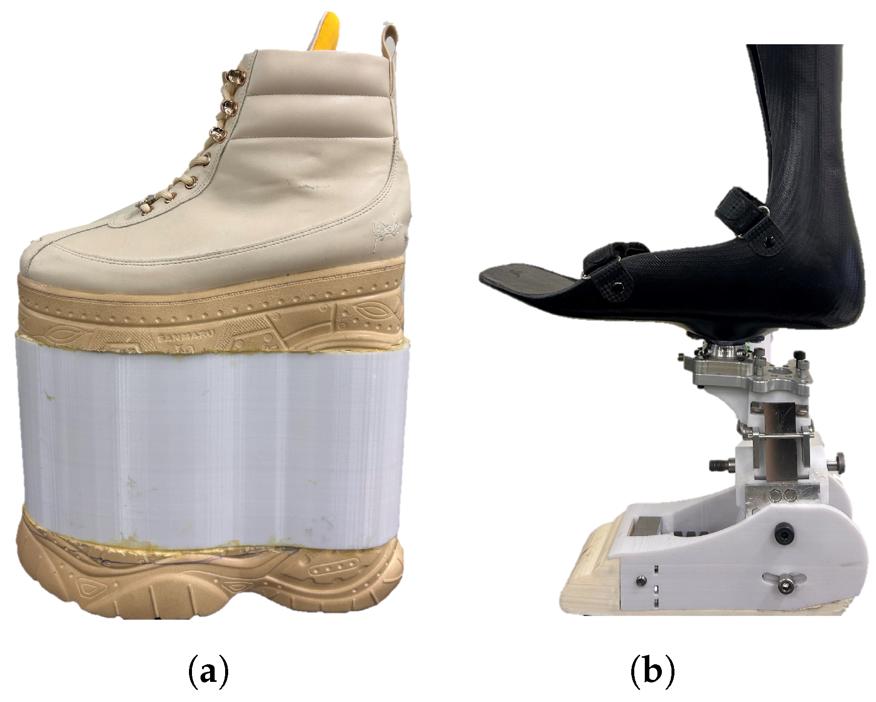
Figure 1. (a) Leg length-extending shoe and, (b) 3-DOF (three degree of freedom) prosthesis mounted on a prosthetic simulator for its use in the gait trials.
Prosthesis 07 00082 g001
Figure 2. Experimental setup for the gait trials with its inputs and outputs. The effects of the presence and absence of toe compliance and compliance in the coronal plane were assessed for gait kinematics, ground reaction forces, spatiotemporal variables, and electromyographic signals.
Prosthesis 07 00082 g002
Figure 3. (a) Frontal, (b) lateral, and (c) isometric CAD of the prosthetic foot and its components.
Prosthesis 07 00082 g003
Figure 4. (a) Stance time and (b) stance/swing ratio boxplots for the different compliance variants over the gait trials (T: toe compliance; L Lateral compliance; NT: no toe compliance; NL: no lateral compliance). * indicates a p-value < 0.05, ** indicates a p-value < 0.01.
Prosthesis 07 00082 g004
Figure 5. Pelvis, knee, hip, and ankle kinematics in the sagittal and coronal planes under the different compliance conditions during the gait trials. The curve shown in black corresponds to barefoot walking. The shaded areas correspond to the standard deviation of the curves.
Prosthesis 07 00082 g005
Figure 6. Peak (a) eversion, (b) pelvis obliquity, and (c) dorsiflexion boxplots for the different compliance variants over the gait trials (T: toe compliance; L: Lateral compliance; NT: no toe compliance; NL: no lateral compliance). * indicates a p-value < 0.05, ** indicates a p-value < 0.01.
Prosthesis 07 00082 g006
Figure 7. (a) Ankle coronal range of motion, (b) hip sagittal range of motion and (c) pelvis coronal range of motion boxplots for the different compliance variants over the gait trials (T: toe compliance; L: Lateral compliance; NT: no toe compliance; NL: no lateral compliance). * indicates a p-value < 0.05, ** indicates a p-value < 0.01.
Prosthesis 07 00082 g007
Figure 8. Sagittal (around the mediolateral axis), coronal (around the anteroposterior axis), and transverse (around the vertical axis) moments along with the ground reaction forces in the anteroposterior, mediolateral, and vertical directions under the different compliance conditions during the gait trials. The control curve, shown in black, corresponds to barefoot walking. The shaded areas correspond to the standard deviation of the curves.
Prosthesis 07 00082 g008
Figure 9. Peak (a) dorsiflexion moment, (b) coronal moment, (c) vertical force, and (d) mediolateral force for the different compliance variants over the gait trials (T: toe compliance; L: Lateral compliance; NT: no toe compliance; NL: no lateral compliance). * indicates a p-value < 0.05, ** indicates a p-value < 0.01.
Prosthesis 07 00082 g009
Figure 10. Average RMS of the (a) non-prosthetic-side gluteus medius and prosthetic-side (b) vastus lateralis and (c) hamstrings for the different compliance variants in the gait trials (T: toe compliance; L: Lateral compliance; NT: no toe compliance; NL: no lateral compliance). * indicates a p-value < 0.05, ** indicates a p-value < 0.01.
Prosthesis 07 00082 g010
Table 1. Obtained spatiotemporal variables for the different foot conditions. The corresponding Friedmann and Wilcoxon p-values are shown. All variables, with the exception of gait speed, were normalized by leg length and time and therefore are adimensional. Statistically significant comparisons (p-value < 0.05) are displayed in bold.
MetricConditions—Median (IQR)FriedmannWilcoxon (p-Value)
NT-NLT-NLNT-LT-LFp-ValueNT-NL
vs.
T-NL
NT-NL
vs.
L-NT
NT-NL
vs.
T-L
Spatiotemporal
Gait Speed (m/s)0.46 (0.17)0.49 (0.12)0.49 (0.20)0.50 (0.17)1.560.6684
Stance Time3.64 (0.88)3.29 (0.66)3.35 (1.10)3.18 (0.92)8.280.04050.0090.3750.0136
Swing Time1.85 (0.42)1.94 (0.39)1.88 (0.38)1.88 (0.41)3.00.391
Ratio
Stance-Swing
1.64 (0.47)1.50 (0.26)1.69 (0.31)1.63 (0.27)8.20.0400.00580.3220.064
Step width0.12 (0.02)0.12 (0.02)0.12 (0.01)0.12 (0.02)1.070.781
Step Length0.43 (0.07)0.44 (0.05)0.45 (0.06)0.44 (0.07)2.640.45
Stride Length0.81 (0.12)0.83 (0.06)0.84 (0.09)0.83 (0.16)0.3590.948
Table 2. Joint peak angles for the different foot conditions. The corresponding Friedmann and Wilcoxon p-values are shown. Statistically significant comparisons (p-value < 0.05) are displayed in bold.
MetricConditions—Median (IQR)FriedmannWilcoxon (p-Value)
NT-NLT-NLNT-LT-LFp-ValueNT-NL
vs.
T-NL
NT-NL
vs.
L-NT
NT-NL
vs.
T-L
Peak Kinematics ( Deg )
Peak dorsiflexion7.22 (4.62)9.01 (3.72)6.00 (4.37)9.18 (5.59)13.790.00310.0190.5560.048
Peak plantarflexion8.29 (6.25)5.95 (4.20)8.57 (3.81)5.69 (4.22)8.760.01690.250.910.128
Peak inversion0.80 (0.20)0.91 (0.55)1.05 (0.42)0.78 (0.42)1.440.696
Peak eversion2.06 (0.48)1.99 (1.02)2.69 (0.59)2.63 (0.55)8.870.03090.9210.0190.013
Peak knee flexion47.65 (7.28)45.16 (8.74)46.28 (7.80)44.16 (11.17)7.560.056
Peak hip flexion26.33 (14.62)22.36 (12.88)23.31 (10.01)24.75 (8.03)2.40.493
Peak hip extension4.64 (9.02)6.40 (6.93)7.88 (3.26)5.98 (5.57)4.680.196
Peak hip abduction2.73 (6.84)1.41 (3.95)3.07 (6.64)3.15 (4.34)3.240.356
Peak hip adduction5.66 (5.92)6.26 (5.45)6.01 (5.88)6.46 (5.81)2.760.43
Max. pelvis tilt5.86 (4.25)4.22 (2.91)5.34 (3.47)6.55 (2.36)0.3560.948
Max. pelvis obliquity4.55 (2.55)4.29 (1.98)4.06 (2.60)3.29 (2.13)8.7560.0320.4310.8450.0019
Table 3. Joint’s range of motion for the different foot conditions. The corresponding Friedmann and Wilcoxon p-values are shown. Statistically significant comparisons (p-value < 0.05) are displayed in bold.
MetricConditions—Median (IQR)FriedmannWilcoxon (p-Value)
NT-NLT-NLNT-LT-LFp-ValueNT-NL
vs.
T-NL
NT-NL
vs.
L-NT
NT-NL
vs.
T-L
ROM ( Deg )
Ankle Sagittal ROM15.76 (1.54)16.02 (2.12)15.04 (1.95)15.39 (3.82)4.320.228
Ankle Coronal ROM2.76 (0.23)3.13 (0.92)3.62 (0.33)3.31 (0.63)17.280.00060.3220.00190.0019
Knee ROM38.00 (8.39)37.78 (7.85)36.74 (7.68)38.03 (9.39)2.280.516
Hip Sagittal ROM33.13 (4.37)29.62 (3.70)32.05 (5.23)30.40 (4.73)12.230.0030.0190.9210.048
Hip Coronal ROM7.96 (2.17)7.96 (1.91)8.22 (3.20)8.61 (1.85)4.190.24
Pelvis Sagittal ROM5.41 (2.03)4.58 (0.76)5.14 (1.34)4.77 (1.25)5.40.144
Pelvis Coronal ROM9.21 (0.97)8.82 (0.75)8.64 (1.81)8.65 (1.26)7.910.0470.02730.0480.019
Table 4. Obtained ground reaction forces and moments for the different foot variants. The corresponding Friedmann and Wilcoxon p-values are shown. Statistically significant comparisons (p-value < 0.05) are displayed in bold.
MetricConditions—Median (IQR)FriedmannWilcoxon (p-Value)
NT-NLT-NLNT-LT-LFp-ValueNT-NL
vs.
T-NL
NT-NL
vs.
L-NT
NT-NL
vs.
T-L
Ground Reaction
Moments ( Nm · kg 1 )
Peak A-P Plantarflexion
Moment
0.48 (0.20)0.39 (0.19)0.59 (0.20)0.44 (0.17)4.910.177
Peak A-P Dorsiflexion
Moment
0.48 (0.20)0.39 (0.19)0.59 (0.20)0.44 (0.17)11.750.0080.3750.0090.2324
Peak M-L Moment1.05 (0.22)0.98 (0.27)0.78 (0.26)0.89 (0.40)10.310.0160.7690.0270.62
Peak Transversal Moment0.16 (0.04)0.17 (0.06)0.15 (0.04)0.16 (0.06)1.190.753
Ground Reaction
Forces ( kN · kg 1 )
Peak A-P, F x Force−0.098 (0.046)−0.091 (0.033)−0.079 (0.044)−0.089 (0.038)5.870.117
Peak A-P, F x + Force0.11 (0.02)0.10 (0.04)0.10 (0.04)0.12 (0.04)2.870.41
Peak Lateral Force0.070 (0.015)0.069 (0.010)0.061 (0.019)0.066 (0.012)12.240.0060.6950.0480.019
Peak Vertical Force1.124 (0.052)1.123 (0.044)1.155 (0.049)1.165 (0.045)21.727 × 10−50.9210.0020.004
Table 5. Obtained RMS from electromyography signals for the different foot conditions. The corresponding Friedmann and Wilcoxon p-values are shown. Statistically significant comparisons (p-value < 0.05) are displayed in bold.
MetricConditions—Median (IQR)FriedmannWilcoxon (p-Value)
NT-NLT-NLNT-LT-LFp-ValueNT-NL
vs.
T-NL
NT-NL
vs.
L-NT
NT-NL
vs.
T-L
EMG Prosthetic side
( μ V )
RMS Gluteus Medius0.60 (0.76)0.66 (0.70)0.58 (0.62)0.80 (0.69)1.940.58
RMS Vastus Lateralis0.95 (0.21)0.87 (0.32)0.92 (0.26)1.05 (0.87)5.40.1440.01950.0830.845
RMS Hamstrings1.63 (0.54)1.42 (0.72)1.13 (0.62)1.56 (0.57)6.240.10.0480.160.083
RMS Gluteus Maximus0.26 (0.12)0.30 (0.08)0.35 (0.10)0.31 (0.08)2.330.506
EMG Non-Prosthetic
( μ V )
RMS Gluteus Medius1.00 (1.10)0.83 (0.63)0.84 (0.40)0.66 (0.45)6.960.070.550.490.037
RMS Vastus Lateralis0.73 (1.10)0.72 (0.49)0.63 (0.56)0.55 (0.62)2.730.434
RMS Hamstrings1.63 (1.38)1.95 (1.60)1.87 (1.15)1.94 (1.03)1.790.614
RMS Gluteus Maximus0.53 (0.32)0.33 (0.25)0.33 (0.34)0.32 (0.27)3.120.373
Table 6. Cohen’s d effect size and estimated power for significant biomechanical variables. Statistically significant comparisons (p-value < 0.05) are displayed in bold. * indicates significance with large effect size and moderate power ( β > 0.55); ** indicates significance with small effect size and low statistical power ( β < 0.55).
VariablePairMean DifferenceEffect Size (Cohen’s d)Power
Stance TimeT-NL0.2661.1080.875
NT-L0.1010.3020.130
T-L0.3170.9370.7509 *
Ratio Stance/SwingT-NL0.1851.2590.942
NT-L0.04940.2710.119
T-L0.1650.71550.523
DorsiflexionT-NL−2.2063−1.06140.8469
NT-L−0.4812−0.19440.0856
T-L−1.2459−0.82490.6425 *
Hip Sagittal RomT-NL2.277350.98020.7874
NT-L0.052490.01350.0502
T-L1.616250.54810.3412 **
Peak Pelvis ObliquityT-NL0.74330.44480.243
NT-L0.5850.2990.148
T-L1.67161.00790.805
Peak EversionT-NL−0.0777−0.18170.081
NT-L−0.5710−0.99070.796
T-L−0.5802−1.14850.897
Ankle Coronal RomT-NL−0.2920−0.64100.441
NT-L−0.7663−2.45081
T-L−0.5983−3.80161
Pelvis Sagittal RomT-NL0.66530.75410.5665 *
NT-L0.67310.80810.6248 *
T-L0.90260.97690.7848
Anteroposterior Peak Ground
Reaction Moment Dorsiflexion
T-NL0.0480.340.16
NT-L−0.162−1.30340.9548
T-L0.0310.19280.0849
Mediolateral Peak Ground
Reaction Moment
T-NL0.0280.150.07
NT-L0.1400.720.53 **
T-L0.0270.120.06
Mediolateral Peak Ground
Reaction Force
T-NL−0.0021−0.330.16
NT-L0.00380.560.35 **
T-L0.00150.250.11 **
Vertical Peak Ground
Reaction Force
T-NL0.00790.340.16
NT-L−0.0231−1.190.92
T-L−0.0244−1.070.85
Vastus Lateralis Activity
Prosthetic side
T-NL0.0981.05410.8421
NT-L0.13420.52370.3164
T-L−0.1341−0.22810.0992
Hamstrings Activity
Prosthetic side
T-NL0.202950.53860.3314 **
NT-L0.307850.528760.3215
T-L0.227330.390460.1977
Gluteus Medius Activity
Non-Prosthetic side
T-NL0.13270.36260.177
NT-L0.02710.04670.052
T-L0.3540.86710.6853 *
Disclaimer/Publisher’s Note: The statements, opinions and data contained in all publications are solely those of the individual author(s) and contributor(s) and not of MDPI and/or the editor(s). MDPI and/or the editor(s) disclaim responsibility for any injury to people or property resulting from any ideas, methods, instructions or products referred to in the content.