Article

An Optimal Synthetic Strategy for Conjugating Folic Acid with Manganese-Doped Silica Nanoparticles to Enhance Their Colloidal Stability


1
Alexander Butlerov Chemistry Institute, Kazan (Volga Region) Federal University, 18 Kremlyovskaya Street, Kazan 420008, Russia
2
Institute of Carbon Neutrality, School of New 401 Energy and Materials, Southwest Petroleum University, Chengdu 610500, China
3
A.E. Arbuzov Institute of Organic and Physical Chemistry, Kazan Scientific Center, Russian Academy of Sciences, 8 Arbuzov Street, Kazan 420088, Russia
4
Department of Nanotechnology in Electronics, Kazan National Research Technical University Named After A.N. Tupolev-KAI, 10 Karl Marx Street, Kazan 420111, Russia
5
Department of Physics, Kazan National Research Technological University, 68 Karl Marx Street, Kazan 420015, Russia
*
Authors to whom correspondence should be addressed.
Chemistry 2026, 8(2), 21; https://doi.org/10.3390/chemistry8020021
Submission received: 29 December 2025 / Revised: 21 January 2026 / Accepted: 29 January 2026 / Published: 11 February 2026

Abstract

The inadequate biosafety of MRI contrast agents (CAs) remains a challenging issue. Both increasing the magnetic relaxivity of CAs and targeting them through conjugation with folates are promising approaches to addressing this issue. Silica nanoparticles (SNs) with Mn2+ ions specifically localized in the outer layer were selected as the target for further surface modification for the covalent attachment of folates. It was shown that when Mn-containing SNs are conjugated with folates via preliminary amino modification of the surface silanol groups, the folate-conjugated SNs suffer from colloidal instability. Thus, precoating Mn-containing SNs with unfolded BSA exposes surface amino groups that successfully conjugate with folates without loss of colloidal stability. Partial washout of surface-localized Mn2+ follows folate conjugation of Mn-containing SNs, although residual Mn2+ ions provide r1(2) relaxivities of 62.1 (160.4) mM−1s−1 at 0.47 T.

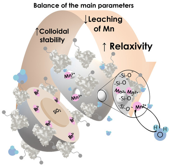
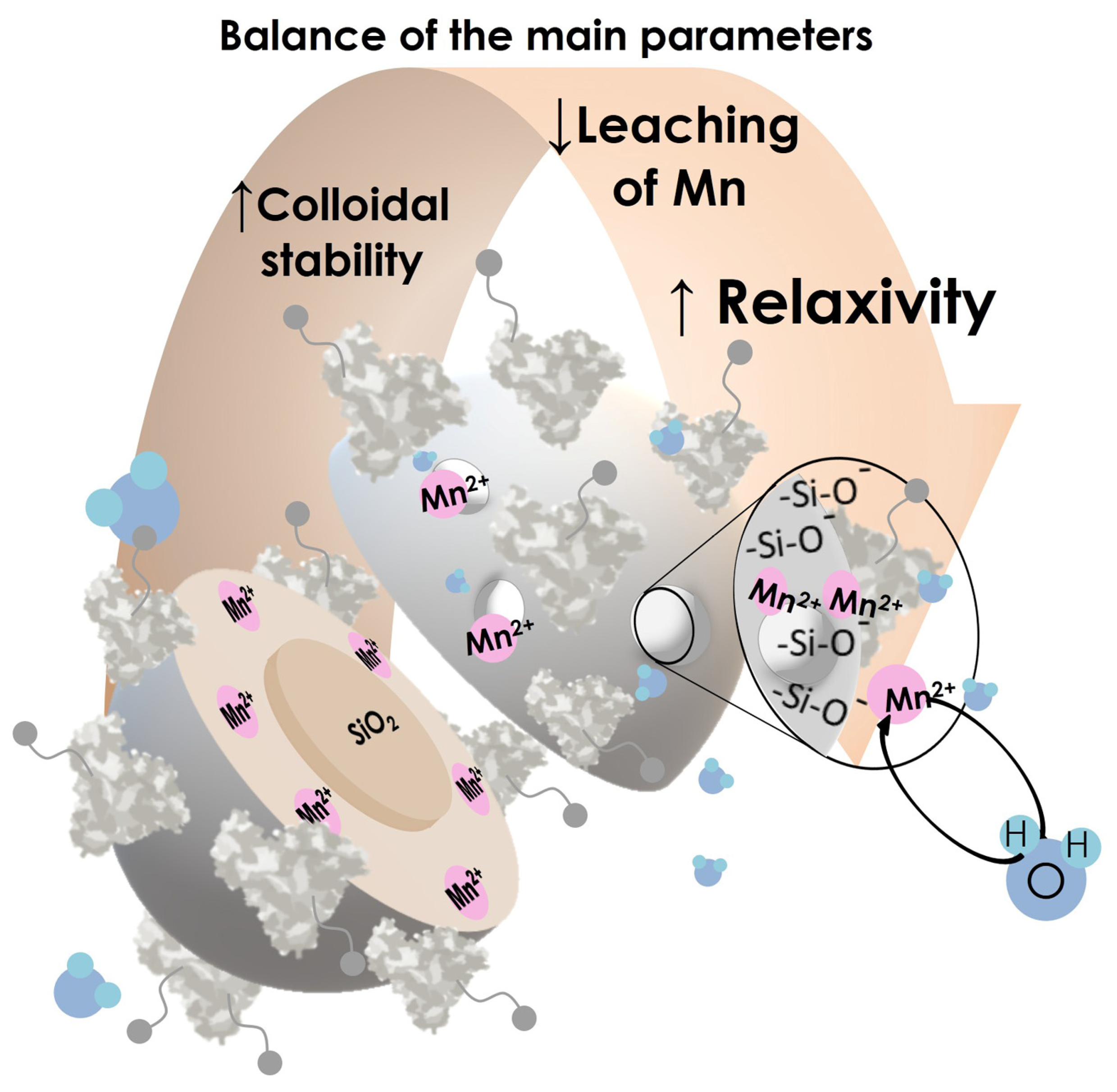
Graphical Abstract

1. Introduction

Manganese ions Mn2+ encapsulated in silica nanoparticles represent a promising basis for the creation of contrast agents for MRI diagnostics. Current commercially available contrast agents are primarily based on gadolinium compounds Gd3+, while contrast agents based on manganese ions Mn2+ are available in smaller quantities [1,2,3,4]. An important characteristic of contrast agents is their ability to improve the visibility of specific tissues or structures during diagnostic imaging by altering the local relaxation properties of the tissues. This characteristic is expressed by a parameter called relaxivity, which primarily depends on the zero-field-splitting contribution, which is significantly higher for Gd3+ ions than for Mn2+. However, manganese ions Mn2+ are biogenic ions compared to gadolinium ions Gd3+ [1,5,6,7,8,9,10,11,12,13]. Therefore, the development of manganese-based contrast agents Mn2+ is a pressing issue. The goal is to produce Mn2+ compounds that are stable under physiological conditions and exhibit high relaxivity values comparable to gadolinium compounds Gd3+.
One way to increase the relaxivity parameters of Mn2+ compounds can be achieved by retarding the translational and rotational motion due to the incorporation of Mn2+ into silica nanoparticles (SNs). A key requirement for effective contrast is the high accessibility of manganese ions to water molecules, which is achieved through their preferential location in the outer layer of SNs [14,15]. Such architecture of Mn2+-loaded SNs was used in this study. These SNs were produced using the method we described previously in [16], which is based on the use of a reverse microemulsion method and a complex of Mn2+ with oxalate ligands. The SNs were designated as Mn(OX)@SNs.
The use of “naked” silica nanoparticles in vivo is undesirable due to their low biocompatibility. Surface silanol groups (–Si–OH) can disrupt protein structure, induce oxidative stress, and, most critically, damage cell membranes, exhibiting pronounced hemolytic activity [13,17,18,19,20,21,22,23,24,25,26]. In this study, we explore two fundamentally different approaches to surface functionalization of silica nanoparticles to create a biocompatible shell. The first is the covalent introduction of amino groups using aminopropyltriethoxysilane (APTES). Modification with amino groups significantly reduces hemolytic activity by neutralizing the negative charge of the silica nanoparticle surface [27]. The second is the non-covalent formation of a shell from denatured bovine serum albumin (BSA). This method allows the creation of a stable protein layer on the particle surface, which effectively shields silanol groups and can mask the nanoparticle from immediate capture by the mononuclear phagocytic system [28].
However, the purpose of surface modification is not limited to improving biocompatibility. To enhance diagnostic efficiency, it is necessary to ensure the accumulation of the contrast agent in the target tissue, i.e., to achieve targeting. Both types of modifications we selected—the amino groups and the protein shell—serve a dual function. In addition to enhancing biocompatibility, they provide functional groups for the subsequent attachment of targeting molecules. In particular, primary amino groups, which are also present in the BSA structure, are a classic reaction site for conjugation with carboxyl groups.
Folic acid (FA) was chosen as the targeting molecule. This decision was motivated by the fact that folate receptors (FRs) are overexpressed on the membranes of a wide range of cancer cells (e.g., ovarian, breast, and lung cancer), while their expression is minimal in healthy tissue [29,30,31,32,33,34,35]. Targeting nanoparticles to these receptors significantly increases their local concentration in the tumor, enhancing signal contrast and diagnostic specificity. Among the many linkers used for the covalent grafting of folic acid [33,36,37] the formation of the amide bond between the activated carboxyl groups of folic acid and the amino groups on the surface of silica nanoparticles is the most widely used method for grafting folic acid residue onto the surface of silica nanoparticles [14,37,38,39].
Therefore, the aim of this study is to select the optimal method for functionalizing silica nanoparticles with folic acid molecules, ensuring minimal loss of manganese content and colloidal stability of the modified nanoparticles in aqueous solutions. In this study, the efficiency of covalent functionalization of silica nanoparticles with amino groups was compared with an alternative strategy using pre-formation of a shell from denatured BSA, and a comprehensive assessment of the obtained results was given, including an analysis of their colloidal stability and magnetic relaxation properties.

2. Materials and Methods

2.1. Materials

Commercial chemicals Triton X-100 (98%), tetraethyl orthosilicate (TEOS, 98%), ammonium hydroxide (28–30%), n-heptanol (98%), cyclohexane (99%), acetone (99%), ethanol (99.5%), 3-aminopropyltriethoxysilane (APTES, 99%), β-alanine, and fluorescamine were purchased from Acros Organics (Geel, Belgium). Mn(NO3)2·4 H2O (98%), folic acid (FA, ≥97%), 1-ethyl-3-(3-dimethylaminopropyl)carbodiimide (EDAC, 99%), N-hydroxysuccinimide (NHS, 98%), and potassium oxalate monohydrate (99%) were purchased from Alfa Aesar (Heysham, Lancashire, England). Bovine serum albumin (≥98%) was purchased from Sigma-Aldrich (Burlington, MA, USA), EtOH was purified by distillation, and phosphate (pH 7.4) buffer was used to adjust the required pH-values. DMSO (SRL, extrapure, 99%) was purified by distillation under reduced pressure.

2.2. Synthesis

Mn(OX)@SNs were synthesized according to the reported procedures [14].
The obtained nanoparticles were functionalized with denatured BSA molecules following [40], resulting Mn(OX)-BSA. Briefly, Ethanol was gradually introduced via syringe pump (1 mL/min) into an aqueous solution of BSA (0.2 g∙L−1) in phosphate buffer (0.05 M, pH 7.4) and Mn(OX)@SNs (1 g∙L−1) while stirring at 850 rpm. This mixture was then sonicated in a water bath (≤45 °C) for 1 h. After sonication, the solution was stirred for an additional 2 h at ambient temperature. Finally, the nanoparticles were isolated and purified through two cycles of centrifugation (15,000 rpm, 20 min each) followed by redispersion.

2.2.1. Synthesis of I-Mn(OX)@SNs-NH2-FA and O-Mn(OX)@SNs-NH2-FA via One-Pot Method

O-Mn(OX)@SNs-NH2-FA were synthesized according to the reported procedures [14] for "Mn(OX)@SNs-NH2-FA" nanoparticles.
I-Mn(OX)@SNs-NH2-FA. Following the procedure described in [14] the reaction was continued for 4 h after the addition of folic acid in a foil-covered vessel, protected from light. Folic acid was activated, following the method described in [14] before introducing in reaction media. The synthesized nanoparticles were precipitated with acetone and then washed one time with the acetone/ethanol mixture (1:1), two times with ethanol, and three times with water.

2.2.2. Conjugation of Folic Acid with APTES

6.67 mg of folic acid was dissolved in 2 mL of DMSO in a dark glass vial. In 15 min, the solution was added to the vessel with a suspension of 7.04 mg of EDAC and stirred until complete dissolution. Then the solution was transferred to a vial with a suspension of 1.74 mg of NHS, where it was stirred for 24 h in a vessel without access to light. Then 0.049 mL of APTES was added to the obtained mixture, and the conjugation continued for 18 h to add to II-Mn(OX)@SNs-NH2-FA or 1 h for III-Mn(OX)@SNs-NH2-FA.

2.2.3. Synthesis of II-Mn(OX)@SNs-NH2-FA and III-Mn(OX)@SNs-NH2-FA via One-Pot Method

5.96 g of Triton X-100, 5.72 mL of n-heptanol, and 1.38 mL of H2O were added to 23.3 mL of cyclohexane. After 15 min of stirring, 0.25 mL of aqueous NH3 (28–30%) was added, followed by the addition of 0.25 mL of TEOS in 15 min. This mixture was stirred for 24 h. On the next day, aqueous potassium oxalate solution (0.69 mL, 1 × 10⁻² M) was added dropwise to the mixture, and after 10 min of stirring, aqueous Mn(NO3)2·4 H2O solution (0.69 mL, 5 × 10⁻³ M) was slowly added. In 10 min, 0.25 mL of TEOS was added. The obtained mixture was stirred for a further 24 h.
The next day, 2 mL of DMSO solution, containing conjugated FA-APTES were added to the obtained mixture, and the synthesis continued for an additional 5 h in a foil-covered vessel without access to light. The synthesized nanoparticles were precipitated with acetone and then were washed one time with the acetone/ethanol mixture (1:1), two times with ethanol, and three times with water.

2.2.4. Conjugation of Mn(OX)@SNs-BSA with Folates

The solution of pre-activated folic acid was added to the 3 mL of dispersion of Mn(OX)@SNs-BSA in ethanol (3 g·L−1). The obtained mixture was stirred for 24 h (800–850 rpm) in a dark glass vial covered with foil. The nanoparticles were separated by centrifugation and then washed two times with distilled water.

2.3. Methods

ICP-OES: The contents of Si and Mn were identified in the colloids using simultaneous inductively coupled plasma optical emission spectrometry (ICP-OES) model iCAP 6300 DUO by Varian Thermo Scientific Company (Palo Alto, CA, USA) equipped with a CID detector. The simultaneous measurements of peak heights within the 166 to 867 nm range were performed with the optical resolution of less than 0.007 nm to 200 nm at the working frequency of 27.12 MHz. The optimal peak height measurements with suppressed spectral noises were done through both radial and axial view configurations. Concentration of Si and Mn ions was determined, respectively, by the spectral lines: 251.61 nm and 257.61 nm.
The percentage of manganese loss during functionalization was calculated by comparing the Si:Mn molar ratio of functionalized silica nanoparticles to the Si:Mn molar ratio of the Mn(OX)@SNs reported previously in [14].
Transmission electron microscopy (TEM): The transmission electron microscopy (TEM) images were obtained by means of a Hitachi HT7700 (Chiyoda-ku, Japan) transmission electron microscope at an acceleration voltage of 100 kV. Silica nanoparticles were ultrasonicated in bidistilled water for 10 min and then dispersed on 200 mesh copper grids with continuous formvar support films.
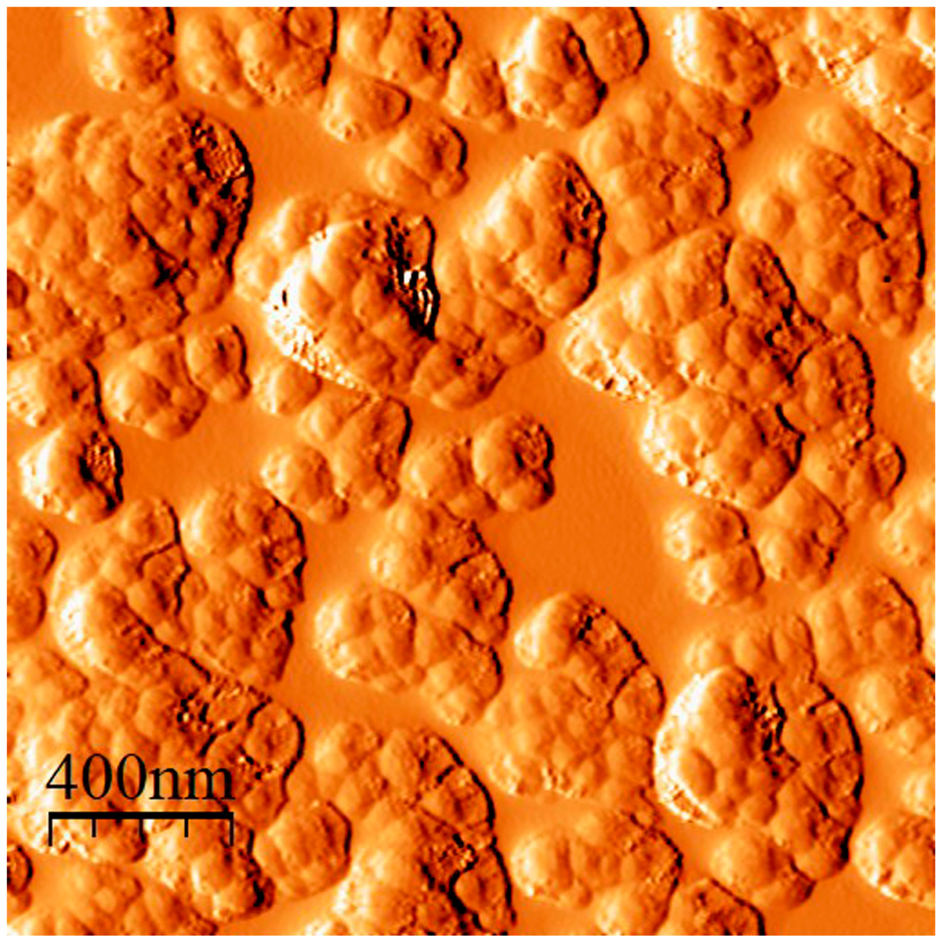
An atomic force microscope (AFM) MultiMode V (Veeco, Plainview, New York, USA) has been used to reveal the morphology of the nanoparticles. The 250–350 kHz RTESP cantilevers (Veeco, Plainview, NY, USA) have been used in all measurements. The aqueous dispersions of nanoparticles (0.2 g∙L−1) were ultrasonicated for 10 min, and then a droplet of the sample was placed on a mica surface. The AFM imaging was performed after water evaporation. The microscopic images were obtained with a 512 × 512 resolution. The scanning rate was 1 Hz. AFM images processing was carried out using WSxM 4.0 (Nanotec Electrónica S.L., Tres Cantos, Spain) [41]. The tip-convolution effect has been minimized by processing the obtained AFM data with the use of a standard algorithm (deconvolution).
Dynamic light scattering (DLS): Dynamic light scattering (DLS) and electrokinetic potential experiments were performed on a Zetasizer Nano instrument (Malvern Instruments, Malvern, UK). Electrokinetic potential values were calculated by the Smoluchowski-Helmholtz equation [42]. Experimental autocorrelation functions were analyzed with the Malvern DTS software (Dispersion Technology Sofware 5.10) and the second-order cumulant expansion methods. The average error was ca. 4%. All samples were prepared in deionized water filtered through a PVDF membrane with a Syringe Filter (0.45 µm). The concentration of silica nanoparticle dispersion was 0.2 g∙L−1. All measurements were performed at least in triplicate at 25 °C. The samples were ultrasonicated for 10 min and then equilibrated at 25 °C prior to size and zeta-potential measurements for 2 min.
Ultraviolet-visible (UV-Vis): The UV–vis measurements were carried out using Specord®50 Plus (Analytik Jena, Jena city, Germany) with 10 mm cuvettes at 25 °C.
Relaxometric measurements: The preparation of the samples and subsequent relaxometric measurements were carried out in accordance with the procedure described in [15].
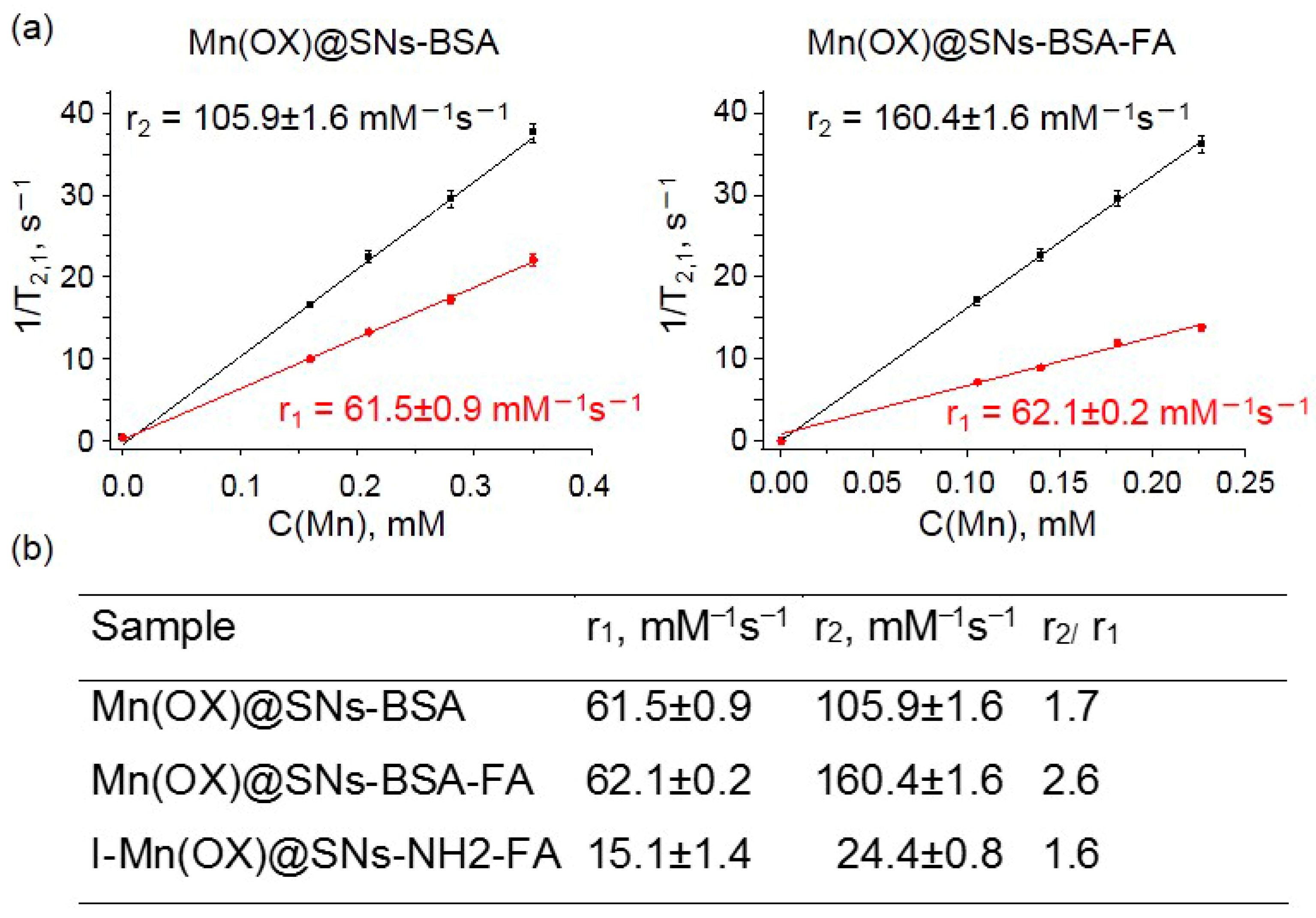
Transverse 1/(T2CMn) and longitudinal 1/(T1CMn) relaxivities (mM−1s−1) have been calculated from the slopes of 1/T2,1 vs. c(Mn) plot. The proton relaxation times T1 and T2 were measured using pulsed NMR-relaxometer Minispec MQ20 (19.65 MHz, 0.47 T), from Bruker (Rheinstetten, Germany), by applying the standard radio frequency pulse sequences: inversion-recovery method (spin-lattice relaxation time T1), and Carr–Purcell sequence [43], modified by Meiboom–Gill (spin-spin relaxation time T2). The longitudinal relaxation times T1 of water molecule protons have been defined utilizing the analyzer’s inversion-recovery pulse sequence, with 20 data points collected for fitting [44] with the measuring accuracy error smaller than 3%. The temperature was maintained with the Thermo/Haake DC10 circulator (Karlsruhe, Germany).

3. Results

3.1. Conjugation of Folic Acid with Amino Groups Grafted to Manganese-Containing SNs by Covalent or Non-Covalent Methods

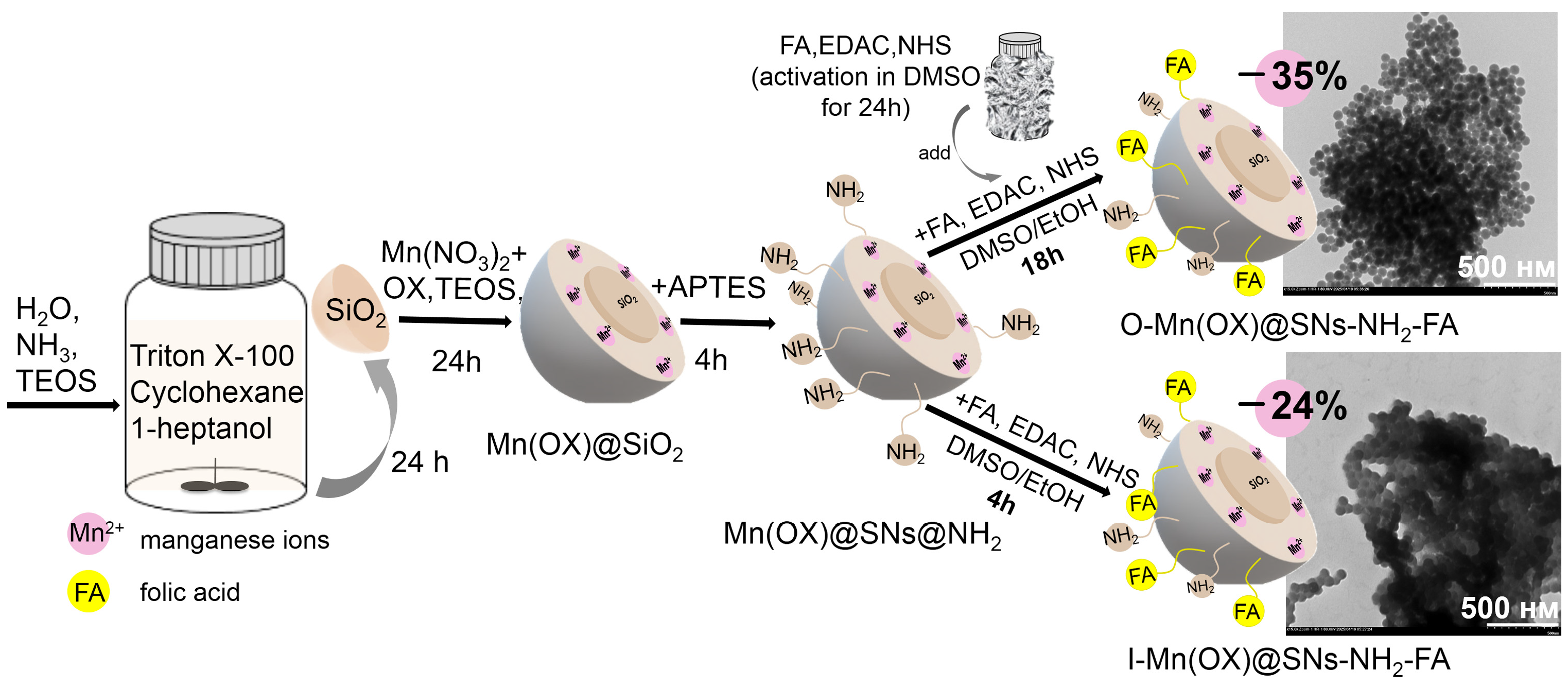
Typically, folate-conjugation is achieved through two steps, which are the surface modification with amino-groups and their cross-linking with folates [37,39]. This method involves preliminary synthesis, isolation, and washing of Mn(OX)@SNs-NH2 nanoparticles, followed by conjugation with activated folic acid. As was previously shown, all this makes this method rather complex and induces a significant loss in the Mn-content [14]. To avoid these shortcomings, a one-pot synthesis method was developed, in which the activated folic acid is introduced to the reaction medium of pre-prepared Mn(OX)@SNs-NH2 nanoparticles without their isolation and washing [14]. Indeed, the use of the one-pot synthesis allows us to obtain the folate-conjugated nanoparticles, which will be further designated as Mn(OX)@SNs-NH2-FA (Figure 1). This synthetic modification allows to reduce the loss of the initial amount of manganese from 60%, as was achieved in [14], to 35% (Figure 1), although for a higher reduction in Mn-losses, the synthesis method should be further modified.
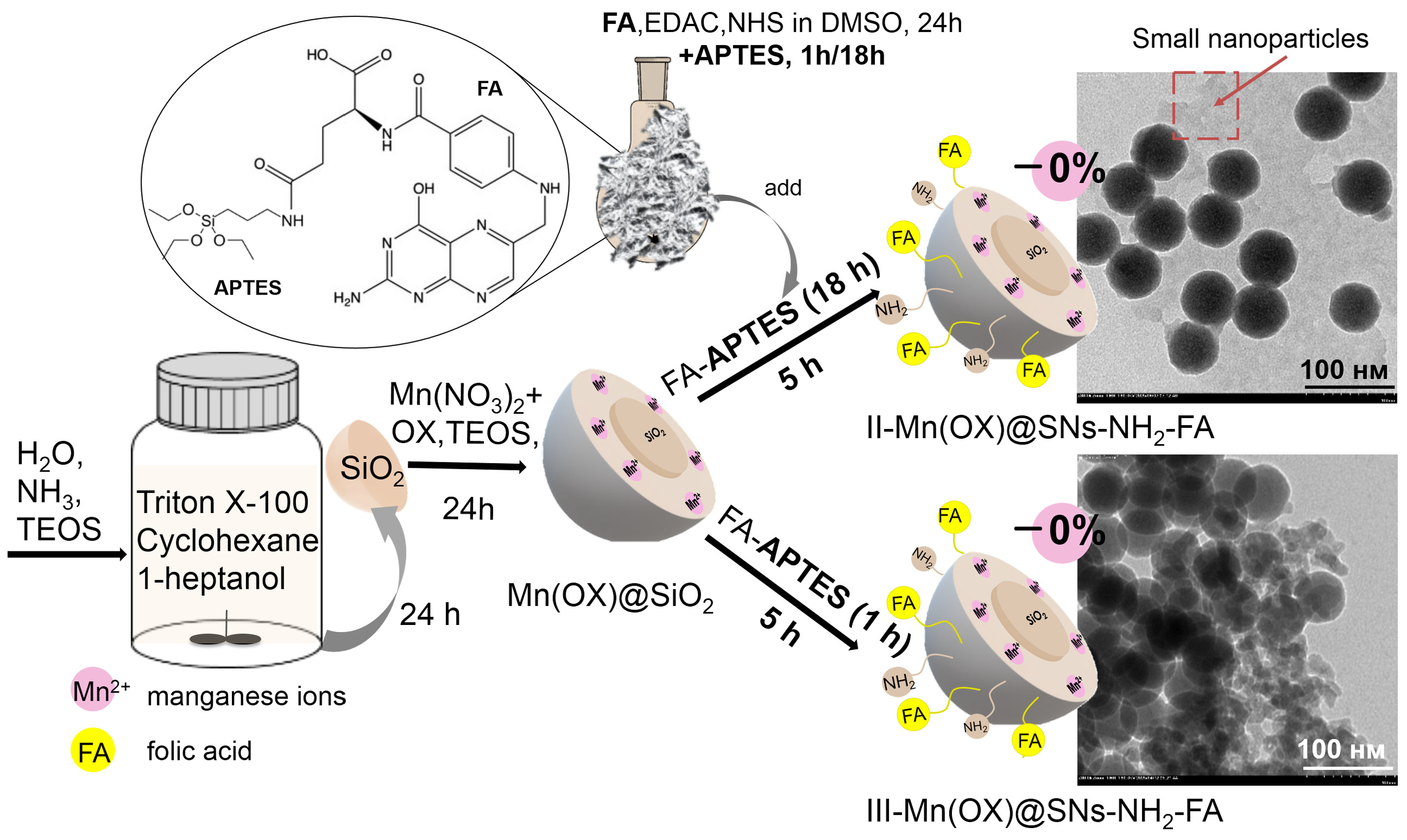
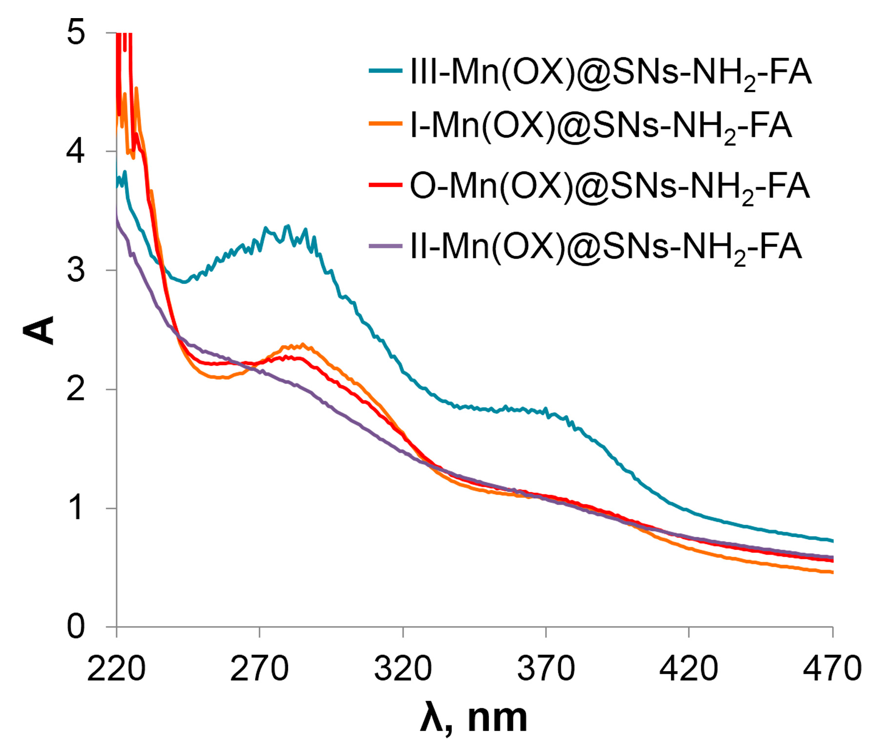
A modification of the one-pot synthesis of folate-conjugated nanoparticles was carried out by varying the time conditions. The first modified method (Figure 1) was similar to the previously published one [14], but the conjugation time of nanoparticles with folates was reduced from 18 to 4 h, since the percentage of manganese ions leaching from silica nanoparticles should be time-dependent. Figure 1 shows that, according to ICP-OES data, Mn losses are slightly reduced (24%) during the synthesis of I-Mn(OX)@SNs-NH2-FA nanoparticles by the modified procedure. To further limit manganese loss, pre-activated folic acid was coupled to aminopropyltriethoxysilane (APTES) as a pre-step before conjugation with Mn(OX)@SNs-NH2-FA (Figure 2). The duration of this pre-step was 1 or 18 h (Figure 2). Next, these reaction mixtures were introduced into the synthesized Mn(OX)@SNs nanoparticles without their preliminary isolation, and the resulting mixture was stirred for 5 h to synthesize II-Mn(OX)@SNs-NH2-FA and III-Mn(OX)@SNs-NH2-FA (Figure 2). The as-prepared nanoparticles were then isolated and washed using the standard method. Although no reduction in manganese content was observed for these nanoparticles compared to Mn(OX)@SNs-NH2 (Table 1), these methods cannot be recommended for modifying particles with folates due to several drawbacks. The drawback of the III-Mn(OX)@SNs-NH2-FA is evident from its TEM image (Figure 2), which shows smaller particles in addition to standard silica nanoparticles. This may be due to the preferential polycondensation of the folate-modified APTES molecules themselves compared to their polycondensation on the Mn(OX)@SN surface. The admixture of smaller-sized particles is less significant for the II-Mn(OX)@SNs-NH2-FA sample (Figure 2), but their deficiency will become apparent upon further analysis of the UV-visible spectra of both samples (Figure 3).
Characteristic absorption bands at ~280 nm and ~375 nm in the electronic absorption spectra of Mn(OX)@SNs-NH2-FA samples indicate the presence of folate residues on their surface (Figure 3). It should be noted that these bands are quite pronounced for all the studied samples, with the exception of sample II-Mn(OX)@SNs-NH2-FA, where these bands are very weak (Figure 3). This indicates that the synthesis conditions used to obtain II-Mn(OX)@SNs-NH2-FA nanoparticles are not suitable for their efficient conjugation with folates.
However, these nanoparticles exhibit low colloidal stability, as evidenced by the increase in the average size and polydispersity index of all studied Mn(OX)@SNs-NH2-FA samples compared to similar values for Mn(OX)@SNs (Table 1). It is well known that the combination of negatively and positively charged groups on the surface of silica nanoparticles is usually accompanied by their enhanced aggregation. The aforementioned aggregation behavior of various Mn(OX)@SNs-NH2-FA samples is in good agreement with their electrokinetic potential values, which range from −10 to +15 mV (Table 1). Thus, after folate modification, no significant surface recharging occurs, since some amino groups remain unbound to folates. The colloidal instability of Mn(OX)@SNs-NH2-FA is manifested in their rapid precipitation in aqueous solutions, which has an undesirable effect on the recording of absorption spectra. As suggested by the works [14,39] the colloidal instability of both amino-modified SNs and their folate-conjugated counterparts can be restricted after their treatment by bovine serum albumin (BSA). In turn, the protein coating of silica nanoparticles allows the use of protein amino groups for conjugation with folates. However, the instability of this protein coating leads to its easy exchange with plasma proteins [45,46], making it unsuitable for the development of targeted contrast agents.

3.2. Conjugation of Silica Nanoparticles Pre-Coated with Denatured BSA with Folic Acid

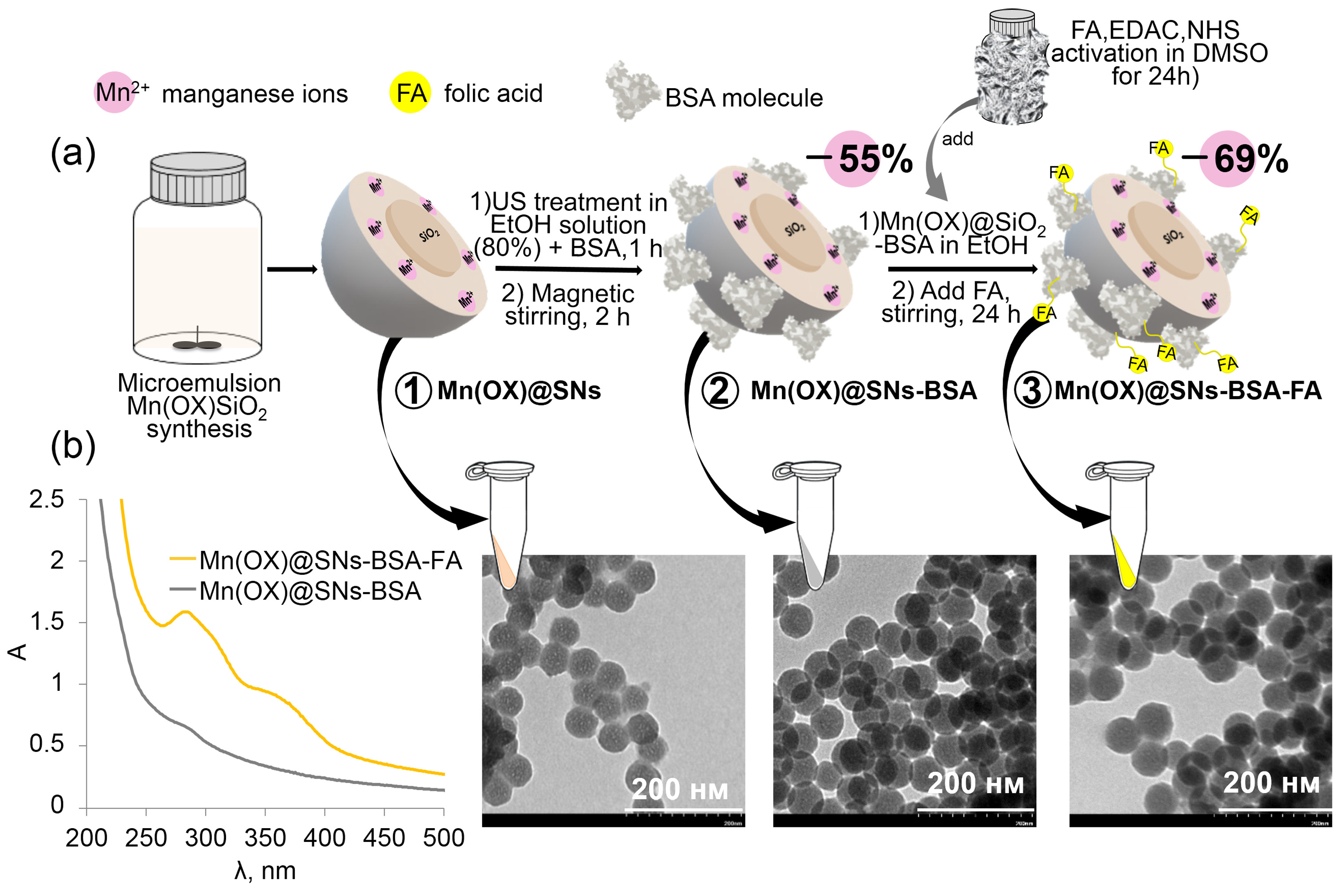
It is well known that protein denaturation makes their adsorption on the silica surface strong enough to avoid easy exchange with plasma proteins. Methods for coatings of silica nanoparticles with unfolded proteins are already known [47,48]. In particular, a modified procedure for BSA-based coating on SNs doped with manganese silicate and manganese oxides [40] was successfully applied to create a similar coating on the surface of Mn(OX)@SNs. Such BSA-modified nanoparticles, hereinafter referred to as Mn(OX)@SNs-BSA, were conjugated with folic acid using the procedure schematically shown in Figure 4. According to the TEM imaging, their size, as well as the size of their folate-conjugated counterparts (Mn(OX)@SNs-BSA-FA), is close to that of the original Mn(OX)@SNs (Figure 4). The Mn(OX)@SNs-BSA sample was also examined by atomic force microscopy (AFM) (Figure A1) to detect the BSA coating, since AFM is more convenient than TEM for examining biological samples. No significant aggregation of Mn(OX)@SNs-BSA and Mn(OX)@SNs-BSA-FA is revealed in both aqueous solutions (Table 1) and in the dried samples (Figure 4).
The UV-visible spectra (Figure 4b) of Mn(OX)@SNs-BSA-FA nanoparticles contain bands characteristic of folic acid fragments with a maximum at ~278 nm and ~360 nm, confirming the presence of folates on the surface of modified silica nanoparticles. Coating of Mn(OX)@SNs with BSA results in a manganese loss of 55% of the initial value, according to ICP-OES data (Table 1). Subsequent modification of Mn(OX)@SNs-BSA with folic acid results in a further manganese loss up to 69%.
To assess the applicability of the developed Mn(OX)@SNs-NH2-FA and Mn(OX)@SNs-BSA-FA nanoparticles as targeted contrast agents, their ability to paramagnetically enhance the longitudinal and transverse relaxation of water molecule protons was assessed through the relaxivity values r1(2), which are the relaxation rates related to one mM of Mn2+ ions. The r1(2) values presented in Figure 5b indicate that the ability of Mn(OX)@SNs-BSA-FA to increase magnetic relaxation rates is significantly higher than that of Mn(OX)@SNs-NH2-FA. Notably, the r1(2) values of Mn(OX)@SNs-BSA and their folate-conjugated analogs are close to each other (Figure 5b), indicating that the BSA coating is the main reason for this increase in relaxivity. Thus, the loss of manganese content in Mn(OX)@SNs-BSA is largely compensated by the increase in relaxation parameters observed upon non-covalent modification with BSA. Notably, these r1(2) values are close to the maximum values previously reported for manganese-containing nanoparticles [14,16,49].
Mn(OX)@SNs-BSA-FA nanoparticles exhibit high colloidal stability due to their high negative electrokinetic potential values (Table 1). The increase in PDI values from 0.1 for Mn(OX)@SNs nanoparticles to 0.2 for their BSA-coated counterparts may be due to some admixture of unfolded protein aggregates (Table 1). Moreover, after one month of refrigerated storage in the dark, the particles remain aggregation-resistant (Table 1). It is noteworthy that the colloidal stability achieved in this work is significantly higher than in [50], where the BSA coating was obtained by covalently grafting the protein via the amino groups. For such particles with a FA-BSA coating, the zeta potential values are +9 mV, since even after conjugation with BSA, the amount of residual propylamine groups is sufficient to neutralize the surface charge [50].
It is noteworthy that the significant increase in the r1(2) values of Mn(OX)@SNs nanoparticles after their coating with unfolded BSA is accompanied by a loss of Mn content (Table 1). This differs significantly from the previously described trend for SNs encapsulated by manganese silicates/oxides [40]. This indicates that the aqueous ethanol solution used for the surface deposition of unfolded BSA on the SNs surface initiates Mn(OX) leaching to a greater extent compared to manganese silicates/oxides. In turn, the high r1(2) values of Mn(OX)@SNs-BSA-FA (Table 1) are comparable to similar values or Mn2+ ions bound to unfolded BSA via surface amino acid residues [40]. Thus, it can be assumed that both the leaching of Mn2+ ions and their subsequent binding to the BSA-based outer layer lead to high r1(2) values of Mn(OX)@SNs-BSA and their folate-conjugated analogs.
Consequently, modification of Mn(OX)@SNs particles with folic acid, either through pre-introduction of amino groups or through coating with denatured BSA, results in a loss of the manganese content of unmodified nanoparticles. In the case of Mn(OX)@SNs-NH2-FA nanoparticles, such losses can be minimized to 24%, although their increased aggregation is a drawback. The colloidal stability and r1(2) values of Mn(OX)@SNs-BSA-FA nanoparticles are significantly higher than those of Mn(OX)@SNs-NH2-FA. However, further research is needed to understand which characteristics of Mn-containing nanoparticles have a greater impact on their in vivo contrast effect.
It is worth discussing the factors responsible for the contrast effect in the target tissue provided by Mn(OX)@SNs-BSA-FA and Mn(OX)@SNs-NH2-FA when used as contrast agents in vivo. As for any Mn-containing nanoparticles, the contrast effect should mainly depend on their concentration in the tissue and the ability of Mn2+ ions, which constitute the nanoparticles, to accelerate the magnetic relaxation of water protons. The tissue concentration should be significantly affected by the nature of the nanoparticle surface, their ability to form the so-called “protein corona”, and the targeting action of folate residues on the surface of these nanoparticles [51,52]. The second factor, i.e., the paramagnetic enhancement of the magnetic relaxation of water protons provided by manganese-containing nanoparticles, may be largely due to the coordination transformations of Mn2+ ions occurring in a specific biological microenvironment. The most important of these transformations are those caused by the acidification resulting from the uptake of manganese-containing nanoparticles by cells. Since the slightly acidic environment of both cancer cells themselves and their specific cellular compartments, lysosomes, is well known as a factor causing partial removal of Mn2+ ions from nanoparticles [53,54]. However, this pH-induced phenomenon is not a disadvantage because the removed Mn2+ ions tend to bind to surrounding proteins, which usually provides a high-contrast effect, which has already been demonstrated by measuring the magnetic relaxation rates of protein-bound Mn2+ ions [49,55]. It should be noted that pH dependence was demonstrated for the original Mn(OX)@SNs nanoparticles [14], whereas some pH-dependent specificity should be expected for Mn(OX)@SNs-BSA-FA and Mn(OX)@SNs-NH2-FA. The above indicates the need for further research to understand which characteristics of Mn-containing nanoparticles have a greater impact on their contrast effect in vivo.

4. Discussion

A prerequisite for conjugating nanoparticles with folates is the introduction of amino groups onto their surface. However, covalent amino modification of nanoparticles invariably reduces their colloidal stability, which remains a problem even after conjugation with folates. This work presents an alternative approach based on the non-covalent introduction of amino groups that does not neutralize the surface charge of the nanoparticles. This was achieved by pre-coating manganese-containing nanoparticles with unfolded BSA. The pre-coating provides multiple surface amino groups available for conjugation with folates, while the negative surface charge of the pre-coated nanoparticles limits their aggregation.
Notably, folate conjugation to manganese-containing BSA-coated SNs is accompanied by significant leaching of Mn2+ from these nanoparticles. However, the leaching of Mn2+ ions and their subsequent binding to the BSA-based outer layer lead to high r1(2) values of BSA-coated SNs doped with Mn(OX) and their folate-conjugated analogs. This indicates that residual Mn2+ ions in BSA-coated SNs and their folate-conjugated analogs are more exposed on the surface than the Mn2+ ions in the parent nanoparticles doped with Mn(OX). Thus, this work reveals a significant difference in the properties of manganese-containing and folate-conjugated nanoparticles, where amine-modified or BSA-coated silica nanoparticles are used as the starting nanoparticles for further conjugation. However, further studies are needed to determine which type of these folate-conjugated nanoparticles is more suitable for use as MRI contrast agents.

Author Contributions

Conceptualization, A.R.M., Y.Z., Z.H. and R.R.Z.; methodology, A.P.B. and O.D.B.; investigation, A.P.B.; A.S.S.; I.R.N., K.V.K. and O.D.B.; writing—review and editing, A.R.M., O.D.B., A.P.B. and R.R.Z.; visualization, A.P.B.; supervision, A.R.M. and Z.H. All authors have read and agreed to the published version of the manuscript.

Funding

This work was supported by the Gennady Komissarov Foundation for the Support of Young Researchers, agreement No. 4-ФП.

Data Availability Statement

The original contributions presented in this study are included in the article. Further inquiries can be directed to the corresponding authors.

Acknowledgments

The authors gratefully acknowledge the Assigned Spectral-Analytical Center of FRC Kazan Scientific Center of RAS for providing the necessary facilities to carry out physical-chemical measurements.

Conflicts of Interest

The authors declare no conflicts of interest.

Abbreviations

The following abbreviations are used in this manuscript:
MRIMagnetic resonance imaging
CAscontrast agents
SNssilica nanoparticles
APTESaminopropyltriethoxysilane
BSAbovine serum albumin
FAfolic acid
FRsfolate receptors

Appendix A. AFM-Image of Mn(OX)@SNs-BSA Nanoparticles

Figure A1. AFM-Image of Mn(OX)@SNs-BSA Nanoparticles.
Chemistry 08 00021 g0a1

References

  1. Caravan, P. Divalent Manganese Complexes as Potential Replacements for Gadolinium-Based Contrast Agents. Investig. Radiol. 2024, 59, 187–196. [Google Scholar] [CrossRef]
  2. Tei, L.; Botta, M.; Geraldes, C.F.G.C. Beyond Gadolinium: The Potential of Manganese Nanosystems in MRI and Multimodal Imaging Agents. Acta Biomater. 2025, 207, 468–494. [Google Scholar] [CrossRef]
  3. Poggiarelli, L.; Bernetti, C.; Pugliese, L.; Greco, F.; Beomonte Zobel, B.; Mallio, C.A. Manganese-Based Contrast Agents as Alternatives to Gadolinium: A Comprehensive Review. Clin. Pract. 2025, 15, 137. [Google Scholar] [CrossRef]
  4. Chow, J.C.L. Nanomaterial-Based Molecular Imaging in Cancer: Advances in Simulation and AI Integration. Biomolecules 2025, 15, 444. [Google Scholar] [CrossRef]
  5. Henoumont, C.; Devreux, M.; Laurent, S. Mn-Based MRI Contrast Agents: An Overview. Molecules 2023, 28, 7275. [Google Scholar] [CrossRef]
  6. Yen, T.-Y.C.; Abbasi, A.Z.; He, C.; Lip, H.-Y.; Park, E.; Amini, M.A.; Adissu, H.A.; Foltz, W.; Rauth, A.M.; Henderson, J.; et al. Biocompatible and Bioactivable Terpolymer-Lipid-MnO2 Nanoparticle-Based MRI Contrast Agent for Improving Tumor Detection and Delineation. Mater. Today Bio 2024, 25, 100954. [Google Scholar] [CrossRef] [PubMed]
  7. Kaur, J.; Sridharr, M. Key Insights on the Classification and Theranostic Applications of Magnetic Resonance Imaging Contrast Agents. ChemMedChem 2024, 19, e202300521. [Google Scholar] [CrossRef] [PubMed]
  8. Wu, M.; Liao, Y.; Guo, D.; Zhai, M.; Xia, D.; Zhang, Z.; Liu, X.; Huang, Y. Manganese-Based Nanomaterials in Diagnostics and Chemodynamic Therapy of Cancers: New Development. RSC Adv. 2024, 14, 14722–14741. [Google Scholar] [CrossRef] [PubMed]
  9. Fu, S.; Younis, M.R.; Cai, Z.; Liu, L.; Gu, H.; Ni, G.; Lui, S.; Ai, H.; Song, B.; Wu, M. One-Pot Fabrication of Kinetically Inert Ultrasmall Manganese(II) Chelate-Backboned Polymer Contrast Agents for High-Performance Magnetic Resonance Imaging. Nano Lett. 2024, 24, 14252–14262. [Google Scholar] [CrossRef]
  10. Vymazal, J.; Rulseh, A.M. MRI Contrast Agents and Retention in the Brain: Review of Contemporary Knowledge and Recommendations to the Future. Insights Imaging 2024, 15, 179. [Google Scholar] [CrossRef]
  11. Geraldes, C.F.G.C. Manganese Oxide Nanoparticles for MRI-Based Multimodal Imaging and Theranostics. Molecules 2024, 29, 5591. [Google Scholar] [CrossRef]
  12. Butt, A.; Bach, H. Nanomedicine and Clinical Diagnostics Part I: Applications in Conventional Imaging (MRI, X-Ray/CT, and Ultrasound). Nanomedicine 2025, 20, 167–182. [Google Scholar] [CrossRef] [PubMed]
  13. Zairov, R.R.; Akhmadeev, B.S.; Fedorenko, S.V.; Mustafina, A.R. Recent Progress in Design and Surface Modification of Manganese Nanoparticles for MRI Contrasting and Therapy. Chem. Eng. J. 2023, 459, 141640. [Google Scholar] [CrossRef]
  14. Bochkova, O.; Stepanov, A.; Bebyakina, A.; Kholin, K.; Nizameev, I.; Voloshina, A.; Parfenov, A.; Tarasov, M.; Budnikova, Y.; Abakumov, M.; et al. Roles of Ligand Structure and Synthetic Methodology in Surface Loading of Mn2+ into Silica Nanoparticles for High Contrast Effect in MRI. Colloids Surf. A Physicochem. Eng. Asp. 2025, 727, 138232. [Google Scholar] [CrossRef]
  15. Bochkova, O.; Stepanov, A.; Khazieva, A.; Akhmadeev, B.; Ismaev, I.; Kholin, K.; Nizameev, I.; Sapunova, A.; Voloshina, A.; Laskin, A.; et al. Magnetic Relaxation of Various Silica-Based Mn-Contrast Agents in Relationship with Mn2+ Chemical Transformations during Fabrication. Mater. Today Chem. 2023, 33, 101706. [Google Scholar] [CrossRef]
  16. Ma, Q.; Luo, Y.; Li, Y.; Xu, S.; Wang, L. Ultrasmall Hydrophilic Gd-Free Contrast Agents via Ligand Directed Hydrolysis for Efficient Magnetic Resonance Imaging. Adv. Funct. Mater. 2025, 35, 2422876. [Google Scholar] [CrossRef]
  17. An, K.; Kwon, S.G.; Park, M.; Na, H.B.; Baik, S.-I.; Yu, J.H.; Kim, D.; Son, J.S.; Kim, Y.W.; Song, I.C.; et al. Synthesis of Uniform Hollow Oxide Nanoparticles through Nanoscale Acid Etching. Nano Lett. 2008, 8, 4252–4258. [Google Scholar] [CrossRef]
  18. Peng, Y.-K.; Lai, C.-W.; Liu, C.-L.; Chen, H.-C.; Hsiao, Y.-H.; Liu, W.-L.; Tang, K.-C.; Chi, Y.; Hsiao, J.-K.; Lim, K.-E.; et al. A New and Facile Method to Prepare Uniform Hollow MnO/Functionalized mSiO2 Core/Shell Nanocomposites. ACS Nano 2011, 5, 4177–4187. [Google Scholar] [CrossRef]
  19. Kim, T.; Momin, E.; Choi, J.; Yuan, K.; Zaidi, H.; Kim, J.; Park, M.; Lee, N.; McMahon, M.T.; Quinones-Hinojosa, A.; et al. Mesoporous Silica-Coated Hollow Manganese Oxide Nanoparticles as Positive T1 Contrast Agents for Labeling and MRI Tracking of Adipose-Derived Mesenchymal Stem Cells. J. Am. Chem. Soc. 2011, 133, 2955–2961. [Google Scholar] [CrossRef] [PubMed]
  20. Giacomelli, C.E.; Norde, W. The Adsorption–Desorption Cycle. Reversibility of the BSA–Silica System. J. Colloid Interface Sci. 2001, 233, 234–240. [Google Scholar] [CrossRef]
  21. Ilinskaya, A.N.; Dobrovolskaia, M.A. Nanoparticles and the Blood Coagulation System. Part II: Safety Concerns. Nanomedicine 2013, 8, 969–981. [Google Scholar] [CrossRef]
  22. Dong, X.; Wu, Z.; Li, X.; Xiao, L.; Yang, M.; Li, Y.; Duan, J.; Sun, Z. The Size-Dependent Cytotoxicity of Amorphous Silica Nanoparticles: A Systematic Review of in Vitro Studies. Int. J. Nanomed. 2020, 15, 9089–9113. [Google Scholar] [CrossRef]
  23. Wang, F.; Gao, F.; Lan, M.; Yuan, H.; Huang, Y.; Liu, J. Oxidative Stress Contributes to Silica Nanoparticle-Induced Cytotoxicity in Human Embryonic Kidney Cells. Toxicol. Vitr. 2009, 23, 808–815. [Google Scholar] [CrossRef]
  24. Dinc, B. Toxicity of Sol-Gel Synthesized Silica Nanoparticles: Insights from Cellular and Model Organism Studies. BioNanoScience 2023, 13, 1922–1932. [Google Scholar] [CrossRef]
  25. Murugadoss, S.; Lison, D.; Godderis, L.; Van Den Brule, S.; Mast, J.; Brassinne, F.; Sebaihi, N.; Hoet, P.H. Toxicology of Silica Nanoparticles: An Update. Arch. Toxicol. 2017, 91, 2967–3010. [Google Scholar] [CrossRef] [PubMed]
  26. Croissant, J.G.; Butler, K.S.; Zink, J.I.; Brinker, C.J. Synthetic Amorphous Silica Nanoparticles: Toxicity, Biomedical and Environmental Implications. Nat. Rev. Mater. 2020, 5, 886–909. [Google Scholar] [CrossRef]
  27. Hemmerich, P.H.; Von Mikecz, A.H. Defining the Subcellular Interface of Nanoparticles by Live-Cell Imaging. PLoS ONE 2013, 8, e62018. [Google Scholar] [CrossRef]
  28. Lazarovits, J.; Chen, Y.Y.; Sykes, E.A.; Chan, W.C.W. Nanoparticle–Blood Interactions: The Implications on Solid Tumour Targeting. Chem. Commun. 2015, 51, 2756–2767. [Google Scholar] [CrossRef] [PubMed]
  29. Fernández, M.; Javaid, F.; Chudasama, V. Advances in Targeting the Folate Receptor in the Treatment/Imaging of Cancers. Chem. Sci. 2018, 9, 790–810. [Google Scholar] [CrossRef] [PubMed]
  30. Lee, H.; Kim, S.; Oh, C.; Khan, I.; Shukla, S.; Bajpai, V.K.; Han, Y.-K.; Huh, Y.S. Folic Acid-Modified Bovine Serum Albumin Nanoparticles with Doxorubicin and Chlorin E6 for Effective Combinational Chemo-Photodynamic Therapy. Mater. Sci. Eng. C 2020, 117, 111343. [Google Scholar] [CrossRef]
  31. Türkeş, E.; Sağ Açıkel, Y. Folic Acid-Conjugated Cancer Drug Curcumin-Loaded Albumin Nanoparticles: Investigation of Curcumin Release Kinetics. J. Drug Deliv. Sci. Technol. 2024, 91, 105178. [Google Scholar] [CrossRef]
  32. Al-Salih, M.Y.A.; Pouresmaeil, V.; Davoodi-Dehaghani, F.; Haghighi, H.N.; Tabrizi, M.H. Study the Anticancer Properties of Thymol-Loaded PEGylated Bovine Serum Albumin Nanoparticles Conjugated with Folic Acid. Chem. Biodivers. 2023, 20, e202301122. [Google Scholar] [CrossRef]
  33. Ahmadi, M.; Ritter, C.A.; Von Woedtke, T.; Bekeschus, S.; Wende, K. Package Delivered: Folate Receptor-Mediated Transporters in Cancer Therapy and Diagnosis. Chem. Sci. 2024, 15, 1966–2006. [Google Scholar] [CrossRef] [PubMed]
  34. Xia, J.-M.; Wei, X.; Chen, X.-W.; Shu, Y.; Wang, J.-H. Folic Acid Modified Copper Nanoclusters for Fluorescent Imaging of Cancer Cells with Over-Expressed Folate Receptor. Microchim. Acta 2018, 185, 205. [Google Scholar] [CrossRef]
  35. McCord, E.; Pawar, S.; Koneru, T.; Tatiparti, K.; Sau, S.; Iyer, A.K. Folate Receptors’ Expression in Gliomas May Possess Potential Nanoparticle-Based Drug Delivery Opportunities. ACS Omega 2021, 6, 4111–4118. [Google Scholar] [CrossRef]
  36. Akhshabi, S.; Biazar, E.; Singh, V.; Heidari Keshel, S.; Geetha, N. The Effect of the Carbodiimide Cross-Linker on the Structural and Biocompatibility Properties of Collagen-Chondroitin Sulfate Electrospun Mat. Int. J. Nanomed. 2018, 13, 4405–4416. [Google Scholar] [CrossRef]
  37. Cammarata, C.R.; Hughes, M.E.; Ofner, C.M. Carbodiimide Induced Cross-Linking, Ligand Addition, and Degradation in Gelatin. Mol. Pharmaceutics 2015, 12, 783–793. [Google Scholar] [CrossRef]
  38. Zhang, D.; Fourie-O’Donohue, A.; Dragovich, P.S.; Pillow, T.H.; Sadowsky, J.D.; Kozak, K.R.; Cass, R.T.; Liu, L.; Deng, Y.; Liu, Y.; et al. Catalytic Cleavage of Disulfide Bonds in Small Molecules and Linkers of Antibody–Drug Conjugates. Drug Metab. Dispos. 2019, 47, 1156–1163. [Google Scholar] [CrossRef]
  39. Mallik, R.; Saha, M.; Ghosh, B.; Chauhan, N.; Mohan, H.; Kumaran, S.S.; Mukherjee, C. Folate Receptor Targeting Mn(II) Complex Encapsulated Porous Silica Nanoparticle as an MRI Contrast Agent for Early-State Detection of Cancer. Small 2025, 21, 2401787. [Google Scholar] [CrossRef]
  40. Bochkova, O.; Bebyakina, A.; Akhmadeev, B.; Stepanov, A.; Khamatgalimov, A.; Nizameev, I.; Kholin, K.; Abakumov, M.; Lubina, A.; Voloshina, A.; et al. Adsorption of Denatured BSA on Bare Mn2+-Doped Silica Nanoparticles as a Tool for Altering Magnetic Relaxation Parameters and Hemocompatibility. Mater. Chem. Phys. 2025, 333, 130368. [Google Scholar] [CrossRef]
  41. Horcas, I.; Fernández, R.; Gómez-Rodríguez, J.M.; Colchero, J.; Gómez-Herrero, J.; Baro, A.M. WSXM: A Software for Scanning Probe Microscopy and a Tool for Nanotechnology. Rev. Sci. Instrum. 2007, 78, 013705. [Google Scholar] [CrossRef]
  42. Delgado, A.V.; González-Caballero, F.; Hunter, R.J.; Koopal, L.K.; Lyklema, J. Measurement and Interpretation of Electrokinetic Phenomena. J. Colloid Interface Sci. 2007, 309, 194–224. [Google Scholar] [CrossRef]
  43. Meiboom, S.; Gill, D. Modified Spin-Echo Method for Measuring Nuclear Relaxation Times. Rev. Sci. Instrum. 1958, 29, 688–691. [Google Scholar] [CrossRef]
  44. Henoumont, C.; Laurent, S.; Vander Elst, L. How to Perform Accurate and Reliable Measurements of Longitudinal and Transverse Relaxation Times of MRI Contrast Media in Aqueous Solutions. Contrast Media Mol. 2009, 4, 312–321. [Google Scholar] [CrossRef]
  45. Klein, G.; Devineau, S.; Aude, J.C.; Boulard, Y.; Pasquier, H.; Labarre, J.; Pin, S.; Renault, J.P. Interferences of Silica Nanoparticles in Green Fluorescent Protein Folding Processes. Langmuir 2016, 32, 195–202. [Google Scholar] [CrossRef]
  46. Park, J.H.; Jackman, J.A.; Ferhan, A.R.; Ma, G.J.; Yoon, B.K.; Cho, N.-J. Temperature-Induced Denaturation of BSA Protein Molecules for Improved Surface Passivation Coatings. ACS Appl. Mater. Interfaces 2018, 10, 32047–32057. [Google Scholar] [CrossRef] [PubMed]
  47. Shourni, S.; Javadi, A.; Hosseinpour, N.; Bahramian, A.; Raoufi, M. Characterization of Protein Corona Formation on Nanoparticles via the Analysis of Dynamic Interfacial Properties: Bovine Serum Albumin—Silica Particle Interaction. Colloids Surf. A Physicochem. Eng. Asp. 2022, 638, 128273. [Google Scholar] [CrossRef]
  48. Meissner, J.; Prause, A.; Bharti, B.; Findenegg, G.H. Characterization of Protein Adsorption onto Silica Nanoparticles: Influence of pH and Ionic Strength. Colloid. Polym. Sci. 2015, 293, 3381–3391. [Google Scholar] [CrossRef] [PubMed]
  49. Zairov, R.R.; Kornev, T.A.; Akhmadeev, B.S.; Dovzhenko, A.P.; Vasilyev, V.A.; Kholin, K.V.; Nizameeva, G.R.; Ismaev, I.E.; Mukhametzyanov, T.A.; Liubina, A.P.; et al. Expanding Mn2+ Loading Capacity of BSA via Mild Non-Thermal Denaturing and Cross-Linking as a Tool to Maximize the Relaxivity of Water Protons. Int. J. Biol. Macromol. 2024, 266, 131338. [Google Scholar] [CrossRef]
  50. Zhang, Y.; Xing, Y.; Xian, M.; Shuang, S.; Dong, C. Folate—Targeting and Bovine Serum Albumin-Gated Mesoporous Silica Nanoparticles as a Redox-Responsive Carrier for Epirubicin Release. New J. Chem. 2019, 43, 2694–2701. [Google Scholar] [CrossRef]
  51. Faizullin, B.A.; Khazieva, A.R.; Akhmadeev, B.S.; Mustafina, A.R. Active Targeting of Manganese Nanoparticles for MRI Contrast Enhancement: Prospects and Challenges. Russ. Chem. Rev. 2025, 94, RCR5193. [Google Scholar] [CrossRef]
  52. Cisneros, E.P.; Morse, B.A.; Savk, A.; Malik, K.; Peppas, N.A.; Lanier, O.L. The Role of Patient-Specific Variables in Protein Corona Formation and Therapeutic Efficacy in Nanomedicine. J. Nanobiotechnol. 2024, 22, 714. [Google Scholar] [CrossRef]
  53. Nazari, M.; Alikhani, M.; Tabasi, H.; Nekooei, S.; Abnous, K.; Taghdisi, S.M.; Ramezani, M.; Alibolandi, M. Dual-Responsive Manganese-Based Metal–Organic Framework Coated with Aptamer-Tagged Macrophage Membrane: An Innovative Approach for Enhanced Chemodynamic Therapy and MR Imaging. Cancer Nanotechnol. 2025, 16, 30. [Google Scholar] [CrossRef]
  54. Yang, X.; Zhao, L.; Zheng, L.; Xu, M.; Cai, X. Polyglycerol Grafting and RGD Peptide Conjugation on MnO Nanoclusters for Enhanced Colloidal Stability, Selective Cellular Uptake and Cytotoxicity. Colloids Surf. B Biointerfaces 2018, 163, 167–174. [Google Scholar] [CrossRef]
  55. Budak, H. Water Proton Relaxation Rate Enhancements and Association Constants for Mn(II) to Serum Proteins Determined by NMR T1 Measurements. Z. Für Naturforschung C 2005, 60, 807–812. [Google Scholar] [CrossRef] [PubMed]
Figure 1. A synthetic scheme for optimizing the exposure time of folic acid for conjugation with nanoparticles to reduce manganese leaching. Results for SNs, designated as O-Mn(OX)@SNs-NH2-FA has been published in [14].
Chemistry 08 00021 g001
Figure 2. Synthetic scheme for obtaining folate-conjugated nanoparticles with preliminary binding to APTES with different exposure times of FA and APTES.
Chemistry 08 00021 g002
Figure 3. UV–vis absorption spectra of III-Mn(OX)@SNs-NH2-FA (cyan, C = 1 g·L−1), I-Mn(OX)@ SNs-NH2-FA (orange, C = 0.5 g·L−1), O-Mn(OX)@SNs-NH2-FA (red, C = 0.5 g·L−1), II-Mn(OX)@SNs-NH2-FA (purple, C = 0.5 g·L−1) in water, containing 0.5 g·L−1 BSA for colloidal stabilization of dispersion.
Chemistry 08 00021 g003
Figure 4. Synthesis scheme with TEM images of nanoparticles at each stage of modification (a), UV–vis absorption spectra of (a) Mn(OX)@SNs-BSA-FA (yellow, C = 1 g·L−1), (b) Mn(OX)@SNs-BSA (gray, C = 1 g·L−1) in water (b).
Chemistry 08 00021 g004
Figure 5. 1/T2,1 vs. Mn(II) concentrations in the Mn(OX)SNs-BSA and Mn(OX)SNs-BSA-FA in water. The measurements are conducted at 25 °C and 0.47 T. (a), values of r1(2), r2/r1 in aqueous solution (b).
Chemistry 08 00021 g005
Table 1. Si:Mn molar ratio according to the ICP-OES method, stability of aqueous dispersions of silica nanoparticles according to the DLS method (polydispersity index PDI, average diameter by number dDLS, and electrokinetic potentials (ζ) values). The data in the table reflect average values from three to five replicate syntheses and measurements of all parameters.
Mn:SiLoss of Mn, %PDIdDLS, nmζ, mVdTEM, nm
Mn(OX)@SNs 10.00253:1 0.109 ± 0.02168.1 ± 1.4−41.5 ± 3.951.4
O-Mn(OX)@SNs-NH2-FA0.00163:1350.701-15.8 ± 0.659.6
O-Mn(OX)@SNs-NH2-FA + BSA 2 0.229225.2 ± 11.0-
I-Mn(OX)@SNs-NH2-FA0.00191:1240.650-15.3 ± 0.464.3
I-Mn(OX)@SNs-NH2-FA + BSA 2 0.188267.7-
II-Mn(OX)@SNs-NH2-FA0.00261:101.000-−9.1163.0
II-Mn(OX)@SNs-NH2-FA + BSA 2 0.191483.1 ± 13.8-
III-Mn(OX)@SNs-NH2-FA0.00231:101.000-−9.8 ± 0.161.3/17.0
III-Mn(OX)@SNs-NH2-FA + BSA 2 0.197593.5 ± 61.2-
Mn(OX)@SNs-BSA0.00114:1550.178 ± 0.01678.0 ± 4.9−29.4 ± 1.4
0.218 ± 0.006 381.4 ± 4.0 3−23.6 ± 2.1 3
Mn(OX)@SNs-BSA-FA0.00078:1690.220 ± 0.013174.3 ± 7.6−36.9 ± 2.3
0.231 ± 0.015 3218.4 ± 4.9 3−28.5 ± 3.6 3
1 data published in [14], 2 solution contains 0.1 g·L−1 of BSA for colloidal stabilization, 3 after 1 month of storage.
Disclaimer/Publisher’s Note: The statements, opinions and data contained in all publications are solely those of the individual author(s) and contributor(s) and not of MDPI and/or the editor(s). MDPI and/or the editor(s) disclaim responsibility for any injury to people or property resulting from any ideas, methods, instructions or products referred to in the content.