
Review

Programmable Hydrogel Biosensors for Cancer Biomarkers
by
Hossein Omidian
1,* and
Kwadwo A. Mfoafo
2
1
Barry and Judy Silverman College of Pharmacy, Nova Southeastern University, Fort Lauderdale, FL 33328, USA
2
School of Pharmacy, William Carey University, Biloxi, MS 39532, USA
*
Author to whom correspondence should be addressed.
J. Nanotheranostics 2026, 7(3), 17; https://doi.org/10.3390/jnt7030017
Submission received: 15 May 2026 / Revised: 17 June 2026 / Accepted: 6 July 2026 / Published: 9 July 2026

Abstract

Cancer diagnosis and longitudinal monitoring increasingly depend on biosensing technologies capable of detecting low-abundance, heterogeneous, and dynamic biomarkers in complex biological samples. Hydrogel-based biosensors offer a distinctive materials platform for this purpose because their hydrated, porous, tunable, and biomolecule-compatible networks can integrate molecular recognition, antifouling protection, nanomaterial-assisted signal amplification, and three-dimensional biological interfaces within a single sensing architecture. Across cancer-focused applications, hydrogels have been engineered to detect nucleic acids, soluble protein markers, tumor-associated enzymes, extracellular vesicles, circulating tumor cells, metabolic products, redox signals, and tumor microenvironmental cues in matrices such as serum, plasma, saliva, urine, sweat, blood, cell lysates, and three-dimensional cancer models. Their functional value extends beyond passive immobilization: hydrogels can serve as programmable recognition networks, gated reservoirs, conductive interfaces, optical and plasmonic scaffolds, degradable enzyme-responsive matrices, and cell-compatible microenvironments. These attributes support sensitive biomarker detection, multiplexed profiling, portable and smartphone-assisted formats, wearable or minimally invasive systems, and dynamic monitoring of tumor behavior and treatment response. Nevertheless, the field remains uneven in translational maturity, with many platforms still requiring broader clinical validation, standardized benchmarking, manufacturable device designs, reproducible fabrication, and practical assessment of assay complexity, storage stability, and patient-sample performance. This review positions biomarker-responsive hydrogels as a convergence point between advanced materials engineering and clinically oriented cancer biosensing, with particular promise for liquid biopsy, decentralized diagnostics, and tumor-state-resolved monitoring.

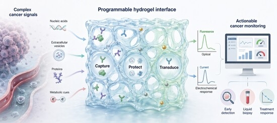
Graphical Abstract

1. Introduction

Early and clinically meaningful cancer detection remains constrained by the need to measure low-abundance, heterogeneous, and temporally dynamic biomarkers in complex biological environments. Conventional laboratory assays have transformed cancer diagnosis and monitoring; however, their reliance on centralized instrumentation, multistep processing, and limited access to longitudinal sampling can restrict their use in decentralized or repeat-testing workflows. Hydrogel-based biosensing platforms help address this gap by combining biomolecular recognition with hydrated, porous, and mechanically tunable matrices that support biological compatibility, target enrichment, antifouling protection, and signal amplification. In cancer-focused studies, these systems have been applied to early biomarker detection, liquid biopsy, tumor-marker surveillance, multiplex profiling, point-of-care diagnostics, and treatment-response assessment [1,2,3,4,5,6,7,8,9,10,11,12,13,14,15].
A major driver of this field is liquid biopsy-compatible sensing. Cancer-associated microRNAs, oncogenic deoxyribonucleic acid (DNA) fragments, extracellular vesicles, and circulating tumor cells can reflect tumor state, disease burden, and molecular subtype in minimally invasive samples. Hydrogel biosensors have been used to detect microRNA-21, microRNA-141, microRNA-381, microRNA-155, microRNA-122, human papillomavirus DNA, mitochondrial DNA, circulating cell-free DNA, circulating tumor DNA carrying the B-Raf proto-oncogene serine/threonine-protein kinase V600E mutation, prostate cancer-derived exosomes, breast cancer-associated extracellular vesicles, and epithelial cell adhesion molecule-positive circulating tumor cells [1,6,8,10,11,13,16,17,18,19,20,21,22,23,24,25,26].
Protein, enzymatic, immune, and receptor-associated biomarkers form a second major axis. These include human epidermal growth factor receptor 2, cancer antigen 15-3, carcinoembryonic antigen, prostate-specific antigen, alpha-fetoprotein, cytokeratin 19 fragment 21-1, tumor necrosis factor alpha, cancer antigen 125, human epididymis protein 4, neuron-specific enolase, squamous cell carcinoma antigen, epithelial cell adhesion molecule, matrix metalloproteinases, hyaluronidase, glutathione S-transferase, and immune-related markers [4,7,9,12,14,15,27,28,29,30,31,32,33,34,35,36,37,38,39,40,41,42]. Because cancer rarely presents as a single-marker disease, multiplexed hydrogel systems are especially relevant for the integrated measurement of tumor markers, circulating biomarkers, cancer-related cell subtypes, metabolic indicators, and combined molecular signatures [10,14,15,26,31,42,43].
The engineering significance of hydrogels lies in their ability to function as active sensing architectures rather than inert supports. Synthetic, natural, peptide, protein, DNA, and hybrid hydrogel matrices have been configured as coatings, films, beads, microstructures, scaffolds, bioinks, optical fibers, microneedle coatings, paper devices, microfluidic components, and electrode interfaces [9,17,19,23,34,37,44,45,46,47,48,49,50]. Nanomaterials, including gold and silver nanostructures, graphene derivatives, MXenes, carbon dots, quantum dots, Prussian blue–platinum nanoparticles, metal–organic frameworks, covalent organic framework composites, and plasmonic probes, can provide optical, electrochemical, catalytic, magnetic, photothermal, or conductive functions within these hydrated networks [18,22,24,31,32,39,41,44,48,51,52,53,54,55]. In this context, hydrogels generally define the biomarker-accessible and biologically compatible interface, whereas embedded or surface-associated nanomaterials support transduction, amplification, and device integration. This distinction is important because hydrogel–nanomaterial composites should be evaluated according to the specific recognition, transduction, and amplification roles assigned to each component rather than by analytical performance alone.
Mechanistically, hydrogel biosensors operate through target-induced network reconfiguration, gated release, enzymatic degradation, catalytic reporter conversion, antifouling interfacial capture, redox responsiveness, optical or plasmonic localization, charge-transfer modulation, and three-dimensional cell-based signal acquisition [5,6,8,10,11,13,17,20,24,25,30,35,40,47,56,57,58,59]. These mechanisms support fluorescence, Förster resonance energy transfer, colorimetry, Raman scattering, surface plasmon resonance and surface plasmon resonance imaging, impedance, amperometry, conductance, electrochemiluminescence, photoelectrochemistry, magnetic resonance, acoustic sensing, photothermal conversion, paper-flow distance, and smartphone-assisted image analysis [2,3,7,9,12,14,15,31,33,39,41,43,60,61,62,63]. The most conceptually important platforms are those in which the hydrogel is inseparable from signal generation. Examples include DNA hydrogels that dissolve or release reporters after nucleic acid recognition, enzyme-responsive hydrogels that alter optical or transport properties after degradation, and conductive or antifouling hydrogels that directly regulate electrochemical readout.
Beyond endpoint detection, hydrogel biosensors are increasingly used to interrogate functional tumor biology. Three-dimensional cultures, spheroids, tumor-like scaffolds, impedance arrays, and cell-compatible electrochemical or optical platforms enable monitoring of proliferation, invasion, colony formation, metabolic activity, oxidative stress, nitrosative signaling, drug response, and subtype-dependent behavior [60,64,65,66,67,68,69,70,71,72]. Accordingly, hydrogel systems capable of measuring hydrogen peroxide, nitric oxide, lactate, potential of hydrogen, reactive oxygen species, glutathione, and drug-induced cellular responses connect biomarker sensing with tumor physiology and preclinical therapeutic evaluation [70,71,72,73,74,75,76,77,78,79,80].
The translational promise of hydrogel cancer biosensors is strongest when analytical performance is demonstrated in clinically relevant matrices and benchmarked against established methods. Representative platforms have detected microRNAs in serum, profiled extracellular vesicles in patient-derived samples, enriched circulating cell-free DNA, correlated exosome readouts with clinical chemiluminescence assays, measured RNAs and proteins directly in plasma for cancer classification, detected human papillomavirus DNA and hyaluronidase in urine or cervical samples, and achieved performance comparable with enzyme-linked immunosorbent assay, reverse transcription polymerase chain reaction, or commercial assays in selected contexts [1,2,3,6,8,9,10,11,13,15,16,23,35,40,43]. However, reported limits of detection, linear ranges, and sample types depend strongly on matrix composition, assay workflow, instrumentation, and validation design, making contextual comparison more informative than direct numerical ranking.
Collectively, these studies establish hydrogel biosensing as a rapidly evolving interface between materials science, analytical chemistry, liquid biopsy, tumor biology, and decentralized cancer diagnostics. At the same time, this review emphasizes that analytical sensitivity alone is insufficient for translation. Clinically meaningful performance also requires matrix compatibility, batch-to-batch fabrication reproducibility, storage stability, cost-aware workflow design, appropriate benchmarking, feasible instrumentation, and clear relevance to cancer diagnosis, monitoring, or treatment-response assessment.

2. Clinical or Biomedical Objective

2.1. Cancer Biomarker Detection as the Central Clinical Objective

The central clinical objective of this literature is to advance hydrogel-based biosensing platforms for cancer biomarker detection. The most relevant studies are those in which hydrogels function as biomarker-responsive, biocompatible, antifouling, or three-dimensional sensing interfaces for detecting cancer-associated nucleic acids, proteins, enzymes, metabolites, extracellular vesicles, circulating tumor cells, and tumor microenvironmental signals. These systems are directed toward earlier diagnosis, minimally invasive sampling, clinically meaningful monitoring, and analysis in complex biological contexts, including serum, plasma, saliva, urine, sweat, blood, and cell-derived samples [1,2,3,4,5,6,7,8,9,10,11,12,13,14,15,22,23,25,81,82,83].
The unifying clinical need is the difficulty of detecting low-abundance or clinically informative cancer biomarkers using methods that are rapid, accessible, reproducible, and compatible with decentralized workflows. Hydrogel biosensors are therefore positioned not only as materials innovations, but also as clinically oriented platforms intended to support cancer screening, disease detection, prognosis, treatment monitoring, and patient surveillance [13,15,24,32,33,34,35,40,57,58,59,84]. For this reason, the clinical value of these platforms depends not only on low detection limits, but also on their performance in realistic matrices, reproducible hydrogel preparation, workflow simplicity, and compatibility with decentralized implementation. This objective is especially important for liquid biopsy and minimally invasive cancer monitoring, where the goal is to detect cancer-relevant signals from accessible biological materials rather than relying exclusively on invasive sampling or centralized laboratory procedures [5,8,10,11,13,23,25,33,84].

2.2. Point-of-Care Translation and Multiplexed Clinical Monitoring

A second major objective is the translation of hydrogel biosensing into practical diagnostic formats, including portable, smartphone-assisted, visual, paper-based, wearable, rapid, low-cost, and point-of-care systems [15,32,40,56,57,59,84,85]. These studies address the clinical challenge that conventional assays may be constrained by instrumentation, cost, processing time, sample preparation, or dependence on specialized laboratory settings. In this context, the biomedical objective is not only improved analytical detection, but also the development of cancer biosensing systems that can operate in more accessible and patient-compatible environments. Accordingly, point-of-care relevance should be evaluated through a cost–performance lens that considers assay time, instrumentation requirements, storage stability, manual operation, and manufacturability alongside sensitivity.
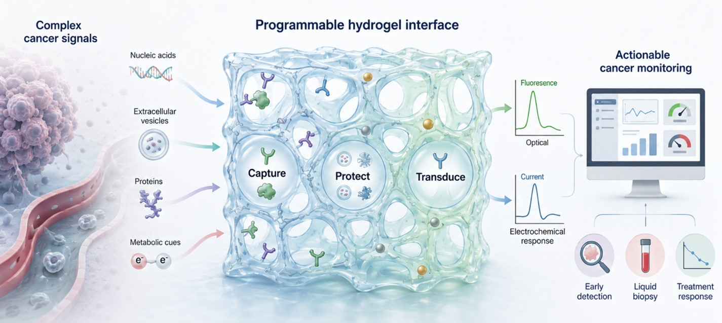
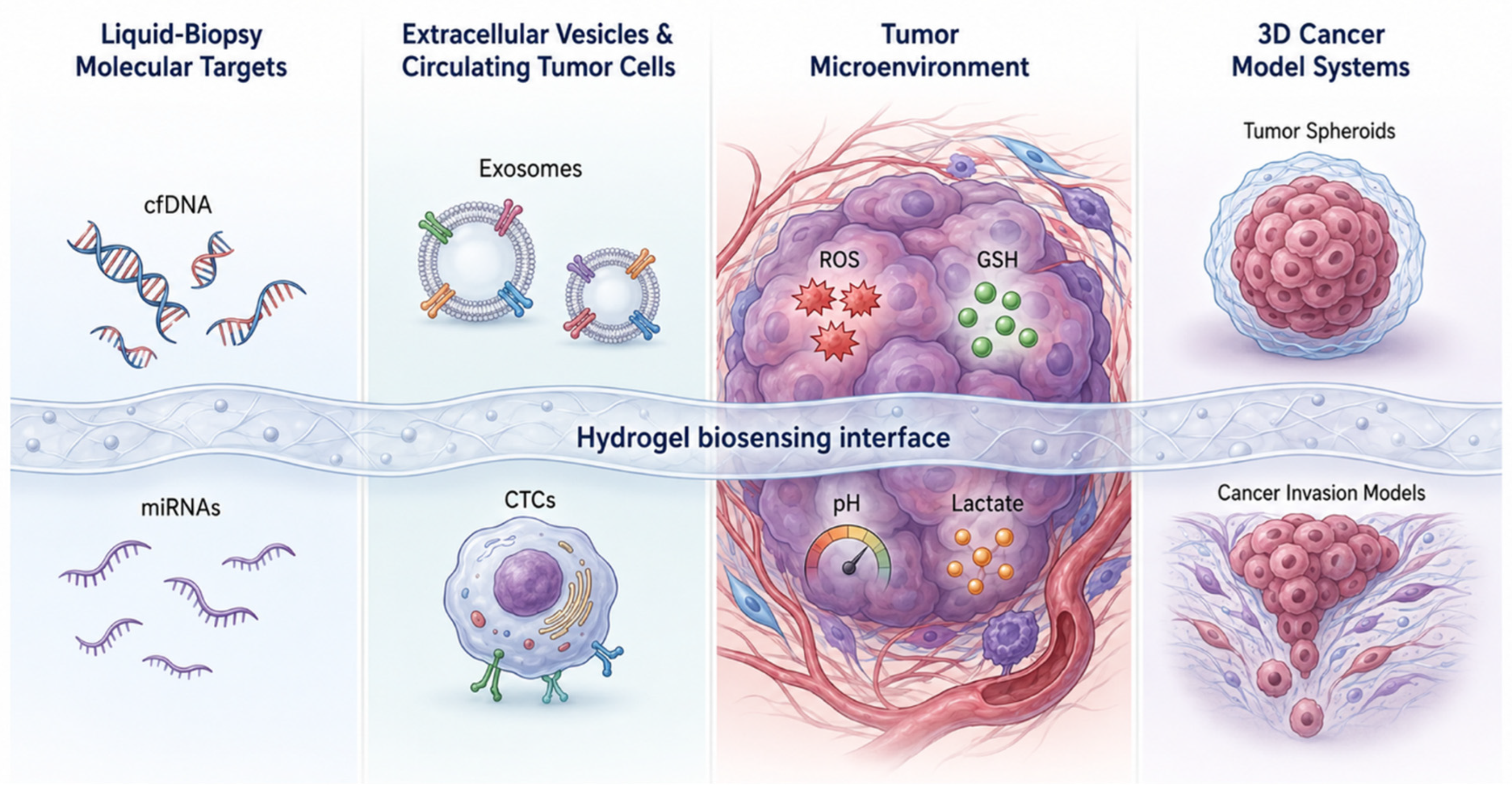
Several studies extend this objective from single-analyte detection to multiplexed or multiparameter biomarker profiling. This direction is important because cancer diagnosis and monitoring often require more clinically informative measurements than isolated biomarker readouts can provide. Hydrogel-based platforms are therefore used to support simultaneous or integrated assessment of tumor markers, circulating biomarkers, cancer-related cell subtypes, and metabolic indicators [10,14,15,26,31,42,43]. Within the scope of this section, these studies are best understood as efforts to make cancer biomarker biosensing more informative, clinically adaptable, and responsive to disease heterogeneity. However, multiplexing also increases the need to clarify which platform elements provide molecular recognition, which enable signal transduction, and which primarily amplify or stabilize the readout. The clinical objective domains and scope are summarized in Table 1, and the overall translation logic is illustrated in Figure 1.

2.3. Tumor Biology, Disease Modeling, and Treatment-Response Assessment

Beyond direct clinical detection, hydrogel biosensing platforms are also used to characterize cancer biology in more physiologically relevant environments. Studies using three-dimensional cultures, tumor spheroids, hydrogel scaffolds, and cell-compatible sensing interfaces aim to monitor cancer cell proliferation, invasion, colony formation, subtype-dependent behavior, metabolic activity, oxidative stress, nitrosative signaling, and drug response [26,60,64,65,66,67,68,69,70,71,72].
These studies remain relevant to biomarker biosensing hydrogels in cancer because they use hydrogel environments to connect measurable biological signals with tumor behavior and therapeutic response. The clinical and biomedical significance of this group lies in improving preclinical cancer models and therapeutic evaluation. Rather than treating biosensing as an endpoint alone, these systems use hydrogel environments to support dynamic and objective assessment of cancer cell behavior under conditions that better approximate tumor biology than conventional two-dimensional or manually interpreted assays [26,60,64,65,66,67,68,69,70,71,72]. Their translational interpretation should therefore account for both biological relevance and technical robustness, including whether the hydrogel matrix maintains consistent structure, cell compatibility, and sensing performance across repeated preparations.

2.4. Therapeutic and Theranostic Extensions

A smaller but important group of studies extends biomarker-responsive hydrogels toward therapeutic or theranostic objectives. These systems integrate biosensing, imaging, controlled drug delivery, photothermal or photodynamic intervention, ribonucleic acid-related modulation, tumor-responsive treatment, or antitumor activity within multifunctional hydrogel platforms [19,44,45,80,86,87,88,89,90,91]. Their relevance to the theme presented here lies in showing how cancer biomarker responsiveness can guide, accompany, or be combined with therapeutic action.
These studies are secondary to the main diagnostic focus rather than a separate dominant theme. They broaden the translational logic of biomarker biosensing hydrogels by demonstrating that cancer-responsive hydrogel systems can link detection, localization, imaging, and treatment within the same material framework while remaining grounded in biomarker-associated cancer applications [80,87,89,90,91]. Because such multifunctional systems often combine recognition, actuation, imaging, and therapeutic components, their practical readiness depends on whether added functional complexity improves clinical utility without compromising reproducibility, stability, or scalable preparation.

2.5. Contextual Biomedical Extensions

Broader biomedical applications, including infectious disease detection, antimicrobial wound care, tissue engineering, regenerative medicine, wearable health monitoring, agricultural diagnostics, food-related antioxidant screening, and general microfluidic or organ model systems, also appear in the reviewed literature [42,49,50,52,53,55,92,93,94,95,96,97,98]. In this section, these studies are retained only as contextual boundary-setting examples.
Overall, the clinical and biomedical objective unifying the cancer-focused literature is the translation of biomarker-responsive hydrogels into practical biosensing systems for early detection, liquid biopsy analysis, tumor-marker monitoring, multiplex profiling, disease modeling, and treatment-response assessment. The strongest contribution of this body of work is its convergence around hydrogel interfaces that can operate in complex biological samples, support miniaturized or decentralized diagnostic formats, and connect cancer-relevant molecular recognition with clinically oriented diagnostic and monitoring needs [1,2,3,5,6,7,8,9,10,11,12,13,14,15,23,84]. At the same time, the field remains uneven in translational maturity, and comparisons across platforms should consider sample matrix, assay complexity, instrumentation, validation depth, fabrication reproducibility, and storage or manufacturing constraints rather than sensitivity alone.

3. Target Analyte, Biological System, and Sample Context

Hydrogel-based cancer biosensors have been applied to a diverse but thematically coherent set of biological targets, including liquid-biopsy nucleic acids, extracellular vesicles, circulating tumor cells, soluble protein markers, tumor-associated enzymes, metabolic and redox signals, and three-dimensional cancer models. The most relevant sample matrices include serum, plasma, saliva, urine, sweat, interstitial fluid mimics, cancer cell lysates, cultured cancer cells, patient-derived specimens, and animal tumor models. Because these matrices differ substantially in biomarker abundance, viscosity, protein content, cellular debris, nuclease activity, and interfering species, analytical performance should be evaluated in relation to sample context rather than as a direct comparison across studies. Within the scope of biomarker biosensing hydrogels in cancer, these studies are best understood not as isolated examples but as evidence for recurring target contexts: molecular liquid biopsy, clinically established cancer biomarkers, functional tumor biology, tumor-microenvironment monitoring, and cell- or tissue-associated cancer models. This biological-context continuum is summarized in Figure 2, while the major target categories and representative sample contexts are organized in Table 2.

3.1. Liquid-Biopsy and Nucleic Acid Targets

Nucleic acid biomarkers represent one of the clearest target classes in hydrogel-based cancer biosensing. miRNAs are especially prominent, with microRNA-21 (miR-21) recurring across serum-derived ribonucleic acid (RNA), human serum, lung cancer-oriented detection, breast cancer cells, extracellular vesicle analysis, crude cancer cell lysates, clinical serum, cellular extract environments, and untreated human serum [1,10,11,13,16,17,18,19]. This repeated use of miR-21 illustrates how hydrogel biosensing platforms have been positioned to detect cancer-associated molecular signatures in both liquid and cell-derived sample contexts. Other miRNA targets broaden this liquid-biopsy orientation, including microRNA-141 for prostate cancer, microRNA-381 from gastric cancer ascites samples, microRNA-16, microRNA-155, microRNA-122 for hepatocellular carcinoma subtype identification, microRNA-19b in patient samples, and broader miRNA biomarker panels for cancer monitoring [20,21,24,26,57,99,100]. For translational interpretation, platforms evaluated in untreated serum, crude lysates, extracellular vesicle preparations, or patient samples provide stronger evidence of matrix compatibility than platforms tested only in simplified or highly controlled sample environments.
The nucleic acid scope extends beyond miRNAs to cancer-associated DNA targets. Human papillomavirus (HPV) DNA and human papillomavirus 16 DNA have been studied in cervical cancer-related screening contexts, including clinical urine and cervical brush samples [6,8]. Other studies have targeted mitochondrial DNA from ovarian cancer patient samples and cfDNA, including B-Raf proto-oncogene serine/threonine-protein kinase V600E-mutant circulating tumor DNA from blood plasma of patients with thyroid cancer or thyroid nodules [22,23]. Extracellular vesicle and circulating-cell systems further strengthen the liquid-biopsy theme: prostate cancer-derived exosomes have been analyzed in human serum, breast cancer-associated extracellular vesicles have been characterized using miR-21 and epithelial cell adhesion molecule (EpCAM), and EpCAM-positive CTCs, including Michigan Cancer Foundation-7 (MCF-7) cells, have been captured from blood for live-cell analysis [2,10,25]. Together, these studies show that hydrogel biosensing targets are not restricted to free molecular analytes, but also include intact vesicular and cellular carriers of cancer-associated information. However, these target formats impose different technical constraints: free nucleic acids emphasize sequence recognition and nuclease resistance, extracellular vesicles require capture or enrichment from complex biofluids, and CTC assays must preserve cell recognition and recovery in blood-derived matrices.

3.2. Protein, Enzymatic, Immune, and Cell-Surface Biomarkers

Classical soluble cancer biomarkers remain a major target category. Breast cancer-related targets include human epidermal growth factor receptor 2, cancer antigen 15-3, tissue inhibitor of metalloproteinases-1, and carcinoembryonic antigen (CEA) in serum or clinically framed breast cancer contexts [4,27,29,31,34]. Prostate-related systems have targeted prostate-specific antigen, sarcosine, and prostate cancer-derived exosomes in serum, urine, or immunoassay settings [2,28,42,51]. Other tumor-associated markers include alpha-fetoprotein for hepatocellular carcinoma, cytokeratin 19 fragment 21-1 (CYFRA-21-1) and tumor necrosis factor alpha in oral cancer-related saliva contexts, C-reactive protein in serum for melanoma assessment, cancer antigen 125 and human epididymis protein 4 in ovarian cancer contexts, human chorionic gonadotropin as a cancer- and pregnancy-related protein target, and lung-related marker panels including neuron-specific enolase, CEA, squamous cell carcinoma antigen, and CYFRA-21-1 [7,9,12,14,15,33,36,37,38,39,41,42,101]. For these clinically familiar markers, the most informative studies are those that move beyond detection-limit optimization and demonstrate selectivity, interference tolerance, and agreement with established clinical or immunoassay-based measurements in relevant samples.
A related group of studies targets functional biomarkers that reflect tumor-associated enzymatic activity, immune signaling, or receptor-defined cell recognition. Matrix metalloproteinases, including matrix metalloproteinase-9 and matrix metalloproteinase-2/9, have been studied in relation to cancer progression or cancer–normal cell differentiation [30,90]. Hyaluronidase appears repeatedly as a tumor-associated enzyme in urine, serum, pancreatic cancer serum, human urine, human serum, and three-dimensional lung cancer cell culture contexts [35,40,47,59,102]. Immune- and therapy-related targets include glutathione S-transferase for cancer chemotherapy drug metabolism, programmed cell death protein 1 antibody, interleukin-2, soluble interleukin-2 receptor alpha, and interleukin-6 in sweat for bone-related disease monitoring [32,52,103,104]. Cell-surface recognition targets include EpCAM in biological fluids and CTCs, folic acid-recognized Henrietta Lacks (HeLa) cells, receptor-positive cancer cells, and biotin receptor-overexpressing K562 leukemia and A549 lung cancer cells [25,54,63,83,105]. This group broadens the section from conventional biomarker quantification to activity-, immune-, and receptor-associated cancer biosensing. In enzyme- and receptor-responsive systems, the hydrogel may contribute directly to recognition through degradable, affinity-based, or cell-interactive structures, whereas labels, electrodes, nanoparticles, or optical probes typically convert these recognition events into measurable signals.

3.3. Metabolic, Redox, and Tumor-Microenvironment Signals

A distinct and cancer-relevant subset of the literature focuses on dynamic biological signals associated with tumor metabolism, oxidative stress, and the tumor microenvironment. Sweat-based analysis has targeted aldehyde biomarkers, glycoproteins, oxidative stress markers, and metabolic fingerprints in lung cancer screening contexts [3]. Other platforms have monitored glucose in serum or interstitial fluid models, extracellular lactate from isolated cancer cells, adenosine triphosphate as a disease-relevant molecule, and pH changes associated with cancer cell metabolites or intracellular environments [42,71,75,94,106,107]. These targets shift the emphasis from biomarker presence alone toward cancer-associated physiological states. Because these signals can change rapidly with cell state, sampling conditions, and local microenvironment, assay time, temporal resolution, and biological model selection are especially important limiting conditions for this category.
Reactive oxygen and nitrogen species are especially recurrent. Hydrogen peroxide (H2O2) and superoxide have been monitored as extracellular products of prostate cells, while H2O2 and nitric oxide have been measured from three-dimensional breast cancer cell cultures under drug stimulation [70,73,74]. H2O2 release has also been compared between luminal A-type MCF-7 and triple-negative M. D. Anderson metastatic breast 231 breast cancer cells [72]. Several systems have used ROS- or GSH-associated responses to distinguish cancer-cell microenvironments, including HeLa, prostate cancer-3, and B16 melanoma F10 cancer cells compared with Chinese hamster ovary-K1 normal cells [76,77,78]. GSH has been studied in living cells, clinical serum, intracellular cancer contexts, and tumor microenvironments [79,80]. Collectively, these studies establish tumor-microenvironmental sensing as a central component of hydrogel-based cancer biosensing, particularly when the monitored signal reflects metabolism, redox state, or stress-related cellular behavior.

3.4. Cancer-Cell, Three-Dimensional, and Animal Model Contexts

Hydrogel biosensing platforms have also been used to study cancer targets within structured cellular and tissue-like systems. These include breast cancer spheroids, lung cancer cell cultures, human hepatoma 7 colony-formation models, human hepatoma 7 and human hepatocellular carcinoma G2 drug-response models, malignant-cell invasion under interleukin-6 stimulation, receptor-positive cancer cell capture, and three-dimensional breast cancer cultures used to monitor tumor evolution, invasion, viability, secretion, or treatment-associated responses [47,60,66,67,68,70,105]. Additional cancer cell contexts include HeLa, prostate cancer-3, B16 melanoma F10, MCF-7, M. D. Anderson metastatic breast 231, A549 lung cancer cells, K562 leukemia cells, and melanoma B16 melanoma F10 models, often compared with normal or nonresponsive cells such as Chinese hamster ovary-K1 or gastric epithelial cell line-1 cells [72,76,77,78,83,89,108]. These systems are valuable because they test sensing under higher biological complexity, but their translational meaning depends on whether hydrogel composition, cell density, culture duration, and readout conditions are sufficiently controlled and reproducible.
Several studies extend the biological context toward engineered tissue models and animal-associated systems. These include cancer microenvironment-mimicking hydrogels, stiff matrices resembling cartilage or cancer microenvironments, organ-on-chip models of healthy and diseased tissue states, cell-loaded bioinks, human embryonic kidney 293 bioprinted constructs, and vascular endothelial growth factor-loaded gelatin methacryloyl in a rat neovascularization model [46,53,95,97,109]. Tumor-bearing and residual-disease settings include residual microtumors and cancer nodules in mouse models, malignant tumor treatment models, and xenografted breast cancer-bearing mice [87,91,110]. In this section, these systems are most relevant when they define the biological target or sample context for cancer biosensing, rather than when they function primarily as broader tissue-engineering or therapeutic platforms. Animal and tumor-model studies provide useful biological context, but they should not be considered equivalent to clinical validation unless performance is also demonstrated in appropriately collected patient-derived specimens.

3.5. Therapeutic-Release, Cancer-Adjacent, and Boundary Contexts

A smaller group of studies involves therapeutic release, antimicrobial systems, infectious disease targets, food analysis, or other noncancer biological settings. These examples are retained only to define the boundaries of the evidence base and to distinguish cancer biomarker biosensing from adjacent hydrogel applications. Cancer-adjacent therapeutic contexts include model protein release under nuclease exposure, small interfering RNA delivery to ovarian cancer cells, insulin release, spectinomycin release with human hepatocellular carcinoma G2 cytotoxicity testing, sustained chemotherapeutic release at tumor sites, anticancer drug release under abnormal pH conditions, and doxorubicin delivery against cancer cells or in xenografted breast cancer-bearing mice [45,88,89,91,111,112]. Chitosan-based hydrogel systems have also been discussed in relation to cancer diagnosis, drug delivery, wound care, drug encapsulation, biosensing, scaffolding, and antimicrobial behavior [96].
Peripheral examples include lipopolysaccharides from Salmonella enterica in food-contamination assessment, nitric oxide release from RAW 264.7 macrophages, Porphyromonas gingivalis in saliva, Escherichia coli and Staphylococcus aureus in wound-infection contexts, severe acute respiratory syndrome coronavirus 2-related targets, glucose and human serum albumin in blood or urine contexts, citrus pathogen DNA in live citrus leaf samples, total antioxidant activity in honey samples, adenosine triphosphate and 3,3′,4,4′-tetrachlorobiphenyl as representative molecular targets, and rhodamine 6G and 4-mercaptopyridine as model in situ analytes [48,50,55,61,62,92,93,98,113,114]. These studies demonstrate the broader reach of hydrogel biosensing, but they remain secondary to the manuscript’s central focus on cancer biomarkers, tumor-microenvironmental signals, and cancer cell model systems. Rare or boundary-setting terms introduced only in this context should be spelled out locally where needed rather than overexpanded in the abbreviation list, to preserve readability and keep the glossary focused on recurring cancer-biosensing terminology.
Table 2. Cancer-relevant target analytes, biological systems, and sample contexts in hydrogel biosensing platforms.
Target Analyte, Biological System, and Sample ContextCancer-Focused Targets or Biological EntitiesBiological Sample, Model, or Patient-Derived ContextRepresentative References
Nucleic acid biomarkersmiR-21, microRNA-141, microRNA-381, microRNA-16, microRNA-155, microRNA-122, and microRNA-19bSerum-derived RNA, human serum, untreated serum, crude cancer cell lysates, breast cancer cells, gastric cancer ascites-derived exosomes, patient samples, and hepatocellular carcinoma subtype contexts[1,10,11,13,16,17,18,20,21,24,26,99,100]
Oncogenic and cancer-associated DNA targetsHPV DNA, human papillomavirus 16 DNA, mitochondrial DNA, cfDNA, and circulating tumor DNA carrying the B-Raf proto-oncogene serine/threonine-protein kinase V600E mutationCervical cancer screening contexts, clinical urine, cervical brush samples, ovarian cancer patient DNA, and blood plasma from patients with thyroid cancer or thyroid nodules[6,8,22,23]
Extracellular vesicles and CTCsProstate cancer-derived exosomes, cancer-associated extracellular vesicles, extracellular vesicle-associated miR-21 and EpCAM, and EpCAM-positive CTCsHuman serum, breast cancer patient versus healthy donor serum, blood samples, MCF-7 CTC models, and 2 µL blood sample contexts[2,10,25]
Soluble protein and antigen biomarkersHuman epidermal growth factor receptor 2, cancer antigen 15-3, CEA, prostate-specific antigen, alpha-fetoprotein, cancer antigen 125, human epididymis protein 4, CYFRA-21-1, tumor necrosis factor alpha, C-reactive protein, human chorionic gonadotropin, neuron-specific enolase, and squamous cell carcinoma antigenSerum, saliva, artificial saliva, urine, patient-derived samples, and breast, prostate, ovarian, oral, hepatocellular, melanoma, and lung cancer contexts[4,7,9,12,14,15,27,28,29,31,33,34,36,37,38,39,41,42,101]
Enzymatic, immune, and receptor-associated biomarkersMatrix metalloproteinase-9, matrix metalloproteinase-2/9, hyaluronidase, glutathione S-transferase, interleukin-2, soluble interleukin-2 receptor alpha, interleukin-6, programmed cell death protein 1 antibody, EpCAM, folate receptor-associated HeLa recognition, and biotin receptor-overexpressing cellsUrine, serum, pancreatic cancer serum, sweat, patient-derived blood, biological fluids, cancer cell surfaces, receptor-positive cancer cells, K562 leukemia cells, and A549 lung cancer cells[25,30,32,35,40,47,52,54,59,63,83,90,103,104]
Metabolic, redox, and tumor-microenvironmental signalsLactate, glucose, adenosine triphosphate, pH, aldehydes, glycoproteins, oxidative stress markers, H2O2, superoxide, nitric oxide, ROS, and GSHSweat, serum, interstitial-fluid mimics, isolated cancer cells, prostate cells, three-dimensional breast cancer cultures, HeLa, prostate cancer-3, B16 melanoma F10, Chinese hamster ovary-K1, MCF-7, and M. D. Anderson metastatic breast 231 models[3,70,71,72,73,74,75,76,77,78,79,80,94,106,107]
Cancer cell, spheroid, and tumor model systemsBreast cancer spheroids, human hepatoma 7 colonies, human hepatoma 7 and human hepatocellular carcinoma G2 drug-response models, malignant invasive cells, MCF-7, M. D. Anderson metastatic breast 231, HeLa, prostate cancer-3, B16 melanoma F10, A549, and K562 cellsThree-dimensional cancer cultures, hydrogel tumor models, invasion models under interleukin-6 stimulation, cancer microenvironment-mimicking phantoms, cell-loaded constructs, and tumor-bearing or xenografted mouse models[53,60,66,67,68,70,72,83,87,89,91,108,109,110]
Therapeutic-release and cancer-adjacent biological systemsModel protein release, small interfering RNA delivery, insulin release, spectinomycin release, chemotherapeutic release, and doxorubicin deliveryNuclease-responsive systems, ovarian cancer cells, human hepatocellular carcinoma G2 cytotoxicity testing, tumor-site release models, cancer cells, and xenografted breast cancer-bearing mice[45,88,89,91,111,112]
Boundary and noncancer biological contextsLipopolysaccharide, Porphyromonas gingivalis, Escherichia coli, Staphylococcus aureus, severe acute respiratory syndrome coronavirus 2-related targets, glucose, human serum albumin, citrus pathogen DNA, honey antioxidant activity, adenosine triphosphate, 3,3′,4,4′-tetrachlorobiphenyl, rhodamine 6G, and 4-mercaptopyridineFood samples, saliva, wound-infection models, biomedical swabs, blood- or urine-related matrices, citrus leaf samples, honey samples, and model in situ analyte systems[48,50,55,61,62,92,93,98,113,114]
Note: Sample contexts are not directly interchangeable across studies. Reported analytical performance should therefore be evaluated alongside matrix complexity, sample volume, assay time, instrumentation, pre-enrichment or amplification requirements, interference testing, and whether validation used spiked, cell-derived, animal-model, or patient-derived samples.

4. Hydrogel–Nanomaterial Platform Design

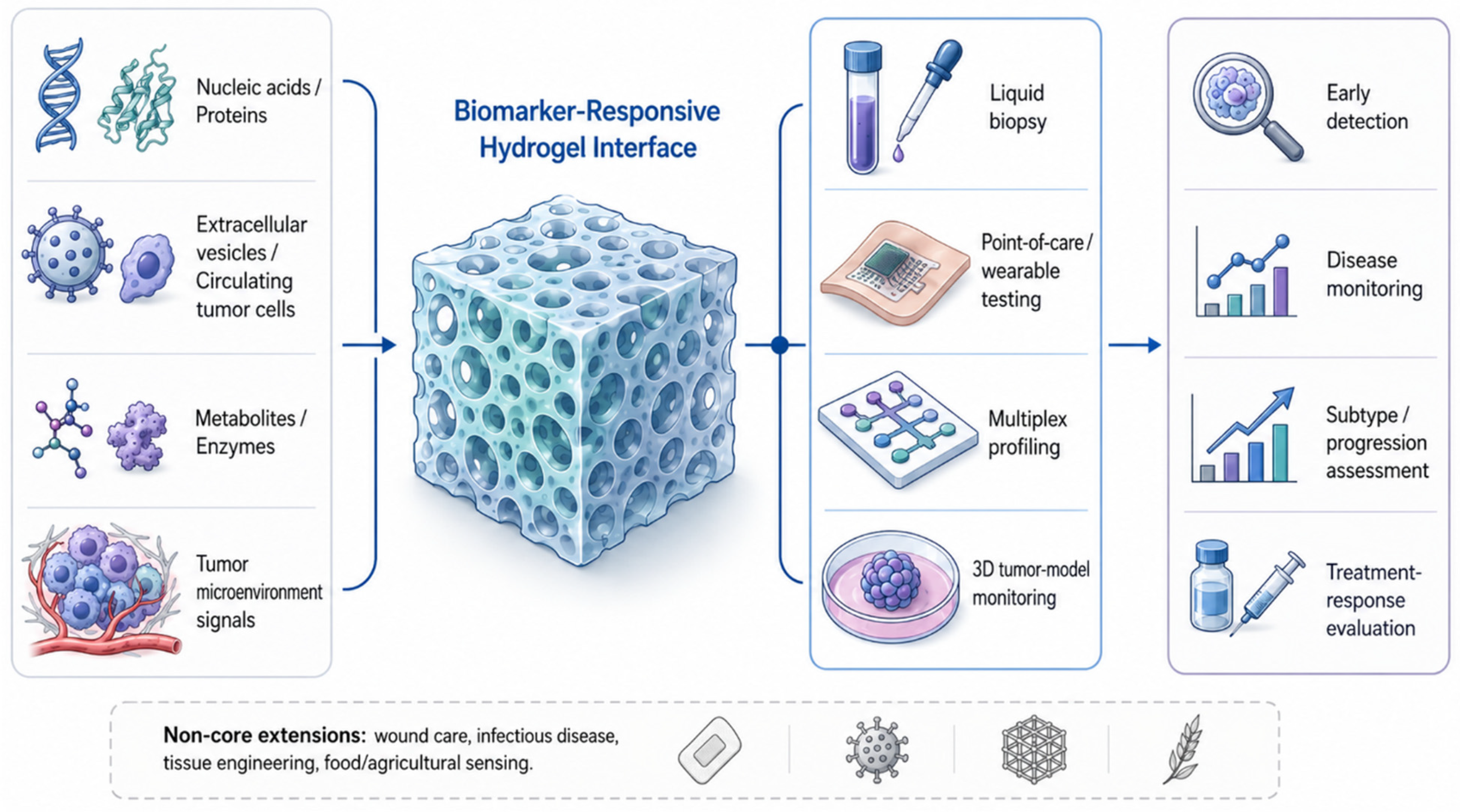
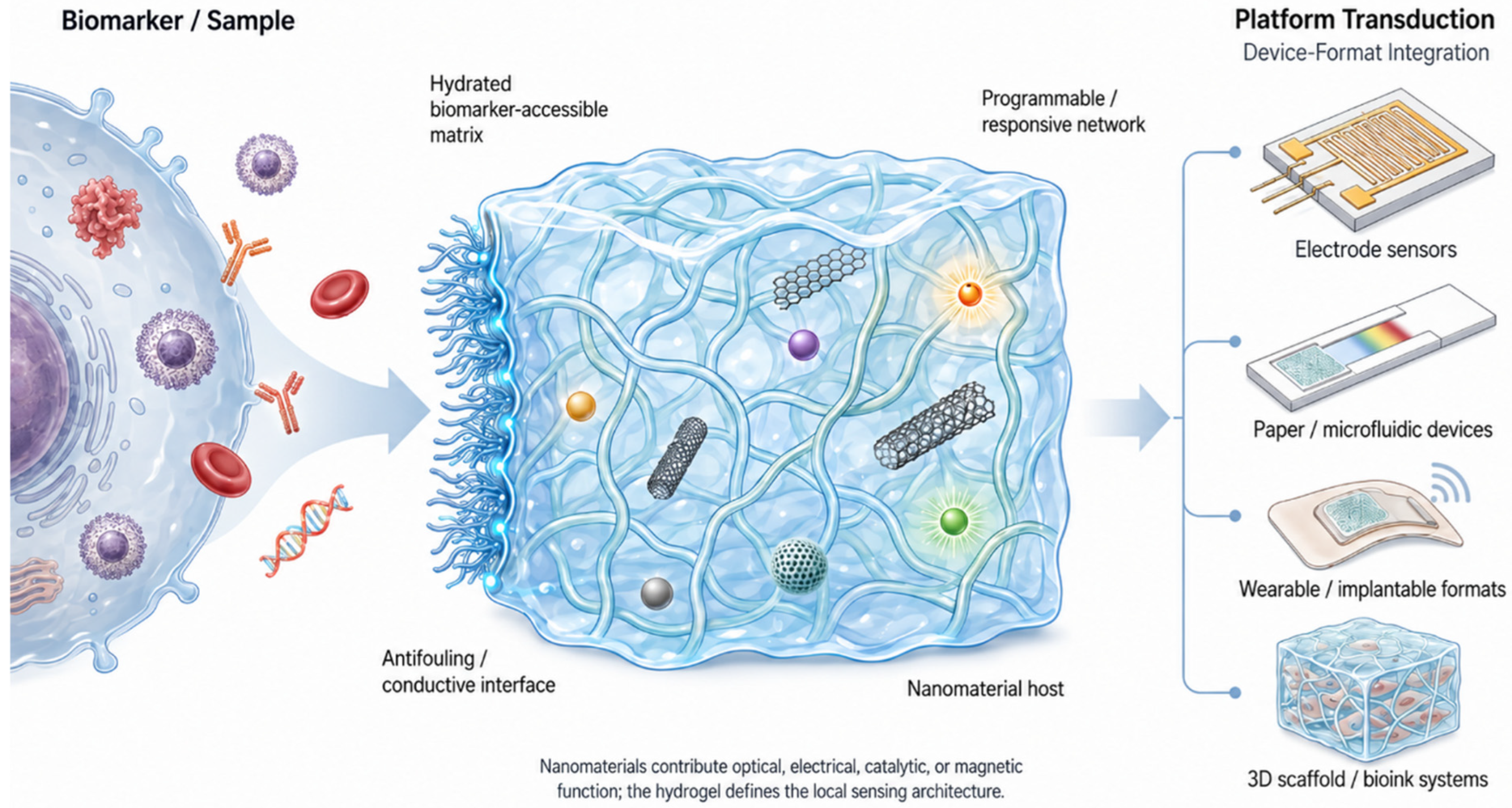
Platform design in biomarker biosensing hydrogels for cancer is defined here by the physical and chemical architecture of the sensing system, including the hydrogel matrix, nanomaterial component, substrate, electrode, paper or microfluidic device, wearable or implantable format, optical interface, three-dimensional scaffold, bioink, or programmable network. Analytical performance, validation, disease rationale, and therapeutic effect are addressed outside this section. Within this boundary, the reviewed studies show that hydrogels serve several distinct engineering roles: they form hydrated sensing interfaces, organize nanomaterials within three-dimensional matrices, provide antifouling or conductive electrode coatings, support portable and tissue-facing devices, and enable programmable or stimulus-responsive material architectures. To avoid conflating material functions, this section distinguishes three recurring roles across composite platforms: molecular recognition, signal transduction, and signal amplification. Hydrogels most often contribute the recognition-accessible, antifouling, biomolecule-compatible, or stimulus-responsive environment, whereas nanomaterials more commonly provide optical, electrical, catalytic, magnetic, plasmonic, conductive, or signal-amplifying functions. Figure 3 summarizes this design logic by positioning the hydrogel as an integrative interface between biomarker-containing samples and device-level transduction formats.

4.1. Hydrogel Matrices as Cancer Biosensing Construction Platforms

The hydrogel matrices used in cancer biomarker biosensing span synthetic, natural, peptide, protein, DNA, and hybrid polymer systems. Synthetic platforms include polyethylene glycol (PEG)-based hydrogels prepared by copper-free click chemistry, poly(ethylene glycol) diacrylate (PEGDA) microparticles and microstructures fabricated by photopatterning, and polyacrylamide-based matrices integrated with DNA or conductive components [17,23,45,48]. Natural and biopolymer systems include chitosan, hyaluronic acid, guar gum, sodium alginate, cellulose derivatives, gelatin-based materials, and bovine serum albumin hydrogels [9,37,44,47,49]. Peptide and supramolecular designs further expand this material space through 9-fluorenylmethoxycarbonyl (Fmoc)-diphenylalanine/alginate bioinks, Fmoc-phenylalanine-glutamic acid-lysine-phenylalanine peptide hydrogel interfaces, peptide–nucleic acid affinity-trap hydrogels, and injectable aldolized hyaluronic acid/quaternized chitosan hydrogels loaded with peptide nanofibers and silver nanoparticles [19,34,46,50].
These matrix choices define the physical form of the biosensing platform, including coatings, films, beads, droplets, fibers, microstructures, scaffolds, and bulk matrices. Several systems reported formulation- and architecture-relevant parameters, including bovine serum albumin hydrogels with a critical strain of approximately 600%, DNA motor gels with tunable shear moduli of 297–3888 pascals, and injectable supramolecular hydrogels formed at a critical gelation concentration of 0.03 weight percent [37,88,115]. These values describe the formulation or mechanical architecture of the hydrogel platform rather than the analytical outcome of the assay. Such construction parameters are important for translational interpretation because hydrogel composition, gelation behavior, and mechanical consistency can influence batch-to-batch reproducibility, storage stability, and device manufacturability.

4.2. Nanomaterial Integration Within Hydrogel Biosensing Architectures

Nanomaterials were incorporated into hydrogel platforms as embedded fillers, interfacial coatings, conductive networks, optical probes, catalytic components, magnetic structures, or signal-associated labels. The hydrogel typically provides the hydrated and porous matrix, while the nanomaterial contributes optical, electrical, plasmonic, catalytic, magnetic, or photothermal function. In functional terms, the hydrogel usually defines where recognition occurs and how the sample accesses the receptor, while the nanomaterial determines how that recognition event is converted into a measurable optical, electrochemical, catalytic, magnetic, or photothermal signal.
Metallic and plasmonic designs include gold nanorods encapsulated in chitosan hydrogel matrices, silver nanoparticle-coated PEG hydrogel microparticles, gold-nanoparticle/three-dimensional graphene hydrogel substrates, gold–silver nanoclusters encapsulated in DNA hydrogels, and silver nanoparticle-deposited graphene oxide nanosheets incorporated into poly(N-isopropylacrylamide) hydrogel probes [31,39,44,48,55]. Carbon-based and conductive composites include graphene oxide/fish-sperm-DNA hydrogel electrodes, thermally exfoliated graphene oxide wearable interfaces, chitosan–polyaniline (PANI)/mesoporous carbon composites, and Ti3C2 MXene nanosheets dispersed in hyaluronic acid/alginate bioinks [22,51,52,53]. Other nanocomposite systems incorporated carbon dots into chitosan hydrogels, PANI nanoparticles into agarose gels, titanium dioxide nanoparticles into photoelectrochemical hydrogels, Prussian blue–platinum nanoparticles into PANI hydrogels, palladium nanoparticles grown in carboxymethyl cellulose-modified covalent organic framework hydrogels, and oxidized Pi@Ce-doped zirconium metal–organic frameworks embedded in polyvinyl alcohol hydrogels [18,24,32,41,54,116].
A useful distinction across these systems is the placement of the nanomaterial relative to the hydrogel. Some platforms embed nanomaterials within the matrix, whereas others coat hydrogel particles, deposit nanomaterials on electrode surfaces, graft them into polymer networks, or integrate them as labels associated with the hydrogel interface. This distinction clarifies how hydrogels structure the biomarker-accessible region while nanomaterials provide the functional components required for biosensing. Thus, in many nanomaterial-loaded hydrogel matrices, the hydrogel primarily supports recognition, antifouling behavior, mass transport, and local biomarker accessibility, whereas the nanomaterial primarily contributes transduction or amplification unless it is explicitly functionalized with a capture probe or recognition element.

4.3. Electrode-Centered and Antifouling Hydrogel Interfaces

Electrode-modified hydrogels represent one of the most recurrent design strategies in cancer biomarker biosensing. Representative systems include PEG-modified chitosan hydrogels on gold surfaces, zwitterionic hydrogels on screen-printed carbon electrodes, PEGDA hydrogel microstructures patterned on gold microelectrodes, and three-dimensional porous polyacrylamide/PANI hydrogel electrodes [4,27,73,92]. Conductive and antifouling interfaces were constructed by combining hydrogels with poly(3,4-ethylenedioxythiophene), PANI, graphene oxide, or zwitterionic polymers. Examples include poly(3,4-ethylenedioxythiophene)/gold-nanoparticle electrodes coated with peptide hydrogels, PANI-modified glassy carbon electrodes bearing sulfobetaine methacrylate/N-isopropylacrylamide copolymer layers, and conductive antifouling nucleoside hydrogel-modified electrodes [12,26,28]. Other electrode architectures include κ-carrageenan hydrogel-coated mesoporous gold, chitosan/titanium dioxide hydrogel-modified glassy carbon electrodes, nanostructured zirconia–guar gum hydrogel composites on indium tin oxide, and ferrocene-tagged DNA–polyacrylamide hydrogels immobilized on indium tin oxide/polyethylene terephthalate electrodes [9,17,36,81].
In these platforms, the hydrogel defines the sensing interface rather than merely supporting the receptor. Its functional role includes forming a hydrated, biomolecule-compatible layer; supporting probe immobilization; reducing nonspecific adsorption; enabling conductive or redox-active coupling; and structuring the contact between the sample and electrode. This distinction helps separate the hydrogel contribution from that of the electrode, receptor, enzyme, amplification chemistry, or nanomaterial. For electrode-centered systems, recognition is typically assigned to immobilized antibodies, aptamers, enzymes, or nucleic acid probes within or on the hydrogel interface; transduction is provided by the electrode and conductive network; and amplification may arise from nanomaterials, redox labels, catalytic nanoparticles, or increased three-dimensional probe loading.

4.4. Portable, Paper-Based, Microfluidic, Wearable, and Optical Formats

Hydrogel–nanomaterial platforms have also been adapted into device formats suited to low-volume, portable, spatially organized, or tissue-facing sensing. Paper-based and hybrid paper systems include fenugreek hydrogel-stabilized nano-hematite microwells, polymethyl methacrylate/paper hybrid disks containing thermoresponsive hydrogel clip units, glass-fiber-paper devices coupled to hyaluronic acid/DNA water-absorption hydrogels, and three-dimensional microfluidic paper devices containing chemiluminescent hydrogel layers [8,15,56,85]. Microfluidic formats include hydrogel-framed electrospun nanofiber matrices inserted into reusable chips, hydrogel-filled microchannels with embedded electrodes, and gel-based microstructures carrying biofunctionalized microparticle arrays within microfluidic chips [30,42,67]. Manufacturing-oriented designs also used stereolithography and hot embossing to produce polydimethylsiloxane microfluidic devices from polymethyl methacrylate molds, with surface roughness below 1 micrometer and fabrication completed in less than 4 h [84].
Wearable, implantable, optical, and minimally invasive formats extend hydrogel design beyond planar sensors. Wearable platforms include conformal silver nanowire–hydrogel composites, hydrogel-coated stainless-steel microneedles containing glucose oxidase–chitosan within alginate–polyvinyl alcohol matrices, and flexible thermally exfoliated graphene oxide immunosensing interfaces [3,52,94]. Optical and implantable architectures include hydrogel matrices containing fibroblasts and nanoprobes, porous hydrogel soft-polymer optical fiber surface-enhanced Raman scattering probes loaded with metal nanoparticles, wet-spun multilayer polysaccharide hydrogel optical fibers, and double optical fiber balls coated with biotin–quantum-dot-grafted alginate/chitosan hydrogels [61,83,110,113]. Flexible needle-integrated systems were represented by polyimide lab-on-a-needle platforms transferred to 1.2 mm medical needles with microscale sensing regions of 150 micrometers × 2 mm [109]. Across these formats, hydrogels provide the soft, hydrated, porous, or conformal interface that connects biosensing chemistry to paper substrates, microfluidic channels, skin-facing devices, optical fibers, needles, or implantable matrices. In portable and tissue-facing systems, the hydrogel contribution is therefore mainly interfacial and operational: it supports sampling, target access, antifouling behavior, local reagent organization, or tissue compatibility, while the device substrate, optical fiber, electrode, camera, or wearable hardware provides the readout pathway. This separation is important because miniaturization alone does not establish point-of-care readiness unless assay workflow, instrumentation, storage, and user operation are also feasible.

4.5. Three-Dimensional, Programmable, and Responsive Hydrogel Systems

Hydrogels were frequently used to organize cancer-relevant biosensing environments in three dimensions. Examples include three-dimensional printed hydrogel scaffolds containing plasmonic nanoparticles and surface-enhanced Raman scattering tags, alginate hydrogels containing green fluorescent protein-expressing MCF-7 cells between vertically aligned electrodes, methyl cellulose hydrogels containing human hepatoma 7 cell colonies in parallel-plate electrode systems, and titanium foam electrodes carrying peptide hydrogel scaffolds with N-(4-aminobutyl)-N-ethylisoluminol assembled through hydrogen bonding and π-stacking [60,65,66,72]. Biofabricated systems include multimaterial PEGDA/gelatin methacryloyl constructs produced through maskless stereolithographic bioprinting, hybrid Fmoc-diphenylalanine/alginate bioinks, and Ti3C2 MXene–hyaluronic acid/alginate bioinks for extrusion-based three-dimensional printing [46,53,95]. In these systems, the hydrogel provides the spatial architecture that positions cells, electrodes, and nanomaterials within a cancer-relevant biosensing format. Here, the hydrogel may contribute not only to sample compatibility but also biological recognition context, because cell position, matrix stiffness, and three-dimensional organization can influence the measured cancer-associated signal.
Programmable and responsive hydrogels formed another important design class. DNA-based platforms include DNA-crosslinked PEG hydrogels, DNA hydrogel microcapsules loaded with quantum dots, DNA nanoflower-loaded hydrogels, ultrathin DNA–gold nanoparticle hydrogel films, sequence-programmable supramolecular DNA hydrogel matrices, and porous three-dimensional DNA hydrogels generated by aptamer-triggered hybridization chain reactions [6,10,20,25,45,57]. Other responsive platforms include mineralized poly(acrylic acid)–manganese dioxide hydrogels, titanium dioxide/MXene conductive hydrogels, dopamine-conjugated carbonized polymer-dot/manganese dioxide hydrogels, and polyethyleneimine–hyaluronic acid hydrogels paired with gold nanobipyramids and platinum nanoparticles [40,76,77,78]. Photofunctional constructs include cadmium selenide quantum dots immobilized in chitosan–poly(methacrylic acid) nanogels, iridium-complex peptide hydrogels, Nile Blue-engineered quantum-dot hydrogels, and PEG hydrogel arrays incorporating cadmium sulfide quantum dots with silver-ion/Rhod-5N components [11,71,89,90]. Responsive and DNA-programmed hydrogels differ from many passive composite matrices because the hydrogel network itself can participate directly in recognition or switching through target-induced assembly, degradation, dissolution, release, or structural reconfiguration. In these cases, the nanomaterial or reporter usually converts the hydrogel response into an optical, electrochemical, or photofunctional signal.
Overall, hydrogel–nanomaterial platform design in cancer biomarker biosensing is defined by modular integration rather than by a single material type. The cited studies collectively show six major construction logics: hydrogel-modified electrode interfaces, nanomaterial-loaded hydrogel matrices, programmable DNA hydrogel architectures, paper and microfluidic hydrogel devices, wearable or optical hydrogel formats, and three-dimensional scaffold or bioink systems. This organization clarifies how hydrogels function as active design elements that structure biomarker-accessible spaces, support nanomaterial integration, and connect recognition environments to device-level biosensing formats. Across these categories, explicitly assigning recognition, transduction, and amplification functions helps distinguish hydrogel-enabled sensing from cases in which the hydrogel primarily serves as a scaffold for nanomaterial-assisted signal generation. Table 3 summarizes these platform-design patterns and the construction details retained in this section.

5. Functional Mechanism and Readout Strategy

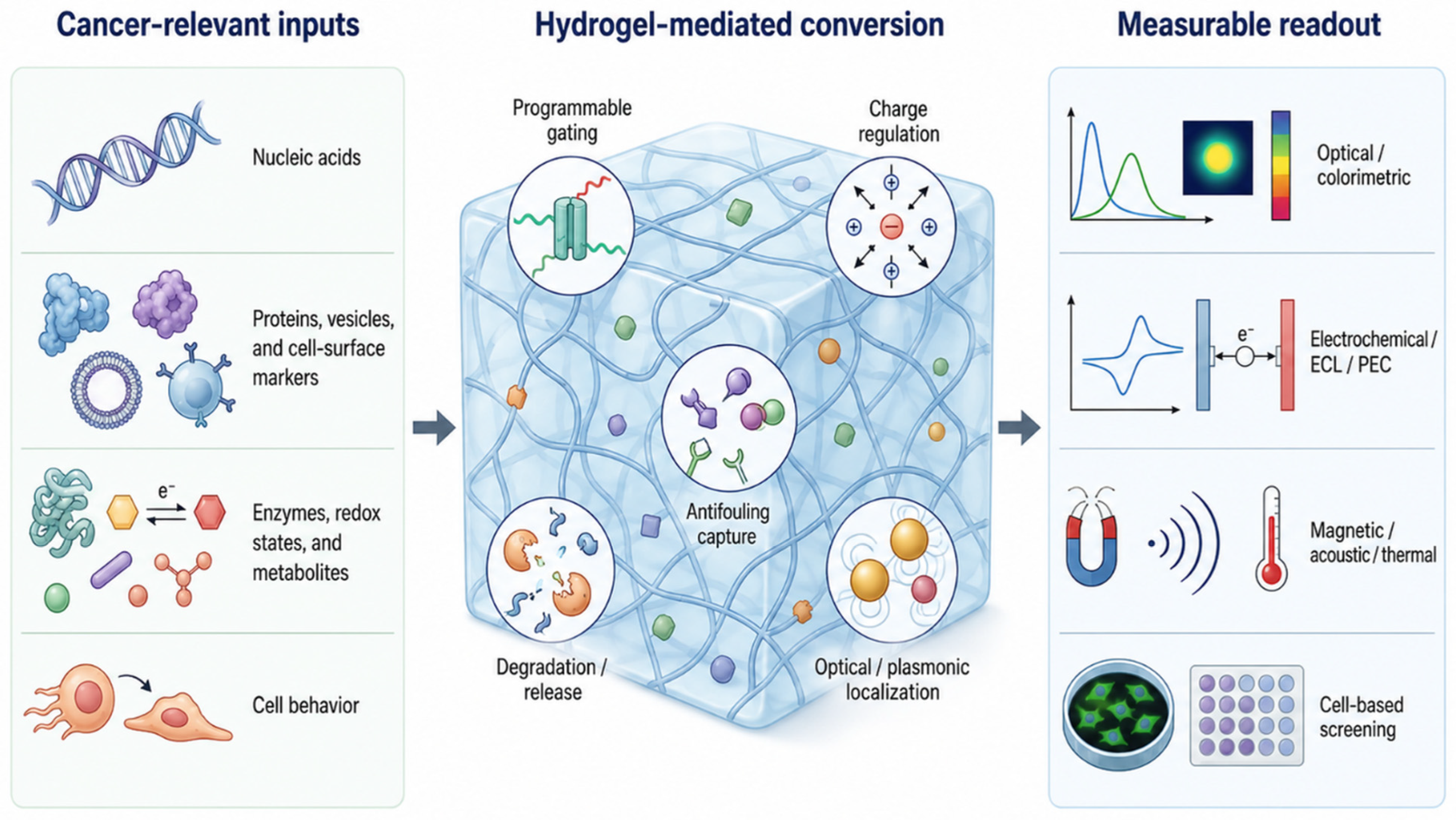
Hydrogel-based cancer biosensors function by converting biomarker recognition or cancer-associated biochemical activity into measurable material, optical, electrochemical, magnetic, acoustic, thermal, or cell-based responses. Their defining feature is not simply the inclusion of a hydrogel, but the specific role the hydrogel plays in signal transduction. Across the reviewed platforms, hydrogels serve as programmable recognition networks, gated release reservoirs, antifouling interfaces, optical or plasmonic scaffolds, three-dimensional cellular matrices, charge-transport regulators, and stimulus-responsive actuators. For mechanistic clarity, the role of the hydrogel should be distinguished from that of nanomaterials, reporters, electrodes, enzymes, or device hardware: the hydrogel often governs recognition accessibility, antifouling protection, gating, degradation, or network reconfiguration, whereas the associated nanomaterial or device component commonly provides transduction and signal amplification.
These functions connect cancer-relevant inputs, including nucleic acids, proteins, extracellular vesicles, enzymes, redox metabolites, pH changes, reactive oxygen or nitrogen species, and cell-surface markers, to readouts such as fluorescence, Förster resonance energy transfer (FRET), colorimetry, Raman scattering, surface plasmon resonance (SPR), surface plasmon resonance imaging (SPRi), electrochemical current, impedance, conductance, electrochemiluminescence (ECL), photoelectrochemistry (PEC), low-field nuclear magnetic resonance, quartz crystal microbalance, surface acoustic wave sensing, photothermal conversion, distance-based paper flow, and smartphone-assisted image analysis. This hydrogel-mediated transduction logic is summarized in Figure 4.

5.1. DNA and Nucleic-Acid Hydrogel Platforms

Nucleic acid-responsive hydrogels provide a direct route from target recognition to signal generation because DNA or RNA binding can alter hydrogel crosslinking, network integrity, or reporter confinement. In hydrogel microcapsules, target microRNA-141 displaced bridging DNA from the capsule shell and released fluorescent QD signals; coupling this displacement process with strand-displacement polymerization and nicking amplification further intensified the fluorescence response [20]. Other miRNA platforms used molecular beacons, peptide nucleic acid probes, single-stranded DNA-functionalized hydrogels, or affinity-trap architectures to convert hybridization into fluorescence, FRET modulation, or fluorescence quenching [16,18,19,99]. Electrochemical DNA hydrogels translated target binding into current changes, as shown in miR-21 sensing, where hybridization dissolved a ferrocene-tagged hydrogel and reduced the redox signal [17]. In these examples, molecular recognition is primarily encoded by the nucleic acid probe or DNA-crosslinked network, while fluorescence labels, QDs, ferrocene tags, or electrodes provide the measurable transduction signal.
A related group of systems used DNA hydrogels as gated reservoirs or programmable reaction modules. HPV DNA disrupted hydrogel crosslinks and released enzyme-loaded DNA nanoflowers for cascade electrochemical signaling [6], while microRNA-155 triggered amplification-mediated release of titanium dioxide nanoparticles for PEC detection [24]. Clustered regularly interspaced short palindromic repeat-associated protein 12a and protein 13a systems, entropy-driven DNA networks, hybridization chain reaction, catalytic hairpin assembly, and DNA nanonet formation expanded hydrogel readouts by producing fluorescence release, AuNP-based colorimetry, distance-based paper flow, or amplified electrochemical current [5,11,13,57,58,100]. In CTC capture, EpCAM recognition initiated aptamer-triggered hybridization chain reaction to form a porous DNA hydrogel cloak around living cells, with adenosine triphosphate used to release the captured cells for downstream analysis [25]. Collectively, these studies establish DNA hydrogels as programmable biosensing materials in which the cancer-associated target controls network reconfiguration, reporter release, or amplification chemistry. Thus, DNA hydrogels represent cases in which the hydrogel is not merely a support matrix but an active recognition and switching element, whereas nanoclusters, AuNPs, titanium.

5.2. Protein, Vesicle, and Cell-Surface Biomarker Interfaces

Protein and vesicle biosensors most often use hydrogels as selective, antifouling, and signal-compatible interfaces. Antibody- or aptamer-functionalized hydrogels support electrochemical, impedimetric, voltammetric, or conductometric detection of human epidermal growth factor receptor 2, prostate-specific antigen, tumor necrosis factor alpha, CYFRA-21-1, interleukin-6, cancer antigen 125, human epididymis protein 4, CEA, and cancer antigen 15-3 [4,7,9,12,28,31,34,37,38,52]. In these systems, the hydrogel-modified interface provides a hydrated environment for capture-probe immobilization and target binding while enabling charge transfer or impedance modulation. Antifouling behavior is mechanistically important in this group because many assays are designed for complex media, where nonspecific adsorption can obscure biomarker-induced signal changes [12,28,34,37]. For this platform class, recognition is usually provided by antibodies, aptamers, or affinity ligands; the hydrogel supports low-fouling capture and local enrichment; and the electrode, conductive additive, optical probe, or nanoparticle converts binding into a measurable signal.
Hydrogel interfaces also enable multimarker and vesicle-level sensing. A DNA hydrogel extracellular vesicle platform used polyvalent aptamers for vesicle capture, while miR-21 and EpCAM triggered cascaded DNA probe assembly to generate three-color fluorescence [10]. In an alpha-fetoprotein-responsive surface-enhanced Raman scattering (SERS) strategy, target binding disentangled an aptamer-linked DNA hydrogel, released immunoglobulin G, and changed Raman-tag distribution after interaction with SERS probes and magnetic beads [33]. SPR and SPRi systems used hydrogel–AuNP structures or bioresponsive nanogels to amplify exosome recognition or multivalent protein binding [2,103,104]. A photothermal-regulated human epididymis protein 4 interface further showed that hydrogel shrinkage can actively tune signal generation: near-infrared-responsive poly(N-isopropylacrylamide) contraction shortened the electrochemical interfacial distance and produced coupled temperature and electrochemical outputs [39]. These examples indicate that, for protein and vesicle biomarkers, hydrogels contribute most strongly by combining selective recognition, local enrichment, antifouling behavior, and interfacial signal control. They also illustrate that nanomaterials may play different roles depending on platform design: AuNPs, Raman tags, magnetic beads, or photothermal elements may amplify or transduce the signal, while molecular recognition remains assigned to aptamers, antibodies, or responsive hydrogel-linked probes.

5.3. Enzymatic, Catalytic, and Tumor-Microenvironment-Responsive Hydrogels

Enzyme-responsive hydrogels convert biochemical activity into measurable structural, optical, or transport changes. Nuclease-degradable DNA hydrogels released encapsulated protein cargo after enzymatic cleavage of DNA crosslinks [45]. Matrix metalloproteinase-responsive systems used proteolytic cleavage of labeled peptide substrates to produce fluorescent flow or luminescence enhancement [30,90]. Hyaluronidase-responsive systems illustrate the breadth of degradation-mediated readouts: hyaluronidase hydrolysis disrupted FRET and increased fluorescence, released water for smartphone-based wet-area measurement, lowered viscosity to alter paper-flow distance, released platinum@silicon dioxide or platinum nanoparticle catalysts for pressure, weight, or colorimetric readout, and enabled H2O2- or 3,3′,5,5′-tetramethylbenzidine (TMB)-mediated etching of gold nanostructures for multicolor plasmonic responses [8,35,40,47,59,102]. In this group, the hydrogel often participates directly in recognition because the target enzyme changes the network itself through cleavage, hydrolysis, viscosity reduction, or matrix disruption; the optical, colorimetric, pressure, paper-flow, or plasmonic readout then reports this hydrogel-level transformation.
Catalytic and nanozyme-based hydrogels link biomarker or cell recognition to reporter conversion. β-Glucan detection used β-glucanase/glucose oxidase-mediated TMB color development quantified by smartphone hue, saturation, and value analysis [56]. Glutathione S-transferase sensing relied on restoration of a metal–organic framework nanoenzyme–TMB color reaction when glutathione S-transferase catalyzed the reaction between GSH and 1-chloro-2,4-dinitrobenzene [32]. A palladium nanoparticle/covalent organic framework hydrogel nanoenzyme catalyzed conversion of the reported NNPH substrate into NPH, generating colorimetric and fluorescent changes after folic acid-mediated HeLa cell recognition [54]. These platforms emphasize that enzymatic and catalytic hydrogels can generate readouts by cleaving the network, releasing confined reagents, changing transport properties, or catalyzing reporter chemistry. Here, the hydrogel may serve as a reagent reservoir or catalytic scaffold, whereas nanozymes, enzymes, or reporter substrates provide chemical amplification.
Cancer-associated biochemical states were also used as functional inputs. ROS-, H2O2-, and GSH-responsive mineralized hydrogels converted redox activity into sol–gel transformation, manganese dioxide degradation, fluorescence recovery, conductivity or resistance shifts, altered adhesiveness, strain–pressure response, or photothermal change [76,77,78]. NPCG-900 combined GSH detection with redox activity by promoting ROS generation while depleting GSH through photothermal-enhanced peroxidase-, catalase-, and oxidase-like functions [80]. These systems differ from conventional capture assays because the signal arises from cancer-associated chemical conditions rather than from binding of a single molecular target. For translational comparison, these platforms should therefore be interpreted according to biological context and dynamic response behavior, because redox and metabolic signals can vary with cell state, sampling conditions, and tumor-microenvironment model design.

5.4. Conductive Hydrogels and Electrochemical Transduction

Conductive, redox-active, or charge-regulating hydrogels translate biomarker binding, enzymatic conversion, or cellular secretion into current, impedance, conductance, chronocoulometric charge, PEC response, or ECL intensity. Enzyme-linked hydrogel microelectrodes detected H2O2 and superoxide using horseradish peroxidase, gold nanoparticles, ferrocene, or superoxide dismutase [73,74]. Hydrogel-modified systems also supported low-potential glucose sensing at −0.2 V, sarcosine oxidase amperometry, and electrochemical monitoring of H2O2 and nitric oxide secretion from three-dimensional breast cancer cultures [51,70,94]. Nucleic acid electrochemical platforms used κ-carrageenan-coated mesoporous gold for hexaammineruthenium(III)-mediated miRNA chronocoulometry and graphene oxide/fish-sperm-DNA hydrogel electrodes for mitochondrial DNA hybridization-associated impedance changes [22,81]. In electrochemical systems, the hydrogel can control probe immobilization, antifouling behavior, charge transport, and local analyte diffusion, while electrodes, conductive fillers, redox tags, or enzymatic labels provide transduction and signal amplification.
PEC and ECL systems used hydrogels to regulate charge movement or luminophore availability. A thrombin aptasensor used silver/titanium dioxide/three-dimensional nitrogen-doped graphene hydrogel to enhance charge separation and plasmon-amplified photocurrent [119]. In human epidermal growth factor receptor 2 PEC sensing, immune recognition generated a manganese dioxide-containing hydrogel layer that blocked electrolyte contact with a tungsten trioxide/tin indium sulfide heterojunction, competed for incident light, and reduced photoelectric conversion [120]. ECL platforms used hydrogel quasi-solid electrolytes, target-triggered nanocluster release, or peptide-hydrogel cell scaffolds to enhance luminescence, decrease ECL after cleavage-mediated release, or monitor endogenous H2O2 from three-dimensional cancer cell cultures [21,72]. Cancer antigen 125 voltammetric sensing used Prussian blue–platinum nanoparticles embedded in a conductive PANI hydrogel, producing an amplified amperometric signal at 0.18 V [41]. Multiplexed screen-printed electrodes further used hydrogel modification to support label-free detection of squamous cell carcinoma antigen, CYFRA-21-1, cancer antigen 125, and neuron-specific enolase [14]. These mechanisms illustrate how electrochemical hydrogels can integrate conductivity, antifouling behavior, redox activity, and local analyte capture within a single interface. However, strong analytical performance in this category should be interpreted alongside instrumentation needs, electrode fabrication reproducibility, manual processing, and compatibility with portable or decentralized electrochemical hardware.

5.5. Optical, Plasmonic, and Smartphone-Readable Hydrogel Biosensors

Optical hydrogel biosensors use the matrix to organize fluorophores, QDs, nanoclusters, plasmonic particles, Raman tags, or colorimetric reagents. Fluorescence-based systems converted pH, adenosine triphosphate, pyrophosphate, GSH, intracellular protein access, or miRNA hybridization into intensity change, ratiometric emission, fluorescence recovery, or quenching [75,79,82,89,107,108]. SERS and plasmonic platforms used hydrogel-supported silver nanowire hotspots, scaffold-integrated SERS tags, porous soft-polymer optical fibers, shape-coded particles, hydrogel–AuNP spheres, or target-responsive hydrogel release to generate Raman fingerprints, multiplexed Raman mapping, SPR/SPRi enhancement, localized surface plasmon resonance shifts, or Raman signal redistribution [2,3,33,48,60,61,63]. In the wearable SERS system, AlexNet-based classification decoded Raman metabolic fingerprints from sweat-derived markers, linking hydrogel plasmonic enrichment with computational readout [3]. In these optical and plasmonic systems, the hydrogel typically localizes reporters, concentrates targets, supports antifouling interaction, or controls release, while QDs, nanoclusters, metal nanostructures, Raman tags, or computational classifiers provide readout and amplification.
Smartphone-readable and visually interpretable outputs formed an important point-of-care-oriented subgroup. β-Glucan detection used enzymatic TMB color change quantified through hue, saturation, and value analysis [56]. DNA–AuNP hydrogel films generated red–green–blue colorimetric signals after target-triggered AuNP release [57]. Hyaluronidase-responsive gold nanorod and nanobipyramid systems produced multicolor outputs through nanoparticle etching and plasmonic peak shifts [35,40]. Polyaniline nanoparticle hydrogels converted pH shifts into visible color changes [116], and glow-type chemiluminescent hydrogels generated a blue signal lasting more than 60 min after H2O2 injection, with smartphone gray-value analysis used for marker quantification [15]. These systems show how hydrogels can translate biomarker or microenvironmental recognition into accessible optical signals without relying exclusively on laboratory-scale instrumentation. Nevertheless, smartphone compatibility should be distinguished from full point-of-care readiness because lighting control, image analysis, reagent stability, sample preparation, and user operation can strongly affect practical performance.

5.6. Magnetic, Acoustic, Photothermal, and Cell-Based Screening Readouts

Hydrogels also enabled less common but mechanistically distinct readout modes. MATCH technology used biomarker recognition to release universal activation molecules that actuated gradient-distributed magnetic nanoparticles in responsive hydrogel pillars, enabling magnetic transduction and cancer classification from RNA and protein signals [43]. Low-field nuclear magnetic resonance sensing used aptamer binding to open a hydrogel coating and expose superparamagnetic particles, thereby altering magnetic resonance signals [62]. Quartz crystal microbalance and surface acoustic wave platforms converted antigen binding at hydrogel-modified interfaces into mass-sensitive or acoustic responses [27,29]. Photothermal systems converted light absorption by gold nanorods, Prussian blue-related nanoparticles, polydopamine probes, or graphene oxide@silver-nanoparticle probes into heat, producing thermoablation, dye release, hydrogel phase transition, or interfacial signal amplification [39,44,85,110]. Across these modes, the hydrogel provides responsive gating, interfacial capture, spatial confinement, or phase-transition behavior, whereas magnetic particles, acoustic devices, mass-sensitive crystals, or photothermal nanomaterials perform the main transduction step.
Cell-based and three-dimensional screening platforms used hydrogels as active biological microenvironments. QD-coded hydrogel cell beads used optical barcodes and live/dead staining to distinguish drug responses [64]. Impedance and capacitance platforms monitored colony formation, invasion, chemosensitivity, drug diffusion, proliferation, apoptosis, and migration in hydrogel-supported three-dimensional cultures [65,66,67,68,69]. Cell-based electrochemical systems measured nitric oxide released from alginate-encapsulated macrophages after lipopolysaccharide exposure and H2O2/nitric oxide secretion from breast cancer cells in peptide-hydrogel scaffolds [70,93]. Multiplexed microfluidic hydrogel arrays spatially organized antibody-coated and enzyme-containing particles to combine protein immunoassays with glucose assays under flow [42]. In these systems, hydrogel contributes to signal generation by controlling cell localization, matrix interaction, diffusion, analyte transport, and spatial encoding. Because these platforms depend on living cells or structured biological models, reproducibility also depends on matrix preparation, cell density, culture duration, diffusion conditions, and consistency of the biological model.
Overall, biomarker biosensing hydrogels in cancer operate through a coherent set of mechanism–readout relationships: target-induced network reconfiguration, gated release, enzymatic degradation, catalytic amplification, antifouling interfacial capture, redox or metabolic responsiveness, optical or plasmonic localization, electrochemical charge modulation, and three-dimensional cell-based signal acquisition. The most informative platforms make the hydrogel integral to transduction, so that recognition of a cancer biomarker or cancer-associated biological state is directly coupled to a measurable diagnostic, screening, or monitoring response. At the same time, mechanistic sophistication should be evaluated together with practical constraints, including assay time, matrix complexity, instrumentation, fabrication reproducibility, storage stability, and whether the system has been tested in spiked, model, or patient-derived samples. Table 4 summarizes these mechanism–readout patterns across the major cancer biomarker biosensing hydrogel classes.

6. Performance Outcomes and Translational Relevance

6.1. Analytical Performance Across Cancer Biomarker Classes

The translational value of biomarker biosensing hydrogels in cancer is reflected first in their ability to detect low-abundance molecular and cellular targets across clinically relevant biomarker classes. Hydrogel-based systems have been applied to nucleic acids, proteins, enzymes, extracellular vesicles, CTCs, metabolic products, and tumor-microenvironment-associated signals. Across these categories, the most informative studies are those in which the hydrogel contributes directly to sensing performance by improving target confinement, antifouling behavior, molecular transport, biocompatibility, three-dimensional cell interfacing, or responsive signal modulation while maintaining measurable performance in complex or disease-relevant samples. Because studies differ substantially in sample matrix, sample volume, assay time, preprocessing, instrumentation, and validation design, reported limits of detection (LODs) and linear ranges are best evaluated as context-dependent performance outcomes rather than direct rankings of technological readiness.
For nucleic acid biomarkers, hydrogel platforms show particularly strong performance in liquid-biopsy-oriented detection. A fibrous hydrogel bioassay detected miRNA biomarkers across more than three orders of magnitude with an LOD of 2.5 attomoles using total RNA extracted from sera of late-stage breast cancer patients [1]. Other representative systems achieved approximately 10 femtomolar detection of miR-21 in human serum using 20 microliters of sample within 1 h [16], 44.9 picomolar detection of amplified microRNA-141 across 102–105 picomolar [20], 0.03 femtomolar detection of miR-21 in breast cancer cells [18], and 3.6083 femtomolar detection of circulating miR-21 in untreated human serum [13]. Hydrogel-enabled liquid-biopsy platforms also extended performance to extracellular vesicle profiling, cfDNA enrichment, and crude lysate analysis, including breast cancer extracellular vesicle detection with reported 100% accuracy in distinguishing patients from healthy donors [10], cfDNA enrichment completed within 5 min with greater than 75% recovery [23], and a PEG hydrogel array that detected miR-21 across 1 femtomolar–500 picomolar, quantified crude cancer-cell lysates down to 10 equivalent cells, and generated clinical serum results comparable to reverse transcription polymerase chain reaction [11]. These examples are strongest translationally when analytical sensitivity is paired with untreated serum, crude lysate, patient-derived material, or comparison with molecular clinical methods, because such contexts better test hydrogel performance under matrix complexity.
Protein and enzyme biomarker systems show similarly broad analytical utility, especially where hydrogels reduce nonspecific adsorption, stabilize recognition interfaces, enrich targets, or support signal amplification in biological media. Representative examples include human epidermal growth factor receptor 2 detection down to 5 picograms per milliliter, approximately 77 femtomolar, with enzyme-linked immunosorbent assay (ELISA) confirmation in serum samples [4]; prostate-specific antigen detection with an LOD of 5.6 picograms per milliliter in human serum [28]; matrix metalloproteinase-9 detection within 30 min at 10 picomolar [30]; EpCAM detection over 0.5–60.0 picograms per milliliter, with LODs of 0.17 picograms per milliliter by SERS and 0.35 picograms per milliliter by fluorescence [63]; tumor necrosis factor alpha detection at 1.20 picograms per milliliter in artificial saliva [7]; CYFRA-21-1 detection across 2.5–80 nanograms per milliliter with a 48-day shelf life and ELISA-validated saliva testing [9]; and CEA detection from 1–30,000 picograms per milliliter with an LOD of 0.04567 picograms per milliliter in serum [58]. Multiplexed hydrogel platforms further strengthen diagnostic relevance by enabling simultaneous marker analysis, including detection of squamous cell carcinoma antigen, CYFRA-21-1, cancer antigen 125, and neuron-specific enolase with LODs of 5.5 picograms per milliliter, 4.8 picograms per milliliter, 0.0054 units per milliliter, and 2.3 picograms per milliliter, respectively [14], as well as lung cancer marker detection of neuron-specific enolase and CEA at 0.032 and 0.036 picograms per milliliter across 0.1–10,000 picograms per milliliter [15]. For these platforms, low LODs are most meaningful when accompanied by selectivity testing, reproducibility or shelf-life data, and validation in serum, saliva, urine, or other disease-relevant matrices.

6.2. Validation in Clinical Samples and Point-of-Care Formats

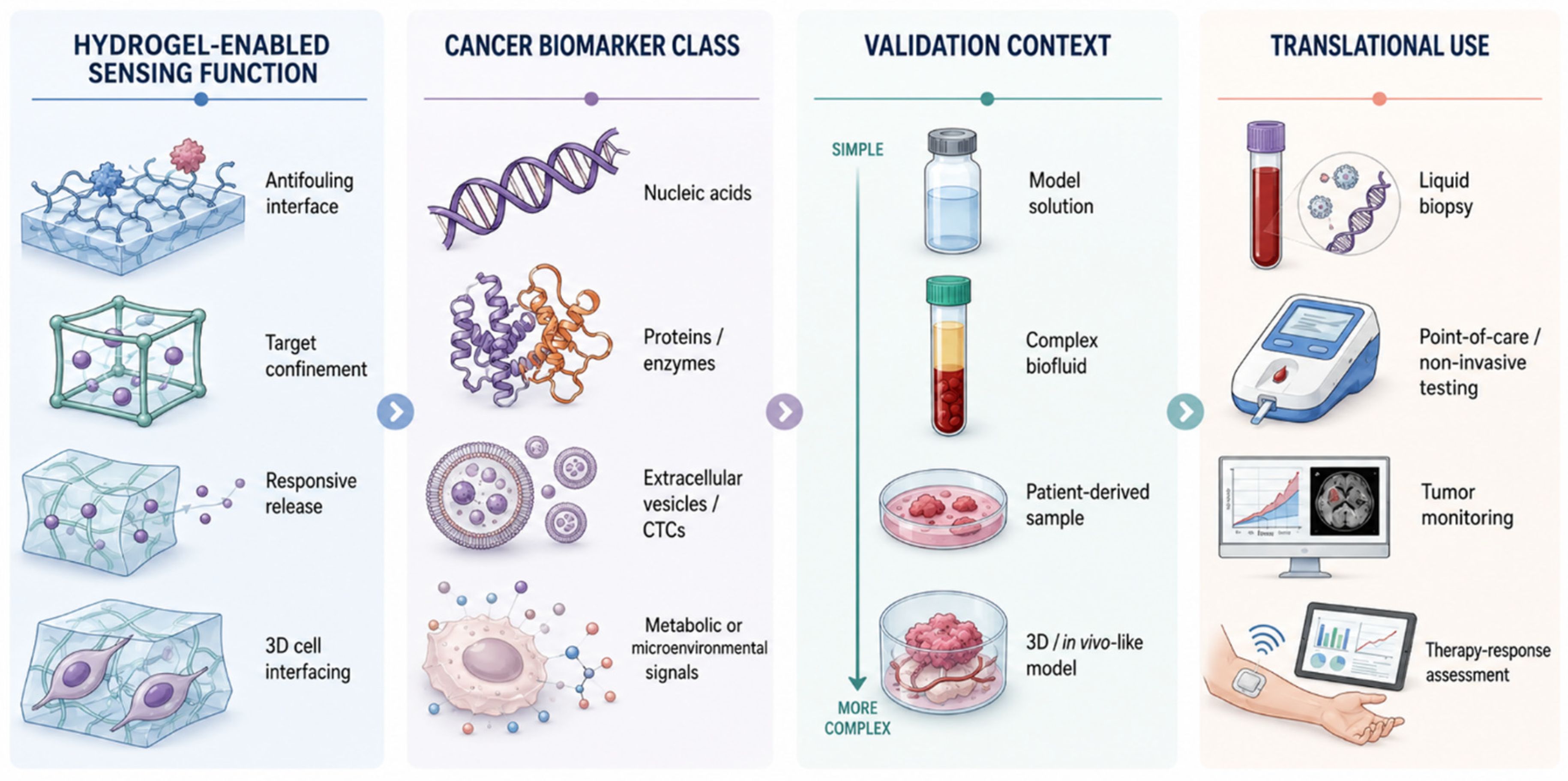
The strongest translational evidence comes from studies that move beyond analytical proof of concept and demonstrate performance in patient-derived or clinically relevant matrices. Hydrogel-based platforms have been tested in serum, saliva, urine, plasma, blood, ascites, cervical brush samples, sweat, artificial interstitial fluid, and cancer-mimicking phantoms. This validation context is central to cancer biomarker biosensing because clinically meaningful cancer biomarkers are rarely measured in clean matrices, and hydrogel interfaces must preserve sensitivity while resisting fouling, dilution effects, and matrix interference. The frequent use of spiked or simplified samples reflects practical barriers to translation, including access to annotated clinical biospecimens, ethical and collection constraints, limited sample volume, the need for standardized preprocessing, and the difficulty of demonstrating batch-to-batch hydrogel reproducibility across realistic matrices. The translational pathway linking hydrogel sensing functions, biomarker classes, validation contexts, and intended uses is summarized in Figure 5.
Several studies directly support this translational trajectory. Hydrogel–AuNP SPRi detection of prostate cancer-derived exosomes correlated with clinical total prostate-specific antigen chemiluminescence immunoassays in human serum [2]. MATCH technology detected RNAs and proteins directly in patient plasma for cancer classification within less than 60 min [43]. Exosomal microRNA-381 was detected in gastric cancer ascites samples [21]. Noninvasive or minimally invasive formats included sweat-based aldehyde biomarker detection with reported 99% diagnostic accuracy for lung cancer screening, hyaluronidase and human papillomavirus 16 DNA detection in clinical urine and cervical brush samples, saliva-based CYFRA-21-1 monitoring in oral cancer patients, hyaluronidase monitoring in pancreatic cancer serum, and Porphyromonas gingivalis detection in 40 clinical saliva samples with an LOD of 6 colony-forming units per milliliter [3,8,9,35,114]. These examples illustrate that the most advanced evidence is not simply the lowest LOD, but agreement with clinical assays, classification in patient plasma, or testing in disease-relevant biofluids.
Point-of-care relevance is most convincing where hydrogel biosensors combine quantitative performance with rapid readout, portability, smartphone compatibility, wearable integration, or reduced sample preparation. Smartphone and paper-based systems detected β-glucan from 1.0–15 nanograms per milliliter with an LOD of 0.5 nanograms per milliliter [56], HPV DNA from 10 femtomolar to 1 nanomolar with an LOD of 3.76 femtomolar in 25 min [6], and hyaluronidase in human urine with an LOD of 0.2 units per milliliter using an equipment-free paper-flow format [59]. A multicolor hyaluronidase biosensor showed a 0.6–40 units per milliliter linear range, an LOD of 0.3 units per milliliter, and ELISA-validated performance in human serum [40]. Wearable and minimally invasive systems further extended this translational logic: a microneedle glucose biosensor achieved LODs of 0.11 millimolar in phosphate-buffered saline, 0.20 millimolar in artificial interstitial fluid, and 0.46 millimolar in interstitial-fluid phantoms, with less than 4% relative standard deviation over 14 days [94], while a lab-on-a-needle platform discriminated cancer from normal tissues in a hydrogel phantom mimicking cancer microenvironments [109]. These examples indicate that portability alone is not the critical outcome; point-of-care value depends on whether the hydrogel platform maintains quantitative performance in realistic matrices while reducing assay complexity or enabling noninvasive access. Practical deployment also requires cost-aware design, storage stability, user-tolerant operation, and compatibility with scalable fabrication or existing point-of-care manufacturing workflows.

6.3. Dynamic Sensing of Tumor Biology and Therapeutic Response

A distinctive contribution of hydrogel biosensors to cancer research is their compatibility with living cells, three-dimensional cultures, and tumor-like microenvironments. This compatibility allows hydrogel platforms to extend beyond endpoint biomarker detection toward dynamic monitoring of metabolic activity, oxidative stress, invasion, drug response, and therapeutic performance. These functions are particularly relevant to cancer biomarker biosensing because many biomarkers reflect changing cellular states rather than static disease signatures. However, cell-based and three-dimensional systems should be evaluated separately from direct clinical diagnostic assays because their performance depends on hydrogel formulation, matrix mechanics, cell density, culture duration, diffusion behavior, and biological-model reproducibility.
Hydrogel systems enabled real-time monitoring of H2O2 and nitric oxide release from three-dimensional breast cancer cells after drug stimulation [70], detected H2O2 released by breast cancer cell subtypes with an ultralow LOD of 0.023 nanomolar [72], and monitored extracellular H2O2 from prostate cells over 2–100 micromolar with an LOD of 0.01 micromolar [73]. Other platforms imaged extracellular lactate metabolism in isolated cancer cells [71], tracked cancer-associated pH changes with low cytotoxicity and 0.1 pH-unit resolution across pH 5.8–7.4 [75], and distinguished cancer cells from normal cells through fluorescence, conductivity, resistance, or photothermal changes in tumor-microenvironment-responsive hydrogels [76,77,78]. These examples position hydrogels not only as sensing supports, but also as biologically compatible interfaces for measuring functional tumor states in three-dimensional or microenvironment-responsive settings. Their translational value is therefore strongest for mechanistic profiling, preclinical model assessment, and treatment-response monitoring rather than immediate replacement of standardized clinical diagnostic assays.
Hydrogel-enabled drug-response and therapeutic-monitoring platforms further support the translational relevance of this material class, although their relevance to this review is strongest when biomarker readout, tumor-microenvironment monitoring, or treatment-response assessment remains central. Impedance-based systems quantified colony formation and enabled doxorubicin half-maximal inhibitory concentration analysis [66], monitored interleukin-6-dependent invasion in real time [67], and compared hepatocellular carcinoma cell chemosensitivity at 24 and 48 h [68]. A vertical impedance electrode array detected location-dependent drug-diffusion effects in three-dimensional tumor models, supporting pharmacodynamic assessment and preclinical screening [69]. Therapeutic hydrogel systems provided controlled release or enhanced treatment outcomes, including antimicrobial hydrogel effectiveness exceeding 99.9% with high endothelial-cell biocompatibility [50], pH-regulated anticancer drug release across pH 5–7.4 with low cytotoxicity in covalently crosslinked nanogels [89], QD DNA hydrogel delivery that increased doxorubicin potency ninefold and demonstrated in vivo efficacy in xenografted breast cancer mice [91], an implantable hydrogel sensor that detected CEA at 1.8 picograms per milliliter, identified cancer nodules with median diameters below 2 mm, and achieved approximately 100% suppression of tumor recurrence and lung metastasis in mouse models [110], and approximately 32 h spectinomycin release with human hepatocellular carcinoma G2 cytotoxicity testing [112]. These examples should be viewed as translationally adjacent to cancer biomarker biosensing unless diagnostic readout, treatment-response measurement, or tumor-state monitoring is a central component of the platform.
Overall, hydrogel-based cancer biosensors have progressed from material feasibility toward platforms capable of sensitive molecular detection, biological-sample compatibility, rapid or portable readout, and functional monitoring in three-dimensional or in vivo-like systems. However, the degree of translational maturity remains variable. Studies validated in patient-derived samples and benchmarked against ELISA, quantitative polymerase chain reaction, reverse transcription polymerase chain reaction, chemiluminescence immunoassays, or clinical classification workflows provide stronger evidence than systems demonstrated only in model solutions or isolated cell models. This distinction is essential for interpreting performance claims without extending them beyond the available evidence. Accordingly, platform comparisons should report not only LOD and linear range, but also matrix type, sample volume, assay duration, instrumentation, preprocessing requirements, storage stability, hydrogel fabrication reproducibility, and whether the system has been tested in spiked, model, or patient-derived samples. Representative platform-level comparisons are summarized in Table 5.

7. Limitations and Future Perspectives

Despite strong reported analytical performance, hydrogel-based cancer biosensors remain at uneven stages of translational readiness. Many systems report ultralow LODs, broad linear ranges, or sophisticated readout mechanisms, yet validation is often performed in model solutions, spiked biological matrices, isolated cell systems, or small patient-derived sample sets. The strongest evidence comes from platforms benchmarked against ELISA, reverse transcription polymerase chain reaction, quantitative polymerase chain reaction, chemiluminescence immunoassays, or clinical classification workflows; however, such comparisons remain inconsistent across the field [2,4,9,10,11,40,41,43,58]. The frequent gap between proof-of-concept performance and patient-cohort validation likely reflects several practical barriers, including limited access to annotated clinical biospecimens, ethical and regulatory requirements for patient-sample testing, small available sample volumes, variable preanalytical handling, the cost of multicenter validation, and the need to demonstrate reproducible hydrogel fabrication across batches. Future studies should prioritize clinically powered cohorts, prospective sample testing, blinded analysis, and direct comparison with standard-of-care assays.
High-performing platforms should therefore be described as being at different stages of translational maturity rather than treated as equally close to clinical deployment. For example, platforms showing excellent analytical sensitivity in serum, untreated serum, or crude lysates still require broader patient-cohort testing, standardized preprocessing, and reproducibility analysis before they can be considered clinically mature. Similarly, portable, smartphone-readable, wearable, or paper-based formats improve accessibility, but they must still demonstrate robust calibration, reagent stability, user-independent operation, and reliable performance across realistic sample conditions.
A central limitation is the frequent mismatch between analytical sensitivity and clinical decision-making. Extremely low LODs are valuable only when they correspond to clinically meaningful biomarker thresholds, disease-stage discrimination, subtype classification, prognosis, recurrence monitoring, or therapeutic-response assessment. Platforms targeting miRNAs, extracellular vesicles, circulating tumor DNA, protein markers, and multiplexed panels should therefore be evaluated not only by analytical figures of merit, but also by receiver operating characteristic performance, biological variability, preanalytical stability, and clinical interpretability [1,2,10,11,13,14,15,16,23,43]. Future hydrogel biosensors will need to move beyond detectability alone toward clinically actionable stratification.
Complex sample compatibility remains a major challenge. Hydrogels can reduce fouling, support target enrichment, and preserve biomolecule accessibility, but serum, plasma, blood, saliva, urine, sweat, ascites, and cell-derived matrices differ substantially in viscosity, protein load, ionic composition, degradation activity, and interfering species. Antifouling and matrix-tolerant interfaces have been demonstrated for several protein, vesicle, and electrochemical platforms [4,9,12,28,34,37,39,52], yet long-term stability, batch-to-batch reproducibility, and matrix-specific calibration remain underdeveloped. These factors help explain why many studies remain at the level of spiked samples or limited biological matrices: moving into genuine patient specimens requires not only sensitivity, but also validated recovery, interference tolerance, standardized sample handling, and reproducible material preparation. Future work should adopt standardized interference panels, recovery testing across donor-variable samples, accelerated aging studies, and manufacturable quality-control metrics.
Another important issue is the need to define the hydrogel contribution more rigorously. In many systems, performance arises from a combination of hydrogel architecture, nanomaterials, receptors, enzymes, electrodes, imaging probes, or amplification chemistries. Future studies should explicitly separate the role of the hydrogel from that of the signal-generating component. This can be achieved through appropriate controls comparing hydrogel-free systems, nonresponsive hydrogel matrices, nanomaterial-only platforms, and hydrogel-integrated platforms under the same sample and readout conditions. Such comparisons are essential for determining whether the hydrogel improves target enrichment, antifouling behavior, molecular transport, mechanical compatibility, cell interfacing, signal amplification, or transduction. A practical reporting format would assign recognition, transduction, and amplification roles separately; for example, the hydrogel may provide the recognition-accessible and antifouling interface, while the nanomaterial, electrode, reporter, or optical element provides signal transduction or amplification.
Multiplexing is a major opportunity, but it also introduces methodological risk. Cancer heterogeneity makes single-marker detection insufficient for many diagnostic and monitoring tasks, and hydrogel systems have begun to support multimarker panels, spatial encoding, microfluidic integration, and parallel electrochemical or optical readouts [10,14,15,26,31,42,43]. However, multiplexed assays introduce challenges in cross-reactivity, signal normalization, differential diffusion, dynamic-range mismatch, and algorithmic interpretation. Future platforms should integrate biomarker-panel design with clearly defined clinical use cases, ensuring that each added marker improves classification, monitoring, or treatment guidance rather than merely increasing assay complexity.
Point-of-care, wearable, paper-based, smartphone-readable, and minimally invasive hydrogel platforms are among the most attractive translational directions, particularly for repeat monitoring and decentralized oncology workflows [3,6,8,15,40,56,57,59,85,94,109]. Nevertheless, portability alone does not establish clinical utility. Field-ready devices must combine quantitative accuracy, robust sample handling, calibration stability, user-independent operation, low-cost fabrication, data security, and compatibility with clinical reporting systems. This creates a cost–performance trade-off: highly engineered hydrogel–nanomaterial systems may achieve superior sensitivity through multistep amplification, but they can also increase fabrication complexity, raw-material cost, storage requirements, manual operation, and dependence on specialized instrumentation. Conversely, simpler hydrogel paper, visual, or wearable formats may be more compatible with decentralized use, even when their analytical performance is less extreme. Future work should emphasize integrated sample preparation, closed-cartridge formats, automated readout, and performance testing under realistic operating conditions.
Hydrogel-based tumor-microenvironmental and three-dimensional cell-sensing systems offer a complementary direction beyond diagnostic detection. Platforms that monitor H2O2, nitric oxide, lactate, pH, ROS, GSH, invasion, proliferation, and drug response in three-dimensional models may improve preclinical screening and support more individualized therapeutic assessment [65,66,67,68,69,70,71,72,73,74,76,77,78,80]. However, these systems require stronger biological standardization, including defined matrix stiffness, diffusion parameters, cell density, culture duration, oxygenation, and comparability to patient-derived tumor models. Future studies should couple biosensing readouts with organoid, tumor explant, or patient-derived xenograft workflows to help bridge the gap between engineered models and clinical tumor behavior. For these systems, reproducibility depends not only on sensor chemistry but also on hydrogel mechanics, cell sourcing, culture conditions, and biological variability across model preparations.
Theranostic and implantable hydrogel systems represent an emerging but still secondary frontier within the scope of cancer biosensing. Platforms linking biomarker recognition with imaging, controlled release, photothermal or photodynamic intervention, RNA modulation, or recurrence suppression demonstrate the possibility of closed-loop cancer-responsive materials [19,44,45,80,87,88,89,91,110]. However, these systems should not be conflated with direct diagnostic biosensors unless biomarker recognition, monitoring, or treatment-response readout is central to the platform. They also face additional barriers, including biocompatibility, biodegradation, immune response, off-target activation, regulatory classification, device retrieval, and long-term safety. Future development should clearly distinguish diagnostic, monitoring, therapeutic, and theranostic endpoints while generating rigorous in vivo evidence for each.
Finally, the field would benefit from harmonized reporting standards. Essential parameters, including sample volume, assay time, LOD, dynamic range, recovery, reproducibility, storage stability, hydrogel composition, mechanical properties, fouling resistance, manufacturing variability, and benchmark comparator, are not consistently reported. Tables that compare LODs and linear ranges should therefore also identify key limiting conditions, such as sample preprocessing, matrix dilution, pre-amplification, specialized instrumentation, manual operation, incomplete interference testing, limited patient-sample validation, or absence of storage and batch-reproducibility data. Standardized reporting would enable more meaningful comparison across DNA hydrogels, antifouling electrodes, nanomaterial-loaded matrices, paper devices, wearables, optical probes, and three-dimensional cell platforms. The next phase of the field should therefore be defined less by isolated demonstrations of sensitivity and more by reproducible, clinically contextualized, manufacturable, and user-compatible hydrogel biosensing systems.

8. Conclusions

Hydrogel-based biosensors have evolved into a versatile, clinically oriented materials platform for cancer biomarker detection and monitoring. Their value lies in the convergence of biomolecular recognition, hydrated and antifouling interfaces, nanomaterial-enhanced transduction, programmable responsiveness, and compatibility with complex biological samples and three-dimensional tumor models. Across liquid biopsy, soluble tumor markers, extracellular vesicles, CTCs, metabolic signals, redox activity, and treatment-response assessment, hydrogels provide more than structural support: they create active sensing microenvironments that translate cancer-associated information into measurable diagnostic and biological readouts.
The most promising future systems will be those that pair analytical sensitivity with clinical validation, multiplexed interpretability, scalable fabrication, and operation in realistic patient-centered workflows. To reach this stage, future platforms must also define hydrogel and nanomaterial roles more explicitly, report practical limiting conditions, and address batch-to-batch reproducibility, storage stability, cost, and compatibility with point-of-care manufacturing. With continued progress in standardization, device integration, and patient-sample validation, programmable hydrogel biosensors are positioned to become an important bridge between advanced biomaterials and next-generation cancer diagnostics.

9. Evidence-to-Practice Roadmap

The evidence presented in this review positions hydrogel-based biosensors not merely as materials innovations but as translational interfaces that connect cancer-relevant biology with measurable diagnostic, monitoring, and modeling outputs. The strongest trajectory runs from biological rationale—low-abundance biomarkers, liquid-biopsy needs, tumor heterogeneity, and decentralized testing—toward hydrogel-enabled mechanisms that support target confinement, antifouling recognition, responsive release, signal amplification, and three-dimensional cell interfacing. However, the field remains unevenly translated: analytical sensitivity and platform creativity are well developed, whereas standardized validation, clinically defined use cases, longitudinal patient testing, and workflow integration remain major barriers. Table 6 below integrates the collected evidence into a concise evidence-to-practice framework spanning rationale, mechanism, validation/trials, populations, outcomes, clinical practice, and future gaps.

Funding

This research received no external funding.

Data Availability Statement

No new data were created or analyzed in this study. Data sharing is not applicable to this article.

Acknowledgments

Non-AI research and reference-management tools, including Web of Science and EndNote, were used to support literature searching, citation organization, and reference management. AI-assisted tools, including OpenAI ChatGPT Plus 5.5, Gemini Pro 3.5, and NotebookLM, were used during manuscript preparation as supportive aids to improve clarity, organization, literature synthesis, and the conceptual drafting and refinement of figures. For the figures, the authors developed the scientific concepts, prepared and refined the prompts, critically evaluated the outputs, and revised the illustrations to ensure consistency with the manuscript and scientific accuracy. Any AI-assisted figures are conceptual illustrations only and do not represent raw experimental data, clinical images, microscopy images, diagnostic materials, or manipulated research outputs. No third-party copyrighted or proprietary images were intentionally uploaded, copied, reproduced, or incorporated. To the best of the authors’ knowledge, the final figures are original conceptual illustrations prepared specifically for this manuscript. The authors reviewed, edited, verified, and finalized all AI-assisted text and figure content and take full responsibility for the accuracy, integrity, and originality of the final manuscript.

Conflicts of Interest

The authors declare no conflict of interest.

Abbreviations

AuNP—gold nanoparticle; CEA—carcinoembryonic antigen; cfDNA—circulating cell-free deoxyribonucleic acid; CTC—circulating tumor cell; CYFRA-21-1—cytokeratin 19 fragment 21-1; DNA—deoxyribonucleic acid; ECL—electrochemiluminescence; ELISA—enzyme-linked immunosorbent assay; EpCAM—epithelial cell adhesion molecule; Fmoc—9-fluorenylmethoxycarbonyl; FRET—Förster resonance energy transfer; GSH—glutathione; H2O2—hydrogen peroxide; HeLa—Henrietta Lacks; HPV—human papillomavirus; LOD—limit of detection; MCF-7—Michigan Cancer Foundation-7; miR-21—microRNA-21; miRNA—microRNA; PANI—polyaniline; PEC—photoelectrochemistry; PEG—polyethylene glycol; PEGDA—poly(ethylene glycol) diacrylate; pH—potential of hydrogen; QD—quantum dot; RNA—ribonucleic acid; ROS—reactive oxygen species; SERS—surface-enhanced Raman scattering; SPR—surface plasmon resonance; SPRi—surface plasmon resonance imaging; TMB—3,3′,5,5′-tetramethylbenzidine.

References

  1. Al Sulaiman, D.; Shapiro, S.J.; Gomez-Marquez, J.; Doyle, P.S. High-Resolution Patterning of Hydrogel Sensing Motifs within Fibrous Substrates for Sensitive and Multiplexed Detection of Biomarkers. ACS Sens. 2021, 6, 203–211. [Google Scholar] [CrossRef] [PubMed]
  2. Chen, W.; Li, J.; Wei, X.; Fan, Y.; Qian, H.; Li, S.; Xiang, Y.; Ding, S. Surface plasmon resonance biosensor using hydrogel-AuNP supramolecular spheres for determination of prostate cancer-derived exosomes. Microchim. Acta 2020, 187, 590. [Google Scholar] [CrossRef] [PubMed]
  3. Chen, Z.X.; Yu, W.R.; Xiong, H.Y.; Liu, S.H.; Yan, S.; Huang, Y.Z. Dual-functionalized wearable SERS substrates enable ultra-sensitive detection of lung cancer biomarkers in sweat. Chem. Eng. J. 2025, 521, 166637. [Google Scholar] [CrossRef]
  4. Chocholova, E.; Bertok, T.; Lorencova, L.; Holazova, A.; Farkas, P.; Vikartovska, A.; Bella, V.; Velicova, D.; Kasak, P.; Eckstein, A.A.; et al. Advanced antifouling zwitterionic layer based impedimetric HER2 biosensing in human serum: Glycoprofiling as a novel approach for breast cancer diagnostics. Sens. Actuators B Chem. 2018, 272, 626–633. [Google Scholar] [CrossRef]
  5. Feng, S.; Chen, H.; Hu, Z.; Wu, T.; Liu, Z. Ultrasensitive Detection of miRNA via CRISPR/Cas12a Coupled with Strand Displacement Amplification Reaction. ACS Appl. Mater. Interfaces 2023, 15, 28933–28940. [Google Scholar] [CrossRef] [PubMed]
  6. He, H.Z.; Cheng, L.J.; He, Y.H.; Chen, J.M.; Song, L.; Yang, Y.Y.; Zhang, Y.; Lin, Z.Y.; Hong, G.L. Dual-enzyme cascade amplification electrochemical biosensor for human papillomavirus based on DNA nanoflower structure. Sens. Actuators B Chem. 2022, 371, 132532. [Google Scholar] [CrossRef]
  7. Kapil, P.; Verma, D.; Pradhan, R.; Kalkal, A.; Packirisamy, G. A bioinspired porous and electroactive reduced graphene oxide hydrogel based biosensing platform for efficient detection of tumor necrosis factor-alpha. J. Mater. Chem. B 2024, 12, 12207–12219. [Google Scholar] [CrossRef] [PubMed]
  8. Li, P.; Xu, S.; Dong, Z.; Liu, H.; Huang, J.; Deng, X.; Tao, Y.; Liu, H.; Lin, Z.; Li, Z. Dual-function paper-based biosensor for sensitive detection of hyaluronidase and human papillomavirus DNA using diffusion wet area as readout. Biosens. Bioelectron. 2025, 271, 117087. [Google Scholar] [CrossRef] [PubMed]
  9. Panwar, S.; Kumar, S. Nanoparticle integrated guar gum hydrogel-based immunosensor for biomedical application. Next Mater. 2025, 7, 100407. [Google Scholar] [CrossRef]
  10. Tang, J.P.; Cui, Z.; Li, Q.; Ke, B.; Liu, P.F.; Yang, D.Y.; Yao, C. DNA Nanotechnology-Engineered Supramolecular Hydrogel Biosensor for Cancer Diagnosis via Extracellular Vesicles. Adv. Funct. Mater. 2025, 36, e12115. [Google Scholar] [CrossRef]
  11. Wu, L.; Wang, Y.; He, R.; Zhang, Y.; He, Y.; Wang, C.; Lu, Z.; Liu, Y.; Ju, H. Fluorescence hydrogel array based on interfacial cation exchange amplification for highly sensitive microRNA detection. Anal. Chim. Acta 2019, 1080, 206–214. [Google Scholar] [CrossRef] [PubMed]
  12. Yang, H.Q.; Wang, P.P.; Geng, F.; Wu, Q.W.; Song, F.X.; Ding, C.F. Copolymerization of zwitterionic sulfobetaine and hydrophobic acrylamide based antifouling electrochemical biosensors for detection of CA125 in clinical serum samples. Sens. Actuators B Chem. 2023, 387, 133820. [Google Scholar] [CrossRef]
  13. Zhang, W.; Xu, H.; Zhao, X.; Tang, X.; Yang, S.; Yu, L.; Zhao, S.; Chang, K.; Chen, M. 3D DNA nanonet structure coupled with target-catalyzed hairpin assembly for dual-signal synergistically amplified electrochemical sensing of circulating microRNA. Anal. Chim. Acta 2020, 1122, 39–47. [Google Scholar] [CrossRef] [PubMed]
  14. Zhao, L.; Han, H.; Ma, Z. Improved screen-printed carbon electrode for multiplexed label-free amperometric immuniosensor: Addressing its conductivity and reproducibility challenges. Biosens. Bioelectron. 2018, 101, 304–310. [Google Scholar] [CrossRef] [PubMed]
  15. Zhou, B.; Hu, S.; Chen, W.; Xiang, S.; Xue, Y.; Zhang, W.; Yang, R. Visualized chemiluminescence immunoassay for simultaneous detection of multiple lung cancer markers based on spatially resolved microfluidic paper chip device. Talanta 2025, 295, 128360. [Google Scholar] [CrossRef] [PubMed]
  16. Caputo, T.M.; Battista, E.; Netti, P.A.; Causa, F. Supramolecular Microgels with Molecular Beacons at the Interface for Ultrasensitive, Amplification-Free, and SNP-Selective miRNA Fluorescence Detection. ACS Appl. Mater. Interfaces 2019, 11, 17147–17156. [Google Scholar] [CrossRef] [PubMed]
  17. Liu, S.; Su, W.; Li, Y.; Zhang, L.; Ding, X. Manufacturing of an electrochemical biosensing platform based on hybrid DNA hydrogel: Taking lung cancer-specific miR-21 as an example. Biosens. Bioelectron. 2018, 103, 1–5. [Google Scholar] [CrossRef] [PubMed]
  18. Mohammadi, S.; Mohammadi, S.; Salimi, A. A 3D hydrogel based on chitosan and carbon dots for sensitive fluorescence detection of microRNA-21 in breast cancer cells. Talanta 2021, 224, 121895. [Google Scholar] [CrossRef] [PubMed]
  19. Yousaf, S.; Amirloo, B.; Aojula, H.S.; Clarke, D.J.; Saiani, A.; Irwin, A.; Bichenkova, E.V. Hydrogel “Affinity Trap” for microRNAs with a Self-Assembling Fluorescent “Split-Probe” to Monitor Their Expression and Degradation. Biomacromolecules 2025, 26, 4595–4611. [Google Scholar] [CrossRef] [PubMed]
  20. Chang, W.H.; Lee, Y.F.; Liu, Y.W.; Willner, I.; Liao, W.C. Stimuli-responsive hydrogel microcapsules for the amplified detection of microRNAs. Nanoscale 2021, 13, 16799–16808. [Google Scholar] [CrossRef] [PubMed]
  21. Guo, Y.C.; Wang, J.L.; Ji, F.Y.; Wang, P.L.; Ma, Q. Natural supramolecular hydrogel/N-dots@COF nanocomposite-based ECL sensor for exosomal miRNA-381 detection. Chem. Eng. J. 2025, 520, 165871. [Google Scholar] [CrossRef]
  22. Sun, L.P.; Hu, N.; Peng, J.; Chen, L.Y.; Weng, J. Ultrasensitive Detection of Mitochondrial DNA Mutation by Graphene Oxide/DNA Hydrogel Electrode. Adv. Funct. Mater. 2014, 24, 6905–6913. [Google Scholar] [CrossRef]
  23. Wei, J.; Zhao, Z.; Gao, J.; Wang, Y.; Ma, L.; Meng, X.; Wang, Z. Polyacrylamide/Phytic Acid/Polydopamine Hydrogel as an Efficient Substrate for Electrochemical Enrichment of Circulating Cell-Free DNA from Blood Plasma. ACS Omega 2020, 5, 5365–5371. [Google Scholar] [CrossRef] [PubMed]
  24. Yang, J.; Fu, S.; Luo, F.; Guo, L.; Qiu, B.; Lin, Z. Homogeneous photoelectrochemical biosensor for microRNA based on target-responsive hydrogel coupled with exonuclease III and nicking endonuclease Nb.BbvCI assistant cascaded amplification strategy. Microchim. Acta 2021, 188, 267. [Google Scholar] [CrossRef] [PubMed]
  25. Ye, D.; Li, M.; Zhai, T.; Song, P.; Song, L.; Wang, H.; Mao, X.; Wang, F.; Zhang, X.; Ge, Z.; et al. Encapsulation and release of living tumor cells using hydrogels with the hybridization chain reaction. Nat. Protoc. 2020, 15, 2163–2185. [Google Scholar] [CrossRef] [PubMed]
  26. Zhang, X.; Zhou, X.; Huang, D.; Chen, Y.; Zhang, Q.; Li, Z.; Feng, L. Nucleoside hydrogel-modified cell subtype biosensor with antifouling and electroconductive properties. Talanta 2025, 285, 127383. [Google Scholar] [CrossRef] [PubMed]
  27. Chang, C.C.; Lin, S.H.; Chu-Su, Y.; Lin, C.W. Using Polyethylene Glycol-Modified Chitosan for Improvement of Carbohydrate Antigen 15-3 Detection on a Quartz Crystal Microbalance Biosensor. Sens. Lett. 2011, 9, 404–408. [Google Scholar] [CrossRef]
  28. Du, Q.; Wang, W.; Zeng, X.; Luo, X. Antifouling zwitterionic peptide hydrogel based electrochemical biosensor for reliable detection of prostate specific antigen in human serum. Anal. Chim. Acta 2023, 1239, 340674. [Google Scholar] [CrossRef] [PubMed]
  29. Gruhl, F.J.; Länge, K. Influence of Surface Preparation Parameters on the Signal Response of an Acoustic Biosensor for the Detection of a Breast Cancer Marker. IEEE Sens. J. 2012, 12, 1647–1648. [Google Scholar] [CrossRef]
  30. Han, S.W.; Koh, W.G. Hydrogel-Framed Nanofiber Matrix Integrated with a Microfluidic Device for Fluorescence Detection of Matrix Metalloproteinases-9. Anal. Chem. 2016, 88, 6247–6253. [Google Scholar] [CrossRef] [PubMed]
  31. Shekari, Z.; Zare, H.R.; Falahati, A. Dual assaying of breast cancer biomarkers by using a sandwich-type electrochemical aptasensor based on a gold nanoparticles-3D graphene hydrogel nanocomposite and redox probes labeled aptamers. Sens. Actuators B Chem. 2021, 332, 129515. [Google Scholar] [CrossRef]
  32. Wang, N.; Zhang, L.; Li, Z.; Zhou, C.; Lv, Y.; Su, X. A sensing platform for on-site detection of glutathione S-transferase using oxidized Pi@Ce-doped Zr-based metal-organic frameworks(MOFs). Talanta 2023, 259, 124537. [Google Scholar] [CrossRef] [PubMed]
  33. Wang, Q.; Hu, Y.; Jiang, N.; Wang, J.; Yu, M.; Zhuang, X. Preparation of Aptamer Responsive DNA Functionalized Hydrogels for the Sensitive Detection of alpha-Fetoprotein Using SERS Method. Bioconjug. Chem. 2020, 31, 813–820. [Google Scholar] [CrossRef] [PubMed]
  34. Wang, W.; Han, R.; Chen, M.; Luo, X. Antifouling Peptide Hydrogel Based Electrochemical Biosensors for Highly Sensitive Detection of Cancer Biomarker HER2 in Human Serum. Anal. Chem. 2021, 93, 7355–7361. [Google Scholar] [CrossRef] [PubMed]
  35. Wang, W.; Xu, Y.; Hou, J.; Li, Z.; Luo, F.; He, D.; Lin, Z. Multicolor biosensor for hyaluronidase based on target-responsive hydrogel and etching of gold nanorods by H2O2. Talanta 2023, 257, 124367. [Google Scholar] [CrossRef] [PubMed]
  36. Yang, H.C.; Yuan, R.; Chai, Y.Q.; Su, H.L.; Zhuo, Y.; Jiang, W.; Song, Z.J. Electrochemical immunosensor for human chorionic gonadotropin based on horseradish peroxidase-functionalized Prussian blue-carbon nanotubes/gold nanocomposites as labels for signal amplification. Electrochim. Acta 2011, 56, 1973–1980. [Google Scholar] [CrossRef]
  37. Yu, X.H.; Li, Y.; Lv, S.P.; Zeng, X.H.; Luo, X.L.; Wang, P.P. Low fouling electrochemical sensing interfaces based on Bovine serum albumin hydrogel for the detection of protein biomarkers in human serum. Microchem. J. 2024, 199, 109872. [Google Scholar] [CrossRef]
  38. Zhang, L.M.; Li, K.T.; Gong, J.Z.; Li, D.; Lin, Z.F.; Li, J.P. Development and application of highly sensitive protein sensor based on spatial conformation of sodium alginate gel-stabilized HE4. Microchem. J. 2025, 213, 113863. [Google Scholar] [CrossRef]
  39. Zhang, S.; Ren, H.; Dai, H.; Lv, L.; Lin, Y. Photothermal-Induced Electrochemical Interfacial Region Regulation Enables Signal Amplification for Dual-Mode Detection of Ovarian Cancer Biomarkers. ACS Appl. Bio Mater. 2021, 4, 6519–6526. [Google Scholar] [CrossRef] [PubMed]
  40. Zhao, Z.; Li, Z.; Huang, J.; Deng, X.; Jiang, F.; Han, R.P.S.; Tao, Y.; Xu, S. A portable intelligent hydrogel platform for multicolor visual detection of HAase. Microchim. Acta 2024, 191, 101. [Google Scholar] [CrossRef] [PubMed]
  41. Zheng, Y.; Wang, H.Q.; Ma, Z.F. A nanocomposite containing Prussian Blue, platinum nanoparticles and polyaniline for multi-amplification of the signal of voltammetric immunosensors: Highly sensitive detection of carcinoma antigen 125. Microchim. Acta 2017, 184, 4269–4277. [Google Scholar] [CrossRef]
  42. Zhu, Q.; Trau, D. Multiplex detection platform for tumor markers and glucose in serum based on a microfluidic microparticle array. Anal. Chim. Acta 2012, 751, 146–154. [Google Scholar] [CrossRef] [PubMed]
  43. Chen, Y.; Zhang, L.; Wu, X.; Sun, X.; Sundah, N.R.; Wong, C.Y.; Natalia, A.; Tam, J.K.C.; Lim, D.W.; Chowbay, B.; et al. Magnetic augmentation through multi-gradient coupling enables direct and programmable profiling of circulating biomarkers. Nat. Commun. 2024, 15, 8410. [Google Scholar] [CrossRef] [PubMed]
  44. Artiga, A.; Garcia-Embid, S.; De Matteis, L.; Mitchell, S.G.; de la Fuente, J.M. Effective In Vitro Photokilling by Cell-Adhesive Gold Nanorods. Front. Chem. 2018, 6, 234. [Google Scholar] [CrossRef] [PubMed]
  45. Barker, K.; Rastogi, S.K.; Dominguez, J.; Cantu, T.; Brittain, W.; Irvin, J.; Betancourt, T. Biodegradable DNA-enabled poly(ethylene glycol) hydrogels prepared by copper-free click chemistry. J. Biomater. Sci. Polym. Ed. 2016, 27, 22–39. [Google Scholar] [CrossRef] [PubMed]
  46. Field, E.H.; Ratcliffe, J.; Johnson, C.J.; Binger, K.J.; Reynolds, N.P. Self-healing, 3D printed bioinks from self-assembled peptide and alginate hybrid hydrogels. Biomater. Adv. 2025, 169, 214145. [Google Scholar] [CrossRef] [PubMed]
  47. Ge, M.; Sun, J.; Chen, M.; Tian, J.; Yin, H.; Yin, J. A hyaluronic acid fluorescent hydrogel based on fluorescence resonance energy transfer for sensitive detection of hyaluronidase. Anal. Bioanal. Chem. 2020, 412, 1915–1923. [Google Scholar] [CrossRef] [PubMed]
  48. Ham, J.; Yun, B.J.; Koh, W.G. SERS-based biosensing platform using shape-coded hydrogel microparticles incorporating silver nanoparticles. Sens. Actuators B Chem. 2021, 341, 129989. [Google Scholar] [CrossRef]
  49. Xue, H.; Zhu, C.; Wang, Y.; Gu, Q.; Shao, Y.; Jin, A.; Zhang, X.; Lei, L.; Li, Y. Stimulus-responsive cellulose hydrogels in biomedical applications and challenges. Mater. Today Bio 2025, 32, 101814. [Google Scholar] [CrossRef] [PubMed]
  50. Zhang, M.; Gu, G.; Xu, Y.; Luan, X.; Liu, J.; He, P.; Wei, G. Injectable Self-Healing Antibacterial Hydrogels with Tailored Functions by Loading Peptide Nanofiber-Biomimetic Silver Nanoparticles. Macromol. Rapid Commun. 2024, 45, e2400173. [Google Scholar] [CrossRef] [PubMed]
  51. Rajarathinam, T.; Jayaraman, S.; Kim, C.S.; Lee, J.; Chang, S.C. Portable Amperometric Biosensor Enhanced with Enzyme-Ternary Nanocomposites for Prostate Cancer Biomarker Detection. Biosensors 2024, 14, 623. [Google Scholar] [CrossRef] [PubMed]
  52. Rani, S.; Bandyopadhyay-Ghosh, S.; Ghosh, S.B. Personalized assessment and monitoring of bone health from sweat: Unveiling TEGO doped wearable, non-invasive hydrogel nanocomposite biosensor empowered by IL-6 detection. Biomed. Mater. 2025, 20, 035010. [Google Scholar] [CrossRef] [PubMed]
  53. Rastin, H.; Zhang, B.; Mazinani, A.; Hassan, K.; Bi, J.; Tung, T.T.; Losic, D. 3D bioprinting of cell-laden electroconductive MXene nanocomposite bioinks. Nanoscale 2020, 12, 16069–16080. [Google Scholar] [CrossRef] [PubMed]
  54. Sun, P.; Hai, J.; Sun, S.; Lu, S.; Liu, S.; Liu, H.; Chen, F.; Wang, B. Aqueous stable Pd nanoparticles/covalent organic framework nanocomposite: An efficient nanoenzyme for colorimetric detection and multicolor imaging of cancer cells. Nanoscale 2020, 12, 825–831. [Google Scholar] [CrossRef] [PubMed]
  55. Zhang, Y.; Hu, C.; Yin, Y.; Ren, K.; He, Y.; Gao, Y.; Han, H.; Zhu, C.; Wang, W. CRISPR/Cas12a-Responsive Smart DNA Hydrogel for Sensitive Electrochemiluminescence Detection of the Huanglongbing Outer Membrane Protein Gene. Anal. Chem. 2024, 96, 11611–11618. [Google Scholar] [CrossRef] [PubMed]
  56. Bagal-Kestwal, D.R.; Chiang, B.H. Portable paper-micro well device composed of agglomerated nano-hematite clusters in enzyme-hydrogel composite for beta glucan detection using smartphone. Sens. Actuators B Chem. 2021, 339, 129836. [Google Scholar] [CrossRef]
  57. Wang, C.; Zhang, Y.; Liu, C.; Gou, S.; Hu, S.; Guo, W. A portable colorimetric point-of-care testing platform for MicroRNA detection based on programmable entropy-driven dynamic DNA network modulated DNA-gold nanoparticle hybrid hydrogel film. Biosens. Bioelectron. 2023, 225, 115073. [Google Scholar] [CrossRef] [PubMed]
  58. Zhang, K.; Pei, M.; Cheng, Y.Y.; Zhang, Z.; Niu, C.; Liu, X.Q.; Liu, J.X.; Guo, F.; Huang, H.G.; Lin, X.J. A novel electrochemical aptamer biosensor based on tetrahedral DNA nanostructures and catalytic hairpin assembly for CEA detection. J. Electroanal. Chem. 2021, 898, 115635. [Google Scholar] [CrossRef]
  59. Zhao, B.; Qi, L.; Tai, W.; Zhao, M.; Chen, X.; Yu, L.; Shi, J.; Wang, X.; Lin, J.M.; Hu, Q. Paper-Based Flow Sensor for the Detection of Hyaluronidase via an Enzyme Hydrolysis-Induced Viscosity Change in a Polymer Solution. Anal. Chem. 2022, 94, 4643–4649. [Google Scholar] [CrossRef] [PubMed]
  60. Garcia-Astrain, C.; Henriksen-Lacey, M.; Lenzi, E.; Renero-Lecuna, C.; Langer, J.; Pineiro, P.; Molina-Martinez, B.; Plou, J.; Jimenez de Aberasturi, D.; Liz-Marzan, L.M. A Scaffold-Assisted 3D Cancer Cell Model for Surface-Enhanced Raman Scattering-Based Real-Time Sensing and Imaging. ACS Nano 2024, 18, 11257–11269. [Google Scholar] [CrossRef] [PubMed]
  61. Guo, J.; Luo, Y.; Yang, C.; Kong, L. In situ surface-enhanced Raman scattering sensing with soft and flexible polymer optical fiber probes. Opt. Lett. 2018, 43, 5443–5446. [Google Scholar] [CrossRef] [PubMed]
  62. Guo, Q.Y.; Ren, S.Y.; Wang, J.Y.; Li, Y.; Yao, Z.Y.; Huang, H.; Gao, Z.X.; Yang, S.P. Low field nuclear magnetic sensing technology based on hydrogel-coated superparamagnetic particles. Anal. Chim. Acta 2020, 1094, 151–159. [Google Scholar] [CrossRef] [PubMed]
  63. Huang, L.; Huang, H.; Zhang, Z.; Li, G. Three-Dimensional DNA Hydrogel Mediated Dual-Mode Sensing Method for Quantification of Epithelial Cell Adhesion Molecule in Biological Fluid Samples. Anal. Chem. 2024, 96, 12254–12261. [Google Scholar] [CrossRef] [PubMed]
  64. Chung, S.H.; Son, S.J.; Min, J. Nano barcoding cell-based biosensor using fluorophor-embedded silica nanotubes. J. Nanosci. Nanotechnol. 2011, 11, 4419–4423. [Google Scholar] [CrossRef] [PubMed]
  65. Lee, S.M.; Han, N.; Lee, R.; Choi, I.H.; Park, Y.B.; Shin, J.S.; Yoo, K.H. Real-time monitoring of 3D cell culture using a 3D capacitance biosensor. Biosens. Bioelectron. 2016, 77, 56–61. [Google Scholar] [CrossRef] [PubMed]
  66. Lei, K.F.; Wu, Z.M.; Huang, C.H. Impedimetric quantification of the formation process and the chemosensitivity of cancer cell colonies suspended in 3D environment. Biosens. Bioelectron. 2015, 74, 878–885. [Google Scholar] [CrossRef] [PubMed]
  67. Lei, K.F.; Tseng, H.P.; Lee, C.Y.; Tsang, N.M. Quantitative Study of Cell Invasion Process under Extracellular Stimulation of Cytokine in a Microfluidic Device. Sci. Rep. 2016, 6, 25557. [Google Scholar] [CrossRef] [PubMed]
  68. Lei, K.F.; Liu, T.K.; Tsang, N.M. Towards a high throughput impedimetric screening of chemosensitivity of cancer cells suspended in hydrogel and cultured in a paper substrate. Biosens. Bioelectron. 2018, 100, 355–360. [Google Scholar] [CrossRef] [PubMed]
  69. Qiu, Y.; Liu, X.; Zhu, Y.; Jiang, D.; Li, F.; Yu, W.; Wan, H.; Zhuang, L.; Pan, Y.; Wang, P. Vertical impedance electrode array for spatiotemporal dynamics monitoring of 3D cells under drug diffusion effect. iScience 2023, 26, 107962. [Google Scholar] [CrossRef] [PubMed]
  70. Wei, X.; Zhao, X.; Sui, D.; Chen, X.; Yang, W. Peptide hydrogel based electrochemical biosensor for simultaneous monitoring of H2O2 and NO released from three-dimensional cultured breast cancer cells. Microchim. Acta 2024, 191, 523. [Google Scholar] [CrossRef] [PubMed]
  71. Zhang, X.; Ding, S.; Cao, S.; Zhu, A.; Shi, G. Functional surface engineering of quantum dot hydrogels for selective fluorescence imaging of extracellular lactate release. Biosens. Bioelectron. 2016, 80, 315–322. [Google Scholar] [CrossRef] [PubMed]
  72. Zhou, Y.; Wei, X.; Chen, J.; Xiong, H.; Sui, D.; Chen, X.; Yang, W. A Three-Dimensional Electrochemiluminescence Sensor Integrated with Peptide Hydrogel for Detection of H2O2 Released from Different Subtypes of Breast Cancer Cells. Anal. Chem. 2024, 96, 13464–13472. [Google Scholar] [CrossRef] [PubMed]
  73. Crulhas, B.P.; Ramos, N.P.; Castro, G.R.; Pedrosa, V.A. Detection of hydrogen peroxide releasing from prostate cancer cell using a biosensor. J. Solid State Electrochem. 2016, 20, 2427–2433. [Google Scholar] [CrossRef]
  74. Crulhas, B.P.; Recco, L.C.; Delella, F.K.; Pedrosa, V.A. A Novel Superoxide Anion Biosensor for Monitoring Reactive Species of Oxygen Released by Cancer Cells. Electroanalysis 2017, 29, 1252–1257. [Google Scholar] [CrossRef]
  75. Liu, H.; Zhang, Y.; Zhao, Y.; Zhao, Y.; Yang, X.; Han, L.; Xin, J.; Yang, B.; Lin, Q. Dual-emission hydrogel nanoparticles with linear and reversible luminescence-response to pH for intracellular fluorescent probes. Talanta 2020, 211, 120755. [Google Scholar] [CrossRef] [PubMed]
  76. Robby, A.I.; Yang, J.H.; Jin, E.J.; Park, S.Y. Tumor Microenvironment-Selective Sol-Gel Mineralization of ROS-Responsive Stretchable and Conductive Hydrogel. Adv. Funct. Mater. 2024, 34, 2402367. [Google Scholar] [CrossRef]
  77. Ryplida, B.; Park, S.Y. Swelling-induced deformation electronic signal of MXene hydrogel for cancer detection. Sens. Actuators B Chem. 2023, 394, 134363. [Google Scholar] [CrossRef]
  78. Subba, S.H.; Park, S.Y. In Situ Cancer-Cell-Triggered Visible Changes in Mechanical Properties, Electroconductivity, and Adhesiveness of a MnO2@PD-Based Mineralized Hydrogel. ACS Appl. Mater. Interfaces 2023, 15, 38357–38366. [Google Scholar] [CrossRef] [PubMed]
  79. Xu, Y.; Yan, J.; Zhu, Y.; Chen, H.; Wu, C.; Zhu, X.; Zhang, Y.; Li, H.; Liu, M.; Yao, S. Self-Cascade Nanoenzyme of Cupric Oxide Nanoparticles (CuO NPs) Induced In Situ Catalysis Formation of Polyelectrolyte as Template for the Synthesis of Near-Infrared Fluorescent Silver Nanoclusters and the Application in Glutathione Detection and Bioimaging. Anal. Chem. 2022, 94, 14642–14651. [Google Scholar] [CrossRef] [PubMed]
  80. Yi, S.W.; Zhao, H.Y.; Xu, X.N.; Guan, B.W.; Zhao, H.F.; Zhang, R.P. A photoresponsive multifunctional carbon-based nanozymes for synergistic detecting and depleting GSH in cancer therapy. Appl. Surf. Sci. 2024, 655, 159568. [Google Scholar] [CrossRef]
  81. Salahuddin, B.; Masud, M.K.; Aziz, S.; Liu, C.H.; Amiralian, N.; Ashok, A.; Hossain, S.M.A.; Park, H.; Wahab, M.A.; Amin, M.A.; et al. κ-Carrageenan Gel Modified Mesoporous Gold Chronocoulometric Sensor for Ultrasensitive Detection of MicroRNA. Bull. Chem. Soc. Jpn. 2022, 95, 198–207. [Google Scholar] [CrossRef]
  82. Sebben, D.; Li, H.Y. Intracellular metal enhanced fluorescence utilizing gold nanoparticles embedded in hydrogel droplets for sensitive protein detection in cells. Talanta Open 2023, 8, 100265. [Google Scholar] [CrossRef]
  83. Zhang, G.S.; Han, A.Q.; Li, N.; Yang, M.Y.; Yan, G.Q.; Cao, Z.G.; Shi, J.H.; Zhou, S.; Shan, L.; Yu, B.L. Specific, sensitive, and reliable sensing of cancer cells based on quantum dot/hydrogel-coated double optical fiber ball. Sens. Actuators B Chem. 2024, 417, 136100. [Google Scholar] [CrossRef]
  84. Lu, H.; Rakhymzhanov, A.; Buttner, U.; Alsulaiman, D. Making Healthcare Accessible: A Rapid Clean-Room-Free Fabrication Strategy for Microfluidics-Driven Biosensors Based on Coupling Stereolithography and Hot Embossing. ACS Omega 2024, 9, 38096–38106. [Google Scholar] [CrossRef] [PubMed]
  85. Fu, G.; Li, X.; Wang, W.; Hou, R. Multiplexed tri-mode visual outputs of immunoassay signals on a clip-magazine-assembled photothermal biosensing disk. Biosens. Bioelectron. 2020, 170, 112646. [Google Scholar] [CrossRef] [PubMed]
  86. Bucatariu, S.; Cosman, B.; Constantin, M.; Ailiesei, G.L.; Rusu, D.; Fundueanu, G. Thermally Solvent-Free Cross-Linked pH/Thermosensitive Hydrogels as Smart Drug Delivery Systems. Gels 2024, 10, 834. [Google Scholar] [CrossRef] [PubMed]
  87. Liu, X.; Ma, M.; Tian, S.; Wang, W.; Li, X. “Domino” cascade reactor based on DNA hydrogel for synergistic treatment of malignant tumor. Eur. J. Med. Chem. 2023, 256, 115441. [Google Scholar] [CrossRef] [PubMed]
  88. Oliveira, C.B.P.; Veloso, S.R.S.; Castanheira, E.M.S.; Figueiredo, P.R.; Carvalho, A.T.P.; Hilliou, L.; Pereira, R.B.; Pereira, D.M.; Martins, J.A.; Ferreira, P.M.T.; et al. An injectable, naproxen-conjugated, supramolecular hydrogel with ultra-low critical gelation concentration-prepared from a known folate receptor ligand. Soft Matter 2022, 18, 3955–3966. [Google Scholar] [CrossRef] [PubMed]
  89. Wu, W.; Shen, J.; Banerjee, P.; Zhou, S. Chitosan-based responsive hybrid nanogels for integration of optical pH-sensing, tumor cell imaging and controlled drug delivery. Biomaterials 2010, 31, 8371–8381. [Google Scholar] [CrossRef] [PubMed]
  90. Xu, J.-W.; Lee, L.C.-C.; Yip, A.M.-H.; Xu, G.-X.; Leung, P.K.-K.; Lo, K.K.-W. Luminescent iridium(iii) 2-cyanobenzothiazole complexes as site-specific labels to afford peptide-based phosphorogenic probes and hydrogels for enzyme activity sensing, cancer imaging and photodynamic therapy. Inorg. Chem. Front. 2025, 12, 2266–2279. [Google Scholar] [CrossRef]
  91. Zhang, L.; Jean, S.R.; Ahmed, S.; Aldridge, P.M.; Li, X.; Fan, F.; Sargent, E.H.; Kelley, S.O. Multifunctional quantum dot DNA hydrogels. Nat. Commun. 2017, 8, 381. [Google Scholar] [CrossRef] [PubMed]
  92. Du, H.; Dang, X.; Chen, R.; Li, Y.; Cui, N.; Yang, H. A universal three-dimensional hydrogel electrode for electrochemical detection of SARS-CoV-2 nucleocapsid protein and hydrogen peroxide. Biosens. Bioelectron. 2024, 259, 116355. [Google Scholar] [CrossRef] [PubMed]
  93. Jiang, H.; Yang, J.; Wan, K.; Jiang, D.; Jin, C. Miniaturized Paper-Supported 3D Cell-Based Electrochemical Sensor for Bacterial Lipopolysaccharide Detection. ACS Sens. 2020, 5, 1325–1335. [Google Scholar] [CrossRef] [PubMed]
  94. Mansouri Majd, S. Hydrogel-coated microneedle biosensor for selective and low-potential glucose detection in skin-mimicking interstitial fluid phantoms. Talanta 2025, 297, 128555. [Google Scholar] [CrossRef] [PubMed]
  95. Miri, A.K.; Nieto, D.; Iglesias, L.; Goodarzi Hosseinabadi, H.; Maharjan, S.; Ruiz-Esparza, G.U.; Khoshakhlagh, P.; Manbachi, A.; Dokmeci, M.R.; Chen, S.; et al. Microfluidics-Enabled Multimaterial Maskless Stereolithographic Bioprinting. Adv. Mater. 2018, 30, e1800242. [Google Scholar] [CrossRef] [PubMed]
  96. Mishra, A.; Omoyeni, T.; Singh, P.K.; Anandakumar, S.; Tiwari, A. Trends in sustainable chitosan-based hydrogel technology for circular biomedical engineering: A review. Int. J. Biol. Macromol. 2024, 276, 133823. [Google Scholar] [CrossRef] [PubMed]
  97. Mughal, S.; López-Muñoz, G.A.; Fernández-Costa, J.M.; Cortés-Reséndiz, A.; De Chiara, F.; Ramón-Azcón, J. Organs-on-Chips: Trends and Challenges in Advanced Systems Integration. Adv. Mater. Interfaces 2022, 9, 2201618. [Google Scholar] [CrossRef]
  98. Zheng, Y.H.; Wu, M.Y.; Han, F.G.; Wu, D.H.; Fu, L. Evaluation of Total Antioxidant Activity of Different Floral Sources of Honeys Using Crosslinked Hydrogels. Int. J. Electrochem. Sci. 2019, 14, 1479–1487. [Google Scholar] [CrossRef]
  99. Lu, H.; Samman, F.; Hasan, E.; Alsulaiman, D. Novabeads: Stimuli-Responsive Signal-Amplifying Hydrogel Microparticles for Enzymeless Fluorescence-Based Detection of microRNA Biomarkers. Small 2025, 21, e2503990. [Google Scholar] [CrossRef] [PubMed]
  100. Mohsenin, H.; Schmachtenberg, R.; Kemmer, S.; Wagner, H.J.; Johnston, M.; Madlener, S.; Dincer, C.; Timmer, J.; Weber, W. Signal-Amplifying Biohybrid Material Circuits for CRISPR/Cas-Based Single-Stranded RNA Detection. Adv. Mater. Technol. 2024, 10, 2400981. [Google Scholar] [CrossRef]
  101. Lin, Z.T.; Li, Y.X.; Gu, J.H.; Wang, H.E.; Zhu, Z.; Hong, X.; Zhang, Z.J.; Lu, Q.Q.; Qiu, J.Y.; Wang, X.F.; et al. A Conductive Nanowire-Mesh Biosensor for Ultrasensitive Detection of Serum C-Reactive Protein in Melanoma. Adv. Funct. Mater. 2018, 28, 1802482. [Google Scholar] [CrossRef]
  102. Li, Z.; Tang, C.; Huang, D.; Qin, W.; Luo, F.; Wang, J.; Guo, L.; Qiu, B.; Lin, Z. Sensitive Hyaluronidase Biosensor Based on Target-Responsive Hydrogel Using Electronic Balance as Readout. Anal. Chem. 2019, 91, 11821–11826. [Google Scholar] [CrossRef] [PubMed]
  103. Yang, H.M.; Teoh, J.Y.; Yim, G.H.; Park, Y.; Kim, Y.G.; Kim, J.; Yoo, D. Label-Free Analysis of Multivalent Protein Binding Using Bioresponsive Nanogels and Surface Plasmon Resonance (SPR). ACS Appl. Mater. Interfaces 2020, 12, 5413–5419. [Google Scholar] [CrossRef] [PubMed]
  104. Yang, H.M.; Yim, B.; Lee, B.H.; Park, Y.; Kim, Y.G.; Kim, J.; Yoo, D. New Tool for Rapid and Accurate Detection of Interleukin-2 and Soluble Interleukin-2 Receptor alpha in Cancer Diagnosis Using a Bioresponsive Microgel and Multivalent Protein Binding. ACS Appl. Mater. Interfaces 2021, 13, 33782–33789. [Google Scholar] [CrossRef] [PubMed]
  105. Li, M.; Liu, X.; Shang, J.; Wang, X.; Zhang, X.B.; Xiong, B. Light-mediated protein functionalization of photoclickable hydrogel interface for selective cell capture and dot blotting assay. Talanta 2024, 267, 125248. [Google Scholar] [CrossRef] [PubMed]
  106. El Housseini, W.; Baiarashov, E.; Gerulskis, R.; Milam, A.; Minteer, S.D. Harnessing Redox Polymer Dynamics for Enhanced Glucose-Oxygen Coupling in Dual Biosensing and Therapeutic Applications. ACS Sens. 2024, 9, 3357–3366. [Google Scholar] [CrossRef] [PubMed]
  107. Mu, J.; Li, X.; Jia, Q. Anchoring Au nanoclusters into coordination polymers: A novel approach toward ATP detection and its application. Talanta 2024, 277, 126306. [Google Scholar] [CrossRef] [PubMed]
  108. Muthuraj, B.; Mukherjee, S.; Chowdhury, S.R.; Patra, C.R.; Iyer, P.K. An efficient strategy to assemble water soluble histidine-perylene diimide and graphene oxide for the detection of PPi in physiological conditions and in vitro. Biosens. Bioelectron. 2017, 89, 636–644. [Google Scholar] [CrossRef] [PubMed]
  109. Park, J.; Sempionatto, J.R.; Kim, J.; Jeong, Y.; Gu, J.; Wang, J.; Park, I. Microscale Biosensor Array Based on Flexible Polymeric Platform toward Lab-on-a-Needle: Real-Time Multiparameter Biomedical Assays on Curved Needle Surfaces. ACS Sens. 2020, 5, 1363–1373. [Google Scholar] [CrossRef] [PubMed]
  110. Geng, S.; Guo, P.; Wang, J.; Zhang, Y.; Shi, Y.; Li, X.; Cao, M.; Song, Y.; Zhang, H.; Zhang, Z.; et al. Ultrasensitive Optical Detection and Elimination of Residual Microtumors with a Postoperative Implantable Hydrogel Sensor for Preventing Cancer Recurrence. Adv. Mater. 2024, 36, e2307923. [Google Scholar] [CrossRef] [PubMed]
  111. Hendrickson, G.R.; Smith, M.H.; South, A.B.; Lyon, L.A. Design of Multiresponsive Hydrogel Particles and Assemblies. Adv. Funct. Mater. 2010, 20, 1697–1712. [Google Scholar] [CrossRef]
  112. Naman, A.; Fatima, A.; Mehmood, N.; Kim, M.; Younas, S. Spectinomycin-Loaded Antibacterial Polyvinyl Alcohol/Silver Oxide Nanobiocomposite Hydrogel: Synthesis, Characterization, Swelling, Cytotoxicity, and Controlled Drug Release Carrier. Bionanoscience 2025, 15, 318. [Google Scholar] [CrossRef]
  113. Guimaraes, C.F.; Ahmed, R.; Mataji-Kojouri, A.; Soto, F.; Wang, J.; Liu, S.; Stoyanova, T.; Marques, A.P.; Reis, R.L.; Demirci, U. Engineering Polysaccharide-Based Hydrogel Photonic Constructs: From Multiscale Detection to the Biofabrication of Living Optical Fibers. Adv. Mater. 2021, 33, e2105361. [Google Scholar] [CrossRef] [PubMed]
  114. Shen, H.W.; Fu, Y.F.; Liu, Z.K.; Pan, S.Y.; Zhou, F.Z.; Peng, Y.; Yang, X.; Zhang, X.M.; Song, J.L. A handheld electrochemical bacterial sensor based on multifunctional composite hydrogels and DNA biomimetic nanowalls for accurate detection of Porphyromonas gingivalis. Chem. Eng. J. 2024, 498, 155344. [Google Scholar] [CrossRef]
  115. Shi, R.; Fern, J.; Xu, W.; Jia, S.; Huang, Q.; Pahapale, G.; Schulman, R.; Gracias, D.H. Multicomponent DNA Polymerization Motor Gels. Small 2020, 16, e2002946. [Google Scholar] [CrossRef] [PubMed]
  116. Lee, T.; Kim, C.; Kim, J.; Seong, J.B.; Lee, Y.; Roh, S.; Cheong, D.Y.; Lee, W.; Park, J.; Hong, Y.; et al. Colorimetric Nanoparticle-Embedded Hydrogels for a Biosensing Platform. Nanomaterials 2022, 12, 1150. [Google Scholar] [CrossRef] [PubMed]
  117. Zeng, Z.; Zhou, R.; Sun, R.; Zhang, X.; Cheng, Z.; Chen, C.; Zhu, Q. Nonlinear hybridization chain reaction-based functional DNA nanostructure assembly for biosensing, bioimaging applications. Biosens. Bioelectron. 2021, 173, 112814. [Google Scholar] [CrossRef] [PubMed]
  118. Nash, A.T.; Foster, D.A.N.; Thompson, S.; Han, S.; Fernandez, M.K.; Hwang, D.K. A New Rapid Microfluidic Detection Platform Utilizing Hydrogel-Membrane under Cross-Flow. Adv. Mater. Technol. 2022, 7, 2101396. [Google Scholar] [CrossRef]
  119. Hao, N.; Hua, R.; Chen, S.; Zhang, Y.; Zhou, Z.; Qian, J.; Liu, Q.; Wang, K. Multiple signal-amplification via Ag and TiO2 decorated 3D nitrogen doped graphene hydrogel for fabricating sensitive label-free photoelectrochemical thrombin aptasensor. Biosens. Bioelectron. 2018, 101, 14–20. [Google Scholar] [CrossRef] [PubMed]
  120. Leng, D.; Yu, Z.; Liu, J.; Jin, W.; Wu, T.; Ren, X.; Ma, H.; Wu, D.; Ju, H.; Wei, Q. Multifunctional Supramolecular Hydrogel Modulated Heterojunction Interface Carrier Transport Engineering Facilitates Sensitive Photoelectrochemical Immunosensing. Anal. Chem. 2024, 96, 8814–8821. [Google Scholar] [CrossRef] [PubMed]
Figure 1. Clinical Translation Logic of Biomarker Biosensing Hydrogels in Cancer. The figure illustrates how cancer-relevant inputs—including nucleic acids, proteins, extracellular vesicles, circulating tumor cells, metabolites, and tumor-microenvironment signals—are processed through a central hydrogel platform and translated into clinically oriented biosensing formats, including liquid biopsy, wearables, multiplex profiling, and three-dimensional tumor-model monitoring. These formats support early detection, disease monitoring, progression assessment, and treatment evaluation. Non-core extensions, including wound care, infectious disease applications, and tissue engineering, are shown as contextual applications outside the central cancer-biomarker pathway (prepared with the assistance of Gemini’s NotebookLM Pro 3.5 and ChatGPT Plus 5.5).
Jnt 07 00017 g001
Figure 2. Biological context continuum in hydrogel-based cancer biomarker biosensing. Hydrogel biosensing platforms span a continuum from molecular liquid-biopsy targets, including circulating cell-free DNA (cfDNA) and microRNAs (miRNAs), to extracellular and circulating entities such as exosomes and circulating tumor cells (CTCs), tumor-microenvironment signals such as reactive oxygen species (ROS), glutathione (GSH), potential of hydrogen (pH), and lactate, and three-dimensional model systems, including tumor spheroids and cancer invasion models. The hydrogel biosensing interface provides a shared sensing environment across these increasing levels of biological complexity (prepared with the assistance of Gemini’s NotebookLM Pro 3.5 and ChatGPT Plus 5.5).
Jnt 07 00017 g002
Figure 3. Hydrogel as the Integrative Interface in Cancer Biomarker Biosensing Platforms. The schematic illustrates the hydrogel as a central functional interface between cancer-relevant biomarkers or sample environments and platform transduction formats. The hydrogel contributes the local sensing architecture through a biomarker-accessible matrix, antifouling or conductive interface, nanomaterial host, and programmable or responsive network, while integrated nanomaterials provide optical, electrical, catalytic, or magnetic functionality. Representative device formats include electrode sensors, paper or microfluidic devices, wearable or implantable systems, and three-dimensional scaffold or bioink platforms. The figure also emphasizes that recognition, transduction, and amplification may be distributed across different components of the same composite platform (prepared with the assistance of Gemini’s NotebookLM Pro 3.5 and ChatGPT Plus 5.5).
Jnt 07 00017 g003
Figure 4. Hydrogel-mediated transduction in cancer biomarker biosensing. Conceptual schematic illustrating how cancer-relevant molecular, cellular, metabolic, and behavioral inputs are converted through an active hydrogel layer into diagnostic signals. The hydrogel functions as a conversion matrix that can support gating, capture, degradation, charge regulation, and plasmonic localization, thereby producing optical, colorimetric, electrochemical, magnetic, acoustic, or cell-based outputs. The schematic also separates recognition, transduction, and amplification functions to clarify whether the hydrogel itself participates in signal generation or primarily supports nanomaterial-assisted readout (prepared with the assistance of Gemini’s NotebookLM Pro 3.5 and ChatGPT Plus 5.5).
Jnt 07 00017 g004
Figure 5. Translational logic of biomarker biosensing hydrogels in cancer. Hydrogel-enabled sensing functions, including antifouling behavior, target confinement, responsive release, and three-dimensional cell interfacing, support detection of cancer-relevant biomarker classes such as nucleic acids, proteins, extracellular vesicles, and microenvironmental signals. Translational relevance increases as platforms progress from model solutions to biofluids, patient samples, and in vivo-like models, enabling liquid biopsy, point-of-care testing, tumor monitoring, and therapy assessment. The figure also emphasizes that translational maturity depends on validation depth, assay complexity, reproducibility, and manufacturable device integration, not sensitivity alone (prepared with the assistance of Gemini’s NotebookLM Pro 3.5 and ChatGPT Plus 5.5).
Jnt 07 00017 g005
Table 1. Clinical and Biomedical Objective Patterns in Biomarker Biosensing Hydrogels for Cancer.
Clinical or Biomedical ObjectiveCancer-Focused Need AddressedPattern Revealed Across the StudiesRepresentative References
Early cancer biomarker detection and diagnostic recognitionNeed for sensitive detection of cancer-associated molecular, cellular, enzymatic, vesicular, and tumor microenvironmental indicators in clinically relevant contexts.This is the core objective of the section: hydrogels are used as biomarker-responsive interfaces to support earlier or more reliable cancer detection.[1,2,3,4,6,7,9,11,12,13,14,15,22,82]
Liquid biopsy and minimally invasive cancer monitoringNeed to detect cancer-relevant signals in accessible biological materials, including blood-derived samples, serum, plasma, saliva, urine, sweat, extracellular vesicles, circulating nucleic acids, and circulating tumor cells.The field is moving from conventional laboratory assays toward patient-compatible monitoring formats that may support repeat testing and clinical surveillance.[5,8,10,11,13,23,25,33,84]
Point-of-care and decentralized cancer diagnosticsNeed for rapid, low-cost, portable, visual, wearable, paper-based, or smartphone-assisted cancer biosensing beyond centralized laboratory settings.Hydrogels frequently support practical diagnostic formats; however, this objective is best framed as translational deployment rather than analytical performance alone.[15,32,40,56,57,59,84,85]
Multiplexed and clinically informative biomarker profilingNeed to move beyond single-marker detection toward simultaneous or multiparameter assessment for cancer classification, subtype identification, or clinically richer monitoring.The pattern is a shift from isolated biomarker measurement toward integrated biomarker panels and multisignal platforms that may better reflect cancer heterogeneity.[10,14,15,26,31,42,43]
Tumor biology, three-dimensional cancer modeling, and drug-response assessmentNeed for hydrogel-based systems that monitor cancer cell behavior, metabolism, invasion, colony formation, subtype-dependent differences, oxidative or nitrosative signaling, and treatment response in more physiologically relevant settings.These studies are not solely diagnostic; they use biosensing hydrogels as experimental platforms for understanding cancer progression and therapeutic response.[26,60,64,65,66,67,68,69,70,71,72]
Biomarker-responsive therapeutic or theranostic integrationNeed to connect cancer detection, imaging, controlled release, photothermal or photodynamic intervention, ribonucleic acid modulation, or tumor-responsive therapy within multifunctional hydrogel systems.This is a secondary but cancer-relevant objective: hydrogels can link biomarker responsiveness with therapeutic action, although these studies should not dominate a biosensing-centered section.[19,44,45,80,86,87,88,89,90,91]
Contextual biomedical and translational extensionsNeed for responsive hydrogel platforms in adjacent areas, including infectious disease detection, antimicrobial wound care, regenerative medicine, tissue engineering, wearable monitoring, agricultural diagnostics, and food-related biomedical screening.These studies demonstrate the broader versatility of hydrogel biosensing, but they serve as contextual boundary-setting examples rather than central evidence for cancer biomarker biosensing.[42,49,50,52,53,55,92,93,94,95,96,97,98]
Translational readiness and practical deploymentNeed to determine whether hydrogel biosensors can move beyond proof-of-concept performance toward reproducible, scalable, and clinically usable formats.Sensitivity is only one dimension of readiness; sample matrix, assay time, instrumentation, batch-to-batch reproducibility, storage stability, cost, and workflow complexity also shape clinical feasibility.[13,15,24,32,33,34,35,40,57,58,59,84,85]
Table 3. Hydrogel–Nanomaterial Platform Design Patterns for Cancer Biomarker Biosensing.
Hydrogel–Nanomaterial Platform DesignCancer Biosensing Design LogicRepresentative Construction FeaturesConstruction DetailsPrimary Recognition, Transduction, and Amplification RolesRepresentative References
Hydrogel-modified electrode interfacesThe hydrogel functions as the hydrated cancer biomarker sensing interface, while the electrode or conductive additive provides electrical coupling.PEG-modified chitosan on gold; zwitterionic hydrogels on screen-printed carbon electrodes; PEGDA hydrogel microstructures on gold microelectrodes; poly(3,4-ethylenedioxythiophene)/gold nanoparticle (AuNP)–peptide hydrogel interfaces; κ-carrageenan on mesoporous gold; chitosan/titanium dioxide and Prussian blue–platinum nanoparticle–PANI hydrogel-modified glassy carbon electrodes.Electrode or substrate identity; hydrogel coating chemistry; conductive or antifouling interfacial layer; immobilization-compatible surface architecture.Hydrogel: hydrated and antifouling recognition interface and probe-support layer; electrode or conductive additive: transduction; nanomaterial, redox label, or catalytic component: amplification where present.[4,12,17,26,27,28,36,41,73,81,92]
Nanomaterial-loaded hydrogel sensing matricesThe hydrogel serves as a hydrated three-dimensional host, while embedded or surface-associated nanomaterials provide optical, catalytic, plasmonic, magnetic, or conductive function.Chitosan hydrogels with gold nanorods; PEG hydrogel microparticles coated with silver nanoparticles; graphene hydrogel/AuNP substrates; carbon dot–chitosan hydrogels; gold–silver nanocluster-loaded DNA hydrogels; palladium nanoparticle/carboxymethyl cellulose–covalent organic framework hydrogels; metal–organic framework-loaded polyvinyl alcohol hydrogels.Nanomaterial class and placement within or on the hydrogel; composite architecture; whether the hydrogel is used as a bulk matrix, film, nanogel, microparticle, or probe.Hydrogel: three-dimensional host, biomarker-accessible matrix, and local recognition environment; nanomaterial: optical, catalytic, plasmonic, magnetic, conductive, or photothermal transduction and amplification.[18,31,32,39,44,48,54,55,89]
DNA-programmed hydrogel biosensing architecturesThe hydrogel provides programmable nucleic acid networks for organizing probes, crosslinks, capsules, films, or three-dimensional DNA structures.DNA-crosslinked PEG hydrogels; DNA hydrogel microcapsules with quantum dots (QDs); DNA nanoflower-loaded hydrogels; ultrathin DNA–AuNP hydrogel films; sequence-programmable supramolecular DNA hydrogels; porous three-dimensional DNA networks.DNA as a crosslinker, scaffold, capsule, film, or network; inclusion of aptamers, DNA probes, QDs, AuNPs, or DNA nanostructures.DNA hydrogel network: recognition, programmable assembly, target-triggered switching, or release; QDs, AuNPs, or reporters: signal generation and amplification.[6,10,13,20,25,33,45,57,91,117]
Paper-based and microfluidic hydrogel devicesThe hydrogel enables portable, low-volume, spatially organized, or flow-controlled assay formats for cancer biomarker biosensing.Fenugreek hydrogel/nano-hematite microfluidic paper-based analytical devices; polymethyl methacrylate/paper disks with thermoresponsive hydrogel clips; hydrogel-framed electrospun nanofiber matrices in microfluidic chips; hydrogel-filled microchannels; three-dimensional microfluidic paper-based analytical devices with chemiluminescent hydrogels; gel-based microparticle arrays.Device format; paper or microfluidic integration; hydrogel role in flow control, spatial patterning, or matrix support; fabrication data where relevant.Hydrogel: flow control, reagent localization, sample-interface support, or matrix-assisted recognition; paper or microfluidic device: assay handling and transport; reporter or nanomaterial: readout and amplification.[8,15,30,42,56,59,67,84,85,118]
Wearable, implantable, optical, and minimally invasive hydrogel formatsThe hydrogel provides soft, conformal, tissue-facing, optical, or needle-compatible interfaces, while device hardware defines the sampling geometry.Silver nanowire–hydrogel wearable composites; hydrogel-coated stainless-steel microneedles; thermally exfoliated graphene oxide-based flexible immunosensors; hydrogel optical fibers; soft-polymer optical fiber surface-enhanced Raman scattering probes; lab-on-a-needle platforms.Wearable or implantable format; fiber, needle, or substrate geometry; conformal or hydrated interface; key dimensions, such as 1.2 mm needle diameter and 150 micrometers × 2 mm sensing region.Hydrogel: soft tissue-facing, conformal, sampling, or optical interface; device hardware: sampling geometry and transduction pathway; nanomaterial: signal enhancement where included.[3,52,61,83,94,109,110,113]
Three-dimensional cell-culture scaffolds and bioink-based platformsThe hydrogel organizes cells, electrodes, and nanomaterials within spatially defined, cancer-relevant three-dimensional biosensing environments.Three-dimensional printed hydrogel scaffolds with plasmonic nanoparticles and surface-enhanced Raman scattering tags; alginate hydrogels with cells between vertical electrodes; methyl cellulose cell-colony hydrogels; PEGDA/gelatin methacryloyl printed constructs; MXene–hyaluronic acid/alginate bioinks; titanium foam/peptide hydrogel scaffolds.Three-dimensional scaffold or bioink composition; cell-compatible hydrogel matrix; electrode or scaffold arrangement; printable or spatially patterned architecture.Hydrogel: three-dimensional biological context, cell positioning, and cell-compatible sensing environment; electrodes, optical probes, or nanoparticles: transduction and signal enhancement.[46,53,60,65,66,69,72,95]
Responsive, supramolecular, and photofunctional hydrogel systemsThe hydrogel is engineered as an active material platform whose structure, mechanics, conductivity, or optical properties contribute directly to the biosensing design.Mineralized poly(acrylic acid)–manganese dioxide hydrogels; titanium dioxide/MXene conductive hydrogels; manganese dioxide@polydopamine hydrogels; cadmium selenide QD–chitosan/poly(methacrylic acid) nanogels; iridium-complex peptide hydrogels; Nile Blue–QD hydrogels; PEG hydrogel arrays with cadmium sulfide QDs.Material responsiveness or photofunctional composition; gelation or mechanical data where construction-relevant, including 0.03 weight percent critical gelation concentration, 297–3888 pascal shear modulus, and approximately 600% critical strain.Hydrogel: stimulus-responsive recognition, switching, degradation, release, or structural reconfiguration; photofunctional materials, conductive fillers, or reporters: transduction and amplification.[11,37,40,71,76,77,78,88,89,90,115]
Table 4. Functional Mechanism and Readout Strategy in Cancer Biomarker Biosensing Hydrogels.
Functional Mechanism and Readout StrategyCancer Biomarker, Stimulus, or Biological InputHydrogel-Mediated ConversionPrincipal Readout PatternRecognition, Transduction, and Amplification RolesRepresentative References
DNA and nucleic acid hydrogel gating and amplificationmiRNAs, HPV DNA, circulating nucleic acids, alpha-fetoprotein-linked aptamer response, and EpCAM-positive cellsTarget hybridization, strand displacement, clustered regularly interspaced short palindromic repeat-associated activation, hybridization chain reaction, catalytic hairpin assembly, DNA nanonet formation, or hydrogel dissolution alters network integrity or releases reporters.Fluorescence, FRET, colorimetric AuNP release, PEC signal, distance-based paper flow, electrochemical current, or cell capture/release readout.Recognition or switching: nucleic acid probes, aptamers, clustered regularly interspaced short palindromic repeat-associated systems, or DNA hydrogel networks; transduction or amplification: fluorophores, AuNPs, titanium dioxide nanoparticles, enzymes, electrochemical labels, or released reporters.[5,6,11,13,17,20,24,25,57,58,99]
Antifouling protein, vesicle, and cell-surface biomarker interfacesHuman epidermal growth factor receptor 2, prostate-specific antigen, cancer antigen 125, human epididymis protein 4, CEA, cancer antigen 15-3, tumor necrosis factor alpha, CYFRA-21-1, interleukin-6, and EpCAM-positive extracellular vesiclesLow-fouling antibody or aptamer interfaces preserve biomarker recognition, support local enrichment, and regulate charge transfer or fluorescence signaling in complex media.Impedance, voltammetry, amperometry, conductance, multicolor fluorescence, SPR/SPRi, or near-infrared-regulated electrochemical interfacial response.Recognition: antibodies, aptamers, or affinity ligands; hydrogel: antifouling capture and enrichment interface; transduction or amplification: electrode response, conductive additives, AuNPs, fluorescence probes, SPR/SPRi structures, or near-infrared-responsive elements.[4,7,9,10,12,28,31,34,37,38,39,52]
Enzymatic, catalytic, and degradation-mediated readoutsHyaluronidase, matrix metalloproteinase-2/9, nucleases, glutathione S-transferase, β-glucan-related enzymatic reactions, and folate receptor-positive cancer cellsEnzymatic cleavage, hydrolysis, viscosity change, reporter release, catalyst release, or nanozyme-mediated reporter conversion produces a measurable signal.Fluorescence, luminescence enhancement, wet-area readout, paper-flow distance, pressure or weight signal, TMB color reaction, colorimetric/fluorescent nanozyme response, or plasmonic nanoparticle etching.Recognition: enzyme-sensitive hydrogel networks, substrates, or folate-mediated cell targeting; transduction or amplification: reporter release, catalyst release, TMB chemistry, nanozyme activity, pressure or flow change, or plasmonic etching.[8,30,32,35,40,45,47,54,56,59,90,102]
Tumor microenvironmental and metabolic responseROS, H2O2, GSH, pH variation, lactate, pyrophosphate, and adenosine triphosphateRedox-, pH-, or metabolite-sensitive hydrogels undergo manganese dioxide degradation, MXene/titanium dioxide transformation, pH-sensitive emission, fluorescence quenching or recovery, conductivity or resistance shifts, FRET modulation, or nanozyme-mediated ROS generation and GSH depletion.Fluorescence, ratiometric emission, conductivity or resistance, colorimetry, photothermal change, or FRET-based lactate imaging.Recognition or stimulus response: redox-, pH-, metabolite-, or adenosine triphosphate-responsive hydrogel chemistry; transduction or amplification: fluorescence recovery, conductive shifts, colorimetric output, photothermal conversion, FRET modulation, or nanozyme activity.[71,75,76,77,78,79,80,89,107,108,116]
Optical, plasmonic, Raman, and smartphone-readable hydrogel outputsCancer cells, exosomes, alpha-fetoprotein, miRNAs, proteins, metabolites, and sweat-derived molecular fingerprintsHydrogels localize QDs, nanoclusters, SERS tags, AuNPs, gold nanorods, chemiluminescent reagents, or plasmonic hotspots to support signal localization and amplification.Fluorescence, SERS, SPR/SPRi, localized surface plasmon resonance shift, colorimetry, chemiluminescence, red–green–blue/hue–saturation–value/gray-value smartphone analysis, or artificial intelligence-classified Raman fingerprints.Recognition or localization: hydrogel-supported capture probes, responsive release, or analyte enrichment; transduction or amplification: QDs, nanoclusters, AuNPs, gold nanorods, SERS tags, chemiluminescent reagents, smartphone analysis, or artificial intelligence-assisted spectral classification.[2,3,10,15,33,48,54,60,61,63,82]
Conductive hydrogel, electrochemical, PEC, and ECL transductionmiRNAs, mitochondrial DNA, thrombin, human epidermal growth factor receptor 2, cancer antigen 125, H2O2, superoxide, nitric oxide, glucose, and sarcosineConductive, redox-active, or charge-transport-regulating hydrogels control charge transfer, redox-label access, hexaammineruthenium(III)-mediated charge accumulation, enzyme catalysis, photocurrent, or luminophore retention/release.Current, impedance, chronocoulometry, PEC photocurrent, ECL intensity, low-potential glucose sensing at −0.2 V, cancer antigen 125 amperometry at 0.18 V, or hydrogel-electrolyte-enhanced ECL.Recognition: probes, enzymes, antibodies, or aptamers at the hydrogel/electrode interface; transduction: electrodes, PEC structures, or ECL systems; amplification: conductive fillers, redox labels, enzymes, luminophores, nanoclusters, or catalytic nanoparticles.[14,21,22,41,51,70,72,73,74,81,94,119,120]
Magnetic, acoustic, mass-sensitive, and photothermal transductionRNA/protein biomarkers, cancer antigen 15-3, tissue inhibitor of metalloproteinases-1, aptamer targets, and residual tumor markersResponsive hydrogels activate magnetic nanoparticles, expose superparamagnetic particles, support antigen-induced mass loading, transmit acoustic responses, or undergo light-driven thermal transitions.Magnetic signal, low-field nuclear magnetic resonance, quartz crystal microbalance, surface acoustic wave sensing, thermal imaging, photothermal therapy, dye release, hydrogel phase transition, or photothermal signal amplification.Recognition or gating: aptamer binding, antigen binding, or responsive hydrogel opening; transduction or amplification: magnetic nanoparticles, superparamagnetic particles, quartz crystal microbalance/surface acoustic wave devices, photothermal nanomaterials, or light-driven phase transitions.[27,29,43,44,62,85,110]
Cell-based and three-dimensional cancer screening readoutsDrug response, colony formation, invasion, cell viability, apoptosis, migration, H2O2/nitric oxide secretion, and metabolic markersHydrogel-supported cells or microfluidic gel compartments control cell localization, diffusion, matrix interaction, analyte transport, and spatial encoding.Impedance, capacitance, live/dead fluorescence, electrochemical secretion monitoring, or spatially encoded multiplex protein/glucose readout.Recognition or context: hydrogel-defined cell microenvironment, spatial organization, and matrix interaction; transduction or amplification: impedance or capacitance electrodes, fluorescence probes, electrochemical secretion monitoring, or encoded microfluidic readouts.[42,64,65,66,67,68,69,70,93]
Note: The readout strategies summarized in Table 4 are mechanistically comparable but not directly equivalent in translational readiness. Matrix complexity, sample volume, assay duration, instrumentation, preprocessing, manual operation, storage stability, and validation in spiked versus patient-derived samples should be considered when comparing platform maturity.
Table 5. Comparative performance and translational evidence across representative hydrogel biosensing platforms.
Performance Outcomes and Translational RelevancePlatform Class/Sensing FormatHydrogel Contribution to BiosensingCancer-Related Biomarker or ApplicationDetection Limit and Linear RangeSample Matrix, Assay Time, and Sample VolumeSelectivity, Reproducibility, or StabilityClinical Validation, Equipment Needs, and Technology MaturityKey Limiting Conditions for ComparisonRepresentative References
High-sensitivity liquid-biopsy nucleic acid sensingHydrogel microgels, DNA hydrogels, PEG hydrogel arrays, DNA nanonets, and hydrogel-assisted enrichment systemsProvides hydrated three-dimensional confinement, probe protection, target enrichment, antifouling interfaces, and responsive release or amplification environments.miR-21, microRNA-141, microRNA-9-2, microRNA-155, cfDNA, mitochondrial DNA, and circulating miRNAsRepresentative values include 44.9 picomolar for amplified microRNA-141, approximately 10 femtomolar for miR-21, 50 attomolar for microRNA-9-2, 0.835 femtomolar for miR-21, 3.6083 femtomolar for circulating miR-21, and 1.0 × 10⁻²⁰ molar for mitochondrial DNA; reported ranges include 102–105 picomolar for amplified microRNA-141 and 1 femtomolar–500 picomolar for miR-21.Validated in serum, crude cancer cell lysates, untreated human serum, ovarian cancer patient DNA samples, and cfDNA workflows; a 20 microliter sample volume and 1 h detection time were reported for one miR-21 microgel system; cfDNA enrichment was completed within 5 min.Reproducibility of ≤5% relative standard deviation was reported for microRNA-9-2 sensing; >75% recovery was reported for cfDNA enrichment; serum miR-21 results were comparable to reverse transcription polymerase chain reaction.Maturity is strongest when platforms are paired with patient-derived samples, minimally processed matrices, or molecular clinical comparisons; equipment ranges from fluorescence and electrochemical readouts to PEC and enrichment polymerase chain reaction workflows.Performance depends strongly on nucleic acid extraction or enrichment, amplification strategy, matrix complexity, and whether testing used untreated serum, crude lysate, or patient-derived samples. Direct LOD comparison is limited by differences in preprocessing and readout instrumentation.[11,13,16,20,22,24,81]
Extracellular vesicle and multianalyte liquid biopsyDNA supramolecular hydrogel biosensors and hydrogel–AuNP SPRi systemsEnables extracellular vesicle capture, enrichment, molecular recognition, and multimarker signal generation within a single assay environment.Extracellular vesicle-associated miR-21, EpCAM, and prostate cancer-derived exosomesThe extracellular vesicle biosensor achieved reported 100% accuracy in distinguishing breast cancer patients from healthy donors; Pearson coefficients were 0.987 for miR-21 and 0.999 for EpCAM; the exosome SPRi system detected 1.00 × 10⁵–1.00 × 10⁷ particles per milliliter, with an LOD of 1.00 × 10⁵ particles per milliliter.Validated in serum samples; extracellular vesicle assay processing time was 30 min; sample volume was not reported in the provided summaries.Multimarker correlation supported analytical reliability; selectivity was linked to extracellular vesicle-associated molecular recognition.Translational maturity is relatively advanced because performance was shown in serum and compared with gold-standard or clinical chemiluminescence workflows; equipment needs include fluorescence or SPRi instrumentation.Vesicle capture efficiency, sample volume, vesicle heterogeneity, and need for fluorescence or SPRi instrumentation limit direct comparison with simpler soluble-marker assays. Patient-cohort size and preprocessing requirements should be considered where reported.[2,10]
Protein and enzyme biomarker immunosensingAntifouling hydrogel electrodes and hydrogel-enhanced electrochemical, PEC, SERS, and fluorescence biosensorsImproves antifouling performance, receptor immobilization, target accessibility, interfacial stability, and signal amplification in biological matrices.Human epidermal growth factor receptor 2, prostate-specific antigen, matrix metalloproteinase-9, EpCAM, tumor necrosis factor alpha, CYFRA-21-1, CEA, human epididymis protein 4, cancer antigen 125, alpha-fetoprotein, and hyaluronidaseRepresentative LODs include human epidermal growth factor receptor 2 at 5 picograms per milliliter and 45 picograms per milliliter, prostate-specific antigen at 5.6 picograms per milliliter, matrix metalloproteinase-9 at 10 picomolar, EpCAM at 0.17–0.35 picograms per milliliter, tumor necrosis factor alpha at 1.20 picograms per milliliter, CEA at 0.04567 picograms per milliliter, and human epididymis protein 4 at 2.37 × 10⁻¹⁴ moles per liter; representative ranges include EpCAM at 0.5–60.0 picograms per milliliter, CYFRA-21-1 at 2.5–80 nanograms per milliliter, and CEA at 1–30,000 picograms per milliliter.Tested in serum, saliva, artificial saliva, urine, pancreatic cancer serum, and other biological matrices; matrix metalloproteinase-9 detection occurred within 30 min; assay times and sample volumes were otherwise not consistently reported.Reported features include antifouling stability, 48-day CYFRA-21-1 shelf life, serum recoveries of 92.4–106.7% for human epididymis protein 4, and validation of several platforms against ELISA or standard assays.Clinical relevance is strongest when low LODs are combined with serum, saliva, or urine validation and standard-method agreement; equipment includes electrochemical, PEC, SERS, fluorescence, and immunoassay readouts.Comparability is limited by differences in antibody or aptamer format, interference testing, matrix dilution, assay time, and dependence on electrochemical, PEC, SERS, fluorescence, or immunoassay instrumentation. Storage stability and batch-to-batch hydrogel fabrication are not consistently reported.[4,7,9,12,28,30,33,34,35,38,58,63]
Multiplexed cancer-marker detectionMultiplex screen-printed carbon electrodes, three-dimensional microfluidic paper-based analytical devices, aptasensors, and microfluidic gel arraysSupports spatial encoding, multireceptor immobilization, parallel signal readout, and integration of multiple assay chemistries in one hydrogel-enabled format.Squamous cell carcinoma antigen, CYFRA-21-1, cancer antigen 125, neuron-specific enolase, CEA, cancer antigen 15-3, human chorionic gonadotropin, prostate-specific antigen, and glucoseMultiplex screen-printed carbon electrodes achieved LODs of 5.5 picograms per milliliter for squamous cell carcinoma antigen, 4.8 picograms per milliliter for CYFRA-21-1, 0.0054 units per milliliter for cancer antigen 125, and 2.3 picograms per milliliter for neuron-specific enolase; a lung-marker microfluidic paper-based analytical device detected neuron-specific enolase and CEA at 0.032 and 0.036 picograms per milliliter over 0.1–10,000 picograms per milliliter; a cancer antigen 125 platform achieved 4.4 milliunits per milliliter over 0.01–5000 units per milliliter.Serum-based or paper-based formats were reported; chemiluminescent signal duration exceeded 60 min for one microfluidic paper-based analytical device system; sample volumes were not reported in the provided summaries.Cancer antigen 125 and neuron-specific enolase sensitivities were reported as fourfold higher than previous works in one platform; cancer antigen 125 performance matched commercial chemiluminescent microparticle immunoassays.Multiplexing increases diagnostic relevance by reducing reliance on single markers; maturity is strongest where serum testing or agreement with commercial assays was shown; equipment ranges from screen-printed electrochemical devices to smartphone-readable microfluidic paper-based analytical devices.Multiplexing increases workflow and calibration complexity. Cross-reactivity, channel-to-channel reproducibility, reagent stability, and need for parallel readout hardware should be considered before comparing LODs with single-marker assays.[14,15,31,41,42]
Point-of-care, portable, and wearable hydrogel biosensorsSmartphone microfluidic paper-based analytical devices, paper-flow devices, microneedles, lab-on-a-needle systems, and wearable sweat sensorsProvides portable sensing interfaces, reagent storage, sample-compatible matrices, mechanical compliance, and responsive visual, electrochemical, or smartphone-readable outputs.β-Glucan, HPV DNA, hyaluronidase, glucose, aldehyde biomarkers, interleukin-6, and tissue metabolic markersHPV DNA detection reached 3.76 femtomolar across 10 femtomolar–1 nanomolar in 25 min; hyaluronidase paper-flow detection reached 0.2 units per milliliter in urine; hyaluronidase serum detection showed a 0.6–40 units per milliliter range with a 0.3 units per milliliter LOD; microneedle glucose sensing showed LODs of 0.11–0.46 millimolar; sweat aldehyde sensing achieved a 0.1 picomolar LOD and reported 99% diagnostic accuracy.Validated in urine, sweat, serum, artificial interstitial fluid, interstitial-fluid phantoms, and hydrogel cancer microenvironment phantoms; HPV detection took 25 min; microneedle repeatability was tested over 14 days.Microneedle glucose sensing showed <4% relative standard deviation over 14 days; ELISA-validated hyaluronidase serum performance was reported; selectivity was reported for sweat aldehyde and interleukin-6 systems.Point-of-care testing maturity is strongest when portability is paired with rapid readout, noninvasive matrices, stability, and realistic sample validation; equipment needs range from paper/smartphone formats to wearable electrochemical readouts.Portable format does not by itself ensure point-of-care testing readiness. Limiting conditions include lighting or imaging control, wearable calibration, reagent storage, user operation, sample collection variability, and whether the system avoids precision laboratory equipment.[3,6,8,40,52,56,59,94,109]
Cell-based and tumor-microenvironment sensingThree-dimensional hydrogel cell scaffolds, impedance arrays, ECL cell sensors, ROS-responsive conductive hydrogels, and metabolic imaging hydrogelsProvides tissue-like three-dimensional environments, cell encapsulation, mechanical responsiveness, and local readout of metabolic or redox activity.H2O2, nitric oxide, lactate, pH, ROS/GSH-associated signals, cancer cell invasion, and three-dimensional viabilityH2O2 detection reached 0.01 micromolar in prostate-cell systems and 0.023 nanomolar in breast cancer subtype monitoring; pH probes resolved 0.1 pH unit across pH 5.8–7.4; cancer cell discrimination was shown through conductivity, resistance, fluorescence, and photothermal responses.Demonstrated in prostate cells, breast cancer subtypes, isolated cancer cells, three-dimensional cultures, tumor-bearing mice, and cancer microenvironment models; assay times and sample volumes were not consistently reported.Selectivity was shown through cancer-versus-normal cell discrimination, subtype-dependent H2O2 release, and resistance or fluorescence responses to tumor-microenvironmental signals.These systems are most relevant for mechanistic cancer biology, tumor-microenvironment profiling, and therapy-response studies rather than immediate replacement of clinical diagnostic assays; equipment includes impedance, ECL, fluorescence, photothermal, and smartphone-linked readouts.Readouts depend on biological model reproducibility, hydrogel mechanics, cell density, culture duration, diffusion gradients, and stimulation conditions. These systems should not be ranked directly against liquid-biopsy assays based on LOD alone.[70,71,72,73,75,76,77,78,110]
Drug-response and therapeutic-monitoring platformsImpedance hydrogel cultures, injectable or responsive hydrogels, DNA hydrogel drug carriers, and bioinksEnables three-dimensional drug-response modeling, controlled release, cell-compatible scaffolding, and combined sensing/intervention in tumor-relevant settings.Doxorubicin response, drug diffusion, controlled release, recurrence suppression, and therapeutic deliverySystems enabled half-maximal inhibitory concentration analysis, 24/48 h chemosensitivity testing, approximately 32 h controlled release, ninefold increased doxorubicin potency, >95% viability in printed constructs, >99.9% antimicrobial effectiveness, and approximately 100% suppression of recurrence and lung metastasis in one mouse model.Validated in three-dimensional cultures, human hepatocellular carcinoma G2 cytotoxicity testing, xenografted breast cancer mice, tumor-bearing mice, endothelial-cell biocompatibility testing, and bioprinted constructs; assay times and sample volumes were not consistently reported.Reported outcomes include low cytotoxicity, high endothelial-cell biocompatibility, and high cell viability in printed constructs.These platforms are translationally adjacent to biomarker biosensing when they support treatment-response assessment, tumor-model monitoring, or integrated detection/intervention; they should not be conflated with direct diagnostic biomarker assays.Therapeutic efficacy and biosensing performance are not interchangeable. Key constraints include animal-model dependence, formulation complexity, release reproducibility, manufacturing scalability, and whether biomarker readout is central or secondary.[50,53,66,67,68,69,89,91,110,112]
Note: The values summarized in Table 5 are intended for contextual comparison rather than direct performance ranking. Matrix composition, sample volume, assay duration, preprocessing, instrumentation, manual operation, storage stability, hydrogel batch reproducibility, and patient-sample validation strongly affect technological readiness.
Table 6. Evidence-to-Practice Roadmap for Biomarker Biosensing Hydrogels in Cancer.
Roadmap StageHigh-Level Take-Home SynthesisWhat We KnowWhat We Do Not Yet KnowFuture Priorities and Practice-Facing Direction
Rationale: why hydrogels matter for cancer biosensingThe field is driven by a clear clinical need: detecting low-abundance, heterogeneous, and dynamic cancer signals in accessible samples without relying exclusively on centralized laboratory workflows.Hydrogel biosensors are repeatedly positioned for early detection, liquid biopsy, tumor-marker monitoring, multiplex profiling, disease modeling, and treatment-response assessment. Their value lies in combining biomarker responsiveness with biocompatibility, antifouling behavior, hydrated transport, and compatibility with complex samples.The clinical niche is not always sharply defined. Many platforms show strong analytical performance, but it is often unclear whether they are intended for screening, triage, prognosis, surveillance, therapy monitoring, or preclinical modeling.Future studies should define the intended clinical decision point before platform optimization, including screening, diagnostic confirmation, recurrence surveillance, therapy selection, or response monitoring.
Mechanism: how hydrogels convert biology into signalHydrogels are most persuasive when they are integral to transduction rather than passive supports.Mechanistic roles include programmable DNA-network reconfiguration, gated release, enzymatic degradation, catalytic amplification, antifouling capture, redox or pH responsiveness, optical/plasmonic localization, electrochemical charge regulation, and three-dimensional cell-based signal acquisition.Mechanisms remain difficult to compare across platforms because studies use different targets, matrices, readouts, amplification chemistries, and reporting standards.Prioritize mechanism-to-use-case matching: DNA hydrogels for nucleic-acid liquid biopsy, antifouling conductive hydrogels for protein biomarkers in biofluids, responsive hydrogels for tumor-microenvironment signals, and three-dimensional hydrogels for dynamic cell-state monitoring.
Platform design: what engineering strategies are convergingThe field is not converging on a single hydrogel material; it is converging on modular design logic.Major design patterns include hydrogel-modified electrodes, nanomaterial-loaded hydrogel matrices, programmable DNA hydrogels, paper/microfluidic systems, wearable or optical formats, and three-dimensional scaffold or bioink platforms. Hydrogels structure the biomarker-accessible environment, while nanomaterials provide optical, electrical, catalytic, magnetic, or photothermal function.It remains unclear which architectures are most scalable, manufacturable, shelf-stable, or compatible with clinical quality-control requirements.Move from “new platform demonstration” to “translation-ready architecture,” with attention to reproducible fabrication, batch-to-batch consistency, storage stability, calibration strategy, user workflow, and compatibility with clinical sample handling.
Validation/trials: how close the evidence is to clinical testingThe evidence base is strongest at the analytical and early translational validation stage, while true clinical-trial-level evidence remains limited.Several platforms have been tested in serum, saliva, urine, plasma, blood, ascites, cervical brush samples, sweat, artificial interstitial fluid, patient-derived samples, and cancer-mimicking phantoms. Some were benchmarked against ELISA, RT-PCR, qPCR, chemiluminescence immunoassays, or clinical classification workflows.Most studies still lack large, prospective, multicenter patient cohorts, blinded validation, standardized preanalytical protocols, and comparison against clinically accepted decision thresholds.The next step should be staged validation: analytical validation, matrix validation, retrospective patient testing, blinded prospective cohorts, and clinical utility studies showing whether the biosensor changes decisions or improves workflow.
Populations and samples: who and what the platforms are meant to testHydrogel biosensing is expanding from isolated biomarkers toward biologically richer cancer contexts.Target contexts include miRNAs, cfDNA, HPV DNA, extracellular vesicles, circulating tumor cells, soluble proteins, tumor enzymes, immune markers, redox/metabolic signals, spheroids, three-dimensional cultures, and animal or tumor-like models. Sample contexts span serum, plasma, saliva, urine, sweat, blood, lysates, patient specimens, and tumor-model systems.Patient populations are often underspecified. Disease stage, tumor subtype, treatment status, comorbidities, benign controls, and longitudinal sampling are not consistently incorporated.Future work should define clinically meaningful populations, including early-stage versus advanced disease, benign versus malignant lesions, treated versus untreated patients, recurrence-monitoring cohorts, and high-risk screening groups.
Outcomes: what success should mean beyond low detection limitsSensitivity is necessary but insufficient; clinical value depends on whether the platform remains accurate, robust, and interpretable in realistic biological and workflow conditions.Reported outcomes include ultralow limits of detection, multiplex detection, rapid assay times, smartphone or visual readouts, wearable formats, non-invasive sampling, three-dimensional tumor monitoring, and agreement with standard methods in selected studies.Many studies do not yet demonstrate clinically actionable thresholds, predictive values, reproducibility across operators, robustness across sample variability, or added value over existing assays.Report performance using clinically interpretable metrics, including sensitivity, specificity, AUC, positive and negative predictive value, reproducibility, interference testing, stability, turnaround time, cost, and decision impact.
Clinical practice: where translation is most plausible firstNear-term clinical adoption is most plausible where hydrogels solve a practical workflow problem, not merely where they improve signal intensity.Strong practice-facing opportunities include minimally invasive liquid biopsy, point-of-care testing, multiplex biomarker panels, saliva-, urine-, or sweat-accessible monitoring, and three-dimensional treatment-response platforms. Point-of-care value is strongest when quantitative performance is preserved in realistic matrices while reducing instrumentation, sample volume, or assay complexity.The clinical workflow fit remains uncertain for many platforms: who performs the test, where it is performed, how results are interpreted, and how results alter care are often not addressed.Develop use-case-specific prototypes, including clinic-based rapid triage tests, home or wearable surveillance systems, laboratory-compatible liquid-biopsy enrichment tools, and oncology drug-response assays for preclinical or personalized testing.
Future gaps: what must be solved to move from evidence to practiceThe central gap is no longer whether hydrogel biosensors can detect cancer-relevant signals; it is whether they can do so reproducibly, at scale, in clinically defined scenarios, and with actionable interpretation.The manuscript shows a broad and technically mature foundation across materials, mechanisms, targets, readouts, and validation contexts.Open questions include standardization, manufacturability, regulatory readiness, long-term storage, biofouling under real-world use, multibiomarker interpretation, clinical thresholds, and integration with existing oncology pathways.The field should prioritize clinically anchored benchmarking, harmonized reporting standards, prospective validation, multiplex interpretation models, manufacturable hydrogel formats, and evidence that hydrogel-enabled testing improves diagnostic or monitoring decisions.
Disclaimer/Publisher’s Note: The statements, opinions and data contained in all publications are solely those of the individual author(s) and contributor(s) and not of MDPI and/or the editor(s). MDPI and/or the editor(s) disclaim responsibility for any injury to people or property resulting from any ideas, methods, instructions or products referred to in the content.