Review

Photophysical Process of Hypocrellin-Based Photodynamic Therapy: An Efficient Antimicrobial Strategy for Overcoming Multidrug Resistance

by
Pazhani Durgadevi
1,
Koyeli Girigoswami
2,* and
Agnishwar Girigoswami
1,*
1
Medical Bionanotechnology, Faculty of Allied Health Sciences (FAHS), Chettinad Hospital & Research Institute (CHRI), Chettinad Academy of Research and Education (CARE), Kelambakkam, Chennai 603103, Tamil Nadu, India
2
Medical Bionanotechnology Lab, Department of Obstetrics & Gynaecology, Centre for Global Health Research, Saveetha Medical College and Hospital, Saveetha Institute of Medical and Technical Sciences, Thandalam, Chennai 602105, Tamil Nadu, India
*
Authors to whom correspondence should be addressed.
Physics 2025, 7(3), 28; https://doi.org/10.3390/physics7030028
Submission received: 19 March 2025 / Revised: 22 May 2025 / Accepted: 16 June 2025 / Published: 15 July 2025
(This article belongs to the Section Biophysics and Life Physics)

Abstract

The emergence of multidrug-resistant (MDR) bacteria and biofilm-associated infections has created a significant hurdle for conventional antibiotics, prompting the exploration of alternative strategies. Photodynamic therapy (PDT), a technique that utilizes photosensitizers activated by light to produce ROS, has emerged as a beacon of hope in the fight against MDR microorganisms. Among the natural photosensitizers, hypocrellins (A and B) have shown remarkable potential with their dual-mode photodynamic action, generating ROS via both Type I (electron transfer) and Type II (singlet oxygen) pathways. This unique action disrupts bacterial biofilms and inactivates MDR pathogens. The amphiphilic nature of hypocrellins further enhances their promise, enabling deep biofilm penetration and ensuring potent antibacterial effects even in hypoxic environments, surpassing the capabilities of synthetic photosensitizers. This study critically examines the antimicrobial properties of hypocrellin-based PDT, emphasizing its mechanisms, advantages over traditional antibiotics, and effectiveness against MDR pathogens. Comparative analysis with other photosensitizers, the role of nanotechnology-enhanced delivery systems, and future clinical applications are explored. Its combination with nanotechnology enhances therapeutic outcomes, providing a viable alternative to conventional antibiotics. Further clinical research is essential to optimize its application and integration into antimicrobial treatment protocols.

1. Introduction

The rise in multidrug-resistant (MDR) bacteria poses a well-recognized challenge by rendering conventional antibiotics ineffective in the current health system. Infections caused by MDR pathogens result in elevated morbidity, mortality, and utilization of antibiotics [1]. It has been estimated that MDR bacteria were explicitly responsible for 1.27 million deaths globally in 2019 [2]. Approximately ten million deaths per year by 2050 have been forecasted if preventive measures are not being taken due to antimicrobial resistance (AMR) [3]. AMR occurs when bacteria, viruses, fungi, and parasites evolve mechanisms to resist antimicrobial drugs, rendering standard treatments ineffective. The primary drivers of AMR include the overuse and misuse of antibiotics in human medicine, excessive antibiotic application in livestock farming, and the urgent need for better infection control and sanitation. The global movement of people and goods also plays a significant role in facilitating the spread of resistant strains. Misuse in agricultural settings is particularly alarming, as approximately 73% of all antibiotics are used in livestock, contributing to the transmission of resistance genes to human pathogens [4]. The key contributors to AMR are the ESKAPE pathogens (Enterococcus faecium, Staphylococcus aureus, Klebsiella pneumoniae, Acinetobacter baumannii, Pseudomonas aeruginosa, and Enterobacter species), which represent a group of aggressive and drug-resistant bacteria creating major obstacles in medical environments by effectively escaping antimicrobial therapies [5,6,7]. Among them, Enterococcus faecium is particularly concerning for its vancomycin-resistant strains, which are a leading cause of hospital-acquired bloodstream infections [8,9,10]. Similarly, Staphylococcus aureus, especially its methicillin-resistant form, is a major pathogen responsible for skin, soft tissue, and bloodstream infections, often leading to severe complications [11]. Klebsiella pneumoniae, known for its carbapenem-resistant strains, is a primary cause of pneumonia and sepsis, particularly in hospitalized patients with weakened immune systems [12]. Acinetobacter baumannii, an opportunistic pathogen, is notorious for its resistance to multiple antibiotics and is commonly associated with ventilator-associated pneumonia, making it a persistent threat in intensive care units [13]. Pseudomonas aeruginosa, another highly adaptable pathogen, is a major cause of chronic lung infections, particularly in cystic fibrosis patients, where its biofilm-forming ability makes treatment exceptionally difficult [14]. Lastly, Enterobacter species have increasingly demonstrated resistance to β-lactam antibiotics, leading to difficult-to-treat infections that further complicate clinical management [15]. One of the most concerning resistance mechanisms employed by these bacteria is biofilm formation, which significantly enhances their survival and persistence in hostile environments, including within the human body [16].
Biofilms are complex, self-produced extracellular polymeric matrices that encase bacterial communities, providing a physical and chemical shield against antibiotics, immune responses, and environmental stressors. Within a biofilm, bacteria exist in a dormant or slow-growing state, reducing their susceptibility to antibiotics that typically target actively dividing cells [17]. Additionally, biofilms serve as genetic reservoirs, facilitating the horizontal transfer of resistance genes among bacterial populations, further compounding the AMR crisis. This protective barrier not only limits drug penetration but also allows bacteria to survive antibiotic exposure and re-emerge as more resistant strains once treatment ceases [18]. Biofilm-associated infections, commonly observed in chronic wounds, implant-related infections, and respiratory diseases such as cystic fibrosis, are particularly challenging to manage due to their recalcitrance to standard antibiotic therapies. The presence of biofilms in MDR pathogens like Pseudomonas aeruginosa, Acinetobacter baumannii, and Staphylococcus aureus underscores the urgent need for alternative therapeutic approaches beyond conventional antibiotics. Addressing biofilm-associated resistance is crucial in mitigating the growing threat of AMR and improving the efficacy of antimicrobial treatments [19]. To address this crisis, international efforts such as the one health approach, which integrates human, animal, and environmental health perspectives, have been implemented to monitor and reduce the spread of resistance [20]. Additionally, the Global Action Plan on AMR (GAP-AMR) and the Global Antimicrobial Resistance and Use Surveillance System (GLASS) aim to enhance global AMR surveillance and antimicrobial stewardship [4].
Several countries have also introduced national action plans to regulate antibiotic use and increase public awareness regarding the risks of AMR. While these initiatives mark significant progress, the success of AMR mitigation efforts depends on sustained collaboration between policymakers, healthcare professionals, researchers, and the public to preserve the effectiveness of the existing antimicrobial treatments. However, despite those global efforts, the continued rise in MDR bacterial infections and biofilm-associated resistance necessitates innovative therapeutic approaches beyond conventional antibiotics. Photodynamic therapy (PDT) has gained attention as a potential treatment, relying on a photosensitizing agent, light exposure, and molecular oxygen to produce ROS that efficiently eradicate bacteria and break down biofilms [21]. Among the multiple photosensitizers investigated for antimicrobial PDT, natural photosensitizers such as curcumin, riboflavin, chlorophyll derivatives, and hypericin have gained traction because they are biocompatible, have relatively good light absorbance, and have the potential to generate ROS [22]. Curcumin, a polyphenol derived from turmeric, has displayed certain antimicrobial qualities by way of ROS creation and membrane damage, and riboflavin (vitamin B2) shows some PDT activity when exposed to UV and blue light [23]. Chlorophyll derivatives, such as chlorin e6, are frequently studied for their potential to penetrate bacterial cells and induce oxidative damage [24]. Likewise, hypericin, derived from Hypericum perforatum, has demonstrated large antibacterial effects, especially against Gram-positive bacteria [25]. Hypocrellin, a perylenequinone derivative and one of the natural photosensitizers, has turned into a highly viable choice due to its good enough ROS production, biofilm penetration, and wide-spectrum antibacterial effects. Nanotechnology has been integrated to improve photosensitizer stability, solubility, and targeted bacterial delivery to a higher degree to further improve the efficacy of hypocrellin-based PDT. Hypocrellin’s bioavailability may be improved through nanoformulations, such as liposomes, polymeric nanoparticles (NPs), and metal-based carriers, and they can also allow deeper biofilm penetration, which overcomes the limits of standard photosensitizers. Through the careful leveraging of nanotechnology, hypocrellin-based PDT may offer an efficient antimicrobial strategy without resistance. Such a strategy sets the stage for its common clinical application in managing multiple MDR bacterial infections [26].

Conventional Treatments for AMR and Their Limitations

For decades, antibiotics have been the primary treatment for bacterial infections, revolutionizing modern medicine and significantly reducing mortality from infectious diseases. The discovery of penicillin initiated the golden age of antibiotics, leading to the development of β-lactams, tetracyclines, macrolides, aminoglycosides, fluoroquinolones, and carbapenems [27]. These antibiotics act by interfering with crucial bacterial mechanisms, including cell wall formation (β-lactams), protein production (aminoglycosides, macrolides), DNA replication (fluoroquinolones), and folic acid synthesis (sulfonamides and trimethoprim) [28]. To enhance the efficacy of antibiotic treatment, combination therapy is frequently employed, where two or more antibiotics with different mechanisms of action are used simultaneously. Reference [29] examined the combined antifungal effects of honokiol (HNK) and fluconazole (FLC) on azole-resistant (azole-R) Candida albicans clinical strains. The study employed in vitro (checkerboard microdilution and time-kill assays) and in vivo (murine candidiasis model) experiments to assess the combination’s efficacy. The fractional inhibitory concentration index (FICI) method showed that HNK and FLC exhibited synergistic effects in all 24 fluconazole-resistant C. albicans isolates, with no antagonistic interactions. In the time-kill assay, combination therapy led to a 2.5 log10 CFU/mL reduction compared to HNK alone at 24 h, confirming synergy. The animal model of systemic candidiasis revealed that mice treated with HNK-plus-FLC had a significantly lower fungal load in the kidneys and a higher survival rate (100%) compared to those given fluconazole alone (80%). The study concluded that HNK enhances FLC efficacy against drug-resistant C. albicans, suggesting its potential use in overcoming antifungal resistance.
Combinations of β-lactams with β-lactamase inhibitors, like amoxicillin–clavulanate and piperacillin–tazobactam, enhance β-lactam efficacy against bacteria that produce β-lactamase. In cases of carbapenem-resistant infections, combinations like colistin–imipenem serve as last-resort treatments [30]. Additionally, efforts to improve infection control have led to the exploration of alternative antimicrobial therapies, such as bacteriophage therapy, which involves the use of viruses that selectively infect and lyse bacteria. Bacteriophages have been investigated for their efficacy against antibiotic-resistant pathogens, particularly Pseudomonas aeruginosa, Acinetobacter baumannii, and Staphylococcus aureus [31]. Reference [32] explored the use of personalized inhaled bacteriophage therapy to combat MDR Pseudomonas aeruginosa infections in cystic fibrosis patients [32]. The study involved nine CF adults treated with a customized phage cocktail, targeting bacterial surface receptors linked to antibiotic resistance and virulence factors. Patients received nebulized phage therapy for 7 to 10 days, and outcomes were assessed based on bacterial load reduction, lung function improvements, and resistance trade-offs. The results demonstrated that sputum P. aeruginosa burden significantly decreased post-phage therapy, with a median 4-log reduction in bacterial count. Additionally, the patients exhibited lung function improvement, and the bacteria that evolved resistance to phages showed trade-offs, such as increased sensitivity to antibiotics (OMKO1 phage targeting efflux pumps) or reduced virulence (TIVP-H6 phage affecting Type IV pili-mediated adherence). Beyond bacteriophage therapy, antimicrobial peptides, including defensins, cathelicidins, and synthetic peptides like LL-37, have emerged as potential alternatives due to their ability to disrupt bacterial membranes [33]. Nanotechnology-based antimicrobials, such as metallic NPs (silver, gold, and copper oxide), lipid-based NPs, and polymeric NPs, have also demonstrated promising antimicrobial activity [34]. In parallel, immunotherapy strategies have been developed to enhance the host immune response against bacterial infections, utilizing monoclonal antibodies and vaccines to neutralize bacterial toxins and prevent infection [35]. Given the increasing recognition of biofilm-associated infections, study has also focused on biofilm-disrupting agents such as DNase, dispersin B, quorum-sensing inhibitors, and matrix-degrading enzymes, which aim to degrade biofilms and enhance antibiotic efficacy [36]. Despite their extensive use and effectiveness, conventional treatments face significant limitations, primarily due to the growing crisis of AMR. Combination therapy, while initially effective, can lead to increased toxicity, gut microbiome disruption, and the selection of pan-resistant bacterial strains [37]. Bacteriophage therapy, though highly specific, requires precise bacterial strain identification, making it challenging for broad clinical application [38]. Similarly, nanotechnology-based antimicrobials suffer from stability issues, potential cytotoxicity, and quite high production costs, which limit their widespread use. Immunotherapy and vaccines, while promising, remain constrained by relatively high costs, antigenic variability, and lengthy development timelines [39]. Among the most concerning challenges in AMR is biofilm-associated infections, which significantly increase bacterial resistance to antibiotics. Given these extensive limitations, there is an urgent need for novel, resistance-free antimicrobial approaches. PDT is considered a highly promising approach due to its wide-ranging effectiveness, capability to penetrate biofilms, and quite low likelihood of triggering bacterial resistance. Unlike antibiotics, PDT relies on light-activated photosensitizers and ROS to eliminate bacteria, making it an attractive solution for tackling MDR infections and biofilm-associated diseases [40]. As AMR continues to rise, integrating PDT into antimicrobial treatment protocols could provide a sustainable and effective alternative to conventional therapies, addressing the pressing need for new strategies in bacterial infection management.

2. Photodynamic Therapy as an Alternative Antimicrobial Strategy

PDT is a novel therapeutic strategy recognized for its broad-spectrum antimicrobial effects and its potential to combat MDR (Table 1). PDT involves a photosensitizer (PS), a specific wavelength of light, and molecular oxygen, which together generate ROS that induce oxidative damage, leading to microbial inactivation [41]. Initially developed for treating cancer, PDT has now expanded into antimicrobial applications due to its ability to kill both planktonic bacteria and biofilm-associated pathogens without promoting resistance [42]. Unlike antibiotics, which often fail against biofilm-associated infections, PDT has demonstrated the ability to penetrate biofilms and disrupt their structure. This makes PDT particularly promising for treating chronic wounds, implant-associated infections, and hospital-acquired infections (HAIs) [43]. The success of PDT in microbial inactivation depends on several factors, including the type of photosensitizer used, light exposure parameters, and oxygen availability. Photosensitizers used in PDT exhibit distinct absorption spectra, often characterized by a strong Soret band (typically in the 400–450 nm range) and weaker Q bands (500–700 nm range), especially in porphyrin derivatives [44]. These bands reflect their light absorption potential and determine the excitation wavelength required for efficient ROS generation. Accurate selection of the excitation wavelength, matched to the absorption maxima, is crucial for optimal photodynamic activity, particularly in targeting deep-seated or biofilm-associated infections.

3. Biophysical Mechanism of PDT in AMR

Figure 1, illustrates the PDT mechanism which consists of three key phases: activation of the photosensitizer, production of ROS, and cellular damage resulting in cell death [56].

3.1. Photosensitizer Activation

PS is a molecule that absorbs light energy and undergoes electronic excitation. Upon the absorption of a photon, the PS transitions from its ground state (singlet state, S0) to an electronically excited singlet state (S1). This state is highly unstable and decays via either non-radiative internal conversion, fluorescence emission, or intersystem crossing (ISC) to a triplet state (T1), which has a longer lifetime and is essential for the photodynamic effect. The triplet state of the PS is crucial as it undergoes photochemical reactions, leading to ROS generation. This transition follows Jablonski diagram principles (Figure 1), where excitation energy is absorbed and transferred to molecular oxygen, facilitating the production of cytotoxic species [43].

3.2. Reactive Oxygen Species Generation

Once in the triplet state, the PS may follow two distinct pathways, classified as Type I and Type II [57] photodynamic reactions:

3.2.1. Type I Mechanism: Electron Transfer-Based Reactions

In the Type I pathway, the excited PS undergoes electron transfer or hydrogen atom abstraction from biomolecules such as lipids, proteins, or nucleic acids, generating free radicals that drive oxidative stress within the target cells. The process commences as the PS captures a photon, leading to its excitation and transition into a higher-energy singlet state (1PS*). Through ISC, the energized singlet form transitions into a more stable triplet configuration (3PS*), which has a longer lifespan and higher reactivity [58]. At this phase, rather than transferring energy straight to molecular oxygen as seen in the Type II pathway, the activated PS engages with surrounding biomolecules, enabling electron transfer or hydrogen abstraction. These interactions produce highly reactive radical species such as the superoxide anion (O2•−), hydroxyl radicals (OH*), and hydrogen peroxide (H2O2). Once formed, these radicals undergo secondary reactions with molecular oxygen, further propagating oxidative stress [59]. The superoxide anion (O2•−) undergoes enzymatic dismutation by superoxide dismutase (SOD) to produce hydrogen peroxide (H2O2), which in turn can participate in Fenton or Haber–Weiss reactions in the presence of Fe2+ or Cu+, leading to the formation of the highly reactive hydroxyl radical (OH•). The hydroxyl radical is among the strongest oxidizing agents in biological systems, capable of inflicting significant oxidative damage on cellular structures, including phospholipid membranes, proteins, and genetic material [60]. Lipid peroxidation, triggered by radical chain reactions, leads to membrane destabilization and cell death. Protein oxidation results in the misfolding or inactivation of crucial enzymes, while oxidative DNA damage contributes to strand breaks and mutations, triggering apoptosis or necrosis. One defining characteristic of Type I PDT is its effectiveness under hypoxic conditions [61]. A number of solid tumors exhibit regions of quite low oxygen tension due to abnormal vascularization and quite high metabolic activity. Since Type I PDT does not solely rely on molecular oxygen but rather on radical formation, it remains effective in these oxygen-deprived microenvironments. This makes Type I reactions particularly important for treating deep-seated, quite poorly oxygenated tumors, where the traditional singlet oxygen-dependent Type II mechanism may be limited [56].

3.2.2. Type II Mechanism: Energy Transfer Reactions

The Type II pathway represents the classical mechanism of PDT and is responsible for most of the cytotoxic effects in aerobic conditions. Unlike Type I reactions, which rely on direct electron transfer, Type II reactions involve energy transfer from the excited triplet state of the PS to molecular oxygen (3O2), converting it into singlet oxygen (1O2) [62]. Singlet oxygen is an electrophilic, highly reactive oxygen species that can quite rapidly oxidize biological macromolecules, leading to irreversible cellular damage and programmed cell death (apoptosis or necrosis). The photophysical process driving Type II PDT starts when the PS absorbs light, entering an energized singlet form. This is followed by ISC to a triplet configuration, which has a prolonged lifespan and a greater probability of interacting with molecular oxygen. Owing to the triplet nature of both the PS and ground-state molecular oxygen, a spin-allowed energy transfer takes place, resulting in the formation of singlet oxygen—a highly cytotoxic species. Singlet oxygen has an extremely short half-life (of about 40 ns) and limited diffusion radius (approximately of 10–20 nm), meaning that its effects are highly localized to regions where the PS is accumulated [63]. This localized oxidative damage enhances the selectivity of PDT, minimizing damage to adjacent healthy tissue. Upon formation, singlet oxygen interacts with unsaturated lipids in cell membranes, triggering lipid peroxidation. This process compromises membrane integrity and facilitates the leakage of intracellular components. Protein oxidation results in enzyme inactivation and the disruption of structural proteins, impairing cell function. In the nucleus, oxidative DNA damage triggers the activation of apoptotic pathways via caspase-dependent and caspase-independent mechanisms [64]. These effects culminate in apoptosis (programmed cell death) or necrosis (unregulated cell death) depending on the extent of oxidative damage and the cell’s ability to repair itself. The effectiveness of Type II PDT is influenced by molecular oxygen availability, rendering it particularly potent in oxygen-rich tissues, including superficial tumors, skin cancers, and ophthalmologic conditions [65]. However, a major limitation arises in hypoxic tumor environments, where oxygen levels are insufficient to sustain high singlet oxygen generation. In such cases, the efficacy of Type II PDT may be compromised, necessitating alternative strategies such as oxygen-releasing nanocarriers, hyperbaric oxygen therapy, or a combination with Type I PDT to maintain therapeutic effectiveness. Although Type I and Type II PDT reactions are distinct, they often occur simultaneously within the same system, with the predominant mechanism determined by multiple factors, including oxygen concentration, photosensitizer properties, light intensity, and subcellular localization of the PS [66]. Under hypoxic or biofilm-rich conditions, Type I reactions dominate, leading to the formation of the superoxide anion (O2•−), hydroxyl radicals (OH•), and hydrogen peroxide (H2O2). Under aerobic and normoxic conditions, singlet oxygen typically dominates due to high molecular oxygen availability, making Type II reactions more prevalent [67]. In well-oxygenated tissues, Type II reactions dominate, as the availability of molecular oxygen allows for efficient singlet oxygen production [68].

3.3. ROS Pathway Divergence Between Antimicrobial and Antitumor PDT

To distinguish between the mechanistic pathways of anticancer and antimicrobial PDT, the biological context significantly influences the dominant ROS and therapeutic efficacy. In oncologic PDT, particularly within hypoxic tumor microenvironments, Type I photochemical mechanisms become increasingly favored due to the limited availability of molecular oxygen, a condition common in solid tumors. Unlike the oxygen-dependent Type II pathway, which generates cytotoxic singlet oxygen, Type I PDT involves electron or hydrogen atom transfer processes that yield superoxide anions (O2•) and hydroxyl radicals (•OH). These radicals damage biological macromolecules, initiate mitochondrial oxidative phosphorylation (OXPHOS) disruption, and trigger immunogenic cell death (ICD). Recent advancements in oxygen-independent PDT strategies, such as semiconductor-based or boron difluoride dipyrromethene-derived photosensitizers, have significantly improved ROS generation under hypoxic conditions, enhancing the cytotoxic efficiency of Type I PDT in tumors [69]. In contrast, antimicrobial PDT typically relies more on Type II mechanisms, where singlet oxygen (1O2) is the dominant ROS. Unlike mammalian cells, most bacteria lack effective antioxidant defense mechanisms against singlet oxygen, rendering them highly susceptible to 1O2-mediated oxidative damage [70]. The short diffusion radius and highly reactive nature of 1O2 ensure localized and rapid enough bacterial inactivation, particularly in the context of surface and shallow infections where oxygen is sufficiently available. This fundamental difference underlines why even relatively low 1O2 concentrations are sufficient to achieve potent bactericidal effects, whereas cancer treatment often demands deeper penetration and oxygen-independent pathways [71].

4. Advantages of PDT over Conventional Antimicrobial Treatments and Limitations

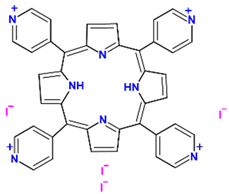
PDT presents multiple benefits over traditional antimicrobial therapies, positioning it as a promising strategy against MDR. A key advantage lies in its distinct mode of action, which involves ROS generation upon light activation. In contrast to antibiotics that act on specific bacterial structures or metabolic pathways, PDT-induced oxidative stress disrupts various cellular components, such as lipids, proteins, and genetic material. This non-specific mode of action drastically reduces the likelihood of resistance development, addressing a major limitation of conventional antibiotics [72]. PDT exhibits rapid bactericidal activity, often achieving significant bacterial reduction within minutes of light exposure. In contrast, antibiotics require prolonged treatment durations, increasing the risk of bacterial adaptation and resistance. This rapid effect is particularly beneficial in critical infections where immediate bacterial eradication is necessary to prevent complications. Reference [73] investigated the synergistic antibacterial effects of antimicrobial photodynamic inactivation (aPDI) using the porphyrin photosensitizer (TMPyP) in combination with silver nanoparticles (AgNPs) against methicillin-resistant Staphylococcus aureus (MRSA) and extended-spectrum beta-lactamase (ESBL)-producing Klebsiella pneumoniae (ESBL-KP). The study determines the optimal conditions for achieving a synergistic antimicrobial effect using light-activation followed by AgNP treatment. The experimental design included pre-incubating bacteria with TMPyP for either 45 min or 5 h, followed by exposure to an LED light source at total fluences of 10 or 20 J/cm2. After light activation, bacterial cultures were introduced into a growth medium containing AgNPs. The study identified the optimal synergy conditions as 1.56–25 μM TMPyP with 3.38 mg/L AgNPs for MRSA and 1.56–50 μM TMPyP with 3.38 mg/L AgNPs for ESBL-KP using a 45 min incubation and 10 J/cm2 fluence. Extended incubation periods and increased irradiation levels lowered the TMPyP concentration required to achieve a synergistic effect. Another crucial advantage of PDT is its broad-spectrum activity. Since ROS generation affects essential cellular components indiscriminately, PDT demonstrates efficacy against diverse bacterial species, encompassing both Gram-positive and Gram-negative bacteria. PDT is also highly effective against bacterial biofilms, which are hard to treat with conventional antibiotics. Biofilms provide a protective extracellular matrix that limits antibiotic penetration and promotes bacterial survival in a dormant state [74]. Antibiotics often fail against biofilms because they primarily target metabolically active bacteria, leaving persister cells intact. PDT, however, generates ROS that degrades the biofilm matrix, increasing bacterial susceptibility and leading to biofilm disruption [75]. PDT can eradicate biofilms more effectively than antibiotics, making it a potential solution for chronic infections such as wound infections, implant-associated infections, and catheter-related bloodstream infections. Another advantage of PDT is its ability to minimize collateral damage to the host microbiota [76]. Reference [77] investigated the efficacy of curcumin-mediated antimicrobial photodynamic therapy (aPDT) in disrupting vancomycin-resistant Staphylococcus aureus (VRSA) biofilms and its role in enhancing phagocytosis by neutrophils in human blood. The study used sub-MIC (minimum inhibitory concentration) of curcumin (2500 µg/mL) followed by blue laser irradiation (20 J/cm2 for 52 s) to evaluate biofilm disruption and bacterial viability. The results showed that curcumin-aPDT led to an 80.83% reduction in biofilm formation, significantly higher than the 28.3% reduction seen with curcumin alone. Congo red assays demonstrated a 39.3% decrease in exopolysaccharide production, confirming its biofilm-disrupting potential. Furthermore, confocal laser scanning microscopy and scanning electron microscopy (SEM) revealed that aPDT-treated biofilms exhibited extensive cell damage with bacterial lysis and structural disintegration. Additionally, singlet oxygen quantification confirmed that curcumin-aPDT primarily operates via a Type II photodynamic mechanism, producing high levels of ROS that contribute to bacterial inactivation. The study also found that when aPDT-treated VRSA biofilms were exposed to whole blood, phagocytosis was significantly enhanced, leading to near-total bacterial clearance. PDT, on the other hand, can be precisely targeted to infected sites, reducing unintended harm to commensal bacteria. This localized action makes PDT an attractive option for infections where microbiome preservation is crucial, such as in gastrointestinal and respiratory tract infections.
Like any treatment, PDT has its challenges. One major limitation is that its effects are limited to the area exposed to light, making it complicated to treat widespread metastatic cancers with current technology. Since PDT relies on oxygen to work, tumors with dense tissue or surrounded by dead cells may not respond well due to low oxygen levels. Another key challenge is ensuring precise light delivery to the target area [78]. Deep-seated tumors, which are harder to reach without surgery, are especially tricky to treat because visible light does not penetrate very far into the body.

5. Natural Sensitizers over Commercial Sensitizers in PDT

With growing concerns over the side effects and limitations of synthetic photosensitizers, natural alternatives have gained significant interest in PDT due to their superior biocompatibility, lower toxicity, and inherent biological activities. Unlike commercial photosensitizers, which often have drawbacks such as prolonged retention in the body, unwanted phototoxicity, and complex synthesis, natural compounds offer a safer and more sustainable alternative. A number of these, including flavonoids, anthraquinones, curcumins, and perylenequinones, have demonstrated quite strong light absorption and efficient ROS generation, making them effective for PDT. Additionally, their inherent biological activities, such as anti-inflammatory and pro-apoptotic properties, can enhance their therapeutic potential. Anthraquinones like emodin and rhein have been extensively studied for their ability to both act as photosensitizers and explicitly inhibit cancer growth. They absorb light in the visible range and have shown promising results in triggering apoptosis in cancer cells by disrupting mitochondrial function. Reference [79] investigated the anticancer effects of emodin-mediated PDT in cervical carcinoma cell lines (SiHa, CaSki, and HaCaT). The study evaluates cell viability, ROS production, apoptosis, and autophagy induction following PDT treatment with emodin. Emodin is a natural anthraquinone compound with known antineoplastic and pro-apoptotic properties. It demonstrated that emodin exhibited concentration- and time-dependent cytotoxicity, with cancer cell viability remaining above 80% at concentrations lower than 30 µM. Fluorescence microscopy confirmed efficient cellular uptake of emodin in all the tested cell lines. After light activation, emodin-PDT significantly reduced cell viability, correlating with an increase in ROS production, caspase-3 activity, and fluorescence intensity of autophagic vacuoles, indicating that cell death occurred through both apoptosis and autophagy. Flavonoids such as quercetin and genistein have also been explored, showing the ability to increase ROS production and enhance the effects of PDT by promoting apoptosis. Genistein, for example, has been found to boost the cytotoxic effects of PDT by activating caspases and causing mitochondrial damage, making it a valuable candidate for combination therapies. Reference [80] investigated the synergistic effect of PDT combined with genistein in enhancing apoptosis in human anaplastic thyroid cancer (SNU 80) cells. The study determines whether the combination of genistein, a soy-derived isoflavone, with photofrin-mediated PDT may improve treatment efficacy compared to either therapy alone. The experimental approach involved treating SNU 80 cells with genistein and varying concentrations of photofrin, followed by PDT irradiation. Cell viability was assessed using MTT assays, and morphological changes were observed via Hoechst 33342 and propidium iodide staining. Additionally, the study evaluated mitochondrial membrane depolarization, ROS generation, and apoptotic protein expression using confocal microscopy and Western blot analysis. The results showed that the combination of genistein and PDT significantly enhanced apoptosis, as evidenced by increased ROS production and mitochondrial membrane depolarization. The Western blot analysis confirmed the upregulation of key apoptotic markers, including caspase-3, caspase-9, caspase-8, caspase-12, and cytochrome c, indicating the activation of both intrinsic and extrinsic apoptotic pathways. The study concluded that while PDT and genistein independently induce apoptosis in thyroid cancer cells, their combination leads to a significantly stronger effect, suggesting that genistein enhances PDT-mediated oxidative stress and apoptotic signaling in anaplastic thyroid cancer cells. Curcumin, a commonly known natural compound, has also been studied for its potential as a photosensitizer [80]. It absorbs blue light and produces singlet oxygen upon irradiation, contributing to effective PDT outcomes. Beyond its photodynamic activity, curcumin naturally inhibits key cancer-related pathways such as NF-κB and STAT3, which adds to its therapeutic effects. However, its quite poor solubility and relatively rapid breakdown in the body have posed challenges for clinical applications, leading to explore modifications such as nanoformulations to improve its stability and delivery. Reference [81] evaluated the efficacy of aPDT using curcumin against methicillin-sensitive Staphylococcus aureus (MSSA), MRSA, and an ATCC S. aureus strain. The study assesses bacterial viability, growth inhibition, ROS production, and adhesion loss following curcumin-mediated aPDT. The experimental setup involved incubating bacterial cultures with curcumin for 20 min, followed by irradiation with an LED light source. Bacterial growth was quantified by colony-forming unit (CFU) counts, while confocal microscopy was used to assess bacterial adhesion, and ROS production was measured using fluorimetry. The results demonstrated that aPDT treatment significantly increased ROS levels, leading to a 4-log reduction in bacterial growth. Additionally, the confocal microscopy analysis confirmed that bacterial adhesion was significantly disrupted in the aPDT-treated groups, suggesting that curcumin-mediated ROS generation compromised cell membrane integrity. Reference [82] investigated the effectiveness of aPDT using curcumin against MRSA) biofilms. The study evaluates biofilm reduction, bacterial viability, ROS production, and curcumin internalization in MRSA cells following PDT treatment. MRSA biofilms were induced and incubated with curcumin for 20 min, followed by irradiation with a 450 nm LED light source (110 mW/cm2, 50 J/cm2, for 455 s). The impact of PDT was assessed using CFU counting, confocal microscopy, SEM, the resazurin metabolic activity assay, and ROS quantification. The results indicated that PDT with curcumin significantly reduced MRSA biofilm growth, with the confocal microscopy confirming curcumin internalization within bacterial cells. SEM images demonstrated biofilm structural disruption, while the ROS quantification revealed increased oxidative stress, leading to bacterial inactivation. Importantly, curcumin alone and the applied irradiation parameters showed no cytotoxicity in the absence of PDT activation.
Perylenequinones, including hypericin and hypocrellins, are among the most promising natural photosensitizers. Hypericin has been extensively studied for its ability to generate singlet oxygen and induce apoptosis in cancer cells. Reference [83] evaluated the effectiveness of aPDT using hypericin against biofilms of Propionibacterium acnes. The study assesses biofilm reduction following different concentrations of hypericin and varying laser irradiation doses. Biofilms were cultivated under anaerobic conditions for 48 h, after which they were treated with hypericin at 5 µg/mL or 15 µg/mL and exposed to low-level laser irradiation (660 nm) at 3 J or 5 J fluences. Bacterial viability was assessed by CFU counting, while structural biofilm changes were analyzed using microscopy techniques. The results demonstrated that aPDT significantly reduced P. acnes biofilms, with the highest biofilm reduction (27.9%) observed when using 15 µg/mL hypericin combined with 5 J laser irradiation. Lower concentrations and energy doses showed moderate antimicrobial effects, with reductions of 9.9–14.1%. The study also confirmed that hypericin-mediated PDT functions by generating ROS, leading to bacterial inactivation. Reference [84] examined the effectiveness of PDT using hypericin and pheophorbide a (Pa) against MRSA infections in both in vitro and in vivo models. The study compares the antibacterial effects of hypericin-PDT and Pa-PDT with clinically approved methylene blue (MB)-mediated PDT, while also assessing wound healing and immune system interactions in a murine MRSA wound infection model. In in vitro studies, hypericin-PDT exhibited the lowest minimum bactericidal concentration (MBC) values (0.625–10 µM) against various MRSA strains, including ATCC RN4220/pUL5054 and community-associated MRSA (CA-MRSA). The study confirmed that hypericin-PDT had superior bactericidal effects compared to Pa-PDT and MB-PDT, with significant (above 1 log10 CFU) reductions in MRSA viability following treatment. In the in vivo murine wound model, topical application of hypericin-PDT led to enhanced wound healing via re-epithelialization, as observed through histological analysis. Moreover, hypericin displayed significantly lower dark toxicity (p < 0.05) on neutrophils compared to Pa or MB, suggesting improved biocompatibility. However, one of the most exciting candidates in this class is hypocrellin because it operates through both Type I and Type II photodynamic mechanisms, making it highly effective even in hypoxic environments where oxygen-dependent photosensitizers struggle. It also clears from the body faster than some commercial photosensitizers, reducing the risk of prolonged skin sensitivity after treatment.

6. Hypocrellin as a Natural Photosensitizer for PDT

Hypocrellins belong to the perylenequinone family of photosensitizers, characterized by a highly conjugated aromatic structure that absorbs visible light efficiently. They are naturally derived secondary metabolites extracted primarily from Hypocrella bambusae and Shiraia bambusicola fungi [85]. The core chemical framework consists of a perylenequinonoid chromophore, which is responsible for its quite strong absorption and photodynamic activity. The four primary members of this class (Figure 2) are hypocrellin A (Hyp-A), hypocrellin B (Hyp-B), hypocrellin C (Hyp-C), and hypocrellin D (Hyp-D). Their structural distinction lies in the functional groups attached to the perylene core. Hyp-A contains hydroxyl (-OH) and methoxy (-OCH3) groups, which contribute to strong hydrogen bonding interactions and enhanced solubility in organic solvents. Hyp-B is formed via the acid/base-mediated dehydration of Hyp-A, resulting in a loss of a hydroxyl group, which modifies its photophysical properties, such as wavelength absorption and ROS yield. Hyp-C and Hyp-D are less extensively studied but structurally related to Hyp-A and Hyp-B [86]. These derivatives feature modifications in oxygenation and methylation patterns, leading to variations in absorption properties, singlet oxygen yield, and photodynamic efficiency. All hypocrellins possess redox-active centers, enabling efficient electron transfer reactions under light exposure, which is essential for their application in PDT. The conjugated system of alternating double bonds and carbonyl groups facilitates strong light absorption, a key requirement for photosensitization [87]. Synthetic modifications of hypocrellins have been explored to fine-tune their photophysical and photochemical properties, particularly by introducing electron-donating or -withdrawing substituents to shift the absorption maximum toward the red/NIR region (around 650–700 nm). These modifications enhance tissue penetration and improve therapeutic efficacy, making them highly relevant for antimicrobial PDT applications [88].

6.1. Photophysical and Photochemical Properties

The photophysical properties of hypocrellins define their photosensitizing efficiency, making them effective agents for photodynamic therapy (Table 2). The key parameters influencing their performance include absorption spectrum, fluorescence quantum yield, and singlet oxygen quantum yield. Hypocrellins exhibit relatively strong absorption in the visible region, with peak absorption between 450 and 550 nm. Their absorption spectrum is dictated by their π-conjugated system, allowing for efficient photon uptake [89]. Hyp-A typically shows a maximum absorption of around 400–520 nm, while Hyp-B is somewhat red-shifted, with absorption peaks around 600–900 nm due to structural dehydration [90,91]. These compounds also exhibit shoulder peaks between 510 and 540 nm, influenced by solvent polarity and aggregation state. Hypocrellins exhibit a higher molar extinction coefficient, allowing efficient photon harvesting even at quite low concentrations. The spectral characteristics enable effective activation by blue (470 nm), green (532 nm), or yellow (580 nm) light sources, which are clinically accessible for antimicrobial PDT. Additionally, the solvent environment and pH significantly influence the spectral characteristics of hypocrellins. In polar solvents or at acidic pH, Hyp-A and Hyp-B may form aggregates through π–π stacking and hydrogen bonding interactions, leading to hypochromic shifts and reduced ROS generation due to self-quenching [92]. Conversely, in organic or amphiphilic environments (such as micelles or liposomes), they exist predominantly as monomers, maintaining relatively high photoactivity. Their photostability under therapeutic light exposure is considered moderate; however, repeated irradiation can lead to photobleaching, especially in oxygen-rich systems. This degradation affects long-term efficacy and highlights the importance of controlled light dosing during therapy [93]. Despite this, hypocrellins demonstrate better photobleaching resistance than hypericin and exhibit minimal dark toxicity, making them favorable candidates for repeated PDT sessions. Their ability to maintain high singlet oxygen production in both aqueous and lipid-rich biological environments strengthens their utility in treating biofilm-associated or membrane-dense infections. The reported fluorescence lifetimes of Hyp-A and Hyp-B are 1.07 and 1.03 ns depending on the solvent polarity and aggregation state [94]. However, compared to porphyrins and bacteriochlorins, the absorption of native hypocrellins is somewhat blue-shifted, limiting their penetration in biological tissues [95]. To overcome this, structural modifications, such as amination and sulfonation, have been used to red-shift their absorption toward the NIR region for deeper tissue penetration [96]. Hypocrellins exhibit moderate fluorescence quantum yields, which enables their dual functionality as theranostic agents, useful for both imaging and therapy. The fluorescence properties are influenced by substituent effects, with electron-withdrawing groups tending to quench fluorescence while enhancing ISC, which is crucial for PDT efficiency. The ability of a photosensitizer to generate singlet oxygen determines its PDT efficacy. Hypocrellins demonstrate high singlet oxygen quantum yields (about 0.7–0.8), comparable to photofrin and chlorin e6, two commonly recognized PDT agents [97]. This high yield is attributed to their efficient ISC and strong enough interaction with molecular oxygen in solution. Hypocrellins possess triplet states (of about 4–6 µs), which enhances their ability to transfer energy to molecular oxygen, facilitating singlet oxygen formation. This property is beneficial for PDT as it increases the probability of ROS generation before non-radiative decay occurs [98]. The singlet oxygen quantum yield of hypocrellin highlights its potent oxidative stress in normoxic tissues. This straightforwardly correlates with membrane lipid peroxidation, protein denaturation, and nucleic acid damage. In contrast, hypocrellin efficient ISC and long-lived triplet state enable radical generation under oxygen-limited environment conditions typically encountered in bacterial biofilms or necrotic tissues. Thus, the photophysical traits of hypocrellin translate right into ROS-mediated antimicrobial and antibiofilm efficacy, confirming its adaptability as a photosensitizer.

6.2. Photodynamic Activity of Hypocrellin

In Type I reactions, hypocrellin participates in electron transfer interactions with nearby biomolecules, including lipids and proteins, resulting in the generation of superoxide anions (O2•) and hydroxyl radicals (•OH). These radicals contribute to oxidative damage, particularly affecting the lipid bilayer, intracellular proteins, and nucleic acids, resulting in cell death. Conversely, in Type II reactions, hypocrellin transfers its energy straight to molecular oxygen, producing singlet oxygen, which is a highly cytotoxic ROS. This singlet oxygen reacts with unsaturated lipids and membrane-bound proteins, disrupting cellular integrity and leading to apoptosis or necrosis. Reference [99] investigated the photodynamic antimicrobial effects of Hyp-A against both Gram-positive (Staphylococcus aureus and Bacillus subtilis) and Gram-negative (Escherichia coli and Salmonella typhimurium) bacterial strains. The study assessed the efficacy of Hyp-A-PDT under different light exposure times and oxygen conditions, alongside the influence of divalent metal ions (Ca2+ and Mg2+) on bacterial susceptibility. The results demonstrated that Hyp-A-mediated PDT led to a 99.98% reduction in S. aureus viability after 120 min of irradiation, indicating a high photodynamic efficiency against Gram-positive bacteria. However, Gram-negative bacteria exhibited higher resistance to PDT, attributed to the outer membrane barrier limiting ROS penetration. The addition of Ca2+ and Mg2+ ions significantly enhanced HA-PDT efficacy against Gram-negative bacteria, most probably by increasing membrane permeability and facilitating photosensitizer uptake. Mechanistic analyses confirmed that Hyp-A induces both Type I reactive species (superoxide and hydroxyl radicals) and Type II singlet oxygen, leading to bacterial cell damage (Figure 3). Oxygen availability was found to play a crucial role in Hyp-A-PDT efficacy, as bacteria treated in low-oxygen conditions showed higher survival rates. Furthermore, ROS scavenger experiments validated the involvement of both photodynamic reaction pathways. Reference [108] developed glutathione (GSH)-responsive photosensitizer (Hyp-B-NBD (7-nitro-2,1,3-benzoxadiazole)) based on Hyp-B to enhance tumor selectivity and PDT efficacy [108]. While Hyp-B possesses a relatively high molar extinction coefficient, quite strong phototoxicity, and quite rapid metabolism, its lack of tumor specificity limits its clinical applications. To address this, NBD was covalently linked to o Hyp-B, creating a prodrug-like system that remains inactive until triggered by GSH-designed Hyp-B-NBD system. NBD acts as both a fluorescence quencher and a GSH-responsive activation group, suppressing singlet oxygen generation through a photoinduced electron transfer (PET) process. As a result, Hyp-B-NBD exhibits negligible fluorescence and phototoxicity in its native state. However, in the presence of intracellular GSH, particularly in cancer cells, Hyp-B-NBD undergoes a reaction that releases free Hyp-B, restoring its fluorescence and ROS-producing capabilities. This selective activation strategy ensures that PDT is only triggered within tumor microenvironments, minimizing off-target effects and enhancing therapeutic specificity. Reference [107] evaluated the antimicrobial and antileishmanial activities of Hyp-A and Hyp-B against fungi, bacteria, and Leishmania donovani. The study determines the therapeutic potential of hypocrellins beyond their known photodynamic effects. The results demonstrated that Hyp-A exhibited significant antifungal activity against Candida albicans, with an IC50 of 0.65 µg/mL and a minimum fungicidal concentration (MFC) of 1.41 µg/mL. However, Hyp-B displayed only weak activity against C. albicans, and neither compound was active against Cryptococcus neoformans. Hyp-A also showed moderate antibacterial effects against Staphylococcus aureus, MRSA, Pseudomonas aeruginosa, and Mycobacterium intracellulare, with IC50 values ranging from 3 to 10 µg/mL, though it did not exhibit bactericidal activity. In contrast, Hyp-B showed little to no antibacterial effects. Regarding antileishmanial activity, Hyp-A was found to be three-fold more potent than amphotericin B and six-fold more potent than pentamidine, with an IC50 of 0.27 µg/mL against L. donovani. Hyp-B exhibited only moderate activity, with an IC50 of 12.7 µg/mL.

7. Comparative Analysis of Hypocrellin with Other Sensitizers

7.1. Biofilm Penetration and Antimicrobial Effectiveness

Bacterial biofilms, composed of extracellular polymeric substances (EPSs), present a significant barrier to antimicrobial agents, including photosensitizers. Effective PDT agents must not only penetrate these biofilms but also generate sufficient ROS to disrupt bacterial cells [109]. Hypocrellin-based PDT demonstrates superior biofilm penetration compared to traditional photosensitizers such as porphyrins, phenothiazinium dyes, and phthalocyanines due to its unique physicochemical properties. The penetration capability of a photosensitizer is influenced by its lipophilicity, molecular size, and charge [110]. Hypocrellin derivatives, particularly Hyp-A and Hyp-B, exhibit an optimal log p-value (partition coefficient) of 3.2–4, indicating balanced hydrophilicity and lipophilicity [111]. This property facilitates diffusion through the hydrophobic regions of biofilms, whereas hydrophilic photosensitizers like methylene blue (log p ≈ 0.1–0.9) exhibit limited penetration and remain mostly surface-bound [47]. The antimicrobial action of PDT is largely dictated by the singlet oxygen quantum yield, which determines the efficiency of ROS production upon light activation. Hypocrellin exhibits a singlet oxygen quantum yield approximately of 0.75 [112], significantly higher than hematoporphyrin derivatives (singlet oxygen quantum yield approximately of 0.64) [113] and toluidine blue O (singlet oxygen quantum yield approximately of 0.49) [114], enabling stronger oxidative damage to bacterial membranes, proteins, and DNA. One of the major limitations of synthetic photosensitizers in PDT is their potential cytotoxicity and adverse effects on host tissues. Hypocrellin-based compounds exhibit lower toxicity and higher selectivity for bacterial cells due to their preferential uptake and ROS-mediated targeting. The selectivity of a photosensitizer is often quantified using the IC50 value (the concentration required to reduce cell viability by 50%). Hypocrellin exhibits an IC50 > 20 µM for mammalian fibroblast cells [115], whereas commonly used photosensitizers such as phenothiazinium dyes (e.g., toluidine blue O) demonstrate IC50 values as low as 10 µM [116], indicating greater potential for cytotoxic side effects. This suggests that hypocrellin-based therapies can effectively eradicate bacterial infections while minimizing damage to host tissues.

7.2. Pharmacokinetics and Systemic Safety

The systemic safety of a photosensitizer is a critical factor in determining its suitability for clinical applications. Key pharmacokinetic parameters such as absorption, distribution, metabolism, and excretion (ADME) play a vital role in minimizing systemic toxicity and ensuring therapeutic effectiveness [117].

Plasma Half-Life and Clearance Rate

One of the major concerns with photosensitizers is prolonged circulation in the bloodstream, which may lead to extended photosensitivity, increasing the risk of phototoxic reactions [118]. Hypocrellin has a plasma half-life (t1/2) of approximately 2.319 ± 0.462 h [119], which is significantly shorter than hematoporphyrin derivatives (t1/2 > 5 h) [89]. This rapid clearance reduces the risk of prolonged photosensitivity, allowing patients to resume normal activities sooner after treatment without the need for extended light protection measures. Additionally, pharmacokinetic studies indicate that hypocrellin follows a biphasic elimination profile, with an initial relatively rapid distribution phase followed by quite a slower elimination phase [120].

8. Nanoformulations of Hypocrellin for Enhanced PDT

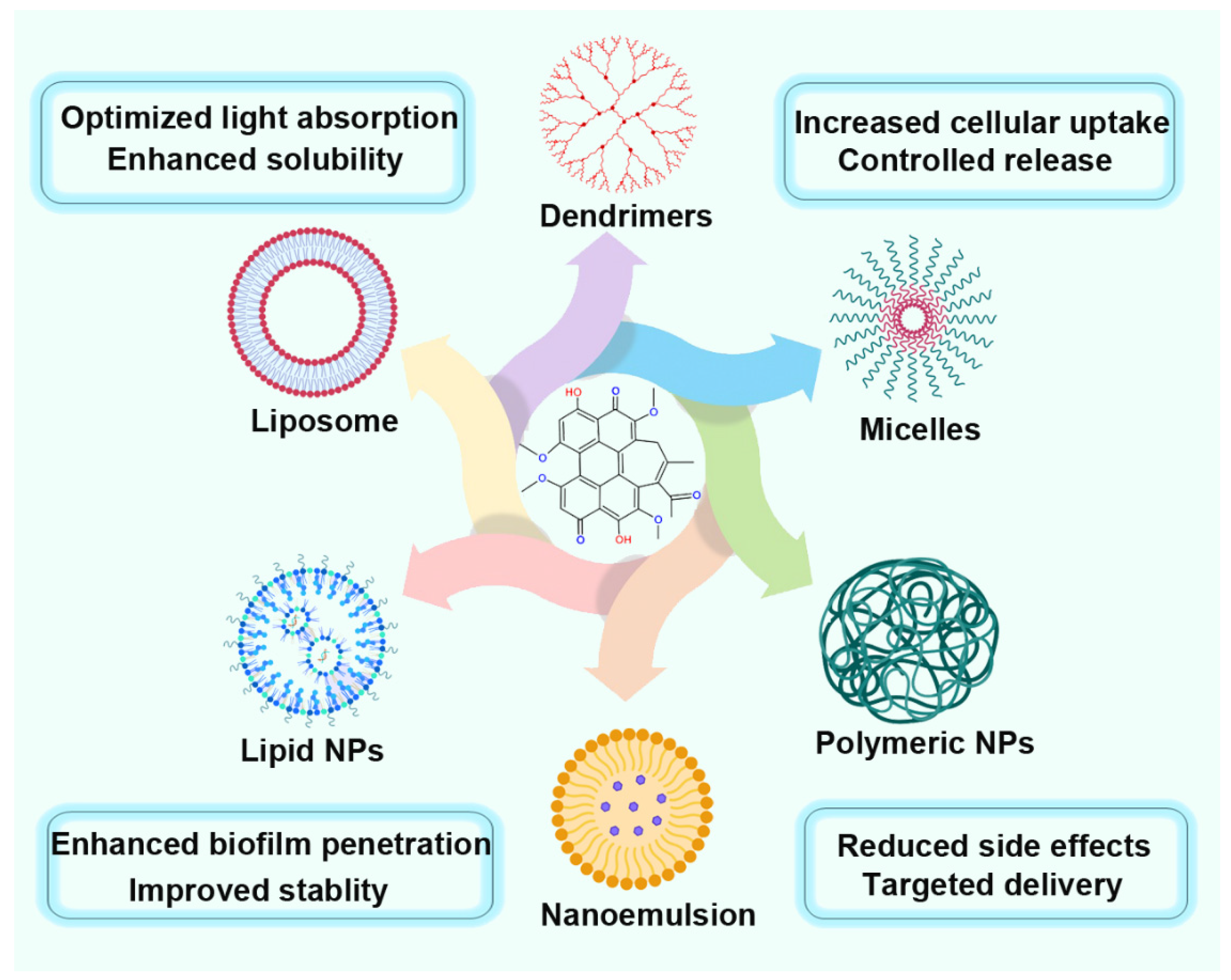
The effectiveness of various phytochemical-based drugs is often compromised due to their high enough molecular weight, relatively poor lipid membrane permeability, and quite low bioavailability, which limits their therapeutic efficacy. On the other hand, a significant proportion of newly developed pharmaceutical compounds exhibit relatively low water solubility, leading to slow absorption rates and ineffective drug delivery. A major challenge in conventional drug formulation is the complication in optimizing drug solubility, stability, and targeted release while ensuring efficient absorption at the intended site of action [121]. To overcome these challenges, nanotechnology-driven drug delivery systems have been extensively investigated in recent years [122], providing notable benefits such as increased drug solubility, prolonged circulation time, enhanced bioavailability, and targeted drug delivery [123]. Nanoformulations (Figure 4) leverage considerably high surface-area-to-volume ratio and quantum confinement effects to improve drug retention, controlled release, and therapeutic efficiency [124]. In these systems, drugs may be encapsulated, dissolved, or conjugated with nanocarriers, typically ranging between 10 and 100 nm in size, to facilitate precise transport from the site of administration to the site of action. Furthermore, nanocarriers protect drug molecules from biochemical degradation, enzymatic inactivation, and pH fluctuations, ensuring that the therapeutic agent reaches its target in an active form with minimal systemic toxicity. Several nanocarrier platforms have been successfully implemented for PS delivery in PDT. Among them, liposomes, polymeric NPs, metallic NPs, nanoemulsions, and micelles (Table 3) are commonly used due to their ability to enhance cellular uptake, improve ROS generation efficiency, and minimize off-target effects.
Nano-based drug delivery systems can employ either passive targeting, where nanocarriers accumulate at the disease site due to physicochemical properties like size and surface charge, or active targeting, which involves surface modifications enabling specific interactions with overexpressed receptors on target cells. Hypocrellin, despite its promising photodynamic properties, faces limitations such as quite poor water solubility, considerably low bioavailability, and short retention time in target tissues. To overcome these challenges, nanoformulation strategies have been employed to improve its solubility, enhance stability, and facilitate targeted delivery for PDT applications [125].
Table 3. Various nanoformulations of hypocrellins in PDT.
Nano CarrierNanoformulationHypocrellin TypeApplicationKey Observations Ref.
Mixed polymer NPsTransferrin-modified Poly(D, L-Lactide-co-glycolide) and corboxymethyl chitosan NPsAPDT and targeted therapyStrong ROS, significant photo-cytotoxicity, and
63% tumor inhibition rate
[126]
Copolymeric NPsNano silver-loaded Poly(lactide-co-glycolide)-
d-α-tocopheryl polyethylene glycol 1000 succinate NPs
BPDTHigher encapsulation efficiency 84.06 ± 11.43%, ROS production 90.62 ± 20.12, significant antiangiogenic effect 89.9%, and superior phototoxic effect 85.5% [127]
Polymer NPsPoly(D,L-loactic-co-glycolic NPsAPDT and cancer therapyEnhanced photostability, reduced dark cytotoxicity, exceptional antitumor property, and ROS production ability[128]
Polymer NPsHyp-B and Paclitaxeel-encapsulated hyaluronic acid–ceramide NPsBPDT and chemotherapyHigher phototoxicity and encapsulation efficiency of 70%, and exceptional antitumor properties[129]
Composite NPs (polymer and metal
NPs)
Poly(D,L-loactic-co-glycolic NPs incorporated nano
silver
BPDTEnhanced singlet oxygen production, higher ROS production 138.02 ± 13.23, superior phototoxic effect 82.2%, and significant anti-angiogenic effect[130]
Polymer NPsPoly(D,L-loactic-co-glycolic Nps ADrug delivery, PDTpH-dependent delivery, higher solubility, superior stability, and enhanced bioavailability[131]
Polymer NPsNeutrophil membrane coated Poly(D,L-loactic-co-glycolic NPs BNIR fluorescence imaging, targeted therapy, and PDTInhibit the expression of JUNB and promote ROS production, exceptional antitumor efficacy, and better anti-inflammation effect [132]
Polymer NPsTransferrin-modified
HepG2 cell membrane-coated hypocrellin bionic NPs
BTargeted therapy and fluorescence imagingLong-term stability, exceptional biocompatibility, lower toxicity, and higher ROS production[133]
CompositeHypocrellin–cisplatin-
intercalated hectorite nanoformulation
AChemo therapy and PDTExceptional biocompatibility, high payload, controlled release, and higher photostability and photobleachability [134]
Mixed
polymer
Self-assembled 1,2-diamino-2-methyl-
propane and PEG-PLGA nanovesicles
BPDT, PTT, fluorescence imaging, and photoacoustic imagingExceptional photothermal stability, higher singlet oxygen production, and photothermal conversion efficiency. [135]
Nanofiber Poly(L-lactic acid)–silk
fibroin nanofiber
AChemotherapy and drug deliveryExceptional pH stability, strong inhibitory effects, and controlled drug release[136]
MicellesFolate-conjugated poly(ethylene glycol)-poly (lactic acid) micelleBTargeted therapy and PDTHigh drug loading capacity, quite good biocompatibility, controlled release, and enhanced targeting and antitumor effect[137]
Nanorods Dopamine-modified
hypocrellin derivative-loaded calcium phosphate nanorods
BFluorescence imaging and
PDT
Lower cytotoxicity, good enough biocompatibility, efficient singlet oxygen production, and enhanced antitumor activity[138]

8.1. Liposomal Hypocrellin

Liposomes are commonly recognized as efficient drug delivery systems due to their biocompatibility, controlled drug release, and ability to enhance the solubility of hydrophobic molecules. The liposemes are composed of lipid bilayers, which can encapsulate both hydrophilic and hydrophobic drugs [57], making them a promising carrier for photosensitizers like hypocrellin. Hypocrellin-loaded liposomes have demonstrated high stability, enhanced circulation time, and increased cellular uptake. These nanoformulations protect hypocrellin from premature degradation, allowing for a controlled release profile and ensuring optimal ROS generation upon light activation. Notably, liposomal hypocrellin formulations retain nearly 70% of their PDT activity, indicating minimal loss of photosensitizing efficiency. Another key advantage of liposome-based hypocrellin is its ability to reduce off-target toxicity and improve selectivity. By taking advantage of the enhanced permeability and retention (EPR) effect, liposomes facilitate preferential accumulation in infected or tumor tissues, making them an effective tool for targeted antimicrobial and anticancer PDT. Furthermore, the liposomal encapsulation of hypocrellin enhances its photochemical reactivity, leading to the production of both singlet oxygen and hydroxyl radicals. This dual mechanism contributes to potent antimicrobial and anticancer effects, making it superior to free hypocrellin in PDT applications. Reference [119] developed a liposomal Hyp-B (LHB) formulation and evaluated its pharmacokinetics, photodynamic efficacy, and safety for potential application in age-related macular degeneration PDT. The LHB formulation, prepared using high-pressure homogenization, exhibited high encapsulation efficiency, enhanced solubility, and prolonged circulation time, addressing the limitations of free Hyp-B. Pharmacokinetic studies showed that LHB had a half-life of 2.319 ± 0.462 h and was fully metabolized within 24 h, reducing the risk of prolonged photosensitivity. In a rat model of choroidal neovascularization (CNV), LHB-PDT at 1 mg/kg under yellow light irradiation led to significant CNV occlusion with minimal retinal damage, outperforming verteporfin, a standard PDT agent, in both efficacy and tissue selectivity. Additionally, LHB caused minimal skin phototoxicity, with only 2 out of 200 mice experiencing mild inflammatory responses at a high dose (4 mg/kg).

8.2. Polymeric Nanoparticles

Polymeric NPs made from biocompatible and biodegradable polymers such as poly (lactic-co-glycolic acid) (PLGA) and polyethylene glycol (PEG) enable sustained and controlled release, thereby maximizing therapeutic efficacy [139]. PEGylated PLGA NPs provide several advantages, including increased circulation time by preventing rapid clearance, enhanced solubility of hypocrellin, improving its bioavailability and controlled and sustained drug release, and minimizing systemic toxicity. The encapsulation of hypocrellin in PEG-PLGA NPs follows an emulsion-solvent evaporation method, which helps retain the PS within a protective matrix, thereby preventing aggregation and photodegradation. This encapsulation significantly enhances ROS generation efficiency upon light activation, making it more effective for antimicrobial and anticancer applications. Polymeric NPs can be surface-functionalized with targeting ligands to improve specific cellular uptake. This approach reduces off-target effects while ensuring efficient hypocrellin accumulation in infected or tumor tissues. The nanoscale size also enables passive accumulation through the EPR effect, improving overall PDT effectiveness. Reference [140] developed a polymeric micelle-based delivery system for Hyp-A to enhance its solubility, stability, and aPDT efficacy. Hyp-A suffers from relatively poor water solubility and aggregation in physiological conditions, which limits its ROS production and antimicrobial potential. To this end, Hyp-A was encapsulated into lipase-sensitive methoxy poly(ethylene glycol)-block-poly(ε-caprolactone) (mPEG-PCL) micelles, which degrade upon contact with bacterial lipase. The in vitro results demonstrated that MIC and MBC of HA-loaded micelles were significantly lower after light irradiation (0.69 mg/L and 1.38 mg/L, respectively) compared to free Hyp-A. Without light, the MIC and MBC values were significantly higher, confirming the photoactivated antimicrobial effect. Fluorescence staining further revealed that ROS production was responsible for bacterial cell death. In the in vivo acute peritonitis model, mice treated with Hyp-A-loaded micelles showed an 86% survival rate, significantly higher than those treated with free Hyp-A or blank micelles. The system effectively eradicated MRSA infections in vivo, demonstrating that nanoformulated Hyp-A improved bioavailability, targeted bacterial degradation, and PDT efficacy while maintaining quite low hemolytic activity and biocompatibility. Dendrimers are highly branched, tree-like polymeric macromolecules with a well-defined core and multiple functional terminal groups, making them versatile nanocarriers for drug delivery [141], including hypocrellin-based PDT applications. These nanostructures facilitate quite high drug-loading efficiency, precise molecular architecture, and tunable surface chemistry, which to be engineered for enhanced solubility, stability, and the targeted delivery of hypocrellin-based photosensitizers. The unique structural design of dendrimers allows them to encapsulate hypocrellin within their core or conjugate it onto their periphery via covalent interactions, improving photostability and bioavailability. Additionally, dendrimer-based hypocrellin delivery systems may be functionalized with targeting ligands such as folic acid or peptides, enabling selective accumulation in bacterial biofilms or infected tissues, thereby minimizing off-target effects. Furthermore, their high degree of surface modification flexibility allows for optimized ROS production and controlled release in response to physiological stimuli, which is crucial for maximizing PDT efficacy while reducing dark toxicity.

8.3. Inorganic Nanoparticles for Hypocrellin-Based PDT

Inorganic NPs, including silver, gold, and mesoporous silica NPs, have gained attention for enhancing the photodynamic properties of sensitizers [142]. These NPs improve solubility, stability, and photochemical efficiency by enhancing light absorption, promoting ROS generation, and enabling targeted drug delivery. Inorganic NPs also bind with photosensitizers by means of various linkers, as shown in Figure 5. Gold NPs have been used as photothermal enhancers, working synergistically with PDT to improve hypocrellin’s antibacterial and anticancer effects [142]. Lipid-coated gold nanocages loaded with hypocrellin exhibited high two-photon absorption efficiency, making them highly effective for deep-tissue PDT applications. Additionally, hierarchical gold/copolymer nanostructures have been developed to improve drug encapsulation and extend retention time in target tissues. Reference [140] developed hierarchical gold/copolymer nanostructures as water-dispersible nanotanks for hydrophobic drug encapsulation, with a focus on Hyp-B for PDT. The nanocomposites were synthesized using gold NPs and poly(dimethylsiloxane)-poly(ethylene glycol) (PDMS-PEG) block copolymers, fabricated via a hydrosilylation reaction to achieve a core–shell structure with amphiphilic properties. The incorporation of long PEG chains allowed these nanostructures to remain well-dispersed in aqueous environments, while hydrophobic interactions between PDMS segments and HB facilitated drug encapsulation. HB-loaded nanocomposites demonstrated significant anticancer activity upon light activation, confirming their potential for PDT-based cancer therapy. Notably, while the gold/copolymer nanocomposites without HB exhibited slight cytotoxicity, the encapsulated drug was found to significantly enhance photodynamic efficacy. The study concluded that these hierarchically structured core–shell nanocomposites offer a versatile and efficient platform for hydrophobic drug delivery, paving the way for the development of multifunctional bio-delivery systems in cancer treatment. Reference [143] designed a bioconjugate nanostructure by incorporating hypocrellin-loaded mixed lipid-coated gold nanocages for dual photothermal and photodynamic cancer therapy. These nanocages were uniformly dispersed with strong near-infrared absorption, as confirmed by SEM, transmission electron microscopy (TEM), energy-dispersive X-ray analysis, and UV–vis spectroscopy. The study demonstrated that gold nanocages effectively quenched the fluorescence of hypocrellin, reducing premature singlet oxygen generation and thereby minimizing off-target phototoxicity. Upon two-photon illumination, the nanostructure facilitated a synergistic effect, where PDT was significantly enhanced by localized photothermal heating, leading to greater anticancer efficacy. The findings suggest that hypocrellin-loaded gold nanocages provide a highly efficient, targeted approach for combined PDT and photothermal therapy (PTT). Hypocrellin-loaded mesoporous AgNps provide pH-responsive drug release, ensuring site-specific activation under acidic tumor or bacterial microenvironments. These formulations enhance drug retention, minimize premature degradation, and improve bioavailability, making them superior carriers for controlled PDT applications. Reference [144] developed pH-responsive silica NPs encapsulating Hyp-B and bromocresol purple (BCP) to enhance selective PDT activity in tumor environments. The study leveraged pH-dependent singlet oxygen generation, utilizing the “inner filter” effect of BCP to regulate Hyp-B activation. In acidic conditions (pH 5), BCP shifted its absorption, allowing the efficient excitation of Hyp-B, leading to enhanced production. In contrast, at basic pH (pH 10), BCP absorbed light competitively, restricting Hyp-B excitation and reducing formation. This design enabled selective PDT activation in the acidic tumor microenvironment, minimizing off-target phototoxicity. In vitro studies confirmed that Hyp-B-loaded NPs effectively killed tumor cells under light irradiation, demonstrating the potential of pH-responsive nanoformulations for precise, tumor-targeted PDT. Hypocrellins also form stable complexes with metal ions such as aluminum (III), magnesium (II), zinc (II), and lanthanum (III), improving solubility and absorption in the phototherapeutic window (600–900 nm). Some Cu (II), Co (III), and oxovanadium (IV) complexes have demonstrated enhanced DNA photodamage, making them particularly effective for antimicrobial and anticancer PDT [145].

8.4. Micelle-Based Hypocrellin Nanoformulations

Micelles represent a promising nanocarrier system for Hyp-A and Hyp-B, offering improved solubility, stability, and tumor-targeted accumulation due to their unique amphiphilic core–shell structure. These self-assembled nanocarriers are formed by surfactant molecules that encapsulate hydrophobic hypocrellin derivatives within their lipophilic core, while the hydrophilic corona facilitates dispersion in biological fluids. Triton X-100-based micelles effectively maintain the photosensitization activity of Hyp-B, ensuring high ROS generation and prolonged circulation time in the bloodstream. The small particle size (5–100 nm) of these micelles enables the EPR effect, allowing passive accumulation in bacterial infections and tumor tissues. Moreover, micelle surface modifications with targeting ligand, PEG, or folic acid have been employed to improve selective cellular uptake, reducing off-target effects. Reference [137] developed folate-conjugated polymeric micelles (FA-PEG-PLA) loaded with Hyp-B for targeted intraperitoneal PDT against ovarian cancer. These micelles exhibited high encapsulation efficiency, improved water solubility, and controlled drug release, overcoming the quite poor bioavailability of free Hyp-B. Cellular uptake studies showed significantly enhanced internalization in folate receptor (FR)-positive SKOV3 cells compared to FR-negative A2780 cells, confirming active targeting. In vivo studies demonstrated that Hyp-B-loaded micelles accumulated 20-fold more in ovarian tumors, leading to superior PDT efficacy compared to non-targeted formulations. Pharmacokinetic analysis revealed prolonged circulation time and slower elimination, while biodistribution studies indicated higher retention in tumors, liver, and kidneys, highlighting the role of folate-mediated receptor binding.

9. Future Perspectives and Challenges

As antibiotic resistance continues to outpace drug development, the need for innovative antimicrobial strategies has never been more urgent. Hypocrellin-based PDT emerges as a promising solution, offering a non-traditional approach to tackling MDR infections. However, while the concept is scientifically exciting, there are real-world hurdles that need to be addressed before this therapy becomes a practical alternative in clinical settings. One of the biggest challenges is improving delivery and stability. Hypocrellin, in its natural form, has relatively poor water solubility and is quickly enough eliminated from the body, making it less effective for systemic infections. Advances in nanotechnology have made it possible to improve bioavailability of hypocrellin and targeted delivery, but large-scale manufacturing, cost efficiency, and long-term stability of these formulations remain open questions. Another key issue is penetration into biofilms and deep-seated infections. Biofilms act as a protective shield for bacteria, significantly reducing the effectiveness of most antimicrobial treatments. While hypocrellin has shown promise in disrupting biofilms, optimizing its ability to reach and fully eradicate these bacterial communities—especially in chronic wounds, implants, and medical devices—requires further innovation. Light penetration is another major limitation. Since hypocrellin absorbs visible light, its effectiveness is reduced for treating infections deep within tissues. Finding ways to shift its absorption towards the NIR region, which penetrates deeper into biological tissues, may open doors to broader medical applications. Technologies like two-photon excitation and upconversion NPs are promising strategies to help overcoming that barrier. Beyond the technical challenges, regulatory approvals and clinical translation present significant hurdles. While preclinical studies are promising, moving from the laboratory to real-world applications requires rigorous safety evaluations, pharmacokinetic studies, and well-designed clinical trials. Establishing standardized treatment protocols, including optimal light doses and photosensitizer concentrations, to be critical to ensuring reproducible and effective outcomes. Finally, cost and accessibility must be considered. The burden of MDR infections is particularly high in low-resource healthcare settings where access to expensive therapies is limited. Ensuring that hypocrellin-based PDT is affordable, straightforward to administer, and adaptable to different healthcare infrastructures is to be essential for its success as a globally viable antimicrobial strategy. Despite these challenges, the potential of hypocrellin-based PDT is undeniable. By integrating insights from microbiology, nanotechnology, and photomedicine, this therapy has the chance to transform the way one struggles with antibiotic-resistant infections. The road ahead requires continued innovation, collaboration, and investment, and, as successful, it redefines the future of antimicrobial treatment.

10. Conclusions

The rise in MDR bacterial infections has rendered conventional antibiotics increasingly ineffective, necessitating the development of alternative antimicrobial strategies. PDT has emerged as a promising approach due to its ability to generate ROS that can effectively eradicate bacteria and disrupt biofilms without promoting resistance. Among the various photosensitizers explored, hypocrellins have demonstrated exceptional potential owing to their strong photophysical properties, broad-spectrum antibacterial activity, and dual Type I/Type II ROS generation mechanisms. Hypocrellin-based PDT offers multiple advantages over traditional antimicrobial treatments, including high efficacy against biofilm-associated infections, reduced likelihood of resistance development, and minimal systemic toxicity. Nonetheless, obstacles such as inadequate solubility, restricted tissue penetration, and the necessity for optimized light delivery to be overcomed to enable successful clinical translation. Recent advancements in nanotechnology, including liposomal encapsulation, polymeric NPs, and inorganic nanocarriers, have significantly improved the stability, bioavailability, and targeted delivery of hypocrellins, enhancing their therapeutic efficacy. Overall, hypocrellin-based PDT represents a highly promising alternative to conventional antibiotics, offering a novel, resistance-free approach to combating MDR pathogens. With continued advancements in formulation strategies, light-based technologies, and clinical validation, the hypocrellin-based PDT approach has the potential to revolutionize antimicrobial therapy and to improve patient outcomes in the struggle against antibiotic resistance.

Author Contributions

Conceptualization, A.G.; data collection, P.D., K.G. and A.G.; data analysis, P.D.; writing—original draft, P.D.; writing—review and editing, A.G.; designing, A.G.; verification, K.G. All authors have read and agreed to the published version of the manuscript.

Funding

We acknowledge CARE for financial support. P.D. acknowledges CARE for research fellowship.

Data Availability Statement

All the data were included in the manuscript.

Acknowledgments

We acknowledge CARE for infrastructural support. P.D. acknowledges CARE for research fellowship.

Conflicts of Interest

The authors declare no conflicts of interest.

Abbreviations

The following abbreviations are used in this manuscript:
ADME absorption, distribution, metabolism, and excretion
AMRantimicrobial resistance
ATCCAmerican Type Culture Collection
aPDIantimicrobial PDI
aPDTantimicrobial PDT
BCPbromocresol purple
C.Candida
CAcommunity-associated
CF Cystic Fibrosys
CFUcolony-formated unit
CNVchoroidal neovascularization
D dyxtro-tartaric acid (isomer form)
DNAdeoxyribonucleic acid
DNasedeoxyribonuclease
EPRenhanced permeability and retention
EPSextracellular polymeric substance
ESBLextended-spectrum beta-lactamase
ESKAPEEnterococcus faecium, Staphylococcus aureus, Klebsiella pneumoniae, Acinetobacter baumannii, Pseudomonas aeruginosa, and Enterobacter (species)
FAfolate
FICI fractional inhibitory concentration index
FLCfluconazole
FRfolate receptor
GAPGlobal Action Plan
GLASSGlobal Antimicrobial Resistance and Use Surveillance System
GSHglutathione
HAHyp-A
HAIhospital-acquired infection
HBHyp-B
HNKhonokiol
HepG2human liver cancer cell line, specifically derived from a hepatoblastoma
Hyphypocrellin
ICD immunogenic cell death
IC50 half-maximum inhibitory concentration
ISCintersystem crossing
JUNB proto-oncogene and a member of the AP-1 (Activator Protein-1) transcription factor family
KPKlebsiella pneumoniae
Llevo-tartaric acid (isomer form)
L.Leishmania
LEDLight Emitting Diode
LHBliposomal Hyp-B
LL-37human antimicrobial peptide
log ppartition coefficient
log10log10CFU (inhibition efficiency)
MBmethylene blue
MBCminimum bactericidal concentratio
MDRmultidrug-resistant
MFCminimum fungicidal concentration
MICminimum inhibitory concentration
MRSAmethicillin-resistant Staphylococcus aureus
MSSAmethicillin-resistant Staphylococcus aureus
MTT3-(4,5-di Methyl Thiazol-2-yl)-2,5-diphenylTetrazolium (bromide)
mmethoxy (for example, mPEG–PCL)
NBD 7-nitro-2,1,3-benzoxadiazole
NF-κB nuclear factor kappa-light-chain-enhancer of activated B cells
NIRnear-infrared
NPnanoparticle
OMKO1opportunistic pathogene that infects and kills bacteria, particularly P. aeruginosa
OXPHOSoxidative phosphorylation
P.Pseudomonas
Papheophorbide a
PCL poly(ε-caprolactone)
PDMSpoly(dimethylsiloxane)
PDTphotodynamic therapy
PDI photodynamic inactivation
PEGpoly(ethylene glycol)
PETPhotoinduced electron transfer
PLGApoly(lactic co-glycilic acid)
PSphotosensitizer
PTT phototermal therapy
pHpotential of hydrogen (density of hydrogen (alkalinity or acidity) in a substance)
Q (band)33–50 GHz electromagnetic spectrum frequency region
ROSreactive oxygen species
S.Staphylococcus
SEMscanning electron microscopy
SKOV3Human ovarian cancel cell line
STAT3Signal Transducer and Activator of Transcription 3
SNU 80Seoul National University cell line
SODsuperoxide dismutase
S0 ground singlet state
S1Electronically excited singlet state
TEM transmission electron microscopy
TIVP-HType IV pili-mediated adherence
TMPyPmeso-tetrakis(N-methyl-4-pyridyl)porphine tetrakis(p-toluenesulfonate)
T1 triplet state
UVultra-violet
VRSAvancomycin-resistant Staphylococcus aureus
vis visible
4-log (reduction)99.9% reductionin the number of microorganisms
π-conjugated systemsystem of connected p-orbitals with delocalized electrons

References

  1. van Duin, D.; Paterson, D.L. Multidrug-resistant bacteria in the community: Trends and lessons learned. Infect. Dis. Clin. N. Am. 2016, 30, 377–390. [Google Scholar] [CrossRef] [PubMed]
  2. Murray, C.J.L.; Ikuta, K.S.; Sharara, F.; Swetschinski, L.; Robles Aguilar, G.; Gray, A.; Han, C.; Bisignano, C.; Rao, P.; Wool, E.; et al. Global burden of bacterial antimicrobial resistance in 2019: A systematic analysis. Lancet 2022, 399, 629–655. [Google Scholar] [CrossRef] [PubMed]
  3. Thirumalai, A.; Girigoswami, K.; Harini, K.; Pallavi, P.; Gowtham, P.; Girigoswami, A. A review of the current state of probiotic nanoencapsulation and its future prospects in biomedical applications. Biocatal. Agric. Biotechnol. 2024, 57, 103101. [Google Scholar] [CrossRef]
  4. Tang, K.W.K.; Millar, B.C.; Moore, J.E. Antimicrobial resistance (AMR). Br. J. Biomed. Sci. 2023, 80, 11387. [Google Scholar] [CrossRef]
  5. De Oliveira, D.M.P.; Forde, B.M.; Kidd, T.J.; Harris, P.N.A.; Schembri, M.A.; Beatson, S.A.; Paterson, D.L.; Walker, M.J. Antimicrobial resistance in ESKAPE pathogens. Clin. Microbiol. Rev. 2020, 33, cmr.00181-19. [Google Scholar] [CrossRef]
  6. Venkatesan, L.S.; Sathishkumar, P. Combination therapy of natural products for the treatment of ESKAPE pathogens. Nat. Prod. Res. 2024. in print. [Google Scholar] [CrossRef]
  7. Dey, N.; Kamatchi, C.; Vickram, A.S.; Anbarasu, K.; Thanigaivel, S.; Palanivelu, J.; Pugazhendhi, A.; Ponnusamy, V.K. Role of nanomaterials in deactivating multiple drug resistance efflux pumps—A review. Environ. Res. 2022, 204, 111968. [Google Scholar] [CrossRef]
  8. Antinate Shilpa, S.; Subbulakshmi, M.S.; Hikku, G.S. Nanoparticles of metal/metal oxide embedded fabrics to impart antibacterial activity to counteract hospital acquired infections. Engin. Res. Express 2022, 4, 032002. [Google Scholar] [CrossRef]
  9. Lebreton, F.; van Schaik, W.; McGuire, A.M.; Godfrey, P.; Griggs, A.; Mazumdar, V.; Corander, J.; Cheng, L.; Saif, S.; Young, S.; et al. Emergence of epidemic multidrug-resistant Enterococcus faecium from animal and commensal strains. mBio 2013, 4, mbio.00534-13. [Google Scholar] [CrossRef]
  10. Alduhaidhawi, A.H.M.; AlHuchaimi, S.N.; Al-Mayah, T.A.; Al-Ouqaili, M.T.S.; Alkafaas, S.S.; Muthupandian, S.; Saki, M. Prevalence of CRISPR-Cas systems and their possible association with antibiotic resistance in Enterococcus faecalis and Enterococcus faecium collected from hospital Wastewater. Infect. Drug Resist. 2022, 15, 1143–1154. [Google Scholar] [CrossRef]
  11. Turner, N.A.; Sharma-Kuinkel, B.K.; Maskarinec, S.A.; Eichenberger, E.M.; Shah, P.P.; Carugati, M.; Holland, T.L.; Fowler, V.G. Methicillin-resistant Staphylococcus aureus: An overview of basic and clinical research. Nat. Rev. Microbiol. 2019, 17, 203–218. [Google Scholar] [CrossRef] [PubMed]
  12. Karampatakis, T.; Tsergouli, K.; Behzadi, P. Carbapenem-resistant Klebsiella pneumoniae: Virulence factors, molecular epidemiology and latest updates in treatment options. Antibiotics 2023, 12, 234. [Google Scholar] [CrossRef] [PubMed]
  13. Munoz-Price, L.S.; Weinstein, R.A. Acinetobacter Infection. N. Engl. J. Med. 2008, 358, 1271–1281. [Google Scholar] [CrossRef] [PubMed]
  14. Sousa, A.M.; Pereira, M.O. Pseudomonas aeruginosa diversification during infection development in cystic fibrosis lungs—A review. Pathogens 2014, 3, 680–703. [Google Scholar] [CrossRef]
  15. Iredell, J.; Brown, J.; Tagg, K. Antibiotic resistance in Enterobacteriaceae: Mechanisms and clinical implications. BMJ 2016, 352, h6420. [Google Scholar] [CrossRef]
  16. Jolivet-Gougeon, A.; Bonnaure-Mallet, M. Biofilms as a mechanism of bacterial resistance. Drug Discov. Today Technol. 2014, 11, 49–56. [Google Scholar] [CrossRef]
  17. Bano, S.; Hassan, N.; Rafiq, M.; Hassan, F.; Rehman, M.; Iqbal, N.; Ali, H.; Hasan, F.; Kang, Y.-Q. Biofilms as battlefield armor for bacteria against antibiotics: Challenges and combating strategies. Microorganisms 2023, 11, 2595. [Google Scholar] [CrossRef]
  18. Michaelis, C.; Grohmann, E. Horizontal gene transfer of antibiotic resistance genes in biofilms. Antibiotics 2023, 12, 328. [Google Scholar] [CrossRef]
  19. Azmy, H.; Sofy, A.; Aboseidah, A.; El-Morsi, E.-S.; Elmorshedy, H.; Hmed, A. Combating multidrug resistance: The potential of antimicrobial peptides and biofilm challenges. Int. J. Innov. Sci. Res. Technol. 2024, 9, 29. [Google Scholar] [CrossRef]
  20. Hernando-Amado, S.; Coque, T.; Baquero, F.; Martínez, J. Defining and combating antibiotic resistance from One Health and Global-Health perspectives. Nat. Microbiol. 2019, 4, 1432–1442. [Google Scholar] [CrossRef]
  21. Youf, R.; Müller, M.; Balasini, A.; Thétiot, F.; Müller, M.; Hascoët, A.; Jonas, U.; Schönherr, H.; Lemercier, G.; Montier, T.; et al. Antimicrobial photodynamic therapy: Latest developments with a focus on combinatory strategies. Pharmaceutics 2021, 13, 1995. [Google Scholar] [CrossRef] [PubMed]
  22. Klausen, M.; Ucuncu, M.; Bradley, M. Design of photosensitizing agents for targeted antimicrobial photodynamic therapy. Molecules 2020, 25, 5239. [Google Scholar] [CrossRef]
  23. Comeau, P.; Manso, A. A systematic evaluation of curcumin concentrations and blue light parameters towards antimicrobial photodynamic therapy against cariogenic microorganisms. Pharmaceutics 2023, 15, 2707. [Google Scholar] [CrossRef]
  24. Suvorov, N.; Pogorilyy, V.; Diachkova, E.; Vasil’ev, Y.; Mironov, A.; Grin, M. Derivatives of natural chlorophylls as agents for antimicrobial photodynamic therapy. Int. J. Mol. Sci. 2021, 22, 6392. [Google Scholar] [CrossRef]
  25. Vollmer, A.; Al-Ahmad, A.; Argyropoulou, A.; Thurnheer, T.; Hellwig, E.; Attin, T.; Vach, K.; Wittmer, A.; Ferguson, K.; Skaltsounis, A.L.; et al. Antimicrobial photoinactivation using visible light plus water-filtered infrared-A (VIS + wIRA) and Hypericum perforatum modifies In Situ oral biofilms. Sci. Rep. 2019, 9, 20325. [Google Scholar] [CrossRef]
  26. Gugu Nkosi, P.W.; Chandran, R.; Abrahamse, H. Hypocrellin: A natural hotosensitizer and nano-formulation for enhanced molecular targeting of PDT of melanoma. WIREs Nanomed. Nanobiotechnol. 2024, 16, e1997. [Google Scholar] [CrossRef]
  27. Muteeb, G.; Rehman, M.T.; Shahwan, M.; Aatif, M. Origin of antibiotics and antibiotic resistance, and their impacts on drug development: A narrative review. Pharmaceuticals 2023, 16, 1615. [Google Scholar] [CrossRef]
  28. Vega, N.M.; Gore, J. Collective antibiotic resistance: Mechanisms and implications. Curr. Opin. Microbiol. 2014, 21, 28–34. [Google Scholar] [CrossRef]
  29. Jin, J.; Guo, N.; Zhang, J.; Ding, Y.; Tang, X.; Liang, J.; Li, L.; Deng, X.; Yu, L. The synergy of honokiol and fluconazole against clinical isolates of azole-resistant Candida albicans. Lett. Appl. Microbiol. 2010, 51, 351–357. [Google Scholar] [CrossRef]
  30. Sullivan, G.J.; Delgado, N.N.; Maharjan, R.; Cain, A.K. How antibiotics work together: Molecular mechanisms behind combination therapy. Curr. Opin. Microbiol. 2020, 57, 31–40. [Google Scholar] [CrossRef]
  31. Wittebole, X.; De Roock, S.; Opal, S.M. A historical overview of bacteriophage therapy as an alternative to antibiotics for the treatment of bacterial pathogens. Virulence 2014, 5, 226–235. [Google Scholar] [CrossRef] [PubMed]
  32. Chan, B.K.; Stanley, G.L.; Kortright, K.E.; Modak, M.; Ott, I.M.; Sun, Y.; Würstle, S.; Grun, C.; Kazmierczak, B.; Rajagopalan, G.; et al. Personalized inhaled bacteriophage therapy decreases multidrug-resistant Pseudomonas aeruginosa. medRxiv 2023. [Google Scholar] [CrossRef]
  33. Mba, I.E.; Nweze, E.I. Antimicrobial peptides therapy: An emerging alternative for treating drug-resistant bacteria. Yale J. Biol. Med. (YJBM) 2022, 95, 445–463. Available online: https://pmc.ncbi.nlm.nih.gov/articles/PMC9765339/ (accessed on 11 June 2025). [PubMed]
  34. Fadaka, A.O.; Sibuyi, N.R.; Madiehe, A.M.; Meyer, M. Nanotechnology-based delivery systems for antimicrobial peptides. Pharmaceutics 2021, 13, 1795. [Google Scholar] [CrossRef]
  35. Seixas, A.M.M.; Sousa, S.A.; Leitão, J.H. Antibody-based immunotherapies as a tool for tackling multidrug-resistant bacterial infections. Vaccines 2022, 10, 1789. [Google Scholar] [CrossRef]
  36. Jiang, Y.; Geng, M.; Bai, L. Targeting biofilms therapy: Current research strategies and development hurdles. Microorganisms 2020, 8, 1222. [Google Scholar] [CrossRef]
  37. Yan, D.; Li, Y.; Liu, Y.; Li, N.; Zhang, X.; Yan, C. Antimicrobial properties of chitosan and chitosan derivatives in the treatment of enteric infections. Molecules 2021, 26, 7136. [Google Scholar] [CrossRef]
  38. Olawade, D.B.; Fapohunda, O.; Egbon, E.; Ebiesuwa, O.A.; Usman, S.O.; Faronbi, A.O.; Fidelis, S.C. Phage therapy: A targeted approach to overcoming antibiotic resistance. Microb. Pathog. 2024, 197, 107088. [Google Scholar] [CrossRef]
  39. Chehelgerdi, M.; Chehelgerdi, M.; Allela, O.Q.B.; Pecho, R.D.C.; Jayasankar, N.; Rao, D.P.; Thamaraikani, T.; Vasanthan, M.; Viktor, P.; Lakshmaiya, N.; et al. Progressing nanotechnology to improve targeted cancer treatment: Overcoming hurdles in its clinical implementation. Mol. Cancer 2023, 22, 169. [Google Scholar] [CrossRef]
  40. Pallavi, P.; Sharmiladevi, P.; Haribabu, V.; Girigoswami, K.; Girigoswami, A. A nano approach to formulate photosensitizers for photodynamic therapy. Curr. Nanosci. 2022, 18, 675–689. [Google Scholar] [CrossRef]
  41. Hansda, S.; Girigoswami, K. Photodynamic therapy in cancer: An overview. In Handbook of Oxidative Stress in Cancer: Therapeutic Aspects; Chakraborti, S., Ed.; Springer Nature Singapore Pte Ltd: Singapore, 2022; Volume 1, pp. 1285–1308. [Google Scholar] [CrossRef]
  42. Malik, R.; Manocha, A.; Suresh, D.K. Photodynamic therapy—A strategic review. Indian J. Dent. Res. 2010, 21, 285–291. [Google Scholar] [CrossRef] [PubMed]
  43. Pallavi, P.; Harini, K.; Mahata, A.; Thirumalai, A.; Girigoswami, K.; Girigoswami, A. Revolutionizing cancer treatment through nanoengineered photosensitizer formulations for advanced photodynamic therapy. Int. J. Nano Dimens. 2024, 15, 152410. [Google Scholar] [CrossRef]
  44. Ion, R.-M. Revisiting tetra-p-sulphonated porphyrin as antimicrobial photodynamic Therapy agent. Coatings 2021, 11, 393. [Google Scholar] [CrossRef]
  45. Sun, Y.; Xing, D.; Shen, L.; Sun, M.; Fang, M.; Bi, L.; Sui, Y.; Zhang, Z.; Cao, W. Bactericidal effects of hematoporphyrin monomethyl ether-mediated photosensitization against pathogenic communities from supragingival plaque. Appl. Microbiol. Biotechnol. 2013, 97, 5079–5087. [Google Scholar] [CrossRef]
  46. Lambrechts, S.A.; Aalders, M.C.; Langeveld-Klerks, D.H.; Khayali, Y.; Lagerberg, J.W. Effect of monovalent and divalent cations on the photoinactivation of bacteria with meso-substituted cationic porphyrins. Photochem. Photobiol. 2004, 79, 297–302. [Google Scholar] [CrossRef]
  47. Tardivo, J.P.; Del Giglio, A.; de Oliveira, C.S.; Gabrielli, D.S.; Junqueira, H.C.; Tada, D.B.; Severino, D.; de Fátima Turchiello, R.; Baptista, M.S. Methylene blue in photodynamic therapy: From basic mechanisms to clinical applications. Photodiagn. Photodyn. Therapy 2005, 2, 175–191. [Google Scholar] [CrossRef]
  48. Wainwright, M.; Byrne, M.N.; Gattrell, M.A. Phenothiazinium-based photobactericidal materials. J. Photochem. Photobiol. B Bio. 2006, 84, 227–230. [Google Scholar] [CrossRef]
  49. Turbay, M.B.; Rey, V.; Argañaraz, N.M.; Morán Vieyra, F.E.; Aspée, A.; Lissi, E.A.; Borsarelli, C.D. Effect of dye localization and self-interactions on the photosensitized generation of singlet oxygen by rose bengal bound to bovine serum albumin. J. Photochem. Photobiol. B 2014, 141, 275–282. [Google Scholar] [CrossRef]
  50. Ragàs, X.; Dai, T.; Tegos, G.P.; Agut, M.; Nonell, S.; Hamblin, M.R. Photodynamic inactivation of Acinetobacter baumannii using phenothiazinium dyes: In vitro and in vivo studies. Lasers Surg. Med. 2010, 42, 384–390. [Google Scholar] [CrossRef]
  51. Vecchio, D.; Bhayana, B.; Huang, L.; Carrasco, E.; Evans, C.L.; Hamblin, M.R. Structure-function relationships of Nile blue (EtNBS) derivatives as antimicrobial photosensitizers. Eur. J. Med. Chem. 2014, 75, 479–491. [Google Scholar] [CrossRef]
  52. Kasimova, K.R.; Sadasivam, M.; Landi, G.; Sarna, T.; Hamblin, M.R. Potentiation of photoinactivation of Gram-positive and Gram-negative bacteria mediated by six phenothiazinium dyes by addition of azide ion. Photochem. Photobiol. Sci. 2014, 13, 1541–1548. [Google Scholar] [CrossRef] [PubMed]
  53. Kustov, A.V.; Berezin, D.B.; Zorin, V.P.; Morshnev, P.K.; Kukushkina, N.y.V.; Krestyaninov, M.A.; Kustova, T.V.; Strelnikov, A.I.; Lyalyakina, E.V.; Zorina, T.E.; et al. Monocationic chlorin as a promising photosensitizer for antitumor and antimicrobial Photodynamic therapy. Pharmaceutics 2023, 15, 61. [Google Scholar] [CrossRef] [PubMed]
  54. Mesquita, M.Q.; Menezes, J.C.J.M.D.S.; Neves, M.G.P.M.S.; Tomé, A.C.; Cavaleiro, J.A.S.; Cunha, Â.; Almeida, A.; Hackbarth, S.; Röder, B.; Faustino, M.A.F. Photodynamic inactivation of bioluminescent Escherichia coli by neutral and cationic pyrrolidine-fused chlorins and isobacteriochlorins. Bioorg. Med. Chem. Lett. 2014, 24, 808–812. [Google Scholar] [CrossRef]
  55. Felifel, N.T.; Sliem, M.A.; Kamel, Z.; Bojarska, J.; Seadawy, M.G.; Amin, R.M.; Elnagdy, S.M. Antimicrobial photodynamic therapy against Escherichia coli and Staphylococcus aureus using nanoemulsion-encapsulated zinc phthalocyanine. Microorganisms 2023, 11, 1143. [Google Scholar] [CrossRef]
  56. Przygoda, M.; Bartusik-Aebisher, D.; Dynarowicz, K.; Cieślar, G.; Kawczyk-Krupka, A.; Aebisher, D. Cellular Mechanisms of Singlet Oxygen in Photodynamic Therapy. Int. J. Mol. Sci. 2023, 24, 16890. [Google Scholar] [CrossRef]
  57. Vimaladevi, M.; Divya, K.C.; Girigoswami, A. Liposomal nanoformulations of rhodamine for targeted photodynamic inactivation of multidrug resistant gram negative bacteria in sewage treatment plant. J. Photochem. Photobiol. B Bio. 2016, 162, 146–152. [Google Scholar] [CrossRef]
  58. Pallavi, P.; Girigoswami, K.; Harini, K.; Gowtham, P.; Thirumalai, A.; Girigoswami, A. Theranostic dye entrapped in an optimized blended-polymer matrix for effective photodynamic inactivation of diseased cells. Naunyn-Schmiedeberg’s Arch. Pharmacol. 2025, 398, 867–880. [Google Scholar] [CrossRef]
  59. Pallavi, P.; Harini, K.; Crowder, S.; Ghosh, D.; Gowtham, P.; Girigoswami, K.; Girigoswami, A. Rhodamine-conjugated anti-Stokes gold nanoparticles with higher ROS quantum yield as theranostic probe to arrest cancer and MDR bacteria. Appl. Biochem. Biotechnol. 2023, 195, 6979–6993. [Google Scholar] [CrossRef]
  60. Correia, J.H.; Rodrigues, J.A.; Pimenta, S.; Dong, T.; Yang, Z. Photodynamic Therapy review: Principles, photosensitizers, applications, and future directions. Pharmaceutics 2021, 13, 1332. [Google Scholar] [CrossRef]
  61. Su, L.J.; Zhang, J.H.; Gomez, H.; Murugan, R.; Hong, X.; Xu, D.; Jiang, F.; Peng, Z.Y. Reactive oxygen species-induced lipid peroxidation in apoptosis, autophagy, and ferroptosis. Oxid. Med. Cell. Longev. 2019, 2019, 5080843. [Google Scholar] [CrossRef]
  62. Castano, A.P.; Demidova, T.N.; Hamblin, M.R. Mechanisms in photodynamic therapy: Part two—Cellular signaling, cell metabolism and modes of cell death. Photodiagn. Photodyn. Therapy 2005, 2, 1–23. [Google Scholar] [CrossRef] [PubMed]
  63. Castano, A.P.; Demidova, T.N.; Hamblin, M.R. Mechanisms in photodynamic therapy: Part one—Photosensitizers, photochemistry and cellular localization. Photodiagn. Photodyn. Therapy 2004, 1, 279–293. [Google Scholar] [CrossRef] [PubMed]
  64. Alves, E.; Faustino, M.A.; Neves, M.G.; Cunha, A.; Tome, J.; Almeida, A. An insight on bacterial cellular targets of photodynamic inactivation. Future Med. Chem. 2014, 6, 141–164. [Google Scholar] [CrossRef] [PubMed]
  65. Zhao, W.; Wang, L.; Zhang, M.; Liu, Z.; Wu, C.; Pan, X.; Huang, Z.; Lu, C.; Quan, G. Photodynamic therapy for cancer: Mechanisms, photosensitizers, nanocarriers, and clinical studies. MedComm 2024, 5, e603. [Google Scholar] [CrossRef]
  66. Alvarez, N.; Sevilla, A. Current advances in photodynamic therapy (PDT) and the future potential of PDT-combinatorial cancer therapies. Int. J. Mol. Sci. 2024, 25, 1023. [Google Scholar] [CrossRef]
  67. Dai, T.; Huang, Y.-Y.; Hamblin, M.R. Photodynamic therapy for localized infections—State of the art. Photodiagn. Photodyn. Therapy. 2009, 6, 170–188. [Google Scholar] [CrossRef]
  68. Fujii, J.; Soma, Y.; Matsuda, Y. Biological action of singlet molecular oxygen from the standpoint of cell signaling, injury and death. Molecules 2023, 28, 4085. [Google Scholar] [CrossRef]
  69. Ming, L.; Cheng, K.; Chen, Y.; Yang, R.; Chen, D. Enhancement of tumor lethality of ROS in photodynamic therapy. Cancer Med. 2021, 10, 257–268. [Google Scholar] [CrossRef]
  70. Alfei, S.; Schito, G.C.; Schito, A.M.; Zuccari, G. Reactive oxygen species (ROS)-mediated antibacterial oxidative therapies: Available methods to generate ROS and a novel option proposal. Int. J. Mol. Sci. 2024, 25, 7182. [Google Scholar] [CrossRef]
  71. Maisch, T. Resistance in antimicrobial photodynamic inactivation of bacteria. Photochem. Photobiol. Sci. 2015, 14, 1518–1526. [Google Scholar] [CrossRef]
  72. Hamblin, M.R.; Hasan, T. Photodynamic therapy: A new antimicrobial approach to infectious disease? Photochem. Photobiol. Sci. 2004, 3, 436–450. [Google Scholar] [CrossRef]
  73. Malá, Z.; Žárská, L.; Bajgar, R.; Bogdanová, K.; Kolář, M.; Panáček, A.; Binder, S.; Kolářová, H. The application of antimicrobial photodynamic inactivation on methicillin-resistant S. aureus and ESBL-producing K. pneumoniae using porphyrin photosensitizer in combination with silver nanoparticles. Photodiagn. Photodyn. Therapy 2021, 33, 102140. [Google Scholar] [CrossRef] [PubMed]
  74. Sperandio, F.F.; Huang, Y.Y.; Hamblin, M.R. Antimicrobial photodynamic therapy to kill Gram-negative bacteria. Recent Pat. Antiinfect. Drug Discov. 2013, 8, 108–120. [Google Scholar] [CrossRef] [PubMed]
  75. Ramakrishnan, R.; Singh, A.K.; Singh, S.; Chakravortty, D.; Das, D. Enzymatic dispersion of biofilms: An emerging biocatalytic avenue to combat biofilm-mediated microbial infections. J. Biol. Chem. 2022, 298, 102352. [Google Scholar] [CrossRef] [PubMed]
  76. Demidova, T.N.; Hamblin, M.R. Photodynamic therapy targeted to pathogens. Int. J. Immunopathol. Pharmacol. 2004, 17, 245–254. [Google Scholar] [CrossRef]
  77. Akhtar, F.; Khan, A.U. Antimicrobial photodynamic therapy (aPDT) against vancomycin resistant Staphylococcus aureus (VRSA) biofilm disruption: A putative role of phagocytosis in infection control. Photodiagn. Photodyn. Therapy 2021, 36, 102552. [Google Scholar] [CrossRef]
  78. Calixto, G.M.; Bernegossi, J.; De Freitas, L.M.; Fontana, C.R.; Chorilli, M. Nanotechnology-based drug delivery systems for photodynamic therapy of cancer: A review. Molecules 2016, 21, 342. [Google Scholar] [CrossRef]
  79. Galiardi-Campoy, A.E.B.; Machado, F.C.; Carvalho, T.; Tedesco, A.C.; Rahal, P.; Calmon, M.F. Effects of photodynamic therapy mediated by emodin in cervical carcinoma cells. Photodiagn. Photodyn. Therapy 2021, 35, 102394. [Google Scholar] [CrossRef]
  80. Ahn, J.-C.; Biswas, R.; Chung, P.-S. Combination with genistein enhances the efficacy of photodynamic therapy against human anaplastic thyroid cancer cells. Lasers Surg. Med. 2012, 44, 840–849. [Google Scholar] [CrossRef]
  81. Freitas, M.A.A.; Pereira, A.H.C.; Pinto, J.G.; Casas, A.; Ferreira-Strixino, J. Bacterial viability after antimicrobial photodynamic therapy with curcumin on multiresistant Staphylococcus aureus. Future Microbiol. 2019, 14, 739–748. [Google Scholar] [CrossRef]
  82. Ribeiro, I.d.P.; Pinto, J.G.; Souza, B.M.N.; Miñán, A.G.; Ferreira-Strixino, J. Antimicrobial photodynamic therapy with curcumin on methicillin-resistant Staphylococcus aureus biofilm. Photodiagn. Photodyn. Therapy 2022, 37, 102729. [Google Scholar] [CrossRef] [PubMed]
  83. Barroso, R.A.; Navarro, R.; Tim, C.R.; de Paula Ramos, L.; de Oliveira, L.D.; Araki, Â.T.; Fernandes, K.G.C.; Macedo, D.; Assis, L. Antimicrobial photodynamic therapy against Propionibacterium acnes biofilms using hypericin (Hypericum perforatum) photosensitizer: In vitro study. Lasers Med. Sci. 2021, 36, 1235–1240. [Google Scholar] [CrossRef]
  84. Chan, B.C.; Dharmaratne, P.; Wang, B.; Lau, K.M.; Lee, C.C.; Cheung, D.W.; Chan, J.Y.; Yue, G.G.; Lau, C.B.; Wong, C.K.; et al. Hypericin and pheophorbide a mediated photodynamic therapy fighting MRSA wound infections: A translational study from In vitro to in vivo. Pharmaceutics 2021, 13, 1399. [Google Scholar] [CrossRef] [PubMed]
  85. Christina Pires Gonçalves, L. Photophysical properties and therapeutic use of natural photosensitizers. J. Photochem. Photobiol. 2021, 7, 100052. [Google Scholar] [CrossRef]
  86. Wu, Y.a.; Yin, X.; Li, M. Mini-review of developments in the chemical modification of plant-derived photosensitizing drug hypocrellin and its biomedical applications. Interdiscip. Med. 2024, 2, e20240027. [Google Scholar] [CrossRef]
  87. Al Subeh, Z.Y.; Waldbusser, A.L.; Raja, H.A.; Pearce, C.J.; Ho, K.L.; Hall, M.J.; Probert, M.R.; Oberlies, N.H.; Hematian, S. Structural diversity of perylenequinones is driven by their redox behavior. J. Org. Chem. 2022, 87, 2697–2710. [Google Scholar] [CrossRef]
  88. Zhang, X.; Wei, Q.; Tian, L.; Huang, Z.; Tang, Y.; Wen, Y.; Yu, F.; Yan, X.; Zhao, Y.; Wu, Z.; et al. Advancements and future prospects in hypocrellins production and modification for photodynamic therapy. Fermentation 2024, 10, 559. [Google Scholar] [CrossRef]
  89. Wang, X.; Wang, L.; Fekrazad, R.; Zhang, L.; Jiang, X.; He, G.; Wen, X. Polyphenolic natural products as photosensitizers for antimicrobial photodynamic therapy: Recent advances and future prospects. Front. Immunol. 2023, 14, 1275859. [Google Scholar] [CrossRef]
  90. Qi, S.; Guo, L.; Yan, S.; Lee, R.J.; Yu, S.; Chen, S. Hypocrellin A-based photodynamic action induces apoptosis in A549 cells through ROS-mediated mitochondrial signaling pathway. Acta Pharm. Sin. B 2019, 9, 279–293. [Google Scholar] [CrossRef]
  91. Kitamura, T.; Nakata, H.; Takahashi, D.; Toshima, K. Hypocrellin B-based activatable photosensitizers for specific photodynamic effects against high H2O2-expressing cancer cells. Chem. Commun. 2022, 58, 242–245. [Google Scholar] [CrossRef]
  92. Zhou, L.; Ge, X.; Zhou, J.; Wei, S.; Shen, J. Modulating the photo-exciting process of photosensitizer to improve in vitro phototoxicity by preparing its self-assembly nanostructures. RSC Adv. 2015, 5, 2794–2805. [Google Scholar] [CrossRef]
  93. Lan, J.; Chen, S.; Chen, Z.; Luo, D.; Yu, C.; Zeng, L.; Sun, W.; Zhang, X.; Yao, X.; Wu, F.; et al. Chemo-photodynamic antitumour therapy based on Er-doped upconversion nanoparticles coated with hypocrellin B and MnO2. Biomater. Advan. 2024, 161, 213891. [Google Scholar] [CrossRef] [PubMed]
  94. Zhou, Z.; Sun, Z.; Zhang, X.; Zhang, Z. Photophysical and photochemical events during the photosensitization of Hypocrellin A on a colloidal CdS semiconductor. Dye. Pigment. 2001, 51, 9–14. [Google Scholar] [CrossRef]
  95. Zhu, W.; Gao, Y.H.; Liao, P.Y.; Chen, D.Y.; Sun, N.N.; Nguyen Thi, P.A.; Yan, Y.J.; Wu, X.F.; Chen, Z.L. Comparison between porphin, chlorin and bacteriochlorin derivatives for photodynamic therapy: Synthesis, photophysical properties, and biological activity. Eur. J. Med. Chem. 2018, 160, 146–156. [Google Scholar] [CrossRef]
  96. Deng, H.; Liu, X.; Xie, J.; Yin, R.; Huang, N.; Gu, Y.; Zhao, J. Quantitative and site-directed chemical modification of hypocrellins toward direct drug delivery and effective photodynamic activity. J. Med. Chem. 2012, 55, 1910–1919. [Google Scholar] [CrossRef]
  97. Ma, J.; Jiang, L. Photogeneration of singlet oxygen (1O2) and free radicals (Sen•−, O2•−) O.−2) by tetra-brominated hypocrellin B derivative. Free Radic. Res. 2001, 35, 767–777. [Google Scholar] [CrossRef]
  98. Diwu, Z. Novel therapeutic and diagnostic applications of hypocrellins and hypericins. Photochem. Photobiol. 1995, 61, 529–539. [Google Scholar] [CrossRef]
  99. Su, Y.; Sun, J.; Rao, S.; Cai, Y.; Yang, Y. Photodynamic antimicrobial activity of hypocrellin A. J. Photochem. Photobiol. B Biol. 2011, 103, 29–34. [Google Scholar] [CrossRef]
  100. Yang, Y.; Wang, C.; Zhuge, Y.; Zhang, J.; Xu, K.; Zhang, Q.; Zhang, H.; Chen, H.; Chu, M.; Jia, C. Photodynamic antifungal activity of hypocrellin A against Candida albicans. Front. Microbiol. 2019, 10, 01810. [Google Scholar] [CrossRef]
  101. Su, Y.; Yin, X.; Rao, S.; Cai, Y.; Reuhs, B.; Yang, Y. Natural colourant from Shiraia bambusicola: Stability and antimicrobial activity of hypocrellin extract. Int. J. Food Sci. Technol. 2009, 44, 2531–2537. [Google Scholar] [CrossRef]
  102. Song, S.; Sun, X.; Meng, L.; Wu, Q.; Wang, K.; Deng, Y. Antifungal activity of hypocrellin compounds and their synergistic effects with antimicrobial agents against Candida albicans. Microb. Biotechnol. 2021, 14, 430–443. [Google Scholar] [CrossRef] [PubMed]
  103. Zhang, M.; Liu, X.; Ran, X.; Gao, R.; Sun, J.; Zhuang, K.; You, Z.; Zhang, Z.; Ran, Y. Hypocrellin A-mediated photodynamic antibacterial activity against Cutibacterium acnes: An in vitro study. Photodiagn. Photodyn. Therapy 2025, 51, 104467. [Google Scholar] [CrossRef] [PubMed]
  104. Otieno, W.; Liu, C.; Deng, H.; Li, J.; Zeng, X.; Ji, Y. Hypocrellin B-ediated photodynamic inactivation of Gram-positive antibiotic-resistant bacteria: An in vitro study. Photobiomodulation Photomed. Laser Surg. 2019, 38, 36–42. [Google Scholar] [CrossRef]
  105. Li, Y.; Xu, Y.; Liao, Q.; Xie, M.; Tao, H.; Wang, H.-L. Synergistic effect of hypocrellin B and curcumin on photodynamic inactivation of Staphylococcus aureus. Microb. Biotechnol. 2021, 14, 692–707. [Google Scholar] [CrossRef]
  106. Jiang, Y.; Leung, A.W.; Wang, X.; Zhang, H.; Xu, C. Inactivation of Staphylococcus aureus by photodynamic action of hypocrellin B. Photodiagn. Photodyn. Therapy 2013, 10, 600–606. [Google Scholar] [CrossRef]
  107. Ma, G.; Khan Shabana, I.; Jacob Melissa, R.; Tekwani Babu, L.; Li, Z.; Pasco David, S.; Walker Larry, A.; Khan Ikhlas, A. Antimicrobial and antileishmanial activities of hypocrellins A and B. Antimicrob. Agents Chemother. 2004, 48, 4450–4452. [Google Scholar] [CrossRef]
  108. Yu, Z.; Liu, T.; Zheng, X.; Wang, Y.; Sha, J.; Shan, L.; Mu, T.; Zhang, W.; Lee, C.S.; Liu, W.; et al. A glutathione responsive photosensitizer based on hypocrellin B for photodynamic therapy. Spectrochim. Acta A Mol. Biomol. Spectrosc. 2025, 325, 125052. [Google Scholar] [CrossRef]
  109. Martins Antunes de Melo, W.C.; Celiešiūtė-Germanienė, R.; Šimonis, P.; Stirkė, A. Antimicrobial photodynamic therapy (aPDT) for biofilm treatments. Possible synergy between aPDT and pulsed electric fields. Virulence 2021, 12, 2247–2272. [Google Scholar] [CrossRef]
  110. Polat, E.; Kang, K. Natural photosensitizers in antimicrobial photodynamic therapy. Biomedicines 2021, 9, 584. [Google Scholar] [CrossRef]
  111. Liu, X.; Xie, J.; Zhang, L.; Chen, H.; Gu, Y.; Zhao, J. A novel hypocrellin B derivative designed and synthesized by taking consideration to both drug delivery and biological photodynamic activity. J. Photochem. Photobiol. B Biol. 2009, 94, 171–178. [Google Scholar] [CrossRef]
  112. Toffoli, D.; Gomes, L.; Vieira, N.; Courrol, L. Photodynamic potentiality of hypocrellin B and its lanthanide complexes. J. Opt. A Pure Appl. Opt. 2008, 10, 104026. [Google Scholar] [CrossRef]
  113. Tanielian, C.; Schweitzer, C.; Mechin, R.; Wolff, C. Quantum yield of singlet oxygen production by monomeric and aggregated forms of hematoporphyrin derivative. Free Radic. Biol. Med. 2001, 30, 208–212. [Google Scholar] [CrossRef] [PubMed]
  114. Ding, R. Enhanced Singlet Oxygen Production from Metal Nanoparticle Based Hybrid Photosensitizers. Ph.D. Thesis, University of Cincinnati, Cincinnati, OH, USA, 2016. Available online: http://rave.ohiolink.edu/etdc/view?acc_num=ucin1447690607 (accessed on 11 June 2025).
  115. Chan-Bacab, M.J.; Reyes-Estebanez, M.M.; Camacho-Chab, J.C.; Ortega-Morales, B.O. Microorganisms as a potential source of molecules to control trypanosomatid diseases. Molecules 2021, 26, 1388. [Google Scholar] [CrossRef]
  116. Najm, M.; Pourhajibagher, M.; Badirzadeh, A.; Razmjou, E.; Alipour, M.; Khoshmirsafa, M.; Bahador, A.; Hadighi, R. Photodynamic therapy using Toluidine Blue O (TBO) dye as a photosensitizer against leishmania major. Iran. J. Public Health 2021, 50, 2111–2120. [Google Scholar] [CrossRef]
  117. Lin, J.; Sahakian, D.C.; de Morais, S.M.; Xu, J.J.; Polzer, R.J.; Winter, S.M. The Role of Absorption, Distribution, Metabolism, Excretion and Toxicity in Drug Discovery. Curr. Top. Med. Chem. 2003, 3, 1125–1154. [Google Scholar] [CrossRef]
  118. Davis, A.E.; Kennelley, G.E.; Amaye-Obu, T.; Jowdy, P.F.; Ghadersohi, S.; Nasir-Moin, M.; Paragh, G.; Berman, H.A.; Huss, W.J. The phenomenon of phototoxicity and long-term risks of commonly prescribed and structurally diverse drugs. J. Photochem. Photobiol. 2024, 19, 100221. [Google Scholar] [CrossRef]
  119. Li, T.; Hou, X.; Deng, H.; Zhao, J.; Huang, N.; Zeng, J.; Chen, H.; Gu, Y. Liposomal hypocrellin B as a potential photosensitizer for age-related macular degeneration: Pharmacokinetics, photodynamic efficacy, and skin phototoxicity in vivo. Photochem. Photobiol. Sci. 2015, 14, 972–981. [Google Scholar] [CrossRef]
  120. Jahangir, M.A.; Khan, S.; Singh, A.D.; Muheem, A.; Soni, A.; Taleuzzaman, M. Nanophytomedicine in clinical management: An introductory evidence-based review. J. Pharm. Res. Sci. Technol. 2022, 6, 26–37. [Google Scholar] [CrossRef]
  121. Mehrdadi, S. Lipid-based nanoparticles as oral drug delivery systems: Overcoming poor gastrointestinal absorption and enhancing bioavailability of peptide and protein therapeutics. Adv. Pharm. Bull. 2024, 14, 48–66. [Google Scholar] [CrossRef]
  122. Thirumalai, A.; Girigoswami, K.; Prabhu, A.D.; Durgadevi, P.; Kiran, V.; Girigoswami, A. 8-anilino-1-naphthalenesulfonate-conjugated carbon-coated ferrite nanodots for fluoromagnetic imaging, smart drug delivery, and biomolecular sensing. Pharmaceutics 2024, 16, 1378. [Google Scholar] [CrossRef]
  123. Prabakaran, L.; Sathyaraj, W.V.; Yesudhason, B.V.; Subbaraj, G.K.; Atchudan, R. Green synthesis of multifunctional silver nanoparticles using Plectranthus amboinicus for sensitive detection of triethylamine, with potential in vitro antibacterial and anticancer activities. Chemosensors 2023, 11, 373. [Google Scholar] [CrossRef]
  124. Harini, K.; Girigoswami, K.; Vajagathali, M.; Bose, D.; Thirumalai, A.; Kiran, V.; Durgadevi, P.; Girigoswami, A. Enhanced behavioral impact of optimized bupropion-encapsulated bilosomes over traditional niosomes treating depression. Naunyn-Schmiedeberg’s Arch. Pharmacol. 2024, 398, 4373–4392. [Google Scholar] [CrossRef] [PubMed]
  125. Zhou, L.; Zhou, J.-H.; Dong, C.; Ma, F.; Wei, S.-H.; Shen, J. Water-soluble hypocrellin A nanoparticles as a photodynamic therapy delivery system. Dyes Pigment. 2009, 82, 90–94. [Google Scholar] [CrossRef]
  126. Lin, X.; Yan, S.-Z.; Qi, S.-S.; Xu, Q.; Han, S.-S.; Guo, L.-Y.; Zhao, N.; Chen, S.-L.; Yu, S.-Q. Transferrin-modified nanoparticles for photodynamic therapy enhance the antitumor efficacy of hypocrellin A. Front. Pharmacol. 2017, 8, 00815. [Google Scholar] [CrossRef]
  127. Krishnaswami, V.; Ponnusamy, C.; Sankareswaran, S.; Paulsamy, M.; Madiyalakan, R.; Palanichamy, R.; Kandasamy, R.; Natesan, S. Development of copolymeric nanoparticles of hypocrellin B: Enhanced phototoxic effect and ocular distribution. Eur. J. Pharm. Sci. 2018, 116, 26–36. [Google Scholar] [CrossRef]
  128. Qi, S.-S.; Lin, X.; Zhang, M.-M.; Yan, S.-Z.; Yu, S.-Q.; Chen, S.-L. Preparation and evaluation of hypocrellin A loaded poly(lactic-co-glycolic acid) nanoparticles for photodynamic therapy. RSC Adv. 2014, 4, 40085–40094. [Google Scholar] [CrossRef]
  129. Chang, J.-E.; Cho, H.-J.; Yi, E.; Kim, D.-D.; Jheon, S. Hypocrellin B and paclitaxel-encapsulated hyaluronic acid–ceramide nanoparticles for targeted photodynamic therapy in lung cancer. J. Photochem. Photobiol. B Biol. 2016, 158, 113–121. [Google Scholar] [CrossRef]
  130. Natesan, S.; Krishnaswami, V.; Ponnusamy, C.; Madiyalakan, M.; Woo, T.; Palanisamy, R. Hypocrellin B and nano silver loaded polymeric nanoparticles: Enhanced generation of singlet oxygen for improved photodynamic therapy. Mater. Sci. Engin. C 2017, 77, 935–946. [Google Scholar] [CrossRef]
  131. Guo, L.-Y.; Yan, S.-Z.; Li, Q.; Xu, Q.; Lin, X.; Qi, S.-S.; Yu, S.-Q.; Chen, S.-L. Poly(lactic-co-glycolic) acid nanoparticles improve oral bioavailability of hypocrellin A in rat. RSC Adv. 2017, 7, 42073–42082. [Google Scholar] [CrossRef]
  132. Zhang, Z.; Li, D.; Cao, Y.; Wang, Y.; Wang, F.; Zhang, F.; Zheng, S. Biodegradable Hypocrellin B nanoparticles coated with neutrophil membranes for hepatocellular carcinoma photodynamics therapy effectively via JUNB/ROS signaling. Int. Immunopharmacol. 2021, 99, 107624. [Google Scholar] [CrossRef]
  133. Zhang, Z.; Li, X.; Zhao, K.; Guo, Z.; Liu, Y.; Zeng, D.; Ban, X.; Zhao, L.; Ma, X.; Zheng, S. Transferrin-modified cancer cell member coating hypocrellin B-derived nanomaterials for enhanced photodynamic therapy efficacy in hepatocellular carcinoma. Med. Drug Discov. 2021, 11, 100088. [Google Scholar] [CrossRef]
  134. Khatoon, N.; Afthab, J.; Zhang, Z.; Chu, M.Q.; Huang, Y.; Li, J.; Wang, B.; Pu, G.; Zhou, C.H. Hypocrellin A-cisplatin-intercalated hectorite nano formulation for chemo-photodynamic tumor-targeted synergistic therapy. J. Mater. Sci. 2024, 59, 2087–2103. [Google Scholar] [CrossRef]
  135. Zheng, X.; Ge, J.; Wu, J.; Liu, W.; Guo, L.; Jia, Q.; Ding, Y.; Zhang, H.; Wang, P. Biodegradable hypocrellin derivative nanovesicle as a near-infrared light-driven theranostic for dually photoactive cancer imaging and therapy. Biomaterials 2018, 185, 133–141. [Google Scholar] [CrossRef]
  136. Lin, P.; Gu, H.; Zhuang, X.; Wang, F.; Hu, X. Controlled release of curcumin and hypocrellin A from electrospun poly(l-lactic acid)/silk fibroin nanofibers for enhanced cancer cell inhibition. ACS Appl. Bio Mater. 2024, 7, 5423–5436. [Google Scholar] [CrossRef]
  137. Li, J.; Yao, S.; Wang, K.; Lu, Z.; Su, X.; Li, L.; Yuan, C.; Feng, J.; Yan, S.; Kong, B.; et al. Hypocrellin B-loaded, folate-conjugated polymeric micelle for intraperitoneal targeting of ovarian cancer in vitro and in vivo. Cancer Sci. 2018, 109, 1958–1969. [Google Scholar] [CrossRef]
  138. Wang, H.; Jia, Q.; Liu, W.; Nan, F.; Zheng, X.; Ding, Y.; Ren, H.; Wu, J.; Ge, J. Hypocrellin derivative-loaded calcium phosphate nanorods as NIR light-triggered phototheranostic Agents with enhanced tumor accumulation for cancer therapy. ChemMedChem 2020, 15, 177–181. [Google Scholar] [CrossRef]
  139. Harini, K.; Girigoswami, K.; Thirumalai, A.; Girigoswami, A. Polymer-based antimicrobial peptide mimetics for treating multi-drug resistant infections: Therapy and toxicity evaluation. Int. J. Pept. Res. Therapy 2024, 30, 64. [Google Scholar] [CrossRef]
  140. Guo, L.-Y.; Yan, S.-Z.; Tao, X.; Yang, Q.; Li, Q.; Wang, T.-S.; Yu, S.-Q.; Chen, S.-L. Evaluation of hypocrellin A-loaded lipase sensitive polymer micelles for intervening methicillin-resistant Staphylococcus aureus antibiotic-resistant bacterial infection. Mater. Sci. Engin. C 2020, 106, 110230. [Google Scholar] [CrossRef]
  141. Thirumalai, A.; Elboughdiri, N.; Karthick, H.; Girigoswami, K.; Girigoswami, A. Phosphorus-carrying cascade molecules: Inner architecture to biomedical applications. Turk. J. Chem. 2023, 47, 667–688. [Google Scholar] [CrossRef]
  142. Chai, S.; Kan, S.; Sun, R.; Zhou, R.; Sun, Y.; Chen, W.; Yu, B. Fabricating polydopamine-coated MoSe2-wrapped hollow mesoporous silica nanoplatform for controlled drug release and chemo-photothermal therapy. Int. J. Nanomed. 2018, 13, 7607–7621. [Google Scholar] [CrossRef]
  143. Gao, L.; Fei, J.; Zhao, J.; Li, H.; Cui, Y.; Li, J. Hypocrellin-loaded gold nanocages with high two-photon efficiency for photothermal/photodynamic cancer therapy in vitro. ACS Nano 2012, 6, 8030–8040. [Google Scholar] [CrossRef] [PubMed]
  144. Li, Z.; Wang, J.; Chen, J.; Lei, W.; Wang, X.; Zhang, B. Hypocrellin B doped and pH-responsive silica nanoparticles for photodynamic therapy. Sci. China Chem. 2010, 53, 1994–1999. [Google Scholar] [CrossRef]
  145. Zeng, Z.; Zhou, J.; Zhang, Y.; Qiao, R.; Xia, S.; Chen, J.; Wang, X.; Zhang, B. Photodynamic properties of hypocrellin A, complexes with rare earth trivalent ions:  Role of the excited state energies of the metal ions. J. Phys. Chem. B 2007, 111, 2688–2696. [Google Scholar] [CrossRef] [PubMed]
Figure 1. Mechanism and advantages of photodynamic therapy. See text for details.
Physics 07 00028 g001
Figure 2. Chemical structures of hypocrellins A, B, C, and D and their properties.
Physics 07 00028 g002
Figure 3. Involvement of hypocrellin (Hyp) in the photophysical process during photodynamic therapy. See text for details.
Physics 07 00028 g003
Figure 4. Nanoformulations of hypocrellin and its advantages in PDT.
Physics 07 00028 g004
Figure 5. Representation of photosensitizer functionalized nanoparticles, possible linkers, and light-induced ROS generation leading to multi-pathway cytotoxicity.
Physics 07 00028 g005
Table 1. Overview of common photosensitizers used in PDT and their antimicrobial photodynamic properties.
ClassPhotosensitizerChemical StructureCharge of the PhotosensitizerBacterial Species TestedSoret (S)/
Q-Band (Q)
(nm)
Quantum Yield of Singlet OxygenRef.
PorphyrinHematoporphyrin monomethyl etherPhysics 07 00028 i001AnionStaphylococcus aureusS = 400
Q = 505, 536, or 630
0.13[45]
5,10,15,20-(tetra-N-methyl-4-pyridyl) porphyrin tetraiodidePhysics 07 00028 i002CationEscherichia coliS = 416
Q = 515, 555, 586, or 640
0.74[46]
PhenothiaziniumMethylene bluePhysics 07 00028 i003CationStaphylococcus aureusQ = 665
or 613
0.49[47]
New methylene bluePhysics 07 00028 i004CationStaphylococcus epidermidisQ = 588
or 632
1.35[48]
Rose BengalPhysics 07 00028 i005AnionStreptococcus mutansQ = 5490.74[49]
Dimethyl methylene bluePhysics 07 00028 i006CationAcinetobacter baumanniiQ = 542
or 647
1.22[50]
5-(Ethylamino)-9-diethylaminobenzophenothiazinium chloridePhysics 07 00028 i007CationStaphylococcus aureusQ = 6540.025[51]
5-(Ethylamino)-9-diethylaminobenzophenothiazinium chloride-COOHPhysics 07 00028 i008CationMethicillin-
resistant Staphylococcus aureus
Q = 6550.023[51]
Toluidine blue OPhysics 07 00028 i009CationStaphylococcus aureus, Escherichia coliQ = 6280.86[52]
ChlorinChlorin e6Physics 07 00028 i010CationPseudomonas aeruginosa, Escherichia coliS = 403
Q = 505, 540, 595, or 667
0.63[53]
IsobacteriochlorinPhysics 07 00028 i011NeutralEscherichia coliS = 386
Q = 506, 540, 580, or 660
0.49[54]
PhthalocyanineZinc phthalocyaninePhysics 07 00028 i012NeutralStaphylococcus aureus, Escherichia coliS = 350
Q = 672
0.56[55]
Table 2. Hypocrellins in antimicrobial photodynamic therapy.
Classification of BacteriaBacterial SpeciesType of HypocrellinOrigin of the CompoundConcentrationExcitation Wavelength
(nm)
Inhibition EfficiencyRef.
Gram-positiveBacillus subtilisHyp-APlant pathogen1 μm650above 99.9%[99]
Gram-positive and negative bacteriaStaphylococcus aureus, Bacillus subtillis, Escherichia coli, and Salmonella typhimuriumHyp-AHuman pathogen1 μM65099.8%[99]
FungusCandida albicansHyp-AHuman pathogen1.0 µg/mL40070.19 ± 4.87%[100]
Gram-positiveMethicillin-resistant
S. aureus
HypocrellinHuman pathogen4 mg/mL464Zone of inhibition 18.5 ± 0.5 µg/mL, MIC 0.75 µg/mL, and MBC 1.5 µg/mL[101]
FungusCandida albicansHyp-A, -B, and -CHuman pathogen10 μM59085%[102]
Gram-positive bacteriaCutibacterium acnesHyp-AHuman pathogen8 µg/mL470MIC 1 µg/mL, and MBC 4 µg/mL[103]
Gram-positive bacteriaStaphylococcus aureus, Enterococcus faecalis, and Streptococcus pneumonisHyp-BHuman pathogen100 µM4927 log10[104]
Gram-positive bacteriaStaphylococcus aureusHyp-BHuman pathogen500 nM4605–6 log10[105]
Gram-positive bacteriaStaphylococcus aureusHyp-BHuman pathogen2.5 µM470-[106]
Fungus, Gram-positive and negative bacteriaCandida albicans, Staphylococcus aureus, Staphylococcus aureus, Pseudomonas aeruginosa, and Mycobacterium intracellularHyp-A and -BHuman pathogen3–10 µg/mL544-[107]
Disclaimer/Publisher’s Note: The statements, opinions and data contained in all publications are solely those of the individual author(s) and contributor(s) and not of MDPI and/or the editor(s). MDPI and/or the editor(s) disclaim responsibility for any injury to people or property resulting from any ideas, methods, instructions or products referred to in the content.