Article

Hydrogen-Induced Effects on Tensile Properties and Impact Toughness in Additively Manufactured vs. Wrought Austenitic Stainless Steels


1
Physics Department, Pryazovskyi State Technical University, Gogolya Str. 29, 49044 Dnipro, Ukraine
2
Division of Metallic Systems, Institute of Materials Research of the Slovak Academy of Sciences, Watsonova Str. 47, 04001 Kosice, Slovakia
3
Department of Strength of Materials and Structures in Hydrogen-Containig Environments, Karpenko Physico-Mechanical Institute of the National Academy of Sciences of Ukraine, Naukova Str. 5, 79060 Lviv, Ukraine
4
Department of Materials Science and Engineering, Lviv Polytechnic National University, S. Bandera Str. 12, 79013 Lviv, Ukraine
*
Author to whom correspondence should be addressed.
Corros. Mater. Degrad. 2026, 7(1), 14; https://doi.org/10.3390/cmd7010014
Submission received: 2 February 2026 / Revised: 19 February 2026 / Accepted: 21 February 2026 / Published: 24 February 2026
(This article belongs to the Special Issue Environment-Assisted Cracking)

Abstract

The growing demand for hydrogen-based energy systems has intensified the need for structural materials with enhanced resistance to hydrogen-induced degradation. This study presents a comparative investigation of hydrogen-induced mechanical behavior and embrittlement susceptibility of laser powder bed fusion (LPBF) manufactured 316L steel and conventionally manufactured (CM) 316H steel. Tensile/Charpy testing, hydrogen charging (up to 115 h), OM, SEM, TEM, and EBSD analysis were employed to assess microstructure, strength, ductility, fracture characteristics, and phase stability. In the uncharged state, LPBF steel exhibited significantly higher strength but lower ductility than CM steel, attributed to its fine cellular sub-grain microstructure. Both steels showed similar hydrogen saturation kinetics, reaching ~9 ppm, with residual hydrogen levels of ~3.3 ppm after 90 days of desorption. Hydrogen exposure led to a more pronounced degradation of the tensile properties of the LPBF steel, with an up to 22% reduction in the ductility-based embrittlement index, while CM steel remained much less affected. Impact toughness in both materials resisted hydrogen embrittlement, retaining over 96% of initial values. Fractographic analysis of tensile specimens revealed subsurface brittle zones consistent with calculated hydrogen diffusion depths. EBSD data indicated that hydrogen-stabilized austenite in LPBF steel was achieved by suppressing deformation-induced martensitic transformation, despite increased dislocation activity. These findings suggest that, while LPBF steel is more vulnerable to hydrogen embrittlement under tensile loading via the HELP mechanism, its microstructure mitigates impact toughness degradation through hydrogen-induced austenite stabilization.

1. Introduction

The increasing global demand for sustainable energy solutions has accelerated the development of hydrogen-based technologies, including fuel cells, high-pressure storage systems, hydrogen pipelines, etc. The structural materials used in these systems must demonstrate advanced resistance to hydrogen-induced degradation. Among candidate materials, austenitic stainless steels, particularly of a 316 grade [1,2], are widely favored in hydrogen environments due to their excellent corrosion resistance, mechanical stability, and relatively low susceptibility to hydrogen embrittlement (HE) [3,4,5].
Hydrogen embrittlement is a complex and multifaceted phenomenon wherein hydrogen atoms diffuse into the metal lattice, interact with microstructural features, and reduce ductility and toughness, often leading to premature failure due to the promotion of crack initiation and propagation under mechanical stress [5,6,7,8,9]. Several mechanisms have been proposed to explain HE, including hydrogen-enhanced localized plasticity (HELP), hydrogen-enhanced decohesion (HEDE), and hydrogen-induced phase transformations, such as strain-induced martensite formation in metastable austenitic steels [10,11,12]. Hydrogen-induced ductilization was observed by Lai et al. [13] in a lightweight TWIP steel and is attributed to the reduced stacking fault energy and enhanced twinning deformation. These mechanisms are highly sensitive to the material’s microstructure, stress state, and hydrogen concentration.
Conventionally manufactured (CM) wrought stainless steels of 304 and 316 grades have been extensively studied in hydrogen environments. Their stable austenitic phase and low hydrogen diffusivity contribute to their resistance to embrittlement under moderate conditions [14,15,16,17,18]. However, factors such as cold working, surface finishing, and grain boundary characteristics can significantly influence hydrogen uptake and mechanical degradation [17,19,20,21]. For instance, Lee et al. [20] demonstrated that surface roughness and finishing treatments affect hydrogen absorption kinetics, while Li et al. [17] showed that cold deformation increases susceptibility to cracking. Okayasu et al. [22] investigated the effect of surface roughness and internal strain on hydrogen absorption and fatigue performance in SUS304, noting that cyclic loading exacerbates hydrogen-induced damage even at low hydrogen concentrations.
In the recent decade, additive manufacturing (AM) technologies (including laser-related techniques such as laser powder bed fusion (LPBF)) have emerged as promising alternatives to conventional processing routes [23,24,25,26]. LPBF enables the fabrication of complex geometries with high precision and material efficiency. LPBF-fabricated 316L stainless steel exhibits a unique hierarchical microstructure characterized by fine cellular morphology, high dislocation density, and residual stresses leading to enhanced strength and hardness compared to its wrought counterpart [27,28,29,30]. The cell boundaries, composed of the dislocation walls and associated elemental (Mo, Cr, Mn) segregation, impede the dislocation motion, acting as strengthening factors with a Hall–Petch effect [31]. Furthermore, dispersed oxide inclusions also contribute to the yield tensile strength of the LPBF 316L [32]. These features can influence hydrogen diffusion, trapping, and embrittlement behavior in ways that differ from wrought counterparts [33,34,35,36]. The cell walls with high-density dislocations and Mn segregation are able to trap hydrogen, thus enhancing the material’s high hydrogen capacity [31]. According to Li et al. [37], the presence of hydrogen in LPBF 316L steel can lead to a softening effect. This is explained by hydrogen’s ability to shield elastic fields, which reduces the interaction forces between dislocations and modifies the steel’s deformation behavior. These findings underscore the need for comprehensive mechanical testing—including tensile and impact assessments—under hydrogen charging to evaluate material performance.
Several investigations have addressed the mechanical performance of hydrogen-charged AM stainless steels. Bertsch et al. [38] demonstrated that directed energy-deposited (DED) 316L showed reduced ductility under hydrogen exposure, while selective laser-melted (SLM) variants were more resistant. Reda et al. [30] evaluated hydrogen embrittlement susceptibility in LPBF 316L under cryogenic conditions, revealing significant differences in fracture behavior between AM and rolled specimens. Behvar et al. [37] provided a comprehensive review of hydrogen embrittlement in AM metals, emphasizing the role of porosity, phase interfaces, and microstructural heterogeneity. The hydrogen and corrosion susceptibility of AM alloys can be mitigated by employing various surface modification techniques and a protective coating [39,40].
Hydrogen embrittlement is usually studied using either a tensile test with the slow strain rate test (SSRT) or via a conventional static tensile test. In this regard, the validity of testing by the Charpy method seems uncertain due to the very high rate of load application. Accordingly, the influence of hydrogen saturation on impact toughness has been studied to a much lesser extent. At the same time, in recent years, a number of papers have been published that consider this aspect [41,42]. As noted in [43], hydrogen embrittlement can manifest not only in the form of specific mechanisms occurring exactly during the test, but also has a preliminary stage where the interaction of hydrogen and dislocations leads to cracking and cavities that induce the bulk material to be compromised before the impact tests. Therefore, when studying the effect of hydrogen saturation on the mechanical properties of structural steel, the use of Charpy tests also seems to be appropriate.
The role of grain boundaries, dislocation structures, and phase composition in hydrogen trapping and embrittlement has also been emphasized in the recent literature [21,33]. Godec et al. [44] investigated fracture behavior in LPBF 316L, showing that hydrogen-assisted cracking is strongly influenced by microstructural anisotropy. Zhang et al. [21] and Mine et al. [45] demonstrated that grain boundary engineering can reduce hydrogen diffusion pathways, potentially enhancing HE resistance. Multiscale modeling approaches have been employed to better understand hydrogen effects in metals. Winzer et al. [46] developed multiscale models to simulate hydrogen-induced damage. These models provide valuable insights into the interplay between hydrogen transport, mechanical stress, and microstructural evolution.
Comparative studies of AM and wrought alloys are essential to understanding the influence of the manufacturing route on hydrogen-induced mechanical behavior. Barrionuevo et al. [47] and Efremenko et al. [48] reported that LPBF 316L exhibited a higher yield strength and microhardness than conventionally processed steel, attributed to its fine cellular microstructure. The post-processing annealing effects on LPBF 316L, showing that heat treatment can modulate tensile and impact properties, may also compromise corrosion resistance [32,49].
Despite these advances, a systematic comparison of tensile and impact properties between hydrogen-charged AM and wrought stainless steel remains lacking. Most studies focus on either AM or wrought materials, without directly evaluating their performance under identical hydrogen-charging conditions. Moreover, the interplay between microstructure, hydrogen diffusion, and mechanical response in these steels is still not fully understood. Hence, there is a clear need for a systematic and comparative study that evaluates the mechanical response of hydrogen-charged austenitic stainless steels produced via different manufacturing routes. In view of the above, this study aims to fill the existing knowledge gap by providing a direct evaluation of hydrogen-induced mechanical behavior in 316 austenitic stainless steels of similar chemical composition produced via two distinct manufacturing routes—additive manufacturing (LPBF) and conventional casting/rolling—subjected to identical hydrogen charging protocols and mechanical testing. The findings are expected not only to clarify the role of microstructure in hydrogen embrittlement but also inform material selection for hydrogen infrastructure, where safety and reliability are paramount.

2. Materials and Methods

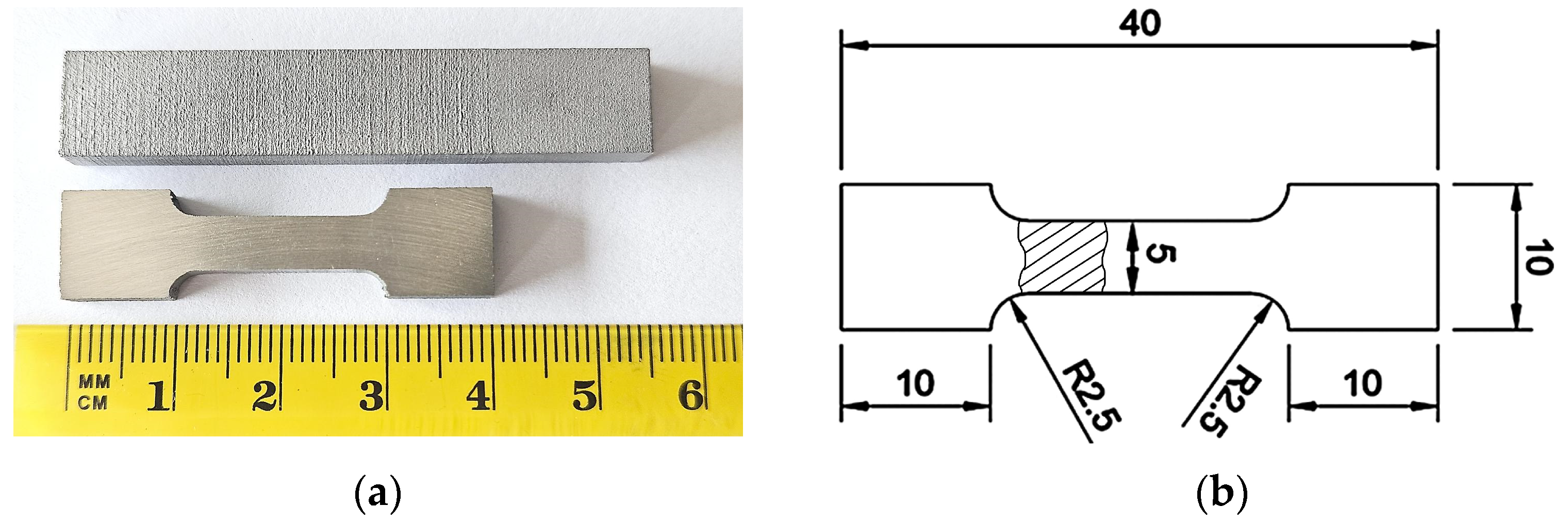
The specimens of 316L steel were fabricated by laser powder bed fusion using the 3D printer Alfa-150D designed by “Additive Laser Technology” (Dnipro, Ukraine). The gas-atomized 316L stainless steel powder of 15–45 µm-sized particles was used as a feedstock. The parameters of printing were as follows: a laser power of 195 W, a scanning speed of 1150 mm/s, a laser spot diameter of 45 µm, a layer thickness of 40 µm, a hatch spacing of 100 µm, a scanning strategy based on stripe, and a rotation angle of 67°. Flat 4 mm-thick tensile specimens of a “dog-bone” configuration were fabricated according to ISO 6892-1:2016 (shown in Figure 1). Charpy specimens were printed as a prism of 5 × 10 × 55 (mm) in size and then V-notched by electrical wire cutting. Under the fabrication, the specimens were oriented longitudinally along the building (Z) direction.
A conventionally manufactured (hereafter mentioned as CM) commercial rolled and annealed plate of 316H steel was used as a counterpart material. The specimens were fabricated from a plate of 5.5 mm thickness, identical to the LPBF-specimens in shape and size. Chemical compositions of the specimens are shown in Table 1.
The research was based on a common approach of electrochemical hydrogen saturation [50,51], which included the following steps: (a) hydrogen charging; (b) immediate mechanical testing after charging completion; (c) immediate after-testing measurement of hydrogen concentration on the fractured sample fragments, and (d) analysis of residual hydrogen content 90 days after the charging.
Before the hydrogen charging, the specimens were polished to Rz = 0.2 mm to remove the stain and technological roughness. The hydrogen charging was performed at room temperature using the VoltaLab40 dynamic electrochemical laboratory (Radiometer Analytical SAS, Villeurbanne, France). The charging was conducted in a galvanostatic regime at ambient temperature using a 0.1 M aqueous solution of H2SO4 [50,52]. To enhance hydrogen uptake, 800 µg/L of H2SeO4 were added as a hydrogen recombination inhibitor. These concentrations were selected to provide stable hydrogen fugacity throughout the long-term charging period. The cathodic current density was set to 10 mA/cm2, and the charging durations were 20 h, 45 h, and 115 h. Surface examination of the hydrogen-charged specimens revealed no evidence of charging-induced microstructural damage, such as blistering or selective attack, which could affect the fracture process distinctly from hydrogen embrittlement.
Hydrogen concentration measurements were conducted using the DH603 analyzer (LECO, St. Joseph, MO, USA) by determining the diffusible and residual hydrogen. For these measurements, the fragments of the fractured samples were utilized as shown in Figure 1b (hatched area).
The specimens in the initial state and the hydrogen-precharged state were comparatively tested using standard tensile tests and impact Charpy tests. Tensile tests were performed on a BISS servo-hydraulic machine (BISS, Bangalore, India) in accordance with the standard (ISO 6892-1:2016, IDT), under the strain rate of 1.5 × 10⁻³ s−1. Impact Charpy tests were conducted on an impact pendulum tester in accordance with ISO 148-1:2016, IDT. Impact toughness (KCV, J/cm2) was evaluated as absorbed impact energy divided by the specimen‘s cross-section area. Three specimens of each type were used for the experimental point, with further averaging of the results.
Microstructure was observed using an optical microscope (OM) GX71 (Olympus Corporation, Tokyo, Japan), a field emission scanning electron microscope (SEM) JSM-7000F (JEOL, Tokyo, Japan), and a transmission electron microscope (TEM) JEM-F200 (JEOL, Tokyo, Japan). The specimens for the microstructural studies were taken as remnants of the tensile test (prepared by standard procedures and etched with an “Aqua Regia” solution (for OM and SEM) or electro-etched in a 10 wt% perchloric acid solution (foils for TEM)). The fracture surface was examined using the SEM JSM-7000F. The electron backscatter diffraction (EBSD) was performed by applying an Apreo S Hivac SEM (Thermo Fisher Scientific, Waltham, MA, USA) equipped with the EBSD Symmetry S3 system (Oxford Instruments, High Wycombe, UK), operated at 20 kV with a 1.0 µm step size.

3. Results

3.1. Susceptibility to Electrolytic Hydrogen Absorption

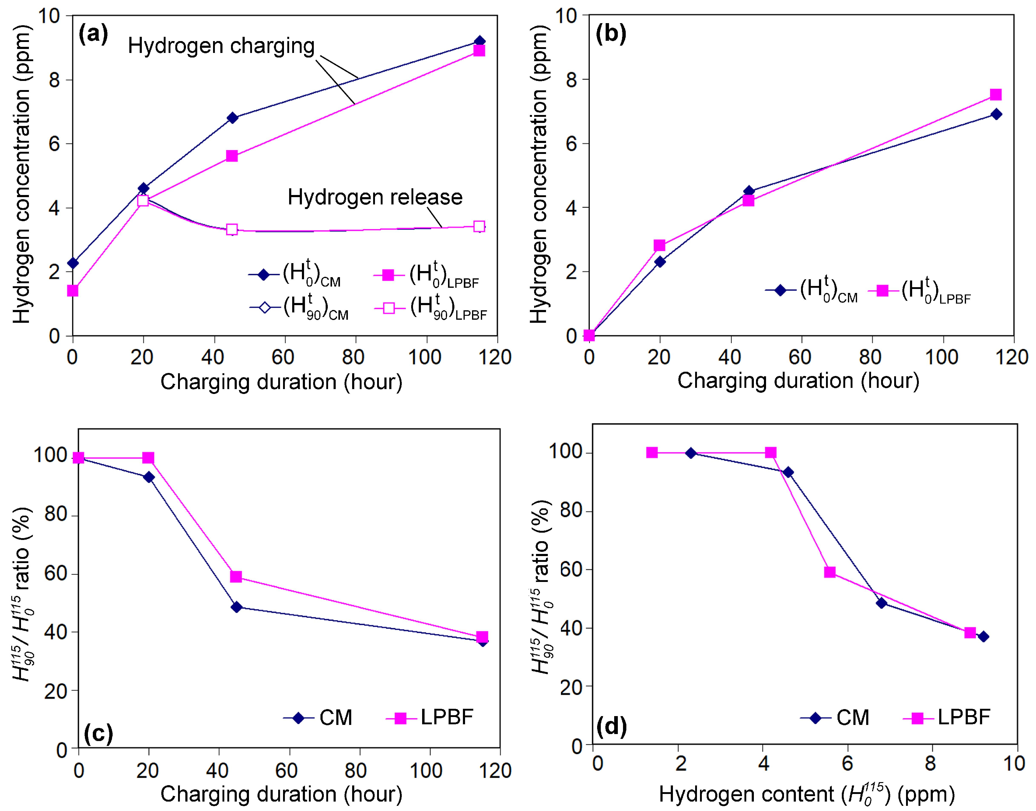
Figure 2 illustrates the dynamics of electrolytic hydrogenation in CM and LPBF specimens. In this section, the hydrogen concentration is denoted as H τ t , where the upper subscript (t) indicates the charging duration and the lower subscript (ф) represents the time elapsed after charging completion. Both steels exhibit susceptibility to hydrogen saturation when exposed to an aqueous solution at room temperature. In the initial state, the baseline hydrogen concentration ( H 0 0 ) in LPBF samples ranged from 1.1 to 1.4 ppm, whereas in the CM samples, it was nearly twice as high (2.2–2.3 ppm), attributed to their metallurgical history (Figure 2a). During the charging period, the current hydrogen concentration in both steels increased following a parabolic trajectory, reaching H 0 115 of 9.2 ppm in CM and 8.9 ppm in LPBF 316L. Given the differing initial hydrogen contents ( H 0 0 ), hydrogenation kinetics are alternatively presented using normalized dependencies that reflect only the hydrogen absorbed during electrolytic charging (Figure 2b). As the normalized curves nearly overlap, CM and LPBF specimens demonstrate nearly identical saturation kinetics, each acquiring approximately 7 ppm of hydrogen by the end of the process.
After 90 days following the hydrogen charging, a reverse process of hydrogen desorption from the samples was observed. As shown by the data in Figure 2a, the hydrogen release behavior is nearly identical for both steels, although they depend on the initial hydrogen content ( H 0 115 ). In specimens saturated for 20 h, no significant decrease in hydrogen concentration was observed even after 90 days of exposure. In contrast, specimens charged for 45 and 115 h exhibited a reduction in hydrogen content to 3.3–3.4 ppm in both cases. According to the data in Figure 2b,c, when the initial hydrogen concentration is up to 4.2–4.6 ppm (after 20 h of charging), no more than 6.5% of the total hydrogen is released from the steel. As the initial concentration increases, the hydrogen desorption accelerates significantly: at 5.6–6.8 ppm (45 h of charging), 49–59% remains after 90 days, while at 8.9–9.2 ppm (115 h), only 37–38% remains. It is noteworthy that, even after 90 days of desorption, the hydrogen content did not return to its initial level. This observation aligns with the findings of Duportal et al. [53] and can be attributed to the phenomenon of hydrogen trapping [54].

3.2. Effect of Hydrogen Charging Duration on Mechanical Properties

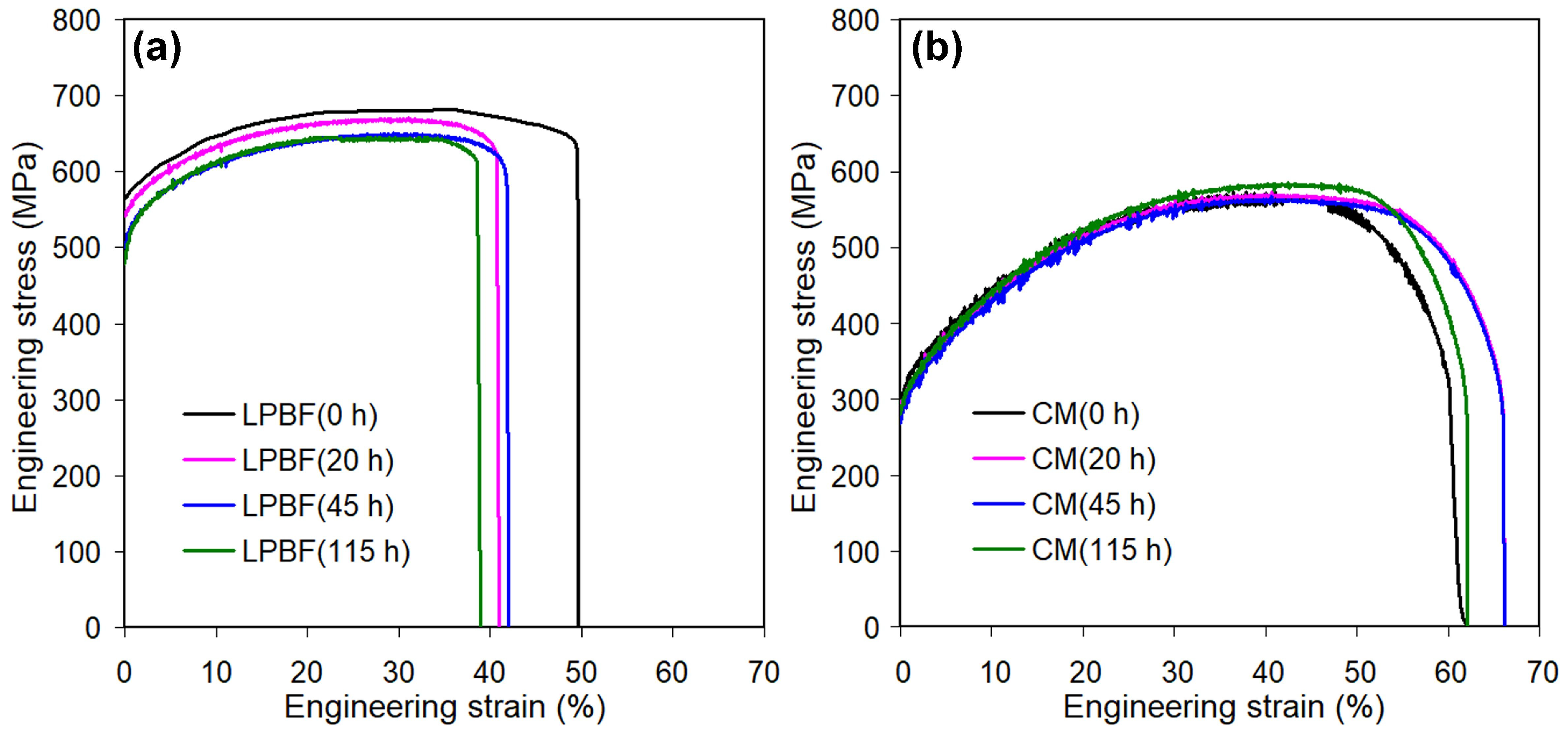
The engineering stress–strain curves of specimens subjected to hydrogen charging for varying durations are shown in Figure 3. Their overall profiles reveal differences in the tensile responses of the two steel groups. The LPBF samples exhibit higher resistance to deformation, as indicated by the greater load required to initiate macroscopic plastic flow (Figure 3a). However, they display reduced ductility, evidenced by lower elongation values prior to fracture. CM specimens begin plastic deformation under considerably lower loads, extending to a greater ultimate elongation (Figure 3b). They also exhibit more pronounced necking prior to fracture.
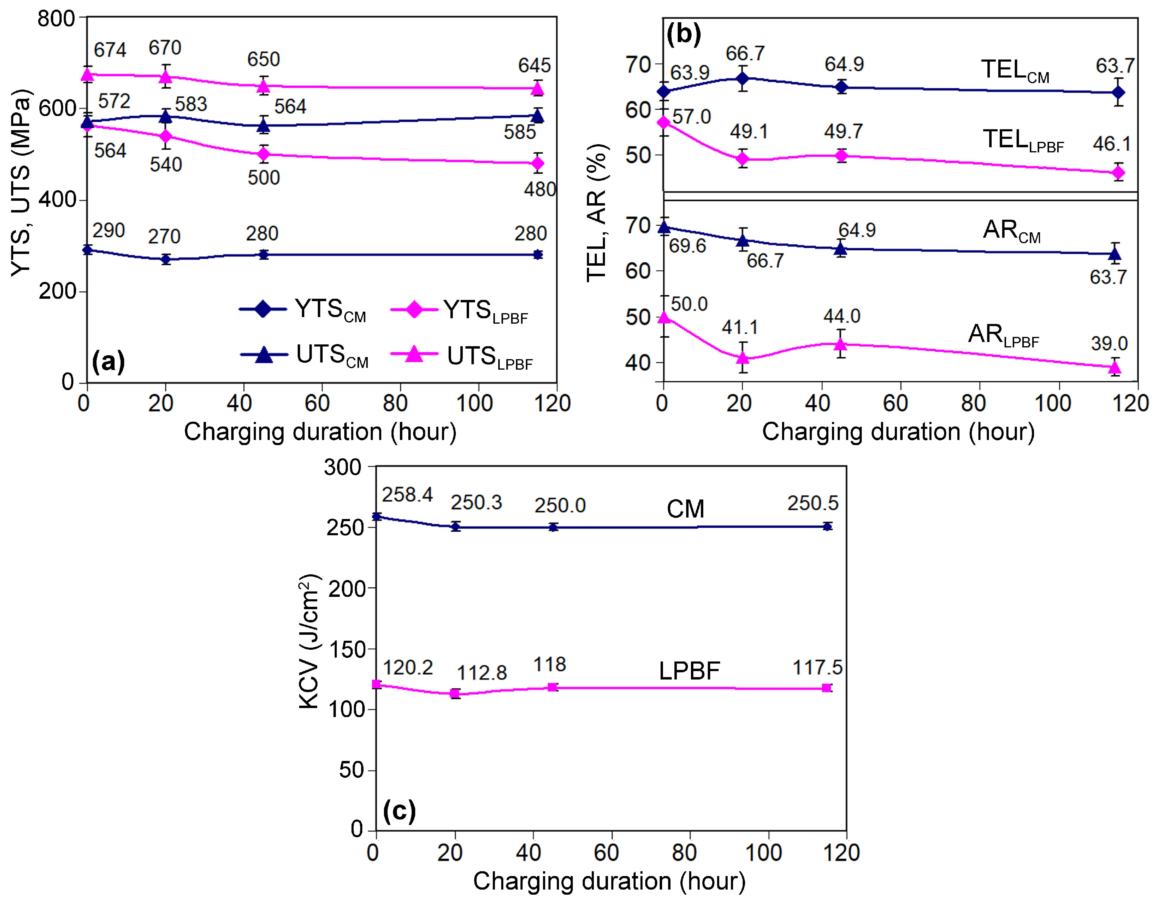
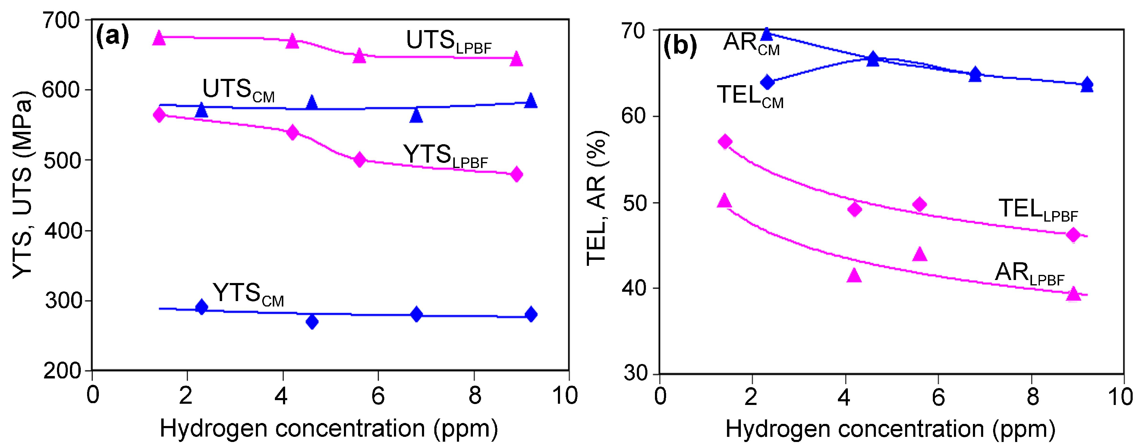
The data presented in Figure 4 illustrate the changes in the mechanical properties of the studied steels as the duration of their hydrogen saturation increases. Figure 4a shows the variation in strength characteristics, i.e., yield tensile strength (YTS) and ultimate tensile strength (UTS). Before hydrogen charging, the CM 316H steel exhibited YTS of 290 MPa and UTS of 572 MPa, respectively. In the LPBF 316L steel, these parameters were significantly higher, at 564 MPa and 674 MPa, respectively, which are consistent with the data of [55]. As the CM steel was saturated with hydrogen, its strength properties varied within narrow limits relative to the initial values (fluctuations of 10–20 MPa for YTS and 8–13 MPa for UTS, corresponding to 3.4–6.9% and 1.4–2.3%, respectively), remaining within the confidence interval of the data scatter. In LPBF samples, with a charging duration up to 115 h, the yield strength gradually decreased from 564 MPa to 480 MPa (84 MPa, 14.9%), which is a statistically significant reduction. A progressive decrease in UTS was also observed, though less pronounced than for YTS (maximally 4.3% to the initial UTS value). In the latter case, the absolute decrease in UTS (29 MPa) remained within the confidence interval.
As shown in Figure 4b, CM steel displays a higher level of ductility compared to the LPBF samples. In the non-charged state, CM exhibits the total elongation (TEL) and area reduction (AR) of 63.9% and 69.6%, respectively, which are 1.1 and 1.4 times higher than those in the LPBF, respectively. With increasing hydrogen saturation, in CM specimens, TEL fluctuates within 63.7–66.7%, with the deviations up to 4.3% relative to the initial state. Meanwhile, AR shows a trend of gradual reduction from 69.6% to 63.7%, i.e., an 8.5% decrease relative to the H 0 0 level. In contrast, the LPBF specimens showed a stronger tendency of ductility, decreasing with the increasing charging duration: for both TEL and AR, a maximum reduction is accounted at approximately 20% to the non-charged state. Thus, the ductility of LPBF steel is found to be more susceptible to hydrogen saturation than that of its rolled counterpart.
This observation does not comply with the impact toughness tendency, as shown in Figure 4c. Before the hydrogen saturation, the impact toughness (KCV) of CM specimens is twice that of LPBF specimens, consistent with previously reported data on the KCV in wrought and additively manufactured 316 steel [32,56]. After 20 h of charging, a slight reduction in KCV was observed for both steels (CM and LPBF), accounting for 8.1 J/cm2 (3.1%) and 7.4 J/cm2 (6.2%), respectively. Notably, with longer hydrogen saturation durations, the impact toughness stabilized at 250.2 J/cm2 (CM 316L) and 117.7 J/cm2 (LPBF). Since the scatter of KCV values does not exceed the confidence interval, it can be concluded that hydrogen charging up to 115 h has practically no effect on the impact toughness of both steels.

3.3. Fractography

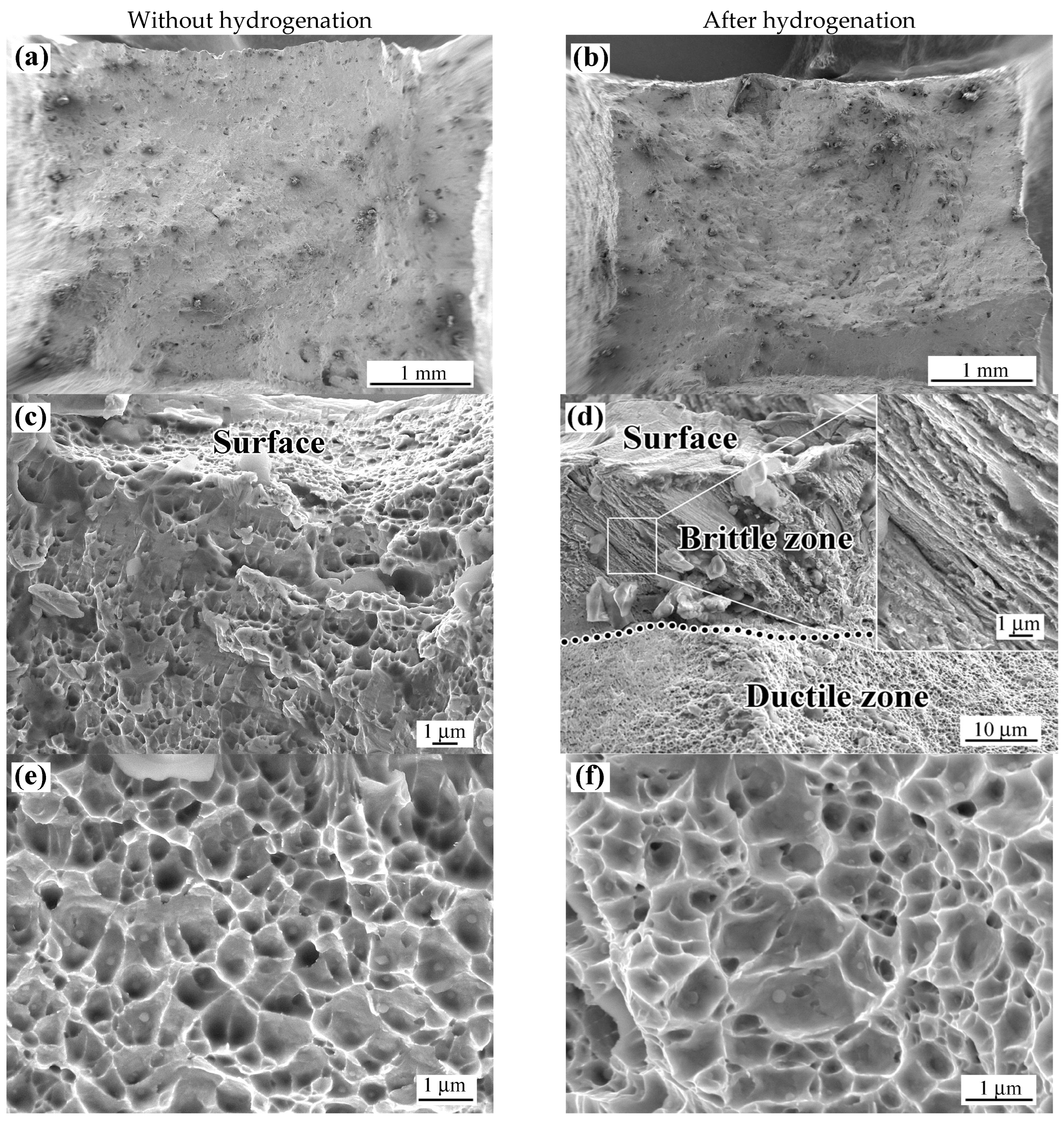
Material behavior under mechanical stress can be partially assessed through a fracture surface analysis. The results of fractographic studies of the broken tensile and impact specimens of both steels are presented in Figure 5, Figure 6, Figure 7 and Figure 8. Figure 5 illustrates the fracture surfaces of LPBF tensile specimens, presenting the general view (Figure 5a,b) and characteristic regions of the near-surface layer (Figure 5c,d) and the central part of the samples (Figure 5e,f).
Based on the general fracture surface images, the specimens appear to be deformed along the diagonals in cross-section, reflecting plastic shape change under tensile loading. In the specimen not subjected to hydrogen charging, a ductile fracture mechanism is observed beneath the surface, extending to a depth of several tens of micrometers. This region is characterized by a fine dimpled pattern and traces of plastic metal flow (Figure 5c). From the surface inward to the specimen’s center, the fracture retains a ductile dimpled morphology, consistent with elevated plasticity indicators (TEL of 57% and AR of 50%). Though the dimples are rather shallow, their sizes vary within one order of magnitude—0.09–1.15 µm. In the LPBF specimen hydrogen charged for 115 h, the fracture near the surface exhibits a distinctly different morphology: a brittle rupture pattern with fibers angled about 40–45° to the surface are clearly seen to the depth of 15–20 µm (Figure 5d). Presumably, this pattern is associated with the planes of maximum shear stress, where the crack propagated along the cell boundaries and the “melt pool” borders (the microstructural features will be detailed below, in Section 3.4).
This fracture morphology appears more brittle compared to the underlying classical dimpled pattern. In the central region of the hydrogen-charged LPBF specimen, the fracture shows a typical ductile dimpled morphology (Figure 5f), similar to that of the uncharged specimen.
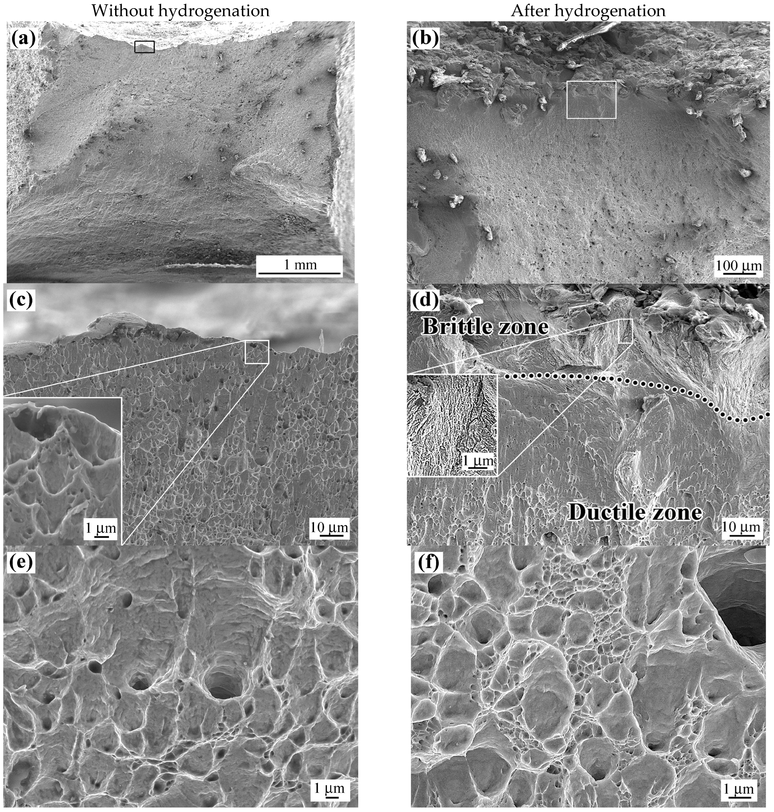
The cross-section of the fractured CM steel specimen is shown in Figure 6. A comparison of Figure 5a and Figure 6a demonstrates a substantial reduction in the cross-sectional area of the CM specimen relative to the LPBF, indicating enhanced ductility in the conventional specimen compared to the LPBF-fabricated specimen. In Figure 6a, a black square denotes the fracture zone, which is magnified in Figure 6c. The latter indicates that, from the surface inward, the fracture exhibits a ductile character, as evidenced by the dimples and ductile tearing extending perpendicularly to the specimen’s outline (see inset in Figure 6c). Deeper into the specimen, the fracture displays a classic ductile pattern, characterized by deep pits with an increased diameter of up to 3 µm, separated by tear ridges, some of which are formed by ultra-disperse (0.05–0.3 µm) voids (Figure 6e). Following hydrogen saturation for 115 h, the fracture surface relief of the CM steel changed similarly to those in LPBF steel. Near the surface, to a depth of 25–30 µm, the fracture exhibits a fibrous, more brittle pattern, as illustrated in Figure 6d, which details a region marked by a square in Figure 6b. The brittle nature of the fibrous fracture is confirmed by the microcracks adjacent to fibers (shown in the inset of Figure 6d). Such cracks are absent in the ductile fracture zone, which begins immediately beneath the brittle zone and extends to the center of the specimen (Figure 6f).
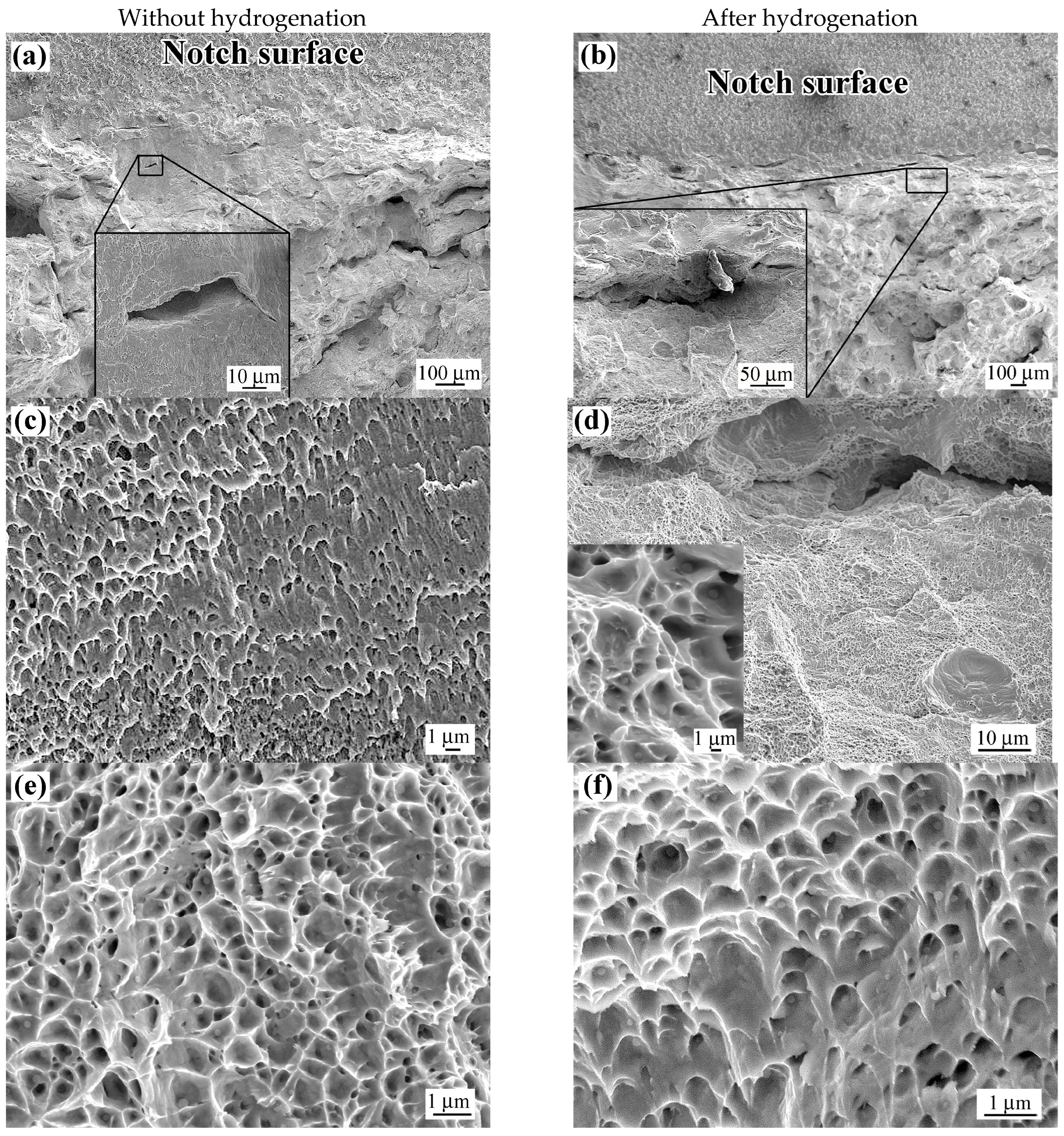
Figure 7a,b show the fracture surface of LPBF steel Charpy specimens located just below the V notch. Irrespective of hydrogen charging, the fracture appears coarse–crystalline at low magnification, with extended microcracks (100–400 µm) oriented parallel to the notch. Notwithstanding this macroscopic fracture characteristic, at the microscopic level, the surface adjacent to the cracks exhibits a dimpled topography, as depicted in Figure 7c,d. This dual nature of the fracture persists deeper into the specimen, extending to its core: a coarse–crystalline appearance at the macroscale and a dimpled pattern at the microscale, with voids that are relatively small and shallow (Figure 7e,f).
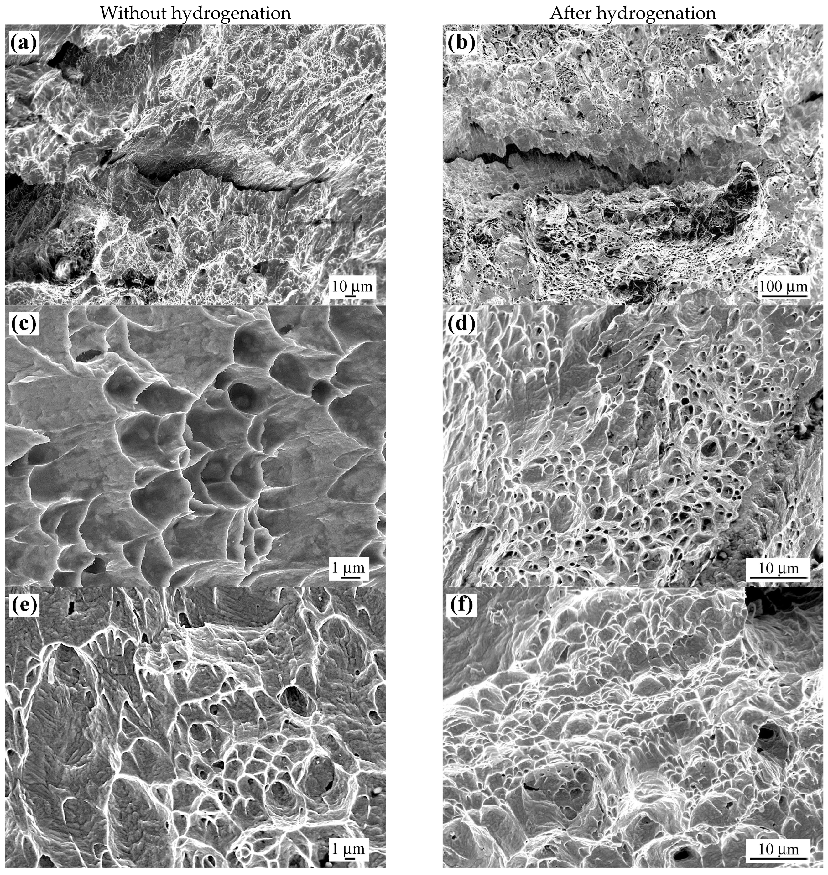
A comparable fracture morphology is observed in the CM steel specimens. In both non-hydrogen-charged (Figure 8a) and hydrogen-charged (Figure 8b) conditions, longitudinal microcracks were detected in the region immediately beneath the notch. The fracture surface surrounding these cracks exhibits a ductile morphology in both cases (Figure 8c and Figure 8d, respectively). The remaining fracture surface area displays a ductile dimpled pattern, with the sizes of the dimples and the elongation of the relief elements exceeding those in LPBF specimens, indicating greater plastic deformation and higher energy absorption during impact fracture (Figure 8e,f).

3.4. Microstructure Observation

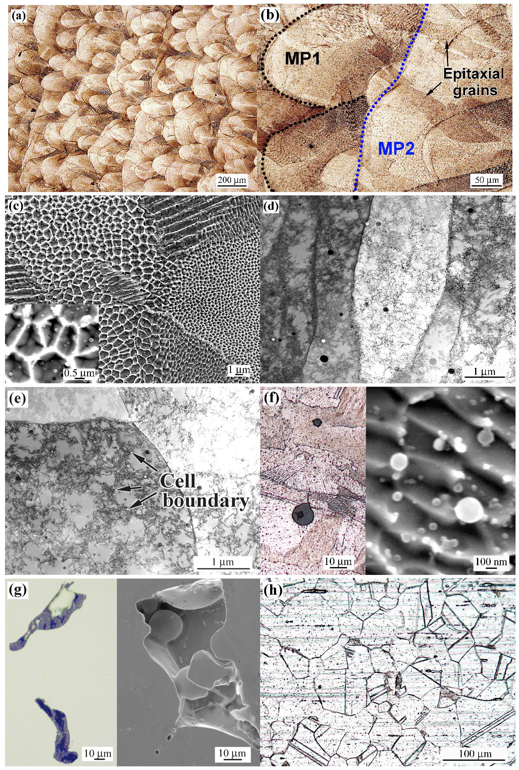
The microstructure of the investigated steels is shown in Figure 9. LPBF 316L steel presents a specific fusion-based cellular microstructure consisting of rows of “melt pools”, which are areas where portions of steel powder were melted under the laser beam (Figure 9a). The “melt pools” (denoted as MP1 in Figure 9b) mainly exhibit an elongated shape with an arc-shaped, rounded contour (indicated by a black dotted line), induced by the Gaussian energy distribution within the laser beam spot [57,58]. The length and width of MP1s reach up to 200 µm in different areas (Figure 9b). In addition to MP1, other “melt pools” (MP2), with a stretched shape and continuous wavy boundaries (indicated by the blue line), are observed in Figure 9b. MP1 and MP2 were formed due to the overlap of the “melt pool” rows created by the laser beam moving in different directions (at a 67° angle). The structure also contains stretched epitaxial grains (tens of micrometers long) intersecting several adjusting “melt pool” rows (Figure 9b). The “melt pools” have a characteristic inherent microstructure consisting of colonies of fine columnar cells, oriented relative to the temperature gradient direction [59] (Figure 9c). The cross-sectional sizes of the cells in different colonies vary by more than an order of magnitude (0.1–1.5 µm). The cell boundaries are composed of the dislocation clusters (dislocation walls), while the cell bodies are divided into smaller subgrains (Figure 9d,e). It should be noted that the LPBF 316L steel microstructure contains a significant amount of predominantly spherical non-metallic inclusions (complex Mn-Si-Al oxide [32]), with sizes ranging widely from tens of micrometers down to the nanoscale (Figure 9f). Additionally, the as-printed specimens contain large discontinuities in the form of lack-of-fusion porosity (Figure 9g). Samples of 316H steel exhibit a typical austenite microstructure, characterized by equiaxed polyhedral twinned grains with an average size of 37.1 ± 4.5 µm (Figure 9h).
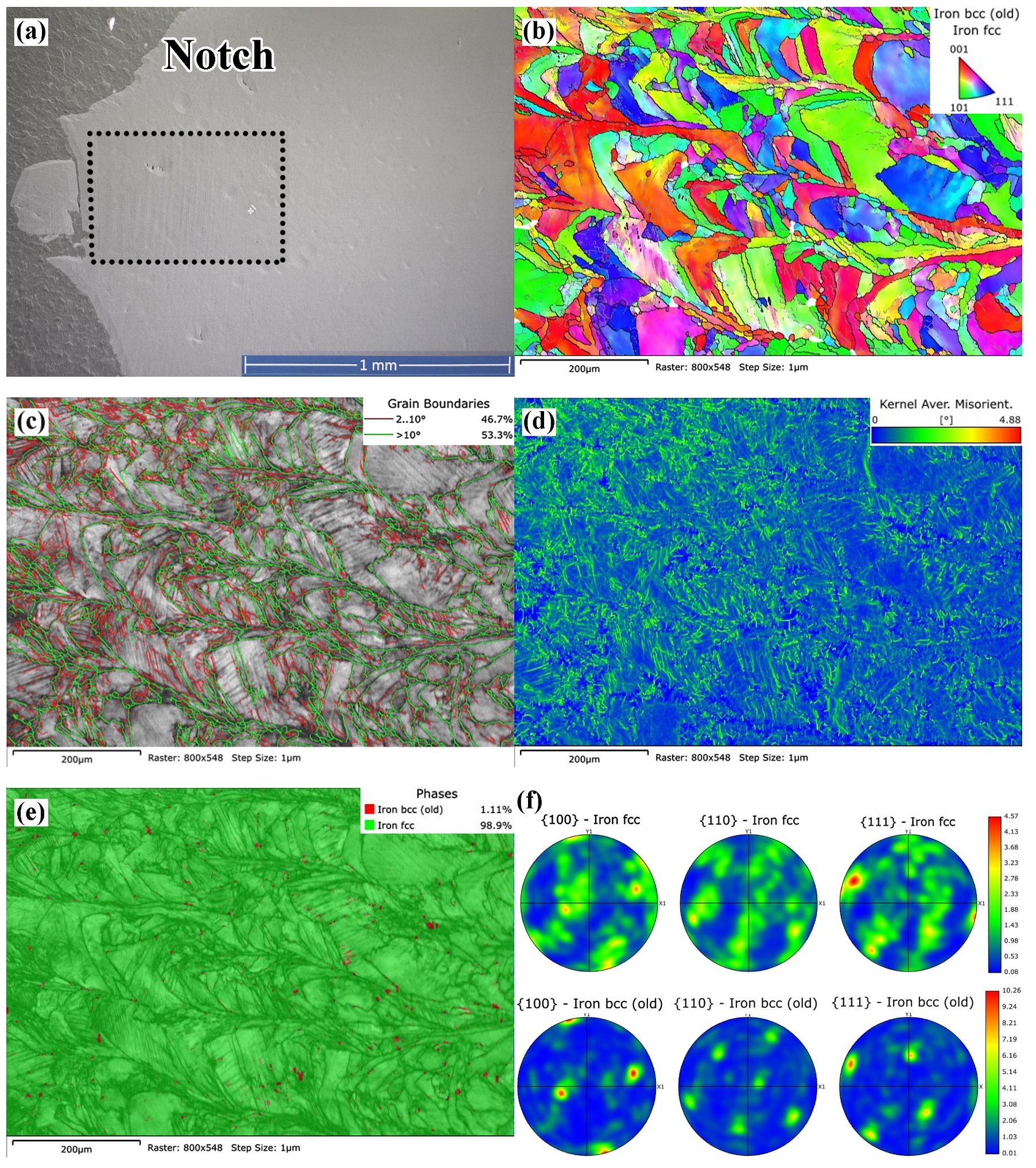
A more detailed analysis of the microstructure was performed using an EBSD analysis. Figure 10 presents data for the LPBF sample, which was not subjected to hydrogen charging. The deformed region of the Charpy sample, located in close proximity to the notch tip (shown as a dark rectangle in Figure 10a, positioned 0.5 mm from the notch tip), was analyzed. Figure 10 shows an inverse pole figure (IPF) map, revealing significant deformation and elongation of “melt pools” in a direction perpendicular to crack propagation. The “melt pools” retained their arc-shaped pattern during elongation, while maintaining a variety of orientations, i.e., no specific texture was formed (Figure 10b). Slip bands formed within the “melt pools.” A large number of low-angle grain boundaries (LAGBs) emerged along the slip bands and at the interfaces of high-angle grain boundaries (HAGBs, boundaries of “melt pools”), with a ratio of HAGBs:LAGBs = 46.7:53.3 (%) (Figure 10c). The formation of LAGBs led to localized elastic lattice distortions, as evidenced by the kernel average misorientation (KAM) map (Figure 10d). It is observed that areas with high KAM values are concentrated closer to the notch tip, indicating a direct correlation between deformation during fracture and the concentration of local distortions. The average KAM value was 0.64°, suggesting significant plastic deformation in the material, as it indicates a higher density of geometrically necessary dislocations (GNDs) causing lattice curvature. The density of GNDs (ρGND) is calculated by the Kubin–Mortensen relationship [60]:
ρ G N D = α θ | b | Δ x
where θ represents the average crystallographic misorientation measured over a distance Δx; b denotes the Burgers vector magnitude (2.5 × 10⁻¹⁰ m [61]), and α is a coefficient that reflects the nature of the grain boundary. To approximate θ and Δx, the mean KAM value (converted to radian [62]) and the EBSD step size of 1.0 µm were employed, respectively, as a first-order approach. The coefficient α was taken as two for a pure tilt boundary [63].
The calculations using Equation (1) give ρGND of 8.9 × 10¹³ m−2. It should be noted that this ρGND value represents only the minimum GND density [64], primarily because 2D EBSD characterization limits the number of components of the Nye dislocation density tensor [65].
According to the phase map (Figure 10e), the analyzed region predominantly exhibits an FCC lattice, indicating the presence of austenite. However, in certain local regions associated with the dislocation slip bands and the boundaries, areas with a BCC lattice are detected, suggesting the presence of the α-Fe phase, likely α′-martensite, formed due to deformation-induced martensitic transformation. This further confirms intense plastic deformation at the crack tip. The amount of α′-martensite in the analyzed region, based on EBSD data, was 1.11 vol.%. The martensite formed on specific planes due to the dislocation slip and accumulation; consequently, the crystallographic orientation of the martensite crystals is less varied than that of the austenite. This accounts for the pronounced texture in the alpha-phase orientation, as evidenced by the BCC-phase pole figures in Figure 10f. The formation of deformation-induced martensite in the 316L LPBF steel under impact loading is consistent with previously published studies [66,67].
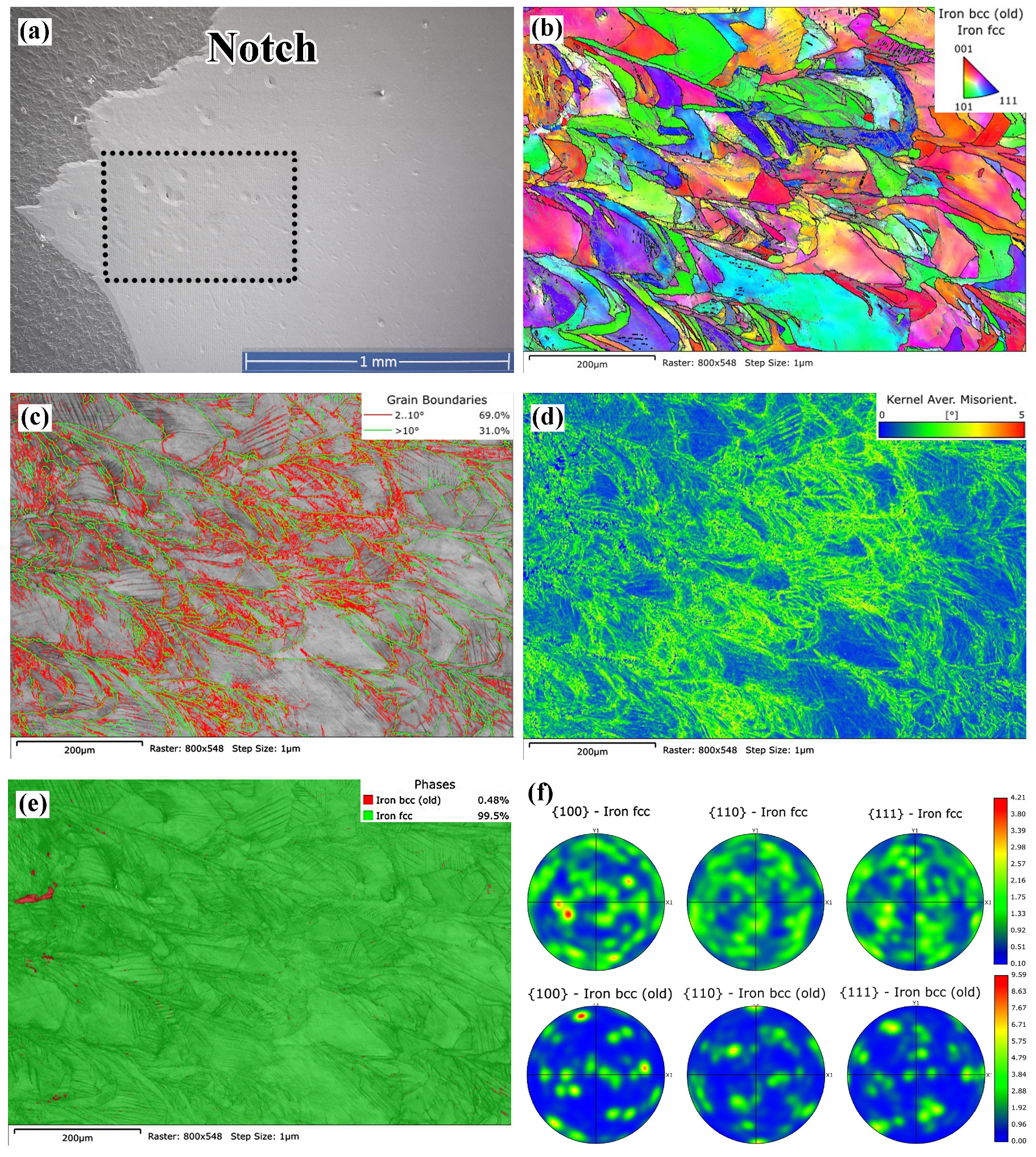
Figure 11 presents EBSD data for the LPBF sample, which was subjected to hydrogen charging. In the hydrogen-charged 316L LPBF steel sample, subjected to 115 h of hydrogenation, significant deformation was observed near the notch-tip (Figure 10a), as evidenced by more elongated “melt pools” (Figure 11b). This deformation was not accompanied by grain rotation or the formation of a preferred texture. The emergence of numerous slip bands resulted in a large extent of grain boundaries (Figure 11c), with low-angle boundaries accounting for the majority (69%), which is 1.5 times higher than in the non-hydrogenated sample. Consequently, there was a significant increase in the density of KAM distribution (Figure 11d), with the average KAM value rising to 1.09°, indicating that more GNDs were accumulated near the grain boundaries during plastic deformation. Using Equation (5), the GNDs density was calculated to be 1.5 × 10¹⁴ m−2, which is 1.7 times higher than that of the non-charged specimen.
Interestingly, despite the significant deformation, the amount of formed martensite was twice-lower than in the non-hydrogenated sample. According to Figure 11e, the volume fraction of the BCC phase is 0.48 vol.%, with nearly all of the martensite concentrated in the left part of the image, i.e., in the region of maximum deformation. This suggests a reduced tendency of hydrogenated austenite to undergo deformation-induced martensitic transformation during impact fracture.
Analysis of the pole figures shown in Figure 10f reveals that, in the non-hydrogen-charged LPBF specimen, the FCC phase exhibits a moderately developed texture relative to the (100), (110), and (111) planes. Its maximum intensity is 4.57, indicating the presence of preferred crystallographic orientations, particularly regarding the (111) plane. The orientation distribution is relatively uniform, typical of plastically deformed FCC structures. For the BCC phase, the texture is significantly stronger, with a maximum intensity of 10.26, especially regarding the (110) and (111) planes. This high intensity suggests anisotropy, which may influence the mechanical properties and hydrogen sensitivity. In the hydrogen-charged LPBF specimen (Figure 11f), the maximum texture intensity in the FCC phase is reduced to 4.21, indicating slight texture scattering, possibly due to hydrogen’s effect on the substructure and local plasticity. The orientation distribution remains nearly uniform, without sharp peaks. For the BCC phase, the texture intensity is notably reduced (maximum of 9.59), particularly with respect to the (100) and (110) planes. A comparison of Figure 10f and Figure 11f shows that, under hydrogen saturation, the austenitic FCC phase demonstrates greater resistance to structural changes, maintaining a more uniform orientation distribution. In contrast, the BCC phase is significantly more sensitive: the reduction in texture intensity and changes in orientation distribution indicate hydrogen’s influence, manifesting as the suppression of directed deformation. Thus, based on the pole figures, hydrogen predominantly affects the BCC phase.

4. Discussion

4.1. H Concentration Dependence of Mechanical Behaviors and Embrittlement Index

The effect of dissolved hydrogen content on the mechanical properties of steels under tensile testing is shown in Figure 12. It can be observed that hydrogen concentrations up to 10 ppm do not have a statistically significant impact on either the strength properties or elongation of CM 316H steel (Figure 12a,b). In contrast, the area reduction decreased systematically with the increasing hydrogen content (Figure 12b), which is described by a power-law expression:
ARCM (%) = 73.4·(H)−0.06  (R2 = 0.99)
where H is the hydrogen concentration (ppm).
Meanwhile, LPBF 316L steel proved to be more susceptible to hydrogen embrittlement. A noticeable reduction in yield strength was observed, especially when the hydrogen concentration exceeded 4 ppm. A similar concentration-dependent behavior refers to ultimate tensile strength, though the variations were lower. In contrast, the ductility characteristics responded more significantly, showing a pronounced downward trend with the increasing hydrogen content, aligning with the power-law trends:
TELLPBF (%) = 58.9·(H)−0.11  (R2 = 0.96),
ARLPBF (%) = 21.8·(H)−0.13  (R2 = 0.84).
The susceptibility of materials to hydrogen-induced embrittlement is typically described by the embrittlement index (EI), determined according to the following formula [50]:
E I ( 0 ; 115 ) = X 0 X 115 X 0 × 100 % ,
where X0 and X115 denote the property values of the material in its initial condition (before charging) and after 115 h of hydrogen charging, respectively.
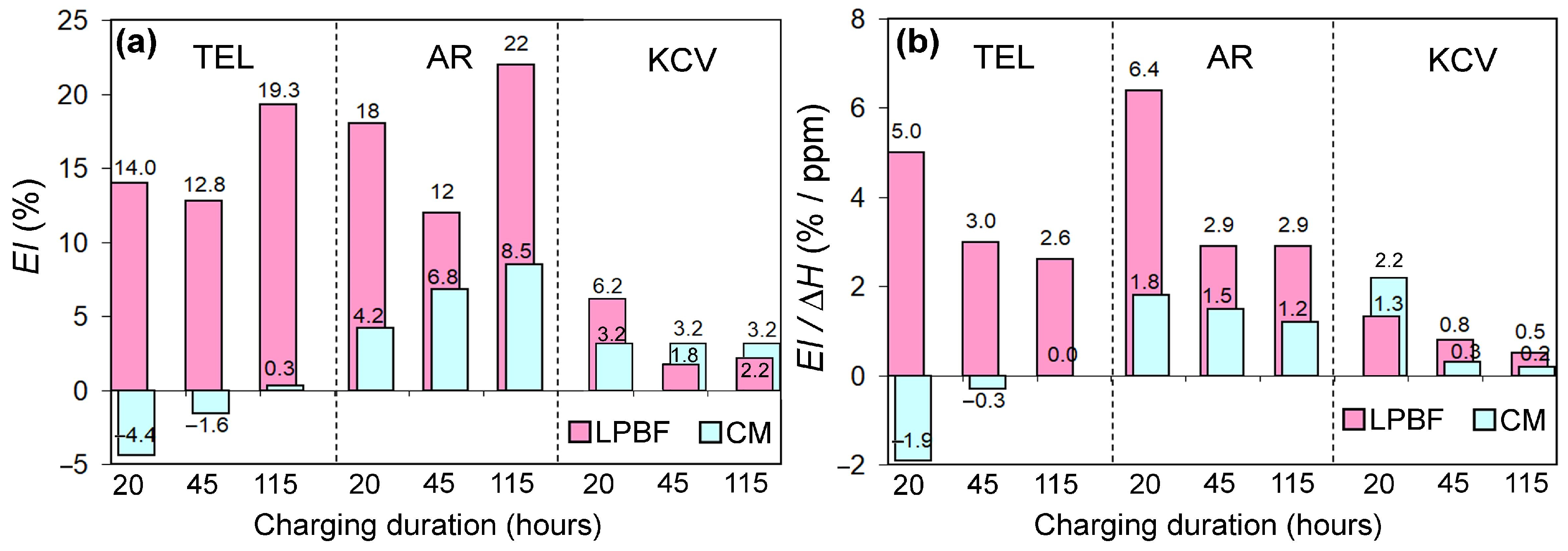
The mechanical property indicators used for calculating the embrittlement index (EI) included the average values of ductility characteristics (TEL, AR) and impact toughness (KCV). The EI values for different properties are presented in Figure 13a. These results reveal a higher susceptibility of LPBF 316L steel to hydrogen embrittlement, as evidenced by the EI values for elongation at break reaching 13–14% after 20–45 h of hydrogen charging, increasing by 1.5 times (up to 19.3%) after the maximum exposure duration. For area reduction, an even higher EI value of 22% is recorded. In contrast, CM 316H steel showed complete insensitivity to hydrogen embrittlement in terms of elongation, with even negative EI values observed. Regarding AR, a certain tendency toward embrittlement was noted, as EI increased from 4.2% to 8.5% with longer exposure, although this effect was less pronounced than in LPBF steel.
The impact toughness in both steels responded only slightly to hydrogen charging. In most cases, the EI remained within 2–3%, except for LPBF steel after 20 h of exposure, where the EI reached 6.2%, which may be attributed to measurement statistical variation.
More insightful are the data presented in Figure 13b, which show the ratio of EIH, where ΔH is an increment of hydrogen content after the charging (ΔH = H 0 115 H 0 0 ). For LPBF steel, a decreasing trend in EIH is observed across all three indicators as the charging duration increases, i.e., as the hydrogen content rises. This indicates that the specific embrittlement effect of hydrogen (per each absorbed ppm of H) diminishes as its concentration grows—with the most pronounced effect occurring at early saturation stages. A similar trend is observed in CM steel for AR and KCV, while no embrittlement is detected for TEL.

4.2. Hydrogen Embrittlement Mechanism

According to the results obtained, at hydrogen concentrations up to 9 ppm, CM 316H steel demonstrated high resistance to hydrogen embrittlement (except for the area reduction). Such behavior is consistent with its reputation as one of the most hydrogen-compatible austenitic alloys. Several factors contribute to this phenomenon: (a) high hydrogen solubility and low diffusivity: the face-centered cubic lattice of austenite provides high hydrogen solubility combined with its very low diffusion coefficient [68]; (b) structural stability and lower susceptibility to hydrogen-driven phase changes, such as the formation of brittle martensite via DIMT [69]: the alloy’s elevated Ni and Mo contents stabilize the austenitic matrix and suppress deformation-induced martensitic transformation, which is often a precursor to hydrogen-assisted cracking; (c) and hydrogen trapping at dislocations and defects in 316 steel is comparatively less detrimental [70] and may even contribute to a hydrogen-induced strength increase [71]. Therefore, the present results align with previous investigations that have shown that stable austenitic stainless steels demonstrate strong resistance to hydrogen embrittlement, maintaining their fundamental deformation mechanisms under hydrogen charging, which results in negligible changes in flow stress and homogeneous plasticity [72,73].
The vulnerability of CM 316H steel to the decline in AR stems from the fact that this parameter reflects the material’s ability to sustain large, localized deformation in the necking region, where triaxial stress states dominate [74]. Hydrogen strongly influences this stage of deformation due to its tendency to accumulate in regions of high hydrostatic tension and at microstructural defects. Such redistribution promotes premature void nucleation and accelerates crack initiation, as demonstrated by numerous studies on hydrogen diffusion, trapping, and damage evolution [75].
In contrast to CM 316H, the LPBF-produced 316L steel exhibited a simultaneous decrease in all tensile properties after hydrogen charging, with the most pronounced degradation observed in the ductility parameters. This can be attributed to the characteristic “as-melted” (fusion-based) LPBF cellular microstructure, containing an increased number of hydrogen diffusion paths such as fusion boundaries and higher dislocation density, as well as hydrogen trapping sites and/or stress concentrators related to various microstructural features, e.g., residual porosity (lack-of-fusion defects, Figure 9g), oxide inclusions (Figure 9f), and highly anisotropic grain structures (Figure 9a–c), as also reported in [76]. As a result, hydrogen promotes early crack initiation and damage accumulation already during uniform deformation. Consequently, hydrogen-assisted embrittlement is not confined to the necking region but affects the entire gauge length.
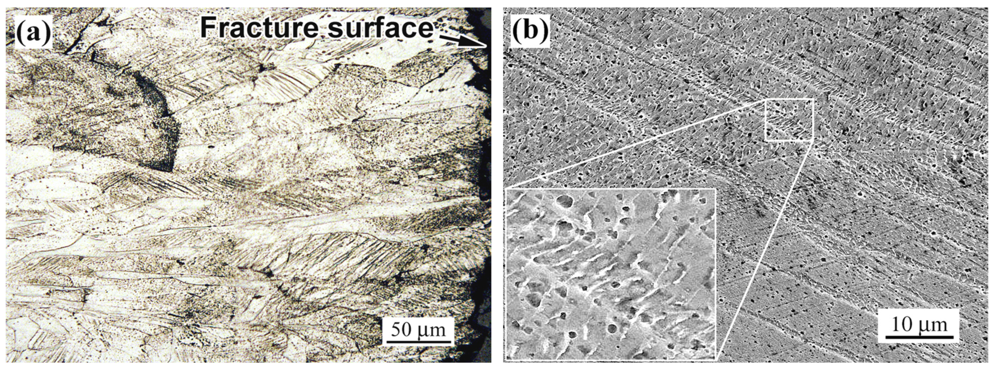
The analysis reveals that hydrogen embrittlement in the investigated steels is most evident under tensile loading. Beyond the reduction in mechanical properties, HE manifests through a distinct brittle fracture zone in the subsurface layer along the perimeter of the fractured tensile specimen, which is particularly pronounced in LPBF steel (Figure 5d). This type of fracture in the hydrogen-saturated regions of 316L steel was previously observed in [77]. Based on the findings of [53], which revealed uneven hydrogen distribution inside 316L steel (following cathodic charging at 50 °C for 144 h), and giving low hydrogen diffusivity (D) in austenitic steels [68,78], it is reasonable to suggest that, in our case, the observed brittle fracture originates from hydrogen accumulation in the near-surface region of the specimen.
As a first approximation, the hydrogen diffusion depth in steel (x) can be evaluated using the expression below [79]:
x = ( 2 D t ) 1 / 2 ,  
where t is the hydrogenation duration.
The diffusion coefficient D for 316L steel, in turn, can be derived from its temperature dependence using an Arrhenius-type fit [78]:
D = D 0 exp ( E D / R T ) ,
where D0 is the pre-exponential factor (3.91 × 10⁻⁶ m2/s [78]), ED is the activation energy (60.4 kJ/mol [78]), R is the gas constant (8.314 J/(K·mol)), and T is the absolute temperature.
The calculations yield a diffusion coefficient D of 1.011 × 10⁻¹⁶ m2/s at 25 °C. Accordingly, the hydrogen penetration depth in 316L steel at room temperature for over 144 h is estimated to be 10.2 µm. This value is comparable in magnitude to the width of the brittle fracture zone observed in tensile specimens saturated with hydrogen (Figure 5d and Figure 6d). However, the calculated depth is 1.5–2 times smaller than the actual brittle zone width in LPBF steel (15–20 µm) and three times smaller than in conventionally manufactured steel. If the width of the brittle fracture zone is indeed governed by hydrogen presence, this implies a higher hydrogen diffusion rate in CM steel compared to LPBF steel. The lower hydrogen penetration rate—along with the higher degree of embrittlement—can be attributed to the unique as-built structural features of LPBF steel, particularly the cellular pattern formed by dislocation walls [32]. Elastic stress fields associated with crystal lattice defects attract mobile hydrogen atoms and promote hydrogen permeation in stainless steels [51]. On the other hand, these same defects also facilitate hydrogen accumulation by acting as hydrogen traps. As reported in [70], additively manufactured 316L contains multiple hydrogen trapping sites, with two primary types: elastic stress fields (dislocation cores) and dislocation cell walls. Hydrogen accumulation enhances the formation of hydrogen-induced defects, resulting in their higher volume fraction compared to conventionally manufactured steel under identical charging conditions [51]. Hydrogen migrates along dislocations and cellular boundaries toward regions of stress concentration, triggering brittle fracture and contributing to pronounced hydrogen embrittlement in the as-built sample. This is reflected in the greater sensitivity of the plastic properties of LPBF steel to hydrogen embrittlement, which is consistent with the findings of Álvarez et al. [36], Khaleghifari et al. [80], and Hong et al. [81]. However, these conclusions stand in contrast to the results reported in [30], where additively manufactured 316L stainless steel demonstrated superior resistance to hydrogen embrittlement compared to its as-rolled counterpart.
Fractographic analysis revealed a fully ductile dimpled fracture in the core of the ruptured tensile specimens of both CM 316H and LPBF 316L steels (Figure 5f and Figure 6f), whereas a thin near-surface rim exhibited fine transgranular facets oriented approximately normal to the specimen surface. This morphology suggests that hydrogen primarily affects a near-surface layer, where it promotes strong strain localization and quasi-brittle cracking, while the bulk material retains its ductile fracture behavior. As already shown in Section 3.4, the higher-magnification SEM images revealed an occurrence of oxide inclusions (Figure 9f) and lack-of-fusion porosity (Figure 9g) in the as-built LPBF specimens prior to hydrogen charging. However, given that these defects are inherent to the entire volume, they are expected to be present at crack initiation sites, thereby influencing the fracture process. This supports the conclusion about the occurrence of mixed mechanisms of hydrogen embrittlement at the cracks’ initiation sites. The HELP mechanism can be generally related to the change in size and morphology of ductile dimples, whereas the HEDE mechanism is generally related to typical brittle fracture features, including transgranular cleavage and/or intergranular decohesion. Djukic et al. [82] reviewed the synergistic action and interplay of HELP and HEDE mechanisms in steels and iron. They also proposed a novel and unified HELP+HEDE model based on the specific microstructural mapping of the dominant HE mechanisms, with implications on the fracture process and resulting hydrogen-assisted fracture modes [82]. However, in the case of our current investigation, the absence of pronounced brittle fracture features within the major fracture area indicates that HELP, rather than HELP+HEDE, is likely the dominant mechanism of hydrogen embrittlement. This presumption is supported by the microstructure of the subsurface zone of the hydrogen-charged LPBF 316L, where the quasi-brittle transgranular rim is found to be associated with the localized slip bands rather than with grain boundary decohesion (Figure 14a). As the slip bands developed, they caused displacements (shears) in the columnar cell bundles without any loss of continuity or the occurrence of fractures (Figure 14b). These observations are consistent with the characteristics of the HELP mechanism [8].
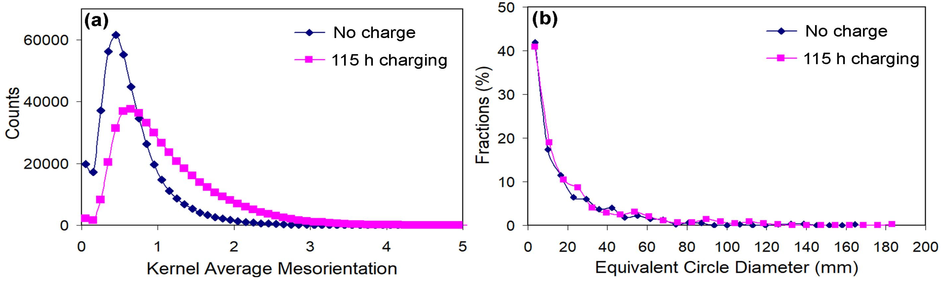
The microstructural evidence obtained from an EBSD analysis further supports the conclusion that hydrogen-assisted damage in LPBF 316L is dominated by the HELP mechanism. In the hydrogen-charged condition, the local lattice misorientation in the fracture initiation region increased significantly, as evidenced by KAM distributions (Figure 15a), with an average KAM value almost doubling (from 0.64° to 1.09°) compared to the non-charged state. Such an increase in lattice curvature indicates a strong accumulation of geometrically necessary dislocations and pronounced strain incompatibility at the microscale. Furthermore, the KAM evolution was corroborated by a ~50% rise in the fraction of high-angle boundaries (from 46.7% to 69.0%), which could result from the deformation-induced transformation of low-angle boundaries into high-angle ones promoted by the local strain concentration [83]. Indirect evidence for boundary transformation lies in the fact that hydrogen charging left the steel’s grain size nearly unchanged, as shown by the grain size distribution (Figure 15b) and average values: 20.51 μm (pre-charging) and 23.42 μm (post-charging). Thus, the rise in the HAGB fraction was not triggered by grain fragmentation. This behavior is characteristic of HELP, where hydrogen facilitates dislocation motion but promotes its localization into narrow slip bands rather than homogeneous plastic flow [8]. In the LPBF microstructure, which contains lack-of-fusion defects, pores, and oxide inclusions, these HELP-induced slip bands readily intersect process-induced stress concentrators, triggering early microcrack initiation throughout the gauge section. As a result, hydrogen affects not only post-necking ductility but also uniform deformation, leading to a simultaneous reduction in yield strength, ultimate tensile strength, and elongation, and a reduction in area. This contrasts sharply with wrought 316H steel, where the cleaner and more homogeneous microstructure confines hydrogen-assisted damage primarily to the necking region, preserving uniform elongation and strength while reducing only the area reduction.

4.3. Role of Hydrogen in Deformation-Induced Martensitic Transformation

As shown above, the embrittling effect of hydrogen on the properties of LPBF 316L steel was more pronounced in tensile tests than in impact bending tests (in alignment with the results of Nguyen et al. [54]). A similar effect was reported in [84], where hydrogen charging was shown to improve the impact toughness of conventionally manufactured 316H steel, attributed to hydrogen-enhanced twinning-induced plasticity (TWIP). The absence of hydrogen embrittlement during Charpy impact tests, despite its clear presence in tensile tests, can be attributed to the high strain rate, which suppressed the diffusional interaction between the hydrogen and the material’s microstructures. Unlike slow tensile testing, the dynamic nature of the impact test does not allow for sufficient time for hydrogen diffusion to critical sites [85]. Furthermore, the severe geometric constraint of the sharp V notch induces a high level of triaxiality that dominates the failure mechanism, effectively masking the detrimental effect of hydrogen on the material’s toughness [86].
The effect of hydrogen on the impact toughness of LPBF 316L steel should also be considered in light of the potential for deformation-induced martensitic transformation (DIMT) during loading. As noted in [87], DIMT reduces the impact toughness of austenitic steels due to the appearance of brittle α′ martensite. The tendency of austenite to undergo DIMT is typically assessed using the martensite start temperature (Md) and the stacking fault energy (SFE) [88,89]. These values for austenitic steel at room temperature can be estimated using equations [90,91]:
SFE (mJ/m2) = 1.2 + 1.4Ni + 0.6Cr + 17.7Mn − 44.7Si,
Md (°C) = 413 − 462(C + N) − 9.5Ni − 13.7Cr − 8.1Mn − 18.5Mo − 9.2Si,
where Md refers to the lowest temperature at which applying 30% true plastic strain induces a 50% transformation to martensite [92].
Using expressions (8) and (9) along with the data from Table 1, the following values were obtained: (a) for CM 316H: SFE = 35.37 mJ/m2, Md = −2.4 °C, and (b) for LPBF 316L: SFE = 11.08 mJ/m2, Md = 5.3 °C. According to [93], SFE governs the dominant plastic deformation mechanism, which transitions from dislocation slip (SFE > 45 mJ/m2) to mechanical twinning (18–45 mJ/m2) and, finally, to strain-induced martensitic transformation (<18 mJ/m2).
Given the calculated Md and SFE values, LPBF 316L should exhibit a higher susceptibility to DIMT at room temperature, whereas CM 316H should tend to deform via mechanisms more favorable to impact toughness, such as dislocation slip and/or mechanical twinning. These conclusions are supported by EBSD analysis, which revealed the presence of deformation-induced martensite in the fractured Charpy specimen of LPBF 316L steel, specifically in the area adjacent to the notch—i.e., the zone that experienced significant plastic deformation during testing (Figure 10e). α′-martensite crystals were observed throughout the analyzed area (540 × 780 µm); they nucleated at high-angle grain boundaries at “melt pool” junctions and at low-angle boundaries along specific slip planes, forming a distinct texture with respect to the (100) and (111) planes (Figure 10f). In the hydrogen-charged LPBF 316L specimen, α′-martensite was detected only in close proximity to the notch, within a distance of no more than 100 µm—the zone of most intense deformation (Figure 11e). This indicates that hydrogen contributes to the stabilization of austenite in LPBF 316L, suppressing DIMT. According to the EBSD data presented above, hydrogen charging caused a sharp increase in the density of geometrically necessary dislocations and the fraction of HAGBs, which suggests that active dislocation slip is the dominant deformation mechanism, serving as an alternative to deformation-induced martensitic transformation in this case.
The effect of hydrogen on deformation-induced martensitic transformations in austenitic stainless steels (which follows the γ⟶ε⟶α′ sequence in SUS304 and similar austenitic stainless steels [94]) remains a central topic of research due to its critical influence on the performance of steels operating in hydrogen-rich environments. According to the prevailing consensus, hydrogen suppresses thermally induced martensitic transformation in low-alloyed steels [95,96] by diffusing to martensite nuclei (“strain embryos” [97]) and reducing the transformation’s driving force. Conversely, in austenitic steels, hydrogen promotes DIMT and increases the content of ε- and α′-martensite in deformed zones [78,95,97,98], largely due to its effect on reducing stacking fault energy [97]. In other works dedicated to the austenitic stainless steels, hydrogen was found either to decrease α′-martensitic transformation (under the high-pressure torsion) [99] or act complexly (promoting nucleation of α′-martensite while suppressing the growth of formed α′-martensite nuclei) [100]. The data obtained in this study suggest the opposite trend—hydrogen appears to inhibit DIMT in LPBF austenitic steel. This is consistent with earlier reports [97,101,102] suggesting that the cellular sub-grain structure of additively manufactured austenitic steel impedes martensitic transformation during plastic deformation under hydrogen charging. According to Hong et al. [101], the cellular microstructure and high density of LAGBs in LPBF 316L restrict twin formation and dislocation slip, thereby reducing the number of martensite nucleation sites. Mechanistically, the sub-grain boundaries—a network of dislocation walls—may impose mechanical constraints on the γ⟶α′ transformation, which involves a volume expansion. Hydrogen atoms penetrating the lattice of LPBF steel may stabilize (pin) these dislocation walls, thereby enhancing the mechanical stabilization of austenite, similarly to other interstitial elements. Consequently, the inherent microstructural features of LPBF steel enhance hydrogen’s austenite-stabilizing effect, leading to suppressed DIMT. By reducing the amount of strain-induced martensite, hydrogen saturation may indirectly improve impact toughness, counteracting hydrogen-induced embrittlement. Based on these considerations, the low susceptibility of LPBF 316L steel to hydrogen embrittlement in terms of impact toughness can be attributed to a balance between its detrimental (embrittlement) and beneficial (DIMT suppression) effects of hydrogen saturation—with the latter prevailing.

5. Conclusions

The article presents a comparative analysis of hydrogen-induced mechanical behavior and propensity to the hydrogen embrittlement (HE) phenomenon of two austenitic stainless steels belonging to the 316 grade family, specifically 316L steel produced via laser powder bed fusion (LPBF) and conventionally manufactured (CM) 316H steel. Based on the experimental results, the following conclusions were drawn:
1. LPBF 316L steel exhibited in the as-LPBF state significantly higher strength but lower ductility and impact toughness than conventionally manufactured 316H steel. In the uncharged state, LPBF steel exhibited higher yield tensile strength (YTS) (564 vs. 290 MPa) and ultimate tensile strength (UTS) (674 vs. 572 MPa) than CM steel but lower total elongation (TEL) (57% vs. 63.9%), area reduction (RA) (50% vs. 69.6%), and impact toughness (KCV) (258.4 J/cm2 vs. 120.2 J/cm2). This behavior is attributed to the specific fine cellular FCC microstructure of the LPBF steel, containing increased dislocation density, some residual porosity, oxide inclusions, and highly anisotropic grain structures in contrast to the coarse-grained austenitic structure of wrought and annealed CM steel with fully recrystallized polyhedral grains;
2. Despite a higher concentration baseline in CM steel, both materials exhibited similar hydrogen absorption/desorption kinetics: hydrogen concentrations reached ~9 ppm after 115 h of charging and dropped to residual 3.3–3.4 ppm after 90 days of desorption;
3. Under tensile loading, the LPBF-processed steel exhibited a higher susceptibility to hydrogen embrittlement compared to its wrought counterpart. After 115 h of charging, LPBF showed a 14.9% drop in YTS and ductility-based embrittlement indices (EI) up to 22%. Conversely, CM steel exhibited minimal changes, with EI values below 8.5% and negligible variations in strength and TEL. The LPBF steel is more susceptible to HE in tensile loading conditions because of the higher density of hydrogen diffusion paths (e.g., fusion boundaries and higher dislocation density) and hydrogen trapping sites and/or stress concentrators in the cellular microstructure, accelerating the hydrogen embrittlement;
4. Hydrogen embrittlement in tensile conditions was driven by the HELP mechanism, as evidenced by the formation of brittle subsurface zones (15–20 µm deep in LPBF and 25–30 µm in CM steel) while the core of both alloys retained a fully ductile fracture. This transition, aligning with the calculated hydrogen diffusion depth (~10.2 µm), confirms that damage is localized to hydrogen-enriched regions. Furthermore, as the total hydrogen content increased, its specific embrittling effect on the tensile properties tended to diminish;
5. The impact toughness in both steels remained nearly unaffected, retaining over 96% of their initial values. Both the LPBF and CM steels show low hydrogen embrittlement sensitivity in impact loading conditions due to suppressed diffusional interaction between the hydrogen and material microstructures at a high mechanical loading rate;
6. Hydrogen stabilizes austenite in LPBF steel, suppressing deformation-induced martensitic transformation (DIMT). The EBSD analysis revealed a 1.7-fold increase in dislocation density and a shift toward low-angle grain boundaries. Despite increased strain, the volume fraction of deformation-induced martensite decreased by more than 50%. These findings suggest that, while LPBF steel is more vulnerable to hydrogen embrittlement under tensile loading via the HELP mechanism, its microstructure mitigates impact toughness degradation through hydrogen-induced austenite stabilization to DIMT.

Author Contributions

Conceptualization, B.E., V.E., Y.C. and L.F.; methodology, V.E., L.F., A.S., I.P. and V.K.; software, B.E. and F.K.; validation, V.K. and A.S.; formal analysis, F.K.; investigation, B.E., Y.C., F.K. and A.S.; resources, V.E., I.P., L.F. and A.S.; data curation, V.K.; writing—original draft preparation, B.E., V.E., Y.C. and L.F.; writing—review and editing, B.E., V.E., Y.C. and L.F.; visualization, B.E.; supervision, V.E. and L.F; project administration, Y.C. and L.F; funding acquisition, V.E., I.P. and L.F. All authors have read and agreed to the published version of the manuscript.

Funding

This research was funded by the Ministry of Education and Science of Ukraine (project No. 0123U101834). This work was supported by the Slovak Research and Development Agency under the contract No. APVV-23-0034 and by the Slovak Scientific Grant Agency within the frame of the project VEGA 2/0072/22.

Data Availability Statement

Data is contained within the article.

Conflicts of Interest

The authors declare no conflicts of interest.

Abbreviations

The following abbreviations are used in this manuscript:
AMAdditive manufacturing
ARArea reduction
BCCBody-centered cubic
CMConventional manufacturing
DEDDirected energy deposited
DIMTDeformation-induced martensite transformation
EBSDElectron backscattered diffraction
EIEmbrittlement index
FCCFace-centered cubic
GNDGeometrically necessary dislocation
HAGBHigh-angle grain boundary
HEHydrogen embrittlement
HEDEHydrogen-enhanced decohesion
HELPHydrogen-enhanced localized plasticity
IPFInverse pole figure
KAMKernel average misorientation
KCVImpact toughness
LAGBLow-angle grain boundary
LPBFLaser powder bed fusion
OMOptical microscopy
SEMScanning electron microscopy
SSRTSlow Strain Rate Test
TELTotal elongation
TEMTransmission electron microscopy
TWIPTwinning-induced plasticity
UTSUltimate tensile strength
YTSYield tensile strength

References

  1. Morsiya, C. A review on parameters affecting properties of biomaterial SS 316L. Aust. J. Mech. Eng. 2020, 20, 803–813. [Google Scholar] [CrossRef]
  2. Liu, Y.; Zheng, Z.; Long, J.; Xu, Z.; Jiao, S.; Qiao, Y.; Zheng, K.; Yin, F. Corrosion behaviour of hot-rolled 316L stainless steel-A6 carbon steel composite steel plate for marine environment. J. Mater. Res. Technol. 2023, 26, 556–570. [Google Scholar] [CrossRef]
  3. Lynch, S.P. Hydrogen embrittlement phenomena and mechanisms. Corros. Rev. 2012, 30, 105–123. [Google Scholar] [CrossRef]
  4. Dadfarnia, M.; Novak, P.; Ahn, D.C.; Liu, J.B.; Sofronis, P.; Johnson, D.D.; Robertson, I.M. Recent advances in the study of structural materials compatibility with hydrogen. Adv. Mater. 2010, 22, 1128–1135. [Google Scholar] [CrossRef]
  5. Nagumo, M. Hydrogen related failure of steels—A new aspect. Mater. Sci. Technol. 2004, 20, 940–950. [Google Scholar] [CrossRef]
  6. Robertson, I.M.; Sofronis, P.; Nagao, A.; Martin, M.L.; Wang, S.; Gross, D.W.; Nygren, K.E. Hydrogen embrittlement understood. Metall. Mater. Trans. B 2015, 46, 1085–1103. [Google Scholar] [CrossRef]
  7. Vasylyev, M.O.; Mordyuk, B.M.; Sydorenko, S.I.; Voloshko, S.M.; Burmak, A.P.; Franchik, N.V. Evolution of a structure-phase state and microhardness of a surface of stainless steel 12Cr18Ni10Ti in the conditions of ultrasonic impact treatment in various mediums. Metallofiz. Noveishie Tekhnol. 2017, 39, 905–928. [Google Scholar] [CrossRef]
  8. Song, J.; Curtin, W.A. Mechanisms of hydrogen-enhanced localized plasticity: An atomistic study using α-Fe. Acta Mater. 2014, 68, 61–69. [Google Scholar] [CrossRef]
  9. Robertson, I.M. The effect of hydrogen on dislocation dynamics. Eng. Fract. Mech. 2001, 68, 671–692. [Google Scholar] [CrossRef]
  10. Choi, Y.H.; Lee, J.H.; Kim, S.M.; Lee, D.-H.; Kim, H.-T.; Kim, J.-H.; Kim, M.; Kim, S.-K.; Lee, J.-M. Temperature-dependent hydrogen embrittlement of austenitic stainless steel on phase transformation. Metals 2023, 13, 35. [Google Scholar] [CrossRef]
  11. Koyama, M.; Akiyama, E.; Lee, Y.-K.; Raabe, D.; Tsuzaki, K. Overview of hydrogen embrittlement in high-Mn steels. Int. J. Hydrogen Energy 2017, 42, 12706–12723. [Google Scholar] [CrossRef]
  12. Wang, Y.; Zhang, Y.; Liu, C.T. Hydrogen effects on high-entropy alloys: A review. J. Mater. Res. 2020, 35, 3151–3165. [Google Scholar] [CrossRef]
  13. Lai, Z.-H.; Lin, Y.-T.; Sun, Y.-H.; Tu, J.-F.; Yen, H.-W. Hydrogen-induced ductilization in a novel austenitic lightweight TWIP steel. Scr. Mater. 2022, 213, 114629. [Google Scholar] [CrossRef]
  14. Okayasu, M.; Wen, H.; Kondo, R. Hydrogen Embrittlement of Austenitic Stainless Steels with Different Surface Treatments. Int. J. Steel Struct. 2024, 24, 477–487. [Google Scholar] [CrossRef]
  15. Li, X.; Liu, Y.; Zhang, Y.; Wang, Y. Hydrogen-induced phase transformation in austenitic steels. Mater. Sci. Eng. A 2013, 586, 408–414. [Google Scholar] [CrossRef]
  16. Wang, H.; Li, H.; Wang, J.; Li, F.; Hu, L. Microstructural Evolution and Strengthening Mechanism in Laser Powder Bed Fusion 316L Stainless Steel with High Strength and Ductility. J. Mater. Eng. Perform. 2025, 34, 7426–7436. [Google Scholar] [CrossRef]
  17. Truschner, M.; Kroll, R.; Eichinger, M.; Keplinger, A.; Engelberg, D.L.; Mori, G. Hydrogen embrittlement characteristics in cold-drawn high-strength stainless steel wires. Corros. Sci. 2023, 216, 111109. [Google Scholar] [CrossRef]
  18. Lee, J.-C.; Yang, D.C.; Sung, M.Y.; Kim, N.-S.; Park, H.-K.; Choi, M.; Kim, Y.D.; Sohn, S.S.; Park, C.-S. Influence of crystallographic textures on the hydrogen embrittlement resistance of austenitic stainless steel. J. Mater. Res. Technol. 2024, 32, 2757–2766. [Google Scholar] [CrossRef]
  19. Nakada, N.; Ito, H.; Matsuoka, Y.; Tsuchiyama, T.; Takaki, S. Effect of grain refinement on hydrogen embrittlement in austenitic stainless steel. Acta Mater. 2010, 58, 895–903. [Google Scholar] [CrossRef]
  20. Lee, J.Y.; Lee, S.; Lee, C.H.; Kim, J.H. Effect of surface finishing on hydrogen absorption in austenitic stainless steels. Corros. Sci. 2019, 149, 54–67. [Google Scholar] [CrossRef]
  21. Zhang, Y.; Wang, Y.; Liu, C.T. Role of grain boundaries in hydrogen diffusion and trapping in stainless steels. Acta Mater. 2022, 229, 117748. [Google Scholar] [CrossRef]
  22. Okayasu, M.; Takeuchi, S.; Mizuguchi, T. Hydrogen absorption and fatigue behavior of SUS304 stainless steel. Int. J. Fatigue 2019, 123, 230–239. [Google Scholar] [CrossRef]
  23. Haghdadi, N.; Laleh, M.; Moyle, M.; Primig, S. Additive manufacturing of steels: A review of achievements and challenges. J. Mater. Sci. 2021, 56, 64–107. [Google Scholar] [CrossRef]
  24. Sukhova, O.V. Structure and properties of Fe-B-Cc powders alloyed with Cr, V, Mo or Nb for plasma-sprayed coatings. Probl. At. Sci. Technol. 2020, 128, 77–83. [Google Scholar] [CrossRef]
  25. Trembach, B.; Silchenko, Y.; Balenko, O.; Hlachev, D.; Kulahin, K.; Heiko, H.; Bellorin-Herrera, O.; Khabosha, S.; Zakovorotnyi, O.; Trembach, I. Study of the hardfacing process using self-shielding flux-cored wire with an exothermic addition with a combined oxidizer of the Al-(CuO/Fe2O3) system. Int. J. Adv. Manuf. Technol. 2024, 134, 309–335, Correction in Int. J. Adv. Manuf. Technol. 2024, 134, 3985. https://doi.org/10.1007/s00170-024-14324-x. [Google Scholar] [CrossRef]
  26. Efremenko, B.V.; Zurnadzhy, V.I.; Chabak, Y.G.; Efremenko, V.G.; Kudinova, K.V.; Mazur, V.A. A comparison study on the effect of counter ball material on sliding wear response of SLM-printed biomedical 316L steel. Mater. Today Proc. 2022, 66, 2587–2593. [Google Scholar] [CrossRef]
  27. D’Andrea, D. Additive Manufacturing of AISI 316L Stainless Steel: A Review. Metals 2023, 13, 1370. [Google Scholar] [CrossRef]
  28. Li, P.; Warner, D.H.; Fatemi, A.; Phan, N. Critical assessment of the fatigue performance of additively manufactured 316L stainless steel. Int. J. Fatigue 2016, 85, 130–143. [Google Scholar] [CrossRef]
  29. Li, W.; Meng, L.; Niu, X.; Zhou, W. Microstructure, mechanical properties, and deformation behaviour of LPBF 316L via post-heat treatment. Virtual Phys. Prototyp. 2024, 19, e2405623. [Google Scholar] [CrossRef]
  30. Reda, R.; Ataya, S.; Ayman, M.; Saad, K.; Mostafa, S.; Elnady, G.; Khan, R.; Elshaghoul, Y.G.Y. Additively Manufactured 316L Stainless Steel: Hydrogen Embrittlement Susceptibility and Electrochemical Gas Production. Appl. Sci. 2025, 15, 5824. [Google Scholar] [CrossRef]
  31. Lin, Y.T.; An, X.; Zhu, Z.; Nai, M.L.S.; Tsai, C.W.; Yen, H.W. Effect of cell wall on hydrogen response in CoCrFeMnNi high-entropy alloy additively manufactured by selective laser melting. J. Alloys Compd. 2022, 925, 166735. [Google Scholar] [CrossRef]
  32. Efremenko, B.; Chabak, Y.; Petryshynets, I.; Zhao, T.; Efremenko, V.; Wu, K.; Xia, T.; Džupon, M.; Arshad, S. Evaluation of the Suitability of High-Temperature Post-Processing Annealing for Property Enhancement in LPBF 316L Steel: A Comprehensive Mechanical and Corrosion Assessment. Metals 2025, 15, 684. [Google Scholar] [CrossRef]
  33. Nabizada, A.; Claeys, L.; Jacques, P.J.; Depover, T.; Verbeken, K. Effects of hydrogen on nanoscale plasticity and creep in heat-treated LPBF 316L austenitic stainless steel. Mater. Charact. 2026, 231, 115891. [Google Scholar] [CrossRef]
  34. Zhou, Z.; Zhang, K.; Hong, Y.; Zhu, H.; Zhang, W.; He, Y.; Zhou, C.; Zheng, J.; Zhang, L. The dependence of hydrogen embrittlement on hydrogen transport in selective laser melted 304L stainless steel. Int. J. Hydrogen Energy 2021, 46, 16153–16163. [Google Scholar] [CrossRef]
  35. Bertsch, K.M.; Nagao, A.; Rankouhi, B.; Kuehl, B.; Thoma, D.J. Hydrogen embrittlement of additively manufactured austenitic stainless steel 316 L. Corros. Sci. 2021, 192, 109790. [Google Scholar] [CrossRef]
  36. Álvarez, G.; Harris, Z.; Wada, K.; Rodríguez, C.; Martínez-Pañeda, E. Hydrogen embrittlement susceptibility of additively manufactured 316L stainless steel: Influence of post-processing, printing direction, temperature and pre-straining. Addit. Manuf. 2023, 78, 103834. [Google Scholar] [CrossRef]
  37. Li, S.-H.; Lee, D.-H.; Zhao, Y.; Ramamurty, U. Hydrogen-induced softening and embrittlement in 316L stainless steel fabricated using laser-powder bed fusion. Acta Mater. 2024, 274, 119959. [Google Scholar] [CrossRef]
  38. Bertsch, K.M.; Shalchi-Amirkhiz, B.; Wegener, K. Hydrogen embrittlement susceptibility of DED 316L stainless steel. Addit. Manuf. 2021, 37, 101737. [Google Scholar] [CrossRef]
  39. Davoodi, F.; Varmaziar, S.; Atapour, M.; Iuliano, L.; Saboori, A. In vitro corrosion and bio-tribocorrosion performance of electron beam powder bed fusion Ti6Al4V specimens with lapping and superfinishing treatments. Prog. Addit. Manuf. 2025, 10, 9117–9132. [Google Scholar] [CrossRef]
  40. Efremenko, B.V.; Shimizu, K.; Espallarg, N.; Efremenko, V.G.; Kusumoto, K.; Chabak, Y.G.; Belik, A.G.; Chigarev, V.V.; Zurnadzhy, V.I. High-temperature solid particle erosion of Cr-Ni-Fe-C arc cladded coatings. Wear 2020, 460–461, 203439. [Google Scholar] [CrossRef]
  41. Cauwels, M.; Depraetere, R.; De Waele, W.; Hertelé, S.; Verbeken, K.; Depover, T. Effect of hydrogen charging on Charpy impact toughness of an X70 pipeline steel. Procedia Struct. Integr. 2022, 42, 977–984. [Google Scholar] [CrossRef]
  42. Villalobos, J.C.; Bedolla-Jacuinde, A.; Torres-Islas, Á.; Velasco-Plascencia, M.; Villanueva, H.; Rojas, H.; Del-Pozo, A. Evaluation of Hydrogen Embrittlement’s Effects on the Impact Toughness of Martensitic Ultra-High-Strength Steels as a Function of the Cathodic Charging Time. Materials 2025, 18, 764. [Google Scholar] [CrossRef]
  43. Dávila-Pérez, M.I.; Reyes-Calderón, F.; Vázquez-Gómez, O.; Vergara-Hernández, H.J.; Villalobos, J.C.; López-Martínez, E. Hydrogen permeation in a Cr–Mo–V medium-carbon steel: Effect of the quenching medium and tempering temperature. Int. J. Hydrogen Energy 2022, 47, 33105–33111. [Google Scholar] [CrossRef]
  44. Godec, M.; Kosec, B.; Podgornik, B. Hydrogen-assisted fracture behavior of LPBF 316L stainless steel. Eng. Fract. Mech. 2022, 268, 108496. [Google Scholar] [CrossRef]
  45. Mine, Y.; Tachibana, K.; Horita, Z. Grain-boundary diffusion and precipitate trapping of hydrogen in ultrafine-grained austenitic stainless steels processed by high-pressure torsion. Mater. Sci. Eng. A 2011, 528, 8100–8105. [Google Scholar] [CrossRef]
  46. Winzer, N.; Mrovec, M. Multiscale Approaches to Hydrogen-Assisted Degradation of Metals. JOM 2014, 66, 1366–1367. [Google Scholar] [CrossRef][Green Version]
  47. Barrionuevo, D.; Montero-Sistiaga, M.L.; Kruth, J.P.; Van Humbeeck, J. Microstructure and mechanical properties of 316L stainless steel produced by selective laser melting. Mater. Sci. Eng. A 2016, 676, 256–261. [Google Scholar] [CrossRef]
  48. Efremenko, B.; Chabak, Y.; Petryshynets, I.; Efremenko, V.; Wu, K.; Arshad, S.; Kromka, F. Microstructure Evolution, Tensile/Nanoindentation Response, and Work-Hardening Behaviour of Prestrained and Subsequently Annealed LPBF 316L Stainless Steel. Materials 2025, 18, 1102. [Google Scholar] [CrossRef] [PubMed]
  49. Montero-Sistiaga, M.L.; Kruth, J.P.; Van Humbeeck, J. Post-processing of LPBF 316L: Effects on mechanical properties. Mater. Des. 2018, 145, 111–121. [Google Scholar] [CrossRef]
  50. Falat, L.; Čiripová, L.; Petruš, O.; Puchý, V.; Petryshynets, I.; Kovaľ, K.; Džunda, R. The Effects of Electrochemical Hydrogen Charging on Charpy Impact Toughness and Dry Sliding Tribological Behavior of AISI 316H Stainless Steel. Crystals 2023, 13, 1249. [Google Scholar] [CrossRef]
  51. Lin, J.; Chen, F.; Liu, F.; Xu, D.; Gao, J.; Tang, X. Hydrogen permeation behavior and hydrogen-induced defects in 316L stainless steels manufactured by additive manufacturing. Mater. Chem. Phys. 2020, 250, 123038. [Google Scholar] [CrossRef]
  52. Drímalová, P.; Nový, F.; Váňová, P.; Straková, D. Hydrogen Embrittlement of Advanced High-strength Steel S960MC Used in Transport and Vehicle Industry and the Influence of Potassium Thiocyanate during Hydrogenation. Period. Polytech. Transp. Eng. 2024, 52, 355–361. [Google Scholar] [CrossRef]
  53. Duportal, M.; Oudriss, A.; Feaugas, X.; Savall, C. On the estimation of the diffusion coefficient and distribution of hydrogen in stainless steel. Scr. Mater. 2020, 186, 282–286. [Google Scholar] [CrossRef]
  54. Chen, Y.-S.; Huang, C.; Liu, P.-Y.; Yen, H.-W.; Niu, R.; Burr, P.; Moore, K.L.; Martínez-Pañeda, E.; Atrens, A.; Cairney, J.M. Hydrogen trapping and embrittlement in metals—A review. Int. J. Hydrogen Energy 2025, 136, 789–821. [Google Scholar] [CrossRef]
  55. Xu, M.; Guo, H.; Wang, Y.; Hou, Y.; Dong, Z.; Zhang, L. Mechanical properties and microstructural characteristics of 316L stainless steel fabricated by laser powder bed fusion and binder jetting. J. Mater. Res. Technol. 2023, 24, 4427–4439. [Google Scholar] [CrossRef]
  56. Nguyen, L.T.H.; Hwang, J.-S.; Kim, M.-S.; Kim, J.-H.; Kim, S.-K.; Lee, J.-M. Charpy Impact Properties of Hydrogen-Exposed 316L Stainless Steel at Ambient and Cryogenic Temperatures. Metals 2019, 9, 625. [Google Scholar] [CrossRef]
  57. Wen, S.; Li, S.; Wei, Q.; Yan, C.; Sheng, Z.; Shi, Y. Effect of molten pool boundaries on the mechanical properties of selective laser melting parts. J. Mater. Process. Technol. 2014, 214, 2660–2667. [Google Scholar] [CrossRef]
  58. Efremenko, V.G.; Lekatou, A.G.; Efremenko, B.V.; Chabak, Y.G.; Petryshynets, I.; Margariti, D.; Emmanouilidou, S.; Puchy, V. Laser beam surface modification of additively manufactured Ni-based superalloy: Correlations of microstructure and tribological/corrosion properties. J. Mater. Res. Technol. 2025, 35, 4390–4411. [Google Scholar] [CrossRef]
  59. Kanagarajah, P.; Brenne, F.; Niendorf, T.; Maier, H.J. Inconel 939 processed by selective laser melting: Effect of microstructure and temperature on the mechanical properties under static and cyclic loading. Mater. Sci. Eng. A 2013, 588, 188–195. [Google Scholar] [CrossRef]
  60. Kubin, L.P.; Mortensen, A. Geometrically Necessary Dislocations and Strain-Gradient Plasticity: A Few Critical Issues. Scr. Mater. 2003, 48, 119–125. [Google Scholar] [CrossRef]
  61. Dong, Y.; Sun, Z.; Xia, H.; Feng, J.; Du, J.; Fang, W.; Liu, B.; Wang, G.; Li, L.; Zhang, X.; et al. The Influence of Warm Rolling Reduction on Microstructure Evolution, Tensile Deformation Mechanism and Mechanical Properties of an Fe-30Mn-4Si-2Al. TRIP/TWIP Steel. Metals 2018, 8, 742. [Google Scholar] [CrossRef]
  62. Li, Y.; Goto, S.; Kostka, A.; Herbig, M. Local measurement of geometrically necessary dislocation densities and their strengthening effect in ultra-high deformed pearlite. Mater. Charact. 2023, 203, 113132. [Google Scholar] [CrossRef]
  63. Moussa, C.; Bernacki, M.; Besnard, R.; Bozzolo, N. About quantitative EBSD analysis of deformation and recovery substructures in pure tantalum. IOP Conf. Ser. Mater. Sci. Eng. 2015, 89, 012038. [Google Scholar] [CrossRef]
  64. Sun, Y.-H.; Chen, S.-W.; Lai, Z.-H.; Lu, S.-L.; Lin, Y.-T.; Tu, J.-F.; Yen, H.-W. Strain-hardening resilience via the cooperation of geometrically necessary dislocations and deformation twins in a strong and ductile lightweight high-entropy steel. Mater. Des. 2024, 244, 113212. [Google Scholar] [CrossRef]
  65. Muránsky, O.; Balogh, L.; Tran, M.; Hamelin, C.J.; Park, J.-S.; Daymond, M.R. On the measurement of dislocations and dislocation substructures using EBSD and HRSD techniques. Acta Mater. 2019, 175, 297–313. [Google Scholar] [CrossRef]
  66. Ni, X.; Kong, D.; Wu, W.; Zhang, L.; Dong, C. Deformation-induced martensitic transformation in 316L stainless steels fabricated by laser powder bed fusion. Mater. Lett. 2021, 302, 130377. [Google Scholar] [CrossRef]
  67. Jeong, S.G.; Kim, E.S.; Kwon, H.; Ahn, S.Y.; Choe, J.; Karthik, G.M.; Heo, Y.-U.; Kim, H.S. Tailoring deformation-induced martensitic transformation through cellular engineering in laser powder bed fusion processed 316L stainless steel. Mater. Sci. Eng. A 2024, 898, 146383. [Google Scholar] [CrossRef]
  68. Lee, S.K.; Kim, H.S.; Noh, S.J.; Han, J.H. Hydrogen permeability, diffusivity, and solubility of SUS 316L stainless steel in the temperature range 400 to 800 °C for fusion reactor applications. J. Korean Phys. Soc. 2011, 59, 3019–3023, Erratum in J. Korean Phys. Soc. 2012, 61, 168. https://doi.org/10.3938/jkps.61.168. [Google Scholar] [CrossRef]
  69. Noh, H.-S.; Kang, J.-H.; Kim, K.-M.; Kim, S.-J. The effects of replacing Ni with Mn on hydrogen embrittlement in Cr-Ni-Mn-N austenitic steels. Corros. Sci. 2019, 152, 93–100. [Google Scholar] [CrossRef]
  70. Metalnikov, P.; Ben-Hamu, G.; Eliezer, D. Hydrogen Trapping in Laser Powder Bed Fusion 316L Stainless Steel. Metals 2022, 12, 1748. [Google Scholar] [CrossRef]
  71. Ogawa, Y.; Takakuwa, O.; Tsuzaki, K. Solid-solution hardening by hydrogen in Fe–Cr–Ni-based austenitic steel: Temperature and strain rate effects. Mater. Sci. Eng. A 2023, 879, 145281. [Google Scholar] [CrossRef]
  72. León-Cázares, F.D.; Zhou, X.; Kagay, B.; Sugar, J.D.; Alleman, C.; Ronevich, J.; San Marchi, C. Hydrogen effects on the deformation and slip localization in a single crystal austenitic stainless steel. Int. J. Plast. 2024, 180, 104074. [Google Scholar] [CrossRef]
  73. Michler, T.; Naumann, J. Hydrogen embrittlement of Cr-Mn-N-austenitic stainless steels. Int. J. Hydrogen Energy 2010, 35, 1485–1492. [Google Scholar] [CrossRef]
  74. Barsoum, I.; Faleskog, J. Rupture mechanisms in combined tension and shear—Experiments. Int. J. Solids Struct. 2006, 44, 1768–1786. [Google Scholar] [CrossRef]
  75. Dadfarnia, M.; Sofronis, P.; Neeraj, T. Hydrogen interaction with multiple traps: Can it be used to mitigate embrittlement? Int. J. Hydrogen Energy 2011, 36, 10141–10148. [Google Scholar] [CrossRef]
  76. Cheng, H.; Luo, H.; Pan, Z.; Wang, X.; Zhao, Q.; Fu, Y.; Li, X. Effects of laser powder bed fusion process parameters on microstructure and hydrogen embrittlement of high-entropy alloy. J. Mater. Sci. Technol. 2023, 155, 211–226. [Google Scholar] [CrossRef]
  77. Brass, A.-M.; Chêne, J. Hydrogen uptake in 316L stainless steel: Consequences on the tensile properties. Corros. Sci. 2006, 48, 3222–3242. [Google Scholar] [CrossRef]
  78. Yamabe, J.; Takakuwa, O.; Matsunaga, H.; Itoga, H.; Matsuoka, S. Hydrogen diffusivity and tensile-ductility loss of solution-treated austenitic stainless steels with external and internal hydrogen. Int. J. Hydrogen Energy 2017, 42, 13289–13299. [Google Scholar] [CrossRef]
  79. Wen, Y.; Koyama, M.; Hojo, T.; Ajito, S.; Akiyama, E. Plasticity-induced Hydrogen Desorptions Associated with Hydrogen-assisted Martensitic Transformation and Deformation Twinning in Austenitic Stainless Steels. ISIJ Int. 2024, 64, 474–481. [Google Scholar] [CrossRef]
  80. Khaleghifar, F.; Razeghi, K.; Heidarzadeh, A.; Mousavian, R.T. Effect of hydrogen on the tensile behavior of austenitic stainless steels 316L produced by laser-powder bed fusion. Metals 2021, 11, 586. [Google Scholar] [CrossRef]
  81. Hong, Y.; Yin, G.; Zheng, Y.; Zhang, W.; Ma, L. Heat treatment effects on hydrogen embrittlement in 316L stainless steel produced by selective laser melting in high-pressure environment. Int. J. Hydrogen Energy 2025, 166, 150896. [Google Scholar] [CrossRef]
  82. Djukic, M.B.; Bakic, G.M.; Zeravcic, V.S.; Sedmak, A.; Rajicic, B. The synergistic action and interplay of hydrogen embrittlement mechanisms in steels and iron: Localized plasticity and decohesion. Eng. Fract. Mech. 2019, 216, 106528. [Google Scholar] [CrossRef]
  83. Hughes, D.A.; Hansen, N. High angle boundaries formed by grain subdivision mechanisms. Acta Mater. 1997, 45, 3871–3886. [Google Scholar] [CrossRef]
  84. Falat, L.; Čiripová, L.; Homolová, V.; Ďurčová, M.; Milkovič, O.; Petryshynets, I.; Džunda, R. Microstructural Dependence of the Impact Toughness of TP316H Stainless Steel Exposed to Thermal Aging and Room-Temperature Electrolytic Hydrogenation. Materials 2024, 17, 4303. [Google Scholar] [CrossRef] [PubMed]
  85. Astafurova, E.; Fortuna, A.; Melnikov, E.; Astafurov, S. The Effect of Strain Rate on Hydrogen-Assisted Deformation Behavior and Microstructure in AISI 316L Austenitic Stainless Steel. Materials 2023, 16, 2983. [Google Scholar] [CrossRef]
  86. Hwang, B.-K.; Cha, S.-J.; Kim, H.-T.; Lee, S.-J.; Kim, J.-H.; Lee, J.-M. Notch Sensitivity of Hydrogen-Charged 316L Stainless Steel: Experimental Insights into Mechanical Degradation and Fracture Mechanics. Materials 2025, 18, 1274. [Google Scholar] [CrossRef]
  87. Hwang, B.; Lee, T.-H.; Park, S.-J.; Oh, C.-S.; Kim, S.-J. Correlation of austenite stability and ductile-to-brittle transition behavior of high-nitrogen 18Cr–10Mn austenitic steels. Mater. Sci. Eng. A 2011, 528, 7257–7266. [Google Scholar] [CrossRef]
  88. Efremenko, V.; Shimizu, K.; Pastukhova, T.; Chabak, Y.; Brykov, M.; Kusumoto, K.; Efremenko, A. Three-body abrasive wear behaviour of metastable spheroidal carbide cast irons with different chromium contents. Int. J. Mater. Res. 2018, 109, 147–156. [Google Scholar] [CrossRef]
  89. Koval’, A.D.; Efremenko, V.G.; Brykov, M.N.; Andrushchenko, M.I.; Kulikovskii, R.A.; Efremenko, A.V. Principles of Development of Grinding Media with Increased Wear Resistance. Part 2. Optimization of Steel Composition to Suit Conditions of Operation of Grinding Media. J. Frict. Wear 2012, 33, 153–159. [Google Scholar] [CrossRef]
  90. Mallick, P.; Tewary, N.K.; Ghosh, S.K.; Chattopadhyay, P.P. Effect of cryogenic deformation on microstructure and mechanical properties of 304 austenitic stainless steel. Mater. Charact. 2017, 133, 77–86. [Google Scholar] [CrossRef]
  91. Angel, T.J. Formation of martensite in austenitic stainless steels. J. Iron Steel Inst. 1954, 177, 165–174. [Google Scholar]
  92. Moshtaghi, M.; Safyari, M. Effect of work hardening mechanisms in asymmetrically cyclic loaded austenitic stainless steels on low-cycle and high-cycle fatigue behavior. Steel Res. Int. 2021, 92, 202000242. [Google Scholar] [CrossRef]
  93. Shen, Y.F.; Li, X.X.; Sun, X.; Wang, Y.D.; Zuo, L. Twinning and martensite in a 304 austenitic stainless steel. Mater. Sci. Eng. A 2012, 552, 514–522. [Google Scholar] [CrossRef]
  94. Hatano, M.; Kubota, Y.; Shobu, T.; Mori, S. Presence of ε-martensite as an intermediate phase during the strain-induced transformation of SUS304 stainless steel. Philos. Mag. Lett. 2016, 96, 220. [Google Scholar] [CrossRef]
  95. Koyama, M.; Iikubo, S.; Varanasi, R.S. Overview of Hydrogen Effects on γ−ε Martensitic Transformation in Steels. ISIJ Int. 2024, 64, 732–741. [Google Scholar] [CrossRef]
  96. Iyer, K.J.L.; Ramachandran, E.G. The Stabilization of Austenite by Hydrogen in Two Tool Steels. Can. Metall. Q. 1979, 18, 483–491. [Google Scholar] [CrossRef]
  97. Hosoya, Y.; Inoue, A.; Masumoto, T. Effect of Hydrogen on Crack Propagation Behavior and Microstructures around Cracks in Austenitic Stainless Steels. Tetsu—Hagané 1978, 64, 769. (In Japanese) [Google Scholar] [CrossRef]
  98. Hao, C.; Koyama, M.; Akiyama, E. Quantitative Evaluation of Hydrogen Effects on Evolutions of Deformation-Inducede-Martensite and Damage in a High-Mn Steel. Metall. Mater. Trans. A 2020, 51, 6184–6194. [Google Scholar] [CrossRef]
  99. Mine, Y.; Horita, Z.; Murakami, Y. Effect of hydrogen on martensite formation in austenitic stainless steels in high-pressure torsion. Acta Mater. 2009, 57, 2993–3002. [Google Scholar] [CrossRef]
  100. San Marchi, C.; Ronevich, J.A.; Sabisch, J.E.C.; Sugar, J.D.; Medlin, D.L.; Somerday, B.P. Effect of microstructural and environmental variables on ductility of austenitic stainless steels. Int. J. Hydrogen Energy 2021, 46, 12338–12347. [Google Scholar] [CrossRef]
  101. Hong, Y.J.; Zhou, C.S.; Zheng, Y.Y.; Zhang, L.; Zheng, J.Y.; Chen, X.Y.; An, B. Formation of strain-induced martensite in selective laser melting austenitic stainless steel. Mater. Sci. Eng. A 2019, 740–741, 420–426. [Google Scholar] [CrossRef]
  102. Kong, D.C.; Dong, C.F.; Ni, X.Q. Superior resistance to hydrogen damage for selective laser melted 316L stainless steel in a proton exchange membrane fuel cell environment. Corros. Sci. 2020, 166, 108425. [Google Scholar] [CrossRef]
Figure 1. Configuration of the specimens used for tensile and Charpy testing: (a) view of the LPBF-fabricated specimens; (b) in-plane shape and dimensions of the tensile specimen (hatched area is intended for hydrogen concentration determination).
Cmd 07 00014 g001
Figure 2. Effect of hydrogen-charging duration on current hydrogen concentration in CM and LPBF steels: (a) the kinetics of hydrogen charging and hydrogen release (after 90 days); (b) normalized kinetic curves; (c) H 90 115 / H 0 115 ratios as a function of charging duration; (d) H 90 115 / H 0 115 ratios as a function of H 0 115 hydrogen content.
Cmd 07 00014 g002
Figure 3. Effect of hydrogen-charging duration on engineering stress–strain curves for the specimens: (a) LPBF and (b) CM.
Cmd 07 00014 g003
Figure 4. Effect of hydrogen-charging duration on mechanical properties of CM and LPBF specimens: (a) YTS and UTS, (b) TEL and AR, (c) KCV.
Cmd 07 00014 g004
Figure 5. Fracture patterns of the tensile LPBF specimens: (a,b) total view, (c,d) near surface zone, (e,f) central zone. (a,c,e)—without hydrogen charging; (b,d,f)—hydrogen charging for 115 h.
Cmd 07 00014 g005
Figure 6. Fracture patterns of the tensile CM specimens: (a,b) total view, (c,d) near surface zone, (e,f) central zone. (a,c,e)—without hydrogen charging; (b,d,f)—hydrogen charging for 115 h.
Cmd 07 00014 g006
Figure 7. Fracture patterns of the Charpy LPBF specimens: (a,b) total view of the zone just under the V notch; (c) and (d) magnified views of the ductile areas surrounding the cracks shown in (a) and (b), respectively; (e,f) ductile pattern of the fracture in the central zone of the samples. (a,c,e)—without hydrogen charging; (b,d,f)—hydrogen charging for 115 h.
Cmd 07 00014 g007
Figure 8. Fracture patterns of the Charpy CM specimens: (a,b) microcracks in zone just under the V notch; (c) and (d) are magnified views of the ductile areas surrounding the cracks shown in (a) and (b), respectively; (e,f) ductile pattern in the central zone of the samples. (a,c,e)—without hydrogen charging; (b,d,f)—hydrogen charging for 115 h.
Cmd 07 00014 g008
Figure 9. Microstructure of the LPBF and CM specimens: (a) rows of the “melt pools”, (b) intrinsic microstructure of the “melt pools” and epitaxial grains, (c) cells and columnar constituents, (d) magnified image of the column colony, (e) dislocation structure of cell boundaries, (f) oxide inclusions, (g) lack-of-fusion porosity, (h) equiaxed austenite grains and twins of CM 316H steel. Images (ag) correspond to LPBF 316L steel. ((a,b,f (left),g (left),h)—OM; (c,f (right),g (right))—SEM; (d,e)—TEM).
Cmd 07 00014 g009
Figure 10. Results of the EBSD study of the LPBF 316L specimen without hydrogen charging: (a) EBSD analysis area, (b) inverse pole figure map, (c) band contrast and grain boundary maps, (d) kernel average misorientation map, (e) phase map, (f) pole figures of FCC and BCC phases.
Cmd 07 00014 g010
Figure 11. Results of the EBSD study of the LPBF 316L specimen hydrogen charged for 115 h: (a) EBSD analysis area, (b) inverse pole figure map, (c) band contrast and grain boundary maps, (d) kernel average misorientation map, (e) phase map, (f) pole figures of FCC and BCC phases.
Cmd 07 00014 g011
Figure 12. Effect of hydrogen concentration on the tensile properties of CM and LPBF steels: (a) YTS and UTS; (b) TEL and AR.
Cmd 07 00014 g012
Figure 13. Effect of hydrogen charging duration on the (a) embrittlement index (EI) for TEL, AR, and KCV; (b) EIH ratios for TEL, AR, and KCV.
Cmd 07 00014 g013
Figure 14. Microstructure of the ruptured edge of the LPBF 316L specimen hydrogen-charged for 115 h: (a) optical overview and (b) enlarged view of the slip bands.
Cmd 07 00014 g014
Figure 15. (a) Kernel average misorientation and (b) grain size distributions for the LPBF 316L in the non-charged state and after 115 h of hydrogen charging.
Cmd 07 00014 g015
Table 1. The chemical compositions (wt. %) of the LPBF and CM specimens.
SteelCSiMnCrNiMoFe
LPBF 316L0.0220.801.0816.3911.922.36balance
CM 316H0.0500.511.7716.7611.132.05balance
Disclaimer/Publisher’s Note: The statements, opinions and data contained in all publications are solely those of the individual author(s) and contributor(s) and not of MDPI and/or the editor(s). MDPI and/or the editor(s) disclaim responsibility for any injury to people or property resulting from any ideas, methods, instructions or products referred to in the content.