Article

In Situ Growth of Cu2O-Coated Cu Aggregates on Wood and Bamboo for Efficient Mold Resistance

1
Department of Chemistry, College of Sciences, Northeastern University, Shenyang 100819, China
2
College of Environmental Science and Engineering, Beijing Forestry University, Beijing 100083, China
*
Author to whom correspondence should be addressed.
Surfaces 2025, 8(3), 66; https://doi.org/10.3390/surfaces8030066
Submission received: 31 July 2025 / Revised: 27 August 2025 / Accepted: 28 August 2025 / Published: 5 September 2025

Abstract

Wood and bamboo products with log-term carbon storage, less energy consumption, and CO2 emission face the challenge of fungal infection. Their antifungal property can be enhanced by Cu-based nanoparticles. Herein, Cu2O-coated Cu (Cu2O@Cu) aggregates were grown in situ on the surface of pine wood (PW), beech wood (BW), oak wood (OW), and bamboo via vacuum impregnation. Morphology, crystalline structure, elemental ratio, and chemical state of Cu2O@Cu and Cu2O@Cu-loaded specimens were characterized. Uniformly distributed agglomerates composed of Cu2O@Cu exhibited an average size of 2 μm (Cu2O@Cu-loaded PW and Cu2O@Cu-loaded BW) and several hundred nanometers (Cu2O@Cu-loaded OW and Cu2O@Cu-loaded bamboo) on the surfaces. A strong mold resistance for Aspergillus niger was achieved after cultivating Cu2O@Cu-loaded specimens for 28 days. Infection values were grade 0 for Cu2O@Cu-loaded PW and grade 1 for Cu2O@Cu-loaded BW, Cu2O@Cu-loaded OW, and Cu2O@Cu-loaded bamboo (p < 0.05), which were significantly better than those of pristine specimens (grade 2 for PW and grade 4 for BW, OW and bamboo). A low leaching rate of 5.23–7.81% with three repetitions presented a monotonically positive relation with the loading atomic content of Cu (12.6–27.1 at. %), demonstrating an excellent stability of Cu2O@Cu-loaded specimens. This study highlighted the potential of Cu-based preservatives in the field of wood and bamboo preservation.

1. Introduction

With the increasing awareness of low carbon and environmental protection, more and more renewable resources including wood and bamboo have been widely used in urban infrastructure construction such as fences, gardens, and floors to reduce and replace the use of non-renewable resources [1,2,3]. As natural polymer composite materials, wood and bamboo have attracted great attention because of their unique colors, patterns, high strength-to-weight ratio, and convenient processing. Under the background of sustainable development, the utilization of wood and bamboo resources is of crucial practical significance for promoting energy conservation and emission reduction to develop a green circular economy and a resource-saving and environment-friendly society. However, sugars and carbohydrates in wood and bamboo are nutrient sources (e.g., extracts and natural oils) for fungi and insects, which provide favorable conditions for the growth of molds [4,5]. Wood and bamboo with a soft texture and large pore size are easily harmed by mold and wood rot fungi to induce mildew and decay, which reduce the service life and cause material loss seriously. It is an urgent issue to preserve wood and bamboo via the mildew-proof treatment.
Multiple methods have been developed to prevent the mildew of wood and bamboo. Among these, physical methods including high-temperature sterilization, water immersion, and smoking are restricted because of their limited durability and mildew prevention effectiveness [6,7,8]. Chemical methods by reorganizing the chemical structure of organic substances and hydrophilic groups in wood and bamboo can enhance the mildew prevention performance [9,10]. Mildew-proof agents including natural mildew-proof agents, oil-borne mildew-proof agents, water-borne mildew-proof agents, and nano mildew-proof agents have been commonly applied to prevent mildew via brushing, dipping, and pressing on/with wood and bamboo materials. However, natural antifungal agents with environmental friendliness are limited for large-scale applications due to their poor heat resistance, poor stability and high cost [11]. Oil-borne mildew-proof agents with excellent mildew/corrosion resistance, anti-loss, dimensional stability, and surface bonding performance are restricted because of their high cost and great harm to people and animals [12,13]. Water-borne mildew-proof agents with high efficiency, low cost, and low environmental harm are restrained by their easy loss in high-humidity environments [14,15]. During the last decade, nano mildew-proof agents stand out owing to their designability, durability, erosion resistance, and good permeability.
As an agent for the sterilization and treatment of infections as early as 2400 BC, copper sulfate (CuSO4) can form a protective layer on the surface of wood from copper compounds [16]. However, the safe concentration range of CuSO4 is small, and the efficacy is greatly influenced by environmental factors. Soluble Cu compounds with high concentrations presented toxic effects in living organisms. It is established that chronic intoxication of the human body with Cu and its salts during manufacture can result in functional disorders of the nervous system, liver, and kidneys [17]. Impairment of Cu metabolism in the human body is associated with pathological manifestations and a number of diseases, including Konovalov–Wilson disease and Menkes disease [18]. Thus, the usage of Cu-containing materials, such as nanoparticles (NPs) or nanoaggregates, are of significant applied interest.
Cu-based NPs including CuO, Cu2O, and Cu NPs have been proposed for use in biomedical applications including antibacterial and biocidal agents [19,20]. Primary mechanisms by these Cu-based NPs exert antibacterial activity have been identified as follows: (1) direct interaction between NPs and the surface of bacterial cells, (2) formation of reactive oxygen species (ROS), (3) release of free Cu ions into the extracellular and intracellular environments, and (4) interaction of these cations with biomolecules. Cu has attracted more attention due to the much lower price than Ag [21]. Antibacterial properties of Cu are obviously improved at the nanoscale level. However, one of the biggest obstacles for the long-term mildew proofing of Cu NPs is the “initial burst and final decay” [22,23,24,25]. It would be reasonable to assume that oxidized Cu in the form of coating shells over the Cu core probably exhibits an antioxidant property and a synergistic superposition effect of components/structures, providing a promising mildew-proof performance. For instance, various types of bacterial species could be eliminated with a ratio of >99.2% by Cu NPs/Cu2O NPs [26]. Cu2O NPs with a smaller size could inactivate E. coli, with a higher antibacterial activity [27].
It would be desirable to evaluate the long-term mildew-proof and leaching resistance of Cu2O-coated Cu (Cu2O@Cu) NPs. Moreover, among various well-known methods for the preparation of Cu NPs, the aqueous solution reduction method is the most widely employed due to the advantages, including high yield and quality of particles, simplicity of operation, limited equipment requirements, and ease of control. Ascorbic acid is a reductant with a weak reducing ability, presenting a low reaction driving force to restrict the significant growth of Cu particles. By immersing woods in Cu salt solutions, it would be available to prepare Cu2O@Cu NPs in situ on the surfaces or in the holes of woods via this aqueous solution reduction approach, with the assistance of ascorbic acid at a given pH value.
In this work, Cu2O@Cu aggregates were grown in situ on the surface of pine wood (PW), beech wood (BW), oak wood (OW), and bamboo, with a reduction in ascorbic acid. Multiple characterizations, including appearance, microstructure, crystalline phase, element distribution and ratio, and elemental chemical state of Cu2O@Cu aggregates and Cu2O@Cu-loaded specimens, were systematically investigated. The inhibition effect of loading Cu2O@Cu aggregates on the growth of Aspergillus niger was evaluated. The leaching resistance of Cu2O@Cu aggregates on the surface of woods and bamboo were explored and compared with reported nano mildew-proof agents.

2. Materials and Methods

2.1. Materials

Anhydrous copper nitrate (Cu (NO3)2, 99.9%), ascorbic acid (99.9%), and sodium hydroxide (NaOH, 99.9%) were purchased from Sigma-Aldrich Co., Ltd. (Darmstadt, Germany). PW, BW, OW, and bamboo specimens were collected from the local wood market (Shenyang China) without mildew, blue stain, knots, and damages by worms. The tree species were Pinus sylvestris var. mongholica Litv. for pine wood, Fagus lucida Rehder & E. H. Wilson for beech wood, Quercus rubra for oak wood, and Phyllostachys edulis for bamboo (Shenyang, Liaoning, China). Block samples were taken from normal parts of growth ring of woods, and the defects such as twill and knuckles were avoided. The cross section, radial section, and chord section were perpendicular to each other as far as possible. Since the sapwood was located on the outside of the trunk near the bark, it generally contained living cells and storage substances (such as starch), which was more likely to breed bacteria and molds. Thus, all the samples for pine and oak were taken from sapwood. No false heartwood was taken. After sawing, the surfaces of samples were sander-grinded to be as close as possible to the use scene of furniture or decorative materials. The initial moisture contents of wood and bamboo samples should be around 13%, which were basically the same as the equilibrium moisture content in the area where Northeastern University was located (i.e., Shenyang city, Liaoning province, China). The sizes of specimens were 25 mm of length × 20 mm of width × 20 mm of height for wood, and 20 mm of length × 20 mm of width × 5 mm of height for bamboo. All chemicals and reagents were of analytical grade without further purification.

2.2. Preparation of Cu2O@Cu-Loaded Woods and Bamboo

The preparation processes of Cu2O@Cu-loaded woods and bamboo are illustrated in Figure 1. Raw wood and bamboo specimens were washed thoroughly with DI water and vacuum-dried at 80 °C for 24 h. Then, pristine samples were immersed into a Cu (NO3)2 solution (100 mL and 0.5 mol/L) and kept in a vacuum chamber with a pressure of 10 Pa for 24 h. After the addition of an ascorbic acid solution (20 mL and 5.0 mol/L) for 2 h, the solution pH value was adjusted to 7.0 using negligible amounts of NaOH solution, and the solution was stirred continuously for 1 h. As-prepared samples were rinsed using DI water and dried at 80 °C for 12 h. The obtained products were termed as Cu2O@Cu/PW, Cu2O@Cu/BW, Cu2O@Cu/OW, and Cu2O@Cu/bamboo, respectively.

2.3. Characterization

Morphologies of Cu2O@Cu aggregates and Cu2O@Cu-loaded woods and bamboo were observed using scanning electron microscopy (SEM, Zeiss Ultra55 system, Carl Zeiss NTS GmbH, Oberkochen, Germany) and transmission electron microscopy (TEM, Hitachi H7000, Tokyo Japan). The elemental species and ratios were acquired through an energy-dispersive X-ray spectroscopy equipped on a TEM (Hitachi SU8600, Tokyo Japan) and SEM, respectively. The crystalline structures of samples were analyzed through an X-ray diffraction (XRD) instrument (D6 Advance X-ray diffractometer, Bruker, Germany). X-ray photoelectron spectroscopy (XPS) measurements were recorded via an ESCALAB Mark II system (VG Scientific, Uppsala Sweden). The moisture contents of samples before and after vacuum-drying at 80 °C for 24 h were measured using a water determination apparatus (MA160, Sartorius, Gottingen Germany). Specifically, the moisture content of wood and bamboo samples were measured by drying it completely. Representative test pieces with a thickness of 10–12 mm along the grain were sawn from the tested wood and bamboo. The degree of dryness and wetness of the test piece was the same as that of the whole wood and bamboo. There were no defects, such as peeling, scabbing, decaying, and moth-eaten defects.

2.4. Mold Resistance Test of Cu2O/Cu-Loaded Woods and Bamboo

The method used was accorded with the Chinese Standard of construction industry JC/T 2039-2010 (“Antibacterial and mildew-proof wooden decorative board”). All samples were weighed and sterilized before the anti-mold experiment. Aspergillus niger was inoculated on the slant of potato dextrose agar (PDA) medium, cultivated at 28 °C for 7 days, and stored at 5 °C as preserved aspergillus niger. Preserved aspergillus niger was firstly inoculated on the slant of the PDA substrate. Then, for the mold resistance test of each Cu2O@Cu-loaded wood or bamboo specimen, two specimens were placed separately on one PDA substrate covered by mycelium. The Petri dishes were cultivated at the temperature of 28 °C and the humidity of 90% for 28 days. The infection values of mold on the wood and bamboo specimens were determined according to Table S1 [28]. A total of 3 samples were prepared in this study for the antifungal experiment of each wood and bamboo.

2.5. Leaching Evaluation of Cu2O@Cu-Loaded Woods and Bamboo

According to the Chinese Standard GB/T 29905-2013, leaching studies of Cu2O@Cu-loaded woods and bamboo were evaluated. Specifically, each Cu2O@Cu-loaded sample (6 specimens) was immersed into DI water with a continuous stirring. After stewing with an interval of 2 days, the leachate was taken out and DI water was renewed. The concentration of Cu in leachate was determined via the inductively coupled plasma-atomic emission spectrometer (iCAP PRO, Thermo Fisher Scientific, Waltham, MA, USA). The leaching rate was calculated as L (%) = Cleachate × Vleachate/mtotal × 100%, where Cleachate (mg/L) was the concentration of Cu in the leachate, V (mL) was the volume of the leachate, and mtotal (mg) was the total mass of Cu2O@Cu aggregates on woods and bamboo.

3. Results and Discussion

3.1. Characterization of Cu2O@Cu-Loaded Woods and Bamboo

The measured moisture contents were 8.6% for PW, 9.4% for BW, 9.7% for OW, and 8.9% for bamboo, respectively. Figure 2 illustrated the digital photos of woods and bamboo samples impregnated with Cu2O@Cu aggregates on the surfaces. Compared with the pristine colors of PW, BW, OW, and bamboo, the colors of in situ-formed Cu2O@Cu-loaded wood and bamboo specimens were darkened, indicating the Cu2O@Cu aggregates were bonded inside and on the surface of these woods and bamboo. Specifically, the surfaces of Cu2O@Cu/PW and Cu2O@Cu/OW presented an ocher color (Figure 2a,c). The surface of Cu2O@Cu/BW was rougher than that of pristine BW (Figure 2b) and exhibited the darkest brown color, which may be attributed to the greater absorption due to its more porous structure. The surface of Cu2O@Cu/bamboo also presented a brown color (Figure 2d), indicating the successful attachment of Cu2O@Cu aggregates. Objective numerical data of these color images were provided in Table S2. Values of the chroma difference (i.e., ∆E2000) were presented as 26.52 for PW, 10.59 for BW, 7.72 for OW, and 32.98 for bamboo.
As shown in Figure 3a,b, Cu2O@Cu aggregates removed from the surface of woods and bamboo dispersed in the reaction solution were approximately of a spherical shape and some agglomeration of aggregates were observed. The TEM image in Figure 3c exhibited that each Cu2O@Cu NP was spherical-like. Besides the parallel crystal lattice of Cu (111) with a lattice distance of 2.06 Å in the main matrix of Cu2O@Cu aggregates, the crystal lattice of Cu2O with a lattice distance of 2.45 Å was clearly shown at the edge of aggregates (Figure 3d) [29]. Given the easy oxidation of Cu nanomaterials, it is reasonable to observe this oxidation phase. TEM/EDS mapping images of Cu2O@Cu aggregates in Figure 3e–g exhibited that the region of the Cu element was obviously smaller than that of the O element, which indicated the clear encapsulation of the Cu core by an oxidation shell. Distribution of particle diameter was fitted by a Gauss function (Figure 3h). The average particle diameter was 41.7 ± 0.1 nm.
Crystalline phase of Cu2O@Cu aggregates was confirmed, as shown in Figure 3i. Diffraction peaks located at 2θ = 43.3°, 50.4°, and 74.2° were observed, which correspond to the (111), (200), and (220) reflections of face-centered cubic Cu crystals (JCPDS No.85-1326) [29]. No distinctive diffraction peak for Cu2O was presented. Thus, the mass content of Cu2O in Cu2O@Cu aggregates was deduced. The Cu LMM spectrum in Figure 3j exhibited two typical peaks at 918.1 eV (Cu) and 916.0 eV (Cu2O), proving the existence of a Cu2O phase on the surface of Cu2O@Cu aggregates [30].
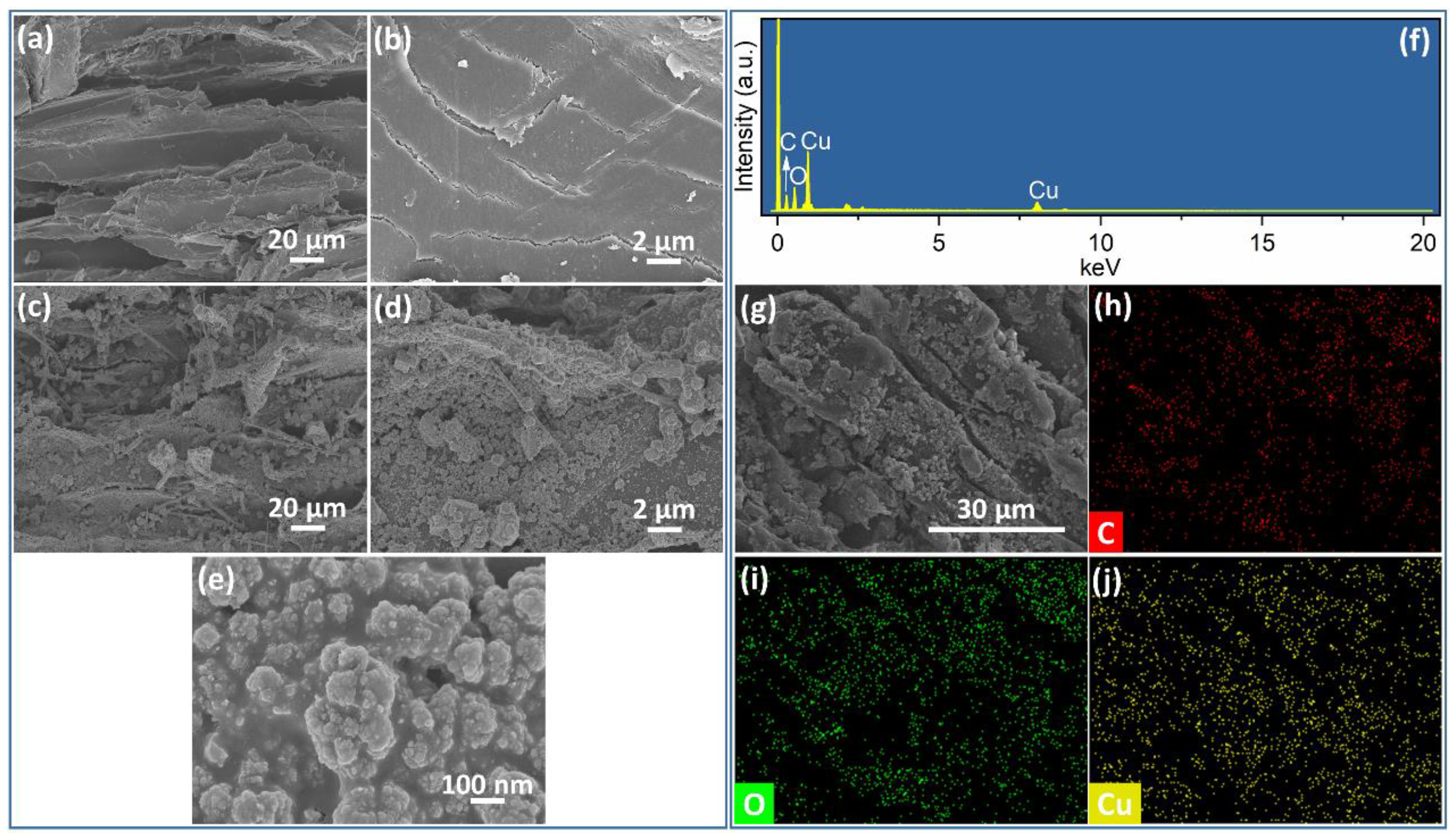
The surface structures of Cu2O@Cu-loaded woods and bamboo were analyzed by the combination of SEM and EDS mapping. As shown in Figure 4a, PW presented an ordered parallel microstructure without obvious distortion. The surface of PW was smooth and clean without the attachment of aggregates or their agglomerations (Figure 4b). For Cu2O@Cu/PW, aggregates were significantly distributed randomly on the surface of PW, and these aggregates existed as an agglomerated state (Figure 4c–e). As displayed in Figure 4f and Table 1, the atomic ratio of Cu on the surface of Cu2O@Cu/PW was 20.1 at.%. Figure 4h–j presented the elemental distributions of C, O, and Cu of Cu2O@Cu/PW, respectively. The Cu element was distributed uniformly on the surface of Cu2O@Cu/PW, indicating the abundance of Cu, followed by C and O elements (Figure 4g–j).
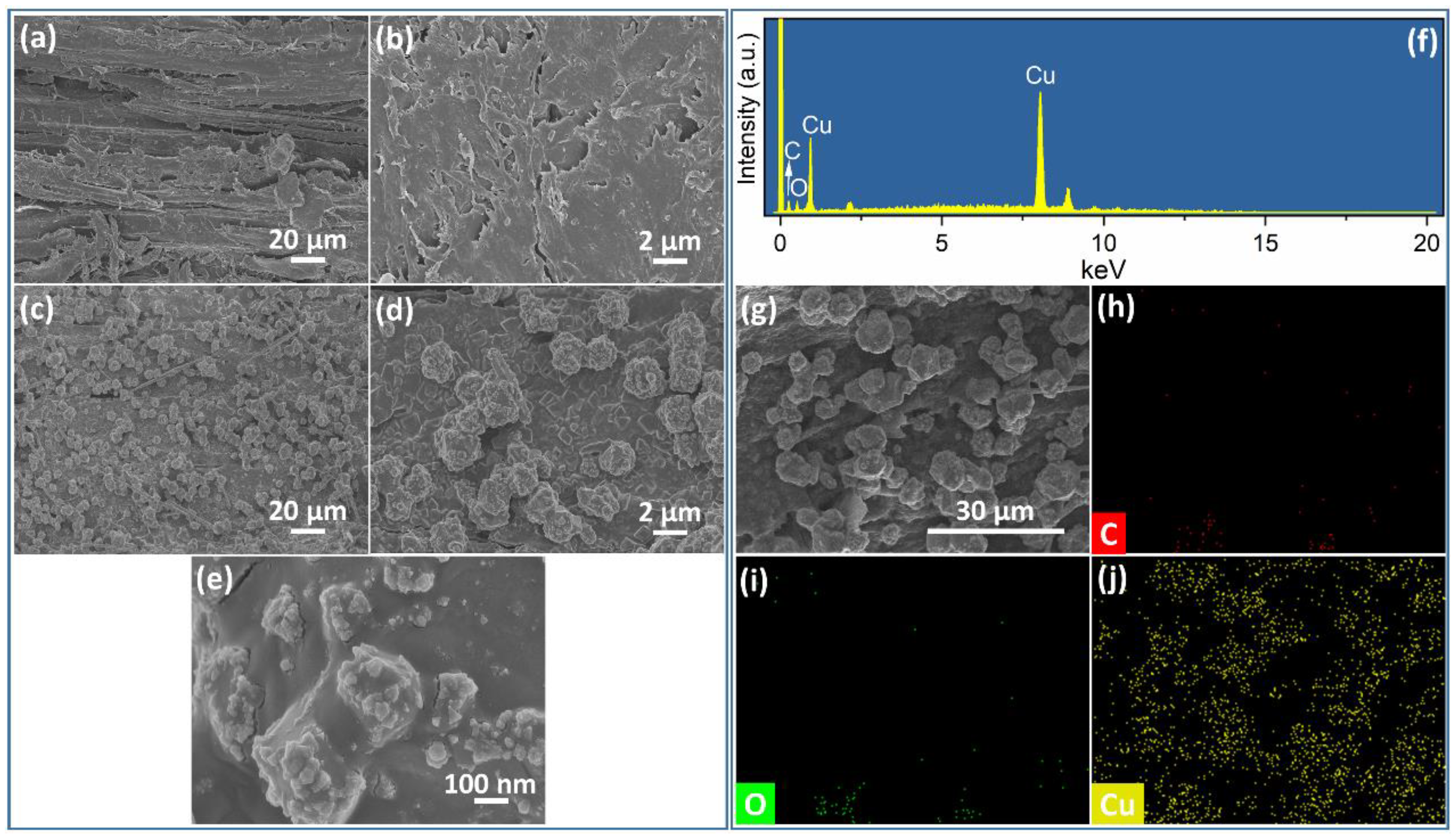
A long fibrous microstructure with some exfoliated debris and voids is shown on the surface of pristine BW (Figure 5a,b). Many agglomerated particles with an average diameter around 2 μm were distributed on the surface of Cu2O@Cu/BW uniformly (Figure 5c,d). These agglomerated particles were composed of nano-sized particles (Figure 5e). Compared with Cu2O@Cu/PW, the peak intensity of Cu in the EDX spectrum was obviously higher (Figure 5f). Moreover, Figure 5h–j presented the elemental distributions of C, O, and Cu of Cu2O@Cu/BW, respectively. The Cu element with a bright yellow color was also more distinctively presented compared with those of C and O elements (Figure 5g–j), indicating the effective modification of Cu2O@Cu aggregates on the surface of BW. The atomic ratio of Cu on the surface of Cu2O@Cu/PW was 27.1 at. %, which was the highest amount, i.e., four Cu2O@Cu-loaded woods and bamboo.
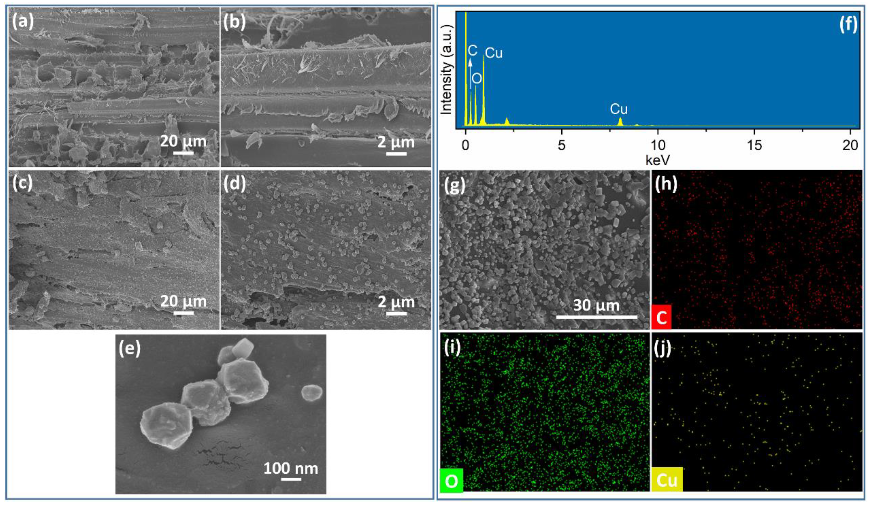
Given the hardness of OW, some exfoliated debris remained on the surface of OW in the absence of obvious voids (Figure 6a,b). After the in situ growth of Cu2O@Cu aggregates, uniformly distributed agglomerated particles with sizes less than 300 nm were observed on the surface of Cu2O@Cu/OW (Figure 6c–e). The sizes of these agglomerated particles were significantly lower than those on Cu2O@Cu/PW and Cu2O@Cu/BW. The atomic ratio of Cu on the surface of Cu2O@Cu/OW was 12.6 at. % (Figure 6f). Figure 6h–j presented the elemental distributions of C, O, and Cu of Cu2O@Cu/OW, respectively. Given the much smaller agglomerated Cu aggregates on the surface, the brightness for the distribution of the Cu element on Cu2O@Cu/OW was relatively weak (Figure 6g–j).
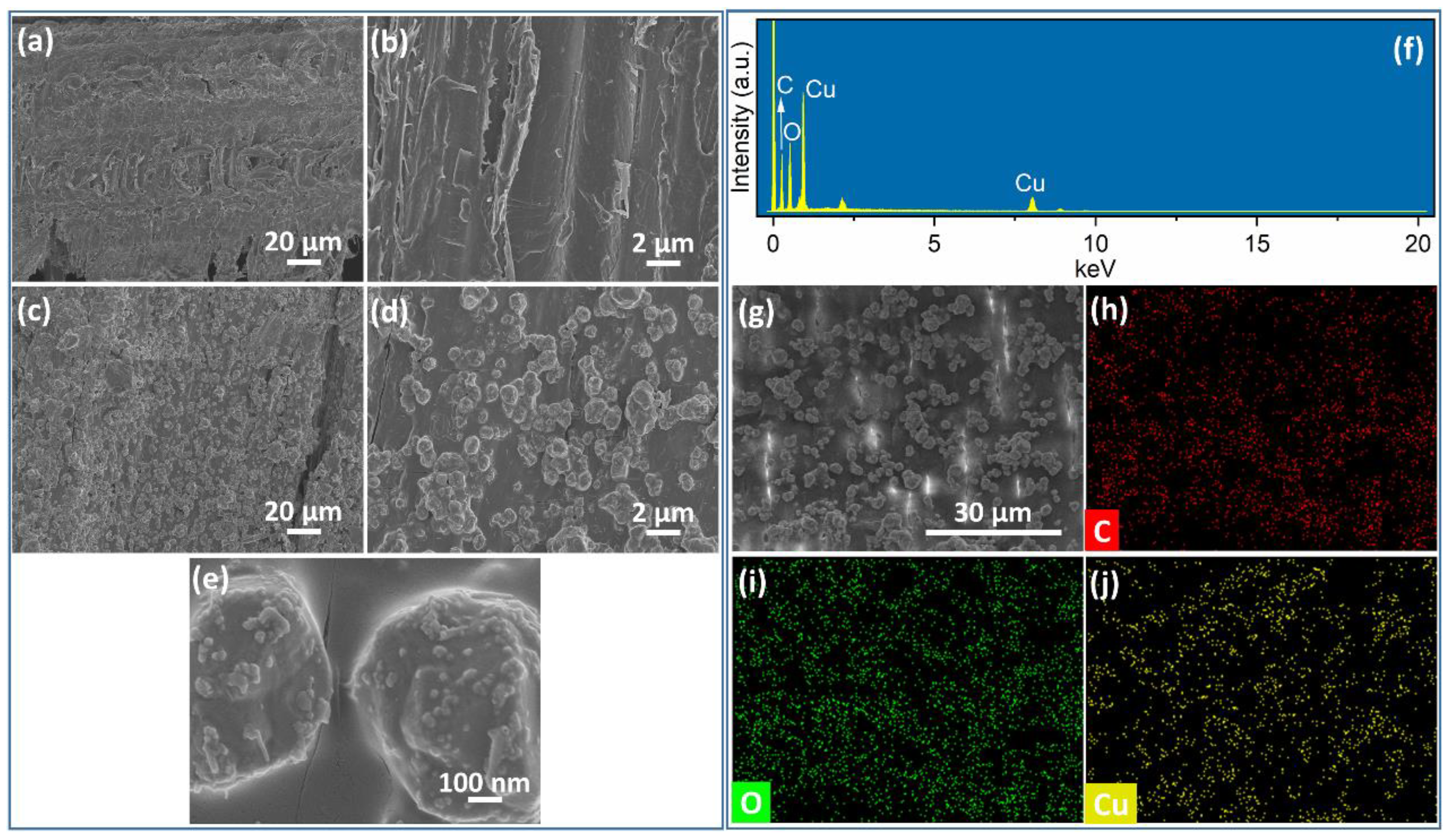
Since the porosity ratio of pristine bamboo materials is in the range of 48–70%, vertical channels were observed on the cross-sectional surface of bamboo (Figure 7a,b). In situ-grown Cu2O@Cu aggregates were distributed uniformly on the surface of Cu2O@Cu/bamboo as agglomerated particles with a size less than 2 μm (Figure 7c,d). These agglomerated particles were composed of Cu2O@Cu aggregates (Figure 7e). The atomic ratio of Cu on the surface of Cu2O@Cu/bamboo was 13.0 at. % (Figure 7f). Figure 7h–j present the elemental distributions of C, O, and Cu of Cu2O@Cu/bamboo, respectively. The uniform distribution of the Cu element was also confirmed by EDS mappings (Figure 7g–j).
The pore volume and average pore diameter of PW were 1.59 cm3/g and 445.0 nm. Given the sizes of Cu2O@Cu nanoaggregates on the surface of Cu2O@Cu/PW, some of these nanoaggregates would be available to enter the holes of PW [31]. The loading morphology of Cu2O@Cu/BW (0.75 cm3/g of pore volume, 70.7% of 2000–58,000 nm pore, 10.3% of 500–2000 nm pore, 4.7% of 80–500 nm pore, and 14.3% of 200–58,000 nm pore) was similar to that of Cu2O@Cu/PW [32]. The pore volume and average pore diameter of OW were larger than those of the other two woods, i.e., 0.53 cm3/g and 16.8 μm [33]. The lower loading of Cu2O@Cu nanoaggregates on Cu2O@Cu/OW should be attributed to the much larger pore sizes of the OW matrix, which were not appropriate to keep nanoaggregates in the pores. The pore volume and average pore diameter of bamboo were 0.63 cm3/g and 35.6 nm, respectively [34]. The pore diameter of bamboo was not available for the obvious entrance of Cu2O@Cu aggregates (several hundred nanometers to around 2 μm). This may explain why the content of Cu was lower than those of Cu2O@Cu/PW and Cu2O@Cu/BW.

3.2. Anti-Mold Evaluation of Four Cu2O@Cu-Loaded Woods and Bamboo

The anti-mold effects of Cu2O@Cu aggregates on PW, BW, OW, and bamboo were evaluated, as shown in Figure 8. A blank sample of PW, BW, OW, and bamboo was applied as the control group for comparison. No obvious mycelium was attached on the surface of PW, which may be attributed to its natural antibacterial effect. Some black spots appeared on the side and top surface of PW, while the surface of Cu2O@Cu/PW was not impacted (Figure 8a). A different situation for the growth of Aspergillus niger was presented for the other three samples. Specifically, abundant mycelia of Aspergillus niger grew around the side of BW (Figure 8c), OW (Figure 8e) and bamboo (Figure 8g), with an infection value of grade 1 after the 7-day cultivation. Some mycelia even arrived on the top surface of OW. Cu2O@Cu/BW, Cu2O@Cu/OW, and Cu2O@Cu/bamboo exhibited significant inhibition zones for the growth of Aspergillus niger, presenting an infection value of grade 0.
The infection content of Aspergillus niger tended to be more serious with the growth time. As the growth time increased to 28 days, the infection area on the side surface of PW reached almost one-third, with an infection value of grade 2 (Figure 8b). Cu2O@Cu/PW still presented an infection value of grade 0, since no distinct mycelium was attached on the surface of Cu2O@Cu/PW. The whole side surfaces of BW, OW, and bamboo were thoroughly infected with a black color (Figure 8d,f), with an infection value of grade 4. The top surface of bamboo exhibited a puce color, which was also a typical color of Aspergillus niger (Figure 8h). Thus, it is indicated that the surface as well as the quality of BW, OW, and bamboo were infected by Aspergillus niger. After the integration of Cu2O@Cu aggregates with these woods and bamboo, small amounts of mycelia were presented on the surface of Cu2O@Cu/BW and Cu2O@Cu/OW, showing an excellent anti-mold effect. The whole side surface of Cu2O@Cu/bamboo was covered by the bundles of mycelia, with an infection value of grade 1. The lower anti-mold effect of Cu2O@Cu/bamboo may be ascribed to the lower content of Cu2O@Cu aggregates on the surface of bamboo. Therefore, it is demonstrated that Cu2O@Cu aggregates located on the surface of PW, BW, OW, and bamboo inhibited the growth of mold with a log-term stability of 28 days. This inhibition effect should be attributed to the Cu2+/Cu+ ions released from Cu2O@Cu aggregates. The p values for infection grades were less than 0.05.
As Cu2+ ions enter fungal cells, intracellular Cu2+ can be oxidized to Cu+ and generate reactive oxygen species (ROS) via a Fenton-like reaction, leading to a series of reactions such as protein denaturation, lipid peroxidation, DNA and RNA damage, and ultimate apoptosis and necrosis for fungal cell death [34,35,36]. As a key signal molecule, ROS participates in cell signal transduction, activates transcription factors, and affects gene expression, thus promoting the cell proliferation and differentiation. Cu2+ ions presented a dose–effect relationship. For instance, after the Cu2+ treatment for 48 h, the proliferation inhibition rate reached or exceeded 87.8% at the cytotoxic concentration level (≥10−4 mol/L), while the proliferation promotion effect of 13.4–16.2% was shown at the micromolar concentration level (i.e., 10−6–10−5 mol/L) [37]. Excessive Cu2+ may combine with all protein sulfhydryl groups, which would inactivate protein components, rapidly destroy the cell membrane structure, and cause cell death, thus losing the ability to produce ROS.

3.3. Leaching Evaluation

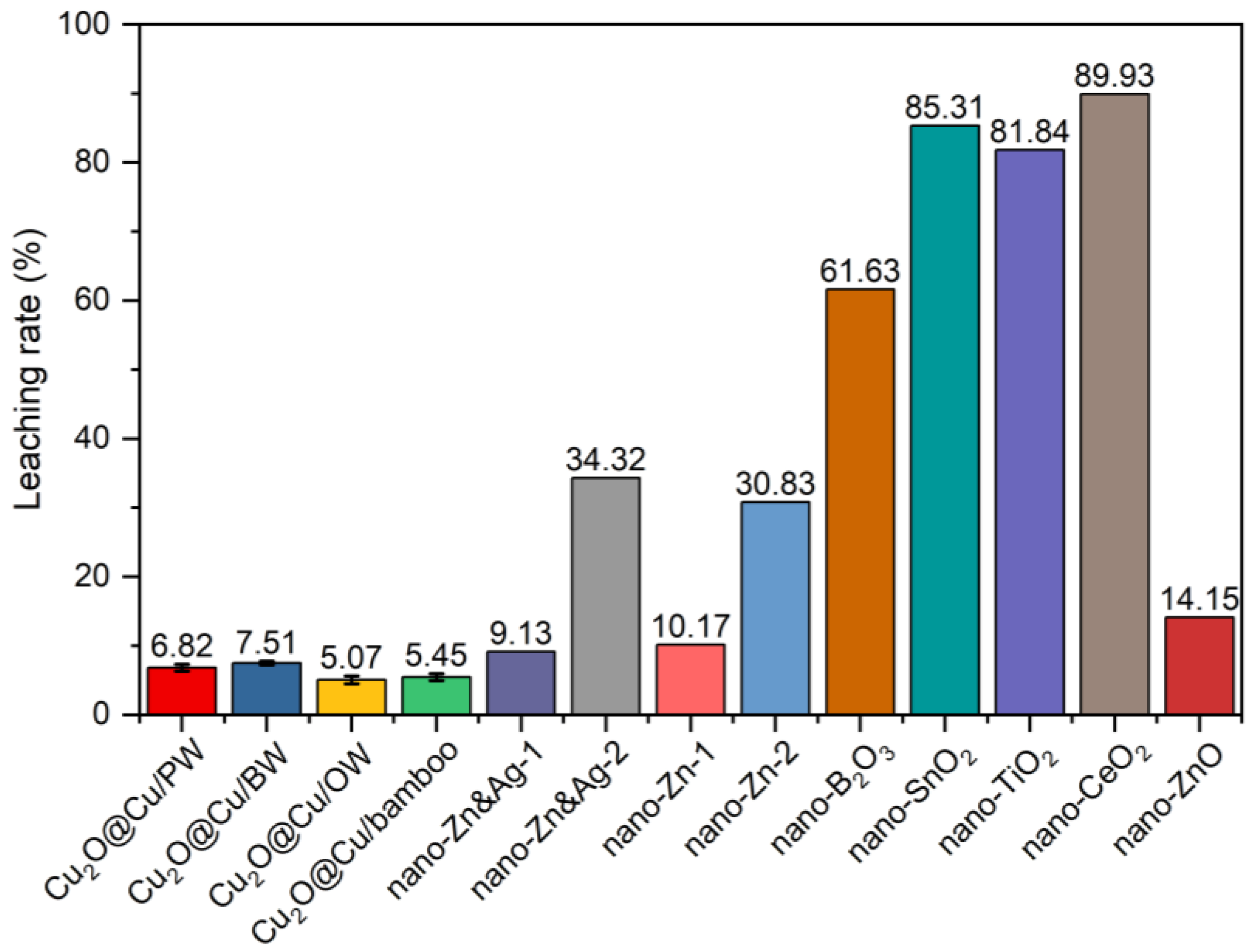
Since nano anti-mold agents loaded on the surface of woods or bamboos are highly leachable because of their water-borne characteristic and nanoscale size, it is necessary to evaluate the leaching behavior of Cu2O@Cu-loaded woods and bamboo during the applications. The leaching rates of Cu2O@Cu-loaded woods and bamboo were 6.82% for Cu2O@Cu/PW, 7.51% for Cu2O@Cu/BW, 5.07% for Cu2O@Cu/OW, and 5.45% for Cu2O@Cu/bamboo (Figure 9). The leaching rate presented a monotonically positive relation with the atomic content of Cu loaded on the surface of these four samples. Wood with a higher porosity would provide more surface-available sites for the capture of Cu2O@Cu aggregates, inducing more agglomerates of Cu2O@Cu aggregates on the surfaces of PW and BW due to the van der Waals forces. However, larger agglomerates would present weaker adhesion of Cu2O@Cu aggregates to the surface of specimens. Thus, Cu2O@Cu/PW and Cu2O@Cu/BW with larger agglomerate sizes would release more Cu2O@Cu aggregates. All experimental data were the averages of triplicate determinations, and the relative errors of the data were less than 5.0%. The standard deviations of the leaching rate were 0.408 for Cu2O@Cu/PW, 0.227 for Cu2O@Cu/BW, 0.468 for Cu2O@Cu/OW, and 0.413 for Cu2O@Cu/bamboo.
The leaching rates of Cu2O@Cu-loaded woods and bamboo in this work were compared with some nano mildew inhibitors reported in the previous literature. Cu2O@Cu-loaded woods and bamboo presented much lower leaching rates among these nano mildew inhibitors. For instance, the leaching rates of B2O3, SnO2, TiO2, CeO2, and ZnO NPs loaded on woods were 14.15–89.93% [38,39]. Clausen et al. improved the leaching rate of southern yellow pine sapwood by impregnating with ZnO NPs (30 and 70 nm) directly [40]. The leaching rate of ZnO-impregnated wood was 10.17%, which was obviously less than that of ZnSO4-treated wood (i.e., 30.83%). Moreover, Zn&Ag NPs also exhibited a similar trend [41]. Compared with vacuum impregnation of NPs on wood or bamboo, vacuum impregnation of Cu2O@Cu aggregates on woods and bamboo with an in situ adsorption–reduction–growth process presented a better leaching resistance.
Cu ions gradually seep out of the coating and enter the environment, posing a threat to the health of organisms. Cu ions show a growth inhibition, which is particularly prominent under high Cu concentration conditions. It not only disturbs the structure and function of the phytoplankton community but also reduces their light energy utilization efficiency and primary productivity [42]. Cu ions present cytotoxicity and neurotoxicity in mammalian cells, which may be related to oxidative stress and mitochondrial dysfunction. Cu ions and NPs induce toxicity in animals—among which mitochondrial damage, oxidative stress, and vertebral deformities are frequently observed [43]. Several studies have been carried out to explore the long-term effects of Cu NPs and/or its degradation production on multiple species, with fully mechanistic investigations still lacking [44]. It remains to be verified whether the toxicity of Cu NPs and their degradation products can be transferred through various food chains to affect the structure and function of ecosystems. Due to the mold-proof performance, Cu NPs have been often applied in coatings to protect woods and bamboo, thus posing health risks to both the woods and bamboo product makers and their consumers. Therefore, further investigations of Cu NPs on human health are warranted to better understand the impacts and associated risks.

4. Conclusions

In summary, agglomerated particles composed of Cu2O@Cu aggregates (average NP diameter of 41.7 nm with Cu2O coating shells over Cu core) were successfully grown in situ on the surfaces of woods and bamboo. These agglomerated particles were distributed uniformly on the surface of specimens, and their average sizes presented a declining trend from two μm (Cu2O@Cu/PW and Cu2O@Cu/BW) to several hundred nanometers (Cu2O@Cu/OW and Cu2O@Cu/bamboo). Four Cu2O@Cu-loaded specimens presented a Cu mass content that ranged from 12.6 to 27.1 at.%. A strong mold resistance towards Aspergillus niger was achieved after the cultivation of Cu2O@Cu-loaded woods and bamboo for 28 days. The infection values were grade 0 for Cu2O@Cu/PW and grade 1 for Cu2O@Cu/BW, Cu2O@Cu/OW, and Cu2O@Cu/bamboo (p < 0.05), which were significantly better than those of pristine specimens (i.e., grade 2 for PW and grade 4 for BW, OW, and bamboo). A low leaching rate for four specimens was carried out three times and was in the range of 5.23–7.81%, exhibiting a monotonically positive relation with the loading atomic content of the Cu element. It demonstrated an excellent stability of Cu2O@Cu-loaded woods and bamboo. This work provided a valuable reference for the application of metallic NPs in the field of wood and bamboo preservation.

Supplementary Materials

The following supporting information can be downloaded at: https://www.mdpi.com/article/10.3390/surfaces8030066/s1, Table S1: Standard method for the evaluation of infection values. Table S2. Objective numerical data of these color images. Table S3. Comparison of antifungal performance for various nano agents for Aspergillus niger. Rreferences [27,38,40,45,46,47] are cited in the Supplementary Materials.

Author Contributions

D.Z.: conceptualization, investigation, validation, data curation, methodology, writing—original draft. F.Z.: writing-review and editing. All authors have read and agreed to the published version of the manuscript. M.C.: supervision, funding acquisition, writing—review and editing.

Funding

This research was funded by National Natural Science Foundation of China (grant number 22274017).

Data Availability Statement

Data presented in this article are available at request from the corresponding author.

Conflicts of Interest

The authors declare no conflicts of interest.

References

  1. Lao, W.; Hu, J.; Huang, Y.; Li, X. Environmental footprints of wood and bamboo reconstituted materials production in China: A cradle-to-gate life cycle assessment. Ind. Crops. Prod. 2025, 232, 121320. [Google Scholar] [CrossRef]
  2. Lima, L.; Trindade, E.; Alencar, L.; Alencar, M.; Silva, L. Sustainability in the construction industry: A systematic review of the literature. J. Clean. Prod. 2021, 289, 125730. [Google Scholar] [CrossRef]
  3. Xu, P.; Zhu, J.; Li, H.; Wei, Y.; Xiong, Z.; Xu, X. Are bamboo construction materials environmentally friendly? A life cycle environmental impact analysis. Environ. Impact Assess. Rev. 2022, 96, 106853. [Google Scholar] [CrossRef]
  4. Schubert, M.; Panzarasa, G.; Burgert, I. Sustainability in wood products: A new perspective for handling natural diversity. Chem. Rev. 2023, 123, 1889–1924. [Google Scholar] [CrossRef]
  5. Gregory, S.A.; McGettigan, C.P.; McGuinness, E.K.; Rodin, D.M.; Yee, S.K.; Losego, M.D. Single-cycle atomic layer deposition on bulk wood lumber for managing moisture content, mold growth, and thermal conductivity. Langmuir 2020, 36, 1633–1641. [Google Scholar] [CrossRef]
  6. Julian, T.C.; Chen, Y.; Wang, Y.; Gao, W.; Sun, Q.; Fukuda, H. Revolutionizing wood construction: Densified integrated wood with voronoi structures. ACS Appl. Eng. Mater. 2023, 1, 3186–3193. [Google Scholar] [CrossRef]
  7. John, B.; Sahoo, S.K. Development of nanofree, silane-free, and fluorine-free cardanol-based multifunctional, superhydrophobic wood coating. ACS Appl. Polym. Mater. 2024, 6, 9226–9237. [Google Scholar] [CrossRef]
  8. Ou, J.; Zhao, G.; Wang, F.; Li, W.; Lei, S.; Fang, X.; Siddiqui, A.R.; Xia, Y.; Amirfazli, A. Durable superhydrophobic wood via one-step immersion in composite silane solution. ACS Omega 2021, 6, 7266–7274. [Google Scholar] [CrossRef] [PubMed]
  9. Fernández-Costas, C.; Palanti, S.; Charpentier, J.P.; Sanromán, M.Á.; Moldes, D. A sustainable treatment for wood preservation: Enzymatic grafting of wood extractives. ACS Sustain. Chem. Eng. 2017, 5, 7557–7567. [Google Scholar] [CrossRef]
  10. Kumar, R.; Ray, S.; Pandey, K.K.; Muthukumar, A. Potential use of Pterocarpus santalinus bark extracts as UV-protective coats on Hevea brasiliensis wood. ACS Sustain. Chem. Eng. 2023, 11, 9965–9978. [Google Scholar] [CrossRef]
  11. Chang, M.; Shen, P.; Wang, L.; Ma, Q.; Jia, Z.; Hu, C.; Zhang, X. Research progress of eco-friendly plant-derived biomass-based wood adhesives: A review. Ind. Crop. Prod. 2024, 222, 120093. [Google Scholar] [CrossRef]
  12. Piao, X.; Zhao, Z.; Guo, H.; Wang, Z.; Jin, C. Improved properties of bamboo by thermal treatment with wood wax oil. Colloids Surf. A Physicochem. Eng. Asp. 2022, 643, 128807. [Google Scholar] [CrossRef]
  13. Li, X.; Ji, S.; Li, T.; Liu, Z.; Hao, X.; Chen, Z.; Zhong, Y.; Li, X. The physical, mechanical and fire performance of bamboo scrimber processed with thermal-treated bamboo bundles. Ind. Crops. Prod. 2023, 205, 117549. [Google Scholar] [CrossRef]
  14. Paris, M.J.; Mani-López, E.; Ramírez-Corona, N.; López-Malo, A. Postharvest mold growth control in raspberries and blackberries using cinnamon essential oil-loaded alginate beads. ACS Food Sci. Technol. 2025, 5, 127–136. [Google Scholar] [CrossRef]
  15. Rahmawati, N.; Sumardi, I.; Dungani, R. Isolation and identification of fungi inhabiting rubber impregnated wood, and their role of quality changing the impregnated wood. BioResources 2020, 15, 2839–2849. [Google Scholar] [CrossRef]
  16. Chaturvedi, K.S.; Henderson, J.P. Pathogenic adaptations to host-derived antibacterial copper. Front. Cell. Infect. Microbiol. 2014, 4, 3. [Google Scholar] [CrossRef] [PubMed]
  17. Malhotra, N.; Ger, T.-R.; Uapipatanakul, B.; Huang, J.-C.; Chen, K.H.C.; Hsiao, C.-D. Review of copper and copper nanoparticle toxicity in fish. Nanomaterials 2020, 10, 1126. [Google Scholar] [CrossRef]
  18. Latorre, M.; Troncoso, R.; Uauy, R. Biological Aspects of Copper. In Clinical and Translational Perspectives on Wilson Disease; Academic Press: Cambridge, MA, USA, 2019; pp. 25–31. [Google Scholar]
  19. Gudkov, S.V.; Burmistrov, D.E.; Fomina, P.A.; Validov, S.Z.; Kozlov, V.A. Antibacterial properties of copper oxide nanoparticles (review). Int. J. Mol. Sci. 2024, 25, 11563. [Google Scholar] [CrossRef]
  20. Gudkov, S.V.; Baimler, I.V.; Uvarov, O.V.; Smirnova, V.V.; Volkov, M.Y.; Semenova, A.A.; Lisitsyn, A.B. Influence of the concentration of Fe and Cu nanoparticles on the dynamics of the size distribution of nanoparticles. Front. Phys. 2020, 8, 622551. [Google Scholar] [CrossRef]
  21. Nataraja, S.T.; Vishnu, R.; Anoop, E.V.; Chathoth, A.M.; Valasseri, J.; Babu, S. Zinc and copper nanoparticles for coconut wood preservation. ACS Appl. Nano Mater. 2025, 8, 4946–4957. [Google Scholar] [CrossRef]
  22. Xie, F.; Pu, Y.; Liang, Y.; Qiu, J.; Zheng, K.; Gao, J. Preparation of highly stable wood with fast curing, ultraviolet, and mildew resistance through copper-montmorillonite nanoparticles hybridized with tung oil. ACS Appl. Mater. Interfaces 2025, 17, 8231–8247. [Google Scholar] [CrossRef] [PubMed]
  23. Pinheiro, L.D.S.M.; Sentena, N.Z.; Sangoi, G.G.; Vizzotto, B.S.; Pinto, E.O.; Pavoski, G.; Espinosa, D.C.R.; Machado, A.K.; Silva, W.L. Copper nanoparticles from acid ascorbic: Biosynthesis, characterization, in vitro safety profile and antimicrobial activity. Mater. Chem. Phys. 2023, 307, 128110. [Google Scholar] [CrossRef]
  24. Pathak, R.; Punetha, V.D.; Bhatt, S.; Punetha, M. A review on copper-based nanoparticles as a catalyst: Synthesis and applications in coupling reactions. J. Mater. Sci. 2024, 59, 6169–6205. [Google Scholar] [CrossRef]
  25. Phan, D.N.; Dorjjugder, N.; Saito, Y.; Khan, M.Q.; Ullah, A.; Bie, X.; Taguchi, G.; Kim, I.S. Antibacterial mechanisms of various copper species incorporated in polymeric nanofibers against bacteria. Mater. Today Commun. 2020, 25, 101377. [Google Scholar] [CrossRef]
  26. Xiong, L.; Yu, H.; Nie, C.; Xiao, Y.; Zeng, Q.; Wang, G.; Wang, B.; Lv, H.; Li, Q.; Chen, S. Size-controlled synthesis of Cu2O nanoparticles: Size effect on antibacterial activity and application as a photocatalyst for highly efficient H2O2 evolution. RSC Adv. 2017, 7, 51822–51830. [Google Scholar] [CrossRef]
  27. Qi, Y.; Dai, X.; Wei, L.; Luo, H.; Liu, Y.; Dong, X.; Yang, D.; Li, Y. Nano-AgCu alloy on wood surface for mold resistance. Nanomaterials 2022, 12, 1192. [Google Scholar] [CrossRef] [PubMed]
  28. Aghdam, H.D.; Bellah, S.M.; Malekfar, R. Surface-enhanced Raman scattering studies of Cu/Cu2O core-shell NPs obtained by laser ablation. Spectrochim. Acta A 2019, 223, 117379. [Google Scholar] [CrossRef]
  29. Ye, M.; Shao, T.; Liu, J.; Li, C.; Song, B.; Liu, S. Phase engineering of Cu@Cu2O core-shell nanospheres for boosting tandem electrochemical CO2 reduction to C2+ products. Appl. Surf. Sci. 2023, 622, 156981. [Google Scholar] [CrossRef]
  30. He, S.; Xu, J.; Wu, Z.; Bao, Y.; Yu, H.; Chen, Y. Compare of porous structure of meso bamboo and Pinus sylvestris L. lumber. J. Nanjing For. Univ. 2017, 41, 157–162. [Google Scholar]
  31. Vitas, S.; Segmehl, J.S.; Burgert, I.; Cabane, E. Porosity and pore size distribution of native and delignified beech wood determined by mercury intrusion porosimetry. Materials 2019, 12, 416. [Google Scholar] [CrossRef]
  32. Gao, L.; Cheng, H.; Chen, J.; Chen, W.; Zhao, J. Pore structure characterization of Oak via X-ray computed temography. BioResources 2020, 15, 3053–3063. [Google Scholar] [CrossRef]
  33. Lee, S.; Jang, J.W.; Ryu, Y.B. Surface oxidation of Cu2O nanoparticles by adsorbed ammonia. Nanomaterials 2022, 12, 4242. [Google Scholar] [CrossRef] [PubMed]
  34. Wu, H.; Zhang, Z.; Cao, Y.; Hu, Y.; Li, Y.; Zhang, L.; Cao, X.; Wen, H.; Zhang, Y.; Lv, H.; et al. A Self-amplifying ROS-responsive nanoplatform for simultaneous cuproptosis and cancer immunotherapy. Adv. Sci. 2024, 11, 2401047. [Google Scholar] [CrossRef] [PubMed]
  35. Murugesan, S.; Balasubramanian, S.; Perumal, E. Copper oxide nanoparticles induced reactive oxygen species generation: A systematic review and meta-analysis. Chem. Biol. Interact. 2025, 405, 111311. [Google Scholar] [CrossRef]
  36. Duan, X.; Peng, D.; Zhang, Y.; Chen, Z.; Huang, Y. Micromolar concentration of copper (ii) ions enhances proliferation in human skin keratinocyte through appropriating generation of reactive oxygen species. J. Army Med. Univ. 2015, 37, 1053–1058. [Google Scholar] [CrossRef]
  37. Ameen, F.; Alsamhary, K.; Alabdullatif, J.A.; ALNadhari, S. A review on metal-based nanoparticles and their toxicity to beneficial soil bacteria and fungi. Ecotoxicol. Environ. Saf. 2021, 213, 112027. [Google Scholar] [CrossRef]
  38. Terzi, E.; Kartal, S.N.; Yılgör, N.; Rautkari, L.; Yoshimura, T. Role of various nano-particles in prevention of fungal decay, mold growth and termite attack in wood, and their effect on weathering properties and water repellency. Int. Biodeterior. Biodegrad. 2016, 107, 77–87. [Google Scholar] [CrossRef]
  39. Clausen, C.A.; Kartal, S.N.; Arango, R.A.; Green, F., III. The role of particle size of particulate nano-zinc oxide wood preservatives on termite mortality and leach resistance. Nanoscale Res. Lett. 2011, 6, 427. [Google Scholar] [CrossRef]
  40. Kartal, S.N.; Green, F., III.; Clausen, C.A. Do the unique properties of nanometals affect leachability or efficacy against fungi and termites? Int. Biodeterior. Biodegrad. 2009, 63, 490–495. [Google Scholar] [CrossRef]
  41. Tang, C.; Qin, X.; Huang, W.; Debi, S.; Zhang, Z.; Guo, J.; Liu, W.; Mo, J. Environmental sources, fates, toxicological effects, and health risks of copper pyrithione: An overview. Front. Environ. Sci. Eng. 2024, 18, 132. [Google Scholar] [CrossRef]
  42. Muhcu, D.; Terzi, E.; Kartal, S.N.; Yoshimura, T. Biological performance, water absorption, and swelling of wood treated with nanoparticles combined with the application of Paraloid B72. J. For. Res. 2017, 28, 381–394. [Google Scholar] [CrossRef]
  43. Lankone, R.S.; Ruggiero, E.; Goodwin, D.G., Jr.; Vilsmeier, K.; Mueller, P.; Pulbere, S.; Challis, K.; Bi, Y.; Westerhoff, P.; Ranville, J.; et al. Evaluating performance, degradation, and release behavior of a nanoform pigmented coating after natural and accelerated weathering. NanoImpact 2020, 17, 100199. [Google Scholar] [CrossRef]
  44. Aguayo, M.G.; Oviedo, C.; Reyes, L.; Navarrete, J.; Gómez, L.; Torres, H.; Gaviño, G.; Trollund, E. Radiata pine wood treated with copper nanoparticles: Leaching analysis and fungal degradation. Forests 2021, 12, 1606. [Google Scholar] [CrossRef]
  45. Mantanis, G.; Terzi, E.; Kartal, S.N.; Papadopoulos, A.N. Evaluation of mold, decay and termite resistance of pine wood treated with zinc- and copper-based nanocompounds. Int. Biodeter. Biodegrad. 2014, 90, 140–144. [Google Scholar] [CrossRef]
  46. Harandi, D.; Walsh-Korb, Z.; Moradienayat, M. CNCs and CeO2 as organic–inorganic additives to enhance HPC bio-polymer wood coatings against photochemical degradation. J. Coat. Technol. Res. 2025, 22, 691–701. [Google Scholar] [CrossRef]
  47. Chen, F.; Yang, X.; Wu, Q. Antifungal capability of TiO2 coated film on moist wood. Build. Environ. 2009, 44, 1088–1093. [Google Scholar] [CrossRef]
Figure 1. Schematic illustration for the preparation of Cu2O@Cu-loaded woods and bamboo.
Surfaces 08 00066 g001
Figure 2. Color change for pristine and vacuum-treated Cu2O@Cu-loaded woods and bamboo: PW and Cu2O@Cu/PW (a), BW and Cu2O@Cu/BW (b), OW and Cu2O@Cu/OW (c), bamboo and Cu2O@Cu/bamboo (d).
Surfaces 08 00066 g002
Figure 3. SEM images (a,b) and TEM images (c,d) of Cu2O@Cu aggregates. EDS mapping images (eg). Distribution of particle diameter (h). XRD pattern (i). Cu LMM spectrum (j).
Surfaces 08 00066 g003
Figure 4. SEM images of PW (a,b) and Cu2O@Cu/PW (ce). EDX spectrum (f) and EDS mapping images (gj) of Cu2O@Cu/PW surface.
Surfaces 08 00066 g004
Figure 5. SEM images of BW (a,b) and Cu2O@Cu/BW (ce). EDX spectrum (f) and mapping images (gj) of Cu2O@Cu/BW surface.
Surfaces 08 00066 g005
Figure 6. SEM images of OW (a,b) and Cu2O@Cu/OW (ce). EDX spectrum (f) and mapping images (gj) of Cu2O@Cu/OW surface.
Surfaces 08 00066 g006
Figure 7. SEM images of bamboo (a,b) and Cu2O@Cu/bamboo (ce). EDX spectrum (f) and mapping images (gj) of Cu2O@Cu/bamboo.
Surfaces 08 00066 g007
Figure 8. Digital photos of PW and Cu2O@Cu/PW (a,b), BW and Cu2O@Cu/BW (c,d), OW and Cu2O@Cu/OW (e,f), and bamboo and Cu2O@Cu/bamboo (g,h) after 7 days and 28 days of mold growth.
Surfaces 08 00066 g008
Figure 9. Leaching rates of Cu2O@Cu/PW, Cu2O@Cu/BW, Cu2O@Cu/OW, and Cu2O@Cu/bamboo, and their comparison with other nano mildew inhibitor reported in the literature.
Surfaces 08 00066 g009
Table 1. Surface element ratios of Cu2O@Cu-loaded woods and bamboo determined by EDS analysis.
SampleC (at. %)O (at. %)Cu (at. %)
PW63.936.1-
BW64.835.2-
OW61.538.5-
Bamboo62.737.3-
Cu2O@Cu/PW50.829.120.1
Cu2O@Cu/BW59.213.727.1
Cu2O@Cu/OW49.138.312.6
Cu2O@Cu/bamboo55.231.813.0
Disclaimer/Publisher’s Note: The statements, opinions and data contained in all publications are solely those of the individual author(s) and contributor(s) and not of MDPI and/or the editor(s). MDPI and/or the editor(s) disclaim responsibility for any injury to people or property resulting from any ideas, methods, instructions or products referred to in the content.